
EMBLetc Magazine Spring 2016
48
The European Molecular Biology Laboratory Magazine Issue 86 Spring 2016 The rhythms in life Synapse Forgetting to learn Nucleus SPIM doctors Cultures Laughing matter
-
Upload
european-molecular-biology-laboratory-embl -
Category
Documents
-
view
217 -
download
1
description
The magazine of the European Molecular Biology Laboratory
Transcript of EMBLetc Magazine Spring 2016

The European Molecular Biology Laboratory Magazine Issue 86 Spring 2016
The rhythms in lifeSynapse Forgetting to learn
Nucleus SPIM doctors
Cultures Laughing matter

Contents
13
The rhythms in life How EMBL scientists are discovering and understanding the waves and rhythms inside us
18 SPIM doctors
EMBL researchers continue to push the boundaries of microscopy
23 In synchrotron
Essential innovations for X-ray crystallography experimentation at EMBL
30 Growth and integration
Social, scientific and technical interactions of EMBL-EBI data services
28 Periodic proteins
Predictive power of the periodic table of protein complexes
32 Lighting up development
EMBL PhD project that puts development in a new light
Nucleus
2 EMBLetc. spring 2016
5 In sync
6 Drugging bacteria 7 Forever young
8 Forgetting to learn
9 Target-validation collaboration welcomes Biogen
10 Parallel profiling
11 Immunology meets single-cell sequencing
36 The search for EMBL’s next Director-General
Regulars
37 From the Archive The Moon Dreamers
38 Branches Laughing matter
39 Reviews Life’s Blueprint by Benny Shilo
40 Q&A What is the one marine mystery you would most like to see solved?
41 Awards & honours
42 Alumni Admin ability
47 Obituary Bernard Jacrot
Synapse
Cultures
The european Molecular Biology laBoraTory Magazine 3

EditorialEven if you cannot hold a beat, it turns out the cells in your body can. This edition, our cover story explores remarkable research by EMBL scientists that sheds light on the rhythms within us. From waves rippling across a single oocyte to thousands of cells synchronising in the developing embryo, tempos, pulses and even ‘dance’ moves play an intrinsic role in our biology (page 13). On a grander scale we also get ‘in sync’ with two EMBL scientists whose work at synchrotrons is proving invaluable to X-ray crystallography experiments (page 23). As we continue scratching deeper beneath the surface at EMBL, we also explore rhythms of collaboration and teamwork – from the compelling role of EMBL researchers in driving the past, present and future of selective plane illumination microscopy (page 18) to the massive interconnections of data resources at EMBL-EBI (page 30), and to the crucial role of EMBL’s administration in driving forward the Laboratory’s missions (page 44). These stories, and many more besides show just some of the ways that work at EMBL is enabling us to see the rhythms in life in a new light.
Adam Gristwood Editor
Publisher
European Molecular Biology Laboratory,
Heidelberg
Office of Information and Public Affairs
Editor-in-chief
Silke Schumacher
Director, International Relations
Editors
Adam Gristwood, magazine
Chloë Cross, digital
Sonia Furtado Neves, science
Isabelle Kling, science
Mehrnoosh Rayner, alumni
Mary Todd Bergman, EMBL-EBI
Design
Edenspiekermann, Amsterdam
Printed by
ColorDruck Solutions, Leimen
Contact
Cover illustration
Aad Goudappel, Rotterdam
EMBLetc. ONLINE
NEwS.EMBL.DE
Word to remember
Compaction Noun, pronunciation: /kəm-păk′shən/
Event in early stage of the formation of the mammalian embryo, when a loose ball of cells draws together and the embryo’s structure begins to emerge.
Ph
ot
o: E
MB
L Ph
ot
oL
aB
/Ma
riE
tta
Sc
hu
PP
4 EMBLetc. spring 2016

This showed that the cells don’t need any outside cues to determine when – and how fast – they blink. But were there specific cells that dictated the rhythm? “We found that the answer is no,” says Tsiairis. “It’s a true democracy: the cells all adjust their blinking rate to reach a compromise.”
Aulehla’s lab is now investigating if the principles they found in the dish apply in the embryo, when the first waves are set up. The group is also looking into how cells read the rhythm at which those around them are blinking (for more, see page 13).
Tsiairis CD & Aulehla A. Cell, 11 February 2016. DOI:10.1016/j.cell.2016.01.028
FuLL STORy AND vIDEO ONLINE:
NEwS.EMBL.DE/?p=6533
In sync What do cells in an embryo have in common with schools of fish, swarms of fireflies, and applauding audiences? By SOnIA FuRTADO neveS
When a mouse embryo is developing, the cells that will form its vertebrae periodically turn specific genes on and off, on and off, in a rhythmic manner. This is a remarkably coordinated process: rows of cells blink in succession, so that if you look at the whole embryo you see the blinking in waves.
Each of these waves ends in the formation of a vertebra. But how does it start? “How do thousands of cells manage to turn genes on in such a coordinated fashion, in space and time?” Alexander Aulehla, a group leader at EMBL Heidelberg, wanted to know. Scientists in the field had pondered several possibilities. There could be an outside cue that sets the rhythm, or a ‘pacemaker’ cell – or group of cells – that tells the
others when are blinking (see page 13). But instead, Aulehla’s postdoc Charisios Tsiairis found that the cells synchronise on their own: they self-organise, as the scientists say.
Studying how these waves start would be extremely difficult to do in an embryo, so the EMBL researchers devised a way to answer the question in a lab dish. In work published in Cell, Tsiairis took tissue from several mouse embryos, separated it into individual cells, and then placed a random mix of cells – from different embryos and different parts of the embryo – together. At first, the cells blinked randomly, but they quickly started blinking in sync. Next, they formed circles where blinking happened in a wave, rolling outwards from the centre.
iMa
GE
: EM
BL
/c t
Sia
iriS
during heart development, researchers may discover new ways to treat heart disease. In the study, published in Cell, Christoph Müller’s group determined the 3D structure of transcription factors TBX5 and NKX2-5, bound to DNA.
Luna-Zurita L et al. Cell, 25 February 2016. DOI: 10.1016/j.cell.2016.01.004
FuLL STORy ONLINE:
NEwS.EMBL.DE/?p=6555
the 3D structure shows how two transcription factors influence one another’s binding to a specific stretch of DNa – an interaction that is crucial for a heart to develop healthily
By DAnA SMITh
Scientists at EMBL Heidelberg and the Gladstone Institutes have discovered that three transcription factors — proteins that turn genes on or off — interact with each other and the genome to influence how a heart forms in an embryo. By understanding how the transcription factors work together
True love iMa
GE
: EM
BL
/c.M
üL
LE
r
The european Molecular Biology laBoraTory Magazine 5
Synapse

led the work at EMBL. “Now think how many drugs are out there, and how many people take several drugs daily: even if only a fraction of drugs have this big an impact, they could still be dramatically shaping people’s gut.”
Forslund K. et al. Nature 10 December 2015. DOI: 10.1038/nature15766
FuLL STORy ONLINE:
NEwS.EMBL.DE/?p=6207
Drugging bacteriaBy SOnIA FuRTADO neveS
Metformin, the drug most often used to treat type 2 diabetes, has a greater effect on gut microbes than the disease itself. The finding, by scientists at EMBL Heidelberg and colleagues in the MetaHIT consortium, has implications for studies searching for links between our microbiomes and disease. Published in Nature, the study points to new approaches for understanding how metformin works, and minimising the side effects of a drug that patients take in high doses for many years.
“It’s surprising that this single drug triggers such a noticeable change in our microbes,” says Peer Bork, who
iMa
GE
: co
ur
tE
Sy
of
Pa
cif
ic N
or
th
wE
St
Na
tio
Na
L La
Bo
ra
to
ry
(By
-Nc
-Sa
)
Gut bacteria are more affected by metformin than by the type 2 diabetes it is prescribed to treat
By MARy TODD BeRgMAn
An in-depth analysis of the value and impact of EMBL-EBI carried out by consultancy Charles Beagrie Ltd provides detailed insights into the return on investment in open data. The report puts benefits to users and their funders at between £1 billion and £5 billion per annum worldwide.The authors applied a robust methodology, drawing on both measurable indicators and interviews with commercial and academic service users and collaborators. The report highlights efficiencies gained and money saved by companies and academic organisations that use EMBL-EBI services, and demonstrates that EMBL-EBI data and services contributed to the wider realisation of future research impacts worth £920 million every year.
“The findings of the value and impact assessment demonstrate that we are indeed providing a healthy return on the public investment in EMBL-EBI, but more importantly they help bring home the point that open data is essential to the success and sustainability of biomedical and life-science research,” said Rolf Apweiler, Director of EMBL-EBI.
Professor Melanie Welham, BBSRC Executive Director of Science added: “EMBL-EBI provides vital life-science infrastructure for researchers in the UK and beyond, generating substantial opportunities for bioscience research and the generation of new knowledge. This report demonstrates clear return on investment for the institute and the research community to the benefit of society and the wider global economy.”
BEAGRIE.COM/STATIC/RESOuRCE/
EBI-IMPACT-REPORT.PDF
Value and impact
6 eMBLetc. SPRIng 2016

By SOnIA FuRTADO neveS
This ‘before and after’ image could be thought of as stem cells’ equivalent of an advert for anti-wrinkle cream: ‘look how cells stay young!’ It shows that a molecule called microRNA-142 allows stem cells to remain unchanged, instead of growing into specialised cell types. Given the right conditions, stem cells with low levels of microRNA-142 (green, left) grow into neurons (pink, right). But stem cells with high levels of the molecule (red, left) remain unchanged (blue, right), Pierre Neveu’s group at EMBL Heidelberg have found.
“It seems that this microRNA leaves the cells deaf to their environment: you can ‘throw’ whatever you want at them, and they don’t care: they carry on being stem cells,” says Neveu. The finding, published in Molecular Systems Biology in December 2015, could have implications for cancer treatment and regenerative medicine, as well as more fundamental research. Hanna Sladitschek, a PhD student in Neveu’s lab, found that this microRNA suppresses a gene that can turn cells into tumour cells. And it blocks two chain reactions
Forever youngthat have also been linked to cancer. “So you could think of delivering the microRNA to cells to prevent tumours from spreading,” Neveu postulates.
Sladitschek hL & neveu PA. Molecular Systems Biology 21 December 2015. DOI 10.15252/msb.20156525
FuLL STORy ONLINE:
NEwS.EMBL.DE/?p=6579
the stem cell equivalent of an anti-wrinkle cream advert
iMa
GE
: EM
BL
/ha
NN
a S
La
Dit
Sc
hE
k
The european Molecular Biology laBoraTory Magazine 7
Synapse

Forgetting to learnScientists discover neural mechanisms in mouse brains that indicate that we actively forget as we learn
By SOnIA FuRTADO neveS
They say that once you’ve learned to ride a bicycle, you never forget how to do it. But new research suggests that while learning, the brain is actively trying to forget. The study, by scientists at EMBL and University Pablo Olavide in Sevilla, Spain, was published in Nature Communications.
“This is the first time that a pathway in the brain has been linked to forgetting, to actively erasing memories,” says Cornelius Gross, who led the work at EMBL.
At the simplest level, learning involves making associations, and remembering them. Working with mice, Gross and colleagues studied the hippocampus, a region of the brain that’s long been known to help form memories. Information enters this part of the brain through three different routes. As memories are cemented, connections between neurons along the ‘main’ route become stronger.
When they blocked this main route, the scientists found that the mice were no longer capable of learning a Pavlovian response – associating a sound to a consequence, and anticipating that consequence. But if the mice had learned that association before the scientists stopped information flow in that main route, they could still retrieve that memory. This confirmed that
this route is involved in forming memories, but isn’t essential for recalling those memories. The latter probably involves the second route into the hippocampus, the scientists surmise.
But blocking that main route had an unexpected consequence: the connections along it were weakened, meaning the memory was being erased. “Simply blocking this pathway shouldn’t have an effect on its strength,” says Agnès Gruart from University Pablo Olavide. “When we investigated further, we discovered that activity in one of the other pathways was driving this weakening.”
Interestingly, this active push for forgetting only happens in learning situations. When the scientists blocked the main route into the hippocampus under other circumstances, the strength of its connections remained unaltered.“One explanation for this is that there is limited space in the brain, so when you’re learning, you have to weaken some connections to make room for others,” says Gross. “To learn new things, you have to forget things you’ve learned before.”
The findings were made using genetically engineered mice, but with help from Maja Köhn’s lab at EMBL the scientists demonstrated that it is possible to produce a drug that activates this ‘forgetting’ route in the brain without the need for genetic engineering. This approach, they say, might be interesting to explore if one were looking for ways to help people forget traumatic experiences.
Madroñal et al. Nature Communications, 18 March 2016. DOI: 10.1038/ncomms10923
the three routes into the hippocampus seem to be linked to different aspects of learning: forming memories (green), recalling them (yellow) and forgetting (red)
iMa
GE
: Joh
N w
oo
D
8 EMBLetc. spring 2016

on target validation,” said Philip Ma, vice president, Digital Health Technology & Data Sciences at Biogen. “We look forward to combining our expertise in data sciences with the leading capabilities of GSK, the Sanger Institute and EMBL-EBI.”
The CTTV covers all aspects of human health and disease. The cornerstone of the collaboration is an agreement that experimental data and information gathered within the CTTV will be shared to benefit the broader scientific community, after basic quality control checks to ensure consistency with the data-sharing guidelines of both institutes. The CTTV welcomes new interest from companies and academic institutions that wish to accelerate the discovery of drug targets through open innovation.
Angermueller C, et al. Nature Methods, 11 January 2016. DOI: 10.1038/nmeth.3728
MORE ONLINE:
TARGETvALIDATION.ORG
By DAn JOneS
Research from the lab of Lars Steinmetz at EMBL Heidelberg has led to the first detailed atlas of the start points where genes are expressed in the genome of the malaria-causing parasite Plasmodium falciparum. The researchers hope this atlas, published in Cell Reports, will provide a new framework for understanding how and why gene expression changes across the parasite’s life cycle
Mapping malariain human blood cells, and may eventually open windows onto new therapeutic avenues.
Adjalley Sh et al. Cell Reports, 3 March 2016. DOI: 10.1016/j.celrep.2016.02.025
FuLL STORy ONLINE:
NEwS.EMBL.DE/?p=6629
colorised scanning electron micrograph of red blood cell infected with malaria parasites (blue); uninfected cells with a smooth red surface
Target-validation collaboration welcomes BiogenIn February 2016, pharmaceutical company Biogen joined the Centre for Therapeutic Target Validation (CTTV), the pioneering public-private collaboration to improve the success rate for discovering new medicines
By MARy TODD BeRgMAn
Biogen’s new membership follows the December 2015 launch of the new CTTV Target Validation Platform, which helps researchers identify therapeutic targets for new medicines.
“We are committed to advancing evidence-based target discovery and opening up the field for researchers to create innovative methods and tools to accelerate the development of new medicines,” says Sally John, vice president of Computational Biology & Genomics at Biogen. “Being part of the CTTV helps us realise this vision and provides a practical, harmonised way to share data with the scientific community.”
“The CTTV is proud to announce the addition of Biogen to our collaboration to improve the process of identifying and validating targets,” elaborates Jeffrey Barrett, CTTV Director. “The pre-competitive nature of CTTV is critical: the collaboration of our members allows us to make the most of commercial R&D practice while making the data and information available to everyone. It is truly exciting to apply so many different areas of expertise, from cell biology to large scale genome analysis, to the challenge of creating better medicines.”
“The importance of accessing and managing searchable, structured data is critical to sharing knowledge
iMa
GE
: Nia
iD (c
c B
y 2.0
)
Synapse
9The euROPeAn MOLeCuLAR BIOLOgy LABORATORy MAgAZIne

The protocol, published in Nature Methods, maximises the amount of biological information that can be obtained from a single cell.
Single-cell sequencing technology has progressed rapidly in recent years, and is widely used to study how gene expression profiles (‘transcriptomes’) vary between cells. Recent advances also allow researchers to explore chemical modification of DNA (‘epigenetics’), for example DNA methylation, which is a driving force behind changes to gene expression. Until now, it has only been possible to study single-
cell transcriptomes and epigenomes separately.
“Our approach provides the first direct view on the relationship between heterogeneity in DNA methylation and variation of expression in specific genes across single cells,” said Stegle.
To test the method, the group used mouse embryonic stem cells (ESCs) at a stage when they switch continuously between different gene-expression states. Just as in the cells of an early stage embryo, the identity of these cells is rather fluid. The researchers used two techniques in parallel: one
Parallel profiling
that reveals detailed information about expression (how much variation there is, where that heterogeneity is coming from) and one to study DNA methylation in the same cells. For each cell, they obtained sufficient coverage to study epigenetic and transcriptome diversity of several thousand genes.
Important insights“To understand development, it is really important that we pin these relationships down, and get them right,” adds Wolf Reik of the Babraham Institute and Wellcome Trust Sanger Institute.
“Our statistical approach revealed hundreds of individual associations between variable epigenetic regions and gene expression,” adds Christof Angermueller of EMBL-EBI. “These associations can provide important insights into how pluripotency is maintained and how cell differentiation is regulated.”
The method was developed within the Sanger Institute/EMBL-EBI Single Cell Genomics Centre, a collaborative effort to develop single-cell technologies and apply them to new biological questions.
Angermueller C, et al. Nature Methods, 11 January 2016. DOI: 10.1038/nmeth.3728
A new technique developed by the Stegle group at EMBL-EBI, in collaboration with researchers at the Sanger Institute and KU Leuven, makes it possible to study the epigenome and transcriptome of an entire single cell at the same time
iLLu
St
ra
tio
N: S
PE
Nc
Er
Ph
iLL
iPS
/EM
BL-E
Bi
iMa
GE
: EM
BL
/N.h
of
fM
aN
N
New method makes it easier to explore relationship between DNa modification and gene expression
Sachse’s groups present the first high-resolution structure of Pol III, achieved with cryo-electron microscopy techniques.
hoffmann et al. Nature, 25 november 2015. DOI: 10.1038/nature16143
FuLL STORy ONLINE:
NEwS.EMBL.DE/?p=6048
By ROSeMARy WILSOn
“The family gallery is now complete!” says Christoph Müller with a grin, having finally succeeded in resolving the 3D atomic structure of the largest and most elusive RNA polymerase, Pol III. In a paper published in Nature, Christoph Müller’s and Carsten
Pol III: Completing the family album
the 3D atomic structure of rNa Poly-merase iii completes the family album
10 EMBLetc. spring 2016

iLLu
St
ra
tio
N: E
MB
L-EB
i/SP
EN
cE
r P
hiL
LiP
S
Immunology meets single-cell sequencingA new method, TraCeR, provides a powerful tool for research into immune response, vaccination, cancer and autoimmunity
By MARy TODD BeRgMAn
What makes one ‘T-cell’ attack an antigen, and another remember it for next time?
The Teichmann group on the Wellcome Genome Campus has developed a new technique that helps scientists understand what makes sibling ‘T-cell’ receptors different from one another. Called TraCeR, the technique lets researchers look at the sequence and gene expression profile of ‘T-cell’ receptors in individual cells at the same time, using single-cell sequencing. This opens up new possibilities for developing rapid diagnostics based on the genetic profile of blood cells.
When your immune system detects an invader – whether that’s a disease or, in the case of autoimmune disease, part of your own body – it starts producing an army of ‘T-cells’ to remove the pathogen, which
itself is producing lots of different proteins.
“It’s a battlefield, with fighters on different fronts, snipers, generals and even journalists bearing witness,” explains Mike Stubbington of EMBL-EBI, now at the Sanger Institute. “What we wanted to know was how different populations of ‘T-cells’ respond to disease – what role they’re playing in the battle.”
Identifying invaders‘T-cells’ are equipped with receptors that can latch on to a particular invader out of a vast array of possible options. This means they are extremely variable, with hundreds of billions of possible DNA sequences. A combination of paired sequences determines what protein a receptor will detect, and to understand what is happening at the molecular level, one must find both sequences in each
cell. TraCeR lets scientists look at the DNA and RNA (expression) profiles of these highly variable ‘T-cell’ receptors at the same time.
“This technique helps us see whether all the ‘children’ of a particular ‘T-cell’ do the same thing at the same time, which is an open question in biology,” adds Tapio Lönnberg of EMBL-EBI. “We can start to see whether the antigen itself plays a role in how a ‘T-cell’ will respond, and even whether it’s possible to determine what the invader is, just based on the sequence of a ‘T-cell’ receptor.”
“This new tool for single-cell sequencing gives us a new approach to the study of ‘T-cells’, and opens up new opportunities to explore immune responses in disease, vaccination, cancer and autoimmunity,” says Sarah Teichmann, formerly of EMBL-EBI and now Head of Cellular Genetics at the Sanger Institute.
Stubbington MJ, Lönnberg T, et al. Nature Methods, 7 March 2016 DOI: 10.1038/nmeth.3800
The european Molecular Biology laBoraTory Magazine 11
Synapse

each cell’s exact family tree. This has enabled them to identify a crucial turning point in the embryo’s life. The findings were published in Nature Methods in December. The technology that enabled them is the latest in a string of light-sheet microscopy developments at EMBL (see page 18).
Strnad P et al. Nature Methods, 14 December 2015. DOI: 10.1038/nmeth.3690
FuLL STORy ONLINE:
NEwS.EMBL.DE/?p=6291
iMa
GE
: Na
SS
Er
ru
Sa
N, N
at
ioN
aL iN
St
itu
tE
S o
f h
Ea
Lth
iMa
GE
: EM
BL
/JuL
iuS
ho
SS
aiN
During cell division, DNa (purple) must be correctly grouped and divided between daughter cells
By ROSeMARy WILSOn
Our cells are continually going through a cycle of growth and division. At each division, DNA must be correctly duplicated and distributed into the daughter cells. Failure to do so can result in cell death, cancer or infertility. A molecular machine crucial for the accurate implementation of this process is cohesin – a ring of proteins that latches onto the cell’s DNA ensuring that identical copies of the DNA stay together and divide correctly into the daughter cells.
A cohesive structureKyle Muir, a PhD student in Daniel Panne’s group at EMBL Grenoble, has now succeeded in shedding some light on the process, in a recent Cell Reports paper.
Muir KW et al. Cell Reports, 25 February 2016. DOI: 10.1016/j.celrep.2016.01.078
FuLL STORy ONLINE:
NEwS.EMBL.DE/?p=6647
full length ctP1L protein (green) in complex with truncated c-terminal domain (violet)
By ROSeMARy WILSOn
In a study published in the Journal of Biological Chemistry, EMBL Hamburg’s Rob Meijers and collaborators from the Institute of Food Research, UK, show that the genes of viral enzymes that degrade the cell walls of Clostridium bacteria produce not the usual one, but two proteins, which form a complex. The
One gene, two proteins, one complexresults give insights into how these enzymes degrade the bacterial cell wall and could be used to combat a range of Clostridium infections.
Dunne M et al. Journal of Biological Chemistry, 18 December 2015. DOI: 10.1074/jbc.M115.671172
FuLL STORy ONLINE:
NEwS.EMBL.DE/?=6443
iMa
GE
: EM
BL
/ro
B M
EiJE
rS
By SOnIA FuRTADO neveS
For the first time, scientists can observe the first two to three days of a mouse embryo’s life, as it develops from a fertilised egg up to the stage when it would implant in its mother’s uterus, thanks to a new light-sheet microscope developed at EMBL Heidelberg. Starting from the first cell, researchers can now track each cell’s daughters, granddaughters, great-granddaughters, and so on, so that, at any given moment, they know
Turning point of a lifetime
Scientists can now view and track the first days of a mouse embryo’s life
12 EMBLetc. spring 2016

The rhythms in life
Waves are all around us. We find them lapping at our feet on the beach, electromagnetic waves carry phone calls and Facebook to our mobile phones, while sound waves let us hear and be heard. Recently, tiny ripples of gravitational waves confirmed again Albert einstein’s theory of general relativity. It turns out waves are inside us too, in organs and embryos and cells
By SaM LEMoNick
The european Molecular Biology laBoraTory Magazine 13
nucleus

iLLu
St
ra
tio
N: a
aD
Go
uD
aP
PE
L 14 EMBLetc. spring 2016

Scientists are just beginning to discover and understand the waves and rhythms that move inside living things.
Some waves you can watch, moving across a cell surface like a ripple in a pond. Others are rhythmic like a heartbeat. One wave could hold the key to helping brains heal after a stroke. Another reveals the surprising similarity between cells and a symphony audience.
Advances in imaging technology and techniques increasingly enable researchers at the EMBL and elsewhere to watch biological processes as they unfold. What they’ve seen shows that rhythm and motion – rather than discrete steps – are intrinsic to some of the most fundamental actions of cells and organs.
TempoIt’s perhaps fitting that an exploration of waves would begin in the ocean. But the attention of Johanna Bischof, a PhD Student in the Lénàrt group at EMBL Heidelberg, is focussed not on stormy seas but the egg cells of starfish. Bischof is watching waves travel across starfish oocytes, zooming in on these individual cells as they shiver and start to divide. She hopes the work will shed light on how oocytes develop into a fertilisable egg.
Oocytes are cells that become eggs. They contain nearly all the instructions and parts to create a new organism and because of that, they’re comparatively large. And
starfish oocytes are built to handle a certain amount of abuse because their mothers release them into the open ocean. That makes them easy to study. It also changes the way they divide. “The normal cell that’s dividing is very, very small. Everything essentially happens at the same time,” Bischof says. Not so in oocytes.
Unlike a typical cell division, which results in two equally sized cells, oocytes split into a large cell that will become the egg, and a much smaller one. That helps conserve the nutrients and cellular machinery the embryo will need to grow. Right before they split, the oocyte’s surface seems to shiver with a contraction that moves across the cell in a wave. Scientists have known about these shivers and waves since the mid-1970s, but Bischof has finally begun to connect them to the cell division process.
The contraction, she says, is a result of the chemical signals that organise the cell for division. In smaller cells, those signals reach all parts of the cell at essentially the same time. But oocytes are larger.
“In these oocytes, the mixing isn’t fast enough for everything to happen at the same time,” explains Bischof. Instead the cells are so big that the waves are visible as a ripple across the cell’s surface that looks like a water balloon moving in slow motion.
By tagging parts of the cell with fluorescent markers, Bischof can watch the oocyte organise itself internally for division. She’s found
that the wave is related to the asymmetry within the oocyte, which creates a gradient of chemical signals that the wave follows. She has even found ways to control the wave, reversing it with chemicals, blocking signals to certain parts of the cell, even changing the shape of the cell and how it moves to its internal rhythm. These waves are helping her learn more about how these important cells divide.
Call and responseReduce the magnification, and a zebrafish larva swims into view. With their transparent bodies, zebrafish, native to stagnant pools, slow moving streams and rice paddies in Asia, are one of biology’s favourite model organisms.
Looking down into the developing brain of a larval zebrafish has shown Francesca Peri’s team at EMBL that a wave of chemical signals is responsible for raising a mayday call to emergency responders in the nervous system.
Developing brains produce far more neurons than needed, while in adult brains neurons die or get diseased. Things would get very messy if it weren’t for the existence of special immune cells called microglia, who race to clear up the debris of dead and damaged cells. They allow new brain tissue to form and work so efficiently that scientists can’t even spot dead neurons because microglia eat them too quickly.
Instead, watching the zebrafish brains directly has shown the team that when neurons are injured, a chord of three chemical signals — >>
“It’s as if the cells are listening to the same song wearing headphones, but each one has pressed play at a different time”
The european Molecular Biology laBoraTory Magazine 15
nucleus

>> glutamate, calcium and adenosine triphosphate (ATP) — rolls out from the injury like a thunderclap. Peri has found that this gradient of chemicals created as the wave moves outward and loses strength is the key to attracting microglia to the correct spot. They are activated by ATP and move to where there’s more of it, leading them to the damaged cells.
Brain injuries in humans prompt the same chemical wave and the same response by microglia, although it’s not yet understood in as much detail.
But in the case of a stroke or other large trauma, microglia can actually be counterproductive.
“When you have a massive response of microglia and other cells, scars can form that prevent regeneration,” explains Peri. Just as when you get a cut on your skin, a scar forms to help protect injured tissue. But it also can prevent regrowth of new tissue.
So Peri’s research on these tiny larval fish could ripple out into human health. Learning more about how microglia find injuries could teach us ways to help them leave the wound sooner. That might give the brain a better chance to regrow and recover from trauma.
Let’s DanceMoving out of the water and into mammals, we find the motion of cells changes. In mice, Takashi Hiiragi’s group at EMBL Heidelberg has observed what they call “cellular dancing” and it could shed new light on embryonic development.
Jean-Léon Maître, a postdoc in the group is watching a crucial phase in the development of the mouse embryo. It’s called compaction: when a loose ball of cells draws together and the embryo’s structure begins to emerge.
At this stage each cell shimmies to an unheard beat, a wave travelling across its surface every 80 seconds. But although these cells dance next to each other, they don’t necessarily dance together. It’s as if they’re listening to the same song wearing headphones, but each one has pressed play at a different time.
One beat every 80 seconds would be pretty slow in a dance class or nightclub but that’s pretty fast from a biology perspective (the contraction waves Bischof studies, for example, can take 10 minutes). To follow it, Maître needs confocal microscopes that can capture images very quickly, and powerful processing software to piece the images together.
One of the mysteries Maître hopes to solve is that while all pre-embryonic cells dance, some of them eventually stop, while others continue. He thinks that may be one of the clues to their significance in embryonic development.
By manipulating the cell’s chemical signals, Maître has discovered that the contractions are caused by a protein called myosin, the same protein that works like a motor in our muscles to make them contract. It is also the same protein that drives the contraction wave in starfish oocytes. He can now control the cell contractions’ size and can turn them on and off. Next he hopes to change their tempo. If we can learn to control their rhythm, he says, he thinks it may shed light on where they’re coming from.
Jean-Léon Maître’s research looks to map surface tensions in a developing mouse embryo
fluorescent markers show the molecules that control the contraction wave in starfish oocytes as it moves along the outside of the cell during the wave
iMa
GE
: EM
BL
/JEa
N-L
éo
N M
aît
rE
“For the cells that make the vertebrae, it’s not any one of these chemicals that tells them what to do, but the relationship between the three”
16 EMBLetc. spring 2016
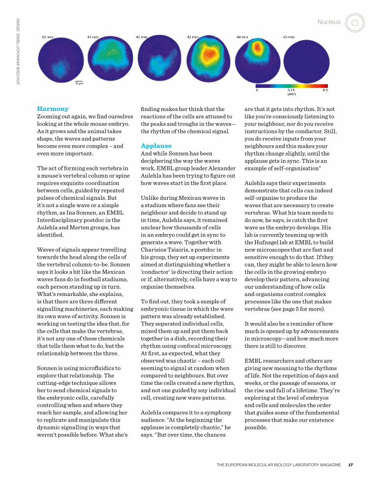
HarmonyZooming out again, we find ourselves looking at the whole mouse embryo. As it grows and the animal takes shape, the waves and patterns become even more complex – and even more important.
The act of forming each vertebra in a mouse’s vertebral column or spine requires exquisite coordination between cells, guided by repeated pulses of chemical signals. But it’s not a single wave or a simple rhythm, as Ina Sonnen, an EMBL Interdisciplinary postdoc in the Aulehla and Merten groups, has identified.
Waves of signals appear travelling towards the head along the cells of the vertebral column-to-be. Sonnen says it looks a bit like the Mexican waves fans do in football stadiums, each person standing up in turn. What’s remarkable, she explains, is that there are three different signalling machineries, each making its own wave of activity. Sonnen is working on testing the idea that, for the cells that make the vertebrae, it’s not any one of these chemicals that tells them what to do, but the relationship between the three.
Sonnen is using microfluidics to explore that relationship. The cutting-edge technique allows her to send chemical signals to the embryonic cells, carefully controlling when and where they reach her sample, and allowing her to replicate and manipulate this dynamic signalling in ways that weren’t possible before. What she’s
finding makes her think that the reactions of the cells are attuned to the peaks and troughs in the waves—the rhythm of the chemical signal.
ApplauseAnd while Sonnen has been deciphering the way the waves work, EMBL group leader Alexander Aulehla has been trying to figure out how waves start in the first place.
Unlike during Mexican waves in a stadium where fans see their neighbour and decide to stand up in time, Aulehla says, it remained unclear how thousands of cells in an embryo could get in sync to generate a wave. Together with Charisios Tsiairis, a postdoc in his group, they set up experiments aimed at distinguishing whether a ‘conductor’ is directing their action or if, alternatively, cells have a way to organise themselves.
To find out, they took a sample of embryonic tissue in which the wave pattern was already established. They separated individual cells, mixed them up and put them back together in a dish, recording their rhythm using confocal microscopy. At first, as expected, what they observed was chaotic – each cell seeming to signal at random when compared to neighbours. But over time the cells created a new rhythm, and not one guided by any individual cell, creating new wave patterns.
Aulehla compares it to a symphony audience. “At the beginning the applause is completely chaotic,” he says. “But over time, the chances
are that it gets into rhythm. It’s not like you’re consciously listening to your neighbour, nor do you receive instructions by the conductor. Still, you do receive inputs from your neighbours and this makes your rhythm change slightly, until the applause gets in sync. This is an example of self-organisation”
Aulehla says their experiments demonstrate that cells can indeed self-organise to produce the waves that are necessary to create vertebrae. What his team needs to do now, he says, is catch the first wave as the embryo develops. His lab is currently teaming up with the Hufnagel lab at EMBL to build new microscopes that are fast and sensitive enough to do that. If they can, they might be able to learn how the cells in the growing embryo develop their pattern, advancing our understanding of how cells and organisms control complex processes like the one that makes vertebrae (see page 5 for more).
It would also be a reminder of how much is opened up by advancements in microscopy—and how much more there is still to discover.
EMBL researchers and others are giving new meaning to the rhythms of life. Not the repetition of days and weeks, or the passage of seasons, or the rise and fall of a lifetime. They’re exploring at the level of embryos and cells and molecules the order that guides some of the fundamental processes that make our existence possible.
iMa
GE
: EM
BL
/Joh
aN
Na
BiS
ch
of
The european Molecular Biology laBoraTory Magazine 17
nucleus

eMBLetc. SPRIng 2016 18
SPIM doctorsAny high school student has probably watched little bugs bumping around under a microscope. Looking inside those organisms is trickier, but eMBL scientists continue to push the boundaries of what microscopes can doBy SaM LEMoNick

iMa
GE
: EM
BL
/aL
ES
Sa
ND
ro
cic
ca
rE
LL
i
nucleus
19The euROPeAn MOLeCuLAR BIOLOgy LABORATORy MAgAZIne
SPIM doctorsNew vision: this Selective Plane illumination Microscopy image by alessandro ciccarelli, a postdoc at EMBL Monterotondo, shows neurons located in the region of the brain enabling perception and voluntary movements. Different colours indicate different cell positions within the brain forest. “the image shows only ten percent of brain cells – if all the cells were visible, they would look like a dense ‘spaghetti bowl’, making it impossible to distinguish their elegant architecture,” says ciccarelli.
the image will feature as part of the 3D exhibition ‘Life in Perspective’ that will premiere in heidelberg this year. the initiative aims to share scientific data from EMBL using several visual techniques in an interactive way. attendees of EMBL’s annual reception on 3 March were given a sneak preview. EMBL scientists Gustavo de Medeiros, Stefan Günther and Giorgia Guglielmi, and EMBL designer Manuela Beck welcome ideas and support – contact [email protected]

The invention of Selective Plane Illumination Microscopy (SPIM), which allowed scientists to watch an embryo
develop for the first time, was hailed as a significant breakthrough. SPIM is still relatively new, only dating back to the mid 1990s. But its huge potential is already clear. What started in a physics lab led to the first groundbreaking insights in biology just a decade later. Now, with new papers and a new start-up, EMBL researchers show they are still adding new dimensions to the technology.
Let there be lightIt was in the late 1980s that Roelof W Wijnaendts-van-Resandt, Ernst Stelzer and members of his lab at EMBL started developing the multi-lens microscopes that would become SPIM. The simplest microscopes shine light on or through a subject, which an observer then looks at through a magnifying lens. Fluorescence microscopy — itself a
relatively new technique — uses dyes or genetic modifications that cause specific parts of a cell to glow when exposed to certain wavelengths of light.
Fluorescence microscopy is powerful, but the light that spurs cells to fluoresce also damages them. Stelzer’s lab found a solution that has unlocked huge opportunities. By confining light to a very thin sheet, they could light up just one plane at a time for the microscope to focus on, protecting the other parts of the sample. Repeating the process over and over gives a complete picture of the specimen.
‘Intellectual breakthrough’Jan Huisken was a PhD student in Stelzer’s lab in the early 2000s. A decade earlier, Stelzer’s lab had developed a fluorescence microscope that used two opposing lenses to get a full view of a sample suspended in a droplet of water. Huisken’s task was to use beams of light to manipulate a sample suspended in a water droplet
20 EMBLetc. spring 2016

— something that turned out to be very difficult. The group decided to look for an easier solution, and SPIM was born.
“There are moments when you can really say, ‘That is an intellectual breakthrough’,” says Stelzer, who is now at the Buchmann Institute for Molecular Sciences at the Goethe University in Frankfurt. It wasn’t long before Stelzer and his biologist colleagues were seeing the possibilities of SPIM.
An advanced version of SPIM was used to create the stunning videos of a growing fish embryo highlighted by Science as one of the breakthroughs of the year in 2008. EMBL biophysicist Phillip Keller’s movie let scientists track each individual cell, answering previously unanswerable questions about how animals develop.
The ability to rapidly image a large three-dimensional sample and the small amount of light used — SPIM’s
unique properties — make it possible to watch an entire living organism in exquisite detail. “People are naturally limited by the tools that they have available,” says Keller, who is now at Howard Hughes Medical Institute’s Janelia Research Campus. With SPIM, he and other researchers can take entirely new approaches to research questions.
Software upgradeInnovation didn’t stop there. SPIM is powerful, but the light can’t penetrate fully into larger samples, making for a blurry picture. Lars Hufnagel, a group leader at EMBL since 2007, helped develop a new technique called multi-view-SPIM, or MuVi-SPIM. It uses two light sheets on opposite sides of the sample and two imaging lenses to create a much clearer picture in much less time than on original SPIMs, where samples had to be rotated to look into them from multiple sides. Hufnagel’s lab has since further improved upon this principle. >>
iMa
GE
: EM
BL
/kE
LL
Er
et
al
.
Phillip keller’s digital zebrafish embryo provided the first complete developmental blueprint of a vertebrate, in 2008
“There are moments when you can really say, ‘That is an intellectual breakthrough’ ”
The european Molecular Biology laBoraTory Magazine 21

Considering SPIM’s growth so far, it seems almost inevitable that somebody will soon solve that problem. It’s the kind of improvement that seems especially possible at a place like EMBL.
Hufnagel says the twin missions of developing new technology and advancing the life sciences are what make EMBL suited to breakthroughs like SPIM. The many improvements and derivations EMBL researchers continue to make are further testament to that.
Strnad P et al. Nature Methods, 14 December 2015. DOI: 10.1038/nmeth.3690
de Medeiros g et al. Nature Communications, 25 november 2015. DOI: 10.1038/ncomms9881
Krzic u et al. Nature Methods, 03 June 2012. DOI:10.1038/nmeth.2064
Keller PJ et al. Science, 14 november 2008. DOI: 10.1126/science.1162493
>> Hufnagel described the key to that advance with his colleagues in a Nature Communications paper last month. In previous versions of this technique, the light sources had to illuminate the sample one at a time or the detector would be overwhelmed by scattered light.
Now, Hufnagel, in collaboration with Hamamatsu Photonics, has written a new computer program for the cameras that makes it possible to have both lights shine at the same time and detect only unscattered light. In addition to improved image quality, this doubles the speed of the experiment and halves the number of images the computer has to store and analyse. “The nice thing about this is it is just a software upgrade on the camera,” he says. Inverted SPIMThe MuVi-SPIM technology is being marketed by a new start-up called Luxendo. Hufnagel and Jan Ellenberg who is Head of the Cell Biology and Biophysics Unit at EMBL Heidelberg, founded the company with EMBL’s technology transfer arm, EMBLEM. “We were contacted by a number of labs that wanted to get access to this disruptive technology,” says Jürgen Bauer, EMBLEM’s deputy managing director. That demand inspired him to work with the EMBL scientists to create Luxendo.
Now Ellenberg and Hufnagel are working to take SPIM even further. Their latest paper, just published in Nature Methods, makes use of a development called inverted SPIM. The standard SPIM setup has the detector and light parallel to the lab bench. That means the sample has to be held in place vertically in a capillary tube or a gel. Embedding the biological sample in that very unnatural environment means fragile samples like mammal embryos couldn’t be imaged.
In inverted SPIM, the lenses point up at an angle from underneath the specimen, which simply rests on a glass slide like a conventional microscope. Without the need to embed and suspend the sample, the system is much easier to use. It might sound like a simple improvement, but it could answer questions about infertility. Many embryos fail in their very early stages after only dividing a few times. Some eggs don’t even make it to fertilisation before they die. “We don’t understand why that is, why the divisions aren’t happening properly. You have to be able to look inside the cells when they are having the problems,” Ellenberg says.
His paper describes the first two days of a mouse embryo’s development imaged with inverted SPIM. Mammal embryos die when embedded in gels or tubes for conventional SPIM, but inverted SPIM overcomes that problem, enabling scientists to watch live a process that they could thus far only speculate upon. The new research is a starting point for learning more about mouse embryos, which will be important to understand a major reason for human infertility.
Double troubleEllenberg and Hufnagel are planning to develop a multi-view version of this technique as well, but for now they say that simply doubling the number of lenses makes things a bit too crowded. Instead, they are working on more advanced optical designs.
“You have to be able to look inside the cells when they are having the problems”
22 EMBLetc. spring 2016

In synchrotronMeet researchers who are pushing the boundaries of X-ray crystallography experimentation at eMBLBy roSEMary wiLSoN
Ph
ot
o: r
oS
EM
ar
y w
iLS
oN
Synchrotrons are advancing research in fields as diverse as biosciences, forensics and particle physics
The european Molecular Biology laBoraTory Magazine 23
nucleus
Ph
ot
o: E
MB
L
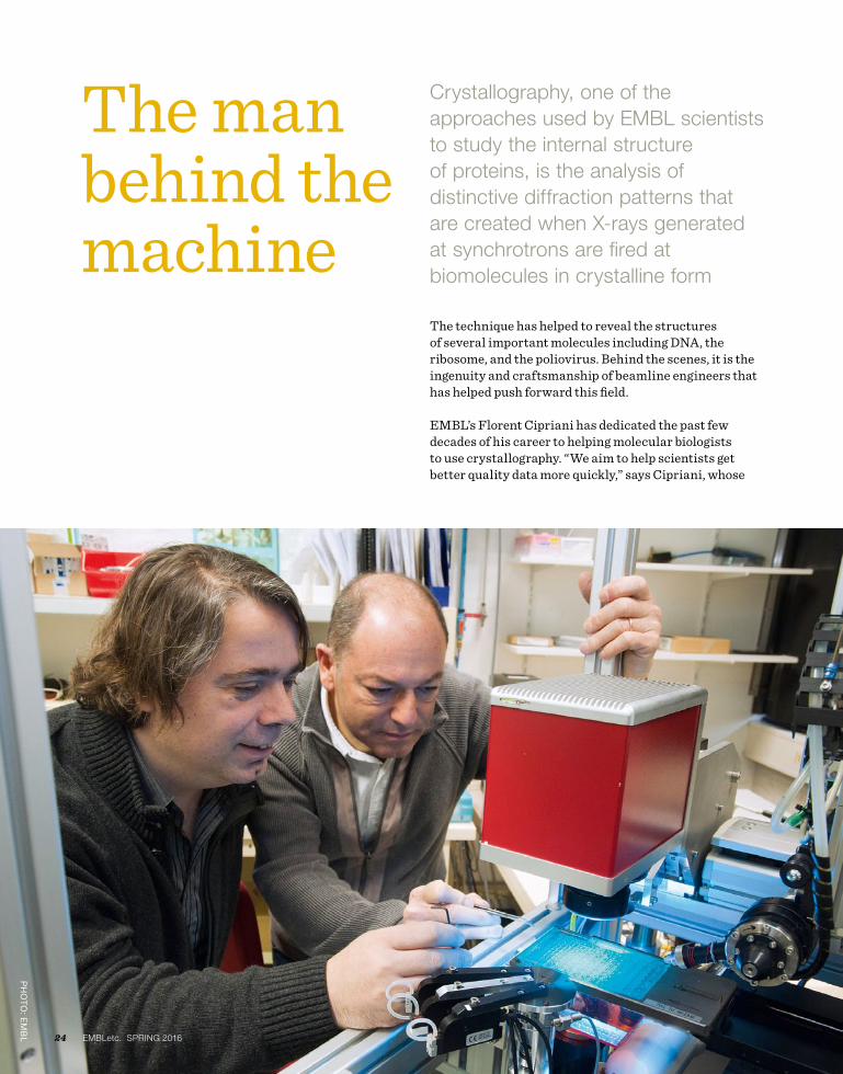
The man behind the machine
The technique has helped to reveal the structures of several important molecules including DNA, the ribosome, and the poliovirus. Behind the scenes, it is the ingenuity and craftsmanship of beamline engineers that has helped push forward this field.
EMBL’s Florent Cipriani has dedicated the past few decades of his career to helping molecular biologists to use crystallography. “We aim to help scientists get better quality data more quickly,” says Cipriani, whose
Crystallography, one of the approaches used by eMBL scientists to study the internal structure of proteins, is the analysis of distinctive diffraction patterns that are created when X-rays generated at synchrotrons are fired at biomolecules in crystalline form
24 eMBLetc. SPRIng 2016

team is located on the EPN – European Photon and Neutron – campus in Grenoble. Their innovations range from devices that enable the harvesting of extremely delicate crystals to helping in the analysis of diffraction patterns. “Beyond a passion for technical challenges, our driving force is really to make our beamline users happy,” he says. “I often say the instrumentation lab is in the basement of the EMBL building because we support the science!”
Cipriani’s team is currently commissioning a new sample changer for one of the ESRF crystallography beamlines run by EMBL in order to increase their capacity and to perform experiments more rapidly.
Handle with careSample changers take care of the biological material set to enter the X-ray beam. Before shutting the heavy lead door of the experimental hutch protecting them from the intense X-rays, scientists first load their precious frozen samples into a container (known as a Dewar) holding liquid nitrogen. Then, one by one, the samples are moved gently from the icy chamber and placed carefully and precisely into the X-ray beam by a robotic arm, ready for measurements that the scientist controls remotely from the other side of the door.
Automation means things get done quickly, safely, accurately and without the need for any human intervention. However, while producing good quality crystals of small and uniformly shaped proteins is difficult enough, crystallisation of larger and more complex proteins is even more of a challenge, usually producing crystals of poorer quality and smaller size. “Structural biologists frequently have to screen several hundred tiny crystals before they find any good ones and obtain the high resolution data needed to build atomic 3D structures to answer related biological questions,” explains Cipriani. And with such small delicate crystals with handling really becomes a problem.
“One of the last parts of the crystallography experiment that is still manual is moving the crystals from the lab to the beamline,” says Cipriani. “So we started wondering how we can automate this too.”
Cipriani’s team was one of the teams that developed CrystalDirect – an automated crystal harvesting technology. The miniscule, delicate crystals grown in the lab ready for beamline experiments must be
painstakingly hooked out of their plates using tiny nylon fishing loops and the naked eye. But working together with Josan Marquez, who leads EMBL’s high-throughput crystallisation laboratory in Grenoble, they developed a method that eliminates crystal handling by growing them on top of a thin film. This can be used directly in the X-ray beam after it is cut with a laser.
The first systems are already working in Grenoble and the first commercial product will be available at EMBL Hamburg in the summer. “Not only is it possible to easily extract single crystals but even entire plates can be put directly into the beamline circumventing the need for any manual handling at all,” Cipriani explains. “The system allows a high level of precision and flexibility and that’s what we always aim for.”
Cipriani is eager to bring these innovations to as many users as possible, and over the years several of his team’s developments have been commercialised and used worldwide. “People seem to appreciate them,” he says, pleased. Crucial to this process is the EMBL’s technology transfer activity, EMBLEM. “It’s sometimes difficult balancing different interests but the global benefit is huge and EMBLEM is a really important partner for opening up discussions with industry.” Next up is a self-cleaning Dewar. “It cleans out the liquid nitrogen, thus preventing ice crystals contaminating and degrading diffraction data,” he says enthusiastically.
Setting the standardRecognising the fact that scientists tend to visit several synchrotrons to conduct their experiments, Cipriani is also keen to see that equipment is standardised across all sites. He has been designing a new compact and precise sample holder as part of the EC-funded project BioStruct-X, proposed as a future European standard. “The standard we developed 10 years ago proved beneficial for scientists who now regularly visit several synchrotrons,” he explains. “The new standard should also speed up crystal processing while reducing handling and transportation costs,” Cipriani explains.
As scientists look to gain ever more detailed images of ever more complex biomolecules, much rests on the shoulders of beamline engineers. But Cipriani is confident that the flow of ingenious solutions will continue to enable researchers to push the boundaries of structural biology. “With so many projects, what is missing is time!” he adds with a smile. “Above all, good instrumentation is the fruit of the work of diverse people who like working together – what we call a team!” he concludes.
READ MORE ABOuT THE LATEST DEvELOPMENTS OF
CRySTALDIRECT ONLINE: NEwS.EMBL.DE/?p=6753
florent cipriani (right) developed crystalDirect together with Josan Marquez (left)
The european Molecular Biology laBoraTory Magazine 25
nucleus

How to make a protein happy
Two recipes developed to determine the ideal conditions for protein stability, purification and storage, making the process of protein handling more efficient are now commercially available. Stephane Boivin, a staff scientist who initiated the project, tells us more about the RUBIC Buffer and Additive Screens, and the journey from idea to commercial product.
What can you do with the rubIC screens? It’s not easy to find the best working conditions for a protein. Information such as how a protein will respond to a certain buffer (a chemical mixture that stabilises pH) and what pH it prefers is very important, but identifying these parameters is often a laborious and frustrating process of trial and error. The RUBIC screens help scientists systematically identify the conditions needed to keep their protein stable and happy. They are also compatible with a wide range of proteins, so one size fits all!
How do they work? The RUBIC Buffer Screen is designed to screen for general parameters that will keep proteins stable, while the RUBIC Additive Screen helps users identify ideal conditions for protein purification and storage. The screens – each prepared in a 96 well plate – can be used alone, or in a two-step approach. The Buffer Screen contains diverse solutions commonly used in structural biology experiments including differing salt concentrations, pH and buffer type. Once small amounts of the protein sample have been mixed with the 96 different conditions, we use a process called fluorescence-based thermal denaturation (or Thermofluor). With increasing temperature, the protein will begin to unfold, exposing its core so that a fluorescent dye included in the screen can bind to it, increasing the visible fluorescent signal. Users can then really see when their protein starts to fall apart and so identify the optimal conditions in terms of pH, salt concentration and so on. Once the general parameters suitable for the protein have been determined, the additive screen plate can be used to pinpoint the ideal conditions for purification and storage.
Tell us about the journey from idea to commercialisation… Over the years I have gained a lot of expertise using Thermofluor as a technique in the lab, and adapting it to specific projects and needs. As a postdoc at the University of Texas Health Science Center in Houston, I used Thermofluor to screen for buffer conditions to overcome antibody instability; I quickly realised it offers an effective way to explore stabilising conditions without using too much precious protein, providing some invaluable insights while saving a lot of frustration and time. I continued to use Thermofluor while at EMBL Grenoble, and when I moved to the SPC Facility at EMBL Hamburg we introduced it as one of a series of steps to help optimise experiments. In order to answer specific questions for in-house users we developed two screens that have now been tested on more than 300 different proteins – so we can safely say they are applicable in a wide range of cases! After publishing a paper, external users started requesting the screen, and so we began to distribute it as a prefilled, ready-to-use plate across EMBL and to other institutes. Biotech company Molecular Dimensions soon enquired about marketing the screen on a global scale.
What was it like to bridge the gap between research and commerce?Thankfully EMBLEM, EMBL’s partner for technology transfer, took care of all the legal and contractual issues. Having said that, we were in regular contact with Molecular Dimensions as they started to produce the screen, and while we tested and validated it before
Scientists at the Sample Preparation and Characterisation Facility (SPC) at eMBL’s hamburg site aim to help their users get the best results by optimising protein samples for structural biology experiments
26 EMBLetc. spring 2016

large-scale production. We shared the protocols and information about how to set it up, how to interpret the results, how to troubleshoot, and we answered any open questions. As a result, the accompanying documentation contains easy-to-follow instructions and explanation. All in all, it has been a very interesting process and it is very satisfying to know the screen is now available to help many more users with their research.
rob Meijers (left), head of the Sample Preparation and characterisation facility, together with Stéphane Boivin (right)
Ph
ot
o: E
MB
L/r
oS
EM
ar
y w
iLS
oN
“It is very satisfying to know the screen is now available to help many more users with their research”
The european Molecular Biology laBoraTory Magazine 27
nucleus

Periodic proteinsThe iconic periodic table of the elements has a new partner in predictive power: the predictable table of protein complexesBy Mary toDD BErGMaN
Just as different ballroom dances can be seen as an endless combination of a small number of basic steps, the ‘dance’ of protein complex assembly can be seen as endless variations on dimerisation (two units link together), cyclisation (three or more subunits for a ring) and heteromeric subunit addition (two different proteins bind to each other).
The new table, created by an interdisciplinary team from the EMBL-EBI, the Wellcome Trust Sanger Centre and the Cavendish Laboratory at the University of
Cambridge, provides a useful tool for biotechnology and the engineering of novel complexes.
The blank spots in Mendelev’s table were, as predicted, neatly filled as new chemical elements were discovered. By setting out the fundamental steps in the evolution of protein complexes, the new table helps researchers visualise the enormous variety of structures proteins can build, see which ones might be discovered next, and predict how entirely novel structures could be engineered.
Data in step“Evolution has given rise to a huge variety of protein complexes, and it can seem a bit chaotic,” explains Joe Marsh, formerly of the Wellcome Genome Campus and now of the MRC Human Genetics Unit at the University of Edinburgh. “But if you break down the steps proteins take to become complexes, there are some basic rules that can explain almost all of the assemblies people have observed so far.”
“By analysing the tens of thousands of protein complexes for which
28 EMBLetc. spring 2016
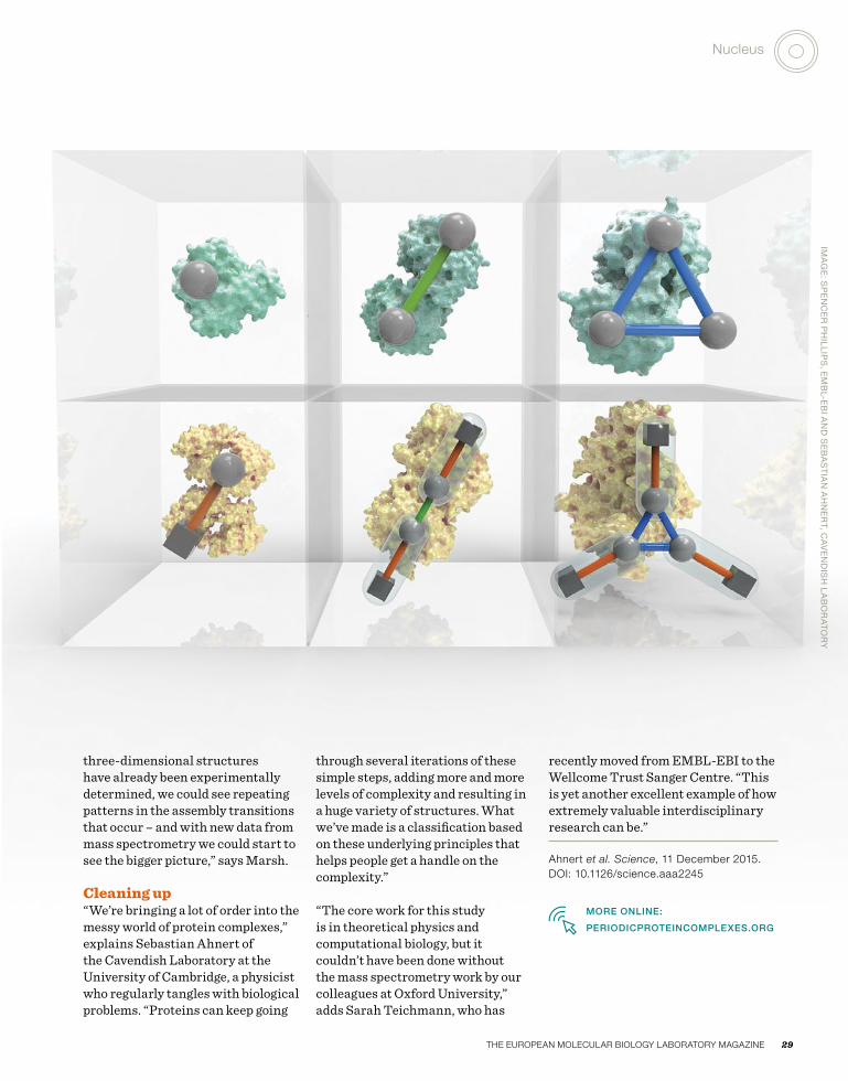
three-dimensional structures have already been experimentally determined, we could see repeating patterns in the assembly transitions that occur – and with new data from mass spectrometry we could start to see the bigger picture,” says Marsh.
Cleaning up “We’re bringing a lot of order into the messy world of protein complexes,” explains Sebastian Ahnert of the Cavendish Laboratory at the University of Cambridge, a physicist who regularly tangles with biological problems. “Proteins can keep going
through several iterations of these simple steps, adding more and more levels of complexity and resulting in a huge variety of structures. What we’ve made is a classification based on these underlying principles that helps people get a handle on the complexity.”
“The core work for this study is in theoretical physics and computational biology, but it couldn’t have been done without the mass spectrometry work by our colleagues at Oxford University,” adds Sarah Teichmann, who has
recently moved from EMBL-EBI to the Wellcome Trust Sanger Centre. “This is yet another excellent example of how extremely valuable interdisciplinary research can be.”
Ahnert et al. Science, 11 December 2015. DOI: 10.1126/science.aaa2245
MORE ONLINE:
PERIODICPROTEINCOMPLExES.ORG
iMa
GE
: SP
EN
cE
r P
hiL
LiP
S, E
MB
L-EB
i aN
D S
EB
aS
tia
N a
hN
Er
t, ca
vE
ND
iSh
La
Bo
ra
to
ry
The european Molecular Biology laBoraTory Magazine 29
nucleus

iLLu
St
ra
tio
N: ‘S
Er
vic
E u
PD
at
E f
ro
M E
MB
L-EB
i 30 EMBLetc. spring 2016
Growth and integration
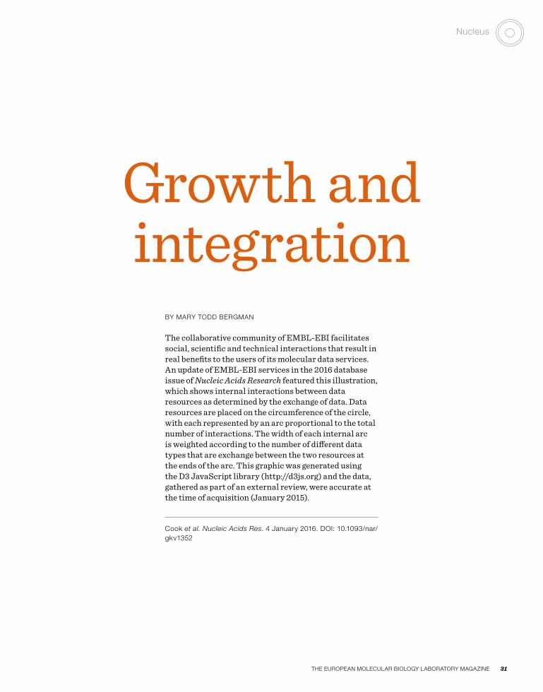
By MARy TODD BeRgMAn
The collaborative community of EMBL-EBI facilitates social, scientific and technical interactions that result in real benefits to the users of its molecular data services. An update of EMBL-EBI services in the 2016 database issue of Nucleic Acids Research featured this illustration, which shows internal interactions between data resources as determined by the exchange of data. Data resources are placed on the circumference of the circle, with each represented by an arc proportional to the total number of interactions. The width of each internal arc is weighted according to the number of different data types that are exchange between the two resources at the ends of the arc. This graphic was generated using the D3 JavaScript library (http://d3js.org) and the data, gathered as part of an external review, were accurate at the time of acquisition (January 2015).
Cook et al. Nucleic Acids Res. 4 January 2016. DOI: 10.1093/nar/gkv1352
The european Molecular Biology laBoraTory Magazine 31
nucleus

Ph
ot
o: E
MB
L Ph
ot
oL
aB
/Ma
riE
tta
Sc
hu
PP
32 EMBLetc. spring 2016

Lighting up development
A PhD student in the De Renzis group at eMBL heidelberg describes how optogenetics helped her to illuminate the path that tissues follow to get into shapeBy GiorGia GuGLiELMi
One of the most challenging experiences of my life was getting started with my PhD at EMBL, to study how organisms develop their shape, a process known as morphogenesis. Trained as a microbiologist, I was used to working with tiny single cells that displayed a rather simple behaviour. In the De Renzis group, I had to deal with fruit fly embryos made of thousands of cells that constantly exchange information and influence each other.
Understanding what is going on when an organism such as a fruit fly develops from egg to larva is a daunting task. Much of what we know about the biological processes that drive development comes from turning specific genes on and off and then watching for the effect. However, the commonly used techniques have a limitation: one has
to wait some time (several hours in the best case) before looking at the consequences of such perturbations and drawing conclusions. As my supervisor Stefano De Renzis puts it, “it’s like arriving at the scene of a car crash: you know something went wrong at some point, but you were not there when it happened, so you cannot tell what exactly caused the accident”.
Light control When I joined Stefano’s lab, my dream was to have a remote control to perturb the activity of single cells, and watch the effects immediately. That way, I thought, it would be much easier to understand when and where specific cell behaviours were needed during complex developmental processes. I thought that a feasible way to create such a remote control would be to use light. Back in 1999, Francis Crick
had already voiced the possibility of controlling neuron activity with light. Just a couple of years later, neuroscientists managed to use light to activate brain cells, and they named the newborn technique ‘optogenetics’. However, when I started my PhD in 2011, no one had ever used this technique to interrogate how cells behave and interact with each other in a whole organism during development.
I have always had a strong interest in biotechnology, and introducing optogenetics as a tool to study embryonic development seemed like the ideal PhD project for me. As an embryo develops, most of the changes in its shape are driven by cells contracting, so I decided to prevent them from doing so, by using light to reduce their ‘muscle fibres’. Work from our and other groups had shown that a particular fatty >>
The european Molecular Biology laBoraTory Magazine 33
nucleus

>> molecule in the cell membrane – a lipid called PI(4,5)P2 – serves as an anchor for the fibres that pull on the membrane when the cell contracts, and is essential for many developmental processes. Inspired by these findings, I set up an approach to use light to remove PI(4,5)P2 from the cell membrane, as this would prevent the fibres from latching on, meaning the fruit fly embryo’s cells would be unable to contract. One of the most exciting moments of my PhD was the day I went to the microscope and confirmed that I could stop individual cells from contracting with just a few millisecond pulses of light: my light-based remote control was working!
In the meantime, working close to morphogenesis enthusiasts in both Stefano’s group and the neighbouring Leptin lab, I got interested in how tissues bend to form folds or tubes. At around four hours old, for instance, cells on the underside of the fruit fly
embryo start moving inwards, folding into the embryo where they will eventually give rise to muscles. By using my brand new optogenetic remote control to block cell’s ability to contract, I found that there is a group of cells that is absolutely required for the tissue to fold inwards. What was even more fascinating was that the geometry in which these cells are organised within the tissue sets up the way they change their shape. It’s like when you bake a cake: it will take the shape of the tin you poured the mixture into. In the same way, the geometry of the tissue imposes some sort of constraint on the way individual cells behave.
Detective workWhen these results came to light, I needed to transform a lot of visual information into meaningful numbers, so I teamed up with Joseph Barry, then a postdoc in Wolfgang Huber’s group. Joe did an amazing job of developing algorithms to quantify cell features and discussing the huge amount of data I had produced: “For me the best parts were sitting together mulling over images, trying to figure out what was going on,” he now recalls.
The paper in which we describe this new optogenetic method to control cells’ ability to contract, and its application to study fruit fly morphogenesis was published in Developmental Cell, but collaborations with labs worldwide have already started. “Other scientists asked for help
“It is possible to blend scientific approaches that have traditionally been separate”
and reagents to modulate stem cell activity and to control neural tube formation in vertebrates,” explains Stefano. “In fact, cell contractility is a highly conserved cell behaviour that drives many morphogenetic processes, so I believe that this optogenetic approach will be applicable to other organisms to address questions about cell-cell interaction and tissue mechanics.”
My adventure as a PhD student at EMBL is almost finished, but Stefano has long-term plans to use light to quiz other key events that shape the embryo: “What we are doing now is engineering this optogenetic system to be able to control signalling between individual cells during morphogenesis and get an insight into cell communication. We want to know what kind of information cells exchange, when they exchange such information and how loud they ‘speak’, for instance.”
Keep an eye out: optogenetics will make you see development in a new light!
guglielmi g et al. Developmental Cell, 19 november 2015. DOI: 10.1016/j.devcel.2015.10.020
GuGLIELMI ExPLAINS THE
TECHNIquE IN THIS vIDEO:
NEwS.EMBL.DE/?p=5696
34 EMBLetc. spring 2016

Cultures
36 The search for EMBL’s next Director-General EMBL Director-General Iain Mattaj discusses the search for his successor
37 From the Archive: The Moon Dreamers Illustrator Edmond Baudoin donates an original artwork to the EMBL Archive
39 Reviews Life’s Blueprint: The science and art of embryo creation, by Benny Shilo
40 Q&A what is the one marine mystery you would most like to see solved?
41 Awards & honours
42 Alumni we meet Gareth Griffiths, the new Chair of EMBL’s Alumni Association and shine a light on EMBL’s administrative alumni as we say “farewell” to Keith williamson, EMBL’s ninth Administrative Director
47 Obituary Bernard Jacrot, Former Head of EMBL Grenoble (1980-89)
38 Branches: Laughing matter Following her EMBL Forum for Science and
Society lecture we caught up with Sophie Scott to discuss the neuroscience of laughter
Ph
ot
o: S
hu
tt
Er
St
oc
k35The euROPeAn MOLeCuLAR BIOLOgy LABORATORy MAgAZIne
Cultures

and implementing the successive five-year scientific programmes. Given the complexity of EMBL, it is important that the Director-General gains knowledge of the institute before developing their plan. The next programme runs from 2017 till 2021, so assuming office in January 2019 is ideal in this respect.
What are the key attributes for an EMbL Director-General?The candidate has to be a strong scientific leader, respected by the internal and external life science community. They should also be a competent administrator, a diplomat and good listener, capable of communicating effectively with the scientific community, staff at all five sites, EMBL Council and member states.
What are the challenges and opportunities for the next Director-General?There are always exciting scientific opportunities in the life sciences and potential for new focal areas for EMBL to identify and pursue. The challenge is to frame EMBL’s goals in such a way that explicitly demonstrates our added value; to show that the Laboratory brings something extra to European life science that would be difficult or impossible for national bodies to achieve. EMBL is an incredibly important institution in Europe and globally, and bears a responsibility to its community to be collaborative and defend the requirement for infrastructure, training and technology development necessary for groundbreaking basic research. EMBL has a freedom of scope that is unusual; it is not without constraint, but we have the privilege to shape accomplishments on a far-reaching world stage.
READ THE FuLL INTERvIEw AT
NEwS.EMBL.DE/?p=6616’
The search for EMBL’s next Director-General Iain Mattaj talks about the search for his successor as leader of Europe’s flagship laboratory for the life sciences
By ChLOë CROSS
Imagine taking the helm of one of the world’s most respected basic research organisations: would you crumble or thrive in a role so formidably demanding, unpredictable, influential and accountable? After over a decade as EMBL Director-General, Iain Mattaj is approaching the end of his mandate and the hunt for his successor is on. He talks about the search and selection process and the must-have qualities required to steer the Laboratory forward.
What is the process for finding the new Director-General?The search is managed by a committee led by the Chair of EMBL Council, comprising members of Council, our Scientific Advisory Committee, the Chair of
EMBO Council, and a small number of selected individuals. Candidates will be canvassed both through advertising and nominations to the search committee. The chosen individual must be approved by EMBL Council but will be a very senior and visible scientist, and that makes it uncertain how quickly they will be able to join EMBL. From my side, I’m very flexible about timing – the most important thing is that we find and help establish an excellent candidate.
With a three-year recruitment and handover timeline, how important is timing?One of the most important parts of the job, and the best chance as Director-General to influence the shape of the Laboratory, is developing
Ph
ot
o: E
MB
L Ph
ot
oL
aB
/uD
o r
iNG
EiS
EN
after more than a decade as EMBL Director-General, iain Mattaj is approaching the end of his mandate
36 EMBLetc. spring 2016

iMa
GE
: ED
Mo
ND
Ba
uD
oiN
/ cé
Dr
ic v
iLL
aN
i/ EM
BL a
rc
hiv
E D
E 23
24 P-B
au
-1
The Moon Dreamers
With the goal of telling the story of molecular biology firsthand, eMBL’s Archive is on the hunt for unique material significant to the Laboratory’s history: our new regular feature explores some of the highlightsBy aNNE-fLorE LaLoë
This edition we highlight an original artwork donated to the EMBL Archive by illustrator Edmond Baudoin. Last year, together with mathematician Cédric Villani, Baudoin published a book that explores the intense personal story of Leó Szilárd, a nuclear physicist who in part initiated the idea of EMBL. Les Rêveurs lunaires – or The Moon Dreamers – highlights socio-ethical issues intertwined with fundamental breakthroughs, also zooming in on the lives of code-breaker Alan Turing, quantum pioneer Werner Heisenberg and military commander Hugh Dowding. I caught up with Baudoin to find out more.
What did the book set out to achieve?While we might know about the discoveries our main characters are famous for, we might not often think about scientists’ human journeys. The four protagonists made significant contributions to modern science, but also had to confront significant moral and ethical questions. How did they feel about their contributions to knowledge when they looked back on their work? Were they proud, ashamed, or confused? Our book aims to illustrate – and even challenge – the role of science in our society through their experiences.
How did you draw each character? I adapted my drawing style depending on how easily I came to understand the individual. For Szilárd, as I learned about him, the different layers of his character and the complexities of his story meant that I wanted to use a special tool that scratched the surface of the paper – much like digging into his personality. During his life, Szilárd seemingly carries the weight of the world on his shoulders, together with the fear and personal tragedy that came part and parcel with being a Hungarian Jew in the first half of the twentieth century. I felt it was important to reflect this
complexity and layered depth in how I represented him.
Describe your dynamic with Villani… I was never really good at maths in school, so it was really magical for me to be approached by Villani, who liked my drawing style. The chemistry that developed between us was tremendous, and after reading his storylines over and over, the pages just flowed out – it felt like musical improvisation! But there was a lot of back-and-forth exchange. In one night scene, I had sketched a crescent moon and emailed the draft to Villani: he wrote back instantly explaining that on the evening of the scene – 6 August 1945 –, the moon would have been waning, not waxing and I redrew the page accordingly. It was a delight to work with someone with such meticulous attention to detail. For Villani, communicating science is a passion – almost an act of activism! Ultimately we both wanted to take advantage of the very popular medium of comic books to tell a narrative that could promote understanding of the role of science in a better world.
From the Archive
Ph
ot
o: t
ho
Ma
S S
aLv
a
The european Molecular Biology laBoraTory Magazine 37
Cultures

Branches Laughing matterFollowing her eMBL Science and Society Forum lecture, neuroscientist and stand-up comic Sophie Scott explains the complexity and social importance of laughter
By LAuRA hOWeS
How do you get an auditorium full of scientists to laugh? For Sophie Scott, the answer is simple: you play audio clips of people laughing uncontrollably and the reaction is infectious.
But for Scott, this is more that just a useful way to break the ice with her audience. She works on the neuroscience of laughter at University College London (UCL) in the UK. Her research looks at the physical process of laughing and also
its social role and how we use laughter to mediate relationships. Laughter isn’t just funny, she says, it’s also incredibly useful.
Imagine you have travelled to a foreign country where you don’t know the language or the culture; how do you communicate? It turns out that there are very few universal expressions and exclamations. Most people will recognise an expression of fear or distaste, but joy? Pleasure? One positive emotion that Scott has
Ph
ot
o: u
Niv
Er
Sit
y c
oL
LE
GE
LoN
Do
N
38 EMBLetc. spring 2016

shown to be universal is laughter. On the savannah of Africa, during Scott’s research, when a hunter feels silly or embarrassed he begins to laugh and soon he and Scott’s research team are laughing together.
Infectious interactionsBack in the auditorium, Scott uses audio clips to get us laughing. It is not just the audio track that’s making us laugh, she says, but also the reactions of our neighbours and colleagues sitting next to us. We might think that we laugh at jokes, but we laugh mainly to interact with other people – a trait that extends across mammalian species, from primates to rats, for whom laughter is associated with play and tickling, just like with us. “It’s a very social behaviour,” Scott explains. We interact with people by talking, which is a solely human skill, but then we use a very old mammalian behaviour, laughter, to show people that we like them.
If you ever hear someone laugh, says Scott, your brain activates neurons and gets ready to laugh as well, because we are primed to join in. There are two types of laughter – the truly involuntary laughter during which you can hardly breathe, and a ‘fake’ kind used as a social lubricant – and Scott has shown that the brain activation still responds to ‘fake’ laughter. This is true even though the two types have different physical and neurological signals and we can tell the difference between the two. Scott’s research has shown not only that both types of laughter trigger neurons prompting us to join in, but also that ‘fake’ laughter prompts our brains to try and understand why the person is laughing and how we should respond.
So laughter is a social tool that we use to convey different emotions, to bond with people and to signify that we are not a threat. And, says Scott,
ReviewsBy gIORgIA gugLIeLMI
Life’s Blueprint: The Science and Art of Embryo Creation Benny Shilo Benny Shilo is a multitalented man: his involvement as a molecular geneticist studying embryo development at the Weizmann Institute of Science is complemented by a sharp eye for patterns and a talent for photography. He combined these two interests into Life’s Blueprint, a work that reveals the wonders of developmental biology to the general public. From the role of genes in determining cell fate to the generation of complex tissues shapes, through to cell-cell communication, Shilo skillfully uses metaphors and simple, yet accurate, language to convey complex notions. He concludes by discussing future perspectives, including the possibility of generating healthy tissues and organs for medical purposes.
The understanding of key notions is facilitated by beautiful and meaningful visual cues: both a scientific image and a photograph of the macroscopic world, taken by Shilo, accompany each concept. Those having a deeper interest in the field can also find a list of recommended readings and web resources, together with a section providing details about the scientific images and the techniques used to acquire them. All these features make Life’s Blueprint indispensable reading for specialists and non-specialists alike.
although we can tell the difference between ‘fake’ and ‘real’ laughter, the ‘fake’ laughter is still useful for these social roles.
Scott recalls an incident she recently saw on the train to illustrate her point. Two men sat down at a table on the train, joining a woman who was working. The woman got up to move away, but as she was explaining why (she said she didn’t like the smell of the men’s coffee), she laughed and so made them laugh as well. This, says Scott, was the perfect way to defuse the situation and make sure that she didn’t offend the two men. Yet, says Scott, she is sure that if she had asked any of the people involved, they wouldn’t have remembered the laughter at all.
Learning from laughterAccording to Scott, we all underestimate how much we laugh even though we use laughter every day to promote bonding and de-escalate confrontations and negative emotions. For those of us who talk about liking people who make us laugh, Scott has a revealing takeaway message. Rather than liking people because they make us laugh, perhaps we laugh with them because we already like them.
Understanding the behavioural and neurobiological bases of laughter, says Scott, means more than thinking about jokes. It could provide a vital link between human language, relationships and emotional states. So don’t just ask for someone with a good sense of humour when writing your dating profile; pay attention to see if you and your date enjoy laughing together.
This article first appeared in issue 34 of Science in School
The european Molecular Biology laBoraTory Magazine 39
Cultures

Q&A What is the one marine mystery you would most like to see solved? What diatoms doI would like to know what diatoms really do in the ocean. Diatoms are photosynthetic microscopic organisms that come in diferent shapes, but always with transparent cell walls; they are present in pretty much all aquatic environments wherever there is sufficient light for photosynthesis. Textbooks claim many thing about diatoms: that they carry out as much photosynthesis as rainforests – about 20% of the global total! -, that they are really important for sequestering CO2 from the atmosphere in the ocean for long periods of time, or that they grow very fast when conditions are favourable and can survive for long periods in harsh conditions. But most of this is mostly based on fragmentary information and extrapolation. I’d like to rigorously address these issues and get a more accurate understanding of the role of diatoms in our ocean and compare it with the other organisms that share their planktonic world.
Chris Bowler, CNRS Director of Research, IBENS
The big questionsWhat strikes me most is how much we still don’t know. The initial results of the Tara Oceans expedition gave us better estimates of the number of different microscopic species in the oceans, and how many and what kind of genes they encode. We also have a clearer idea of the composition and interactions within the planktonic communities, and which environmental factors influence them. However, we still need to put all this information into context and answer some big questions: how much carbon can the oceans still absorb and how will this affect its biodiversity and factors like photosynthesis, pH, and dissolved oxygen? Is there a tipping point beyond which the ocean ecosystem cannot cope anymore, and will we be able to recognise (and act against) it? As a scientist, I feel very fortunate to be exploring this immense treasure chest of opportunities.
Shinichi Sunagawa, staff scientist, EMBL Heidelberg
how life evolvedI’d like to know how life evolved in the ocean. What did the first animals look like? What did the first central nervous system look like? And how did we get from that to a human brain capable of asking these questions? We can’t go back in time to see, but we can look for clues in what lives in the ocean today. We can look at marine organisms that we believe are similar to the animals that lived in the ocean millions of years ago, when the first nervous systems were evolving. By doing so, we have recently found hints that the first vertebrate skeleton may have evolved from muscle, and that melatonin – the hormone behind jet-lag – has probably been governing animal’s daily rhythms for hundreds of millions of years, first controlling the daily upwards swimming of marine larvae. I’d like to keep diving deeper into these questions, using the sea as a window into our distant past.
Detlev Arendt, group leader, EMBL Heidelberg
Ph
ot
o: E
MB
L Ph
ot
oL
aB
Ph
ot
o: E
MB
L Ph
ot
oL
aB
Ph
ot
o: iB
EN
S
40 EMBLetc. spring 2016

Ph
ot
o: E
MB
L Ph
ot
oL
aB
Ph
ot
o: E
MB
L Ph
ot
oL
aB
Awards & honours
The highest distinction in France, the Order of Légion d’Honneur, has been awarded to Anne Ephrussi, Senior Scientist and Head of the Developmental Biology Unit. She also heads the EMBL International Center for Advanced Training (EICAT), which comprises the EMBL PhD and Postdoctoral Programmes, as well as the Laboratory’s advanced scientific training activities, including the Courses and Conferences programme. The French Légion d’Honneur is an order of chivalry established by Napoleon Bonaparte to recognise eminent accomplishments of service to France; for two centuries, the Order has been presented annually on behalf of the French Head of State to deserving individuals in all fields of activity. Ephrussi is conferred with the rank of Chevalier de la Legion d’Honneur (Knight of the Legion of Honour), for outstanding merit acquired in 23 years’ service to the nation. Ephrussi received the medal of the Order from Professor Georges Cohen at a ceremony at the Institut Pasteur, in November 2015. “Such recognition from my home country to which I feel very committed is a tremendous honour both professionally and personally,” she says. “The intimate ceremony with a few close family members, colleagues and friends, and the fact that the medal was pinned on me by Georges, an old friend and mentor, made this a very happy and special event.”
Jan Ellenberg, Head of the Cell Biology and Biophysics Unit and coordinator of the preparatory phases of the Euro-BioImaging project, will be recognised as an Honorary Doctor of Philosophy by Åbo Akademi University in Turku, Finland. The title is awarded for scientific merit within cell biology, and commitment to the biosciences and Åbo Akademi University; specifically, his role in advancing the University’s cellular research and Turku BioImaging, which is part of the Euro-BioImaging project. The honorary doctorate will be formally conferred at a ceremony in May 2016.
This year’s Chica and Heinz Schaller Research Award has been presented to EMBL group leader Edward Lemke and Hai-Kun Liu from German Cancer Research Center (Deutsches Krebsforschungszentrum, DKFZ). The award, named after its donors, endows each recipient with 100,000 euros in research funding, in recognition of outstanding, innovative work in the field of biomedical research.
Robert Petryszak, team leader at EMBL-EBI, had been acknowledged for leadership in gender equality on the Wellcome Genome Campus with the Best Practice for Supporting Women in Science Award 2016. The award is part of the campus Sex in Science programme, a joint initiative with the Wellcome Trust Sanger Institute to generate discussion and raises awareness about issues traditionally facing women in science, and drive policy and practice changes to redress them. Petryszak was nominated for building flexible working into his team structure from the outset.
Ph
ot
o: c
hS
St
ift
uN
GP
ho
to
: Ma
rc
fo
LL
aN
D/S
aN
GE
r iN
St
itu
tE
from left to right: iain Mattaj, Edward Lemke, hai-kun Liu, and hannah Monyer at the ceremony of the chica and heinz Schaller research award
robert Petryszak and Sumana Sharma (centre, with certificates) received their awards from EMBL-EBi Directors rolf apweiler (far left) and Ewan Birney (second from right) and Sanger institute Director Mike Stratton (far right)
The european Molecular Biology laBoraTory Magazine 41
Cultures

This edition features a warm welcome to the new Chair of the EMBL Alumni Association, Gareth Griffiths, and a fond farewell to EMBL’s ninth Administrative Director Keith Williamson as he retires. We also take a look at the achievements of alumnus Jozef Ševčik, who is helping us build a community in the Slovak Republic, catch up with administrative alumni and announce the winners of this year’s John Kendrew and Lennart Philipson awards. Last but not least, we would like to draw your attention to two treasure troves in which we have compiled valuable information for the EMBL community: ‘Alumni Resources’ (embl.org/alumni/resources) and ‘Make a Difference’ (embl.org/alumni/makeadifference). Please view, use and share them!
Mehrnoosh RaynerHead of Alumni Relations
AlumniP
ho
to
: EM
BL P
ho
to
La
BP
ho
to
: uw
E D
Et
tM
ar
, Go
Et
hE
uN
ivE
rS
ity
Ph
ot
o: M
ar
JaN
vE
rM
aa
S
Former EMBL Genome Biology PhD student, Jop Kind, has been selected as this year’s John Kendrew Young Scientist Award winner for pioneering postdoctoral work in establishing novel technologies to map chromatin domains in single cells. Jop, now Group Leader at the Hubrecht Institute in Utrecht, has also received the Antoni van Leeuwenhoek prize for young researchers at the Netherlands Cancer Institute. “I am deeply
Ernst Stelzer, former EMBL Cell Biology and Biophysics Group Leader, has been selected as the 2016 Lennart Philipson Award winner. The award, which recognises outstanding contributions to translational research and innovation, was given to Ernst for the development and application of new light microscopy methods. Ernst is now Professor at the Goethe University in the Life Sciences Department and in
The ceremony will take place at EMBL Lab Day at EMBL Heidelberg on 22 July. All staff and alumni are invited to attend.
the Buchmann Institute for Molecular Life Sciences, in Frankfurt. “Light sheet-based fluorescence microscopy is the result of more than 25 years of hard work,” Ernst says. “As a member of several Units, I saw the limits of ‘flat biology’, the challenges of studying specimens in 3D, and the necessity for time-lapse imaging. But, crucially, I also had the resources to tackle challenges in a biological manner.”
Light sheets ahead
Charting the wayhonoured to find my name among this select group of excellent scientists who have won this award,” Jop says. “The innovative and out-of-the-box approach to research at EMBL really shaped me as a scientist and was hugely important en route towards independence.”
42 EMBLetc. spring 2016

Ph
ot
o: E
MB
L/M
.ra
yN
Er
Close associationFew scientists have experienced EMBL from as many perspectives as the new Chair of EMBL’s Alumni Association, Gareth Griffiths
Beginning his EMBL career in 1977, shortly after the Lab was founded, he spent more than three decades in the Cell Biology and Biophysics Unit at EMBL Heidelberg leading a group at the heart of the cryo-based electron microscopy revolution. Gareth Griffiths also trained thousands of scientists, and launched memorable networking and social events on campus. In 2009 a new challenge awaited at the University of Oslo, where he took up a professorship – but the chance to remain connected with EMBL proved irresistible. After four years as a member of the Alumni Association Board, Gareth now takes over the reins as Chair.
How do you feel to have been elected Chair?It’s an honour! Many people who leave the good life at EMBL find it hard to adjust elsewhere, and I recently joked that joining the Alumni Association
Board is a platform to overcome the withdrawal symptoms! It provides a means to keep in touch with former friends and colleagues – and of course EMBL as a whole. We on the board feel that we gained so much from the privilege of working at EMBL that we are delighted to give something back.
What are your main priorities? We are ambassadors for EMBL, and as such we want to further develop connections between alumni, the Laboratory, its member states, current staff and the public at large. We want to spread the word to those not in the know about EMBL, to drive forward initiatives that support the Laboratory’s missions, and connect further with the wealth of experience and knowledge of EMBL alumni spread across Europe and the rest of the world. The Alumni Association has provided the seeds for important initiatives, including EMBL’s Archive and the John Kendrew and Lennart Philipson Awards, and we want to continue to develop programmes that support and encourage lifelong connections to EMBL.
Describe some memories from your time at EMbL… New for me when I joined EMBL was the near absence of any barriers or hierarchy between the scientific groups, a feature that still holds true to this day. Also crucial to the early success of the Cell Biology Unit was a diverse group of talent combined with an active networking and social programme. A role that I took responsibility for from the earliest days was as a kind of ‘social secretary’, which in practice meant organising Unit retreats, visits to wine festivals, and infamous leaving and national parties. The goal was simple – to bring people together and have fun, and lots of it. This mix of science and social life meant that we managed to create an atmosphere that was unique. Many visitors who had a flavour of this ‘club’ ended up applying for positions here. And what this meant in practice was that wherever there was an international meeting attended by present or former EMBL members, they would naturally find each other, like family members tend to do.
How does this compare to today?Today’s EMBL has retained the essence of this special atmosphere: a sense of family, personal friendships, helping one another, strong networking and a passion for excellent science. We look forward to continuing this.Gareth played a key role in some of EMBL’s legendary social events,
such as David Meyer’s leaving party – pictured John tooze, kai Simons and Lennart Philipson
Ph
ot
o: E
MB
L/M
.ra
yN
Er
The european Molecular Biology laBoraTory Magazine 43

it was with typical humour that EMBL’s ninth administrative director donated a sculpture to the Laboratory called ‘the missing G’, with the caption “a Gift from keith williamson, who always felt the atc (an abbreviation for EMBL’s advanced training centre) was missing a G”
Admin ability“Working in administration, perhaps the best you can hope for is that no one notices you are there!” Keith Williamson, eMBL’s ninth administrative director, often jokes.
By ADAM gRISTWOOD
Yet during a special event at the end of last year at EMBL Heidelberg, behind-the-scenes business went centre stage as staff and alumni turned out in force to celebrate Keith’s retirement. Beginning his EMBL career as head of personnel, leading the finance department and eventually taking over as administrative director, Keith’s extensive contribution – combined with collegiality, friendliness and a wry sense of humour – defined 14 successful years at the Laboratory. Amidst the mirth and music we turn the spotlight on EMBL’s administrative alumni.
What is special about EMbL’s administration? The science we see coming out of EMBL is truly exceptional, but for administration such inventiveness can create a lot of challenges! Our ultimate goal is to make administration, which
is an inherently rigid activity, responsive to the fluid nature of science. The enriching international environment, spread across five different sites, also presents administrative challenges. One way we respond to this is by combining long-term strategic planning with an intimate understanding of what individual scientists want to achieve in their projects. Essentially, we want to minimise bureaucracy so that scientists can focus on their science. Intertwined with these goals is an emphasis on openness, helpfulness and developing a friendly and cooperative atmosphere – administration is a key part of a very special culture that embodies the whole Laboratory.
What are the most important things you learned at EMbL? The melting pot of cultures and languages creates varying expectations and aspirations.
It is important to take time to understand how people and processes work – getting to know people, and building relationships enable us to support the Laboratory’s activities in a very effective way. At the same time, if something doesn’t help science move forward, we stop it, and find another solution that does. I also learned it is important to have fun along the way!
What are your lasting memories? Anyone who’s worked at, or visited, EMBL knows that it’s a special place, and you feel that as soon as you walk through the door. Having people from more than 80 countries in a single institute means that there is a constant flow of ideas and different ways of thinking. Furthermore, the very constructive relationship between EMBL and it’s governing Council means that the Laboratory can continue to play a leading role in the molecular life sciences, and the new scientific programme is testament to that. The people who make EMBL EMBL are friendly, committed and enthusiastic. If you have a great idea, you have the freedom to turn it into reality. I retire with a heavy heart, but I am looking forward to watching this next chapter from the sidelines.
Ph
ot
o: E
MB
L/u
Do
riN
GE
iSE
N
44 EMBLetc. spring 2016

Rosie Maccagnano Then: Senior Administrative Officer, EMBL Monterotondo (1999–2007) Now: Research Support Services Manager, university of Edinburgh
yann Chabod Then: Budget officer, Head of Budget Office, Head of Human Resources (2006–8) Now: Director, Human Resources Customer Interface, European Patent Office, Munich
The birth of an Italian outstation
Open opportunities
I was there at the birth of EMBL Monterotondo, and it was incredible to be involved in setting up an outstation from scratch. Although there were many challenges initially, I built up a strong working relationship with my colleagues in Heidelberg and in the other outstations, and relied on them to keep my sanity! This was key to maintaining that ‘EMBL family’ feeling – in the sense of close-knit colleagues but also in the sense of ‘family-centric’. Most organisations are still trying to catch up with the
family-friendly policies, support and services that EMBL has had for years. Looking back, I feel proud to have been a part of it. No day was the same – my role covered human resources, health and safety, finance and more besides. I learned many valuable skills, and it was a wonderful place to work.
Professional services I am now building a support team that easily responds and adapts to any new requirements or changes as well as offering support for core
service activities such as academic support and research communications. There is a lot of flexibility to this role, and I am thankful to be able to draw on many of the skills that I developed in Monterotondo to address these new challenges. There are exciting opportunities here to support scientists in ever-creative ways, while also developing professionally.
Secret societyRecently I met another former EMBLer. We discovered we were both at EMBL at the same time – she as a postdoc in Frank Gannon’s team and I in Monterotondo. What was surprising was the revered way we both spoke of EMBL. Almost anyone who is part of the lifelong network of alumni cannot help getting sentimental: it is like being part of a secret society, which includes everyone who has ever been part of the EMBL story.
EMBL is a place with lots of creative young people who are very passionate about science and research. It is also a place where people can continuously develop themselves professionally. This is also true for people working in the Lab’s administration. I took the unusual step of moving from the budget office into human resources, and I was supported at every turn – it was a very important time in my career and paved the way to my
current role at the European Patent Office (EPO). What is striking about EMBL’s administration is how well it fits to the needs of the organisation – it is the open culture and strong service support that makes this dynamic work so well.
International interchangeI am now responsible for the frontline delivery of the human resource service to staff at the EPO. Like EMBL, the EPO has many
different sites across Europe and we provide support to line managers as well as being a first point of contact for staff members. There are a lot of other parallels to EMBL in the sense that the EPO has a strong focus on driving innovation, and the working environment is truly international. It is a challenging job, but highly motivating and interesting.
Continued connectionsComing back to visit as an alumnus, I see many new faces and buildings – but the vibrant and friendly atmosphere remains the same. I have kept in contact with several friends and colleagues, and although many have their own ‘new’ lives now I feel very connected through the alumni network. There is a fantastic energy to the place. À la prochaine!
Ph
ot
o: E
MB
L
The european Molecular Biology laBoraTory Magazine 45
Cultures

Ph
ot
o: r
oS
tiS
La
v S
kr
aB
aN
a
Slovakian synergiesResearchers from EMBL and the Slovak Republic, which joined EMBL as prospect member state in 2014, convened at the Institute of Molecular Biology (IMB) in Bratislava on 25 February to explore opportunities for further cooperation and collaboration
As a prospect member state, the Slovak Republic holds observer status in EMBL’s governing body – the EMBL Council –, and Slovak scientists are granted access to EMBL’s facilities and services, while the country’s early career researchers are also eligible for scholarships in the PhD and Postdoc programmes. The driving force behind the meeting was EMBL
Hamburg alumnus Jozef Ševčik, who mobilised his contacts at the IMB to raise awareness of the opportunities of EMBL prospect membership.
Why is cooperation between EMbL and Slovakia important?The EMBL event in Slovakia highlighted to our research community the opportunities that collaboration with EMBL can offer scientists in the country – including access to incredible facilities, services and training programmes. Connections with EMBL provide great potential to speed up the progress of molecular biology in our country. Slovakia has attracted an increasing number of grants to help reduce the gap between our research and that carried out in Western Europe, and greater collaboration with EMBL can only enhance this further – these are exciting times!
Tell us your EMbL story… My career in protein crystallography – and my introduction to EMBL – started at the University of York, where I solved the structure of RNase Sa and met Keith Wilson who later became the head of EMBL Hamburg. There were no facilities for protein crystallography when I returned to Slovakia, so Keith invited me to work as a postdoc in collaboration with EMBL. During the 1990s, I visited Hamburg 14 times, spending 16 months there in total. Our goal was to fully understand the catalytic mechanism performed by RNase Sa, and to determine the structures that enabled this at the highest possible resolution. Our cooperation resulted in 10 publications. During this time, I met excellent scientists at EMBL and experienced a truly inspiring and creative atmosphere. It was an experience that helped me build an independent protein crystallography laboratory at my home institute in Bratislava.
Have you been back since? After returning to my home institute in 1999, I visited Hamburg 11 more times accompanied by my PhD students to collect synchrotron data from our crystals, which helped us determine the structures of several proteins, and which in turn resulted in around 20 scientific papers. As I am now retired, I look forward to observing the exciting developments at EMBL and in Slovakia from the sidelines, while I continue to read books, spend time at the computer, and enjoy leisure activities.
MORE ONLINE:
NEwS.EMBL.DE /?p=6811
Jozef Ševcik at the X13 beamline at the DoriS ring in hamburg
46 eMBLetc. SPRIng 2016

Obituary Bernard JacrotBernard Jacrot, who was Head of EMBL Grenoble from 1980 to 1989, passed away on 21 January 2016 at the age of 89
By SOnIA FuRTADO neveS
Jacrot’s appointment as Head of EMBL Grenoble in 1980 brought him from Cambridge – where he had been at the MRC Laboratory of Molecular Biology – back to the campus where he had been Director of ILL a decade earlier.
A biophysicist with a keen interest in applying neutron science to biology, Jacrot was ideally placed to lead
the outstation at this time. EMBL had just established its labs next to the ILL reactor, and under Jacrot the two institutes signed a number of agreements that are still in place and which bear fruits to this day.
Under Jacrot, EMBL Grenoble reinforced its technology development and service functions, while building a tradition of strong in-house research – including Jacrot’s own structural studies of viruses.
Alongside his scientific contributions, Jacrot authored two books that reflect his passion for two core values that he shared with EMBL: interdisciplinarity and international
collaboration. Neutrons for science describes the history of ILL as an example of the latter, while Physics and Biology explores the links that can be made between these two sciences.
“Bernard was both demanding and human, and knew how to bring out the best in his staff,” says Joseph Sedita from EMBL Grenoble Facility Management. “After leaving the outstation, he’d regularly come back to visit us, and left nothing but good memories.”
EMBL pays tribute to those memories and offers condolences to Jacrot’s family and friends.
Ph
ot
o: iL
L
Bernard Jacrot in the late 1960s
The european Molecular Biology laBoraTory Magazine 47
Cultures

Events
vIEw THE COMPLETE
LIST OF EvENTS ONLINE
EMBL.ORG/EvENTS
EMBL HeidelbergEMBL conference: European conference of Life Science funders and foundations
EMBL Monterotondo EMBL Distinguished visitor Lecture: Epigenetics and the determination of phenotype – Emma whitelaw, La trobe institute for Molecular Science
EMBL-EBI course: Networks and Pathways
14 april: MDC Career Day with EMBL Alumni, Berlin 23 May: Local Chapter UK, University of Cambridge27 May: Local Chapter meeting Italy, Sapienza University, Rome
June
1
EMBL HeidelbergScience and Society: Experiments in democracy: why should we care about public engagement in science? – James wilsdon, university of Sheffield
EMBL HeidelbergEMBo | EMBL Symposium: New Model Systems for Linking Evolution and Ecology
The Cambridge Union Society, Cambridge Science and Society: rewriting the code of Life: the science and ethics behind genome editing
April
26-27EMBL GrenobleEMBL Distinguished visitor Lecture: alex Bortvin, carnegie institution for Science
April
25
May
8-11May
9-13
upcoming meetings
Alumni
April 29May
4
iMa
GE
: Dr
Ea
MS
tiM
EiM
aG
E: E
MB
o/a
Dit
ya
ku
Su
Ma
Jat
i
iMa
GE
: EM
BL-E
Bi/ S
PE
Nc
Er
Ph
iLL
iPS
iMa
GE
: EN
zy
MLo
Gic
(cc
-By
-2.0)
iMa
GE
: