EM Sample Preparation - SEMAT 16/EM SAMPLE... · EM Sample Preparation Techniques and pathways By...
46
EM Sample Preparation Techniques and pathways By Leica Nanotechnology Santiago Sevillano Page 1 Workshop SEMAT 2012 17/10/2012
Transcript of EM Sample Preparation - SEMAT 16/EM SAMPLE... · EM Sample Preparation Techniques and pathways By...

EM Sample Preparation
Techniques and pathways By Leica Nanotechnology
Santiago Sevillano
Page 1 Workshop SEMAT 2012 17/10/2012

Page 2 Workshop SEMAT 2012 17/10/2012
Workshop SEMAT 2012
ULTRAMICROTOMY / CRYO-ULTRAMICROTOMY LEICA UC6-FC6
TRIMMING/ KNIFE-MAKER LEICA KMR2
CRITICAL POINT DRYING IN BRAGA?
CONTRASTING /LABELLING PROCESSING/ DEHIDRATATION CHEMICAL FIXATION RESIN EMBEDING
COATING HIGH VACUUM EVAPORATOR POLARON CRESSINGTON SPUTTER
FREEZE ETCHING / FREEZE FRACTURE
CRYO-TRANSFER FREEZE SUBSTITUTION
CRYO-FIXATION HIGH PRESSURE FREEZING
CRYO-FIXATION BARE GRID TECHNIQUE PIN INMERSION
CRYO-FIXATION SELF PRESSURE FREEZING
TARGET PREPARTION (CUTTING, MILLING, SAWING, POLISHING, GRINDING) IN MINHO?
ION BEAM CUTTING ION MILLING SYSTEM
SOME TECHNIQUES….

Page 3 Workshop SEMAT 2012 17/10/2012
Tokuyasu technique
RT processing
(Cryo)TEM/SEM
High pressure freezing
Plunge freezing
Metal mirror
At High Pressure
At Ambient Pressure
Cryosectioning
LT processing
Chemical fixation Cryofixation Living Specimen

Knifemaking Trimming Ultrathin sectioning
Contrasting or Immunogold
labelling
KMR3 Trim2 UC7 IGL
AC20
Fast
Standard
TP
AMW
EXAMPLE: Workflow of Room Temperature Specimen Preparation for TEM
Tissue Processing
Page 4 Workshop SEMAT 2012 17/10/2012

EM UC7:Ultramicrotome
EM KMR3 : Knifemaker
Page 5 Workshop SEMAT 2012 17/10/2012

The Leica Ultracut EM UC7
Page 6 Workshop SEMAT 2012 17/10/2012
• is used for ultrathin sectioning for TEM
• 3D reconstruction (tomography, serial sectioning)
• Semithin sections for LM and FT-IR
• Surface planing of AFM and SEM samples. Industrial
material manufacturers and research (polymer, rubber,
and materials), as well as cosmetic samples.

Leica EM UC7 application
Ultra thin section: (< 200nm) TEM
Sample surface: SEM, AFM, SIMS
Thin section: (0.2-15µm) LM, FT-IR
HIPS
cellulose
FT-IR EDX AFM
Page 7 Workshop SEMAT 2012 17/10/2012

Trimming and /or Targeting
Page 8 Workshop SEMAT 2012 17/10/2012
TRIM2
Why using a trimming tool?
1)Save time and money 2)Save diamond/glass knives

Synergies - Room temperature SEM workflow
Page 9 Workshop SEMAT 2012 17/10/2012
H2O concentration
Liquid CO2
concentration
Ethanol, Acetone concentration
Carbon, Gold, Platinum...
Material Research Samples

Tissue Processing
WHY ? SAFETY REASONS !!!
1) Automatic processing of tissue:
Minimizes contact with hazardous reagents (eg. Uranyl, Osmiun or Glutaraldehyde during the CHEMICAL FIXATION).
2) Closed processing chamber with fume extraction system
TP
AMW
Page 10 Workshop SEMAT 2012 17/10/2012

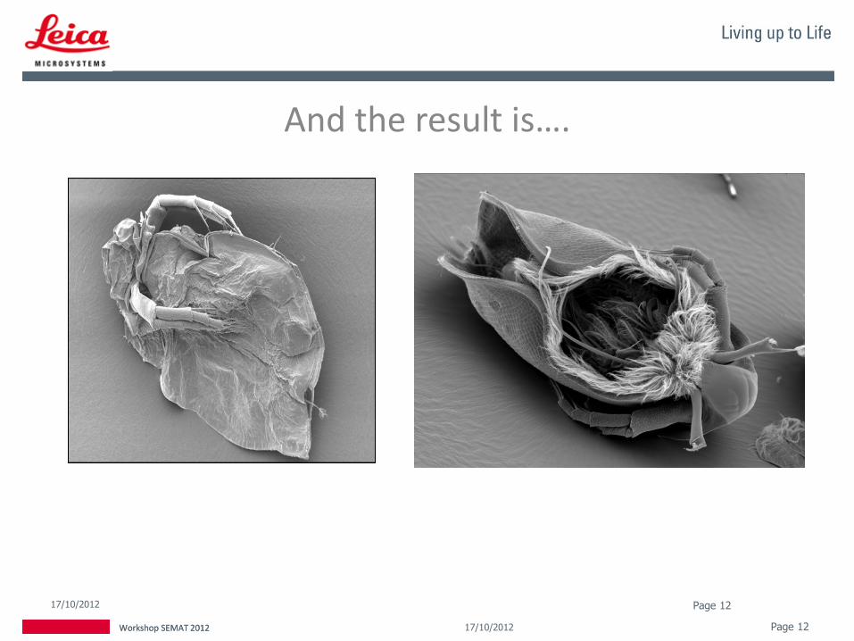
Critical Point Drying
Page 11 Workshop SEMAT 2012 17/10/2012
CPD300
Surface Tension of Water Damages Sample, but not CO2
You should avoid water in the EM column
Why ?
Cells
Insects
Tissues
Wafers
Etc…
Page 12 Workshop SEMAT 2012 17/10/2012
And the result is….
17/10/2012 Page 12
Workshop SEMAT 2012

Page 13 Workshop SEMAT 2012 17/10/2012
Water Flea (Daphnia sp.)
Courtesy of D. Gruber, University of Vienna, Austria

Page 14 Workshop SEMAT 2012 17/10/2012
W. Müller, University of Utrecht, Netherlands

Page 15 Workshop SEMAT 2012 17/10/2012
Cryo-ACE600
ACE200
ACE600
Coating
Sputter Cressington
Evaporator Polaron
At SEMAT:
Why do we coat ? SEM
• Reduce charging, inhibits charging
• Improves the secondary electron signal
• Higher electron yield from specimen surface
• Prevent specimen thermal damage
TEM
• Support films for TEM-grids (carbon on formvar)
• Generate contrast on thin samples (low-angle shadowing)
• Freeze-fracture replica technique
Or, to produce conductive layers in Micro-electronics research.
Page 16 Workshop SEMAT 2012 17/10/2012

Different Coatings • WHY METAL SPUTTERING COATING?
• Chemically inert, does not react with specimens or acids
• Stable in the electron beam
• Transparent to the electron beam (does not create contrast!)
• Conductive - reduces charging
• WHY CARBON COATING?
• Reduce charging
• Localize secondary electron (SE) and back-scattered electron (BSE) signal to the surface in SEM
• Reduce heating of non-conductive specimens to increase potential exposure time to the electron beam
• Reveal topography with shadowing (TEM or SEM)
Page 17 Workshop SEMAT 2012 17/10/2012

- Sputtering metals like gold, gold/palladium, silver, platinum etc… and additionally to LowVac, HigVac sputtering for materials like Aluminum, Chromium, Iridium, Molybdenum, Titanium Tungsten etc.
- Conductive carbon films for X-ray analyses, grids and backing of collodion
and formvar films for biological EM via carbon thread /rod evaporation - Thermal resistance evaporation of metal and carbon. - e-beam evaporation. The finest layers, either carbon or metals. - Freeze drying, freeze etching, in combination with a VCT100: Freeze
fracturing double replica, cryo-transfer.
Page 18 Workshop SEMAT 2012 17/10/2012
Other techniques in your coater:

Evaporation of metals using the electron beam gun
For materials with high melting point
For very fine grained layers (W, Ta/W, Cr, Pt/C)
Shadowing
Homogeneous layers with DARS (Double Axis Rotary Shadowing)
Low heat transfer to specimen
No charged particles to your sample
Metal coating: Electron beam evaporation
Pt
CW
Ta
e- e-
e-
Page 19 Workshop SEMAT 2012 17/10/2012

Coating quality depends on:
• Coating technique
• Layer thickness
• Coating material
• Vacuum conditions
• Specimen temperature
• The specimen itself (“decoration effects”)
• Effects after coating
Page 20 Workshop SEMAT 2012 17/10/2012
• Reorganization of the coating material depends on the surface temperature.
• Colder temperature reduces mobility of the coating material causing:
• Smaller distance between grains
• Finer structure
Coating quality - Specimen temperature WHY CRYO-COATING ???
Page 21 Workshop SEMAT 2012 17/10/2012

Synergies - Cryo SEM workflow
Page 22 Workshop SEMAT 2012 17/10/2012

Page 23 Workshop SEMAT 2012 17/10/2012
Biology Electron microscope
Resistant to high vacuum
Resistant in electron beam
Thin – permeable for electrons
Contrast
Soft and “Large”
Aqueous/hydrated
Light elements (C, O, H, N, S, P etc.)
Any treatment induces changes in the specimen
High vacuum
Electron beam
Sensitive to vibration (High magnifications)

Page 24 Workshop SEMAT 2012 17/10/2012
Tokuyasu technique
RT processing
(Cryo)TEM/SEM
High pressure freezing
Plunge freezing
Metal mirror
At High Pressure
At Ambient Pressure
Cryosectioning
LT processing
Chemical fixation Cryofixation Living Specimen
Page 25 Workshop SEMAT 2012 17/10/2012
Why cryofixation?
• The cellular constituent is rapidly immobilized
• Cryofixation is a physical fixation of all cellular components simultaneously
• Enzymes and antigens are not denatured (antigenecity)
• Cells retained in their ‘native’ state • The physical properties of a frozen sample allow cryo-
sectioning without any additional support by embedding medium (CEMOVIS).
Page 26 Workshop SEMAT 2012 17/10/2012
Why High Pressure Freezing?
• No changes in the physical equilibrium because of fast and precise correlation between pressure and temperature
• No cryo-protection needed, so no alteration of the cellular processes
• Vitrification of sample thickness up to 200 µm

Page 27 Workshop SEMAT 2012 17/10/2012
High pressure freezing
EM HPM100 EM PACT2
•LN2 at 2100bar used for pressure build-up AND cooling
•Alcohol is synchronizing pressure build-up and cooling
•Pressure is the same everywhere
•Pressure build-up and cooling separated
•LN2 at 10 bar used for cooling only
•Pressure transferred only to specimen (2100bar) by filler
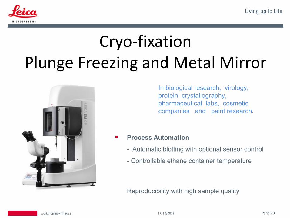
Cryo-fixation Plunge Freezing and Metal Mirror
Process Automation
- Automatic blotting with optional sensor control
- Controllable ethane container temperature
Reproducibility with high sample quality
Page 28 Workshop SEMAT 2012 17/10/2012
In biological research, virology,
protein crystallography,
pharmaceutical labs, cosmetic
companies and paint research.

Three different cryo-condition modes:
– standard mode
– high gas-flow (to reduce ice contaminations)
– wet sectioning (for polymer sectioning at wet condition
using DMSO), wide range of different temperature
settings of knife / specimen and gas (-40°C to -160°C)
Page 29 Workshop SEMAT 2012 17/10/2012

SPECIMEN MOUNTING AND TRANSFER Why? How could I transfer my sample?
SHUTTLE
LOADING STATION
Page 30 Workshop SEMAT 2012 17/10/2012

High pressure freezing (for cells or tissues)
Cryo-SEM
Freeze-fracturing
+
Coating
Transfer
Transfer
HPM 100 EM PACT
VCT 100
ACE600FF BAF 060
VCT 100
Technique Instrument
CRYO-SEM
Page 31 Workshop SEMAT 2012 17/10/2012
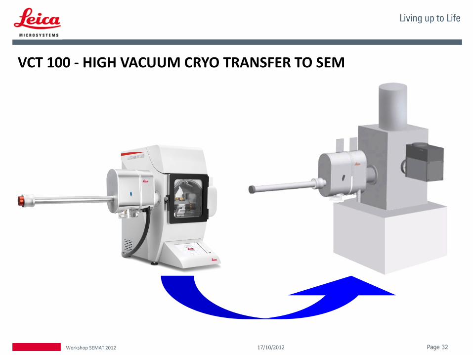
VCT 100 - HIGH VACUUM CRYO TRANSFER TO SEM
Page 32 Workshop SEMAT 2012 17/10/2012

Pathway with Freeze Substitution
TEM
Page 33 Workshop SEMAT 2012 17/10/2012
Dehydration of a cryofixed specimen by exchange of ice with an organic solvent
Freeze substitution is a link between cryofixation and room temperature TEM observation.

Page 34 Workshop SEMAT 2012 17/10/2012
Why Freeze Substitution?
• Enables sectioning at room temperature
• Avoids artifacts produced by conventional room temperature procedures
• Preserves antigenicity

Page 35 Workshop SEMAT 2012 17/10/2012
Contrasting (EM AFS2+FSP)
TEM
(EM HPM 100)
(EM TRIM2) (EM AC20)
Sectioning
High Pressure Freezing
Trimming
Freeze substitution LT embedding

Page 36 Workshop SEMAT 2012 17/10/2012
• Minimized aggregation and redistribution of diffusible elements
• Fixatives are uniformly distributed throughout the sample
Freeze Substitution
Cryotechniques in Biological Electron Microscopy, 1987, Steinbrecht RA, Zierold K)

Page 37 Workshop SEMAT 2012 17/10/2012
Freeze Substitution Processor
for EM AFS2
• Automatic reagent handling
• Special embedding moulds for
– FS of high pressure frozen samples
– PLT samples
“Load and Leave”
FSP

Page 38 Workshop SEMAT 2012 17/10/2012
MATERIAL SCIENCES

Page 39 Workshop SEMAT 2012 17/10/2012
EM TXP Target Surfacing Device for SEM, LM, TEM

Page 40 Workshop SEMAT 2012 17/10/2012
– For site specific cross sections (target preparation)
– Surfacing of small samples
– Pre-preparation prior to ion beam polishing (SEM)
– Pre-preparation prior to ion beam thinning (TEM)
– Pre-preparation prior to ion beam slope cutting (SEM)
– Pre-preparation priot to ultramicrotomy (SEM, TEM) TRIMMING
Microelectronic market
Watch industry
Material reseach (Nano technoloy)

Ion Milling- Slope Cutting FOR SEM
Page 41 Workshop SEMAT 2012 17/10/2012
Triple Ion Beam Cutter TIC3X Used for SEM microstructure analysis
(EDS, WDS, EBSD, CL) and AFM investigations

Page 42 Workshop SEMAT 2012 17/10/2012
Triple Ion Source

Page 43 Workshop SEMAT 2012 17/10/2012
Principle
• Three ion beams hitting the sample from different directions
• Fixed sample
Does not substitute a FIB on SEM, just because the area here is huge.
1x4mm prepared
sample
mask
ion beams
slope
prepared
area

Ion Milling – Only for TEM
Page 44 Workshop SEMAT 2012 17/10/2012
RES 101

Page 45 Workshop SEMAT 2012 17/10/2012
TEM
Plan view preparation
Cross-sectional sample preparation
FIB Cleaning
SEM
Surface cleaning
Polishing
Contrast enhancement
Ion beam slope cutting (35° and 90°)
LM
Applications of the RES 101

Muito obrigado pela vossa atenção!
Page 46 Workshop SEMAT 2012 17/10/2012
+34 673 00 42 57