Elongation factor 4 remodels the A-site tRNA on the ribosome
6
Elongation factor 4 remodels the A-site tRNA on the ribosome Matthieu G. Gagnon a,b,1 , Jinzhong Lin a,b,1 , and Thomas A. Steitz a,b,c,2 a Department of Molecular Biophysics and Biochemistry, Yale University, New Haven, CT 06520-8114; b Howard Hughes Medical Institute, Yale University, New Haven, CT 06520-8114; and c Department of Chemistry, Yale University, New Haven, CT 06520-8107 Edited by Harry F. Noller, University of California, Santa Cruz, CA, and approved March 23, 2016 (received for review November 20, 2015) During translation, a plethora of protein factors bind to the ribosome and regulate protein synthesis. Many of those factors are guanosine triphosphatases (GTPases), proteins that catalyze the hydrolysis of guanosine 5′-triphosphate (GTP) to promote con- formational changes. Despite numerous studies, the function of elongation factor 4 (EF-4/LepA), a highly conserved translational GTPase, has remained elusive. Here, we present the crystal struc- ture at 2.6-Å resolution of the Thermus thermophilus 70S ribosome bound to EF-4 with a nonhydrolyzable GTP analog and A-, P-, and E-site tRNAs. The structure reveals the interactions of EF-4 with the A-site tRNA, including contacts between the C-terminal domain (CTD) of EF-4 and the acceptor helical stem of the tRNA. Remark- ably, EF-4 induces a distortion of the A-site tRNA, allowing it to interact simultaneously with EF-4 and the decoding center of the ribosome. The structure provides insights into the tRNA-remodel- ing function of EF-4 on the ribosome and suggests that the dis- placement of the CCA-end of the A-site tRNA away from the peptidyl transferase center (PTC) is functionally significant. elongation factor 4 | ribosome | tRNA | remodeling | protein–RNA interactions T ranslation of the genetic information requires protein factors that interact with the ribosome sequentially, regulate its ac- tivity, and guide it through the protein synthesis cycle in a concerted manner. Many of those factors are guanosine tri- phosphatases (GTPases), proteins that use energy from guano- sine 5′-triphosphate (GTP) to promote conformational changes that lead to transitions between ribosome functional states (1, 2). In bacteria, for instance, initiation of protein synthesis is largely regulated by initiation factor 2 (IF-2), a GTPase that stabilizes the initiator tRNA in the P site of the ribosome (3). Subsequently, the elongation step is catalyzed by two universally conserved GTPases, elongation factor Tu (EF-Tu) and elongation factor G (EF-G). The ternary complex, consisting of EF-Tu, GTP, and the aminoacyl-tRNA, interacts with the ribosome to decode the codon in the A site of the ribosome. Following accommodation of the aminoacyl-tRNA in the A site of the ribosome and subsequent peptide bond formation, the tRNA–mRNA duplex is translocated by one codon—a process catalyzed by EF-G and GTP (4–6). Termination of protein synthesis is triggered when a stop codon is reached, upon which the newly synthesized protein is released with the help of release factor 3 (RF-3), yet another GTPase (7). Elongation factor 4 (EF-4/LepA) is a highly conserved protein structurally similar to EF-G (8) and has a ribosome-dependent GTPase activity (9–13). However, despite numerous studies, its function has remained elusive (9–20). Fast kinetic studies showed that EF-4 competes with EF-G during elongation for binding to the pretranslocation (PRE) ribosome, with tRNAs in the A and P sites (17). Despite this, EF-4 was also shown to increase the rate of protein synthesis at high intracellular ionic strength (16), without any effect on translational accuracy (16, 18). Conversely, EF-4 was also reported to bind to the posttranslocation (POST) ribosome and catalyze back-translocation of tRNAs from the E and P sites to the P and A sites, respectively (9–11). Recently, ribosome profiling data suggested that EF-4 reduces ribosomal pausing at certain glycine codons and contributes to translation initiation (13). Because of these sparse and controversial experimental data, combined with limited high-resolution snapshots of EF-4 in complex with the ribosome, the mechanism of action and func- tion of EF-4 have remained unclear. Recently, the crystal structure of EF-4 with GDP bound to the ribosome was reported (14). In this structure, the ribosome is clockwise ratcheted and the C-terminal domain (CTD) of EF-4 occupies the A site in the 50S subunit, where it reaches into the peptidyl transferase center (PTC) and interacts with the accep- tor-stem of the peptidyl-tRNA in the P site. A previous cryo- electron microscopy (cryo-EM) reconstruction of EF-4 bound to the ribosome in the presence of the nonhydrolysable GTP analog GDPNP reported a new conformation of the tRNA bound in the A site, allegedly being an intermediate step trapped in the pro- cess of back-translocation (11). However, the low resolution of this cryo-EM reconstruction limits the conclusions that can be drawn from it about the structure and function of EF-4 on the ribosome. To gain further insights into the function of EF-4, we de- termined its crystal structure in complex with the Thermus ther- mophilus 70S ribosome in the presence of the nonhydrolyzable GTP analog, GDPCP, and the A-, P-, and E-site tRNAs. The structure provides a detailed account of the contacts between EF-4, the ribosome, and the A-site tRNA, in particular revealing the network of interactions of the CTD region of EF-4 that stabilize the distorted conformation of the tRNA bound in the A site. Significance Many protein factors interact with the ribosome during protein synthesis. Elongation factor 4 (EF-4/LepA) is a widely distrib- uted and highly conserved translational GTPase for which several physiological roles have been proposed. Despite this, the function of EF-4 remains unknown. We have determined a high-resolution crystal structure of the ribosome bound to EF-4 in its GTP-bound state and A-, P-, and E-site tRNAs. Notably, EF-4 induces a distinct conformation of the tRNA bound in the A site, which deviates substantially from that of a canonical A-tRNA. EF-4 interacts with both helical domains of the A-site tRNA, indicating that EF-4 recognizes the L-shaped conformation of tRNA. Our results provide insights into the tRNA remodeling capacity of EF-4 on the ribosome. Author contributions: M.G.G. and J.L. designed research; M.G.G. and J.L. performed re- search; M.G.G., J.L., and T.A.S. analyzed data; and M.G.G., J.L., and T.A.S. wrote the paper. The authors declare no conflict of interest. This article is a PNAS Direct Submission. Freely available online through the PNAS open access option. Data deposition: The atomic coordinates and structure factors have been deposited in the Protein Data Bank, www.pdb.org (PDB ID code 5J8B). 1 M.G.G. and J.L. contributed equally to this work. 2 To whom correspondence should be addressed. Email: [email protected]. This article contains supporting information online at www.pnas.org/lookup/suppl/doi:10. 1073/pnas.1522932113/-/DCSupplemental. 4994–4999 | PNAS | May 3, 2016 | vol. 113 | no. 18 www.pnas.org/cgi/doi/10.1073/pnas.1522932113
Transcript of Elongation factor 4 remodels the A-site tRNA on the ribosome
Elongation factor 4 remodels the A-site tRNA onthe ribosomeMatthieu G. Gagnona,b,1, Jinzhong Lina,b,1, and Thomas A. Steitza,b,c,2
aDepartment of Molecular Biophysics and Biochemistry, Yale University, New Haven, CT 06520-8114; bHoward Hughes Medical Institute, Yale University,New Haven, CT 06520-8114; and cDepartment of Chemistry, Yale University, New Haven, CT 06520-8107
Edited by Harry F. Noller, University of California, Santa Cruz, CA, and approved March 23, 2016 (received for review November 20, 2015)
During translation, a plethora of protein factors bind to theribosome and regulate protein synthesis. Many of those factorsare guanosine triphosphatases (GTPases), proteins that catalyzethe hydrolysis of guanosine 5′-triphosphate (GTP) to promote con-formational changes. Despite numerous studies, the function ofelongation factor 4 (EF-4/LepA), a highly conserved translationalGTPase, has remained elusive. Here, we present the crystal struc-ture at 2.6-Å resolution of the Thermus thermophilus 70S ribosomebound to EF-4 with a nonhydrolyzable GTP analog and A-, P-, andE-site tRNAs. The structure reveals the interactions of EF-4 with theA-site tRNA, including contacts between the C-terminal domain(CTD) of EF-4 and the acceptor helical stem of the tRNA. Remark-ably, EF-4 induces a distortion of the A-site tRNA, allowing it tointeract simultaneously with EF-4 and the decoding center of theribosome. The structure provides insights into the tRNA-remodel-ing function of EF-4 on the ribosome and suggests that the dis-placement of the CCA-end of the A-site tRNA away from thepeptidyl transferase center (PTC) is functionally significant.
elongation factor 4 | ribosome | tRNA | remodeling |protein–RNA interactions
Translation of the genetic information requires protein factorsthat interact with the ribosome sequentially, regulate its ac-
tivity, and guide it through the protein synthesis cycle in aconcerted manner. Many of those factors are guanosine tri-phosphatases (GTPases), proteins that use energy from guano-sine 5′-triphosphate (GTP) to promote conformational changesthat lead to transitions between ribosome functional states (1, 2).In bacteria, for instance, initiation of protein synthesis is largelyregulated by initiation factor 2 (IF-2), a GTPase that stabilizesthe initiator tRNA in the P site of the ribosome (3). Subsequently,the elongation step is catalyzed by two universally conservedGTPases, elongation factor Tu (EF-Tu) and elongation factor G(EF-G). The ternary complex, consisting of EF-Tu, GTP, and theaminoacyl-tRNA, interacts with the ribosome to decode the codonin the A site of the ribosome. Following accommodation of theaminoacyl-tRNA in the A site of the ribosome and subsequentpeptide bond formation, the tRNA–mRNA duplex is translocatedby one codon—a process catalyzed by EF-G and GTP (4–6).Termination of protein synthesis is triggered when a stop codon isreached, upon which the newly synthesized protein is releasedwith the help of release factor 3 (RF-3), yet another GTPase (7).Elongation factor 4 (EF-4/LepA) is a highly conserved protein
structurally similar to EF-G (8) and has a ribosome-dependentGTPase activity (9–13). However, despite numerous studies, itsfunction has remained elusive (9–20). Fast kinetic studies showedthat EF-4 competes with EF-G during elongation for binding tothe pretranslocation (PRE) ribosome, with tRNAs in the A and Psites (17). Despite this, EF-4 was also shown to increase the rate ofprotein synthesis at high intracellular ionic strength (16), withoutany effect on translational accuracy (16, 18). Conversely, EF-4 wasalso reported to bind to the posttranslocation (POST) ribosomeand catalyze back-translocation of tRNAs from the E and P sitesto the P and A sites, respectively (9–11). Recently, ribosomeprofiling data suggested that EF-4 reduces ribosomal pausing at
certain glycine codons and contributes to translation initiation(13). Because of these sparse and controversial experimentaldata, combined with limited high-resolution snapshots of EF-4 incomplex with the ribosome, the mechanism of action and func-tion of EF-4 have remained unclear.Recently, the crystal structure of EF-4 with GDP bound to the
ribosome was reported (14). In this structure, the ribosome isclockwise ratcheted and the C-terminal domain (CTD) of EF-4occupies the A site in the 50S subunit, where it reaches into thepeptidyl transferase center (PTC) and interacts with the accep-tor-stem of the peptidyl-tRNA in the P site. A previous cryo-electron microscopy (cryo-EM) reconstruction of EF-4 bound tothe ribosome in the presence of the nonhydrolysable GTP analogGDPNP reported a new conformation of the tRNA bound in theA site, allegedly being an intermediate step trapped in the pro-cess of back-translocation (11). However, the low resolution ofthis cryo-EM reconstruction limits the conclusions that can bedrawn from it about the structure and function of EF-4 onthe ribosome.To gain further insights into the function of EF-4, we de-
termined its crystal structure in complex with the Thermus ther-mophilus 70S ribosome in the presence of the nonhydrolyzableGTP analog, GDPCP, and the A-, P-, and E-site tRNAs. Thestructure provides a detailed account of the contacts betweenEF-4, the ribosome, and the A-site tRNA, in particular revealingthe network of interactions of the CTD region of EF-4 thatstabilize the distorted conformation of the tRNA bound in theA site.
Significance
Many protein factors interact with the ribosome during proteinsynthesis. Elongation factor 4 (EF-4/LepA) is a widely distrib-uted and highly conserved translational GTPase for whichseveral physiological roles have been proposed. Despite this,the function of EF-4 remains unknown. We have determined ahigh-resolution crystal structure of the ribosome bound to EF-4in its GTP-bound state and A-, P-, and E-site tRNAs. Notably,EF-4 induces a distinct conformation of the tRNA bound in theA site, which deviates substantially from that of a canonicalA-tRNA. EF-4 interacts with both helical domains of the A-sitetRNA, indicating that EF-4 recognizes the L-shaped conformationof tRNA. Our results provide insights into the tRNA remodelingcapacity of EF-4 on the ribosome.
Author contributions: M.G.G. and J.L. designed research; M.G.G. and J.L. performed re-search; M.G.G., J.L., and T.A.S. analyzed data; and M.G.G., J.L., and T.A.S. wrote the paper.
The authors declare no conflict of interest.
This article is a PNAS Direct Submission.
Freely available online through the PNAS open access option.
Data deposition: The atomic coordinates and structure factors have been deposited in theProtein Data Bank, www.pdb.org (PDB ID code 5J8B).1M.G.G. and J.L. contributed equally to this work.2To whom correspondence should be addressed. Email: [email protected].
This article contains supporting information online at www.pnas.org/lookup/suppl/doi:10.1073/pnas.1522932113/-/DCSupplemental.
4994–4999 | PNAS | May 3, 2016 | vol. 113 | no. 18 www.pnas.org/cgi/doi/10.1073/pnas.1522932113

ResultsCrystallization of the L9–EF-4 Fusion Protein with the Ribosome. Thecrystallization of the wild-type 70S ribosome largely depends onthe inter-ribosome packing mediated by ribosomal protein uL9in the asymmetric unit of the crystal (21). We therefore tookadvantage of the fact that ribosomes isolated from a Thermusthermophilus strain that carries a truncated endogenous ribo-somal protein uL9 (70S:L91–58) altogether lack protein uL9 anddo not crystallize under previously published conditions (6, 14).To rescue crystal growth of ribosomes lacking uL9, we incubatedthe 70S:L91–58 ribosomes with the N-terminal domain of proteinuL9, which has been covalently linked to EF-4 and crystallizedthose ribosomes as described previously (14) (Materials andMethods). Varying the length of the linker between uL9 and EF-4allows for selection of only those protein fusions that yield crystals,indicating proper docking of EF-4 and uL9 to their respectivebinding sites on the ribosome. This engineered crystallization ap-proach has recently been used successfully to determine structuresof the ribosome bound to EF-4–GDP and EF-G–GDP (6, 14).
Overview of the Structure. The structure, refined to a resolution of2.6 Å, contrasts with the previous EF-4–GDP–ribosome structurecomplex (14) in that it has three tRNAs bound in the A, P, and Esites of the ribosome, thereby mimicking a PRE ribosomesubstrate (Fig. 1 and Fig. S1A). Although the tip of the CTD (ordomain VI) of EF-4 reaches into the PTC and contacts theacceptor-end of the peptidyl-tRNA in the P site as previouslyobserved in the presence of GDP (14), a significant portion ofthe CTD interacts with the acceptor- and D-stems of the A-sitetRNA (Fig. 1). Additional interactions with the A-tRNA are me-diated by domain IV of EF-4, which contacts the anticodon-stemregion.In this structure, the ribosome is in a classical state of ratch-
eting. This also contrasts with the previous complex structure ofthe ribosome bound with EF-4–GDP in which the ribosome isclockwise ratcheted (14). As a result, the conformation of thedecoding center is such that it encloses the anticodon-stem loopof the A-site tRNA in the same manner as seen during standard
decoding (22, 23), allowing the universally conserved nucleotidesA1492 (Escherichia coli nucleotide numbering is used throughoutthe text) and A1493 in helix 44 (h44), and G530 in h18 of 16Sribosomal RNA (rRNA), to form canonical interactions with theminor groove of the codon–anticodon minihelix of the A-sitetRNA (Fig. S2A).
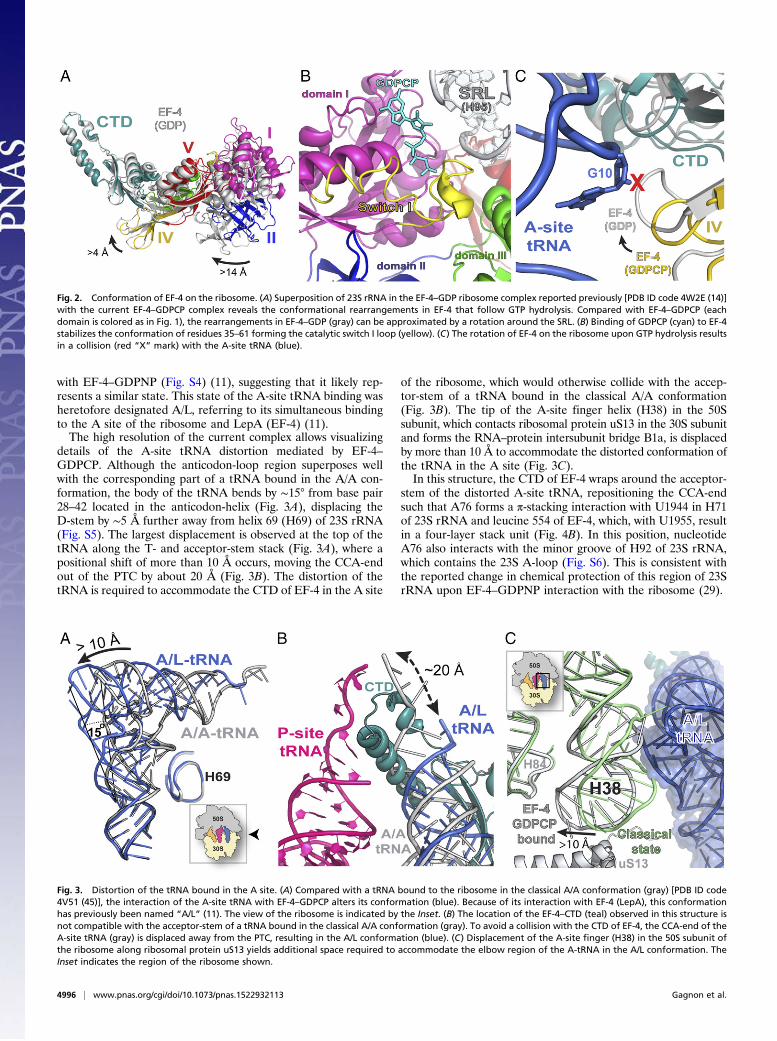
Structural Comparison with the GDP-Bound Form. The relativeinterdomain arrangement in EF-4 in this ribosome complex issimilar to its GDP-bound form reported previously (14) (Fig. 2A)and to that seen in the previous low-resolution cryo-EM re-construction of EF-4–GDPNP in complex with the ribosome(11). The latter observation indicates that the chimeric fusionbetween proteins uL9 and EF-4 does not interfere with theconformation of EF-4 on the ribosome.Similar to EF-G and RF-3 bound to a GTP analog on the
ribosome (24–28), the presence of GDPCP bound to EF-4 sta-bilizes the conformation of residues 35–61 forming the catalyticswitch I loop (Fig. 2B and Fig. S3), consistent with its role inGTP hydrolysis. Switch I loop interacts with the sarcin–ricin loop(SRL) of 23S rRNA and with domain III of EF-4 (Fig. 2B),resulting in a more compact EF-4 structure. Hydrolysis of GTPtriggers interdomain rearrangements that result in a more openconformation of EF-4. In agreement with this and comparedwith the position of EF-4–GDPCP on the ribosome, the overallpositioning of EF-4–GDP on the ribosome changes slightly,which can be approximated by a rotation of the G domain aroundthe SRL (Fig. 2A). As a result, domain IV in the EF-4–GDP–ribosome structure shifts by more than 4 Å toward the A site andwould collide with the tRNA bound in the A site (Fig. 2 A and C),explaining why the presence of the A-site tRNA is not compatiblewith the previous EF-4–GDP–ribosome structure (14).
EF-4 Remodels the A-Site tRNA.Whereas the tRNA in the P site hasthe classical P/P conformation, the tRNA in the A site is dis-torted relative to the position of a canonical A-tRNA (Fig. 3)(Materials and Methods, Note). The overall conformation of theA-site tRNA in the current complex is very similar to the onepreviously observed in a low-resolution cryo-EM reconstruction
Fig. 1. The structure of EF-4–GDPCP bound to the ribosome. (A) Overview of EF-4–GDPCP bound to the 70S ribosome. tRNAs in the E, P, and A sites aredisplayed in orange, pink, and blue, respectively. The 50S and 30S subunits are shown in light blue and yellow, respectively. Portions of the ribosome areomitted for clarity. (B) Close-up view of the C-terminal domain of EF-4 (CTD) (teal) that wraps around the acceptor-stem of the A-site tRNA (blue).
Gagnon et al. PNAS | May 3, 2016 | vol. 113 | no. 18 | 4995
BIOCH
EMISTR
Y
with EF-4–GDPNP (Fig. S4) (11), suggesting that it likely rep-resents a similar state. This state of the A-site tRNA binding washeretofore designated A/L, referring to its simultaneous bindingto the A site of the ribosome and LepA (EF-4) (11).The high resolution of the current complex allows visualizing
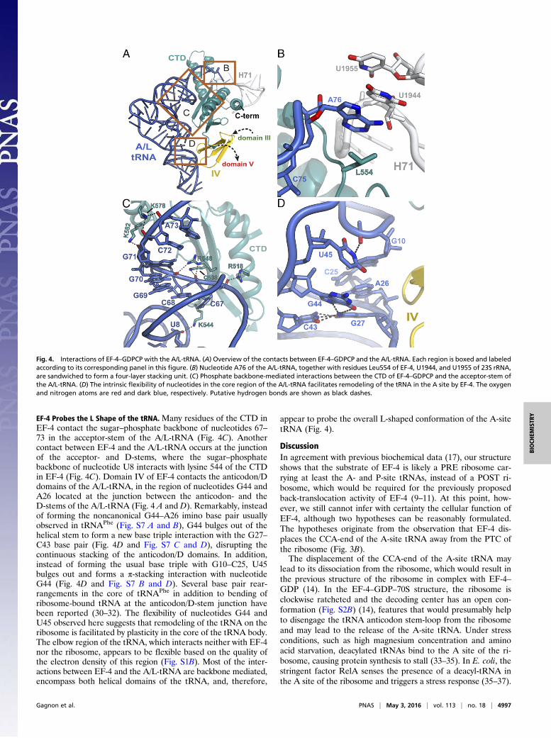
details of the A-site tRNA distortion mediated by EF-4–GDPCP. Although the anticodon-loop region superposes wellwith the corresponding part of a tRNA bound in the A/A con-formation, the body of the tRNA bends by ∼15° from base pair28–42 located in the anticodon-helix (Fig. 3A), displacing theD-stem by ∼5 Å further away from helix 69 (H69) of 23S rRNA(Fig. S5). The largest displacement is observed at the top of thetRNA along the T- and acceptor-stem stack (Fig. 3A), where apositional shift of more than 10 Å occurs, moving the CCA-endout of the PTC by about 20 Å (Fig. 3B). The distortion of thetRNA is required to accommodate the CTD of EF-4 in the A site
of the ribosome, which would otherwise collide with the accep-tor-stem of a tRNA bound in the classical A/A conformation(Fig. 3B). The tip of the A-site finger helix (H38) in the 50Ssubunit, which contacts ribosomal protein uS13 in the 30S subunitand forms the RNA–protein intersubunit bridge B1a, is displacedby more than 10 Å to accommodate the distorted conformation ofthe tRNA in the A site (Fig. 3C).In this structure, the CTD of EF-4 wraps around the acceptor-
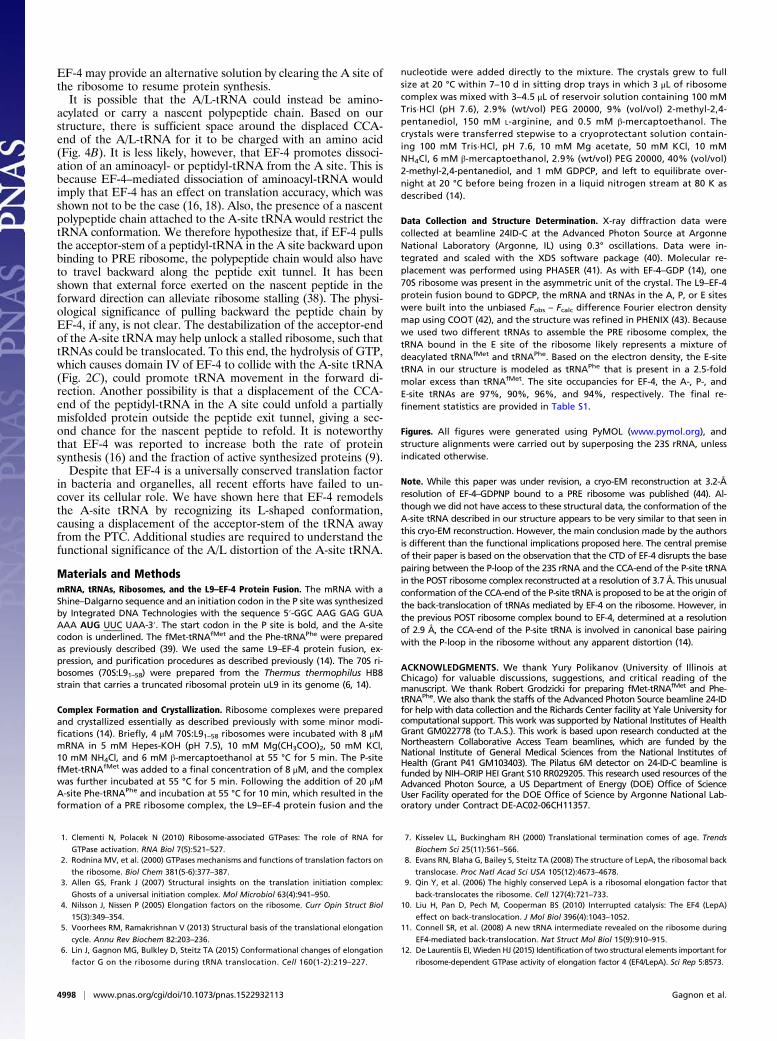
stem of the distorted A-site tRNA, repositioning the CCA-endsuch that A76 forms a π-stacking interaction with U1944 in H71of 23S rRNA and leucine 554 of EF-4, which, with U1955, resultin a four-layer stack unit (Fig. 4B). In this position, nucleotideA76 also interacts with the minor groove of H92 of 23S rRNA,which contains the 23S A-loop (Fig. S6). This is consistent withthe reported change in chemical protection of this region of 23SrRNA upon EF-4–GDPNP interaction with the ribosome (29).
Fig. 3. Distortion of the tRNA bound in the A site. (A) Compared with a tRNA bound to the ribosome in the classical A/A conformation (gray) [PDB ID code4V51 (45)], the interaction of the A-site tRNA with EF-4–GDPCP alters its conformation (blue). Because of its interaction with EF-4 (LepA), this conformationhas previously been named “A/L” (11). The view of the ribosome is indicated by the Inset. (B) The location of the EF-4–CTD (teal) observed in this structure isnot compatible with the acceptor-stem of a tRNA bound in the classical A/A conformation (gray). To avoid a collision with the CTD of EF-4, the CCA-end of theA-site tRNA (gray) is displaced away from the PTC, resulting in the A/L conformation (blue). (C) Displacement of the A-site finger (H38) in the 50S subunit ofthe ribosome along ribosomal protein uS13 yields additional space required to accommodate the elbow region of the A-tRNA in the A/L conformation. TheInset indicates the region of the ribosome shown.
Fig. 2. Conformation of EF-4 on the ribosome. (A) Superposition of 23S rRNA in the EF-4–GDP ribosome complex reported previously [PDB ID code 4W2E (14)]with the current EF-4–GDPCP complex reveals the conformational rearrangements in EF-4 that follow GTP hydrolysis. Compared with EF-4–GDPCP (eachdomain is colored as in Fig. 1), the rearrangements in EF-4–GDP (gray) can be approximated by a rotation around the SRL. (B) Binding of GDPCP (cyan) to EF-4stabilizes the conformation of residues 35–61 forming the catalytic switch I loop (yellow). (C) The rotation of EF-4 on the ribosome upon GTP hydrolysis resultsin a collision (red “X” mark) with the A-site tRNA (blue).
4996 | www.pnas.org/cgi/doi/10.1073/pnas.1522932113 Gagnon et al.
EF-4 Probes the L Shape of the tRNA. Many residues of the CTD inEF-4 contact the sugar–phosphate backbone of nucleotides 67–73 in the acceptor-stem of the A/L-tRNA (Fig. 4C). Anothercontact between EF-4 and the A/L-tRNA occurs at the junctionof the acceptor- and D-stems, where the sugar–phosphatebackbone of nucleotide U8 interacts with lysine 544 of the CTDin EF-4 (Fig. 4C). Domain IV of EF-4 contacts the anticodon/Ddomains of the A/L-tRNA, in the region of nucleotides G44 andA26 located at the junction between the anticodon- and theD-stems of the A/L-tRNA (Fig. 4 A and D). Remarkably, insteadof forming the noncanonical G44–A26 imino base pair usuallyobserved in tRNAPhe (Fig. S7 A and B), G44 bulges out of thehelical stem to form a new base triple interaction with the G27–C43 base pair (Fig. 4D and Fig. S7 C and D), disrupting thecontinuous stacking of the anticodon/D domains. In addition,instead of forming the usual base triple with G10–C25, U45bulges out and forms a π-stacking interaction with nucleotideG44 (Fig. 4D and Fig. S7 B and D). Several base pair rear-rangements in the core of tRNAPhe in addition to bending ofribosome-bound tRNA at the anticodon/D-stem junction havebeen reported (30–32). The flexibility of nucleotides G44 andU45 observed here suggests that remodeling of the tRNA on theribosome is facilitated by plasticity in the core of the tRNA body.The elbow region of the tRNA, which interacts neither with EF-4nor the ribosome, appears to be flexible based on the quality ofthe electron density of this region (Fig. S1B). Most of the inter-actions between EF-4 and the A/L-tRNA are backbone mediated,encompass both helical domains of the tRNA, and, therefore,
appear to probe the overall L-shaped conformation of the A-sitetRNA (Fig. 4).
DiscussionIn agreement with previous biochemical data (17), our structureshows that the substrate of EF-4 is likely a PRE ribosome car-rying at least the A- and P-site tRNAs, instead of a POST ri-bosome, which would be required for the previously proposedback-translocation activity of EF-4 (9–11). At this point, how-ever, we still cannot infer with certainty the cellular function ofEF-4, although two hypotheses can be reasonably formulated.The hypotheses originate from the observation that EF-4 dis-places the CCA-end of the A-site tRNA away from the PTC ofthe ribosome (Fig. 3B).The displacement of the CCA-end of the A-site tRNA may
lead to its dissociation from the ribosome, which would result inthe previous structure of the ribosome in complex with EF-4–GDP (14). In the EF-4–GDP–70S structure, the ribosome isclockwise ratcheted and the decoding center has an open con-formation (Fig. S2B) (14), features that would presumably helpto disengage the tRNA anticodon stem-loop from the ribosomeand may lead to the release of the A-site tRNA. Under stressconditions, such as high magnesium concentration and aminoacid starvation, deacylated tRNAs bind to the A site of the ri-bosome, causing protein synthesis to stall (33–35). In E. coli, thestringent factor RelA senses the presence of a deacyl-tRNA inthe A site of the ribosome and triggers a stress response (35–37).
Fig. 4. Interactions of EF-4–GDPCP with the A/L-tRNA. (A) Overview of the contacts between EF-4–GDPCP and the A/L-tRNA. Each region is boxed and labeledaccording to its corresponding panel in this figure. (B) Nucleotide A76 of the A/L-tRNA, together with residues Leu554 of EF-4, U1944, and U1955 of 23S rRNA,are sandwiched to form a four-layer stacking unit. (C) Phosphate backbone-mediated interactions between the CTD of EF-4–GDPCP and the acceptor-stem ofthe A/L-tRNA. (D) The intrinsic flexibility of nucleotides in the core region of the A/L-tRNA facilitates remodeling of the tRNA in the A site by EF-4. The oxygenand nitrogen atoms are red and dark blue, respectively. Putative hydrogen bonds are shown as black dashes.
Gagnon et al. PNAS | May 3, 2016 | vol. 113 | no. 18 | 4997
BIOCH
EMISTR
Y
EF-4 may provide an alternative solution by clearing the A site ofthe ribosome to resume protein synthesis.It is possible that the A/L-tRNA could instead be amino-
acylated or carry a nascent polypeptide chain. Based on ourstructure, there is sufficient space around the displaced CCA-end of the A/L-tRNA for it to be charged with an amino acid(Fig. 4B). It is less likely, however, that EF-4 promotes dissoci-ation of an aminoacyl- or peptidyl-tRNA from the A site. This isbecause EF-4–mediated dissociation of aminoacyl-tRNA wouldimply that EF-4 has an effect on translation accuracy, which wasshown not to be the case (16, 18). Also, the presence of a nascentpolypeptide chain attached to the A-site tRNA would restrict thetRNA conformation. We therefore hypothesize that, if EF-4 pullsthe acceptor-stem of a peptidyl-tRNA in the A site backward uponbinding to PRE ribosome, the polypeptide chain would also haveto travel backward along the peptide exit tunnel. It has beenshown that external force exerted on the nascent peptide in theforward direction can alleviate ribosome stalling (38). The physi-ological significance of pulling backward the peptide chain byEF-4, if any, is not clear. The destabilization of the acceptor-endof the A-site tRNA may help unlock a stalled ribosome, such thattRNAs could be translocated. To this end, the hydrolysis of GTP,which causes domain IV of EF-4 to collide with the A-site tRNA(Fig. 2C), could promote tRNA movement in the forward di-rection. Another possibility is that a displacement of the CCA-end of the peptidyl-tRNA in the A site could unfold a partiallymisfolded protein outside the peptide exit tunnel, giving a sec-ond chance for the nascent peptide to refold. It is noteworthythat EF-4 was reported to increase both the rate of proteinsynthesis (16) and the fraction of active synthesized proteins (9).Despite that EF-4 is a universally conserved translation factor
in bacteria and organelles, all recent efforts have failed to un-cover its cellular role. We have shown here that EF-4 remodelsthe A-site tRNA by recognizing its L-shaped conformation,causing a displacement of the acceptor-stem of the tRNA awayfrom the PTC. Additional studies are required to understand thefunctional significance of the A/L distortion of the A-site tRNA.
Materials and MethodsmRNA, tRNAs, Ribosomes, and the L9–EF-4 Protein Fusion. The mRNA with aShine–Dalgarno sequence and an initiation codon in the P site was synthesizedby Integrated DNA Technologies with the sequence 5′-GGC AAG GAG GUAAAA AUG UUC UAA-3′. The start codon in the P site is bold, and the A-sitecodon is underlined. The fMet-tRNAfMet and the Phe-tRNAPhe were preparedas previously described (39). We used the same L9–EF-4 protein fusion, ex-pression, and purification procedures as described previously (14). The 70S ri-bosomes (70S:L91–58) were prepared from the Thermus thermophilus HB8strain that carries a truncated ribosomal protein uL9 in its genome (6, 14).
Complex Formation and Crystallization. Ribosome complexes were preparedand crystallized essentially as described previously with some minor modi-fications (14). Briefly, 4 μM 70S:L91–58 ribosomes were incubated with 8 μMmRNA in 5 mM Hepes-KOH (pH 7.5), 10 mM Mg(CH3COO)2, 50 mM KCl,10 mM NH4Cl, and 6 mM β-mercaptoethanol at 55 °C for 5 min. The P-sitefMet-tRNAfMet was added to a final concentration of 8 μM, and the complexwas further incubated at 55 °C for 5 min. Following the addition of 20 μMA-site Phe-tRNAPhe and incubation at 55 °C for 10 min, which resulted in theformation of a PRE ribosome complex, the L9–EF-4 protein fusion and the
nucleotide were added directly to the mixture. The crystals grew to fullsize at 20 °C within 7–10 d in sitting drop trays in which 3 μL of ribosomecomplex was mixed with 3–4.5 μL of reservoir solution containing 100 mMTris·HCl (pH 7.6), 2.9% (wt/vol) PEG 20000, 9% (vol/vol) 2-methyl-2,4-pentanediol, 150 mM L-arginine, and 0.5 mM β-mercaptoethanol. Thecrystals were transferred stepwise to a cryoprotectant solution contain-ing 100 mM Tris·HCl, pH 7.6, 10 mM Mg acetate, 50 mM KCl, 10 mMNH4Cl, 6 mM β-mercaptoethanol, 2.9% (wt/vol) PEG 20000, 40% (vol/vol)2-methyl-2,4-pentanediol, and 1 mM GDPCP, and left to equilibrate over-night at 20 °C before being frozen in a liquid nitrogen stream at 80 K asdescribed (14).
Data Collection and Structure Determination. X-ray diffraction data werecollected at beamline 24ID-C at the Advanced Photon Source at ArgonneNational Laboratory (Argonne, IL) using 0.3° oscillations. Data were in-tegrated and scaled with the XDS software package (40). Molecular re-placement was performed using PHASER (41). As with EF-4–GDP (14), one70S ribosome was present in the asymmetric unit of the crystal. The L9–EF-4protein fusion bound to GDPCP, the mRNA and tRNAs in the A, P, or E siteswere built into the unbiased Fobs – Fcalc difference Fourier electron densitymap using COOT (42), and the structure was refined in PHENIX (43). Becausewe used two different tRNAs to assemble the PRE ribosome complex, thetRNA bound in the E site of the ribosome likely represents a mixture ofdeacylated tRNAfMet and tRNAPhe. Based on the electron density, the E-sitetRNA in our structure is modeled as tRNAPhe that is present in a 2.5-foldmolar excess than tRNAfMet. The site occupancies for EF-4, the A-, P-, andE-site tRNAs are 97%, 90%, 96%, and 94%, respectively. The final re-finement statistics are provided in Table S1.
Figures. All figures were generated using PyMOL (www.pymol.org), andstructure alignments were carried out by superposing the 23S rRNA, unlessindicated otherwise.
Note. While this paper was under revision, a cryo-EM reconstruction at 3.2-Åresolution of EF-4–GDPNP bound to a PRE ribosome was published (44). Al-though we did not have access to these structural data, the conformation of theA-site tRNA described in our structure appears to be very similar to that seen inthis cryo-EM reconstruction. However, the main conclusion made by the authorsis different than the functional implications proposed here. The central premiseof their paper is based on the observation that the CTD of EF-4 disrupts the basepairing between the P-loop of the 23S rRNA and the CCA-end of the P-site tRNAin the POST ribosome complex reconstructed at a resolution of 3.7 Å. This unusualconformation of the CCA-end of the P-site tRNA is proposed to be at the origin ofthe back-translocation of tRNAs mediated by EF-4 on the ribosome. However, inthe previous POST ribosome complex bound to EF-4, determined at a resolutionof 2.9 Å, the CCA-end of the P-site tRNA is involved in canonical base pairingwith the P-loop in the ribosome without any apparent distortion (14).
ACKNOWLEDGMENTS. We thank Yury Polikanov (University of Illinois atChicago) for valuable discussions, suggestions, and critical reading of themanuscript. We thank Robert Grodzicki for preparing fMet-tRNAfMet and Phe-tRNAPhe. We also thank the staffs of the Advanced Photon Source beamline 24-IDfor help with data collection and the Richards Center facility at Yale University forcomputational support. This work was supported by National Institutes of HealthGrant GM022778 (to T.A.S.). This work is based upon research conducted at theNortheastern Collaborative Access Team beamlines, which are funded by theNational Institute of General Medical Sciences from the National Institutes ofHealth (Grant P41 GM103403). The Pilatus 6M detector on 24-ID-C beamline isfunded by NIH–ORIP HEI Grant S10 RR029205. This research used resources of theAdvanced Photon Source, a US Department of Energy (DOE) Office of ScienceUser Facility operated for the DOE Office of Science by Argonne National Lab-oratory under Contract DE-AC02-06CH11357.
1. Clementi N, Polacek N (2010) Ribosome-associated GTPases: The role of RNA for
GTPase activation. RNA Biol 7(5):521–527.2. Rodnina MV, et al. (2000) GTPases mechanisms and functions of translation factors on
the ribosome. Biol Chem 381(5-6):377–387.3. Allen GS, Frank J (2007) Structural insights on the translation initiation complex:
Ghosts of a universal initiation complex. Mol Microbiol 63(4):941–950.4. Nilsson J, Nissen P (2005) Elongation factors on the ribosome. Curr Opin Struct Biol
15(3):349–354.5. Voorhees RM, Ramakrishnan V (2013) Structural basis of the translational elongation
cycle. Annu Rev Biochem 82:203–236.6. Lin J, Gagnon MG, Bulkley D, Steitz TA (2015) Conformational changes of elongation
factor G on the ribosome during tRNA translocation. Cell 160(1-2):219–227.
7. Kisselev LL, Buckingham RH (2000) Translational termination comes of age. Trends
Biochem Sci 25(11):561–566.8. Evans RN, Blaha G, Bailey S, Steitz TA (2008) The structure of LepA, the ribosomal back
translocase. Proc Natl Acad Sci USA 105(12):4673–4678.9. Qin Y, et al. (2006) The highly conserved LepA is a ribosomal elongation factor that
back-translocates the ribosome. Cell 127(4):721–733.10. Liu H, Pan D, Pech M, Cooperman BS (2010) Interrupted catalysis: The EF4 (LepA)
effect on back-translocation. J Mol Biol 396(4):1043–1052.11. Connell SR, et al. (2008) A new tRNA intermediate revealed on the ribosome during
EF4-mediated back-translocation. Nat Struct Mol Biol 15(9):910–915.12. De Laurentiis EI, Wieden HJ (2015) Identification of two structural elements important for
ribosome-dependent GTPase activity of elongation factor 4 (EF4/LepA). Sci Rep 5:8573.
4998 | www.pnas.org/cgi/doi/10.1073/pnas.1522932113 Gagnon et al.
13. Balakrishnan R, Oman K, Shoji S, Bundschuh R, Fredrick K (2014) The conserved
GTPase LepA contributes mainly to translation initiation in Escherichia coli. Nucleic
Acids Res 42(21):13370–13383.14. Gagnon MG, Lin J, Bulkley D, Steitz TA (2014) Crystal structure of elongation factor 4
bound to a clockwise ratcheted ribosome. Science 345(6197):684–687.15. Zhang D, Qin Y (2013) The paradox of elongation factor 4: Highly conserved, yet of no
physiological significance? Biochem J 452(2):173–181.16. Pech M, et al. (2011) Elongation factor 4 (EF4/LepA) accelerates protein synthesis at
increased Mg2+ concentrations. Proc Natl Acad Sci USA 108(8):3199–3203.17. Liu H, et al. (2011) The conserved protein EF4 (LepA) modulates the elongation cycle
of protein synthesis. Proc Natl Acad Sci USA 108(39):16223–16228.18. Shoji S, Janssen BD, Hayes CS, Fredrick K (2010) Translation factor LepA contributes to
tellurite resistance in Escherichia coli but plays no apparent role in the fidelity of
protein synthesis. Biochimie 92(2):157–163.19. Li L, et al. (2014) Ribosomal elongation factor 4 promotes cell death associated with
lethal stress. MBio 5(6):e01708.20. Yang F, Li Z, Hao J, Qin Y (2014) EF4 knockout E. coli cells exhibit lower levels of
cellular biosynthesis under acidic stress. Protein Cell 5(7):563–567.21. Selmer M, Gao YG, Weixlbaumer A, Ramakrishnan V (2012) Ribosome engineering to
promote new crystal forms. Acta Crystallogr D Biol Crystallogr 68(Pt 5):578–583.22. Ogle JM, Murphy FV, Tarry MJ, Ramakrishnan V (2002) Selection of tRNA by the ri-
bosome requires a transition from an open to a closed form. Cell 111(5):721–732.23. Ogle JM, et al. (2001) Recognition of cognate transfer RNA by the 30S ribosomal
subunit. Science 292(5518):897–902.24. Zhou J, Lancaster L, Donohue JP, Noller HF (2013) Crystal structures of EF-G-ribosome
complexes trapped in intermediate states of translocation. Science 340(6140):
1236086.25. Tourigny DS, Fernández IS, Kelley AC, Ramakrishnan V (2013) Elongation factor G
bound to the ribosome in an intermediate state of translocation. Science 340(6140):
1235490.26. Pulk A, Cate JH (2013) Control of ribosomal subunit rotation by elongation factor G.
Science 340(6140):1235970.27. Zhou J, Lancaster L, Trakhanov S, Noller HF (2012) Crystal structure of release factor
RF3 trapped in the GTP state on a rotated conformation of the ribosome. RNA 18(2):
230–240.28. Jin H, Kelley AC, Ramakrishnan V (2011) Crystal structure of the hybrid state of ri-
bosome in complex with the guanosine triphosphatase release factor 3. Proc Natl
Acad Sci USA 108(38):15798–15803.
29. Walter JD, Hunter M, Cobb M, Traeger G, Spiegel PC (2012) Thiostrepton inhibitsstable 70S ribosome binding and ribosome-dependent GTPase activation of elonga-tion factor G and elongation factor 4. Nucleic Acids Res 40(1):360–370.
30. Dunkle JA, et al. (2011) Structures of the bacterial ribosome in classical and hybridstates of tRNA binding. Science 332(6032):981–984.
31. Byrne RT, Konevega AL, Rodnina MV, Antson AA (2010) The crystal structure of un-modified tRNAPhe from Escherichia coli. Nucleic Acids Res 38(12):4154–4162.
32. Schmeing TM, et al. (2009) The crystal structure of the ribosome bound to EF-Tu andaminoacyl-tRNA. Science 326(5953):688–694.
33. Moazed D, Noller HF (1989) Interaction of tRNA with 23S rRNA in the ribosomal A, P,and E sites. Cell 57(4):585–597.
34. Rojiani MV, Jakubowski H, Goldman E (1989) Effect of variation of charged and un-charged tRNA(Trp) levels on ppGpp synthesis in Escherichia coli. J Bacteriol 171(12):6493–6502.
35. Haseltine WA, Block R (1973) Synthesis of guanosine tetra- and pentaphosphate re-quires the presence of a codon-specific, uncharged transfer ribonucleic acid in theacceptor site of ribosomes. Proc Natl Acad Sci USA 70(5):1564–1568.
36. Agirrezabala X, et al. (2013) The ribosome triggers the stringent response by RelA viaa highly distorted tRNA. EMBO Rep 14(9):811–816.
37. Haseltine WA, Block R, Gilbert W, Weber K (1972) MSI and MSII made on ribosome inidling step of protein synthesis. Nature 238(5364):381–384.
38. Goldman DH, et al. (2015) Mechanical force releases nascent chain-mediated ribo-some arrest in vitro and in vivo. Science 348(6233):457–460.
39. Jünemann R, et al. (1996) In vivo deuteration of transfer RNAs: Overexpression andlarge-scale purification of deuterated specific tRNAs. Nucleic Acids Res 24(5):907–913.
40. Kabsch W (1993) Automatic processing of rotation diffraction data from crystals ofinitially unknown symmetry and cell constants. J Appl Cryst 26(6):795–800.
41. McCoy AJ, et al. (2007) Phaser crystallographic software. J Appl Cryst 40(Pt 4):658–674.42. Emsley P, Cowtan K (2004) Coot: Model-building tools for molecular graphics. Acta
Crystallogr D Biol Crystallogr 60(Pt 12 Pt 1):2126–2132.43. Adams PD, et al. (2002) PHENIX: Building new software for automated crystallo-
graphic structure determination. Acta Crystallogr D Biol Crystallogr 58(Pt 11):1948–1954.
44. Zhang D, et al. (2016) EF4 disengages the peptidyl-tRNA CCA end and facilitates back-translocation on the 70S ribosome. Nat Struct Mol Biol 23(2):125–131.
45. Selmer M, et al. (2006) Structure of the 70S ribosome complexed with mRNA andtRNA. Science 313(5795):1935–1942.
46. Polikanov YS, Steitz TA, Innis CA (2014) A proton wire to couple aminoacyl-tRNAaccommodation and peptide-bond formation on the ribosome. Nat Struct Mol Biol21(9):787–793.
Gagnon et al. PNAS | May 3, 2016 | vol. 113 | no. 18 | 4999
BIOCH
EMISTR
Y