Elevation of Resting Mitochondrial Membrane Potential
9
279:C852-C859 , 2000. Am J Physiol Cell Physiol Alicia J. Kowaltowski, Soraya S. Smaili, James T. Russell and Gary Fiskum Bcl-2 of neural cells by cyclosporin A, BAPTA-AM, and Elevation of resting mitochondrial membrane potential You might find this additional info useful... 51 articles, 18 of which can be accessed free at: This article cites http://ajpcell.physiolo gy.org/content/2 79/3/C852.full. html#ref-list-1 1 other HighWire hosted articles This article has been cited by [PDF] [Full Text] [Abstract] , April 1, 2003; 23 (7): 2735-2743. J. Neurosci. Brian M. Polster, Gorka Basañez, Michael Young, Motoshi Suzuki and Gary Fiskum Mitochondria by Dibucaine and Propranolol Release from Neural Cell and Brain c Inhibition of Bax-Induced Cytochrome including high resolution figures, can be found at: Updated information and services http://ajpcell.physiolo gy.org/content/2 79/3/C852.full. html can be found at: AJP - Cell Physiology about Additional material and information http://www.the-aps.org/publications/ajpcell This infomation is current as of February 1, 2012. American Physiological Society. ISSN: 0363-6143, ESSN: 1522-1563. Visit our website at http://www.the-aps.org/ . a year (monthly) by the American Physiological Socie ty, 9650 Rockville Pike, Bethesda MD 20814-3991. Copyright © 2000 by the is dedicated to innovative approaches to the study of cell and molecular physiology. It is published 12 times AJP - Cell Physiology o F e b r u a r y 1 , 2 0 1 2 a j p c e l l p h y s i o l o g y o r g D o w l o a d e d f r o
-
Upload
anil-kumar -
Category
Documents
-
view
223 -
download
0
Transcript of Elevation of Resting Mitochondrial Membrane Potential

8/3/2019 Elevation of Resting Mitochondrial Membrane Potential
http://slidepdf.com/reader/full/elevation-of-resting-mitochondrial-membrane-potential 1/9
279:C852-C859, 2000. Am J Physiol Cell PhysiolAlicia J. Kowaltowski, Soraya S. Smaili, James T. Russell and Gary FiskumBcl-2of neural cells by cyclosporin A, BAPTA-AM, andElevation of resting mitochondrial membrane potential
You might find this additional info useful...
51 articles, 18 of which can be accessed free at:This article cites
http://ajpcell.physiology.org/content/279/3/C852.full.html#ref-list-1
1 other HighWire hosted articlesThis article has been cited by
[PDF][Full Text][Abstract]
, April 1, 2003; 23 (7): 2735-2743. J. Neurosci.Brian M. Polster, Gorka Basañez, Michael Young, Motoshi Suzuki and Gary FiskumMitochondria by Dibucaine and Propranolol
Release from Neural Cell and Brain cInhibition of Bax-Induced Cytochrome
including high resolution figures, can be found at:Updated information and services
http://ajpcell.physiology.org/content/279/3/C852.full.html
can be found at: AJP - Cell PhysiologyaboutAdditional material and information
http://www.the-aps.org/publications/ajpcell
This infomation is current as of February 1, 2012.
American Physiological Society. ISSN: 0363-6143, ESSN: 1522-1563. Visit our website at http://www.the-aps.org/.a year (monthly) by the American Physiological Society, 9650 Rockville Pike, Bethesda MD 20814-3991. Copyright © 2000 by the
is dedicated to innovative approaches to the study of cell and molecular physiology. It is published 12 times AJP - Cell Physiology

8/3/2019 Elevation of Resting Mitochondrial Membrane Potential
http://slidepdf.com/reader/full/elevation-of-resting-mitochondrial-membrane-potential 2/9
Elevation of resting mitochondrial membrane potentialof neural cells by cyclosporin A, BAPTA-AM, and Bcl-2
ALICIA J. KOWALTOWSKI,1,2,* SORAYA S. SMAILI,3,4,*
JAMES T. RUSSELL,3 AND GARY FISKUM1
1 Department of Anesthesiology, The University of Maryland Baltimore, Baltimore, Maryland 21201;2 Departamento de Patologia Clınica, Faculdade de Ciencias Medicas, Universidade Estadual de
Campinas, Campinas, SP, Brazil; 3 Section on Neuronal Secretory Systems, National Institute of
Child Health and Human Development, National Institutes of Health, Bethesda, Maryland 20892; and4 Departamento de Farmacologia, Universidade Federal de Sao Paulo, UNIFESP, Sao Paulo, Brazil
Received 20 May 1999; accepted in final form 30 March 2000
Kowaltowski, Alicia J., Soraya S. Smaili, James T.Russell, and Gary Fiskum. Elevation of resting mitochon-drial membrane potential of neural cells by cyclosporin A,BAPTA-AM, and Bcl-2. Am J Physiol Cell Physiol 279:C852–C859, 2000.—This study tested the hypothesis that
the activity of the mitochondrial membrane permeabilitytransition pore (PTP) affects the resting mitochondrial mem-brane potential () of normal, healthy cells and that theanti-apoptotic gene product Bcl-2 inhibits the basal activityof the PTP. was measured by both fluorometric andnonfluorometric methods with SY5Y human neuroblastomacells and with GT1–7 hypothalamic cells and PC12 pheochro-mocytoma cells in the absence and presence of Bcl-2 geneoverexpression. The resting of Bcl-2 nonexpressing PC12and wild-type SY5Y cells was increased significantly by thepresence of the PTP inhibitor cyclosporin A (CsA) orby intracellular Ca2 chelation through exposure to theacetoxymethyl ester of 1,2-bis(2-aminophenoxy)ethane-
N,N,N ,N -tetraacetic acid (BAPTA-AM). The of Bcl-2-overexpressing PC12 cells was larger than that of Bcl-2-negative cells and not significantly increased by CsA or byCa2 chelation. CsA did not present a significant effect on the monitored in unstressed GT1–7 cells but did inhibit thedecrease in elicited by the addition of t-butyl hydroper-oxide, an oxidative inducer of the mitochondrial permeabilitytransition. These results support the hypothesis that anendogenous PTP activity can contribute to lowering the basal of some cells and that Bcl-2 can regulate the endogenousactivity of the mitochondrial PTP.
calcium; mitochondrial permeability transition; energy me-tabolism
EXPOSURE OF ISOLATED MITOCHONDRIA to Ca2 ions can
cause a nonselective permeabilization of the inner mi-tochondrial membrane due to the opening of the mito-chondrial permeability transition pore (PTP) (30, 53).The PTP promotes a drop in mitochondrial membranepotential () and a loss of accumulated Ca2 and
even induces large amplitude swelling of mitochondria(30, 53). These phenomena are stimulated by the presence of inorganic phosphate, oxidative stress, or dithioreagents and are typically inhibited by cyclosporin A
(CsA) (25, 27, 30, 53). Although the PTP has been studied extensively using isolated mitochondria or permeabilized cells, theseexperiments have rarely been conducted under physiologically relevant conditions (2). In some cells andtissues, the PTP has been implicated as an early eventin both apoptotic and necrotic cell death (17, 29, 30). Inaddition, the anti-apoptotic protein Bcl-2 inhibits thePTP and prevents mitochondrial release of cytochromec, a trigger for apoptosis (26, 28, 52). However, fewstudies have detected the activity of the PTP inintact cells in the absence of potentially lethal stressful conditions (11, 16, 19, 46), e.g., in the presenceof greatly elevated intracellular Ca2 or toxic hydro
peroxides.Classically, PTP opening has been associated withgeneralized mitochondrial dysfunction, which is consistent with a role of the PTP in cell death but would beincompatible with a physiological role for this poreSome studies suggest that, under certain conditionsthe PTP mediates a limited transport of small ionswhich could allow for the maintenance of viable mitochondrial energy-transducing activities (13, 49). Thisactivity state of the PTP has been referred to as the“low-conductance state” (19, 20, 37) but can also beinterpreted as a transient opening of the PTP thatunlike a relatively high-conductance state, is insufficient to cause high-amplitude swelling and irreversible
mitochondrial destruction (38).Recent elucidation of the multiple roles that mito
chondria play in normal cellular Ca2 homeostasis hasprovided additional evidence for a physiological PTPactivity. Upon mobilization of Ca2 from the endoplasmic reticulum by the second messenger inositol 1,4,5
*A. J. Kowaltowski and S. S. Smaili contributed equally to thiswork.
Address for reprint requests and other correspondence: G. Fiskum,Univ. of Maryland, Baltimore, Dept. of Anesthesiology, 685 W. Bal-timore St., Baltimore, MD, 21201 (E-mail: [email protected]).
The costs of publication of this article were defrayed in part by thepayment of page charges. The article must therefore be herebymarked ‘‘advertisement’’ in accordance with 18 U.S.C. Section 1734solely to indicate this fact.
Am J Physiol Cell Physiol279: C852–C859, 2000.
0363-6143/00 $5.00 Copyright © 2000 the American Physiological Society http://www.ajpcell.orgC852

8/3/2019 Elevation of Resting Mitochondrial Membrane Potential
http://slidepdf.com/reader/full/elevation-of-resting-mitochondrial-membrane-potential 3/9
trisphosphate (IP3), mitochondria adjacent to the Ca2
release sites play an important role in clearance of cytosolic Ca2 (16, 39, 43). Under these circumstances,activation of mitochondrial Ca2 influx can modulateIP3 receptors and cytosolic Ca2 signaling (43). More-over, it has been shown that mitochondrial Ca2 up-take triggers mitochondrial Ca2 release, which, inturn, leads to an amplification of the cytosolic Ca2
signals (19). A low-conductance PTP would present atendency to flicker between the opened and closedstates as Ca2 is taken up and released (Ca2-inducedCa2 release), generating and conveying Ca2 signals(20, 44). Thus the low-conductance PTP may be respon-sible for the mitochondrial participation in modulatingand shaping Ca2 transients during Ca2 signaling.
In this report, we investigated the contribution of PTP activity to mitochondrial in healthy, un-stressed neural cells. We found that the PTP contrib-utes significantly toward the reduction in mitochon-drial in two of three different cell lines. The anti-apoptotic gene product Bcl-2, which has been shown toinhibit the PTP in stressed cells, was also found to
minimize the contribution of the PTP to the resting of unstressed cells.
MATERIALS AND METHODS
Cell cultures. Immortalized PC12 adrenal pheochromocy-toma cells, GT1–7 hypothalamic tumor cells, and SY5Y hu-man neuroblastoma cells were maintained as described pre-
viously (34, 3). PC12 and GT1–7 cells were transfected withthe human bcl-2 gene (Bcl-2) or with a control retroviralconstruct (Bcl-2) (24). Experiments were performed eitherwith cells plated on coverslips or with cells that were grownnormally, trypsinized, and suspended in the incubation me-dium. All cells presented 98% viability at the time theywere used, as assayed by trypan blue staining.
Standard incubation conditions. All assays were con-ducted at 37°C, in medium containing 130 mM NaCl, 5.6 mMKCl, 0.8 mM MgSO4, 1 mM Na2PO4, 25 mM glucose, 20 mMHEPES (pH 7.3), 1.5 mM CaCl2, 2.5 mM NaHCO3, 1.5 mg/mlBSA, and 1 mM ascorbic acid. Cells in suspension werecontinuously stirred while cells on coverslips were continu-ously superfused with medium. All additions during experi-ments were made to the suspension or superfusion mediumand did not involve a change in media.
Determination of mitochondrial using TMRE. Cellswere maintained during the experimental assays in mediacontaining tetramethylrhodamine ethyl ester (TMRE, 50nM), a cationic dye that is rapidly and reversibly accumu-lated by mitochondria, due to their (12, 31). As TMRE-based measurements of may underestimate the absolute
value of the membrane potential, these determinations wereused to compare relative levels rather than assigning specific
values (41). Moreover, because TMRE can, under some con-ditions, produce superoxide radicals and even induce thepermeability transition when photodynamically excited (18),all incubations were conducted in the dark, and light expo-sure was kept to the minimum necessary for accurate mea-surements.
TMRE fluorescence of cells in suspension was measuredwith a Perkin-Elmer LS-3 fluorescence spectrophotometerequipped with continuous stirring, operating at excitationand emission wavelengths of 546 and 573 nm, respectively.Measurements of are expressed as the difference in
fluorescence of the suspension before and after the additionof the protonophore uncoupler carbonyl cyanide p-trifluoromethoxyphenylhydrazone (FCCP, 10 M). In the presence oa respiration-driven mitochondrial , the fluorescence oTMRE in mitochondrial or cellular suspensions is quenchedas a result of TMRE accumulation within mitochondria, aneffect which is rapidly reversed by the addition of FCCPTMRE measurements of are generally conducted withhigh-resolution fluorescent microscopy at the single cell level
where mitochondrial images can be discerned (11, 12, 18, 1931, 46). To assess the possible contribution of changes inTMRE fluorescence due to changes in plasma membranepotential, we tested the effects of FCCP in the absence andpresence of the combination of the respiratory inhibitor antimycin A and the mitochondrial ATP hydrolase inhibitooligomycin. The change in fluorescence observed after theaddition of FCCP in the presence of these mitochondriaspecific poisons was 4% of that observed in their absenceindicating that TMRE fluorescence changes observed undeour experimental conditions were due almost exclusively tochanges in mitochondrial , rather than fluctuations in theplasma membrane electrical potential. Although we cannoexclude the possibility that the effects of drugs used in ourexperiments, e.g., CsA, are at least partially due to their
influence on plasma membrane potential, we believe this tobe unlikely, because no effects of these compounds on cellularmembrane potentials have been reported. Results are expressed as the average SE of 3–5 individual determinations. Comparisons between cell types (e.g., Bcl-2) andbetween experimental conditions (e.g., CsA) were madeusing a Tukey’s test multiple pairwise comparison procedurerun by Sigmastat.
In addition to TMRE measurements of with cells insuspension, TMRE fluorescence was recorded using imagesof individual cells acquired with a charge-coupled device(CCD) camera equipped with an intensifier (51). In theseexperiments, cells were plated on coverslips that were thenplaced in a perfusion chamber at 37°C and positioned on thestage of an inverted microscope. In contrast to the decrease in
fluorescence of TMRE that occurs with an increased ocells in suspension, TMRE fluorescence of individually imaged cell bodies under the microscope is proportional to the, due to the concentration of TMRE into the cells inresponse to the respiration-dependent . Cells on coverslips were superfused with TMRE-containing media at aconstant rate. Added drugs were diluted in the perfusionmedium and applied by switching the reservoirs of the perfusion system. Therefore, cells were under the same perfusion conditions during the experiment, and additions did notrepresent any change in the equilibrium of TMRE uptakeCells were exposed to perfusion medium containing TMRE(50 nM) 10 min before the initial recordings and during aldata acquisition. Fluorescence images were acquired every20 s for 30 min at 525-nm excitation and 610-nm emission
wavelengths. Data were extracted using Synapse image processor (Synergy Research Systems, Silver Spring, MD), andresults were plotted as arbitrary units or normalized fluorescence (F/F) for comparison. Fluorescence intensities in thenonzero pixels within each slice were averaged (F) and plotted as normalized fluorescence against time. F is calculatedas the difference between the mean value of the first 20 datapoints before stimulation of the cell and F. TMRE fluorescence signals were not calibrated to membrane potential andrepresent relative values (46). All calculations were performed using Synapse.
Determination of using TPP. Cells were incubated instandard media supplemented with 0.5 M tetraphenylphos
C853RESTING MITOCHONDRIAL PERMEABILITY TRANSITION PORE ACTIVITY

8/3/2019 Elevation of Resting Mitochondrial Membrane Potential
http://slidepdf.com/reader/full/elevation-of-resting-mitochondrial-membrane-potential 4/9
phonium (TPP), and the concentration of TPP was contin-uously monitored in the extracellular medium using a TPP-selective electrode constructed according to Kamo et al. (23).
TPP
uptake by cells treated with antimycin A plus oligo-mycin was 1% of that in respiring cells, again indicatingthat TPP measurements reflect the mitochondrial rather than the plasma membrane potential.
Materials. Tert-butyl hydroperoxide (t-bOOH), FCCP, an-timycin A, oligomycin, and TPPwere purchased from SigmaChemical. The acetoxymethyl ester of 1,2-bis(2-aminophe-noxy)ethane- N,N,N ,N -tetraacetic acid (BAPTA-AM) waspurchased from Calbiochem, CsA was from Alexis, andTMRE was obtained from Molecular Probes. FK-506 was agift from Fujisawa, Japan. CsA and BAPTA were diluted inethanol or DMSO. The final concentration of these vehicleswas 0.001%, which was determined to have no effect onTMRE fluorescence intensity or the response of the TPP
electrode.
RESULTS
The mitochondrial in normal, intact cells wasinitially evaluated by fluorescence microscopy usingTMRE, a fluorescent probe of . Although the base-
line TMRE fluorescence of Bcl-2 PC12 cells remainedconstant over the first 5 min of measurements (Fig. 1and for at least 15 min thereafter (not shown), cellstreated with the PTP inhibitor CsA exhibited a substantial increase in TMRE fluorescence that appearedto reach a plateau 10–15 min after the addition of CsA(Fig. 1).
To ascertain that the increase in TMRE responseobserved could be attributed to the PTP, we treated thecells with BAPTA-AM (Fig. 2), which chelates intracellular Ca2, a necessary trigger for PTP opening (53)We observed that PC12 cells treated with BAPTA-AMalso presented an increase in TMRE response overtime, similar to that observed with CsA. Thus bothintracellular Ca2 chelation, which prevents PTPopening, and CsA, which inhibits the PTP, increased
the resting mitochondrial in PC12 cells.The anti-apoptotic protein Bcl-2, which inhibits PTP
opening induced by Ca2 and pro-oxidants, has beenreported to elevate the resting of isolated mitochondria (42, 28). As a test of the hypothesis that Bcl-2 can
Fig. 1. Fluorescence images of mitochondrialmembrane potential () in PC12 Bcl-2 cellsbefore () and after () exposure to cyclo-sporin A (CsA). Cells were stained with 50 nM
tetramethylrhodamine ethyl ester (TMRE) formitochondrial visualization using acharge-coupled device (CCD) camera. After 5min, 1 M CsA was added, and fluorescencewas monitored for an additional 15 min. Im-ages are representative of 3 experiments.
Fig. 2. Fluorescence images of mito-chondrial in PC12 Bcl-2 cells be-fore () and after () exposure to the
acetoxymethyl ester of 1,2-bis(2-amino-phenoxy)ethane- N,N,N ,N -tetraaceticacid (BAPTA-AM). Cells were stainedwith 50 nM TMRE for mitochondrial visualization using a CCD camera.Images were acquired for 5 min beforeand 15 min after the addition of 10 MBAPTA-AM. Images are representativeof 3 experiments.
C854 RESTING MITOCHONDRIAL PERMEABILITY TRANSITION PORE ACTIVITY
8/3/2019 Elevation of Resting Mitochondrial Membrane Potential
http://slidepdf.com/reader/full/elevation-of-resting-mitochondrial-membrane-potential 5/9
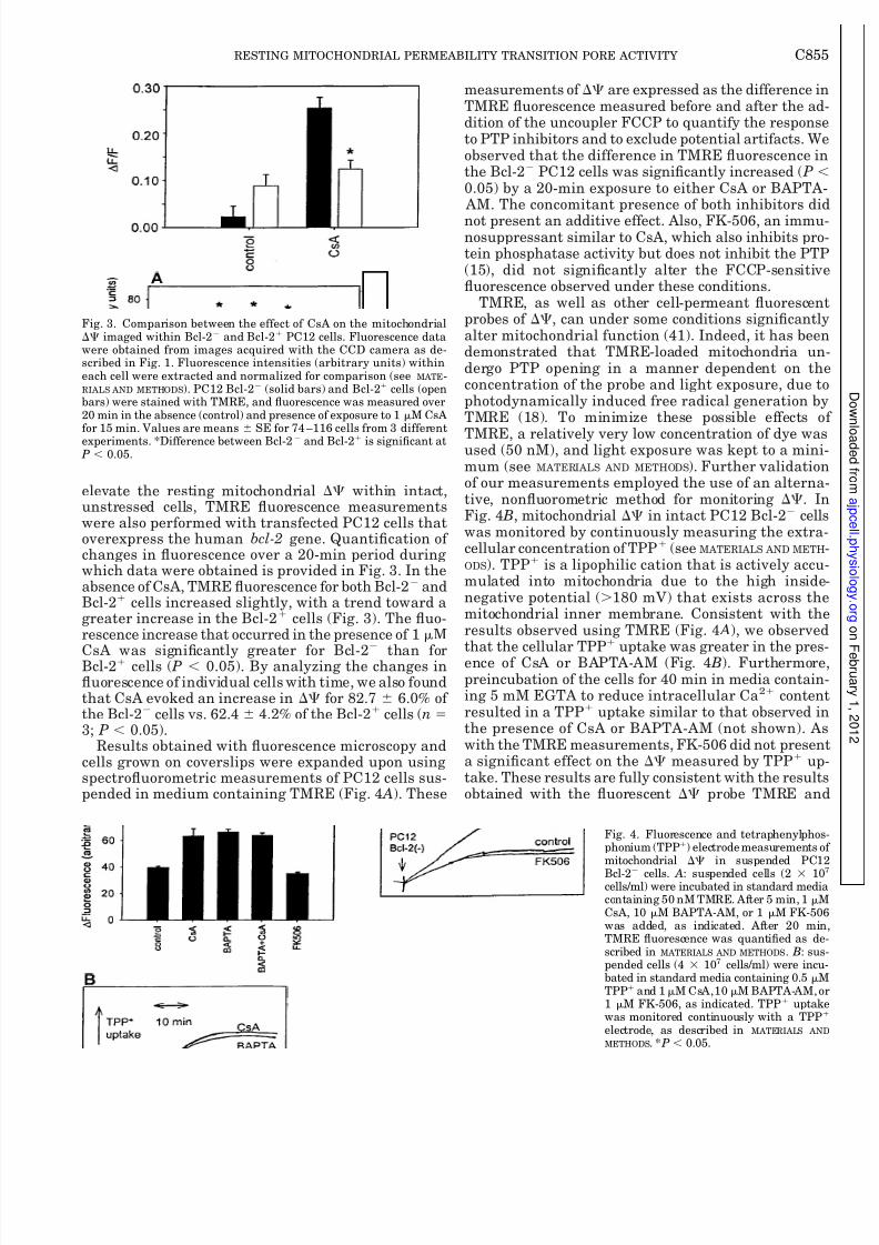
elevate the resting mitochondrial within intact,unstressed cells, TMRE fluorescence measurementswere also performed with transfected PC12 cells thatoverexpress the human bcl-2 gene. Quantification of changes in fluorescence over a 20-min period duringwhich data were obtained is provided in Fig. 3. In theabsence of CsA, TMRE fluorescence for both Bcl-2 andBcl-2 cells increased slightly, with a trend toward agreater increase in the Bcl-2 cells (Fig. 3). The fluo-rescence increase that occurred in the presence of 1 M
CsA was significantly greater for Bcl-2
than forBcl-2 cells ( P 0.05). By analyzing the changes influorescence of individual cells with time, we also foundthat CsA evoked an increase in for 82.7 6.0% of the Bcl-2 cells vs. 62.4 4.2% of the Bcl-2 cells (n 3; P 0.05).
Results obtained with fluorescence microscopy andcells grown on coverslips were expanded upon usingspectrofluorometric measurements of PC12 cells sus-pended in medium containing TMRE (Fig. 4 A). These
measurements of are expressed as the difference inTMRE fluorescence measured before and after the addition of the uncoupler FCCP to quantify the responseto PTP inhibitors and to exclude potential artifacts. Weobserved that the difference in TMRE fluorescence inthe Bcl-2 PC12 cells was significantly increased ( P 0.05) by a 20-min exposure to either CsA or BAPTA- AM. The concomitant presence of both inhibitors did
not present an additive effect. Also, FK-506, an immunosuppressant similar to CsA, which also inhibits protein phosphatase activity but does not inhibit the PTP(15), did not significantly alter the FCCP-sensitivefluorescence observed under these conditions.
TMRE, as well as other cell-permeant fluorescenprobes of , can under some conditions significantlyalter mitochondrial function (41). Indeed, it has beendemonstrated that TMRE-loaded mitochondria undergo PTP opening in a manner dependent on theconcentration of the probe and light exposure, due tophotodynamically induced free radical generation byTMRE (18). To minimize these possible effects o
TMRE, a relatively very low concentration of dye wasused (50 nM), and light exposure was kept to a mini-mum (see MATERIALS AND METHODS). Further validationof our measurements employed the use of an alternative, nonfluorometric method for monitoring . InFig. 4 B, mitochondrial in intact PC12 Bcl-2 cellswas monitored by continuously measuring the extracellular concentration of TPP (see MATERIALS AND METH
ODS). TPP is a lipophilic cation that is actively accumulated into mitochondria due to the high insidenegative potential (180 mV) that exists across themitochondrial inner membrane. Consistent with theresults observed using TMRE (Fig. 4 A), we observedthat the cellular TPP uptake was greater in the presence of CsA or BAPTA-AM (Fig. 4 B). Furthermorepreincubation of the cells for 40 min in media containing 5 mM EGTA to reduce intracellular Ca2 contenresulted in a TPP uptake similar to that observed inthe presence of CsA or BAPTA-AM (not shown). Aswith the TMRE measurements, FK-506 did not presena significant effect on the measured by TPP uptake. These results are fully consistent with the resultsobtained with the fluorescent probe TMRE and
Fig. 3. Comparison between the effect of CsA on the mitochondrial imaged within Bcl-2 and Bcl-2 PC12 cells. Fluorescence datawere obtained from images acquired with the CCD camera as de-scribed in Fig. 1. Fluorescence intensities (arbitrary units) withineach cell were extracted and normalized for comparison (see MATE-RIALS AND METHODS). PC12 Bcl-2 (solid bars) and Bcl-2 cells (openbars) were stained with TMRE, and fluorescence was measured over20 min in the absence (control) and presence of exposure to 1 M CsAfor 15 min. Values are means SE for 74 –116 cells from 3 differentexperiments. *Difference between Bcl-2 and Bcl-2 is significant at P 0.05.
Fig. 4. Fluorescence and tetraphenylphosphonium (TPP) electrode measurements omitochondrial in suspended PC12
Bcl-2
cells. A: suspended cells (2 10cells/ml) were incubated in standard mediacontaining 50 nM TMRE. After 5 min, 1 MCsA, 10 M BAPTA-AM, or 1 M FK-506was added, as indicated. After 20 minTMRE fluorescence was quantified as described in MATERIALS AND METHODS. B: suspended cells (4 107 cells/ml) were incubated in standard media containing 0.5 MTPP and 1M CsA,10 M BAPTA-AM, o1 M FK-506, as indicated. TPP uptakwas monitored continuously with a TPP
electrode, as described in MATERIALS AND
METHODS. * P 0.05.
C855RESTING MITOCHONDRIAL PERMEABILITY TRANSITION PORE ACTIVITY
8/3/2019 Elevation of Resting Mitochondrial Membrane Potential
http://slidepdf.com/reader/full/elevation-of-resting-mitochondrial-membrane-potential 6/9
support the conclusion that PTP opening lowers theresting in this cell line.
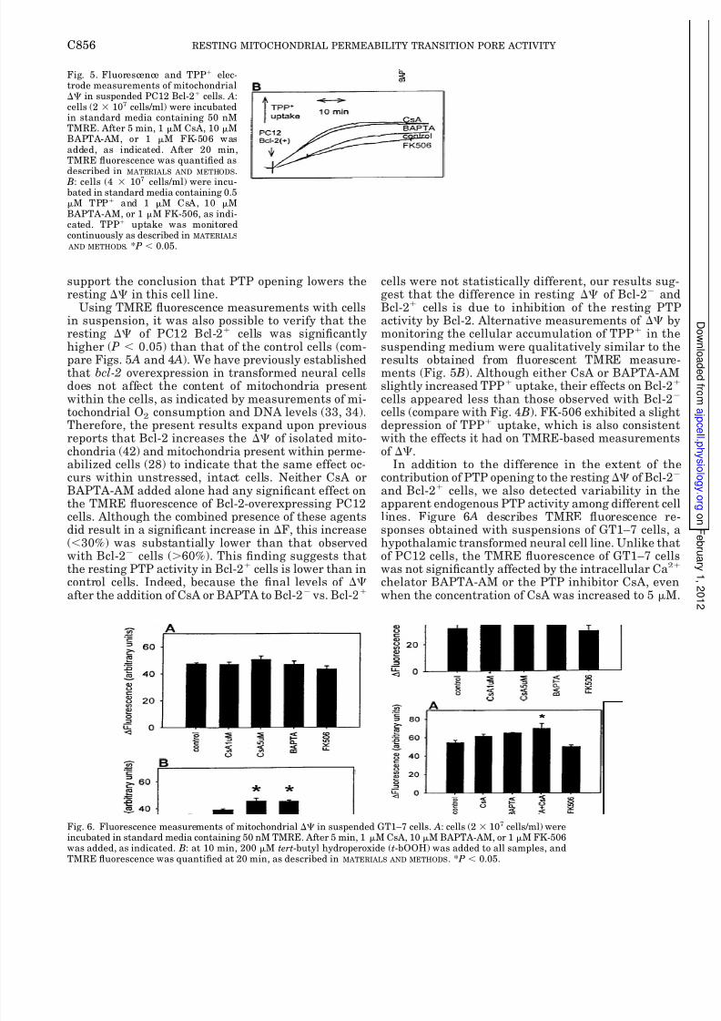
Using TMRE fluorescence measurements with cellsin suspension, it was also possible to verify that theresting of PC12 Bcl-2 cells was significantlyhigher ( P 0.05) than that of the control cells (com-
pare Figs. 5 A and 4 A). We have previously establishedthat bcl-2 overexpression in transformed neural cellsdoes not affect the content of mitochondria presentwithin the cells, as indicated by measurements of mi-tochondrial O2 consumption and DNA levels (33, 34).Therefore, the present results expand upon previousreports that Bcl-2 increases the of isolated mito-chondria (42) and mitochondria present within perme-abilized cells (28) to indicate that the same effect oc-curs within unstressed, intact cells. Neither CsA orBAPTA-AM added alone had any significant effect onthe TMRE fluorescence of Bcl-2-overexpressing PC12cells. Although the combined presence of these agentsdid result in a significant increase in F, this increase(30%) was substantially lower than that observedwith Bcl-2 cells (60%). This finding suggests thatthe resting PTP activity in Bcl-2 cells is lower than incontrol cells. Indeed, because the final levels of after the addition of CsA or BAPTA to Bcl-2 vs. Bcl-2
cells were not statistically different, our results suggest that the difference in resting of Bcl-2 andBcl-2 cells is due to inhibition of the resting PTPactivity by Bcl-2. Alternative measurements of bymonitoring the cellular accumulation of TPP in thesuspending medium were qualitatively similar to the
results obtained from fluorescent TMRE measurements (Fig. 5 B). Although either CsA or BAPTA-AMslightly increased TPP uptake, their effects on Bcl-2
cells appeared less than those observed with Bcl-2
cells (compare with Fig. 4 B). FK-506 exhibited a slightdepression of TPP uptake, which is also consistentwith the effects it had on TMRE-based measurementsof .
In addition to the difference in the extent of thecontribution of PTP opening to the resting of Bcl-2
and Bcl-2 cells, we also detected variability in theapparent endogenous PTP activity among different cellines. Figure 6 A describes TMRE fluorescence responses obtained with suspensions of GT1–7 cells, ahypothalamic transformed neural cell line. Unlike thatof PC12 cells, the TMRE fluorescence of GT1–7 cellswas not significantly affected by the intracellular Ca2
chelator BAPTA-AM or the PTP inhibitor CsA, evenwhen the concentration of CsA was increased to 5 M
Fig. 5. Fluorescence and TPP elec-trode measurements of mitochondrial in suspended PC12 Bcl-2 cells. A:cells (2 107 cells/ml) were incubatedin standard media containing 50 nMTMRE. After 5 min, 1 M CsA, 10 MBAPTA-AM, or 1 M FK-506 wasadded, as indicated. After 20 min,TMRE fluorescence was quantified asdescribed in MATERIALS AND METHODS. B: cells (4 107 cells/ml) were incu-bated in standard media containing 0.5M TPP and 1 M CsA, 10 MBAPTA-AM, or 1 M FK-506, as indi-cated. TPP uptake was monitoredcontinuously as described in MATERIALS
AND METHODS. * P 0.05.
Fig. 6. Fluorescence measurements of mitochondrial in suspended GT1–7 cells. A: cells (2 107 cells/ml) wereincubated in standard media containing 50 nM TMRE. After 5 min, 1 M CsA, 10 M BAPTA-AM, or 1 M FK-506was added, as indicated. B: at 10 min, 200 M tert-butyl hydroperoxide (t-bOOH) was added to all samples, andTMRE fluorescence was quantified at 20 min, as described in MATERIALS AND METHODS. * P 0.05.
C856 RESTING MITOCHONDRIAL PERMEABILITY TRANSITION PORE ACTIVITY

8/3/2019 Elevation of Resting Mitochondrial Membrane Potential
http://slidepdf.com/reader/full/elevation-of-resting-mitochondrial-membrane-potential 7/9
The of unstressed GT1–7 cells was also not affectedby exposure to FK-506. However, if these cells weretreated with t-bOOH, a compound capable of enhanc-ing Ca2-induced PTP in both isolated mitochondriaand cells by oxidizing mitochondrial pyridine nucleo-tides (4, 6, 14, 21, 28, 36), then an average value for Fwas obtained (32 4; Fig. 6 B) that is significantlylower than that obtained in the absence of t-bOOH(47 1; Fig. 6 A). The fluorescence values obtained inthe presence of t-bOOH were significantly increased byexposure of cells to CsA (5 M) or BAPTA but notFK-506. Thus GT1–7 cells do not present a detectableresting PTP activity but do exhibit a drop in con-sistent with PTP activity when exposed to the pro-oxidant t-bOOH.
In contrast to the insensitivity of GT1–7 cells toalterations in resting caused by CsA or BAPTA- AM, but in agreement with the sensitivity of PC12cells, the resting of human SY5Y neuroblastomacells was significantly elevated by the addition of theMPT inhibitor CsA or by exposure to the intracellularCa2 chelator BAPTA-AM (Fig. 7). In addition, wefound that exposure of these cells to the extracellularCa2 chelator EGTA for 40 min resulted in an increasein TMRE fluorescence. Thus, with SY5Y cells, we ob-served an increase in the normal after treatment
with three different conditions that can inhibit PTPactivity by two different mechanisms. As with theother cell lines, the of SY5Y cells was not elevatedby exposure to FK-506.
DISCUSSION
Taken together, our results strongly suggest that thePTP is active and contributes toward a decrease inmitochondrial in resting PC12 and SY5Y cells butnot in unstressed GT1–7 cells. We have determinedthat the PTP influences the resting in PC12 andSY5Y cells by demonstrating that can be elevated
by the presence of CsA or Ca2 chelators but not by theimmune suppressor FK-506, which does not inhibiPTP activity in isolated mitochondria (15). In additionfluorescence microscopy measurements on individuacells indicate that the increase in induced by CsAor BAPTA-AM in PC12 cells is a common phenomenonand occurs in the majority of cells that are analyzedThis finding argues against the possibility that the
observed responses are due to a fraction of cells thatare undergoing PTP as part of a cell death process. TheGT1–7 cell line that did not exhibit sensitivity of resting to PTP inhibitors nevertheless did demonstratea CsA- and BAPTA-AM-sensitive fraction of in thepresence of the PTP inducer t-bOOH. The finding thatt-bOOH does not completely eliminate the of GT1–7 cells, as it does in hepatocytes (4, 21, 36), may relateto the reason why GT1–7 cells do not express a detectable endogenous PTP activity. For example, variabilityof endogenous and induced PTP activity could be due to variability in cellular redox state or sensitivity of mitochondrial pyridine nucleotides to oxidation (14).
The PTP has been studied most extensively with
isolated mitochondria with the use of conditions in vitro that bear little resemblance to those that exiswithin intact cells. Within the last few years, howeverevidence obtained with models of cell and tissue injuryhas supported the involvement of the PTP in necroticand apoptotic cell death (8, 17, 29, 30). Moreover, it hasbeen proposed that a Ca2- and proton-selective, lowconductivity state of the PTP may be active in normaEhrlich tumor cells, generating and conveying electrical and Ca2 signals (19). In the study reported byIchas et al. (19), the PTP is activated upon IP3-inducedCa2 mobilization from the endoplasmic reticulumwhereupon PTP-mediated mitochondrial Ca2 efflux
contributes to the amplification of cytosolic Ca
2
signals. These findings are supported by studies showingthat binding of IP3 to its receptors results in discreteareas of elevated intracellular Ca2 that are sensed byneighboring mitochondria (39). The ensuing increasein intramitochondrial Ca2 can lead to an activation omitochondrial dehydrogenases and, therefore, ATPproduction (40). However, the rapid stimulation of mitochondrial Ca2 uptake by focal spikes in extramitochondrial Ca2 concentrations could result in transientreductions in mitochondrial (9, 47), which wouldpromote activation of the PTP. Opening of the PTPwould be expected to prolong the period of mitochon-drial depolarization and induce the release of at least
some fraction of the accumulated Ca2
. Additionasupport for this scenario comes from observations thatCsA increases mitochondrial Ca2 accumulation innormal cardiomyocytes (1) and decreases Ca2-inducedmitochondrial Ca2 release in Ehrlich ascites tumorcells and endothelial cells (10, 50). In addition, blockade of MPT inhibits agonist-evoked Ca2 oscillations inglial cells, reinforcing the hypothesis that, duringphysiological stimulation, transient PTP openings support Ca2 signaling (46). Further evidence for the baseline activity of a low-conductance state of the PTP hascome from TPP uptake measurements (5), fluorescent
Fig. 7. Fluorescence measurements of mitochondrial in sus-pended SY5Y cells. Cells (2 107 cells/ml) were incubated in stan-dard media containing 50 nM TMRE. After 5 min, 1 M CsA, 10 MBAPTA-AM, or 1 M FK-506 was added, as indicated. After 20 min,TMRE fluorescence was quantified as described in MATERIALS AND
METHODS. EGTA-treated cells were preincubated for 40 min in reac-tion media containing 5 mM EGTA, before the addition of 50 nMTMRE. * P 0.05.
C857RESTING MITOCHONDRIAL PERMEABILITY TRANSITION PORE ACTIVITY

8/3/2019 Elevation of Resting Mitochondrial Membrane Potential
http://slidepdf.com/reader/full/elevation-of-resting-mitochondrial-membrane-potential 8/9
flow cytometry and microscopic imaging of in SY5Yneuroblastoma cells (11), and rat oligodendrocyte pro-genitors (46).
The activity of the MPT under physiological condi-tions would qualify it as an endogenous uncoupler of oxidative phosphorylation. However, the degree of un-coupling and energy expenditure by the movement of ions through the PTP would, by necessity, need to be
very limited so that metabolic homeostasis could bepreserved. Like the activity of well-characterized tis-sue-specific uncoupling proteins (22), the resting statePTP activity may act similarly to increase energy con-sumption without obstructing ATP synthesis. Studiesare in progress to determine the extent to which theendogenous PTP contributes to the basal rate of respi-ration by the PC12 and SY5Y cells used in our exper-iments. Increased energy utilization associated withthe cycling of protons, Ca2, and other ions mediatedby PTP activity should be manifested as heat genera-tion. Indeed, CsA has been demonstrated to decreasethe heat output of normal lymphocytes (25). In addition
to the influence PTP activity can exert on intracellularCa2 signaling, oxygen utilization, and heat produc-tion, the associated reduction in would reduce theformation of superoxide due to “leakage” of electronsfrom the ubiquinone region of the electron transport(45). It is therefore possible that a controlled endoge-nous PTP activity could actually protect mitochondriaagainst self-inflicted oxidative stress (45).
Bcl-2 overexpression has previously been shown toincrease in isolated mitochondria (42) and perme-abilized cells (28). Our findings further indicate thatBcl-2 can elevate the resting of intact, unstressedcells by inhibiting the endogenous activity of the PTP,as previously suggested by JC-1 fluorescence probe
measurements of in another strain of PC12 cells(7). It is possible that the differences in uncoupler-sensitive TMRE fluorescence and TPP uptake be-tween Bcl-2 and Bcl-2 cells are due to differences inmitochondrial volume, even though maximal rates of respiration and mitochondrial DNA contents areequivalent. However, the observation that overexpres-sion of Bcl-2 minimizes the effects of PTP inhibitors onresting constitutes evidence that Bcl-2 actuallyincreases possibly via inhibition of endogenousPTP activity. Bcl-2 has been proposed to act as a H
channel that contributes to rather than detracts fromthe mitochondrial electrochemical gradient of protons
(42). However, considering the known ability of Bcl-2 toinhibit the stress-evoked PTP opening in isolated mi-tochondria and permeabilized cells (28, 32, 42, 48) andthe ability of Bcl-2 to inhibit endogenous PTP activityin our experiments, its endowment for MPT inhibitionmay be its primary mechanism of action. Although thephysiological role for Bcl-2 is generally thought to beone of protection against cytotoxicity (24, 26, 32, 34, 35,48, 52), the present results suggest that Bcl-2 may alsoserve as an enhancer of the efficiency of mitochondrialenergy coupling by decreasing the endogenous PTPactivity.
We thank S. J. Russell for the excellent technical assistance.Transfected cells were kindly provided by Dr. Dale Bredesen
(Burnham Research Institute, La Jolla, CA), and FK-506 was pro vided by Fujisawa, Japan.
This work was supported by the FAPESP, by National Institutesof Health Grant NS-34152, and by the Bayer Corporation.
REFERENCES
1. Altschuld RA, Hohl CM, Castillo LC, Garleb AA, StarlingRC, and Brierley GP. Cyclosporin inhibits mitochondrial calcium efflux in isolated adult rat ventricular cardiomyocytes Am J Physiol Heart Circ Physiol 262: H1699–H1704, 1992.
2. Andreyev A and Fiskum G. Calcium induced release of mitochondrial cytochrome c by different mechanisms selective fobrain versus liver. Cell Death Differ 6: 825–832, 1999.
3. Baumgold J, Paek R, and Fiskum G. Calcium independenceof phosphoinositide hydrolysis-induced increase in cyclic AMPaccumulation in SK-N-SH human neuroblastoma cells. J Neurochem 58: 1754–1759, 1992.
4. Byrne AM, Lemasters JJ, and Nieminen AL. Contribution oincreased mitochondrial free Ca2 to the mitochondrial permeability transition induced by tert-butylhydroperoxide in rat hepatocytes. Hepatology 29: 1523–1531, 1999.
5. Cassarino DS, Swerdlow RH, Parks JK, Parker WDJ, andBennett JPJ. Cyclosporin A increases resting mitochondria
membrane potential in SY5Y cells and reverses the depressedmitochondrial membrane potential of Alzheimer’s disease cybrids. Biochem Biophys Res Commun 248: 168–173, 1998.
6. Castilho RF, Kowaltowski AJ, Meinicke AR, Bechara EJand Vercesi AE. Permeabilization of the inner mitochondriamembrane by Ca2 ions is stimulated by t-butyl hydroperoxideand mediated by reactive oxygen species generated by mitochondria. Free Radic Biol Med 18: 479–486, 1995.
7. Dispersyn G, Nuydens R, Connors R, Borgers M, andGeerts H. Bcl-2 protects against FCCP-induced apoptosis andmitochondrial membrane potential depolarization in PC12 cells Biochim Biophys Acta 1428: 357–371, 1999.
8. Dubinsky JM and Levi Y. Calcium-induced activation of themitochondrial permeability transition in hippocampal neurons J Neurosci Res 53: 728–741, 1998.
9. Duchen MR, Leyssens A, and Crompton M. Transient mitochondrial depolarizations reflect focal sarcoplasmic reticular cal
cium release in single rat cardiomyocytes. J Cell Biol 142: 975–988, 1998.
10. Evtodienko Y, Teplova V, Khawaja J, and Saris NE. ThCa2-induced permeability transition pore is involved in Ca2
induced mitochondrial oscillations. A study on permeabilisedEhrlich ascites tumour cells. Cell Calcium 15: 143–152, 1994.
11. Fall CP and Bennett JPJ. Visualization of cyclosporin A andCa2-sensitive cyclical mitochondrial depolarizations in cell culture. Biochim Biophys Acta 1410: 77–84, 1999.
12. Farkas DL, Wei MD, Febbroriello P, Carson JH, and LoewLM. Simultaneous imaging of cell and mitochondrial membranepotentials. Biophys J 56: 1053–1069, 1989. [published erratumappears in Biophys J 1990 Mar 57(3): following p. 684]
13. Fiskum G and Lehninger AL. Regulated release of Ca2 fromrespiring mitochondria by Ca2 /2H antiport. J Biol Chem 2546236–6239, 1979.
14. Fiskum G and Pease A. Hydroperoxide-stimulated release ocalcium from rat liver and AS-30D hepatoma mitochondriaCancer Res 46: 3459–3463, 1986.
15. Friberg H, Ferrand-Drake M, Bengtsson F, Halestrap APand Wieloch T. Cyclosporin A, but not FK 506, protects mitochondria and neurons against hypoglycemic damage and implicates the mitochondrial permeability transition in cell death J Neurosci 18: 5151–5159, 1998.
16. Hajnoczky G, Robb-Gaspers LD, Seitz MB, and Thoma AP. Decoding of cytosolic calcium oscillations in the mitochondria. Cell 82: 415–424, 1995.
17. Halestrap AP, Kerr PM, Javadov S, and Woodfield KYElucidating the molecular mechanism of the permeability transition pore and its role in reperfusion injury of the heart. Biochim Biophys Acta 1366: 79–94, 1998.
C858 RESTING MITOCHONDRIAL PERMEABILITY TRANSITION PORE ACTIVITY

8/3/2019 Elevation of Resting Mitochondrial Membrane Potential
http://slidepdf.com/reader/full/elevation-of-resting-mitochondrial-membrane-potential 9/9
18. Huser J, Rechenmacher CE, and Blatter LA. Imaging thepermeability pore transition in single mitochondria. Biophys J 74: 2129–2137, 1998.
19. Ichas F, Jouaville LS, and Mazat JP. Mitochondria are ex-citable organelles capable of generating and conveying electricaland calcium signals. Cell 89: 1145–1153, 1997.
20. Ichas F and Mazat JP. From calcium signaling to cell death:two conformations for the mitochondrial permeability transitionpore. Switching from low- to high-conductance state. Biochim Biophys Acta 1366: 33–50, 1998.
21. Imberti R, Nieminen AL, Herman B, and Lemasters JJ.Mitochondrial and glycolytic dysfunction in lethal injury tohepatocytes by t-butylhydroperoxide: protection by fructose, cy-closporin A and trifluoperazine. J Pharmacol Exp Ther 265:392–400, 1993.
22. Jezek P, Engstova H, Zackova M, Vercesi AE, Costa AD, Arruda P, and Garlid KD. Fatty acid cycling mechanism andmitochondrial uncoupling proteins. Biochim Biophys Acta 1365:319–327, 1998.
23. Kamo N, Muratsugu M, Hongoh R, and Kobatake Y. Mem-brane potential of mitochondria measured with an electrodesensitive to tetraphenyl phosphonium and relationship betweenproton electrochemical potential and phosphorylation potentialin steady state. J Membr Biol 49: 105–121, 1979.
24. Kane DJ, Sarafian TA, Anton R, Hahn H, Gralla EB, Val-entine JS, Ord T, and Bredesen DE. Bcl-2 inhibition of neuraldeath: decreased generation of reactive oxygen species. Science
262: 1274–1277, 1993.25. Karlsson H, DePierre JW, and Nassberger L. Energy levels
in resting and mitogen-stimulated human lymphocytes duringtreatment with FK506 or cyclosporin A in vitro. Biochim Biophys Acta 1319: 301–310, 1997.
26. Kluck RM, Bossy-Wetzel E, Green DR, and Newmeyer DD.The release of cytochrome c from mitochondria: a primary sitefor Bcl-2 regulation of apoptosis. Science 275: 1132–1136, 1997.
27. Kowaltowski AJ and Vercesi AE. Mitochondrial damage in-duced by conditions of oxidative stress. Free Radic Biol Med 26:463–471, 1999.
28. Kowaltowski AJ, Vercesi AE, and Fiskum G. Bcl-2 preventsmitochondrial permeability transition and cytochrome c release via maintenance of reduced pyridine nucleotides. Cell Death Different. In press.
29. Kroemer G, Dallaporta B, and Resche-Rigon M. The mito-
chondrial death/life regulator in apoptosis and necrosis. Annu Rev Physiol 60:619–42: 619–642, 1998.30. Lemasters JJ, Nieminen AL, Qian T, Trost LC, Elmore SP,
Nishimura Y, Crowe RA, Cascio WE, Bradham CA, Bren-ner DA, and Herman B. The mitochondrial permeability tran-sition in cell death: a common mechanism in necrosis, apoptosisand autophagy. Biochim Biophys Acta 1366: 177–196, 1998.
31. Loew LM, Carrington W, Tuft RA, and Fay FS. Physiologicalcytosolic Ca2 transients evoke concurrent mitochondrial depo-larizations. Proc Natl Acad Sci USA 91: 12579–12583, 1994.
32. Marzo I, Brenner C, Zamzami N, Susin SA, Beutner G,Brdiczka D, Remy R, Xie ZH, Reed JC, and Kroemer G. Thepermeability transition pore complex: a target for apoptosisregulation by caspases and bcl-2-related proteins. J Exp Med187: 1261–1271, 1998.
33. Murphy AN, Bredesen DE, Cortopassi G, Wang E, andFiskum G. Bcl-2 potentiates the maximal calcium uptake ca-
pacity of neural cell mitochondria. Proc Natl Acad Sci USA 93:9893–9898, 1996.34. Myers KM, Fiskum G, Liu Y, Simmens SJ, Bredesen DE,
and Murphy AN. Bcl-2 protects neural cells from cyanide/ aglycemia-induced lipid oxidation, mitochondrial injury, andloss of viability. J Neurochem 65: 2432–2440, 1995.
35. Newmeyer DD and Green DR. Surviving the cytochrome seas Neuron 21: 653–655, 1998.
36. Nieminen AL, Byrne AM, Herman B, and Lemasters JJMitochondrial permeability transition in hepatocytes induced byt-BuOOH: NAD(P)H and reactive oxygen species. Am J PhysioCell Physiol 272: C1286–C1294, 1997.
37. Novgorodov SA and Gudz TI. Permeability transition pore othe inner mitochondrial membrane can operate in two openstates with different selectivities. J Bioenerg Biomembr 28: 139–146, 1996.
38. Petronilli V, Miotto G, Canton M, Brini M, Colonna RBernardi P, and Di Lisa F. Transient and long-lasting openings of the mitochondrial permeability transition pore can bemonitored directly in intact cells by changes in mitochondriacalcein fluorescence. Biophys J 76: 725–734, 1999.
39. Rizzuto R, Brini M, Murgia M, and Pozzan T. Microdomainwith high Ca2 close to IP3-sensitive channels that are sensed byneighboring mitochondria. Science 262: 744–747, 1993.
40. Robb-Gaspers LD, Burnett P, Rutter GA, Denton RM, Rizzuto R, and Thomas AP. Integrating cytosolic calcium signalinto mitochondrial metabolic responses. EMBO J 17: 4987–5000, 1998.
41. Scaduto RCJ and Grotyohann LW. Measurement of mitochondrial membrane potential using fluorescent rhodamine derivatives. Biophys J 76: 469–477, 1999.
42. Shimizu S, Eguchi Y, Kamiike W, Funahashi Y, Mignon A
Lacronique V, Matsuda H, and Tsujimoto Y. Bcl-2 preventapoptotic mitochondrial dysfunction by regulating proton flux Proc Natl Acad Sci USA 95: 1455–1459, 1998.
43. Simpson PB and Russell JT. Mitochondria support inosito1,4,5-trisphosphate-mediated Ca2 waves in cultured oligodendrocytes. J Biol Chem 271: 33493–33501, 1996.
44. Simpson PB and Russell JT. Role of mitochondrial Ca2
regulation in neuronal and glial cell signalling. Brain Res Brain Res Rev 26: 72–81, 1998.
45. Skulachev VP. Membrane-linked systems preventing superoxide formation. Biosci Rep 17: 347–366, 1997.
46. Smaili SS and Russell JT. Permeability transition pore regulates both mitochondrial membrane potential and agonistevoked Ca2 signals in oligodendrocyte progenitors. Cell Calcium 26: 121–130, 1999.
47. Sparagna GC, Gunter KK, Sheu SS, and Gunter TE. Mito
chondrial calcium uptake from physiological-type pulses of calcium. A description of the rapid uptake mode. J Biol Chem 27027510–27515, 1995.
48. Susin SA, Zamzami N, Castedo M, Hirsch T, Marchetti PMacho A, Daugas E, Geuskens M, and Kroemer G. Bcl-2inhibits the mitochondrial release of an apoptogenic protease. J Exp Med 184: 1331–1341, 1996.
49. Vercesi AE. Stimulation of mitochondrial Ca2 efflux byNADP with maintenance of respiratory control. An Acad BrasCienc 57: 369–375, 1985.
50. Wood PG and Gillespie JI. Evidence for mitochondrial Ca2
induced Ca2 release in permeabilised endothelial cells. Biochem Biophys Res Commun 246: 543–548, 1998.
51. Yagodin SV, Holtzclaw L, Sheppard CA, and Russell JTNonlinear propagation of agonist-induced cytoplasmic calciumwaves in single astrocytes. J Neurobiol 25: 265–280, 1994.
52. Yang J, Liu X, Bhalla K, Kim CN, Ibrado AM, Cai J, PengTI, Jones DP, and Wang X. Prevention of apoptosis by Bcl-2release of cytochrome c from mitochondria blocked. Science 2751129–1132, 1997.
53. Zoratti M and Szabo I. The mitochondrial permeability transition. Biochim Biophys Acta 1241: 139–176, 1995.
C859RESTING MITOCHONDRIAL PERMEABILITY TRANSITION PORE ACTIVITY