Elevated expression of miR-142-3p is related to the pro ...
11
RESEARCH ARTICLE Open Access Elevated expression of miR-142-3p is related to the pro-inflammatory function of monocyte-derived dendritic cells in SLE Yilun Wang 1 , Jun Liang 1* , Haihong Qin 1 , Yan Ge 2 , Juan Du 1 , Jinran Lin 1 , Xiaohua Zhu 1 , Jie Wang 3 and Jinhua Xu 1* Abstract Background: Recent studies have shown that alterations in the function of dendritic cells (DCs) are involved in the pathogenesis of systemic lupus erythematosus (SLE). However, the mechanism of the alteration remains unclear. Methods: We cultured monocyte-derived DCs (moDCs) in vitro and examined the cytokines and chemokines in the supernatants of moDCs in negative controls (NC) and SLE patients in active phase. We then profiled microRNAs (miRNAs) of LPS-stimulated moDCs in SLE patients and used real-time PCR to verify the differentially expressed miRNAs. A lentiviral construct was used to overexpress the level of miR-142-3p in moDCs of NC. We examined the cytokines and chemokines in the supernatants of moDCs overexpressing miR-142-3p and used Transwell test, flow cytometric analysis and cell proliferation to observe the impact on CD4 + T cells in moDC-CD4 + T cell co-culture. Results: moDCs in patients with SLE secreted increased level of IL-6, CCL2 and CCL5, with attraction of more CD4 + T cells compared with NC. We found 18 differentially expressed microRNAs in moDCs of SLE patients by microarray, and target gene prediction showed some target genes of differentially expressed miRNAs were involved in cytokine regulation. miR-142-3p was verified among the highly expressed miRNAs in the SLE group and overexpressing miR-142-3p in moDCs of the NC group caused an increase of SLE-related cytokines, such as CCL2, CCL5, CXCL8, IL-6 and TNF-α. Moreover, moDCs overexpressed with miR-142-3p resulted in attraction of an increased number of CD4 + T cells and in suppression of the proportion of Tregs in DC-CD4 + T cell co-culture whereas the proliferation of CD4 + T cells was not altered. Conclusions: The results demonstrated a role for miR-142-3p in regulating the pro-inflammatory function of moDCs in the pathogenesis of SLE. These findings suggested that miR-142-3p could serve as a novel therapeutic target for the treatment of SLE. Keywords: SLE, Monocyte-derived DCs, MicroRNA Background Systemic lupus erythematosus (SLE) is a complicated autoimmune disease impairing multiple organs. The disease predominantly affects women aged 15-40 years [1] with a female to male ratio of 9:1 [2]. Both genetic and environmental factors contribute to human SLE pathogenesis [3], but the etiology of SLE is not fully understood. Impaired clearance of dying cells may repre- sent a central pathogenic process in human lupus [4]. The accumulated dying cells release autoantigens, which are presented by dendritic cells, further breaking down the immune tolerance of T and B cells and triggering SLE. Dendritic cells (DCs) are so far the most potent antigen- presenting cells (APCs) with important functions in the immune system. Immunodysregulation in SLE involves the complex interplay of various immune cells and DCs are the master regulators for initiation, amplification, and perpetuation of the disease [5]. DCs could influence SLE in several ways including: presentation of self-antigens to autoreactive T cells; oversecretion of pro-inflammatory cytokines; and suppression of regulatory T cells and pro- motion of B cell autoantibody production, either directly * Correspondence: [email protected]; [email protected] 1 Department of Dermatology, Huashan Hospital, Fudan University, 12 Wulumuqi Zhong Road, Shanghai 200040, People’s Republic of China Full list of author information is available at the end of the article © The Author(s). 2016 Open Access This article is distributed under the terms of the Creative Commons Attribution 4.0 International License (http://creativecommons.org/licenses/by/4.0/), which permits unrestricted use, distribution, and reproduction in any medium, provided you give appropriate credit to the original author(s) and the source, provide a link to the Creative Commons license, and indicate if changes were made. The Creative Commons Public Domain Dedication waiver (http://creativecommons.org/publicdomain/zero/1.0/) applies to the data made available in this article, unless otherwise stated. Wang et al. Arthritis Research & Therapy (2016) 18:263 DOI 10.1186/s13075-016-1158-z
Transcript of Elevated expression of miR-142-3p is related to the pro ...
Wang et al. Arthritis Research & Therapy (2016) 18:263 DOI 10.1186/s13075-016-1158-z
RESEARCH ARTICLE Open Access
Elevated expression of miR-142-3p isrelated to the pro-inflammatory function ofmonocyte-derived dendritic cells in SLE
Yilun Wang1, Jun Liang1*, Haihong Qin1, Yan Ge2, Juan Du1, Jinran Lin1, Xiaohua Zhu1, Jie Wang3 and Jinhua Xu1*Abstract
Background: Recent studies have shown that alterations in the function of dendritic cells (DCs) are involved in thepathogenesis of systemic lupus erythematosus (SLE). However, the mechanism of the alteration remains unclear.
Methods: We cultured monocyte-derived DCs (moDCs) in vitro and examined the cytokines and chemokines in thesupernatants of moDCs in negative controls (NC) and SLE patients in active phase. We then profiled microRNAs(miRNAs) of LPS-stimulated moDCs in SLE patients and used real-time PCR to verify the differentially expressedmiRNAs. A lentiviral construct was used to overexpress the level of miR-142-3p in moDCs of NC. We examined thecytokines and chemokines in the supernatants of moDCs overexpressing miR-142-3p and used Transwell test, flowcytometric analysis and cell proliferation to observe the impact on CD4+ T cells in moDC-CD4+T cell co-culture.
Results: moDCs in patients with SLE secreted increased level of IL-6, CCL2 and CCL5, with attraction of more CD4+
T cells compared with NC. We found 18 differentially expressed microRNAs in moDCs of SLE patients by microarray,and target gene prediction showed some target genes of differentially expressed miRNAs were involved in cytokineregulation. miR-142-3p was verified among the highly expressed miRNAs in the SLE group and overexpressingmiR-142-3p in moDCs of the NC group caused an increase of SLE-related cytokines, such as CCL2, CCL5, CXCL8,IL-6 and TNF-α. Moreover, moDCs overexpressed with miR-142-3p resulted in attraction of an increased numberof CD4+ T cells and in suppression of the proportion of Tregs in DC-CD4+T cell co-culture whereas the proliferationof CD4+T cells was not altered.
Conclusions: The results demonstrated a role for miR-142-3p in regulating the pro-inflammatory function of moDCsin the pathogenesis of SLE. These findings suggested that miR-142-3p could serve as a novel therapeutic target forthe treatment of SLE.
Keywords: SLE, Monocyte-derived DCs, MicroRNA
BackgroundSystemic lupus erythematosus (SLE) is a complicatedautoimmune disease impairing multiple organs. Thedisease predominantly affects women aged 15-40 years[1] with a female to male ratio of 9:1 [2]. Both geneticand environmental factors contribute to human SLEpathogenesis [3], but the etiology of SLE is not fullyunderstood. Impaired clearance of dying cells may repre-sent a central pathogenic process in human lupus [4]. The
* Correspondence: [email protected]; [email protected] of Dermatology, Huashan Hospital, Fudan University, 12Wulumuqi Zhong Road, Shanghai 200040, People’s Republic of ChinaFull list of author information is available at the end of the article
© The Author(s). 2016 Open Access This articInternational License (http://creativecommonsreproduction in any medium, provided you gthe Creative Commons license, and indicate if(http://creativecommons.org/publicdomain/ze
accumulated dying cells release autoantigens, which arepresented by dendritic cells, further breaking down theimmune tolerance of T and B cells and triggering SLE.Dendritic cells (DCs) are so far the most potent antigen-
presenting cells (APCs) with important functions in theimmune system. Immunodysregulation in SLE involvesthe complex interplay of various immune cells and DCsare the master regulators for initiation, amplification, andperpetuation of the disease [5]. DCs could influence SLEin several ways including: presentation of self-antigens toautoreactive T cells; oversecretion of pro-inflammatorycytokines; and suppression of regulatory T cells and pro-motion of B cell autoantibody production, either directly
le is distributed under the terms of the Creative Commons Attribution 4.0.org/licenses/by/4.0/), which permits unrestricted use, distribution, andive appropriate credit to the original author(s) and the source, provide a link tochanges were made. The Creative Commons Public Domain Dedication waiverro/1.0/) applies to the data made available in this article, unless otherwise stated.
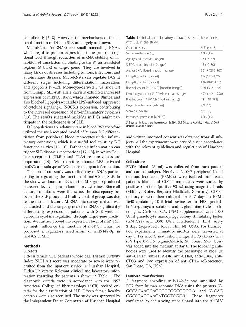
Table 1 Clinical and laboratory characteristics of the patientswith SLE in the study
Characteristics SLE (n = 15)
Sex [male/female (n)] 0/15 (15)
Age (years) [median (range)] 33 (17–57)
SLEDAI score [median (range)] 15 (10–30)
Anti-dsDNA (IU/ml) [median (range)] 391.9 (25.9–800)
C3 (g/l) [median (range)] 0.6 (0.22–1.02)
C4 (g/l) [median (range)] 0.07 (0.06–0.15)
Red cell count (*10^12/l) [median (range)] 3.91 (3.16–4.44)
Lymphocyte count (*10^9/l) [median (range)] 4.74 (1.56–19.78)
Platelet count (*10^9/l) [median (range)] 181 (25–382)
Organ involvement [Y/N (n)] 6/9 (15)
Steroids [Y/N (n)] 9/6 (15)
Immunosuppressant [Y/N (n)] 0/15 (15)
SLE systemic lupus erythematosus, SLEDAI SLE Disease Activity Index, dsDNAdouble-stranded DNA
Wang et al. Arthritis Research & Therapy (2016) 18:263 Page 2 of 11
or indirectly [6–8]. However, the mechanisms of the al-tered function of DCs in SLE are largely unknown.MicroRNAs (miRNAs) are small noncoding RNAs,
which regulate protein expression at the posttranscrip-tional level through reduction of mRNA stability or in-hibition of translation via binding to the 3′ un-translatedregions (3′UTR) of target genes. They are involved inmany kinds of diseases including tumors, infections, andautoimmune diseases. MicroRNAs can regulate DCs atdifferent stages including differentiation, maturation,and apoptosis [9–12]. Monocyte-derived DCs (moDCs)from Blimp1 SLE-risk allele carriers exhibited increasedexpression of miRNA let-7c, which inhibited Blimp1 andalso blocked lipopolysaccharide (LPS)-induced suppressorof cytokine signaling-1 (SOCS1) expression, contributingto the increased expression of pro-inflammatory cytokines[13]. The results suggested miRNAs in DCs might par-ticipate in the pathogenesis of SLE.DC populations are relatively rare in blood. We therefore
utilized the well-accepted model of human DC differen-tiation from peripheral blood monocytes under inflam-matory conditions, which is a useful tool to study DCfunctions ex vivo [14–16]. Pathogenic inflammation cantrigger SLE disease exacerbations [17, 18], in which Toll-like receptor 4 (TLR4) and TLR4 responsiveness areimportant [19]. We therefore choose LPS-activatedmoDCs as a subtype of DCs generated upon inflammation.The aim of our study was to find any miRNAs partici-
pating in regulating the function of moDCs in SLE. Inthe study, we found moDCs in the SLE group producedincreased levels of pro-inflammatory cytokines. Since allculture conditions were the same, the discrepancy be-tween the SLE group and healthy control group was dueto the intrinsic factors. MiRNA microarray analysis wasconducted and the target genes of miRNAs significantlydifferentially expressed in patients with SLE were in-volved in cytokine regulation through target gene predic-tion. We further proved the expression level of miR-142-3p might influence the function of moDCs. Thus, weproposed a regulatory mechanism of miR-142-3p inmoDCs of SLE.
MethodsSubjectsFifteen female SLE patients whose SLE Disease ActivityIndex (SLEDAI) score was moderate to severe were re-cruited from the inpatient service in Huashan Hospital,Fudan University. Relevant clinical and laboratory infor-mation regarding the patients is shown in Table 1. Thediagnostic criteria were in accordance with the 1997American College of Rheumatology (ACR) revised cri-teria for the classification of SLE. Fifteen female healthycontrols were also recruited. The study was approved bythe Independent Ethics Committee of Huashan Hospital
and written informed consent was obtained from all sub-jects. All the experiments were carried out in accordancewith the relevant guidelines and regulations of HuashanHospital.
Cell cultureEDTA blood (25 ml) was collected from each patientand control subject. Nearly 1–2*10^7 peripheral bloodmononuclear cells (PBMCs) were isolated from eachpatient’s blood and CD14+ monocytes were sorted bypositive selection (purity > 90 %) using magnetic beads(Miltenyi Biotec, Bergisch Gladbach, Germany). CD14+
monocytes were then cultured for 5–7 days in RPMI1640 containing 10 % fetal bovine serum (FBS), penicil-lin/streptomycin solution and L-glutamine (Life Tech-nologies, Carlsbad, CA, USA) supplemented with 1000U/ml granulocyte-macrophage colony-stimulating factor(GM-CSF) and 1000 U/ml interleukin-4 (IL-4) every2 days (PeproTech, Rocky Hill, NJ, USA). For transfec-tion experiments, immature moDCs were harvested atday 5. For moDC maturation, 1 μg/ml LPS (Escherichiacoli type 055:B6; Sigma-Aldrich, St. Louis, MO, USA)was added into the medium at day 6. The following anti-bodies were used to identify the phenotype of moDCs:anti-CD11c, anti-HLA-DR, anti-CD40, anti-CD86, anti-CD83 and low expression of anti-CD14 (eBioscience,San Diego, CA, USA).
Lentiviral transfectionsA fragment encoding miR-142-3p was amplified byPCR from human genomic DNA using the primers 5′-GCCACAAGGAGGGCTGGGGGGC-3′ and 5′-GAGCGCCGAGGAAGATGGTGGC-3′. Those fragmentsconfirmed by sequencing were cloned into the pHBLV
Wang et al. Arthritis Research & Therapy (2016) 18:263 Page 3 of 11
vector respectively (Hanbio, Shanghai, China). moDCstransfected with the empty lentiviral vector (designatedas VEC) or RPMI 1640 medium (designated as NC)were used as controls. After 3 days, the transfectedmoDCs were collected.
Microarray analysis and real-time PCRTotal RNA was extracted from moDCs using the miR-Neasy Mini Kit (Qiagen, Hilden, Germany). miRNAexpression profiling was determined by miRNA micro-array analysis using the Agilent Human miRNA ArrayV19.0 ID:046064 (Agilent Technologies, Santa Clara,CA, USA) that included 2006 mature human miRNAs.Differentially expressed miRNAs were identified usingthe paired t test with the cutoff criteria of P < 0.05.Reverse transcription was performed to obtain the
cDNA for miRNA using the All-in-One miRNA qRT-PCR Detection Kit (Genecopoiea, Rockville, MD, USA).Quantitative real-time PCR was carried out with theRotor-Gene Q (Qiagen) using the All-in-One miRNAqRT-PCR Detection Kit (Genecopoiea). The house-keeping gene U6 was used as the internal control. Theprimers for microRNAs and U6 were purchased fromGenecopoiea directly.
Target gene predictionThe target genes of differentially expressed miRNAswere predicted by at least two databases of the follow-ing five usual prediction databases: TargetScan (http://www.targetscan.org), miRanda (http://www.microrna.org/microrna/home.do), PicTar (http://pictar.mdc-berlin.de/),MirTarget2 from miRDB (http://mirdb.org/miRDB/ download.html), and PITA (http://genie.weizmann.ac.il). More-over, the Gene Ontology (GO) functional and pathway en-richment analysis were conducted for the target genesusing the Database for Annotation, Visualization and Inte-grated Discovery (DAVID) online tools with the cutoff cri-terion of a false discovery rate (FDR) < 0.05.
moDCs-CD4+ T cells co-culturemoDCs in each group were collected and overloadedwith OVA peptide (Sigma-Aldrich) for 2 h, used asstimulator cells. They were suspended in RPMI 1640medium to a final concentration of 5 × 10⁵/ml. Allogen-eic CD4+ T cells were obtained from positive selectionof PBMCs as responding cells. The density of respond-ing CD4+ T cells was adjusted to 5 × 10⁶/ml. Stimulatorcells and responding cells were added to each well onthe 96-well plates. Each sample was tested in triplicate.The stimulator and responding cells were cultured to-gether in an incubator (37 °C, 5 % CO2) for 3 days.
CD4+CD25+Foxp3+ Tregs analysisAfter co-culture for 3 days, cell suspensions were incu-bated with FITC-conjugated anti-human CD4 and PE-conjugated anti-human CD25 (Biolegend, San Diego,CA, USA) for 30 min at 4 °C and washed twice with2 ml of phosphate-buffered saline (PBS) pH 7.4 con-taining 1 % bovine serum albumin (BSA). Intracellularstaining for Foxp3 was then performed with APC-conjugated anti-human Foxp3 (eBioscience, San Diego,CA, USA) for 60 min and then washed with PBS/BSA.The supernatants were discarded and cells were resus-pended in 0.2 ml PBS/BSA. Data were acquired with aFACSCanto system (Becton Dickinson, Franklin Lakes,NJ, USA) and analyzed using Flowjo software (TreeStar, Inc., Ashland, OR, USA). The expression levels ofCD4, CD25 and Foxp3 were evaluated by calculatingthe percentage of cells expressing each protein.
CD4+ T cells proliferationCD4+ T cells were labeled with 5 μM carboxyfluoresceindiacetate succinimidyl ester (CFSE, Molecular Probes,Eugene, OR, USA) first and then co-cultured withmoDCs. After 3 days, CFSE dilution was analyzed usingflow cytometric analysis. The proliferation experimentwas evaluated using a division index in Flowjo software(Tree Star, Inc.).
Chemotaxis assayCD4+ T cells were placed on the upper chamber of aTranswell plate, 6.5 mm in diameter, with 5-μm polycar-bonate filters (Corning, Corning, NY, USA). The lowerchamber contained either diluted moDC supernatant (1:1with medium) or control medium. After culture of 3 h at37 °C, the cells that had migrated to the lower chamberwere harvested and counted under a light microscope.
Chemokine and cytokine assaysChemokines [C-X-C motif ligand (CXCL)8, C-C motifligand (CCL)2 and CCL5] and cytokines [IL-6, tumornecrosis factor alpha (TNF-α), IL-10 and IL-17] insupernatants of moDCs or supernatants of CD4+ Tcells-moDCs co-culture were simultaneously quantifiedusing the Cytometric Bead Array (CBA) reagent kits(BD Biosciences Pharmingen, San Diego, CA, USA).
StatisticsContinuous variables were expressed as mean (SD) andcategorical variables as frequencies (%). The Student t testor one-way analysis of variance was used to comparecontinuous variables. All P values were estimated in atwo-tailed fashion. Differences were considered to bestatistically significant at P < 0.05. Data were analyzedusing SPSS 13.0 (SPSS Inc., Chicago, IL, USA).
Wang et al. Arthritis Research & Therapy (2016) 18:263 Page 4 of 11
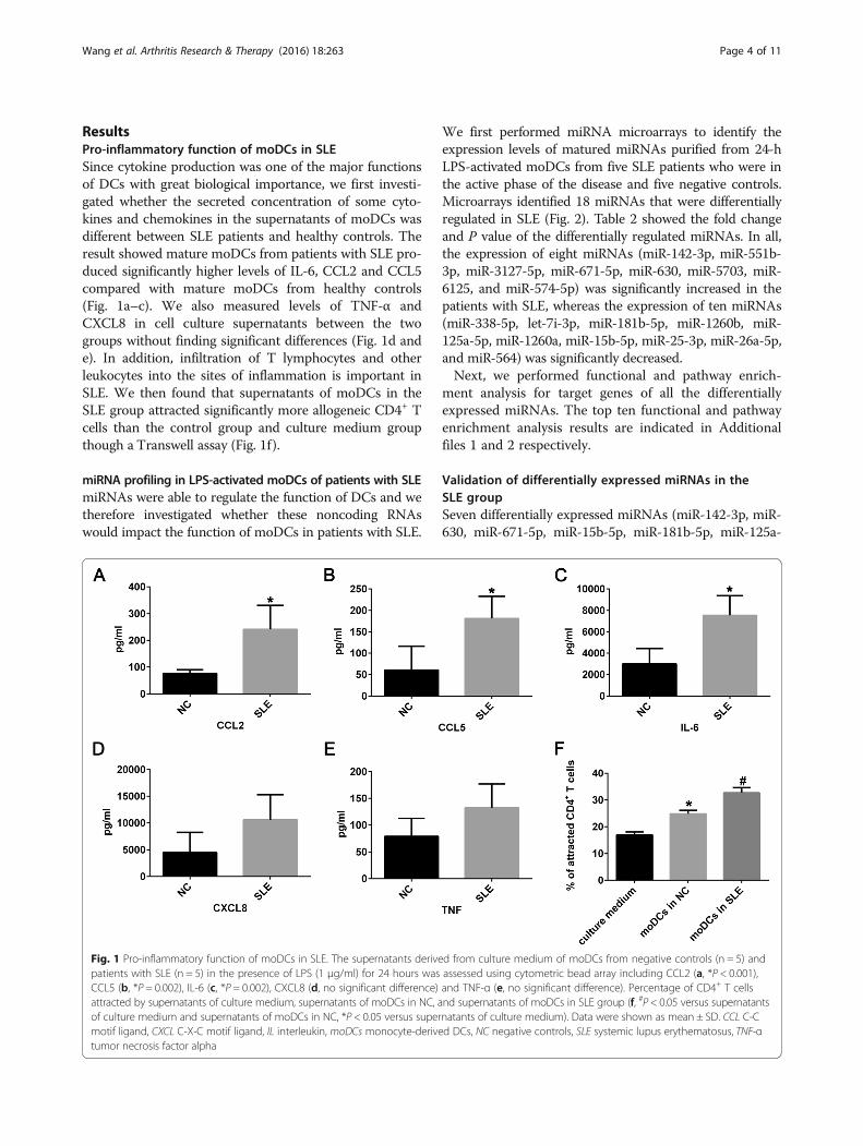
ResultsPro-inflammatory function of moDCs in SLESince cytokine production was one of the major functionsof DCs with great biological importance, we first investi-gated whether the secreted concentration of some cyto-kines and chemokines in the supernatants of moDCs wasdifferent between SLE patients and healthy controls. Theresult showed mature moDCs from patients with SLE pro-duced significantly higher levels of IL-6, CCL2 and CCL5compared with mature moDCs from healthy controls(Fig. 1a–c). We also measured levels of TNF-α andCXCL8 in cell culture supernatants between the twogroups without finding significant differences (Fig. 1d ande). In addition, infiltration of T lymphocytes and otherleukocytes into the sites of inflammation is important inSLE. We then found that supernatants of moDCs in theSLE group attracted significantly more allogeneic CD4+ Tcells than the control group and culture medium groupthough a Transwell assay (Fig. 1f).
miRNA profiling in LPS-activated moDCs of patients with SLEmiRNAs were able to regulate the function of DCs and wetherefore investigated whether these noncoding RNAswould impact the function of moDCs in patients with SLE.
Fig. 1 Pro-inflammatory function of moDCs in SLE. The supernatants derivepatients with SLE (n = 5) in the presence of LPS (1 μg/ml) for 24 hours wasCCL5 (b, *P = 0.002), IL-6 (c, *P = 0.002), CXCL8 (d, no significant difference)attracted by supernatants of culture medium, supernatants of moDCs in NC, aof culture medium and supernatants of moDCs in NC, *P < 0.05 versus supermotif ligand, CXCL C-X-C motif ligand, IL interleukin, moDCs monocyte-derivetumor necrosis factor alpha
We first performed miRNA microarrays to identify theexpression levels of matured miRNAs purified from 24-hLPS-activated moDCs from five SLE patients who were inthe active phase of the disease and five negative controls.Microarrays identified 18 miRNAs that were differentiallyregulated in SLE (Fig. 2). Table 2 showed the fold changeand P value of the differentially regulated miRNAs. In all,the expression of eight miRNAs (miR-142-3p, miR-551b-3p, miR-3127-5p, miR-671-5p, miR-630, miR-5703, miR-6125, and miR-574-5p) was significantly increased in thepatients with SLE, whereas the expression of ten miRNAs(miR-338-5p, let-7i-3p, miR-181b-5p, miR-1260b, miR-125a-5p, miR-1260a, miR-15b-5p, miR-25-3p, miR-26a-5p,and miR-564) was significantly decreased.Next, we performed functional and pathway enrich-
ment analysis for target genes of all the differentiallyexpressed miRNAs. The top ten functional and pathwayenrichment analysis results are indicated in Additionalfiles 1 and 2 respectively.
Validation of differentially expressed miRNAs in theSLE groupSeven differentially expressed miRNAs (miR-142-3p, miR-630, miR-671-5p, miR-15b-5p, miR-181b-5p, miR-125a-
d from culture medium of moDCs from negative controls (n = 5) andassessed using cytometric bead array including CCL2 (a, *P < 0.001),and TNF-α (e, no significant difference). Percentage of CD4+ T cellsnd supernatants of moDCs in SLE group (f, #P < 0.05 versus supernatantsnatants of culture medium). Data were shown as mean ± SD. CCL C-Cd DCs, NC negative controls, SLE systemic lupus erythematosus, TNF-α
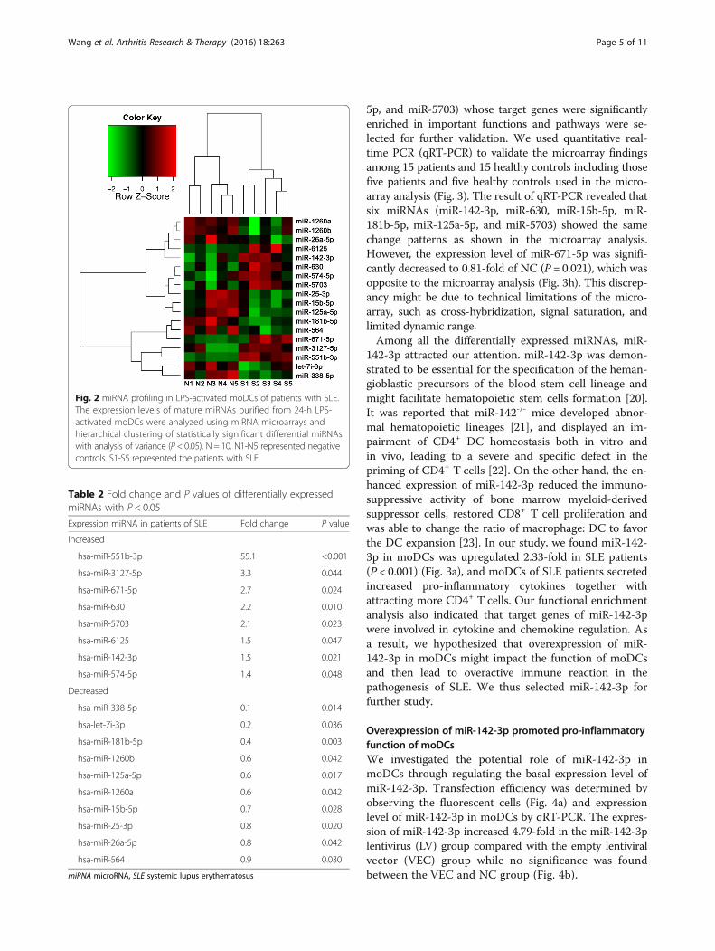
Fig. 2 miRNA profiling in LPS-activated moDCs of patients with SLE.The expression levels of mature miRNAs purified from 24-h LPS-activated moDCs were analyzed using miRNA microarrays andhierarchical clustering of statistically significant differential miRNAswith analysis of variance (P < 0.05). N = 10. N1-N5 represented negativecontrols. S1-S5 represented the patients with SLE
Table 2 Fold change and P values of differentially expressedmiRNAs with P < 0.05
Expression miRNA in patients of SLE Fold change P value
Increased
hsa-miR-551b-3p 55.1 <0.001
hsa-miR-3127-5p 3.3 0.044
hsa-miR-671-5p 2.7 0.024
hsa-miR-630 2.2 0.010
hsa-miR-5703 2.1 0.023
hsa-miR-6125 1.5 0.047
hsa-miR-142-3p 1.5 0.021
hsa-miR-574-5p 1.4 0.048
Decreased
hsa-miR-338-5p 0.1 0.014
hsa-let-7i-3p 0.2 0.036
hsa-miR-181b-5p 0.4 0.003
hsa-miR-1260b 0.6 0.042
hsa-miR-125a-5p 0.6 0.017
hsa-miR-1260a 0.6 0.042
hsa-miR-15b-5p 0.7 0.028
hsa-miR-25-3p 0.8 0.020
hsa-miR-26a-5p 0.8 0.042
hsa-miR-564 0.9 0.030
miRNA microRNA, SLE systemic lupus erythematosus
Wang et al. Arthritis Research & Therapy (2016) 18:263 Page 5 of 11
5p, and miR-5703) whose target genes were significantlyenriched in important functions and pathways were se-lected for further validation. We used quantitative real-time PCR (qRT-PCR) to validate the microarray findingsamong 15 patients and 15 healthy controls including thosefive patients and five healthy controls used in the micro-array analysis (Fig. 3). The result of qRT-PCR revealed thatsix miRNAs (miR-142-3p, miR-630, miR-15b-5p, miR-181b-5p, miR-125a-5p, and miR-5703) showed the samechange patterns as shown in the microarray analysis.However, the expression level of miR-671-5p was signifi-cantly decreased to 0.81-fold of NC (P = 0.021), which wasopposite to the microarray analysis (Fig. 3h). This discrep-ancy might be due to technical limitations of the micro-array, such as cross-hybridization, signal saturation, andlimited dynamic range.Among all the differentially expressed miRNAs, miR-
142-3p attracted our attention. miR-142-3p was demon-strated to be essential for the specification of the heman-gioblastic precursors of the blood stem cell lineage andmight facilitate hematopoietic stem cells formation [20].It was reported that miR-142-/- mice developed abnor-mal hematopoietic lineages [21], and displayed an im-pairment of CD4+ DC homeostasis both in vitro andin vivo, leading to a severe and specific defect in thepriming of CD4+ T cells [22]. On the other hand, the en-hanced expression of miR-142-3p reduced the immuno-suppressive activity of bone marrow myeloid-derivedsuppressor cells, restored CD8+ T cell proliferation andwas able to change the ratio of macrophage: DC to favorthe DC expansion [23]. In our study, we found miR-142-3p in moDCs was upregulated 2.33-fold in SLE patients(P < 0.001) (Fig. 3a), and moDCs of SLE patients secretedincreased pro-inflammatory cytokines together withattracting more CD4+ T cells. Our functional enrichmentanalysis also indicated that target genes of miR-142-3pwere involved in cytokine and chemokine regulation. Asa result, we hypothesized that overexpression of miR-142-3p in moDCs might impact the function of moDCsand then lead to overactive immune reaction in thepathogenesis of SLE. We thus selected miR-142-3p forfurther study.
Overexpression of miR-142-3p promoted pro-inflammatoryfunction of moDCsWe investigated the potential role of miR-142-3p inmoDCs through regulating the basal expression level ofmiR-142-3p. Transfection efficiency was determined byobserving the fluorescent cells (Fig. 4a) and expressionlevel of miR-142-3p in moDCs by qRT-PCR. The expres-sion of miR-142-3p increased 4.79-fold in the miR-142-3plentivirus (LV) group compared with the empty lentiviralvector (VEC) group while no significance was foundbetween the VEC and NC group (Fig. 4b).
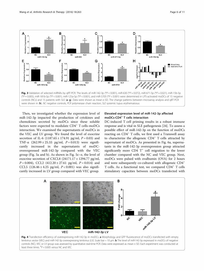
Fig. 3 Validation of selected miRNAs by qRT-PCR. The levels of miR-142-3p (*P < 0.001), miR-630 (*P = 0.072), miR-671-5p (*P = 0.021), miR-15b-5p(*P = 0.005), miR-181b-5p (*P < 0.001), miR-125a-5p (*P < 0.001), and miR-5703 (*P < 0.001) were determined in LPS-activated moDCs of 15 negativecontrols (NCs) and 15 patients with SLE (a–g). Data were shown as mean ± SD. The change patterns between microarray analysis and qRT-PCRwere shown in (h). NC negative controls, PCR polymerase chain reaction, SLE systemic lupus erythematosus
Wang et al. Arthritis Research & Therapy (2016) 18:263 Page 6 of 11
Then, we investigated whether the expression level ofmiR-142-3p impacted the production of cytokines andchemokines secreted by moDCs since these solublefactors were expected to modulate CD4+ T cells-moDCsinteraction. We examined the supernatants of moDCs inthe VEC and LV group. We found the level of exocrinesecretion of IL-6 (1187.65 ± 174.91 pg/ml, P = 0.03) andTNF-α (262.99 ± 25.33 pg/ml, P = 0.013) were signifi-cantly increased in the supernatants of moDC-overexpressed miR-142-3p compared with the VECgroup (Fig. 5a and b). As shown in Fig. 5c–e, the level ofexocrine secretion of CXCL8 (24171.17 ± 1294.77 pg/ml,P = 0.004), CCL2 (412.20 ± 27.61 pg/ml, P = 0.014) andCCL5 (126.46 ± 4.25 pg/ml, P < 0.001) was also signifi-cantly increased in LV group compared with VEC group.
Fig. 4 Transfection efficiency of overexpressing miR-142-3p in moDCs. a Mlentivirus vector (VEC) and miR-142-3p overexpressing lentivirus (LV). Scale bacontrols (NC), VEC or LV group was assessed by quantitative real-time PCR. Daleast three times. #P < 0.005 versus NC and VEC
Elevated expression level of miR-142-3p affectedmoDCs-CD4+ T cells interactionDC-induced T cell priming results in a robust immuneresponse and is vital in SLE pathogenesis [24]. To assess apossible effect of miR-142-3p on the function of moDCsreacting on CD4+ T cells, we first used a Transwell assayto characterize the allogeneic CD4+ T cells attracted bysupernatant of moDCs. As presented in Fig. 6a, superna-tants in the miR-142-3p overexpression group attractedsignificantly more CD4 T+ cell migration to the lowerchamber compared with the NC and VEC group. Next,moDCs were pulsed with ovalbumin (OVA) for 2 hoursand were subsequently co-cultured with allogeneic CD4+
T cells. As a functional test, we compared CD4+ T cellsstimulatory capacities between moDCs transfected with
orphology and GFP fluorescence of moDCs transfected with emptyr = 10 μm. b The level of miR-142-3p expressed in moDCs of negativeta were expressed as mean ± SD. Each experiment was conducted at
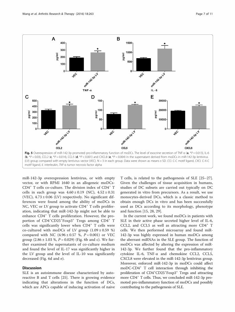
Fig. 5 Overexpression of miR-142-3p promoted pro-inflammatory function of moDCs. The level of exocrine secretion of TNF-α (a, *P = 0.013), IL-6(b, *P = 0.03), CCL2 (c, *P = 0.014), CCL5 (d, *P < 0.001) and CXCL8 (e, *P = 0.004) in the supernatant derived from moDCs in miR-142-3p lentivirus(LV) group compared with empty lentivirus vector (VEC). N = 3 in each group. Data were shown as means ± SD. CCL C-C motif ligand, CXCL C-X-Cmotif ligand, IL interleukin, TNF-α tumor necrosis factor alpha
Wang et al. Arthritis Research & Therapy (2016) 18:263 Page 7 of 11
miR-142-3p overexpression lentivirus, or with emptyvector, or with RPMI 1640 in an allogeneic moDCs-CD4+ T cells co-culture. The division index of CD4+ Tcells in each group was 4.60 ± 0.19 (NC), 4.52 ± 0.31(VEC), 4.73 ± 0.06 (LV) respectively. No significant dif-ferences were found among the ability of moDCs inNC, VEC or LV group to activate CD4+ T cells prolifer-ation, indicating that miR-142-3p might not be able toenhance CD4+ T cells proliferation. However, the pro-portion of CD4+CD25+Foxp3+ Tregs among CD4+ Tcells was significantly lower when CD4+ T cells wereco-cultured with moDCs of LV group (1.09 ± 0.59 %)compared with NC (4.96 ± 0.57 %, P = 0.001) or VECgroup (2.86 ± 1.03 %, P = 0.029) (Fig. 6b and c). We fur-ther examined the supernatants of co-culture mediumand found the level of IL-17 was significantly higher inthe LV group and the level of IL-10 was significantlydecreased (Fig. 6d and e).
DiscussionSLE is an autoimmune disease characterized by auto-reactive B and T cells [25]. There is growing evidenceindicating that alterations in the function of DCs,which are APCs capable of inducing activation of naive
T cells, is related to the pathogenesis of SLE [25–27].Given the challenges of tissue acquisition in humans,studies of DC subsets are carried out typically on DCgenerated in vitro from precursors. As a result, we usemonocytes-derived DCs, which is a classic method toobtain enough DCs in vitro and has been successfullyused as DCs according to its morphology, phenotypeand function [15, 28, 29].In the current work, we found moDCs in patients with
SLE in their active phase secreted higher level of IL-6,CCL2, and CCL5 as well as attracting more CD4+ Tcells. We then performed microarray and found miR-142-3p was highly expressed in human moDCs amongthe aberrant miRNAs in the SLE group. The function ofmoDCs was affected by altering the expression of miR-142-3p. We further found that the pro-inflammatorycytokine IL-6, TNF-α and chemokine CCL2, CCL5,CXCL8 were elevated in the miR-142-3p lentivirus group.Moreover, enforced miR-142-3p in moDCs could affectmoDC-CD4+ T cell interaction through inhibiting theproliferation of CD4+CD25+Foxp3+ Tregs and attractingmore CD4+ T cells. Thus, we concluded miR-142-3p pro-moted pro-inflammatory function of moDCs and possiblycontributing to the pathogenesis of SLE.
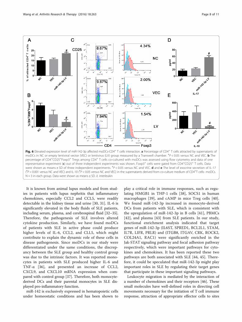
Fig. 6 Elevated expression level of miR-142-3p affected moDCs-CD4+ T cells interaction. a Percentage of CD4+ T cells attracted by supernatants ofmoDCs in NC or empty lentiviral vector (VEC) or lentivirus (LV) group measured by a Transwell chamber. #P < 0.05 versus NC and VEC. b Thepercentage of CD4+CD25+Foxp3+ Tregs among CD4+ T cells co-cultured with moDCs was assessed using flow cytometry and data of onerepresentative experiment (c) out of three independent experiments was shown. Foxp3+ cells were gated from CD4+CD25+ T cells. Datawere shown as means ± SD of three independent experiments. #P < 0.05 versus NC and VEC. d and e The level of exocrine secretion of IL-17(#P < 0.001 versus NC and VEC) and IL-10 (#P < 0.05 versus NC and VEC) in the supernatants derived from co-culture medium of CD4+T cells- moDCs.N = 3 in each group. Data were shown as means ± SD. IL interleukin
Wang et al. Arthritis Research & Therapy (2016) 18:263 Page 8 of 11
It is known from animal lupus models and from stud-ies in patients with lupus nephritis that inflammatorychemokines, especially CCL2 and CCL5, were readilydetectable in the kidney tissue and urine [30, 31]. IL-6 issignificantly elevated in the body fluids of SLE patients,including serum, plasma, and cerebrospinal fluid [32–35].Therefore, the pathogenesis of SLE involves alteredcytokine production. Similarly, we have found moDCsof patients with SLE in active phase could producehigher levels of IL-6, CCL2, and CLL5, which mightcontribute to explain the dynamic role of these cells indisease pathogenesis. Since moDCs in our study weredifferentiated under the same conditions, the discrep-ancy between the SLE group and healthy control groupwas due to the intrinsic factors. It was reported mono-cytes in patients with SLE produced higher IL-6 andTNF-α [36], and presented an increase of CCL2,CXCL9, and CXCL10 mRNA expression when com-pared with control group [37]. Therefore, both monocyte-derived DCs and their parental monocytes in SLE dis-played pro-inflammatory function.miR-142 is exclusively expressed in hematopoietic cells
under homeostatic conditions and has been shown to
play a critical role in immune responses, such as regu-lating HMGB1 in THP-1 cells [38], SOCS1 in humanmacrophages [39], and cAMP in mice Treg cells [40].We found miR-142-3p increased in monocyte-derivedDCs from patients with SLE, which is consistent withthe upregulation of miR-142-3p in B cells [41], PBMCs[42], and plasma [43] from SLE patients. In our study,functional enrichment analysis indicated that targetgenes of miR-142-3p (IL6ST, SPRED1, BCL2L1, STAM,IL7R, LIFR, PRLR) and (ITGB8, ITGAV, CRK, ROCK2,COL24A1, RAC1) were significantly enriched in theJak-STAT signaling pathway and focal adhesion pathwayrespectively, which were important pathways for cyto-kines and chemokines. It has been reported these twopathways are both associated with SLE [44, 45]. There-fore, it could be speculated that miR-142-3p might playimportant roles in SLE by regulating their target genesthat participate in these important signaling pathways.Leukocyte migration is mediated by the interaction of
a number of chemokines and their receptors [46]. Thesesmall molecules have well-defined roles in directing cellmovements necessary for the initiation of T cell immuneresponse, attraction of appropriate effector cells to sites
Wang et al. Arthritis Research & Therapy (2016) 18:263 Page 9 of 11
of inflammation, and regulation of differential recruit-ment of T helper lymphocytes. Matured DCs could se-crete an abundant source of both inflammatory andlymphoid chemokines, sustaining interaction of naiveand activated T cells with antigen-presenting matureDCs. In our experiments, we found the level of CXCL8,CCL2, and CCL5 increased in the supernatants ofmoDCs with enforced expression of miR-142-3p. Thethree chemokines have also been reported in biologicalfluids from SLE [31, 47], so they might act synergistic-ally on the chemotactic activity inducing CD4+ T cellmigration. Since miR-142-3p is elevated in moDCsfrom SLE patients, it suggests that the upregulation ofmiR-142-3p in moDCs of SLE patients might be attrib-uted to attracting more CD4+ T cells, probably involvedin the pathogenesis of disease.We also found the overexpression of miR-142-3p
cause the elevation of IL-6 and TNF-α, leading to a de-crease of CD4+CD25+Foxp3+ Tregs which are anti-inflammatory, and an imbalance of IL-17 and IL-10.Treg deficiency in the periphery is sufficient to evokechronic T cell-mediated autoimmunity and immunopa-thology, which has been associated with SLE [48]. Themechanisms of Treg-mediated suppression includingsecretion of immunosuppressive cytokines, cell contact-dependent suppression, and functional modification orkilling of APC [48]. It has been demonstrated in lupus-prone mice that IL-6 produced by DCs inhibits Tregs[49], because Foxp3+ Treg cells lose Foxp3 expressionand undergo conversion into Th17 cells under theeffect of IL-6 [50]. Therefore, IL-6 triggered an immunedisorder by breaking the balance between Th17 andTreg. Our results indicate that the increase of miR-142-3p in moDCs of SLE made the cells producing in-creased IL-6 and could induce CD4+ T cells to secretemore IL-17 and less IL-10, thus leading to an imbalanceof the immune response. Therefore, overexpression ofmiR-142-3p in moDCs suppressed the increase in Tregs,which correlated with a reduced capacity to suppressresponder T cell proliferation and might thereby con-tribute to the development of SLE. Similarly, mousedendritic cells matured by ingestion of apoptotic blebscould stimulate allogeneic T cells which produced IFNand especially high levels of IL-17, representing an im-portant driving force in SLE [51].We have suggested that elevated expression of miR-
142-3p is related to the pro-inflammatory function ofmoDCs in SLE. However, the limitation of our studyis that we have not investigated the effect of decreas-ing the level of miR-142-3p in moDCs of SLE. Futurestudy would focus on the exact target genes of miR-142-3p in moDCs and whether downregulating miR-142-3p could improve the overactive inflammationphase of SLE.
ConclusionTaken together, our findings suggested a pro-inflammatoryfunction of moDCs in SLE patients partially mediated bymiR-142-3p: (1) microRNAs were differentially expressedand miR-142-3p was increased in moDCs of SLE patients.(2) moDCs in patients with SLE produced higher levels ofsome pro-inflammatory cytokines and chemokines thanhealthy controls. (3) Overexpressing miR-142-3p in moDCsof healthy controls promoted cytokine and chemokineproduction to attract more CD4+ T cells and decreaseTreg expansion. These findings suggested that miR-142-3p might be meaningful in the pathogenesis of SLE andcould serve as a novel therapeutic target for treatment.
Additional files
Additional file 1: Table S1. The top ten enriched Gene Ontology(GO) terms for target genes of all the 18 differentially expressed miRNAs.(DOCX 72 kb)
Additional file 2: Table S2. The top ten enriched pathways for targetgenes of all the 18 differentially expressed miRNAs. (DOCX 68 kb)
Abbreviations3′UTR: 3′ un-translated regions; APC: Antigen-presenting cells; BSA: Bovineserum albumin; CCL: C-C motif ligand; CFSE: Carboxyfluorescein diacetatesuccinimidyl ester; CXCL: C-X-C motif ligand; DCs: Dendritic cells; FBS: Fetalbovine serum; FDR: False discovery rate; GM-CSF: Granulocyte-macrophagecolony-stimulating factor; IL: Interleukin; LPS: Lipopolysaccharide; LV: Lentivirus;miRNAs: microRNAs; moDCs: monocyte-derived DCs; NC: Negative controls;OVA: Ovalbumin; PBMCs: Peripheral blood mononuclear cells; PBS: Phosphate-buffered saline; qRT-PCR: quantitative real-time PCR; SLE: Systemic lupuserythematosus; SOCS1: Suppressor of cytokine signaling-1; TLR: Toll-likereceptor; TNF-α: Tumor necrosis factor alpha; Tregs: Regulatory T cells;VEC: Empty lentiviral vector
AcknowledgementsNot applicable.
FundingThe research was supported by National Natural Science Foundation ofChina (Grant No. 81373212, 81371745, 81402605) and Development Projectof Shanghai Peak Disciplines-Integrated Chinese and Western Medicine.
Availability of data and materialsThe data of microarray was deposited in Gene Expression Omnibus (GEO)and the accession number was GSE79240.
Authors’ contributionsWYL cultured the cells, performed the microarray and transfection experiments,carried out the flow cytometric analysis, and drafted the manuscript. LJparticipated in the design of the study and drafted the manuscript. QHHparticipated in study design and target gene prediction, and revised themanuscript. GY performed the statistical analysis of data and revised themanuscript. DJ participated in qPCR experiments, chemotaxis assay, andmanuscript drafting. LJR and ZXH collected the specimens, participatedin qPCR experiments, and drafted the manuscript. WJ contributed todata interpretation and manuscript revision. XJH conceived of the study,participated in its design and coordination, and revised the manuscript.All authors approved the final version to be published.
Competing interestsThe authors declare that they have no competing interests.
Wang et al. Arthritis Research & Therapy (2016) 18:263 Page 10 of 11
Consent for publicationWritten informed consents were obtained from the patients for publicationof their individual details and any accompanying images in this manuscript.The consent form is held by the authors and is available for review by theEditor-in-Chief.
Ethics approval and consent to participateThe study was approved by the Independent Ethics Committee of HuashanHospital and written informed consent was obtained from all subjects.
Author details1Department of Dermatology, Huashan Hospital, Fudan University, 12Wulumuqi Zhong Road, Shanghai 200040, People’s Republic of China.2Department of Neurology, Huashan Hospital, Fudan University, Shanghai,People’s Republic of China. 3Department of Human Anatomy andHistoembryology, School of Basic Medical Science, Fudan University,Shanghai, People’s Republic of China.
Received: 18 July 2016 Accepted: 19 October 2016
References1. Zhu Z, Liang Z, Liany H, Yang C, Wen L, Lin Z, Sheng Y, Lin Y, Ye L, Cheng
Y, et al. Discovery of a novel genetic susceptibility locus on X chromosomefor systemic lupus erythematosus. Arthritis Res Ther. 2015;17:349.
2. Feng JB, Ni JD, Yao X, Pan HF, Li XP, Xu JH, Pan FM, Xu SQ, Ye DQ.Gender and age influence on clinical and laboratory features in Chinesepatients with systemic lupus erythematosus: 1,790 cases. Rheumatol Int.2010;30(8):1017–23.
3. Patel DR, Richardson BC. Epigenetic mechanisms in lupus. Curr Opin Rheumatol.2010;22(5):478–82.
4. Gaipl US, Munoz LE, Grossmayer G, Lauber K, Franz S, Sarter K, Voll RE,Winkler T, Kuhn A, Kalden J, et al. Clearance deficiency and systemiclupus erythematosus (SLE). J Autoimmun. 2007;28(2-3):114–21.
5. Chan VS, Nie YJ, Shen N, Yan S, Mok MY, Lau CS. Distinct roles of myeloidand plasmacytoid dendritic cells in systemic lupus erythematosus.Autoimmun Rev. 2012;11(12):890–7.
6. Teichmann LL, Ols ML, Kashgarian M, Reizis B, Kaplan DH, Shlomchik MJ.Dendritic cells in lupus are not required for activation of T and B cellsbut promote their expansion, resulting in tissue damage. Immunity.2010;33(6):967–78.
7. Miyara M, Amoura Z, Parizot C, Badoual C, Dorgham K, Trad S, Nochy D,Debre P, Piette JC, Gorochov G. Global natural regulatory T cell depletionin active systemic lupus erythematosus. J Immunol. 2005;175(12):8392–400.
8. Ding D, Mehta H, McCune WJ, Kaplan MJ. Aberrant phenotype and functionof myeloid dendritic cells in systemic lupus erythematosus. J Immunol.2006;177(9):5878–89.
9. Hashimi ST, Fulcher JA, Chang MH, Gov L, Wang S, Lee B. MicroRNAprofiling identifies miR-34a and miR-21 and their target genes JAG1 andWNT1 in the coordinate regulation of dendritic cell differentiation. Blood.2009;114(2):404–14.
10. Zhou H, Huang X, Cui H, Luo X, Tang Y, Chen S, Wu L, Shen N. miR-155 and its star-form partner miR-155* cooperatively regulate type Iinterferon production by human plasmacytoid dendritic cells. Blood.2010;116(26):5885–94.
11. Lu C, Huang X, Zhang X, Roensch K, Cao Q, Nakayama KI, Blazar BR, Zeng Y,Zhou X. miR-221 and miR-155 regulate human dendritic cell development,apoptosis, and IL-12 production through targeting of p27kip1, KPC1, andSOCS-1. Blood. 2011;117(16):4293–303.
12. Dunand-Sauthier I, Santiago-Raber ML, Capponi L, Vejnar CE, Schaad O,Irla M, Seguin-Estevez Q, Descombes P, Zdobnov EM, Acha-Orbea H, et al.Silencing of c-Fos expression by microRNA-155 is critical for dendritic cellmaturation and function. Blood. 2011;117(17):4490–500.
13. Kim SJ, Gregersen PK, Diamond B. Regulation of dendritic cell activation bymicroRNA let-7c and BLIMP1. J Clin Invest. 2013;123(2):823–33.
14. Wang P, Xue Y, Han Y, Lin L, Wu C, Xu S, Jiang Z, Xu J, Liu Q, Cao X. TheSTAT3-binding long noncoding RNA lnc-DC controls human dendritic celldifferentiation. Science. 2014;344(6181):310–3.
15. Sallusto F, Lanzavecchia A. Efficient presentation of soluble antigen bycultured human dendritic cells is maintained by granulocyte/macrophage
colony-stimulating factor plus interleukin 4 and downregulated by tumornecrosis factor alpha. J Exp Med. 1994;179(4):1109–18.
16. Vogelsang P, Karlsen M, Brun JG, Jonsson R, Appel S. Altered phenotypeand Stat1 expression in Toll-like receptor 7/8 stimulated monocyte-deriveddendritic cells from patients with primary Sjogren’s syndrome. Arthritis ResTher. 2014;16(4):R166.
17. Gottschalk TA, Tsantikos E, Hibbs ML. Pathogenic inflammation and itstherapeutic targeting in systemic lupus erythematosus. Front Immunol.2015;6:550.
18. Rodriguez-Pla A, Patel P, Maecker HT, Rossello-Urgell J, Baldwin N, Bennett L,Cantrell V, Baisch J, Punaro M, Gotte A, et al. IFN priming is necessary butnot sufficient to turn on a migratory dendritic cell program in lupus monocytes.J Immunol. 2014;192(12):5586–98.
19. Jiang W, Gilkeson G. Sex Differences in monocytes and TLR4 associatedimmune responses; implications for systemic lupus erythematosus (SLE).J Immunother Applic. 2014;1:1.
20. Nimmo R, Ciau-Uitz A, Ruiz-Herguido C, Soneji S, Bigas A, Patient R, Enver T.MiR-142-3p controls the specification of definitive hemangioblasts duringontogeny. Dev Cell. 2013;26(3):237–49.
21. Kramer NJ, Wang WL, Reyes EY, Kumar B, Chen CC, Ramakrishna C, CantinEM, Vonderfecht SL, Taganov KD, Chau N, et al. Altered lymphopoiesis andimmunodeficiency in miR-142 null mice. Blood. 2015;125(24):3720–30.
22. Mildner A, Chapnik E, Manor O, Yona S, Kim KW, Aychek T, Varol D, Beck G,Itzhaki ZB, Feldmesser E, et al. Mononuclear phagocyte miRNome analysisidentifies miR-142 as critical regulator of murine dendritic cell homeostasis.Blood. 2013;121(6):1016–27.
23. Sonda N, Simonato F, Peranzoni E, Cali B, Bortoluzzi S, Bisognin A, Wang E,Marincola FM, Naldini L, Gentner B, et al. miR-142-3p prevents macrophagedifferentiation during cancer-induced myelopoiesis. Immunity. 2013;38(6):1236–49.
24. Fransen JH, van der Vlag J, Ruben J, Adema GJ, Berden JH, Hilbrands LB.The role of dendritic cells in the pathogenesis of systemic lupus erythematosus.Arthritis Res Ther. 2010;12(2):207.
25. Blanco P, Palucka AK, Gill M, Pascual V, Banchereau J. Induction of dendriticcell differentiation by IFN-alpha in systemic lupus erythematosus. Science.2001;294(5546):1540–3.
26. Scheinecker C, Zwolfer B, Koller M, Manner G, Smolen JS. Alterations ofdendritic cells in systemic lupus erythematosus - phenotypic and functionaldeficiencies. Arthritis Rheum. 2001;44(4):856–65.
27. Sisirak V, Ganguly D, Lewis KL, Couillault C, Tanaka L, Bolland S, D’Agati V,Elkon KB, Reizis B. Genetic evidence for the role of plasmacytoid dendriticcells in systemic lupus erythematosus. J Exp Med. 2014;211(10):1969–76.
28. Chapuis F, Rosenzwajg M, Yagello M, Ekman M, Biberfeld P, Gluckman JC.Differentiation of human dendritic cells from monocytes in vitro. Eur JImmunol. 1997;27(2):431–41.
29. Pickl WF, Majdic O, Kohl P, Stockl J, Riedl E, Scheinecker C, Bello-Fernandez C,Knapp W. Molecular and functional characteristics of dendritic cells generatedfrom highly purified CD14+ peripheral blood monocytes. J Immunol.1996;157(9):3850–9.
30. Hasegawa H, Kohno M, Sasaki M, Inoue A, Ito MR, Terada M, Hieshima K,Maruyama H, Miyazaki J, Yoshie O, et al. Antagonist of monocytechemoattractant protein 1 ameliorates the initiation and progression of lupusnephritis and renal vasculitis in MRL/lpr mice. Arthritis Rheum. 2003;48(9):2555–66.
31. Lit LC, Wong CK, Tam LS, Li EK, Lam CW. Raised plasma concentration andex vivo production of inflammatory chemokines in patients with systemiclupus erythematosus. Ann Rheum Dis. 2006;65(2):209–15.
32. Abdel Galil SM, Ezzeldin N, El-Boshy ME. The role of serum IL-17 and IL-6as biomarkers of disease activity and predictors of remission in patientswith lupus nephritis. Cytokine. 2015;76(2):280–7.
33. Talaat RM, Mohamed SF, Bassyouni IH, Raouf AA. Th1/Th2/Th17/Tregcytokine imbalance in systemic lupus erythematosus (SLE) patients:correlation with disease activity. Cytokine. 2015;72(2):146–53.
34. Lyn-Cook BD, Xie C, Oates J, Treadwell E, Word B, Hammons G, Wiley K.Increased expression of Toll-like receptors (TLRs) 7 and 9 and other cytokinesin systemic lupus erythematosus (SLE) patients: ethnic differences andpotential new targets for therapeutic drugs. Mol Immunol. 2014;61(1):38–43.
35. Fragoso-Loyo H, Richaud-Patin Y, Orozco-Narváez A, Dávila-Maldonado L,Atisha-Fregoso Y, Llorente L, Sánchez-Guerrero J. Interleukin-6 andchemokines in the neuropsychiatric manifestations of systemic lupuserythematosus. Arthritis Rheum. 2007;56(4):1242–50.
36. Henriques A, Ines L, Carvalheiro T, Couto M, Andrade A, Pedreiro S,Laranjeira P, Morgado JM, Pais ML, da Silva JA, et al. Functional
Wang et al. Arthritis Research & Therapy (2016) 18:263 Page 11 of 11
characterization of peripheral blood dendritic cells and monocytesin systemic lupus erythematosus. Rheumatol Int. 2012;32(4):863–9.
37. Carvalheiro T, Rodrigues A, Lopes A, Ines L, Velada I, Ribeiro A, Martinho A,Silva JA, Pais ML, Paiva A. Tolerogenic versus inflammatory activity of peripheralblood monocytes and dendritic cells subpopulations in systemic lupuserythematosus. Clin Dev Immunol. 2012;2012:934161.
38. Yuan Z, Luo G, Li X, Chen J, Wu J, Peng Y. PPARγ inhibits HMGB1 expressionthrough upregulation of miR-142-3p in vitro and in vivo. Cell Signal.2016;28(3):158–64.
39. Su S, Zhao Q, He C, Huang D, Liu J, Chen F, Chen J, Liao J-Y, Cui X, Zeng Y,et al. miR-142-5p and miR-130a-3p are regulated by IL-4 and IL-13 andcontrol profibrogenic macrophage program. Nat Commun. 2015;6:8523.
40. Huang B, Zhao J, Lei Z, Shen S, Li D, Shen G-X, Zhang G-M, Feng Z-H. miR-142-3p restricts cAMP production in CD4(+)CD25(−) T cells and CD4(+)CD25(+)T(REG) cells by targeting AC9 mRNA. EMBO Rep. 2009;10(2):180–5.
41. Te JL, Dozmorov IM, Guthridge JM, Nguyen KL, Cavett JW, Kelly JA, Bruner GR,Harley JB, Ojwang JO. Identification of unique microRNA signature associatedwith lupus nephritis. PLoS One. 2010;5(5):e10344.
42. Dai Y, Huang YS, Tang M, Lv TY, Hu CX, Tan YH, Xu ZM, Yin YB. Microarrayanalysis of microRNA expression in peripheral blood cells of systemic lupuserythematosus patients. Lupus. 2007;16(12):939–46.
43. Carlsen AL, Schetter AJ, Nielsen CT, Lood C, Knudsen S, Voss A, Harris CC,Hellmark T, Segelmark M, Jacobsen S, et al. Circulating microRNA expressionprofiles associated with systemic lupus erythematosus. Arthritis Rheum.2013;65(5):1324–34.
44. Tang Y, Ma X, Zhang H, Gu Z, Hou Y, Gilkeson GS, Lu L, Zeng X, Sun L. Geneexpression profile reveals abnormalities of multiple signaling pathwaysin mesenchymal stem cell derived from patients with systemic lupuserythematosus. Clin Dev Immunol. 2012;2012:826182.
45. Kawasaki M, Fujishiro M, Yamaguchi A, Nozawa K, Kaneko H, Takasaki Y,Takamori K, Ogawa H, Sekigawa I. Possible role of the JAK/STAT pathwaysin the regulation of T cell-interferon related genes in systemic lupuserythematosus. Lupus. 2011;20(12):1231–9.
46. Sallusto F, Lanzavecchia A. Understanding dendritic cell and T-lymphocytetraffic through the analysis of chemokine receptor expression. Immunol Rev.2000;177:134–40.
47. Laborde EA, Vanzulli S, Beigier-Bompadre M, Isturiz MA, Ruggiero RA,Fourcade MG, Catalan Pellet AC, Sozzani S, Vulcano M. Immune complexesinhibit differentiation, maturation, and function of human monocyte-derived dendritic cells. J Immunol. 2007;179(1):673–81.
48. Sakaguchi S, Yamaguchi T, Nomura T, Ono M. Regulatory T cells andimmune tolerance. Cell. 2008;133(5):775–87.
49. Wan S, Xia C, Morel L. IL-6 produced by dendritic cells from lupus-prone miceinhibits CD4 + CD25+ T cell regulatory functions. J Immunol. 2006;178(1):271–9.
50. Komatsu N, Okamoto K, Sawa S, Nakashima T, Oh-hora M, Kodama T,Tanaka S, Bluestone JA, Takayanagi H. Pathogenic conversion of Foxp3+T cells into TH17 cells in autoimmune arthritis. Nat Med. 2014;20(1):62–8.
51. Fransen JH, Hilbrands LB, Ruben J, Stoffels M, Adema GJ, van der Vlag J,Berden JH. Mouse dendritic cells matured by ingestion of apoptotic blebsinduce T cells to produce interleukin-17. Arthritis Rheum. 2009;60(8):2304–13.
• We accept pre-submission inquiries
• Our selector tool helps you to find the most relevant journal
• We provide round the clock customer support
• Convenient online submission
• Thorough peer review
• Inclusion in PubMed and all major indexing services
• Maximum visibility for your research
Submit your manuscript atwww.biomedcentral.com/submit
Submit your next manuscript to BioMed Central and we will help you at every step: