Electrostatic interaction of myristoylated proteins with membranes: simple physics, complicated...
5
Minireview 985 Electrostatic interaction of myristoylated proteins with membranes: simple physics, complicated biology Diana Murray 1 , Nir Ben-Tal 2 , Barry Honig 3 and Stuart McLaughlin 1 * Cell membrane association by several important peripheral proteins, such as Src, MARCKS, HIV-1 Gag, and K-Ras, requires nonspecific electrostatic interactions between a cluster of basic residues on the protein and acidic phospholipids in the plasma membrane. A simple theoretical model based on the nonlinear Poisson–Boltzmann equation describes well the experimentally measured electrostatic association between such proteins and the cell membrane. Addresses: 1 Department of Physiology and Biophysics, SUNY Stony Brook, Stony Brook, NY 11794-8661, USA, 2 Department of Biochemistry, Tel Aviv University, Ramat Aviv, Tel Aviv 69978, Israel and 3 Department of Biochemistry and Molecular Biophysics, Columbia University, 630 West 168 St., New York, NY 10032, USA. *Corresponding author. E-mail: [email protected] Structure 15 August 1997, 5:985–989 http://biomednet.com/elecref/0969212600500985 © Current Biology Ltd ISSN 0969-2126 Hydrophobic and electrostatic interactions act in concert to anchor to cell membranes several important proteins that are either myristoylated (e.g. Src, HIV-1 Gag and myristoy- lated alanine-rich C kinase substrate, MARCKS) or farnesy- lated (e.g. K-Ras). Two recent reviews focused on how the attachment of acyl and isoprenyl groups to proteins influ- ences their association with membranes [1,2]. Here, we consider the role electrostatic interactions play in enhancing the partitioning of myristoylated proteins onto membranes, using the proto-oncogene product c-Src as an example [1,3]. Figure 1 shows the domain organization of the tyrosine kinase c-Src. Recent structural studies have revealed how the intramolecular binding of a C-terminal phosphorylated tyrosine (Tyr527) to the SH2 domain juxtaposes the SH3 domain with the polyproline type-II helix that links the SH2 and kinase domains [3]. The intramolecular interac- tion between the SH3 and kinase domains maintains the protein in an inactive conformation. Because the viral, onco- genic version, v-Src, lacks the C-terminal tyrosine, it is con- stitutively active. The N-terminal portion was not included in the recently determined crystal structure of Src [3]. This region contains the two motifs required for membrane asso- ciation (shown in Figure 1a) — the myristate (colored green) and a cluster of basic residues (blue plus signs), which are shown in more detail in Figure 1b. Membrane binding increases the effective concentration of Src in a thin (d ~1 nm) surface layer adjacent to the membrane. For a spherical cell with a radius r equal to a few micrometers, the volume of the surface phase (V = 4πr 2 d) is about 1/1000 the volume of the cell (V = 4πr 3 /3); thus, anchoring Src to the membrane increases its effective concentration 1000-fold, thereby greatly enhancing its ability to phosphorylate its membrane-bound substrates. Two factors contribute to Src membrane binding Myristate (14-carbon fatty acid) is attached cotranslation- ally through an amide bond to the N-terminal glycine of Src by the enzyme N-myristoyl transferase [1,2]. Myristate is required for Src membrane binding which, in turn, is required for Src to function — nonmyristoylated v-Src mutants are found in the cytoplasm and do not transform cells, even though the kinase activity of the protein is unaffected [1,4,5]. Small myristoylated peptides bind to electrically neutral phospholipid vesicles with a unitary binding energy of 8 kcal/mol (or a molar partition coeffi- cient of 104 M–1) [6,7]. Measurements of the membrane partitioning of acylated peptides show that the binding energy increases 0.8 kcal/mol for each CH2 group added to the acyl chain [6], which is in agreement with measure- ments of the hydrophobic partitioning of fatty acids into oil from water [8]. The model in Figure 1 is consistent with these results: 8 kcal/mol ÷ 0.8 kcal/mol per CH2 = 10 CH2 groups, which penetrate the hydrocarbon core of the membrane; the remaining four traverse the polar head group region and the N-terminal glycine is located just outside the envelope of the polar head group region (Fig- ure 1b). Monolayer and circular dichroism measurements [7] and, more importantly, direct structural electron para- magnetic resonance (EPR) measurements of spin-labeled peptides (D Cafiso, personal communication) confirm that the N-terminal residues of Src do not penetrate the mem- brane and indicate that the peptide has an extended con- formation, as illustrated in Figure 1. Although myristate is required for Src membrane binding, when alone it is not sufficient to anchor Src to its target membranes [1,2,9]. The Src protein partitions onto electri- cally neutral membranes with a molar partition coefficient of 10 3 M –1 [10]; the concentration of lipid in the plasma membrane of a cell of 10 μm radius is about 10 –3 M, so myristate alone would anchor only half of the number of protein molecules to the membrane. As discussed else- where [1,2], other Src family kinases (e.g. Lck and Fyn) augment the binding due to myristate with an N-terminal palmitate (16-carbon fatty acid). Src, in contrast, has an N- terminal cluster of basic residues. Studies with peptides corresponding to the N terminus of Src show that adding
-
Upload
diana-murray -
Category
Documents
-
view
214 -
download
0
Transcript of Electrostatic interaction of myristoylated proteins with membranes: simple physics, complicated...

Minireview 985
Electrostatic interaction of myristoylated proteins withmembranes: simple physics, complicated biologyDiana Murray1, Nir Ben-Tal2, Barry Honig3 and Stuart McLaughlin1*
Cell membrane association by several importantperipheral proteins, such as Src, MARCKS, HIV-1 Gag, andK-Ras, requires nonspecific electrostatic interactionsbetween a cluster of basic residues on the protein andacidic phospholipids in the plasma membrane. A simpletheoretical model based on the nonlinearPoisson–Boltzmann equation describes well theexperimentally measured electrostatic associationbetween such proteins and the cell membrane.
Addresses: 1Department of Physiology and Biophysics, SUNY StonyBrook, Stony Brook, NY 11794-8661, USA, 2Department ofBiochemistry, Tel Aviv University, Ramat Aviv, Tel Aviv 69978, Israeland 3Department of Biochemistry and Molecular Biophysics, ColumbiaUniversity, 630 West 168 St., New York, NY 10032, USA.
*Corresponding author.E-mail: [email protected]
Structure 15 August 1997, 5:985–989http://biomednet.com/elecref/0969212600500985
© Current Biology Ltd ISSN 0969-2126
Hydrophobic and electrostatic interactions act in concert toanchor to cell membranes several important proteins thatare either myristoylated (e.g. Src, HIV-1 Gag and myristoy-lated alanine-rich C kinase substrate, MARCKS) or farnesy-lated (e.g. K-Ras). Two recent reviews focused on how theattachment of acyl and isoprenyl groups to proteins influ-ences their association with membranes [1,2]. Here, weconsider the role electrostatic interactions play in enhancingthe partitioning of myristoylated proteins onto membranes,using the proto-oncogene product c-Src as an example [1,3].
Figure 1 shows the domain organization of the tyrosinekinase c-Src. Recent structural studies have revealed howthe intramolecular binding of a C-terminal phosphorylatedtyrosine (Tyr527) to the SH2 domain juxtaposes the SH3domain with the polyproline type-II helix that links theSH2 and kinase domains [3]. The intramolecular interac-tion between the SH3 and kinase domains maintains theprotein in an inactive conformation. Because the viral, onco-genic version, v-Src, lacks the C-terminal tyrosine, it is con-stitutively active. The N-terminal portion was not includedin the recently determined crystal structure of Src [3]. Thisregion contains the two motifs required for membrane asso-ciation (shown in Figure 1a) — the myristate (coloredgreen) and a cluster of basic residues (blue plus signs),which are shown in more detail in Figure 1b. Membranebinding increases the effective concentration of Src in a thin(d ~1 nm) surface layer adjacent to the membrane. For a
spherical cell with a radius r equal to a few micrometers, thevolume of the surface phase (V = 4πr2d) is about 1/1000 thevolume of the cell (V = 4πr3/3); thus, anchoring Src to themembrane increases its effective concentration 1000-fold,thereby greatly enhancing its ability to phosphorylate itsmembrane-bound substrates.
Two factors contribute to Src membrane bindingMyristate (14-carbon fatty acid) is attached cotranslation-ally through an amide bond to the N-terminal glycine ofSrc by the enzyme N-myristoyl transferase [1,2]. Myristateis required for Src membrane binding which, in turn, isrequired for Src to function — nonmyristoylated v-Srcmutants are found in the cytoplasm and do not transformcells, even though the kinase activity of the protein isunaffected [1,4,5]. Small myristoylated peptides bind toelectrically neutral phospholipid vesicles with a unitarybinding energy of 8 kcal/mol (or a molar partition coeffi-cient of 104 M–1) [6,7]. Measurements of the membranepartitioning of acylated peptides show that the bindingenergy increases 0.8 kcal/mol for each CH2 group addedto the acyl chain [6], which is in agreement with measure-ments of the hydrophobic partitioning of fatty acids intooil from water [8]. The model in Figure 1 is consistent withthese results: 8 kcal/mol ÷ 0.8 kcal/mol per CH2 = 10 CH2groups, which penetrate the hydrocarbon core of themembrane; the remaining four traverse the polar headgroup region and the N-terminal glycine is located justoutside the envelope of the polar head group region (Fig-ure 1b). Monolayer and circular dichroism measurements[7] and, more importantly, direct structural electron para-magnetic resonance (EPR) measurements of spin-labeledpeptides (D Cafiso, personal communication) confirm thatthe N-terminal residues of Src do not penetrate the mem-brane and indicate that the peptide has an extended con-formation, as illustrated in Figure 1.
Although myristate is required for Src membrane binding,when alone it is not sufficient to anchor Src to its targetmembranes [1,2,9]. The Src protein partitions onto electri-cally neutral membranes with a molar partition coefficientof 103 M–1 [10]; the concentration of lipid in the plasmamembrane of a cell of 10 µm radius is about 10–3 M, somyristate alone would anchor only half of the number ofprotein molecules to the membrane. As discussed else-where [1,2], other Src family kinases (e.g. Lck and Fyn)augment the binding due to myristate with an N-terminalpalmitate (16-carbon fatty acid). Src, in contrast, has an N-terminal cluster of basic residues. Studies with peptidescorresponding to the N terminus of Src show that adding

33% acidic lipid to electrically neutral membranes increasesthe binding 1000-fold [7]. The same 1000-fold enhance-ment is seen with the intact Src protein [10]. Mutations thatremove the N-terminal basic residues weaken the partition-ing of Src onto phospholipid vesicles containing acidiclipids and produce non-transforming phenotypes in livingcells [1,10,11]. These observations provide strong evidencethat the N-terminal basic residues contribute to the mem-brane binding of Src by interacting electrostatically withacidic lipids.
Energetics of membrane bindingTo a first approximation, the hydrophobic and electrosta-tic binding energies can be added together to give thetotal binding energy of Src (or the molar partition coeffi-cients can be multiplied). This observation follows frommodels that consider the acyl chain and basic cluster aspoints connected by a flexible string of length L [7,12].Binding of myristate to the membrane confines the basiccluster to a hemisphere of radius L above the membranesurface and facilitates its adsorption to the membrane. Al-though these simple models account for the synergismbetween electrostatic and hydrophobic interactions, theyare descriptive rather than predictive. Using atomic modelsof Src’s N terminus and phospholipid bilayers (Figure 1)and a continuum representation of the solvent [13,14], wecan predict the electrostatic partitioning by solving thenonlinear Poisson–Boltzmann equation [15,16]. This mean
field theory ignores ion–ion correlation effects and thefinite size of ions in the aqueous phase; theoretical workhas justified these assumptions for physiological condi-tions [17]. The assumption that water molecules adjacentto a membrane can be treated theoretically as a dielectriccontinuum is supported by surface force, X-ray diffractionand other experiments [18]. The theoretical methods accu-rately describe the electrostatic potentials adjacent both toproteins [13] and to phospholipid bilayers [19].
We illustrate the theoretical model by considering theinteraction of a nonmyristoylated (nonmyr) peptide corre-sponding to the N terminus of Src, nonmyr-Src(2–19), witha phospholipid membrane containing 33% acidic lipid in100 mM salt solution. The peptide is docked in the aque-ous solution above the membrane (e.g. in an orientationsimilar to that depicted in Figure 1). Each atom is assigneda radius and a partial charge, and the peptide–membranemodel is mapped onto a three-dimensional lattice of points[20]. The nonlinear Poisson–Boltzmann equation is solvednumerically [20] for the electrostatic potential due to thepeptide and the membrane when they are far apart andwhen they are close together, as described by Ben-Tal et al.[15]. These potentials are used to calculate the changes inthe electrostatic free energy as the peptide approaches themembrane [21]. Figure 2a shows the electrostatic freeenergy of interaction as a function of the distance R be-tween the van der Waals surfaces of the peptide and
986 Structure 1997, Vol 5 No 8
Figure 1
+++ +++
Kinase
SH3
SH2
pY
(a) (b)C
Domain structure of c-Src. (a) Cartoon (approximately to scale)illustrating the domain structure of c-Src as determined by recentX-ray crystallographic analysis [3]. See text for details. (b) Explodedview of Src’s N terminus interacting with a 2:1 phosphatidylcholine:phosphatidylserine membrane. The conformation of Src(2–19),
myristate–GSSKSKPKDPSQRRRSLE, is consistent withexperimental measurements (see text). The myristate is coloredgreen, basic residues blue and acidic residues red. In the membrane,the acidic lipid, phosphatidylserine, is identified by its exposednitrogen, colored blue.
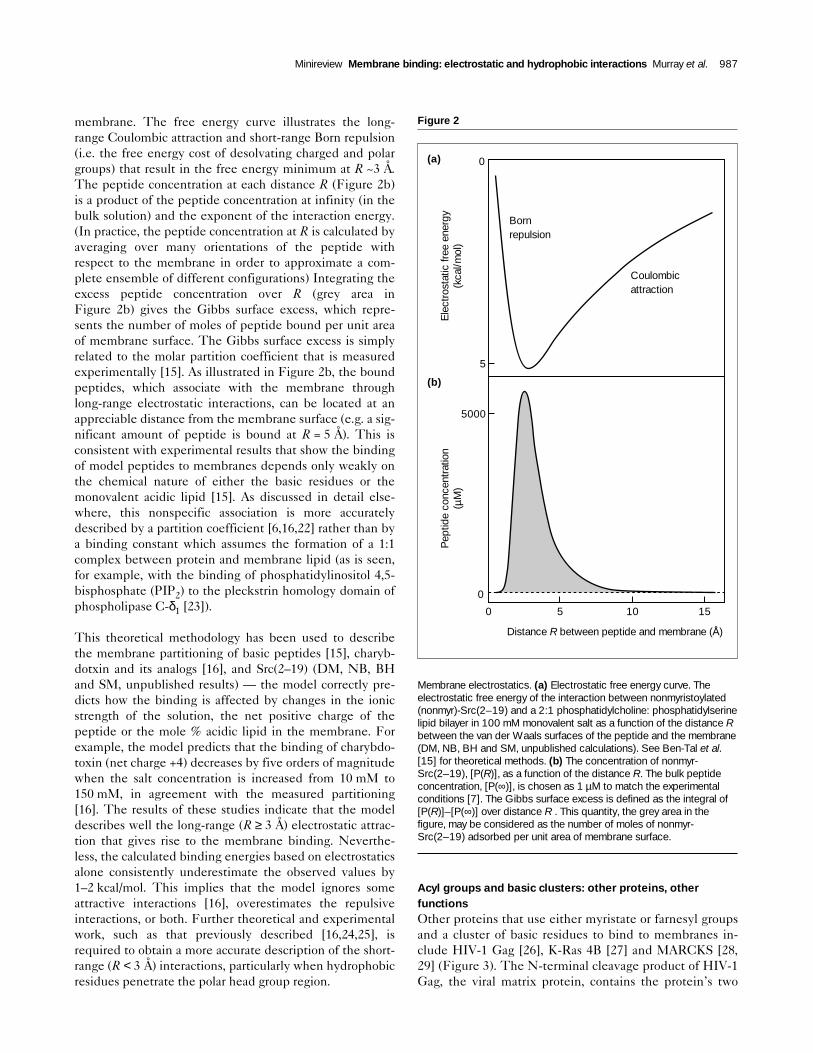
membrane. The free energy curve illustrates the long-range Coulombic attraction and short-range Born repulsion(i.e. the free energy cost of desolvating charged and polargroups) that result in the free energy minimum at R ~3 Å.The peptide concentration at each distance R (Figure 2b)is a product of the peptide concentration at infinity (in thebulk solution) and the exponent of the interaction energy.(In practice, the peptide concentration at R is calculated byaveraging over many orientations of the peptide withrespect to the membrane in order to approximate a com-plete ensemble of different configurations) Integrating theexcess peptide concentration over R (grey area inFigure 2b) gives the Gibbs surface excess, which repre-sents the number of moles of peptide bound per unit areaof membrane surface. The Gibbs surface excess is simplyrelated to the molar partition coefficient that is measuredexperimentally [15]. As illustrated in Figure 2b, the boundpeptides, which associate with the membrane throughlong-range electrostatic interactions, can be located at anappreciable distance from the membrane surface (e.g. a sig-nificant amount of peptide is bound at R = 5 Å). This isconsistent with experimental results that show the bindingof model peptides to membranes depends only weakly onthe chemical nature of either the basic residues or themonovalent acidic lipid [15]. As discussed in detail else-where, this nonspecific association is more accuratelydescribed by a partition coefficient [6,16,22] rather than bya binding constant which assumes the formation of a 1:1complex between protein and membrane lipid (as is seen,for example, with the binding of phosphatidylinositol 4,5-bisphosphate (PIP2) to the pleckstrin homology domain ofphospholipase C-δ1 [23]).
This theoretical methodology has been used to describethe membrane partitioning of basic peptides [15], charyb-dotxin and its analogs [16], and Src(2–19) (DM, NB, BHand SM, unpublished results) — the model correctly pre-dicts how the binding is affected by changes in the ionicstrength of the solution, the net positive charge of thepeptide or the mole % acidic lipid in the membrane. Forexample, the model predicts that the binding of charybdo-toxin (net charge +4) decreases by five orders of magnitudewhen the salt concentration is increased from 10 mM to150 mM, in agreement with the measured partitioning[16]. The results of these studies indicate that the modeldescribes well the long-range (R ≥ 3 Å) electrostatic attrac-tion that gives rise to the membrane binding. Neverthe-less, the calculated binding energies based on electrostaticsalone consistently underestimate the observed values by1–2 kcal/mol. This implies that the model ignores someattractive interactions [16], overestimates the repulsiveinteractions, or both. Further theoretical and experimentalwork, such as that previously described [16,24,25], isrequired to obtain a more accurate description of the short-range (R < 3 Å) interactions, particularly when hydrophobicresidues penetrate the polar head group region.
Acyl groups and basic clusters: other proteins, otherfunctions Other proteins that use either myristate or farnesyl groupsand a cluster of basic residues to bind to membranes in-clude HIV-1 Gag [26], K-Ras 4B [27] and MARCKS [28,29] (Figure 3). The N-terminal cleavage product of HIV-1Gag, the viral matrix protein, contains the protein’s two
Minireview Membrane binding: electrostatic and hydrophobic interactions Murray et al. 987
Figure 2
Membrane electrostatics. (a) Electrostatic free energy curve. Theelectrostatic free energy of the interaction between nonmyristoylated(nonmyr)-Src(2–19) and a 2:1 phosphatidylcholine: phosphatidylserinelipid bilayer in 100 mM monovalent salt as a function of the distance Rbetween the van der Waals surfaces of the peptide and the membrane(DM, NB, BH and SM, unpublished calculations). See Ben-Tal et al.[15] for theoretical methods. (b) The concentration of nonmyr-Src(2–19), [P(R)], as a function of the distance R. The bulk peptideconcentration, [P(∞)], is chosen as 1 µM to match the experimentalconditions [7]. The Gibbs surface excess is defined as the integral of[P(R)]–[P(∞)] over distance R . This quantity, the grey area in thefigure, may be considered as the number of moles of nonmyr-Src(2–19) adsorbed per unit area of membrane surface.
Bornrepulsion
Coulombicattraction
0
5
5000
0
0 5 10 15
Distance R between peptide and membrane (Å)
Pep
tide
conc
entr
atio
n (µ
M)
Ele
ctro
stat
ic fr
ee e
nerg
y (k
cal/m
ol)
(a)
(b)
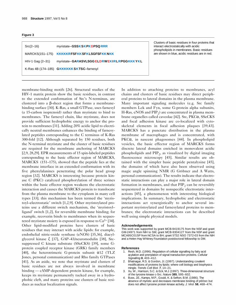
membrane-binding motifs [26]. Structural studies of theHIV-1 matrix protein show the basic residues, in contrastto the extended conformation of Src’s N-terminus, areclustered into a β-sheet region that forms a membrane-binding surface [30]. K-Ras, a small GTPase, uses farnesyl(a 15-carbon isoprenoid) rather than myristate to bind tomembranes. The farnesyl chain, like myristate, does notprovide sufficient hydrophobic energy to anchor the pro-tein to membranes [31]. Adding 20% acidic lipid to electri-cally neutral membranes enhances the binding of farnesy-lated peptides corresponding to the C terminus of K-Ras300-fold [12]. Although separated by 150 residues, boththe N-terminal myristate and the cluster of basic residuesare required for the membrane anchoring of MARCKS[2,9, 28,29]. EPR measurements of 15 spin-labeled peptidescorresponding to the basic effector region of MARCKS,MARCKS (151–175), showed that the peptide lies at themembrane interface in an extended conformation with itsfive phenylalanines penetrating the polar head groupregion [32]. MARCKS is interesting because protein kin-ase C (PKC) catalyzed phosphorylation of three serineswithin the basic effector region weakens the electrostaticinteraction and causes the MARCKS protein to translocatefrom the plasma membrane to the cytoplasm in many celltypes [33]; this mechanism has been termed the ‘myris-toyl–electrostatic’ switch [1,2,9]. Other myristoylated pro-teins use a different switch mechanism, the ‘myristoyl–ligand’ switch [1,2], for reversible membrane binding; forexample, recoverin binds to membranes when its seques-tered myristate moiety is exposed in response to Ca2+ [34].Other lipid-modified proteins have clusters of basicresidues that may interact with acidic lipids: for example,endothelial nitric-oxide synthase (eNOS) [35,36], diacyl-glycerol kinase ζ [37], GAP–43/neuromodulin [38], Src-suppressed C kinase substrate (SSeCKS) [39], some G-protein coupled receptor kinase (GRK) family members[40], the heterotrimeric G-protein subunit α12 (TLZJones, personal communication) and Rho family GTPases[41]. As an aside, we note that myristate and clusters ofbasic residues are not always involved in membranebinding — cAMP-dependent protein kinase, for example,keeps its myristate permanently tucked away in a hydro-phobic cleft, and many proteins use clusters of basic resi-dues as nuclear localization signals.
In addition to attaching proteins to membranes, acylchains and clusters of basic residues may direct periph-eral proteins to lateral domains in the plasma membrane.Many important signaling molecules (e.g. Src familymembers Lck and Fyn, some G-protein alpha subunits,H-Ras, eNOS and PIP2) are concentrated in plasma mem-brane organelles called caveolae [42]. Src, PKCα, SSeCKSand focal adhesion kinase are co-localized with cyto-skeletal elements in focal adhesion plaques [39,43].MARCKS has a punctate distribution in the plasmamembrane of macrophages and is concentrated, withPKCα, in nascent phagosomes [44]. In phospholipidvesicles, the basic effector region of MARCKS formsdiscrete lateral domains enriched in monovalent acidicphospholipids and PIP2, as visualized by digital imagingfluorescence microscopy [45]. Similar results are ob-tained with the simpler basic peptide pentalysine [45],the domains of which have also been observed usingmagic angle spinning NMR (G Gröbner and A Watts,personal communication). The results indicate that electro-static interactions can play a major role in lateral domainformation in membranes, and that PIP2 can be reversiblysequestered in domains by nonspecific electrostatic inter-actions [45], a phenomenon with interesting biologicalimplications. In summary, hydrophobic and electrostaticinteractions act synergistically to anchor several im-portant myristoylated and farnesylated proteins to mem-branes; the electrostatic interactions can be describedwell using simple physical models.
AcknowledgementsThis work was supported by grant MCB-9419175 from the NSF and grantGM-24971 from NIH to SM; grant MCB-9304127 from the NSF and grantMCA95C01SP from NCSA to BH; grant 5T32 NSO 7372-03 from the NIHand a Helen Hay Whitney Foundation postdoctoral fellowship to DM.
References1. Resh, M.D. (1996). Regulation of cellular signalling by fatty acid
acylation and prenylation of signal transduction proteins. CellularSignalling 8, 403–412.
2. Bhatnagar, R.S. & Gordon, J.I. (1997). Understanding covalentmodifications of proteins by lipids: where cell biology and biophysicsmingle. Trends Cell Biol. 7, 14–21.
3. Xu, W., Harrison, S.C. & Eck, M.J. (1997). Three-dimensional structureof the tyrosine kinase c-Src. Nature 385, 595–602.
4. Buss, J.E., Kamps, M.P., Gould, K. & Sefton, B.M. (1986). Theabsence of myristic acid decreases membrane binding of p60src butdoes not affect tyrosine protein kinase activity. J. Virol. 58, 468–474.
988 Structure 1997, Vol 5 No 8
Figure 3
Clusters of basic residues in four proteins thatinteract electrostatically with acidicphospholipids in membranes. Basic residuesare shown in blue, acidic residues are in red.
Src(2–16)
MARCKS(151–175)
HIV-1 Gag (2–31)
K-Ras 4B (174–185)
myristate– GSSKSKPKDPSQ RRR
KKKKKRFSFKKSFKLSGFSFKKNKK
myristate– GARASVLSGG ELDRWEKIRLRPGGKKKYKL
GKKKKKKSKTSC–farnesyl

5. Kamps, M.P., Buss, J.E. & Sefton, B.M. (1985). Mutation of NH2-terminal glycine of p60src prevents both myristoylation andmorphological transformation. Proc. Natl Acad. Sci. USA 82,4625–4628.
6. Peitzsch, R.M. & McLaughlin, S. (1993). Binding of acylated peptidesand fatty acids to phospholipid vesicles: pertinence to myristoylatedproteins. Biochemistry 32, 10436–10443.
7. Buser, C.A., Sigal, C.T., Resh, M.D. & McLaughlin, S. (1994).Membrane binding of myristylated peptides corresponding to theNH2-terminus of Src. Biochemistry 33, 13093–13101.
8. Tanford, C. (1991). The Hydrophobic Effect: Formation of Micellesand Biological Membranes. Krieger Publishing Co. Malabar, FL, USA.
9. McLaughlin, S. & Aderem, A. (1995). The myristoyl-electrostaticswitch: a modulator of reversible protein–membrane interactions.Trends Biochem. Sci. 20, 272–276.
10. Sigal, C.T., Zhou, W., Buser, C.A., McLaughlin, S. & Resh, M.D.(1994). The amino terminal basic residues of Src mediate membranebinding through electrostatic interaction with acidic phospholipids.Proc. Natl Acad. Sci. USA 91, 12253–12257.
11. Kaplan, J.M., Varmus, H.E. & Bishop, J.M. (1990). The src proteincontains multiple domains for specific attachment to membranes. Mol.Cell. Biol. 10, 1000–1009.
12. Ghomashchi, F., Zhang, X., Liu, L. & Gelb, M.H. (1995). Binding ofprenylated and polybasic peptides to membranes: affinities andintervesicle exchange. Biochemistry 34, 11910–11918.
13. Honig, B.H. & Nicholls, A. (1995). Classical electrostatics in biologyand chemistry. Science 268, 1144–1149.
14. Honig, B.H., Sharp, K.A. & Yang, A.S. (1993). Macroscopic models ofaqueous solutions: biological and chemical applications. J. Phys.Chem. 97, 1101–1109.
15. Ben-Tal, N., Honig, B., Peitzsch, R.M., Denisov, G. & McLaughlin, S.(1996). Binding of small basic peptides to membranes containingacidic lipids: theoretical models and experimental results. Biophys. J.71, 561–575.
16. Ben-Tal, N., Honig, B., Miller, C. & McLaughlin, S. (1997).Electrostatic binding of proteins to membranes: theoretical predictionand experimental results with charybdotoxin and phospholipidvesicles. Biophys. J., in press.
17. Carnie, S.L. & Torrie, G.M. (1984). The statistical mechanics of theelectrical double layer. In Advances in Chemical Physics. (Prigogine, I.& Rice, S.A., eds), pp. 141–253, John Wiley and Sons, New York, USA.
18. McLaughlin, S. (1989). The electrostatic properties of membranes.Annu. Rev. Biophys. Chem. 18, 113–136.
19. Peitzsch, R.M., Eisenberg, M, Sharp, K.A. & McLaughlin, S. (1995).Calculations of the electrostatic potential adjacent to modelphospholipid bilayers. Biophys. J. 68, 729–738.
20. Gilson, M.K., Sharp, K.A & Honig, B.H. (1987). Calculating theelectrostatic potential of molecules in solution: method and errorassessment. J. Comp. Chem. 9, 327–335.
21. Sharp, K.A. & Honig, B.H. (1990). Calculating total electrostaticenergies with the nonlinear Poisson-Boltzmann equation. J. Phys.Chem. 94, 7684–7692.
22. White, S.H., Wimley, W.C. & Ladokhin, A.S. (1997). Protein folding inmembranes: determining the energetics of peptide-bilayerinteractions. Methods Enzymol., in press.
23. Lemmon, M.A., Ferguson, K.M. & Schlessinger, J. (1996). PHdomains: diverse sequences with a common fold recruit signalingmolecules to the cell surface. Cell 85, 621–624.
24. Wimley, W.C. & White, S.H. (1996). Experimentally determinedhydrophobicity scale for proteins at membrane interfaces. Nat. Struct.Biol. 3, 842–848.
25. Thorgeirsson, T.E., Russell, C.J., King, D.S. & Shin, Y.K. (1996). Directdetermination of the membrane affinities of individual amino acids.Biochemistry 35, 1803–1809.
26. Zhou, W., Parent, L.J., Wills, J.W. & Resh, M.D. (1994). Identificationof a membrane-binding domain within the amino-terminal region ofhuman immunodeficiency virus type 1 Gag protein which interactswith acidic phospholipids. J. Virol. 68, 2556–2569.
27. Hancock, J.F., Paterson, H. & Marshall, C.J. (1990). A polybasicdomain or palmitoylation is required in addition to the CAAX motif tolocalize p21ras to the plasma membrane. Cell 63, 133–139.
28. Swierczynski, S.L. & Blackshear, P.J. (1996). Myristoylation-dependentand electrostatic interactions exert independent effects on themembrane association of the myristoylated alanine-rich C-kinasesubstrate. J. Biol. Chem. 271, 23424–23430.
29. Seykora, J.T., Myat, M.M., Allen, L.A., Ravetch, J.V. & Aderem, A.(1996). Molecular determinants of the myristoyl-electrostatic switch ofMARCKS. J. Biol. Chem. 271, 18797–18802.
30. Hill, C.P., Bancroft, D.P., Christensen, A.M. & Sundquist, W.I. (1996).Crystal structures of the trimeric human immunodeficiency virus type Imatrix protein: implications for membrane binding and assembly. Proc.Natl. Acad. Sci. USA 93, 3099–3104.
31. Silvius, J.R. & l’Heureux, F. (1994). Fluorimetric evaluation of theaffinities of isoprenylated peptides for lipid bilayers. Biochemistry 33,3014–3022.
32. Qin, Z. & D.S. Cafiso. (1996). Membrane structure of the proteinkinase C and calmodulin-binding domain of myristoylated alanine richC kinase substrate determined by site-directed spin labeling.Biochemistry 35, 2917–2925.
33. Thelen, M., Rosen, A., Nairn, A.C. & Aderem, A. (1991). Regulation byphosphorylation of reversible association of a myristoylated proteinkinase C substrate with the plasma membrane. Nature 351, 320–322.
34. Tanaka, T., Ames, J.B., Harvey, T.S., Stryer, L. & Ikura, M. (1995).Sequestration of the membrane-targeting myristoyl group of recoverinin the calcium-free state. Nature 376, 444–447.
35. Matsubara, M., Titani, K. & Taniguchi, H. (1996). Interaction ofcalmodulin-binding domain peptides of nitric oxide synthase withmembrane phospholipids: regulation of protein phosphorylation andCa2+-calmodulin. Biochemistry 35, 14651–14658.
36. Shaul, P.W., et al., & Michel, T. (1996). Acylation targets endothelialnitric-oxide synthase to plasmalemmal caveolae. J. Biol. Chem. 271,6518–6522.
37. Bunting, M., Tang, W., Zimmerman, G.A., McIntyre, T.M. & Prescott,S.M.. (1996). Molecular cloning and characterization of a novel humandiacylglycerol kinase. J. Biol. Chem. 271, 10230–10236.
38. Hayashi, N., Matsubara, M., Titani, K. & Taniguchi, H. (1997). Circulardichroism and 1H nuclear magnetic resonance studies on the solutionand membrane structure of GAP-43 calmodulin-binding domain. J.Biol. Chem. 272, 7639–7645.
39. Lin, X., Tombler, E., Nelson, P.J., Ross, M. & Gelman, I.H.. (1996). Anovel src- and ras-suppressed protein kinase C substrate associatedwith cytoskeletal architecture. J. Biol. Chem. 271, 28430–28438.
40. Stoffel III, R.H., Pitcher, J.A. & Lefkowitz, R.J. (1997). Targeting Gprotein-coupled receptor kinases to their receptor substrates. J.Memb. Biol. 157, 1–8.
41. Gosser, Y.Q., et al., & M. Rosen. (1997). C-terminal binding domain ofRho GDP-dissociation inhibitor directs N-terminal inhibitory peptide toGTPases. Nature 387, 814–819.
42. Simons, K. & Ikonen, E. (1997). Functional rafts in cell membranes.Nature 387, 569–572.
43. Gilmore, A.P. & Burridge, K. (1996). Molecular mechanisms for focaladhesion assembly through regulation of protein–protein interactions.Structure 4, 647–651.
44. Allen, L.A. & Aderem, A. (1996). Mechanisms of phagocytosis. Curr.Opin. Immunol. 8, 36–40.
45. Glaser, M., et al., & McLaughlin, S. (1996). MARCKS producesreversible inhibition of phospholipase C by sequesteringphosphatidylinositol 4,5-bisphosphate in lateral domains. J. Biol.Chem. 271, 26187–26193.
Minireview Membrane binding: electrostatic and hydrophobic interactions Murray et al. 989