Electronic Supporting Information (ESI) › suppdata › c9 › cc › c9cc00922a ›...
10
S1 Electronic Supporting Information (ESI) Light-driven control of the composition of a supramolecular network Patricia Remón, a David González, a Shiming Li, b Nuno Basílio, c Joakim Andréasson b and Uwe Pischel* a a CIQSO - Centre for Research In Sustainable Chemistry and Department of Chemistry, Campus de El Carmen s/n, E-21071 Huelva, Spain b Department of Chemistry and Chemical Engineering, Chemistry and Biochemistry, Chalmers University of Technology, SE-41296 Gteborg, Sweden c Laboratorio Associado para a Química Verde (LAQV), Rede de Química e Tecnologia (REQUIMTE), Departamento de Química, Faculdade de Ciências e Tecnología, Universidade NOVA de Lisboa, 2829- 516 Caparica, Portugal Electronic Supplementary Material (ESI) for ChemComm. This journal is © The Royal Society of Chemistry 2019
Transcript of Electronic Supporting Information (ESI) › suppdata › c9 › cc › c9cc00922a ›...

S1
Electronic Supporting Information (ESI)
Light-driven control of the composition of a supramolecular network
Patricia Remón,a David González,a Shiming Li,b Nuno Basílio,c Joakim Andréassonb and Uwe
Pischel*a
a CIQSO - Centre for Research In Sustainable Chemistry and Department of Chemistry, Campus de El
Carmen s/n, E-21071 Huelva, Spain
b Department of Chemistry and Chemical Engineering, Chemistry and Biochemistry, Chalmers
University of Technology, SE-41296 Goteborg, Sweden
c Laboratorio Associado para a Química Verde (LAQV), Rede de Química e Tecnologia (REQUIMTE),
Departamento de Química, Faculdade de Ciências e Tecnología, Universidade NOVA de Lisboa, 2829-
516 Caparica, Portugal
Electronic Supplementary Material (ESI) for ChemComm.This journal is © The Royal Society of Chemistry 2019

S2
General methods and materials
The DTE derivative 1 was prepared by a modified published procedure (see below). All chemicals for
the synthesis were used as received without further purification, unless stated otherwise. CH2Cl2 was
distilled over CaH2. 1H NMR (400 MHz) and 13C NMR (101 or 125 MHz) spectra for the
characterization of 1 and its precursor were recorded on Varian Unity 400 or 500 spectrometers at
25 C. In the 1H and 13C NMR spectra, chemical shifts (/ppm) are referenced to the residual solvent
peak: CDCl3, 7.26 ppm (1H NMR) and 77.20 ppm (13C NMR); D2O, 4.80 (1H NMR); MeOH-d4, 49.00
ppm (13C NMR). Thin-layer chromatography to monitor the reactions was performed on silica gel
plates (Merck Kieselgel 60, F254).
All measurements (at room temperature) were done in water (miliQ quality) or in deuterium oxide
(D2O, >99 atom% D) at pH or pD 5.0, respectively. The pH/pD was adjusted by additions of HCl/DCl or
NaOH/NaOD. The pD values were obtained after correction for isotope effects (pD = pH* + 0.4).1
Cucurbit[7]uril (CB7) was prepared by following a published procedure.2 The water content was
taken as 20 weight-%, determined by 1H NMR spectroscopy using malonic acid as internal standard.
Cucurbit[8]uril (CB8; 20 weight-% water) and geranylamine (2) are commercial products and were
used in the highest quality available.
1H NMR experiments to characterize the network were done with a Varian Mercury 500 MHz NMR
spectrometer. For the irradiation two light sources were used: 365-nm light was generated by a UVP
handheld UV lamp (Model VL-4.LC, 4 W) and light at > 590 nm by a 150 W Xenon lamp (Oriel
GmbH & Co.KG), using a long-pass optical filter.
Note: For the four-component mixture (1, 2, CB7, and CB8; all at 500 M) a small amount of
precipitate was observed on prolonged standing of the solution. This can be re-dissolved by heating
gently with a hairdryer.

S3
Experimental procedure for the synthesis of 1
Scheme S1. Synthesis of 1.
Synthesis of 1,2-bis(2-methyl-5-(4-pyridyl)-3-thienyl)cyclopentene
A published method3 was modified to synthesize 1,2-bis(2-methyl-5-(4-pyridyl)-3-
thienyl)cyclopentene. 1,2-Bis(5-chloro-2-methyl-3-thienyl)cyclopentene4 (493 mg, 1.5 mmol), 4-
pyridinylboronic acid (406 mg, 3.3 mmol), Na2CO3 (720 mg, 3 mmol) and Pd(PPh3)4 (174 mg, 0.15
mmol) were placed in a flask under Ar. Dimethoxyethane (DME, 20 mL, degassed) and water (5 mL,
degassed) were subsequently added and the reaction mixture was refluxed (90 C) for 48 h under
argon. After cooling to room temperature, the reaction was quenched with water (40 mL) and Et2O
(100 mL). The organic layer was separated, and the water phase was extracted with Et2O (2 × 100
mL). The combined organic phases were dried (Na2SO4), filtered, and the solvent was evaporated in
vacuo. The crude product was purified by column chromatography (SiO2, MeOH/CH2Cl2 = 2:98) to
afford the product (173 mg, 28% yield). The 1H and 13C NMR data are in agreement with the
published data.5 1H NMR (400 MHz, CDCl3): 8.53 (m, 4H, pyridine-H). 7.35 (m, 4H, pyridine-H), 7.22 (s, 2H, thiophene-
H), 2.86 (t, J = 7.4 Hz, 4H, CH2), 2.18-2.06 (m, 2H, CH2), 2.03 (s, 6H, CH3). 13C NMR (101 MHz, CDCl3): 150.4, 141.5, 137.5, 137.3, 136.9, 135.0, 126.5, 119.5, 38.7, 23.2, 14.8.
Synthesis of 1
2-Bis(2-methyl-5-(4-pyridyl)-3-thienyl)cyclopentene (69 mg, 0.093 mmol) was dissolved in dry CH2Cl2
(3 mL) under argon and CH3I (0.3 mL) was injected. The solution was stirred at room temperature for
3 h. The greenish precipitate was collected by filtration, washed repeatedly with dry CH2Cl2 afforded
the NMR-pure 1 (55 mg, 55% yield). The 1H NMR data are in agreement with the published data.6
1H NMR (400 MHz, D2O) 8.50 (d, J = 6.8 Hz, 4H, pyridine-H), 7.97 (d, J = 7.2 Hz, 4H, pyridine-H), 7.84
(s, 2H, thiophene-H), 4.22 (s, 6H, CH3), 2.88 (t, J = 7.4 Hz, 4H, CH2), 2.18-2.06 (m, 2H, CH2), 2.05 (s, 6H,
CH3). 13C NMR (125 MHz, MeOH-d4): 150.1, 146.4, 145.9, 140.4, 136.8, 134.6, 134.3, 122.8, 47.8, 39.5,
24.0, 15.2.

S4
1H and 13C NMR spectra
Figure S1. 1H NMR spectrum (400 MHz, CDCl3) of 1,2-bis(2-methyl-5-(4-pyridyl)-3-
thienyl)cyclopentene.
Figure S2. 13C NMR spectrum (101 MHz, CDCl3) of 1,2-bis(2-methyl-5-(4-pyridyl)-3-
thienyl)cyclopentene.

S5
Figure S3. 1H NMR spectrum (400 MHz, D2O) of 1.
Figure S4. 13C NMR spectrum (125 MHz, MeOH-d4) of 1.

S6
UV/vis absorption spectra of the four-component mixture on irradition with light
200 300 400 500 600 700 800 9000.0
0.2
0.4
0.6
0.8
1.0
A
/ nm
Figure S5. UV/vis absorption spectra of the four-component mixture (1, 2, CB7, and CB8; all at 15
M) at pH 5 before (red) and after (black) irradiation at 365 nm.
S7
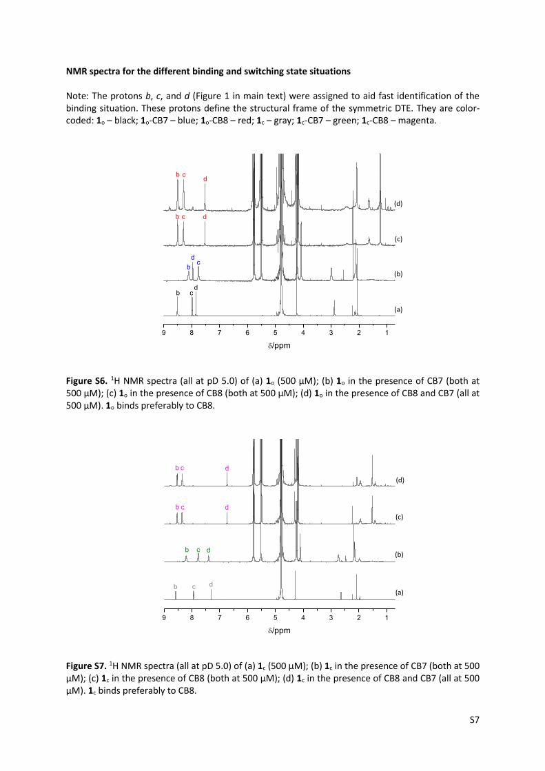
NMR spectra for the different binding and switching state situations Note: The protons b, c, and d (Figure 1 in main text) were assigned to aid fast identification of the binding situation. These protons define the structural frame of the symmetric DTE. They are color-coded: 1o – black; 1o-CB7 – blue; 1o-CB8 – red; 1c – gray; 1c-CB7 – green; 1c-CB8 – magenta.
9 8 7 6 5 4 3 2 1
dc
(d)
(c)
(b)
(a)
/ppm
b
dc
b
b c d
b cd
Figure S6. 1H NMR spectra (all at pD 5.0) of (a) 1o (500 µM); (b) 1o in the presence of CB7 (both at 500 µM); (c) 1o in the presence of CB8 (both at 500 µM); (d) 1o in the presence of CB8 and CB7 (all at 500 µM). 1o binds preferably to CB8.
9 8 7 6 5 4 3 2 1
(d)
(c)
(b)
(a)
/ppm
b c d
b c d
b c d
b c d
Figure S7. 1H NMR spectra (all at pD 5.0) of (a) 1c (500 µM); (b) 1c in the presence of CB7 (both at 500 µM); (c) 1c in the presence of CB8 (both at 500 µM); (d) 1c in the presence of CB8 and CB7 (all at 500 µM). 1c binds preferably to CB8.

S8
9 8 7 6 5 4 3 2 1
(e)
(d)
(c)
(b)
(a)
ppm
b c d
bc
d
b c
d
Figure S8. 1H NMR spectra (all at pD 5.0) of (a) 2 (1 mM); (b) 2 in the presence of CB7 (both at 500
M); (c) 1o (500 M); (d) 1o in the presence of CB7 (both at 500 µM); (e) mixture 1o and 2 in the presence of CB7 (all at 500 µM). 2 displaces 1o completely from CB7.
9 8 7 6 5 4 3 2 1
(a)
(e)
(d)
(c)
(b)
/ppm
b c d
bc
d
b cd
Figure S9. 1H NMR spectra (all at pD 5.0) of (a) 2 (1 mM); (b) 2 in the presence of CB7 (both at 500
M); (c) 1c (500 M); (d) 1c in the presence of CB7 (both at 500 µM); (e) mixture of 1c and 2 in the presence of CB7 (all at 500 µM). 2 displaces 1c completely from CB7.

S9
9 8 7 6 5 4 3 2 1
(a)
(b)
(c)
(d)
(e)
/ppm
b cd
b c d
b c d
Figure S10. 1H NMR spectra (all at pD 5.0) of (a) 2 (1 mM); (b) 2 in the presence of CB8 (both at 500
M); (c) 1o (500 M); (d) 1o in the presence of CB8 (both at 500 µM); (e) mixture 1o and 2 in the presence of CB8 (all at 500 µM). 2 displaces 1o completely from CB8.
9 8 7 6 5 4 3 2 1
(a)
(b)
(c)
(d)
(e)
/ppm
b c d
b c d
db
b cd
bc d
Figure S11. 1H NMR spectra (all at pD 5.0) of (a) 2 (1 mM); (b) 2 in the presence of CB8 (both at 500
M); (c) 1c (500 M); (d) 1c in the presence of CB8 (both at 500 µM); (e) mixture of 1c and 2 in the presence of CB8 (all at 500 µM). 2 displaces 1c partially from CB8.

S10
References 1. P. K. Glasoe and F. A. Long, J. Phys. Chem., 1960, 64, 188-190. 2. C. Márquez, F. Huang and W. M. Nau, IEEE Trans. Nanobiosci., 2004, 3, 39-45. 3. S. Hermes, G. Dassa, G. Toso, A. Bianco, C. Bertarelli and G. Zerbi, Tetrahedron Lett., 2009,
50, 1614-1617. 4. L. N. Lucas, J. J. D. de Jong, J. H. van Esch, R. M. Kellogg and B. L. Feringa, Eur. J. Org. Chem.,
2003, 155-166. 5. A. Presa, G. Vázquez, L. A. Barrios, O. Roubeau, L. Korrodi-Gregório, R. Pérez-Tomás and P.
Gamez, Inorg. Chem., 2018, 57, 4009-4022. 6. X. Yao, T. Li, S. Wang, X. Ma and H. Tian, Chem. Commun., 2014, 50, 7166-7168.