Electronic structure of epitaxial thin films of bay-substituted perylene bisimide dyes
6
Appl Phys A (2009) 95: 285–290 DOI 10.1007/s00339-008-5013-1 Electronic structure of epitaxial thin films of bay-substituted perylene bisimide dyes Markus Scholz · Rüdiger Schmidt · Stefan Krause · Achim Schöll · Friedrich Reinert · Frank Würthner Received: 12 August 2008 / Accepted: 24 November 2008 / Published online: 31 December 2008 © Springer-Verlag 2008 Abstract We have investigated epitaxial thin films of three air-stable organic n-type semiconducting perylene tetracar- boxylic acid bisimides (PBIs) with fluoroalkyl groups in the imide position with a combination of X-ray, UV, and in- verse photoelectron spectroscopy. We explore the applica- bility of film preparation by organic molecular beam depo- sition (OMBD) under ultra-high vacuum conditions to these compounds. By means of fluorine and chlorine substituents in the bay area a systematic torsion of the π -conjugated core of these three PBIs is achieved. Accordingly, these mole- cules offer a model system to analyze the interplay between the molecular conformation, the film structure, and the elec- tronic structure. Our results show that the PBIs can be de- posited intact and contamination free by OMBD and rela- tively smooth films with layer-to-layer growth can be estab- lished. Moreover, the valence spectra reveal the effect of the electro-negative bay substituents as well as of the twisting of the perylene core, which leads to energy shifts of the oc- cupied and unoccupied frontier orbitals. Common address: Röntgen Research Center for Complex Material Systems, Universität Würzburg, Am Hubland, 97074 Würzburg, Germany. M. Scholz · S. Krause · A. Schöll ( ) · F. Reinert Experimentelle Physik II, Universität Würzburg, Am Hubland, 97074 Würzburg, Germany e-mail: [email protected] R. Schmidt · F. Würthner Institut für Organische Chemie, Universität Würzburg, Am Hubland, 97074 Würzburg, Germany A. Schöll · F. Reinert Gemeinschaftslabor für Nanoanalytik, Forschungszentrum Karlsruhe, 76021 Karlsruhe, Germany PACS 71.20.Rv · 73.21.Ac · 79.60.Dp · 81.15.Ef 1 Introduction The commercial use of organic semiconductors for light emitting diodes and the strong research efforts of academic and industrial groups in the field of organic field effect tran- sistors [1] indicate that this research area is presently grow- ing out of its infancy and will probably be able to comple- ment established silicon based microelectronics technology. While a variety of p-type organic semiconductors are avail- able with custom made properties, air-stable n-conducting materials are still a bottleneck to the progress of organic electronics. Many n-channel semiconductors suffer of poor air stability because electron traps can be formed by am- bient oxygen and moisture. Perylene tetracarboxylic acid bisimides (PBI) [2] are among the best available n-type organic semiconductors and PBIs with fluorinated alkyl groups have shown field effect mobility in the range of 0.5 to 1 cm 2 V −1 s −1 in thin film devices [3–5]. Remarkably, some recently developed PBI derivatives, i.e., PBI-H 4 and PBI-F 2 (for structural formulae see Fig. 1) exhibit high n-channel mobility even after exposure to air for more than 50 days. It is assumed that this improved air-stability results from the particularly dense PBI film structure with the closely-packed fluorinated chains, which hinders air and moisture from pen- etrating the channel region [6]. Whilst such dense packing is supported by the flat π -conjugated core of PBI-H 4 and PBI-F 2 (core twisting between 0 and 5 degree) a sig- nificantly distorted π -conjugated core (>30 degree) is observed for in bay area tetrachorinated derivatives like PBI-Cl 4 [7]. Nevertheless, also for such tetrachlorinated PBIs notable n-type charge carrier mobility values have been reported [8, 9].
-
Upload
markus-scholz -
Category
Documents
-
view
213 -
download
0
Transcript of Electronic structure of epitaxial thin films of bay-substituted perylene bisimide dyes

Appl Phys A (2009) 95: 285–290DOI 10.1007/s00339-008-5013-1
Electronic structure of epitaxial thin filmsof bay-substituted perylene bisimide dyes
Markus Scholz · Rüdiger Schmidt · Stefan Krause ·Achim Schöll · Friedrich Reinert · Frank Würthner
Received: 12 August 2008 / Accepted: 24 November 2008 / Published online: 31 December 2008© Springer-Verlag 2008
Abstract We have investigated epitaxial thin films of threeair-stable organic n-type semiconducting perylene tetracar-boxylic acid bisimides (PBIs) with fluoroalkyl groups in theimide position with a combination of X-ray, UV, and in-verse photoelectron spectroscopy. We explore the applica-bility of film preparation by organic molecular beam depo-sition (OMBD) under ultra-high vacuum conditions to thesecompounds. By means of fluorine and chlorine substituentsin the bay area a systematic torsion of the π-conjugated coreof these three PBIs is achieved. Accordingly, these mole-cules offer a model system to analyze the interplay betweenthe molecular conformation, the film structure, and the elec-tronic structure. Our results show that the PBIs can be de-posited intact and contamination free by OMBD and rela-tively smooth films with layer-to-layer growth can be estab-lished. Moreover, the valence spectra reveal the effect of theelectro-negative bay substituents as well as of the twistingof the perylene core, which leads to energy shifts of the oc-cupied and unoccupied frontier orbitals.
Common address: Röntgen Research Center for Complex MaterialSystems, Universität Würzburg, Am Hubland, 97074 Würzburg,Germany.
M. Scholz · S. Krause · A. Schöll (�) · F. ReinertExperimentelle Physik II, Universität Würzburg, Am Hubland,97074 Würzburg, Germanye-mail: [email protected]
R. Schmidt · F. WürthnerInstitut für Organische Chemie, Universität Würzburg,Am Hubland, 97074 Würzburg, Germany
A. Schöll · F. ReinertGemeinschaftslabor für Nanoanalytik, ForschungszentrumKarlsruhe, 76021 Karlsruhe, Germany
PACS 71.20.Rv · 73.21.Ac · 79.60.Dp · 81.15.Ef
1 Introduction
The commercial use of organic semiconductors for lightemitting diodes and the strong research efforts of academicand industrial groups in the field of organic field effect tran-sistors [1] indicate that this research area is presently grow-ing out of its infancy and will probably be able to comple-ment established silicon based microelectronics technology.While a variety of p-type organic semiconductors are avail-able with custom made properties, air-stable n-conductingmaterials are still a bottleneck to the progress of organicelectronics. Many n-channel semiconductors suffer of poorair stability because electron traps can be formed by am-bient oxygen and moisture. Perylene tetracarboxylic acidbisimides (PBI) [2] are among the best available n-typeorganic semiconductors and PBIs with fluorinated alkylgroups have shown field effect mobility in the range of 0.5 to1 cm2 V−1 s−1 in thin film devices [3–5]. Remarkably, somerecently developed PBI derivatives, i.e., PBI-H4 and PBI-F2
(for structural formulae see Fig. 1) exhibit high n-channelmobility even after exposure to air for more than 50 days. Itis assumed that this improved air-stability results from theparticularly dense PBI film structure with the closely-packedfluorinated chains, which hinders air and moisture from pen-etrating the channel region [6]. Whilst such dense packingis supported by the flat π -conjugated core of PBI-H4 andPBI-F2 (core twisting between 0 and 5 degree) a sig-nificantly distorted π -conjugated core (>30 degree) isobserved for in bay area tetrachorinated derivatives likePBI-Cl4 [7]. Nevertheless, also for such tetrachlorinatedPBIs notable n-type charge carrier mobility values have beenreported [8, 9].

286 M. Scholz et al.
Fig. 1 Molecular structures and AM1 calculated geometries of theinvestigated molecules PBI-H4, PBI-F2, and PBI-Cl4. The CH2C3F7chains were replaced by hydrogens in the calculations
The effect of substituents at the 1,6,7, and 12 positions(the so-called bay positions) is indeed quite complex. Apartfrom the structural effect, it has been shown that bay sub-stituents also affect the electronic and the optical proper-ties [2]. In general, electron withdrawing substituents willlower the molecular HOMO and LUMO levels although thiseffect is quite modest in case of fluorine and chlorine sub-stitution at the bay positions of PBIs [2, 8, 9]. Moreover,the arrangement of the molecules in condensates and the in-termolecular coupling can be expected to react sensitively tothe changes of the molecular structure. The latter aspect is ofcrucial importance for the optimization of the charge trans-port properties of thin film devices processed from these ma-terials and motivates this investigation on the preparation ofepitaxial thin film by vacuum sublimation and the determi-nation of the electronic levels.
In this work, we investigate the growth of PBI films pre-pared by organic molecular beam deposition (OMBD) onAg(111) substrates and the electronic structure of these sys-tems by a combination of photoelectron spectroscopy withX-ray and UV excitation (XPS and UPS, respectively), andinverse photoelectron spectroscopy (IPES). For this study,three N,N ′-di(2,2,3,3,4,4,4-heptafluorobutyl)-3,4 :9,10-tetracarboxylic acid bisimide (PBIs) were chosen that beardifferent substituents in the 1,6,7,12 bay positions(Fig. 1).
2 Experimental
The UPS and IPES measurements were performed ina VG ESCALAB UHV chamber at a base pressure of1 × 10−10 mbar. For UPS measurements, a gas discharge
lamp operated with He I (21.22 eV) and a Mk II electronanalyzer were used. The energy resolution was determinedto 50 meV from a fit to the substrate Fermi level. For theXPS experiments addressing the growth mode a Mg Kα-labsource was utilized. The IPES setup consists of a low-energyelectron gun according to Ciccacci et al. [10] with a BaOcathode and a Geiger–Müller-type detector with SrF2 win-dow and filled with Ar and I2. The detector thus operatesat a fixed photon energy of about 9.5 eV and the band passwidth was determined to 0.4 eV. The high resolution XPSmeasurements were performed at the UE52-PGM beamlineat BESSY with a SCIENTA R4000 electron energy analyzerand with a total energy resolution (analyzer plus beamline)of about 40 meV [11]. All PES experiments presented herewere done in normal emission.
The organic thin films were prepared under UHV condi-tions by sublimation from a home-made Knudsen cell. ThePBIs were synthesized according to recently reported pro-cedures [3–5, 8, 9] and purified by zone-sublimation un-der HV conditions twice prior to sample preparation. Thesublimation was monitored with a quadrupole mass spec-trometer and no impurities could be detected. The Ag(111)substrate was sputter-cleaned and annealed before film de-position and the surface quality checked by XPS, UPS andLEED. The deposition rate was carefully calibrated. Thewell-known Ag3d and C1s XPS signals of a PTCDA mono-layer on Ag(111) were used as a reference for very thin PBIfilms, assuming a similar electron mean free path for the rel-atively similar compounds. This approach was corroboratedby work function measurements, which showed a distinctshift in the sample work function when the first closed layerhas been established.
3 Results and discussion
The attenuation behavior of a substrate signal in the XPSspectra with increasing nominal layer thickness is a feasi-ble tool for the investigation of the film growth mode [12].This behavior allows distinguishing between three differentgrowth modes. One extreme in growth is the layer-by-layer(or Frank–van-der-Merve) growth in which the substratesignal is exponentially attenuated with increasing film thick-ness. Layer-by-layer means that a new layer only grows afterthe previous one is completely closed. The second extremeis the growth of islands (Vollmer–Weber growth) where thesubstrate between the islands still gives a strong signal andtherefore is weakly attenuated (linear with small slope) withincreasing nominal film thickness. The mix of both modes isthe Stranski–Krastanov growth, which ranges from a weakroughening of the surface to a few closed layers with islandson top.

Electronic structure of epitaxial thin films of bay-substituted perylene bisimide dyes 287
Fig. 2 Intensity of the Ag3d substrate signal plotted against the nom-inal layer thickness (given in nm) for films of PBI-H4 (a), PBI-F2 (b),and PBI-Cl4 (c) prepared on Ag(111) at room temperature (bottomcurves, black rectangle) and at 150 K (top curves, grey square).
The data sets were normalized to the intensity of the clean substrate.For better comparison, the 150 K data were off set in intensity. Theintensity curves were fitted with exponential functions according to
I = I0e− d
λ and the λ values are given together with the respective data
Figure 2 displays the intensity of the Ag3d XPS signalwith increasing nominal coverage for all three PBI mole-cules. The Ag3d intensities were determined from the in-tegral area of the 3d3/2 and 3d5/2 lines after normaliza-tion on the low binding energy background. Each growthexperiment was performed with the Ag(111) substrate atroom temperature (bottom curves, black rectangle) and at150 K (top curves, grey square). The data clearly showthat the substrate signal attenuates quickly with increasingPBI deposition. Each experimental curve can be fitted with
the exponential function I = I0e− d
λ (where d is the nom-inal layer thickness) with the constant λ (given togetherwith the respective curves in Fig. 2). In the case of per-fect layer-by-layer growth, λ corresponds to the inelasticmean free path (IMFP) of the photoelectrons in the respec-tive materials. The reasonable values of λ (if, e.g., com-pared to PTCDA, see below) indicate a preferential layer-by-layer growth for all three molecules at room temperatureas well as at 150 K. Whereas the films prepared at roomtemperature behave relatively similar for the three PBIs(λ = 2.6 nm (PBI-H4), 2.6 nm (PBI-F2) and 2.5 nm (PBI-Cl4)), the PBI-H4 and PBI-F2 films prepared at 150 K dif-fer significantly from their room temperature counterparts.The smaller IMFP (λ = 2.0 nm for PBI-H4 and λ = 2.4 nmfor PBI-F2) indicate less roughening of the respective filmsat low substrate temperature. For PBI-Cl4 the only slightlyhigher λ = 2.6 nm indicates a similar type of growth at150 K as compared to room temperature.
If the data obtained for the PBI molecules are comparedto the well-investigated model system PTCDA/Ag(111), forwhich layer-by-layer growth was demonstrated at sampletemperatures below 200 K by Kilian et al. [13], a goodagreement can be found. The slightly bigger IMFP for thePBIs with respect to PTCDA (λ = 2.35 nm) [14] indicateeither a generally larger roughness of the PBI films or a lessdense packing of the molecules, which may be a result ofthe steric hindrance due to the CH2C3F7 chains.
The question remains, whether the relatively large PBIcompounds can be deposited intact by OMBD, or if the ther-mal evaporation at the applied temperatures of 530 K leadsto a decomposition of the molecules, particularly of the flu-orinated alkyl chains. Figure 3 presents the high-resolutionC1s spectra of multilayer films (10–15 layers prepared atroom temperature) of PBI-H4 (a), PBI-F2 (b), and PBI-Cl4(c) recorded at a photon energy of hν = 335 eV. These spec-tra were normalized to the low energy background, the en-ergy axis was calibrated with a gold reference sample, andan exponential as well as a Shirley background was sub-tracted. The spectra show a rich signature with various peaksand shoulders, which can be associated with the photoe-mission from the chemically different carbon species withinthe PBI molecules and additional satellite lines. All peakscan be assigned considering the chemical shifts known fromthe literature [15]. For PBI-H4 the most prominent peakat lowest binding energy (284.6 eV) clearly belongs to thephotoemission from the carbon atoms in the perylene core.Moreover, according to the literature, also the carbon atoms
288 M. Scholz et al.
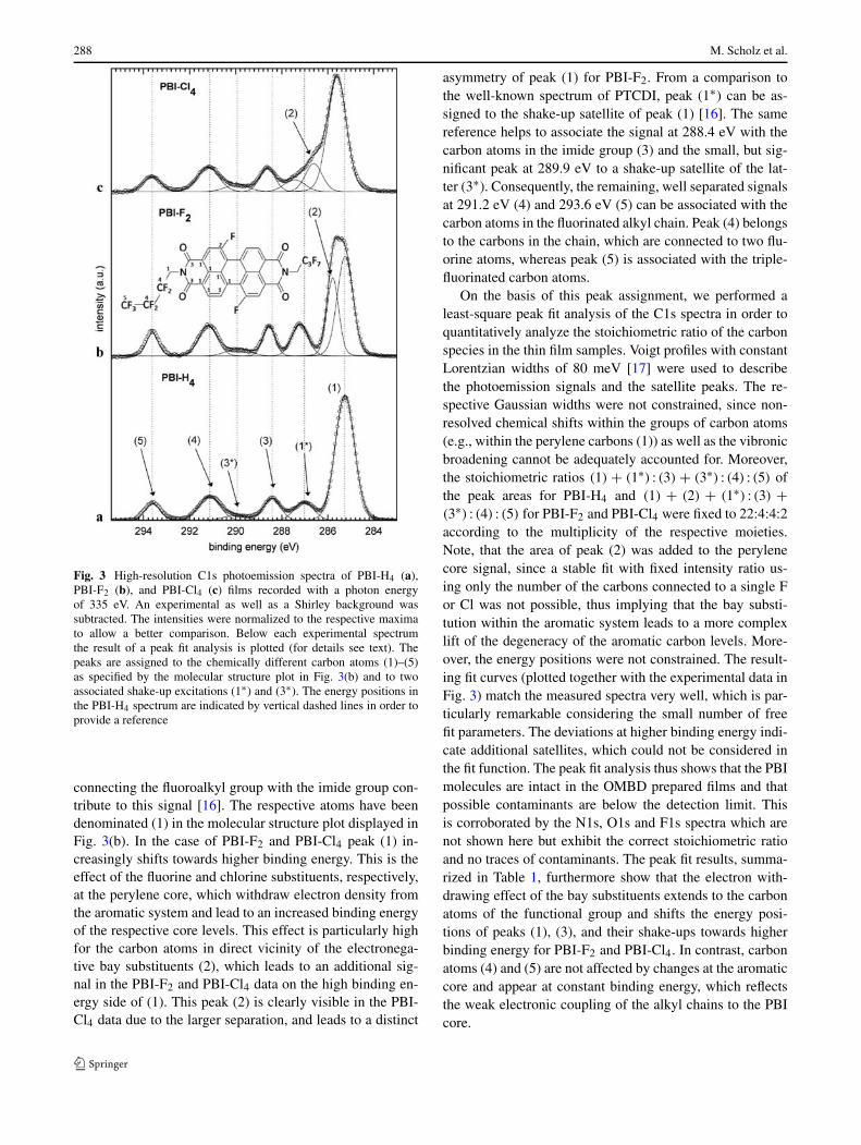
Fig. 3 High-resolution C1s photoemission spectra of PBI-H4 (a),PBI-F2 (b), and PBI-Cl4 (c) films recorded with a photon energyof 335 eV. An experimental as well as a Shirley background wassubtracted. The intensities were normalized to the respective maximato allow a better comparison. Below each experimental spectrumthe result of a peak fit analysis is plotted (for details see text). Thepeaks are assigned to the chemically different carbon atoms (1)–(5)as specified by the molecular structure plot in Fig. 3(b) and to twoassociated shake-up excitations (1∗) and (3∗). The energy positions inthe PBI-H4 spectrum are indicated by vertical dashed lines in order toprovide a reference
connecting the fluoroalkyl group with the imide group con-tribute to this signal [16]. The respective atoms have beendenominated (1) in the molecular structure plot displayed inFig. 3(b). In the case of PBI-F2 and PBI-Cl4 peak (1) in-creasingly shifts towards higher binding energy. This is theeffect of the fluorine and chlorine substituents, respectively,at the perylene core, which withdraw electron density fromthe aromatic system and lead to an increased binding energyof the respective core levels. This effect is particularly highfor the carbon atoms in direct vicinity of the electronega-tive bay substituents (2), which leads to an additional sig-nal in the PBI-F2 and PBI-Cl4 data on the high binding en-ergy side of (1). This peak (2) is clearly visible in the PBI-Cl4 data due to the larger separation, and leads to a distinct
asymmetry of peak (1) for PBI-F2. From a comparison tothe well-known spectrum of PTCDI, peak (1∗) can be as-signed to the shake-up satellite of peak (1) [16]. The samereference helps to associate the signal at 288.4 eV with thecarbon atoms in the imide group (3) and the small, but sig-nificant peak at 289.9 eV to a shake-up satellite of the lat-ter (3∗). Consequently, the remaining, well separated signalsat 291.2 eV (4) and 293.6 eV (5) can be associated with thecarbon atoms in the fluorinated alkyl chain. Peak (4) belongsto the carbons in the chain, which are connected to two flu-orine atoms, whereas peak (5) is associated with the triple-fluorinated carbon atoms.
On the basis of this peak assignment, we performed aleast-square peak fit analysis of the C1s spectra in order toquantitatively analyze the stoichiometric ratio of the carbonspecies in the thin film samples. Voigt profiles with constantLorentzian widths of 80 meV [17] were used to describethe photoemission signals and the satellite peaks. The re-spective Gaussian widths were not constrained, since non-resolved chemical shifts within the groups of carbon atoms(e.g., within the perylene carbons (1)) as well as the vibronicbroadening cannot be adequately accounted for. Moreover,the stoichiometric ratios (1) + (1∗) : (3) + (3∗) : (4) : (5) ofthe peak areas for PBI-H4 and (1) + (2) + (1∗) : (3) +(3∗) : (4) : (5) for PBI-F2 and PBI-Cl4 were fixed to 22:4:4:2according to the multiplicity of the respective moieties.Note, that the area of peak (2) was added to the perylenecore signal, since a stable fit with fixed intensity ratio us-ing only the number of the carbons connected to a single For Cl was not possible, thus implying that the bay substi-tution within the aromatic system leads to a more complexlift of the degeneracy of the aromatic carbon levels. More-over, the energy positions were not constrained. The result-ing fit curves (plotted together with the experimental data inFig. 3) match the measured spectra very well, which is par-ticularly remarkable considering the small number of freefit parameters. The deviations at higher binding energy indi-cate additional satellites, which could not be considered inthe fit function. The peak fit analysis thus shows that the PBImolecules are intact in the OMBD prepared films and thatpossible contaminants are below the detection limit. Thisis corroborated by the N1s, O1s and F1s spectra which arenot shown here but exhibit the correct stoichiometric ratioand no traces of contaminants. The peak fit results, summa-rized in Table 1, furthermore show that the electron with-drawing effect of the bay substituents extends to the carbonatoms of the functional group and shifts the energy posi-tions of peaks (1), (3), and their shake-ups towards higherbinding energy for PBI-F2 and PBI-Cl4. In contrast, carbonatoms (4) and (5) are not affected by changes at the aromaticcore and appear at constant binding energy, which reflectsthe weak electronic coupling of the alkyl chains to the PBIcore.

Electronic structure of epitaxial thin films of bay-substituted perylene bisimide dyes 289
Table 1 Energy position of the peaks identified in the C1s XPS spectraof PBI-H4, PBI-F2 and PBI-Cl4 in Fig. 3. The values were derivedfrom a curve fit analysis as described in the text. For peak assignmentsee Fig. 3(b) and text
Peak PBI-H4 PBI-F2 PBI-Cl4EB (eV) EB (eV) EB (eV)
CC (1) 285.25 285.25 285.59
CF (2) – 285.78 –
CCl (2) – – 286.60
Shake-up (1∗) 287.02 287.23 287.44
CON (3) 288.40 288.57 288.63
Shake-up (3∗) 289.93 289.90 289.90
CF2(4) 291.12 291.17 291.16
CF3(5) 293.65 293.66 293.66
Table 2 HOMO and LUMO onset and resulting transport gap Eg,transdetermined for PBI-H4, PBI-F2 and PBI-Cl4 from the UPS and IPESdata after correction for the experimental broadening in Fig. 4. Theerror bar for the onsets is about ±50 meV, for Eg,trans about 100 meV
PBI-H4 PBI-F2 PBI-Cl4
HOMO (eV) −2.00 −2.05 −1.72
LUMO (eV) 0.68 0.53 0.77
Eg,trans (eV) 2.68 2.58 2.49
In the following, we will address the frontier orbitals ofthe PBI films and their dependence on bay substitution. Theoccupied and unoccupied transport levels were determinedby UPS and IPES, respectively.
In order to minimize the influence of the Ag substrate aswell as of chemically and structurally different interfaciallayers, we chose film thicknesses between 10 and 15 lay-ers. Besides, these films are thin enough to prevent chargingduring IPES experiment. Since radiation damage is an is-sue particularly for IPES measurements on organic samples,we like to note that we took great care to avoid any effectof degradation by aborting scans as soon as any peak shiftor broadening occurred. Nevertheless, sufficient statistics inthe IPES spectra was achieved by averaging the data of dif-ferent samples.
Figure 4 shows the combination of UPS (left side) andIPES spectra (right) of PBI-H4 (a), PBI-F2 (b), and PBI-Cl4 (c). In the UPS, the highest occupied molecular orbitals(HOMOs) appear between −2 and −3 eV and are well sep-arated from the lower lying occupied molecular energy lev-els. The lowest unoccupied molecular orbitals (LUMOs) canbe observed on the IPES side as clearly distinguishable sig-nals between 1 and 2 eV on the leading edge of the unoc-cupied states. The Fermi level was determined with a poly-crystalline Au foil. Interestingly, for PBI-F2 the HOMO and
Fig. 4 Combined UPS (left) and IPES spectra (right) of PBI-H4 (a),PBI-F2 (b), and PBI-Cl4 (c). The transport levels according to theHOMO and LUMO onsets positions were determined from the inter-section of the interpolated background and lines through the inflectionpoints. The transport gaps were derived from the onset separation andare given together with the respective data sets
LUMO levels are slightly shifted to higher binding energieswith respect to PBI-H4, whereas a more pronounced effectin the opposite direction is observed for PBI-Cl4. While theincreased ionization potential and electron affinity upon flu-orination can be attributed to the two electronegative fluo-rine substituents, the explanation for the opposite behaviorin case of PBI-Cl4 seems to be more complicated and hasto take the lifted degeneracy of the valence states due to thelarge twist angle into account.
The HOMO and LUMO onsets provide the energy posi-tions of the frontier orbitals in presence of a hole or electron,respectively, and after relaxation of the electronic system.These states can thus be associated with the transport levels[18]. The transport gaps (Eg,trans) are consequently the dis-tances between the onsets. Note that the IPES onsets haveto be corrected for the relative strong experimental broaden-ing of 400 meV. This leads to a shift of about 50 meV [18].The Eg,trans values, given together with the respective datasets in Fig. 4, demonstrate a decrease of the transport gapdue to the bay-substitution. In the case of PBI-F2 Eg,trans isreduced by 100 meV, whereas the chlorination leads to a re-
290 M. Scholz et al.
duction of about 200 meV for PBI-Cl4 with respect to theunsubstituted PBI-H4 reference.
4 Conclusions
We have demonstrated that thin films of perylene tetracar-boxylic acid bisimides with fluoroalkyl groups in the imideposition can be grown by organic molecular beam deposi-tion on Ag(111) substrates. The XPS data reveal that rel-atively smooth films can be obtained with layer-by-layergrowth at room temperature as well as on cooled substrates.Moreover, the various signals in the high-resolution C1sXPS data can be assigned in detail and a quantitative analy-sis shows that the relatively large compounds do not showsigns of decomposition during thermal sublimation. In thevalence spectra, the effects of the electro-negative bay sub-stituents as well as of the twisting of the perylene core shiftthe occupied and unoccupied frontier orbitals of PBI-F2 andPBI-Cl4 with respect to PBI-H4. In addition, the transportlevels and transport gaps were determined from the com-bination of UPS and IPES data. The data provides a spec-troscopic reference for these novel materials. In particular,a detailed comparison of these results with the data fromoptical spectroscopy, cyclic voltammetry, and X-ray absorp-tion can help to understand the interplay between geomet-ric structure, intermolecular coupling and electronic struc-ture.
Acknowledgements This work was financially supported by theDFG (GRK1221).
References
1. H. Klauk (ed.), Organic Electronics (Wiley-VCH, Weinheim,2006)
2. F. Würthner, Pure Appl. Chem. 78(12), 2341–2349 (2006)3. J.H. Oh S. Liu, Z. Bao, R. Schmidt, F. Würthner, Appl. Phys. Lett.
91(21), 3 (2007)4. R. Schmidt, M.-M. Ling, J.H. Oh, M. Winkler, M. Könemann,
Z. Bao, F. Würthne, Adv. Mater. 19(21), 3692 (2007)5. B.A. Jones, M.J. Ahrens, M.H. Yoon, A. Facchetti, T.J. Marks,
M.R. Wasielewski, Angew. Chem. Int. Ed. 43, 6363 (2004)6. C.D. Dimitrakopoulos, P.R.L. Malenfant, Adv. Mater. 14(2), 99
(2002)7. P. Osswald, F. Würthner, J. Am. Chem. Soc. 129, 14319–14326
(2007)8. Z.J. Chen, M.G. Debije, T. Debaerdemaeker, P. Osswald,
F. Würthner, ChemPhysChem 5(1), 137–140 (2004)9. M.-M. Ling, P. Erk, M. Gomez, M. Koenemann, J. Locklin,
Z. Bao, Adv. Mater. 19, 1123–1127 (2007)10. F. Ciccacci, E. Vescova, S. De Rossi, M. Tosca, Nucl. Instrum.
Methods Phys. Res. Sect. B—Beam Interact. Mater. Atoms 53(2),218–222 (1991)
11. M.L.M. Rocco, M. Häming, D.R. Batchelor, R. Fink, A. Schöll,E. Umbach, J. Chem. Phys. 129, 074702 (2008)
12. H. Lüth, Surfaces and Interfaces of Solid Materials (Springer,Berlin, 1995)
13. L. Kilian, E. Umbach, M. Sokolowski, Surf. Sci. 573(3), 359–378(2004)
14. T. Graber et al., in preparation15. A. Schöll Y. Zou, T. Schmidt, R. Fink, E. Umbach, J. Chem. Phys.
121(20), 10260–10267 (2004)16. D.R.T. Zahn, G.N. Gavrila, G. Salvan, Chem. Rev. 107(4), 1161–
1232 (2007)17. K.C. Prince, M. Vondrácek, J. Karvonen, M. Coreno, R. Camil-
loni, L. Avaldi, M. de Simone, J. Electron Spectrosc. Relat. Phe-nom. 103, 141–147 (1999)
18. S. Krause, M.B. Casu, A. Schöll, E. Umbac, New J. Phys. 10(8),085001 (2008)