ELECTRON-MICROSCOPE OBSERVATIONS OF MITOSIS AND CYTOKINESIS IN
18
J. Cell Sci. 18, 491-507 (1975) 491 Printed in Great Britain ELECTRON-MICROSCOPE OBSERVATIONS OF MITOSIS AND CYTOKINESIS IN MULTINUCLEATE PROTOPLASTS OF SOYBEAN L. C. FOWKE AND C. W. BECH-HANSEN Department of Biology, University of Saskatchewan, Saskatoon, Saskatchewan, Canada SyN 0W0 AND O. L. GAMBORG AND F. CONSTABEL Prairie Regional Laboratory, National Research Council, Saskatoon, Saskatchewan, Canada SjN 0W9 SUMMARY Multinucleate soybean protoplasts produced by spontaneous fusion during enzyme digestion of the cell wall initiated cell division after approximately 40 h in culture. The structure of these protoplasts during mitosis and cytokinesis was studied with both light and electron microscopes. Most nuclei did not fuse but divided synchronously. Interphase nuclei were commonly con- nected by short narrow nuclear bridges. At prophase and metaphase the nuclei appeared typical of those in most higher plants; technical difficulties prevented an adequate examination of protoplasts at anaphase. Telophase was characterized by cytokinesis involving phragmoplast and cell plate formation; however, complete partitioning of the cytoplasm by cell plates was not observed. Numerous coated vesicles were present near to or continuous with the cell plate and plasmalemma. The presence of a few dividing protoplasts with at least double the normal chromosome number suggests that some nuclear fusion occurred prior to mitosis. Very little cell wall material was detected at the margin of the dividing protoplasts. INTRODUCTION Multinucleate protoplasts have been examined with both the light microscope (e.g. Eriksson & Jonasson, 1969; Bawa & Torrey, 1971; Miller, Gamborg, Keller & Kao, 1971; Motoyoshi, 1971; Grambow, Kao, Miller & Gamborg, 1972; Van der Valk & Wessels, 1973; Reinert & Hellmann, 1973; Fowke, Bech-Hansen & Gamborg, 19746) and electron microscope (Withers & Cocking, 1972; Fowke, Bech-Hansen, Gamborg & Shyluk, 1973). Mitosis has been reported in multinucleate protoplasts but has only been clearly illustrated at the light-microscope level (Miller et al. 1971; Motoyoshi, 1971; Van der Valk & Wessels, 1973). It is not apparent from these studies whether nuclear division and cytokinesis in multinucleate protoplasts are typical of division in other plant cells. A recent electron-microscope study indicated that the division process in uninucleate soybean protoplasts is essentially the same as in cultured soybean cells and cells of most higher plants (Fowke, Bech-Hansen, Constabel & Gamborg, 1974a). The present report describes the fine structure of mitosis and cytokinesis in multinucleate soybean protoplasts. 31 C EL iS
Transcript of ELECTRON-MICROSCOPE OBSERVATIONS OF MITOSIS AND CYTOKINESIS IN

J. Cell Sci. 18, 491-507 (1975) 491
Printed in Great Britain
ELECTRON-MICROSCOPE OBSERVATIONS OF
MITOSIS AND CYTOKINESIS IN
MULTINUCLEATE PROTOPLASTS OF
SOYBEAN
L. C. FOWKE AND C. W. BECH-HANSENDepartment of Biology, University of Saskatchewan,Saskatoon, Saskatchewan, Canada SyN 0W0
AND
O. L. GAMBORG AND F. CONSTABEL
Prairie Regional Laboratory, National Research Council,Saskatoon, Saskatchewan, Canada SjN 0W9
SUMMARY
Multinucleate soybean protoplasts produced by spontaneous fusion during enzyme digestionof the cell wall initiated cell division after approximately 40 h in culture. The structure of theseprotoplasts during mitosis and cytokinesis was studied with both light and electron microscopes.Most nuclei did not fuse but divided synchronously. Interphase nuclei were commonly con-nected by short narrow nuclear bridges. At prophase and metaphase the nuclei appeared typicalof those in most higher plants; technical difficulties prevented an adequate examination ofprotoplasts at anaphase. Telophase was characterized by cytokinesis involving phragmoplastand cell plate formation; however, complete partitioning of the cytoplasm by cell plates wasnot observed. Numerous coated vesicles were present near to or continuous with the cell plateand plasmalemma. The presence of a few dividing protoplasts with at least double the normalchromosome number suggests that some nuclear fusion occurred prior to mitosis. Very littlecell wall material was detected at the margin of the dividing protoplasts.
INTRODUCTION
Multinucleate protoplasts have been examined with both the light microscope (e.g.Eriksson & Jonasson, 1969; Bawa & Torrey, 1971; Miller, Gamborg, Keller & Kao,1971; Motoyoshi, 1971; Grambow, Kao, Miller & Gamborg, 1972; Van der Valk &Wessels, 1973; Reinert & Hellmann, 1973; Fowke, Bech-Hansen & Gamborg, 19746)and electron microscope (Withers & Cocking, 1972; Fowke, Bech-Hansen, Gamborg& Shyluk, 1973). Mitosis has been reported in multinucleate protoplasts but has onlybeen clearly illustrated at the light-microscope level (Miller et al. 1971; Motoyoshi,1971; Van der Valk & Wessels, 1973). It is not apparent from these studies whethernuclear division and cytokinesis in multinucleate protoplasts are typical of division inother plant cells. A recent electron-microscope study indicated that the divisionprocess in uninucleate soybean protoplasts is essentially the same as in culturedsoybean cells and cells of most higher plants (Fowke, Bech-Hansen, Constabel &Gamborg, 1974a). The present report describes the fine structure of mitosis andcytokinesis in multinucleate soybean protoplasts.
31 C EL iS

492 L. C. Fowke, C. W. Bech-Hansen, O. L. Gamborg and F. Constabel
MATERIALS AND METHODS
Isolation and culture of protoplasts
Cells of soybean (Glycine vtax (L.) Merr.) were cultured as described previously (Fowke et al.1974a). Protoplasts were produced from cells sampled 45 h after transfer to fresh B5 medium(Gamborg, Miller & Ojima, 1968). Two millilitres of suspension culture were mixed with 2 mlof enzyme solution consisting of 2 % cellulase (Onozuka SS 1500, desalted on Sephadex G-25),1 % pectinase and 05 M sorbitol in B5 medium without sucrose and growth hormones. Theincubation was carried out in Petri dishes on a slowly rotating shaker at room temperature for5 h. The released protoplasts were collected by centrifugation at 100 g for 3 min. After pipettingoff the supernatant, the pellet was washed twice in B5 medium containing 0-3 M sorbitol. Theprotoplasts were then cultured in 200-//I droplets of B5 medium with 036 M sorbitol in Falcondishes. The dishes were sealed with Parafilm and stored in humidified plastic boxes at roomtemperature.
Sampling for microscopy
Protoplast samples were collected immediately after enzyme treatment and at regular intervalsthereafter, fixed in glacial acetic:ethanol (1:3, v/v), stained with carbolfuchsin in 45 % aceticacid (Miller et al. 1971) and examined in the light microscope to determine the frequency ofmitoses in multinucleates. Protoplasts were sampled and prepared for subsequent light andelectron microscopy when multinucleate divisions were most frequent.
Microscopy
Protoplasts were fixed in 1 % glutaraldehyde for 1 h at room temperature followed by 3 %glutaraldehyde for 2 h at room temperature. The glutaraldehyde was prepared in B5 medium +03 M sorbitol. They were then washed briefly with the B5 medium + sorbitol, transferred to005 M sodium phosphate buffer (pH 6-8), cooled to o °C and given 2 changes of buffer over2-3 h. The protoplasts were postfixed in 1 % osmium tetroxide in the same buffer overnightat o °C.
Following a brief wash in distilled water at o °C the protoplasts were dehydrated slowly inethanol to propylene oxide at o °C and infiltrated with Araldite at room temperature (seeFowke et al. 1973; Fowke, 1975, for details). They were finally embedded in glass Petri disheswhich had previously been coated with releasing agent ARC7 (Acheson Colloids Canada Ltd,Brantford, Ontario), dried, and wiped thoroughly with tissues. Following polymerization, thePetri dishes usually had to be broken to remove the thin hardened Araldite blocks. Theseblocks were examined directly in the light microscope and dividing multinucleates were selectedfor subsequent electron microscopy.
RESULTS
Multinucleate protoplasts were observed immediately after enzyme treatment.Mitoses were first noted after approximately 40 h and were recorded for a further40-h period.
Interphase
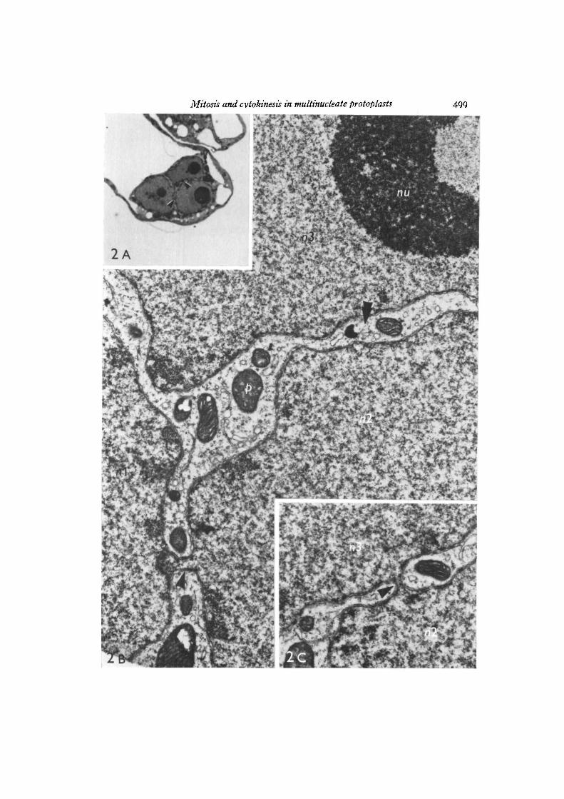
Eight multinucleate protoplasts containing interphase nuclei (7 binucleates, 1trinucleate) were examined and in all cases the nuclei were linked by short narrownuclear bridges. The nuclei in the binucleates were joined by 1-3 bridges while in thetrinucleate 2 bridges connected the 3 nuclei in series (Figs. ZA-C). All 13 bridgesobserved in this study were approximately 0-2 /im in diameter. The nuclei of one of

Mitosis and cytokinesis in multinucleate protoplasts 493
the binucleates and the trinucleate may have been in early prophase rather than ininterphase since chromosome condensation had occurred and microtubules wereobserved in the cytoplasm surrounding the nuclei.
Prophase
Almost all multinucleate protoplasts entered mitosis synchronously and main-tained synchrony throughout the division cycle. Only binucleates were observed atprophase. Prophase nuclei were almost entirely surrounded by a distinct clear zonecontaining microtubules, endoplasmic reticulum and a few larger organelles (Figs.3 A, B). Chromosome condensation was accompanied by disappearance of the nucleoli,breakdown of the nuclear envelope and movement of microtubules into the nuclei(Fig- 3B).
Metaphase
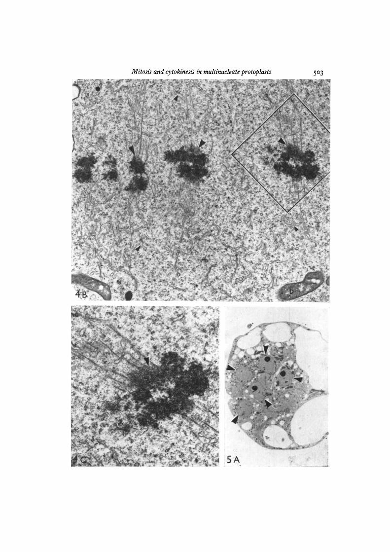
In multinucleate protoplasts at metaphase the individual spindles did not appearto be in contact with one another. Serial sections of the protoplast in Fig. 4 A indicatedthe presence of 6 such spindles. Each spindle contained paired chromosomes alignedon the equatorial plates, microtubules and dilated endoplasmic reticulum profiles(Fig. 4B). The chromosomal microtubules were embedded in rather diffuse granularkinetochores (Fig. 4 c).
Anaphase
Only one multinucleate protoplast at anaphase was found. At least three planes ofdivision were present but the limited number of micrographs obtained were difficultto interpret.
Telophase
Four different multinucleate protoplasts at telophase were obtained and in all casesdaughter nuclei were completely formed. At least 4 nuclei were present in each andserial sections indicated that the protoplast in Fig. 5 A contained 10 nuclei. Cell plateswhich appeared to form by phragmoplasts were present between most of the nucleiin the 4 telophase multinucleates examined (Figs. 5 A, B). The cell plates containedfine fibrils and were irregularly thickened (Fig. 5 c); the phragmoplasts at the edgesof the cell plates consisted of vesicles, microtubules and some electron-dense granularmaterial (Fig. 5D). Numerous coated vesicles were observed in the cytoplasm nearthe forming cell plate and were often continuous with vesicles of the cell plate (Fig. 5E).
Other observations
A few multinucleate protoplasts at metaphase and anaphase appeared to containat least twice the normal number of chromosomes (Fig. 6). These protoplasts seemedto undergo quite normal mitosis.
31-2

494 £• C. Fowke, C. W. Bech-Hansen, O. L. Gamborg and F. Constabel
Very little cell wall material was detected at the surface of the dividing multi-nucleate protoplasts. Fig. 7 illustrates the cell wall present on a multinucleate proto-plast at metaphase after 65 h in culture. Coated vesicles were frequently observedadjacent to or continuous with the plasmalemma.
The sequence of events leading from cells in culture through formation and subse-quent division of multinucleate protoplasts is summarized in diagrammatic form inFig. 1.
Fig. 1. Diagrammatic representation of multinucleate protoplast formation anddivision in soybean. A, cultured cells; B, spontaneous fusion during enzyme treatment;c, multinucleate protoplast with interphase nuclei linked by bridges; D, prophase;E, metaphase; F, anaphase; G, telophase; Di, protoplast containing fused nuclei.
Mitosis and cytokinesis in multinucleate protoplasts 495
DISCUSSION
The flat-embedding technique used in this study proved essential for subsequentselection of dividing multinucleate protoplasts with the light microscope. Embeddingin glass Petri dishes yielded thin blocks of Araldite with both surfaces glassy smooth;such blocks showed a minimum of distortion when examined in the light microscopeand facilitated selection of protoplasts for electron microscopy. Even when using thistechnique, dividing multinucleates were difficult to identify, particularly metaphasesand anaphases. In addition the overall frequency of dividing multinucleate protoplastswas very low. As a result of these problems the current study does not describe mitosisin multinucleate protoplasts in the same detail as was reported for uninucleate proto-plasts of soybean (Fowke et al. 1974a).
There appears to be some variability in the mode of origin of multinucleate proto-plasts with different cell types. Nuclear division without cytokinesis has been reportedby Eriksson & Jonasson (1969), Van der Valk& Wessels (1973) and Reinert & Hellmann(1973). This study and previous work (Miller et al. 1971) indicates that multinucleateprotoplasts of soybean arise by spontaneous fusion during the enzyme treatment.Similarly, multinucleate protoplasts of Ammi (Fowke et al. 1973, 19746), carrot(Grambow et al. 1972), tobacco (Withers & Cocking, 1972) and maize (Motoyoshi,1971) are formed during protoplast isolation. The process of spontaneous fusion intobacco has been examined at the ultrastructural level (Withers & Cocking, 1972).Our results demonstrate that multinucleates resulting from spontaneous fusion canundergo mitosis which resembles that occurring in cultured cells and uninucleateprotoplasts of soybean (Fowke et al. 1974a).
The significance of the nuclear bridges connecting interphase nuclei is not known.It is possible that these bridges may play a role in synchronizing the nuclei prior tomitosis. The vast majority of multinucleates examined divided synchronously; syn-chronous divisions seem to be common in multinucleate protoplasts (Miller et al.1971; Motoyoshi, 1971; Van der Valk & Wessels, 1973). In fact synchronous divisionsare typical of most naturally occurring and artificially created multinucleate cells(Johnson & Rao, 1971). The basis of this synchrony is not really understood. Evidencefrom a number of systems suggests that proteins in the cytoplasm may inducesynchrony.
It is also possible that the bridges may precede and facilitate nuclear fusion. Nuclearbridges have been reported between fusing nuclei during fertilization in plants(Jensen, 1964; Van Went, 1970).
Prophase and metaphase in the multinucleates seemed identical to prophase andmetaphase in the uninucleate protoplasts of soybean (cf. Fowke et al. 1974a). Acomparison was not possible for anaphase since we were able to detect this stage onlyrarely in the multinucleates. This may be due to the difficulty in recognizing anaphasesin the Araldite block. In addition, anaphase is usually a rapid phase in mitosis (Mazia,1961) and this would further reduce the chances of detecting multinucleates at thisstage.
In a number of plant protoplasts nuclear division is apparently not followed by

496 L. C. Fowke, C. W. Bech-Hansen, O. L. Gamborg and F. Constabel
cytokinesis (see above). Our results demonstrate clearly that cytokinesis, involvingtypical cell plate and phragmoplast formation, occurs in multinucleate protoplasts.Cytokinesis seems to resemble the process in uninucleate soybean protoplasts (Fovvkeet al. 1974a). We have not been able to determine whether all nuclei are finally separ-ated by cell plates with the subsequent formation of a multicellular aggregate.
Miller et al. (1971) illustrated that nuclear fusion can occur prior to mitosis insoybean multinucleates. The protoplasts containing division figures with at leastdouble the number of chromosomes (Fig. 6) are thus believed to be a result of suchfusions. There was no evidence of polyploidy in the cultured cells from which themultinucleates were derived.
It seems quite evident that both uninucleate protoplasts (Fowke et al. 1974a) andmultinucleate protoplasts of soybean are capable of nuclear division and cytokinesiswith little detectable external cell wall. At telophase it is likely that wall material isbeing synthesized for 2 distinct sites, external cell walls and cell plates. Coatedvesicles are thought to be involved in cell wall deposition (Bonnett & Newcomb, 1966)and the observed association of numerous coated vesicles with both the cell plate andplasmalemma is consistent with this idea.
This work was supported by the National Research Council of Canada, Grant A6304. Wewish to thank Mr J. P. Shyluk for excellent technical assistance.
REFERENCES
BAWA, S. B. & TORREY, J. G. (1971). 'Budding' and nuclear division in cultured protoplastsof corn, Convolvulus and onion. Bot. Gaz. 132, 240-245.
BONNETT, H. T. & NEWCOMB, E. H. (1966). Coated vesicles and other cytoplasmic componentsof growing root hairs of radish. Protoplasma 62, 50-75.
ERIKSSON, T . & JONASSON, K. (1969). Nuclear division in isolated protoplasts from cells ofhigher plants grown in vitro. Planta 89, 85-89.
FOWKE, L. C. (1975). Electron microscopy of protoplasts. In Plant Tissue Culture Methods (ed.O. L. Gamborg & L. R. Wetter). Saskatoon: National Research Council of Canada.
FOWKE, L. C , BECH-HANSEN, C. W., CONSTABEL, F. & GAMBORG, O. L. (1974a). A com-parative study on the ultrastructure of cultured cells and protoplasts of soybean during celldivision. Protoplasma 81, 189-203.
FOWKE, L. C , BECH-HANSEN, C. W. & GAMBORG, O. L. (19746). Electron microscope observa-tions of cell regeneration from cultured protoplasts of Ammi visnaga. Protoplasma 79, 235-248.
FOWKE, L. C , BECH-HANSEN, C. W., GAMBORG, O. L. & SHYLUK, J. P. (1973). Electronmicroscopic observations of cultured cells and protoplasts of Ammi visnaga. Am. J. Bot.60, 304-312.
GAMBORG, O. L., MILLER, R. A. & OJIMA, K. (1968). Nutrient requirements of suspensioncultures of soybean root cells. Expl Cell Res. 50, 151-158.
GRAMBOW, H. J., KAO, K. N., MILLER, R. A. & GAMBORG, O. L. (1972). Cell division andplant development from protoplasts of carrot cell suspension cultures. Planta 103, 348-355.
JENSEN, W. A. (1964). Observations on the fusion of nuclei in plants. J. Cell Biol. 23, 669-672.JOHNSON, R. T. & RAO, P. N. (1971). Nucleo-cytoplasmic interactions in the achievement of
nuclear synchrony in DNA synthesis and mitosis in multinucleate cells. Biol. Rev. 46, 97-155.MAZIA, D. (1961). Mitosis and the physiology of cell division. In The Cell, vol. 3 (ed. J. Brachet
& A. E. Mirsky), pp. 77—412. New York and London: Academic Press.MILLER, R. A., GAMBORG, O. L., KELLER, W. A. & KAO, K. N. (1971). Fusion and division of
nuclei in multinucleated soybean protoplasts. Can.J. Genet. Cytol. 13, 347-353.

Mitosis and cytokinesis in multinucleate protoplasts 497
MOTOYOSHI, F. (1971). Protoplasts isolated from callus cells of maize endosperm. Expl CellRes. 68, 452-456.
REINERT, J. & HELLMANN, S. (1973). Aspects of nuclear division and cell wall formation inprotoplasts of different origin. Colloq. int. Cent. nat. Rech. sci. 212, 273-279.
VAN DER VALK, P. & WESSELS, J. G. H. (1973). Mitotic synchrony in multinucleate Schizo-phyllum protoplasts. Protoplasma 78, 427-432.
VAN WENT, J. L. (1970). The ultrastructure of the fertilized embryo sac of petunia. Ada bot.ncerl. 19, 468-480.
WITHERS, L. A. & COCKING, E. C. (1972). Fine-structural studies on spontaneous and inducedfusion of higher plant protoplasts. J. Cell Sci. n , 59-75.
{Received 14 January 1975)ABBREVIATIONS ON PLATES
ch chromosome n nucleuscz clear zone mi nucleolusm mitochondrion p plastid

498 L. C. Fowke, C. W. Bech-Hatisen, O. L. Gamborg and F. Constabel
The figures in a series (e.g. 2 A, 2B) represent micrographs of the same protoplast.
Fig. 2 A. Light micrograph showing a trinucleate protoplast. Adjacent thin sections(Figs. 2B, c) illustrate bridges connecting the nuclei in the regions indicated by thearrowheads, x 1200.Fig. 2B. Electron micrograph illustrating same nuclei as in Fig. 1. Nucleus 1 (ni) andnucleus 2 (nz) are connected by a nuclear bridge (single arrow). The second bridgelinking nucleus 2 (nz) with nucleus 3 {TIT,) in the region of the double arrow is shownin Fig. 2C. x 17700.Fig. 2C. Serial section in the region indicated by the double arrowhead in Fig. 2Bshowing nuclear bridge (arrow) connecting nz with ir$. x 18000.
Mitosis and cytokinesis in multinucleate protoplasts

500 L. C. Fowke, C. W. Bech-Hansen, O. L. Gamborg and F. Constabel
Fig. 3 A. Light micrograph of a binucleate protoplast at prophase showing parts ofboth nuclei (arrowheads). A clear zone is present around the nuclei and some chromo-some condensation is evident. An electron micrograph of the nucleus on the right isshown in Fig. 3B. x 1000.Fig. 3B. Electron micrograph of the prophase nucleus indicated by the doublearrowhead in Fig. 3 A. The nucleus is almost entirely surrounded by a clear zone(cz) containing endoplasmic reticulum (large arrowheads) and many microtubules.Chromosomes (ch) and a few microtubules (small arrowheads) are evident within thenucleus, x 13600.Fig. 4A. Light micrograph of a multinucleate protoplast at metaphase showing 4separate metaphase plates (arrowheads). Note that the individual spindles do notappear to be in contact with one another. Serial sections demonstrated 2 additionalmetaphase plates in this protoplast, x 1000.

Mitosis and cytokinesis in multinucleate protoplasts Sax
? v V £ / * * • ; • ••

502 L. C. Fowke, C. W. Bech-Hansen, 0. L. Gamborg and F. Constabel
Fig. 4B. Electron micrograph of one of the spindles shown in Fig. 4A. Note thekinetochores (large arrowheads) with associated microtubules and the endoplasmicreticulum (small arrowheads) within the spindle. An enlargement of the area outlinedin black is shown below in Fig. 4 c. x 15000.
Fig. 4 c. Enlargement from Fig. 4B showing microtubules embedded in the diffusekinetochores (arrowhead), x 28600.
Fig. s A. Light micrograph of a multinucleate protoplast at telophase. Five nuclei(arrowheads) from a total of 10 are evident in this section, x 1000.
Mitosis and cytokinesis in multinucleate protoplasts

504 L. C. Fowke, C. W. Bech-Hansen, O. L. Gamborg and F. Constabel
Fig. SB. Electron micrograph of the protoplast in Fig. 5 A showing 7 nuclei (n). Cellplates (large arrowheads) are present amongst the nuclei. Note the phragmoplasts (smallarrowheads) at the margins of the cell plates. Enlargements of the areas outlined inblack are shown in Figs. 5 C, D. x 3600.Fig. 5 c. Enlargement from Fig. 5B showing the irregularly thickened cell plate(arrowheads), x 17000.

C> .
Mitosis and cytokinesis in multinucleate protoplasts 505

506 L. C. Fotcke, C. W. Bech-Hansen, O. L. Gamborg and F. Constabel
Fig. 5D. Enlargement from Fig. 5B showing a phragmoplast containing vesicles,microtubules (arrowheads) and some electron-dense granular material, x 18400.Fig. s E. Electron micrograph showing part of a cell plate in face view (large arrow-heads) from the same protoplast as in Fig. 4. Numerous coated vesicles are evident(small arrowheads) some of which appear continuous with vesicles of the cell plate(double arrowheads), x 28700.Fig. 6. Light micrograph of a protoplast at anaphase. Nuclear fusion prior to mitosis isthought to be responsible for the unusually large number of chromosomes, x 1000.Fig. 7. Electron micrograph showing loosely organized cell wall material (arrowheads)at the margin of a multinucleate protoplast at metaphase. x 25 000.

Mitosis and cytokinesis in multinucleate protoplasts 5°7

![Maize VKS1 Regulates Mitosis and Cytokinesis During Early … · Maize VKS1 Regulates Mitosis and Cytokinesis During Early Endosperm Development[CC-BY] Yongcai Huang,a,b,1 Haihai](https://static.fdocuments.us/doc/165x107/5f7918fe486d6132ec1d9cd5/maize-vks1-regulates-mitosis-and-cytokinesis-during-early-maize-vks1-regulates-mitosis.jpg)