Electrical Response and Thermal Damage Assessment … · disease, lipedema or cellulite A...
18
Ana González-Suárez 1,2 , Joel N. Jiménez-Lozano 1* and Walfre Franco 1 1 Wellman Center for Photomedicine, Massachusetts General Hospital and Harvard Medical School, USA 2 Biomedical Synergy, Electronic Engineering Department, Universitat Politècnica de València, Spain * Currently at ZELTIQ Aesthetic, Inc., Pleasanton, California, USA {[email protected], [email protected]} October 10th, 2013 Electrical Response and Thermal Damage Assessment of Cutaneous and Subcutaneous Tissues to Noninvasive Radiofrequency Heating: A Computational Modeling Study
Transcript of Electrical Response and Thermal Damage Assessment … · disease, lipedema or cellulite A...

Ana González-Suárez1,2, Joel N. Jiménez-Lozano1* and Walfre Franco1 1Wellman Center for Photomedicine, Massachusetts General Hospital and Harvard Medical School, USA
2Biomedical Synergy, Electronic Engineering Department, Universitat Politècnica de València, Spain * Currently at ZELTIQ Aesthetic, Inc., Pleasanton, California, USA
{[email protected], [email protected]}
October 10th, 2013
Electrical Response and Thermal Damage
Assessment of Cutaneous and
Subcutaneous Tissues to Noninvasive
Radiofrequency Heating: A Computational
Modeling Study

Contents
1. Introduction
2. Mathematical modeling
3. Results
4. Conclusions

Content
1. Introduction
2. Mathematical modeling
3. Results
4. Conclusions

1 Introduction
Epidermis: outer cellular layer (~0.1 mm) Dermis: A dense connective tissue layer perfused with micro-vessels (~1 mm) Hypodermis (or subcutaneous tissue): a fine, collagenous and fibrous septa network with clusters of fat cells (1 cm to 10 cm)
Skin-fat structure Radiofrequency (RF) heating
.
.
Subcutaneous fat diseases:
Lipomatosis, Madelung’s
disease, lipedema or
cellulite
A non-invasive technique can be
used to produce selective heating
of subcutaneous tissue
Clinical applications

1 Introduction
Objectives
Model a real structure of subcutaneous tissue
Assess the electrical and thermal effect of fibrous septa within
subcutaneous tissue during RF hyperthermic heating (< 55ºC)
Quantify and compare the thermal damage occurred in two
subcutaneous tissue structures (one composed by fat only and
another by fat and fibrous septa)
. COMSOL Multiphysics 4.3b

Content
1. Introduction
2. Mathematical modeling
3. Results
4. Conclusions

Mathematical modeling
Domain Geometry
Fibrous septa
RF monopolar applicator (Franco, W. et al. LSM 42, 2010)
Upper view
MRI skin of a female (Mirrashed,
F., et al., Skin Research and Technology,
10, 2004).
2

1
Arrhenius Equation: Domain ODEs and DAEs
Thermal problem: Heat Transfer
2
Mathematical modeling
Governing Equations
Coupled electric-thermal problem
Thermal damage problem
3
dteAt
t
RT
E
0
)( A and ΔE for skin (Weaver and Stoll 1969)
Ω = 1 lesion contour (transepidermal
necrosis, 63% reduction in cell viability)
Electric problem: Electric Currents
QTTcQTkt
Tc bbm
)(
0 V
2EQ
2
Mathematical modeling
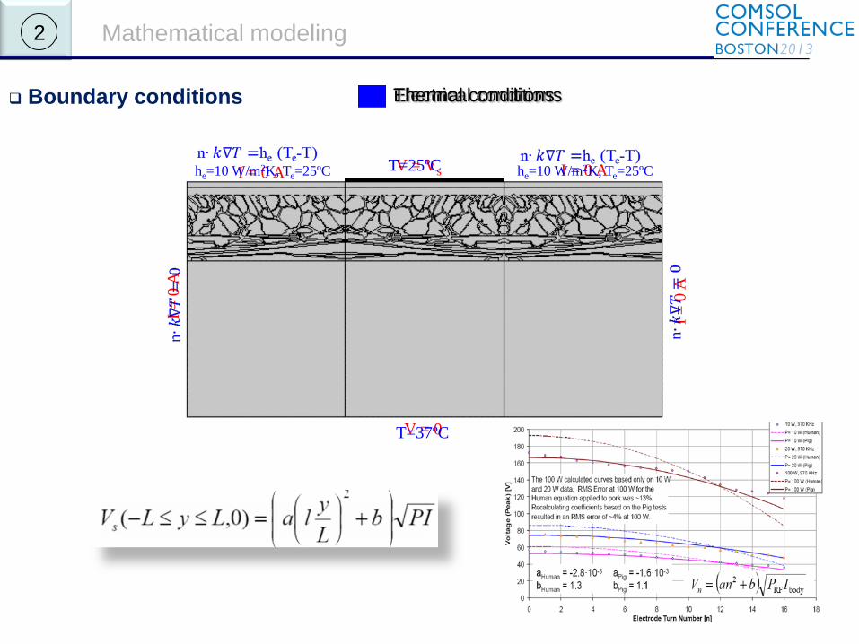
Electrical conditions
I =
0 A
I =
0 A
V = 0
I = 0 A I = 0 A V = Vs
Thermal conditions
T=37ºC
T=25ºC he=10 W/m2K, Te=25ºC
he=10 W/m2K, Te=25ºC
Boundary conditions
2

Mathematical modeling
Thermal and electrical characteristics of the model elements
Element εr σ
(S/m) k (W/m·K) ρ (kg/m3)
c
(J/kg·K)
ω
(kg/m3·s)
Skin 1832.8 0.22 0.53 1200 3800 2
Fat 27.22 0.025 0.16 850 2300 0.6
Muscle 1836.4 0.5 0.53 1270 3800 0.5
Septa 1832.8 0.22 0.53 1200* 3800 0
Main Physical assumptions
Homogeneous tissues
Tissues have isotropic electric and thermal properties
Constant k, c and ω variations are not significant within the 35-50°C range
Properties of the fibrous septa similar to those of the dermis
The perfusion term in the septa is neglected (i.e. fibrous septa as solid)
2

Content
1. Introduction
2. Mathematical modeling
3. Results
4. Conclusions

Results
Electric field
. Subcutaneous tissue with fat only
(no fibrous septa) .
Subcutaneous tissue with fat and
fibrous septa
3

Results
. Subcutaneous tissue with fat only
(no fibrous septa)
Total electric power absorption (Q) and Electric currents (arrows)
. Subcutaneous tissue with fat and
fibrous septa
3

Results
Temperature distribution and thermal damage
. Subcutaneous tissue with fat only
(no fibrous septa) .
Subcutaneous tissue with fat and
fibrous septa
3

Results
Subcutaneous
tissue with fat only
Thermal damage quantification
Subcutaneous tissue with
fat and fibrous septa
The lesion volume is ~ 7 times higher considering fibrous septa
3

Content
1. Introduction
2. Mathematical modeling
3. Results
4. Conclusions

Conclusions
Our results demonstrate the importance of including the fibrous septa when
modeling RF heating of subcutaneous tissue:
The intensity and extent of the electric field in the subcutaneous tissue is increased
considering fibrous septa network
Fibrous septa favors the flux of electric current increasing the intensity of the electric
field, which in turn increases power absorption within subcutaneous tissue
Neglecting the electric and thermal energy contributions of the fibrous septa results in
underestimating thermal damage
4
Our findings would be useful to design and develop novel devices and treatments
to subcutaneous fat diseases during RF hyperthermic heating
. Knowledge of correct dosimetry

Electrical Response and Thermal Damage
Assessment of Cutaneous and
Subcutaneous Tissues to Noninvasive
Radiofrequency Heating: A Computational
Modeling Study
. Acknowledgments
R. Rox Anderson and Walfre Franco for the opportunity to train and conduct my
research at the Wellman Center for Photomedicine
Conselleria d’Educació of the Generalitat Valenciana: Predoctoral visiting
fellowship BEFPI-2013 and grant ValI+D (ACIF/2011/194)