Ekstrakorporal Afinite Tedavisi - Hacettepe · kstrakorporal tedaviler bilirubin, patojenik...
18
Extracorporeal Affinity Therapy Ekstrakorporal Afinite Tedavisi Review Article / Derleme A. Denizli / Hacettepe J. Biol. & Chem., 2011, 39 (2), 93–110 Adil Denizli Hacettepe University, Department of Chemistry, Biochemistry Division, Beytepe, Ankara, Turkey ÖZET E kstrakorporal tedaviler bilirubin, patojenik antibadiler, dolaşımdaki çeşitli immün kompleksler, kolesterol, toksik metal iyonları gibi olası toksik maddelerin insan plazmasından uzaklaştırılmasına yöneliktir. Plazma değişimi, hemodiyaliz, hemofiltrasyon ve hemoperfüzyonu içeren geleneksel ekstrakorporal tedaviler seçici ol- mayan yöntemlerdir. Ayrıca, albümin gibi plazma desteklerine olan ihtiyaç oldukça fazladır. Dahası, plazma ve plazma destekleri kullanılırken hepatit veya immün reaksiyonların ortaya çıkma tehlikesi bulunmaktadır. Günü- müzde ekstrakorporal tedavinin en umut verici uygulaması spesifik afinite adsorpsiyonudur. Afinite taşıyıcılar, kanın bu taşıyıcılarla doldurulmuş kolondan doğrudan geçirildiği hemoperfüzyon sisteminde kullanılabilir. Bu tür uygulama etkili, basit ve ucuzdur. Bu derlemede, ekstrakorporal tedavinin insan plazmasından toksik mad- delerin uzaklaştırılması için kullanıldığı bazı ilginç uygulamalar kısaca verilmiştir. Anahtar Kelimeler Afinite tedavisi, afinite taşıyıcılar, hemoperfüzyon, ekstrakorporal tedavi, bilirubin uzaklaştırma. ABSTRACT E xtracorporeal therapies are directed at the removal of potential toxic substances including bilirubin, pathogenic antibodies, various circulating immune-complexes, cholesterol, toxic metal ions, etc. from human plasma. The conventional extracorporeal therapies including plasma exchange, hemodialysis, hemofiltration and hemoperfusion are non-selective techniques. In addition the requirement for plasma substitutes such as albumin is very high. Moreover, the dangers of hepatitis or immune reactions accompany these therapies while using plasma for plasma products. Today, one of the most promising procedures for extracorporeal therapy is specific affinity adsorption. Affinity carriers may be used in hemoperfusion system, where blood is directly perfused through the column filled with these carriers. This type of application is effective, simple and inexpensive. In this article, some interesting applications on extracorporeal affinity therapies for removal of toxic substances from human plasma are briefly reviewed. Key Words Affinity therapy, affinity carriers, hemoperfusion, extracorporeal treatment, bilirubin removal. Article History: Received December 13, 2010; Revised January 15, 2011; Accepted February 25, 2011; Avaliable Online: April 1, 2011. Correspondence to: Adil Denizli, Hacettepe University, Department of Chemistry, Biochemistry Division, Beytepe, Ankara, Turkey Tel: +90312 297 7983 Fax: +90312 299 2163 E-Mail: [email protected]
Transcript of Ekstrakorporal Afinite Tedavisi - Hacettepe · kstrakorporal tedaviler bilirubin, patojenik...
Extracorporeal Affinity Therapy
Ekstrakorporal Afinite Tedavisi
Review Article / Derleme
A. Denizli / Hacettepe J. Biol. & Chem., 2011, 39 (2), 93–110
Adil Denizli Hacettepe University, Department of Chemistry, Biochemistry Division, Beytepe, Ankara, Turkey
ÖZ E T
Ekstrakorporal tedaviler bilirubin, patojenik antibadiler, dolaşımdaki çeşitli immün kompleksler, kolesterol, toksik metal iyonları gibi olası toksik maddelerin insan plazmasından uzaklaştırılmasına yöneliktir. Plazma
değişimi, hemodiyaliz, hemofiltrasyon ve hemoperfüzyonu içeren geleneksel ekstrakorporal tedaviler seçici ol-mayan yöntemlerdir. Ayrıca, albümin gibi plazma desteklerine olan ihtiyaç oldukça fazladır. Dahası, plazma ve plazma destekleri kullanılırken hepatit veya immün reaksiyonların ortaya çıkma tehlikesi bulunmaktadır. Günü-müzde ekstrakorporal tedavinin en umut verici uygulaması spesifik afinite adsorpsiyonudur. Afinite taşıyıcılar, kanın bu taşıyıcılarla doldurulmuş kolondan doğrudan geçirildiği hemoperfüzyon sisteminde kullanılabilir. Bu tür uygulama etkili, basit ve ucuzdur. Bu derlemede, ekstrakorporal tedavinin insan plazmasından toksik mad-delerin uzaklaştırılması için kullanıldığı bazı ilginç uygulamalar kısaca verilmiştir.
Anahtar KelimelerAfinite tedavisi, afinite taşıyıcılar, hemoperfüzyon, ekstrakorporal tedavi, bilirubin uzaklaştırma.
A B S T R AC T
Extracorporeal therapies are directed at the removal of potential toxic substances including bilirubin, pathogenic antibodies, various circulating immune-complexes, cholesterol, toxic metal ions, etc. from human
plasma. The conventional extracorporeal therapies including plasma exchange, hemodialysis, hemofiltration and hemoperfusion are non-selective techniques. In addition the requirement for plasma substitutes such as albumin is very high. Moreover, the dangers of hepatitis or immune reactions accompany these therapies while using plasma for plasma products. Today, one of the most promising procedures for extracorporeal therapy is specific affinity adsorption. Affinity carriers may be used in hemoperfusion system, where blood is directly perfused through the column filled with these carriers. This type of application is effective, simple and inexpensive. In this article, some interesting applications on extracorporeal affinity therapies for removal of toxic substances from human plasma are briefly reviewed.
Key Words
Affinity therapy, affinity carriers, hemoperfusion, extracorporeal treatment, bilirubin removal.
Article History: Received December 13, 2010; Revised January 15, 2011; Accepted February 25, 2011; Avaliable Online: April 1, 2011.
Correspondence to: Adil Denizli, Hacettepe University, Department of Chemistry, Biochemistry Division, Beytepe, Ankara, Turkey
Tel: +90312 297 7983 Fax: +90312 299 2163 E-Mail: [email protected]

A. Denizli / Hacettepe J. Biol. & Chem., 2011, 39 (2), 93–11094
INTRODUCTION
Conventional hemoperfusion is an extracor-poreal treatment method in which the blood
from the patient is circulated through adsorption column in order to remove exogeneous and endogeneous toxic substances from the blood (Figure 1) [1]. The blood removed from patient must be anticoagulated and generally separated into cells and plasma. The treated plasma is then combined with the previously separated cells and returned to the patient. Hemoperfusion is frequently utilized for acute blood purification as in the case of drug overdose, in which case the carrier is charcoal covered with a biocompatible coating layer [2]. The charcoal adsorbs medium to high molecular weight blood components due to the non-selectivity of conventional hemoperfusion. It would be most desirable to selectively remove any undesired substances from human blood. For this purpose, affinity adsorption was suggested as an alternative to conventional hemoperfusion for removing undesired substances from the
plasma of patient. Plasma exchange is the most commonly used treatments for extracorporeal therapy. Table 1 summarizes the advantages of extracorporeal therapy with affinity carriers compared with plasma exchange. Table 2 gives several diseases potentially suitable for treatment by extracorporeal affinity therapy.
Affinity therapy in hyperbilirubinemiaBilirubin is a negatively charged pigment formed in the normal metabolism of heme proteins in senescent red blood cells. Figure 2 shows the chemical structure of bilirubin. There are two kinds of serum bilirubin. One is mono- or di-glucuronide conjugated at the carboxylic acid groups, and the other is the non-conjugated type, free bilirubin [4]. The non-conjugated bilirubin is not water soluble and predominantly bound to albumin. It is transported to the liver as a complex with albumin and excreted into the bile [5]. Indirect bilirubin corresponds to non-conjugated bilirubin, and direct bilirubin corresponds to conjugated bilirubin. The free bilirubin is highly toxic to many cell types, intracellular organelles and physiological processes [6]. It is believed that the toxicity of bilirubin results from its hydrophobicity, which may lead to its aggregate in phospholipid membranes and subsequently damage the integrity of the membrane and inhibit the chemical performance of membrane bound enzymes. High concentration of free bilirubin can evoke hepatic or biliary tract dysfunction and permanent brain damage or death in more severe case [5]. Neurological dysfunctions as kernicterus or bilirubin encephalophathy may develop if the
Table 1. Benefits of extracorporeal affinity therapy in comparison to plasma exchange [3].
��������������� ����������������������������� ������for substitution materials.
�������������� ������ ��������������������������averted.
������ ��������� �����
��������������� ������ ������!� ��� ��� ������������components, including clotting factors.
������������ ������ ����������� ������������combination with the affinity therapy is possible.
Table 2. Diseases potentially suitable for treatment by extracorporeal affinity therapy.
�����"����#���#����
�����$������������� ����� ����
������� ����������������������������������erythematosus, rheumatoid arthritis
�����"�� �������� ��������#����# ���� ��� � �factors VIII or IX
�������� �# �� ����
��������������� � �������������%�������� ��!���multiple sclerosis)
Figure 1.��������� ��������� �� ������&��� �� �����affinity therapy.

A. Denizli / Hacettepe J. Biol. & Chem., 2011, 39 (2), 93–110 95
bilirubin concentration in the plasma rises above ���������� ����� ���������������� ���������������may cause a yellow discoloration of the skin and other tissues.
There have been many bilirubin removal methods reported in literature. Table 3 shows various kinds of methods that have been applied clinically for the treatment of hyperbilirubinemia. Phototherapy is one of the most commonly used treatments for mild cases. However, the effectiveness of phototherapy is limited by the fact that the light could only penetrate a few millimeters of skin and not reach a large proportion of the total bilirubin pool. It is also found that phototherapy may induce DNA damage [7]. Treatment with plasma exchange, however, requires large volumes of fresh frozen plasma, which is expensive and difficult to obtain. In exchange transfusion, infant’s blood is replaced with bilirubin-free adult blood. This procedure have been under concerns with the fact its relatedness to hypoglycemia, hypocalcemia, acidosis, and more importantly the transmission of infectious diseases like hepatitis or acquired immune deficiency syndrome [8]. Hemodialysis relays upon the diffusive transport, therefore, is a rather slow process. This method is not specific also. It should be noted that these systems are still complex and expensive. Hemoperfusion, i.e., circulation of blood through extracorporeal column containing affinity carriers for bilirubin removal, has become the most promising technique [9-18].
�������� ��� ��� ���� ���� ���� ������������ ���hemoperfusion to the removal of the bilirubin from jaundiced newborn babies by using albumin deposited macroreticular carrier [9]. The first clinical trial of hemoperfusion with anion exchange synthetic fibers for bilirubin removal was reported by Idezuki et al in 1981 [10]. Brown prepared oligo-peptide conjugated polyacrylamide particles as affinity carrier for bilirubin removal [11]. Polyacrylamide resin with oligopeptide sequences bound to the functional sites form effective carrier for bilirubin. It was shown that the main driving force for the binding of bilirubin at the active sites is an electrostatic interaction between the carboxylate group of bilirubin and the positively charge of the oligopeptide pendants on the carrier. Chandy et al. used polylysine attached chitosan particles for selective bilirubin removal ���������������!�� �������!�! ���������������������covalently onto chitosan particles, using nitrogen glow discharge plasma. It seemed that the surface modified chitosan particles could possibly provide an improved supportive therapy for hepatic failure especially for patients with hepatic coma and hyperbilirubinemia. Yamazaki et al. developed poly(styrene-divinyl benzene) based carrier and succesfully applied in the treatment of more than 200 patients with hyperbilirubinemia [13]. Morimoto et al. used plasma exchange and plasma adsorption with styrene-divinyl benzene carrier and removed bilirubin from hepatectomized patients [14]. In the clinical evaluation of patients receiving plasma adsorption with these columns containing styrene-divinyl benzene particles, there was a remarkable improvement of total bilirubin depletion in one case. This plasma adsorption system provided a possibility for an improved supportive therapy for hepatic failure, especially for patients with hepatic coma and
Figure 2.� ����������� �������� ��� ���������� "#33
H36
N4O
6,
molar mass 584.66 g/mol).
Table 3. Clinically applied bilirubin removal methods [4].
Phototherapy
Plasma exchange (plasmapheresis)
Exchange transfusion
Hemodialysis
Hemodialysis using highly permeable membrane
Hemofiltration
Hemoperfusion using activated charcoal
Hemoperfusion using anion exchange resin
A. Denizli / Hacettepe J. Biol. & Chem., 2011, 39 (2), 93–11096
hyperbilirubinemia. Avramescu et al. conjugated bovine serum albumin with ethylene vinyl alcohol ������� �� ���������� ���� �� ����� ��� ���high bilirubin binding capacity [15]. They also ��������������� ��� ������������� ������ �����preparation of mixed matrix adsorber membranes by incorporation of ion exchange particles. The mixed adsorber systems combine the advantages of membrane technology (easy scale up, low pressure drop and high throughputs) with those of column chromatography (high selectivity and high binding capacity). Yu et al. prepared amine-containing cross-linked chitosan carrier and investigated adsorption behavior of conjugated bilirubin [16]. They reported that electrostatic and hydrophobic interactions are the main driving forces for the binding. Kuroda et al. studied selective adsorption of bilirubin by macroporous poly(glycidyl methacrylate-divinyl-benzene) particles [17]. When the surface of the particles was previously coated with albumin, discriminating binding of the bilirubin in the serum was attained and the adsorption of albumin was reduced. Ahmad et al. demonstrated the suitability of rat serum albumin loaded poly(lactide-glycolide) biodegradable microspheres in removal of bilirubin from systemic circulation of hyperbilirubinemic rats [18]. On evaluating the potential of microspheres in depletion of bilirubin from the systemic circulation, rat serum albumin carrying microspheres were found to be competent in removing bilirubin from the serum.
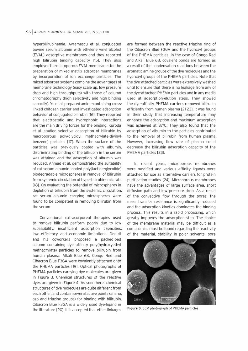
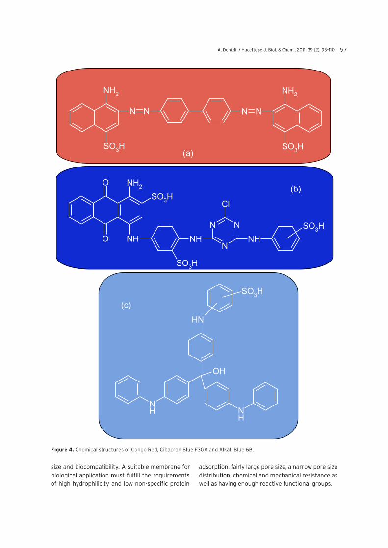
Conventional extracorporeal therapies used to remove bilirubin perform poorly due to low accessibility, insufficient adsorption capacities, low efficiency and economic limitations. Denizli and his coworkers proposed a packed-bed column containing dye affinity poly(hydroxyethyl methacrylate) particles to remove bilirubin from human plasma. Alkali Blue 6B, Congo Red and Cibacron Blue F3GA were covalently attached onto the PHEMA particles [19]. Optical photographs of PHEMA particles carrying dye molecules are given in Figure 3. Chemical structures of the reactive dyes are given in Figure 4. As seen here, chemical structures of dye molecules are quite different from each other, and contain several active points (amino, azo and triazine groups) for binding with bilirubin. Cibacron Blue F3GA is a widely used dye-ligand in the literature [20]. It is accepted that ether linkages
are formed between the reactive triazine ring of the Cibacron Blue F3GA and the hydroxyl groups of the PHEMA particles. In the case of Congo Red and Alkali Blue 6B, covalent bonds are formed as a result of the condensation reactions between the aromatic amine groups of the dye molecules and the hydroxyl groups of the PHEMA particles. Note that the dye-attached particles were extensively washed until to ensure that there is no leakage from any of the dye-attached PHEMA particles and in any media used at adsorption-elution steps. They showed the dye-affinity PHEMA carriers removed bilirubin efficiently from human plasma [21-23]. It was found in their study that increasing temperature may enhance the adsorption and maximum adsorption was achieved at 37oC. They also found that the adsorption of albumin to the particles contributed to the removal of bilirubin from human plasma. However, increasing flow rate of plasma could decrease the bilirubin adsorption capacity of the PHEMA particles [23].
In recent years, microporous membranes were modified and various affinity ligands were attached for use as alternative carriers for protein purification studies [24]. Microporous membranes have the advantages of large surface area, short diffusion path and low pressure drop. As a result of the convective flow through the pores, the mass transfer resistance is significantly reduced and the adsorption kinetics dominates the binding process. This results in a rapid processing, which greatly improves the adsorption step. The choice of the membrane material may be difficult as a compromise must be found regarding the reactivity of the material, stability in polar solvents, pore
Figure 3.����������� ������� ����� ������!
A. Denizli / Hacettepe J. Biol. & Chem., 2011, 39 (2), 93–110 97
size and biocompatibility. A suitable membrane for biological application must fulfill the requirements of high hydrophilicity and low non-specific protein
adsorption, fairly large pore size, a narrow pore size distribution, chemical and mechanical resistance as well as having enough reactive functional groups.
Figure 4. Chemical structures of Congo Red, Cibacron Blue F3GA and Alkali Blue 6B.

A. Denizli / Hacettepe J. Biol. & Chem., 2011, 39 (2), 93–11098
Şenel et al used polyamide hollow-fiber membrane carrying dye molecules for bilirubin molecules [25]. Polyamide hollow-fiber membrane may meet most of these requirements, since they have a narrow pore size distribution and good mechanical rigidity. The pore radii of the polyamide hollow fiber membranes changed between 200 nm and 450 nm. This indicated that the hollow fiber membranes contained mainly macropores. The ��������� ��� ������ �� �� ������ �� � �������given in Figure 5 show the surface structure and the cross-section of the polyamide hollow-fiber membranes. As seen in these photographs, the smallest pore structure of the hollow fiber was highly asymmetric. Furthermore, the smallest pores occurred at the lumen side of the hollow fiber while the pore size at the shell side was much larger.
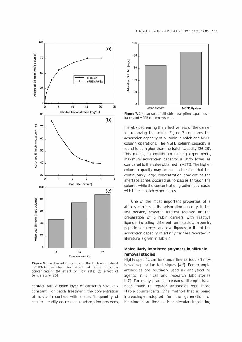
Uzun and Denizli used magnetically stabilized �������� ��� ������� � ���� � ��������� ����������� ������� ������ �������� ��������poly(hydroxyethyl methacrylate) particles for selective bilirubin removal from human plasma for ��� ������ ���� ��� �������� � !"#� $�%� ���� �����as a hemoperfusion column for in-vitro bilirubin �� &�#� ���� '��� ���� ��� ��� �������%� ������ � ���������� �������� �� ��������� � ���#� ���� ���the natural carrier for bilirubin in the blood. It is ����%�������������������� �����%���&�as many as 12 binding sites for bilirubin, but only two of the sites bind bilirubin molecules strongly. The association constant is 9.5 x 10⁷ M [27]. They reported that the non-specific bilirubin adsorption on the mPHEMA particles was 0.47 mg/g. Higher bilirubin adsorption amounts, up to 88.3 mg/g, '�� �������'����������(��� �������)�����particles (Figure 6A). Bilirubin adsorption capacity decreased significantly from 75.0 mg/g to 40.0 mg/g polymer with the increase of the flow-velocity from 0.5 ml/min to 4.0 ml/min (Figure 6B). Bilirubin adsorption increased with increasing temperature (Figure 6C). Adsorption behavior of bilirubin could ��� ������������*���������� ����#
Chromatographic columns appear to have a distinct advantage over batch systems because the adsorption rate depends on the concentration of solute in solution being treated. For column system the carriers are continuously in contact with a fresh solution. As a result the concentration of solute in
Figure 5.�+��������&������� � ������� ��� %�����hollow-fibre membranes; (a) Inner surface; (b) Outer surface; (c) Cross-section [25].
A. Denizli / Hacettepe J. Biol. & Chem., 2011, 39 (2), 93–110 99
contact with a given layer of carrier is relatively constant. For batch treatment, the concentration of solute in contact with a specific quantity of carrier steadily decreases as adsorption proceeds,
thereby decreasing the effectiveness of the carrier for removing the solute. Figure 7 compares the ��������������� ������������������������������� ���������� ���� ����� ������ ������ � ��found to be higher than the batch capacity [26,28]. This means, in equilibrium binding experiments, maximum adsorption capacity is 35% lower as ����������������������������������������������column capacity may be due to the fact that the continuously large concentration gradient at the interface zones occured as to passes through the column, while the concentration gradient decreases with time in batch experiments.
One of the most important properties of a affinity carriers is the adsorption capacity. In the last decade, research interest focused on the preparation of bilirubin carriers with reactive ligands including different aminoacids, albumin, peptide sequences and dye ligands. A list of the adsorption capacity of affinity carriers reported in literature is given in Table 4.
Molecularly imprinted polymers in bilirubin removal studiesHighly specific carriers underline various affinity-based separation techniques [46]. For example antibodies are routinely used as analytical re-agents in clinical and research laboratories [47]. For many practical reasons attempts have been made to replace antibodies with more stable counterparts. One method that is being increasingly adopted for the generation of biomimetic antibodies is molecular imprinting
Figure 6.������� ��������� ���� ���� ���� ���������mPHEMA particles; (a) effect of initial bilirubin concentration; (b) effect of flow rate; (c) effect of temperature [26].
Figure 7. Comparison of bilirubin adsorption capacities in ��������������������� ������

A. Denizli / Hacettepe J. Biol. & Chem., 2011, 39 (2), 93–110100
Table 4. Adsorption capacities for bilirubin of various carriers.
Material ������ Adsorption capacity (mg/g) [R]
Polyacrylamide particles ������� ����������� 0.2-75 [8]
Macroreticular resin Albumin 2-24 [9]
Chitosan particles ���������� 1.5 [12]
Poly(ethylene vinyl alcohol) Bovine serum albumin 25.0 [15]
Poly(GMA-DVB) copolymer Albumin 30 [17]
PHEMA particles Dye molecules 6.8-32.5 [19-21]
Polyamide hollow fiber Cibacron Blue F3GA 48.9 [25]
������ ������������� Human serum albumin 88.3 [26]
mPHEMA particles/Batch Human serum albumin 64.7 [28]
Chitosan coupled nylon membrane Cibacron Blue F3GA 63.4 [29]
��������� ��� !��"��� Quaternary ammonium salt 4.0-80 [30]
Poly(tetrafluoroethylene) membrane Cibaron Blue F3GA 76.2 [31]
Polyamide resin Aminoacid 5-80 [32]
IONEX Polypropylene fiber Tertiary amine 7.7 [33]
Polyacrylonitrile membrane Hepatoycte receptor 2.8 [34]
Poly(butadien-HEMA) gels Bovine serum albumin 3.1 [35]
Partially aminated polyacrylamide -cyclodextrin 42.2 [36]
Cellulose acetate fiber Cibaron Blue F3GA 4.0 [37]
Polyamide/chitosan membrane Polylysine 28.6 [38]
Polyamide membrane Polylysine 32.4 [39]
Poly(GMA-AAm-MBA) Polyethyleneimine 16.6 [40]
Poly(tetrafluoroethylene) membrane Human serum albumin 71.2 [41]
Poly(tetrafluoroethylene) fiber Bovine serum albumin 9.6 [42]
Aluminum oxide-silica membrane ���� 17.6 [43]
Poly(pyrrole)-alumina membrane ���� 32.4 [44]
Poly(glycidyl methacrylate) Cibaron Blue F3GA 241.5 [45]

A. Denizli / Hacettepe J. Biol. & Chem., 2011, 39 (2), 93–110 101
of polymers [48]. Given the advantage of easy preparation and chemical stability, molecularly imprinted polymers (MIPs) possess a high potential for use in a variety of applications such as chromatographic stationary phases, immunoassay-type analyses and sensor development [49-52]. Generally molecular imprinting is a synthetic strategy that is used to assemble a molecular receptor via template-guided synthesis (Figure 8). To prepare MIP, a template molecule is used to guide the assembly of functional monomers. Polymerization reaction is then employed to fix the preassembled binding groups around the template molecule. Following removal of the template molecule, the polymer revealed retains specific binding sites that can selectively rebind the original template molecule. Depending on the interactions between the template molecule and the functional monomers/groups involved at the imprinting and rebinding step, molecular imprinting has two different approaches: non-covalent and covalent [53-55]. In the non-covalent approach, various non-
covalent interactions such as hydrogen bond, ionic interactions and hydrophobic effects are utilized. Given the fact that the non-covalent molecular interactions are prevalent in the biological world, exploitation of these binding forces, as it has turned out, has proven to be the most efficient and preferred method for generating robust, biomimetic materials [53].
During the development of biocompatible materials, aminoacids are main substances for the improvement of biocompatibility. For example, the molecular design and synthesis of a new aminoacid based monomer with a tyrosine and its copolymer had been reported [56]. These new bilirubin-imprinted polymeric particles were prepared for selective removal of bilirubin from hyperbilirubinemic human plasma. In the first ������ ���� ��� � ���� ����������� �������tyrosine methylester (MAT) was synthesized using ��������� ������ ��������������� ����������������as a complexing monomer (Figure 9). Then, bilirubin was complexed with MAT and the bilirubin-imprinted
Figure 8.������������������� ��� �������������������� ��
A. Denizli / Hacettepe J. Biol. & Chem., 2011, 39 (2), 93–110102
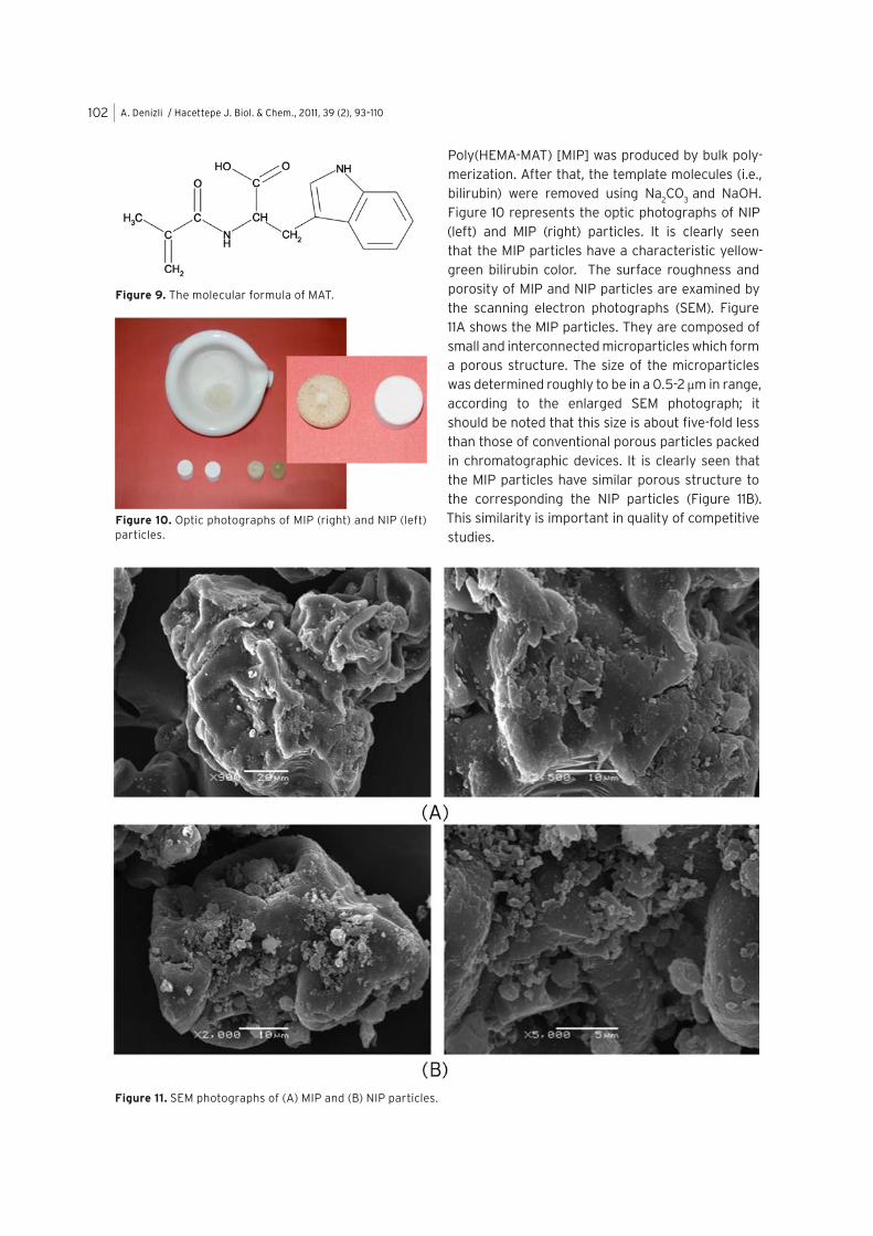
Poly(HEMA-MAT) [MIP] was produced by bulk poly-merization. After that, the template molecules (i.e., bilirubin) were removed using Na
2CO
3 and NaOH.
Figure 10 represents the optic photographs of NIP (left) and MIP (right) particles. It is clearly seen that the MIP particles have a characteristic yellow-green bilirubin color. The surface roughness and porosity of MIP and NIP particles are examined by ���� ����� ����� �� ������ ����� ������� ��� ��11A shows the MIP particles. They are composed of small and interconnected microparticles which form a porous structure. The size of the microparticles was determined roughly to be in a 0.5-2 μm in range, ���� ��� ��� ���� ��� ���� ���� ������ ����� ��should be noted that this size is about five-fold less than those of conventional porous particles packed in chromatographic devices. It is clearly seen that the MIP particles have similar porous structure to the corresponding the NIP particles (Figure 11B). This similarity is important in quality of competitive studies.
Figure 9. The molecular formula of MAT.
Figure 10. Optic photographs of MIP (right) and NIP (left) particles.
Figure 11.������������ ����������������������������� ������
A. Denizli / Hacettepe J. Biol. & Chem., 2011, 39 (2), 93–110 103
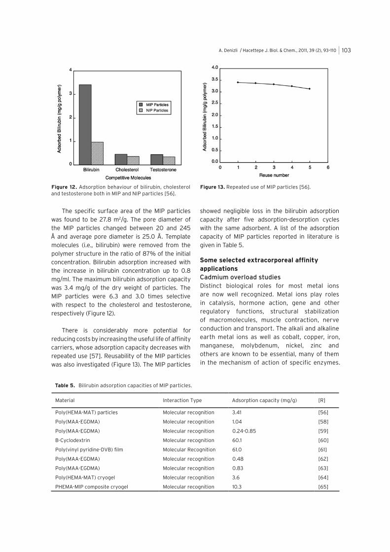
The specific surface area of the MIP particles was found to be 27.8 m2/g. The pore diameter of the MIP particles changed between 20 and 245 Å and average pore diameter is 25.0 Å. Template molecules (i.e., bilirubin) were removed from the polymer structure in the ratio of 87% of the initial concentration. Bilirubin adsorption increased with the increase in bilirubin concentration up to 0.8 mg/ml. The maximum bilirubin adsorption capacity was 3.4 mg/g of the dry weight of particles. The MIP particles were 6.3 and 3.0 times selective with respect to the cholesterol and testosterone, respectively (Figure 12).
There is considerably more potential for reducing costs by increasing the useful life of affinity carriers, whose adsorption capacity decreases with repeated use [57]. Reusability of the MIP particles was also investigated (Figure 13). The MIP particles
showed negligible loss in the bilirubin adsorption capacity after five adsorption-desorption cycles with the same adsorbent. A list of the adsorption capacity of MIP particles reported in literature is given in Table 5.
Some selected extracorporeal affinity applicationsCadmium overload studiesDistinct biological roles for most metal ions are now well recognized. Metal ions play roles in catalysis, hormone action, gene and other regulatory functions, structural stabilization of macromolecules, muscle contraction, nerve conduction and transport. The alkali and alkaline earth metal ions as well as cobalt, copper, iron, manganese, molybdenum, nickel, zinc and others are known to be essential, many of them in the mechanism of action of specific enzymes.
Figure 12. Adsorption behaviour of bilirubin, cholesterol and testosterone both in MIP and NIP particles [56].
Figure 13. Repeated use of MIP particles [56].
Table 5. Bilirubin adsorption capacities of MIP particles.
Material Interaction Type Adsorption capacity (mg/g) [R]
Poly(HEMA-MAT) particles Molecular recognition 3.41 [56]
Poly(MAA-EGDMA) Molecular recognition 1.04 [58]
Poly(MAA-EGDMA) Molecular recognition 0.24-0.85 [59]
B-Cyclodextrin Molecular recognition 60.1 [60]
Poly(vinyl pyridine-DVB) film Molecular Recognition 61.0 [61]
Poly(MAA-EGDMA) Molecular recognition 0.48 [62]
Poly(MAA-EGDMA) Molecular recognition 0.83 [63]
Poly(HEMA-MAT) cryogel Molecular recognition 3.6 [64]
PHEMA-MIP composite cryogel Molecular recognition 10.3 [65]

A. Denizli / Hacettepe J. Biol. & Chem., 2011, 39 (2), 93–110104
However, the adverse effects of metal ions will manifest when a metal ion level exceeds e certain threshold level. The toxicities of heavy metals may be caused by the following mechanisms: blocking the essential functional groups of biomolecules; displacing essential metal ions from biomolecules; modifying the active conformation of biomolecules; disrupting the integrity of biomembranes and modifying some other biologically active agents [66]. Toxic metals are absorbed through the air passages and alimentary canal with food and drinking water. They disturb the economy of endogenous metals and biochemical equilibrium. They have an etiological effect on hypertension, cancer, decrease the ventila of the lungs and other lung diseases. Toxic metal ions are the source of degeneration, reduction of pancreatic efficiency and disfunction of kidneys [67].
The chronic toxicity of cadmium compounds includes kidney damage with proteinuria of low-molecular-weight molecules. An epidemic of Japanese itai-itai disease is believed to be the result of chronic ingestion of Cd2+, with altered renal tubular function, impaired regulation of calcium and phosphorus, manifesting bone demineralization, osteomalacia, and pathological fractures. No specific treatments for acute or chronic cadmium poisoning are available. However, in addition to supportive therapy and hemodialysis, heavy metal poisoning is often treated with a chelating agent. Different chelating agents that are available commercially for the treatment of cadmium poisoning are British anti-lewisite and calcium disodium ethylene diamine tetraacetic acid (EDTA). But there is histopathological evidence for increased toxicity in animals when calcium disodium EDTA is utilized [68]. Recently, one of the most promising technique for blood detoxification is extracoporeal ������������ �������������������������� �������������adsorbents were reported for metal detoxification [70,71].
Metallothioneins are a group of non-enzymatic (6-7 kD) low molecular mass proteins with 61-68 aminoacid residues including 20 cysteins, bound to certain bivalent ions such as Zn2+, Cd2+, Hg2+, Bi2+�� ��2+, Ni2+, Rb2+ or Tc2+ with high affinity [72]. The binding is also observed with univalent
ions such as Cu+, Ag+ and Au+. Metallothionein and its analogues are widely distributed among organisms, from bacteria and fungi to plants and mammals. Metalloproteins play an important role in the metabolism and kinetics of metals including transport of metals, removal of metals, protection from metal toxicity, free radical scavenger, storage of metals, metabolism of essential metal ions, immune response and genotoxicity and carcinogenecity. Cysteinyl hexapeptide (CysHP) is a small metallo-peptide (molecular mass: 627.8) composed of six amino acids, three of them being cysteine residues. The thiol groups in the CysHP structure permit it to form metal clusters including Cd2+, Zn2+, Hg2+, Cu+ and some other bivalent and divalent metal ions. This makes them attractive ligands and metalloproteins are among the most selective chelators known for metal ions for biomedical applications [73].
Denizli et al. prepared thionein carrying PHEMA particles for Cd2+ removal from human plasma both in batch and packed-bed column system [74-76]. Monochlorotriazinyl dye ligand Cibacron Blue F3GA ��� �������� ����� ���� ������ ��������� ������ ��metallo-peptide (i.e., cysteinylhexapeptide) was attached to the dye-attached particles. Then, they were used for Cd2+ removal from human plasma poisoned with Cd2+ in a column system. PHEMA particles were selected as the basic polymer matrix which carries functional hydroxyl groups for further modification. PHEMA particles have hydrophilic character, good blood-compatibility properties, minimal non-specific protein interactions, high chemical and mechanical stabilities for column applications and resistance towards microbial and
Figure 14. Effects of CysHP loading on Cd2+ adsorption [76].
A. Denizli / Hacettepe J. Biol. & Chem., 2011, 39 (2), 93–110 105
enzymatic attacks [77-79]. It was reported that obtained results made these thionein carrying PHEMA particles potential candidates for future detoxification studies. It has been found that the cysteinylhexapeptide loading has a great effect in the capacity of particles for adsorbing Cd2+ ions from human plasma (Figure 14).
Iron overload studiesIron is an essential trace element for almost all organisms for a broad spectrum of biological processes which include electron transfer, transport, storage and activation of oxygen, nitrogen fixation and DNA synthesis [80]. The toxic effects of iron overload are well known especially since the human body has no physiological route for the elimination of excess iron [81]. Chronic iron overload may occur in a variety of diseases where the administration of parental iron is necessary (e.g. thalassemia, aplastic anemia). Acute iron intoxication is also a frequent, sometimes life-threating form of poisoning, especially among young children. The toxicity of iron is related to its ability to induce oxidative stress in cells [82]. In an occupational setting, inhalation exposure to iron oxide may cause siderosis. In the nonoccupational population, ingestion of large quantities of iron salts may cause nausea, vomiting and intestinal bleeding. There is accumulating evidence suggesting that an increase an iron storage may be associated with an increasing ����� ��� ��� ���� ������ ������ ������ ����demonstrated that there is an increased risk for developing colorectal carcinoma following ingestion of high amounts of iron [84]. There is also an increase in hepatocellular carcinoma in patients with hereditary hemochromatosis, an inherited disorder in which there is hyper-absorption of iron from the intestinal tract and in lung cancer from exposure to asbestos fibers, which contains approximately 30% iron by weight [85].
For transfusional iron overload and acute iron poisoning, the only available supportive treatment is chelation therapy and the only available clinical drug for this treatment is desferrioxamine B (DFO) a linear hydroxamate, a natural siderophore [84]. The use of DFO has already been shown to result in prolonged life expectancy, reduced liver iron and
the establishment of negative iron balance. However, the major limitation to the use of DFO is its lack of effectiveness when administered orally, the short half-life time in plasma and its potential toxicity when present in high concentrations [86]. DFO is highly expensive also. For this reason a number of orally active iron chelators are being tested but none of them are still satisfactory [85,87]. To overcome the drawbacks of soluble iron chelators in the treatment of iron overload, attachment of iron chelating ligands have been studied [88]. Comparing to soluble iron chelators, iron chelating resins might have advantages in stability, reusability and minimal damage to biological substances.
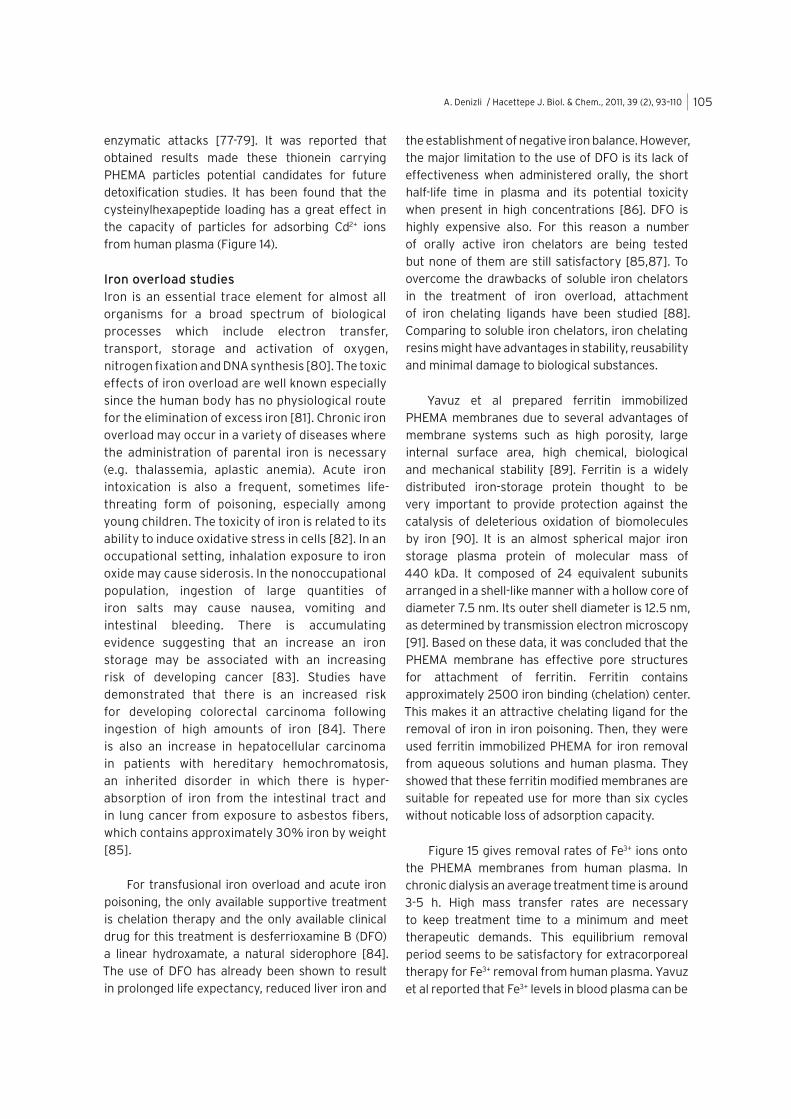
Yavuz et al prepared ferritin immobilized PHEMA membranes due to several advantages of membrane systems such as high porosity, large internal surface area, high chemical, biological and mechanical stability [89]. Ferritin is a widely distributed iron-storage protein thought to be very important to provide protection against the catalysis of deleterious oxidation of biomolecules by iron [90]. It is an almost spherical major iron storage plasma protein of molecular mass of 440 kDa. It composed of 24 equivalent subunits arranged in a shell-like manner with a hollow core of diameter 7.5 nm. Its outer shell diameter is 12.5 nm, as determined by transmission electron microscopy [91]. Based on these data, it was concluded that the PHEMA membrane has effective pore structures for attachment of ferritin. Ferritin contains approximately 2500 iron binding (chelation) center. This makes it an attractive chelating ligand for the removal of iron in iron poisoning. Then, they were used ferritin immobilized PHEMA for iron removal from aqueous solutions and human plasma. They showed that these ferritin modified membranes are suitable for repeated use for more than six cycles without noticable loss of adsorption capacity.
Figure 15 gives removal rates of Fe3+ ions onto the PHEMA membranes from human plasma. In chronic dialysis an average treatment time is around 3-5 h. High mass transfer rates are necessary to keep treatment time to a minimum and meet therapeutic demands. This equilibrium removal period seems to be satisfactory for extracorporeal therapy for Fe3+ removal from human plasma. Yavuz et al reported that Fe3+ levels in blood plasma can be

A. Denizli / Hacettepe J. Biol. & Chem., 2011, 39 (2), 93–110106
reduced significantly with relatively fast adsorption rates by using ferritin modified PHEMA membranes developed in this study (Figure 15).
Aluminum removal studiesAluminum has recently been considered as a causative agent in dialysis encephalopathy, osteodystrophy, and microytic anemia occuring in patients with chronic renal failure who undergo long-term hemodialysis. Only a small amount of Al3+ ions in dialysis solutions may cause these disorders. Encephalopathy has also occured in children consuming aluminum hydroxide as a phosphate binder for renal disorders. Al3+ has also been implicated in neurotoxicity associated with amyotrophic lateral sclerosis, a form of parkinsonism and in Alzheimer’s disease. Demircelik et al prepared Al3+-imprinted PHEMA beads [MIP] with the assistant functional monomer
��� ������ ���������������� ����� ������� �� ���used in selective removal of Al3+ out of human plasma overdosed with Al3+ ions [92]. MAGA was synthesized as the metal complexing monomer by �� ������� ��� ���������� ����� ���� ���� ������chloride, with the goal preparing a solid-phase which has the high selectivity for Al3+ ions (Figure 16). The carboxyl groups of MAGA monomer were complexed with Al3+ ions.
The surface morphology and bulk structure of ����������� ������������ !�����"��� ��#$%�&��MIP beads have a spherical form with the size range of 40-50 μm and rough surface due to the large pores, which formed during the polymerization process. The photograph in Figure 2B shows the presence of macropores within the bead bulk structure due to the formation of ionic cavities. The roughness of the bead surface should be considered as a factor providing an increase in the surface area.
Figure 15. Iron removal rate using PHEMA membranes [89].
Figure 16.� ������� ' ��������� ��� �� ����������binding mode of Al3+ hexagonally coordinated to 2 equivalents of MAGA
2- and 2 equivalents of ROH solvent
molecules, where R is H or C2H
5- or a combination of both.
Figure 17.� !��'��� �'��������������(�)�*� � ������� '����������)+*����,�� ��� �%

A. Denizli / Hacettepe J. Biol. & Chem., 2011, 39 (2), 93–110 107
In addition, the large pores reduce convective mass transfer resistance and facilitate mass. The specific surface area of MIP beads was found to be 55.6 m2/g on the average.
The template Al3+ ions could be reversibly detached from the matrix using a 50 mM solution of EDTA. The MIP and non-imprinted [NIP] beads were then tested in a set of adsorption experiments for their capabilities to remove Al3+ selectively out of human plasma overdosed with Al3+ in the presence of Fe3+, Cu2+ and Zn2+ ions. The relative selectivity coefficients (k’) of the MIP beads for Al3+/Fe3+, Al3+/Cu2+ and Al3+/Zn2+ were found to be almost 4.5, 9.0 and 32.5 times greater than those of the NIP beads, respectively. In addition to these results, Figure 18 illustrates the adsorbed template and competitive ions both in the MIP (dark grey) and the NIP (light grey) beads. As clearly seen here, the competitive adsorption amount for Al3+ ions in the MIP beads is 0.762 mg/g polymer in the presence of competitive ions (Fe3+, Cu2+ and Zn2+). They recovered these MIP beads and reused many times, with no significant decrease in their adsorption capacities.
Other several applicationsImmunoadsorption therapy based on the latter principles of affinity is thus an attractive method chiefly used for cleaning biological fluids such as blood and plasma [93]. Jia et al immobilized protein A on the membrane cartridge for extracorporeal IgG and immune complex removal from blood. The protein A membrane cartridge (6 mg protein A/g dry matrix) adsorbed 23.5 mg IgG per g dry matrix from human plasma. Experiments in vitro and in vivo confirmed that protein A membrane
cartridge mainly adsorbed IgG and little of other plasma proteins, and that blood cell damage was negligible. Phenylalanine or tryptophan-containing imunoadsorbents based on poly(vinyl alcohol) particles have been used for rheumatism and myasthenia gravis therapy [94,95]. Protein A has been used to remove IgG from serum [96]. Protein A apheresis has also been applied successfully to remove autoantibodies in the treatment of severe forms of various autoimmune diseases and after chemotherapy [97]. In some haemophilia cases, antibodies to factor VIII are present and various neurological, nephrological and haemological conditions have been treated using extracorporeal affinity therapy using immunoadsorption columns, packed with sepharose-protein A [98].
Concluding remarksHemoperfusion is a process in which the blood is passed over a carrier in a packed bed column. Hemoperfusion is frequently used for acute blood purification as in the case of drug overdose using charcoal as an adsorbent. The charcoal adsorps medium- to high molecular weight blood components. Charcoal hemoperfusion is non-specific in nature, removing many desirable blood components as well as toxic substances. Consequently, a number of scientists have examined specific extracorporeal removal of toxic substances including bilirubin, metal ions, etc. In this review, we outlined the developments in the extracorporeal affinity therapies. It has been demonstrated that a variety of affinity carriers including both specific and pseudospecific can be used for the removal of toxic substances from human blood.
R E F E R E N C E S
1. �������������� �������������������������������������� ������� �������� ��!"���� ��#���"���� $��%���"��1986.
2. ��� '��� ��� �%*�� ��� :����� ��;�� <������ ��%%���molecule uremic toxin removal via hemodialysis augmented with an immunosorbent packed bed, Ind. Eng. Chem. Res., 49 (2010) 1359.
3. ��� ��"�� ��� �=��=��"� �� >������ �������=�� �%�specific adsorbents for medical therapy, Int. J. Artif. Organs, 12 (1989) 1.
7,62 ppm
1,86 ppm1,65 ppm
0,16 ppm
0
2
4
6
8
Ad
sorb
ed A
l3+ (
pp
m)
Al F e C u Zn
MIP NIP
Figure 18. Adsorbed template and competitive ions both in PHEMAGA-Al3+ and PHEMAGA beads. Flow rate: 0.3 �:@����������������K�LQ��*@:S�U�������������Q�L�g ; T:20°C.

A. Denizli / Hacettepe J. Biol. & Chem., 2011, 39 (2), 93–110108
4. Y. Takenaka, Bilirubin adsorbent column for plasma perfusion, Therapeutic Apheresis, 2 (1998) 129.
5. ��������������� ����������������� ��������������������Tan, Predicting the decrease of conjugated bilirubin ����� ������ �� ����� ��� ���� ����!���� �"#�� ���$�the predialysis molar ratio of conjugated bilirubin to ��� �������%���&������������ ���'�()**)+�,-.�
6. J.D. Ostrow, Bile Pigments and Jaundice: Molecular, Metabolic and Medical Aspects, New York, Dekker, 1986.
7. C. Tiribelli, J.D. Ostrow, The molecular basis of bilirubin encephalopathy and toxicity, J. Hepatology, 43 (2005) 156.
8. /�/��0� ����#��1� ���������2����34�������"�� ���� �� 5�bilirubin with polypeptide-coated resin, Biomat. Artif. Cells, Artif. Organs, 18 (1990) 75.
9. ��� ��������� ��� � ��� ��� � �� � %����� ��� �������� 6��Zinder and J.M. Brandes, In vivo hemoperfusion studies of unconjugated bilirubin removal by ion ������$���������&������"���� ���"���5��7�������6�$�����
27 (1981) 434.10. 8�� 7��9 :��� ��� ����$ ����� ��� �������� ��� � ��!��� &��
;�$�������� ��� ��������� &�� � � ���� ��� &���� � ��T. Kikuchi, H. Tanzawa, Removal of bilirubin and bile ������������������� ��������$���������&�����"���� ���Artif. Intern. Organs, 27 (1981) 428.
11. G.R. Brown, Oligo-peptide functionalized polymeric sorbents for bilirubin, Int. J. Biochromatogr., 1 (1994) 73.
12. &�� �����!�� ��4�� �������� 4 �!�!����� ��� ����9���chitosan beads as adsorbents for bilirubin, Artif. Organs, 16 (1992) 568.
13. Z. Yamazaki, N. Inoue, T. Wada, T. Oda, K. Atsumi, K. Kataoka and Y. Fujisaki, Use of an AR-1 resin column to reduce bilirubin level in modified ascitic fluid, Trans. "���� ���"���5��7�������6�$�����),�� (.-<-+�='*��
14. &�� � ��� � �� ��� ���� ������� ;�� � ���� ��� 7��� ������� ����������� 4������ ��� ���� �� ���$� ����� ���3�adsorbent materials as a treatment for patients with hepatic failure, Artif. Organs, 13 (1989) 447.
15. ��2�� "%������ �� �>���� ��$���� 0�� 1 �������� ���Wessling, Adsorptive membranes for bilirubin removal, J. Chromatogr. B., 803 (2004) 215.
16. Y. Yu, B. He, H. Gu, Adsorption of bilirubin by amine-containing crosslinked chitosan resin, Artif. Cells 1� ��� ����7�� ���1� �������)'�()***+�?*<�
17. ���� � ����&��#���:���0��6�������������%����� ���� ��of bilirubin by macroporous poly(GMA-DVB) beads, Die Angew. Makromol Chem, 237 (1996) 143.
18. ;��"���������"��5�������>������������;�!�9�����&�!!������� 6������ 4��"3���� ������� ��������� ����������of bilirubin in temporarily hyperbilirubinemic rats, Biochim. Biophys. Acta, 1760 (2006) 227.
19. A. Denizli, M. Kocakulak, E. Pişkin, Bilirubin removal from human plasma in a packed bed column system with dye-affinity beads, J. Chromatogr. B, 707 (1998) 25.
20. M. Kocakulak, A. Denizli, A.Y. Rad, E. Pişkin, New sorbent for bilirubin removal from human plasma: cibacron blue-immobilized poly(EGDMA-HEMA) beads, J. Chromatogr. B., 693 (1997) 271.
21. A. Denizli, E. Pişkin, Dye-ligand affinity systems, J. Biochem. Biophys. Methods, 49 (2001) 391.
22. A. Denizli, M. Kocakulak, E. Pişkin, Alkali blue 6B-derivatized poly(EGDMA-HEMA) beads for bili-� ������� %���5� ��� ������������������ � ��������Pure Appl. Chem., A35 (1998) 137.
23. "������9�������� ��: ��:��2��4�A:���������5���� �������for bilirubin removal from human plasma: congo red modified poly(EGDMA-HEMA) beads, J. Appl. Polym. ������B'�(.--'+�?<?�
24. A. Kassab, H. Yavuz, M. Odabaşı, A. Denizli, Human serum albumin chromatography by cibacron blue F3GA-derived microporous polyamide hollow fibre membranes, J. Chromatogr B, 746 (2000) 123.
25. ��� C������ >�� ����9���� ��� 8�% 9�� "�� ����9���� 1���� ����removal from human plasma by dye affinity micropo-� ��� �� ��5�����������������&���� ����?<�()**)+�.-'-��
26. ��� D9 ��� "�� ����9���� 1���� ���� ��� %��� ���5 �������of immobilized albumin in a magnetically stabilized 5� ���9����������1� ������������4 �!���2����.<�()**B+�791.
27. T. Peters, All about albumin, Biochemistry, Genetics and Medical Applications, New York, Academic Press, 1996.
28. A.Y. Rad, H. Yavuz, M. Kocakulak, A. Denizli, Bilirubin removal from human plasma with albumin immobilized magnetic poly(hydroxyethyl methacrylate) beads, Macromol. Biosci., 3 (2003) 471.
29. B. Xia, G. Zhang, F. Zhang, Bilirubin removal by Cibacron Blue F3GA attached nylon-based hydrophilic �55����!��������������������������))B�()**?+�-�
30. ��#�� ��%����� 4���� �������:!� ���� ����� �������Temperature and albumin effects on adsorption of bilirubin from standard solution using anion-exchange resin, Artif Organs, 14 (1990) 14.
31. ��� 0���$�� ��� ���� 1���� ���� ��� %��� 5� �� � ����plasma by Cibacron Blue F3GA using immobilized microporous affinity membranaus capillary method, J. Chromatogr. B., 821 (2005) 112.
32. �����������$����#��1� ���������2����34�������4 �!����resins with amino acid containing pendants for sorption of bilirubin, Int. J. Artif. Organs, 9 (1986) 33.
33. F. Kanai, T. Takahama, I. Iizuka, M. Hiraishi, Z. Yamazaki, 8��> E�� ����8����� !�����&����������"��� ��&��� � ����Artificial Organs, 9 (1985) 75.
34. 7�"������� %�����8��� ��%�������� �� �������� ���� ��characteristics of bioaffinity membranes, J. Membr. ������..?�(.--B+�.B.�
35. ��� "�%���9�� ��� ��� ����� ��� 1��� ���� �� 7�5� ����� 5�the polymeric morphology of sorbents on their properties in affinity chromatography, J. Biochem. Biophys. Methods, 49 (2001) 649.

A. Denizli / Hacettepe J. Biol. & Chem., 2011, 39 (2), 93–110 109
36. H. Wang, J. Ma, Y. Zhang, B. He, Adsorption of bilirubin on the polymeric -cyclodextrin supported by partially aminated polyacrylamide gel, React. Functl. Polym., 32 (1997) 1.
37. ��� ���� ��� ������ �� ������������� ����������cellulose nanofiber as affinity membrane, J. Membr. ��������������!�"���
38. #�� ����$������%��'������%��*+������4�5�����5���6���polylysine carrying chitosan-coated nylon affinity membranes, J. Chromatogr. B., 819 (2005) 301.
39. #�� ����$������%��'������%��;��'���<������%��*+������4� 5�����5��� �� ��=>?>�=����� �������%� �=���membranes, Polym. Int., 54 (2005) 790.
40. E��'���G��?����?��K���%��L������� �+��������������%�and adsorption property of grafting terpolymer microbeads of PEI-GMA/AM/MBA for bilirubin, J. Chromatogr. B., 853 (2007) 62.
41. ?�� ����%�� '�� K���� S�6� ��5��� 4�� 5�����5��� ���T���4�����������������U�������U�����?����"�������!�1495.
42. X.Y. Han, Z.P. Zhang, Preparation of grafted polytetrafluoroethylene fibers and adsorption of bilirubin, Polym. Int., 58 (2009) 1126.
43. #�� ����L�� �����G��K���%��U�� �%��L������K������E��L��%��;��'����?=����>�����+���+������������V�+�>�������affinity membrane for bilirubin removal, J. Membr. �����[�\����"�!�[[[�
44. #�� ����G��U���U�� �%��G��K]��%��K��#��%�� ��K���%��K��Tu, D. Ge, Poly(pyrrole-3-carboxylic acid) alumina composite membrane for affinity adsorption of 5�����5����K�����5��� �����[�[����"�!�"�"�
45. E.B. Altıntaş, D. Türkmen, V. Karakoç, A. Denizli, Efficient removal of bilirubin from human serum by ����q�� +=�>�44���=� 5��+��� K�� E������� ����� ���(2011) 957.
46. K. Takeda, A. Kuwahara, K. Ohmon, T. Takeuchi, Molecularly imprinted tunable binding sites based on conjugated prosthetic groups and ion-paired �4������K��*���U����� ����"["�����\!�}}[[�
47. E�� �����%���� ��+!�� ���������=� �������+� ��=����~�man-made mimics of antibodies and their application in analytical chemistry. Elsevier, Amsterdam (2001).
48. ��� ����� ��� ������ G�� ��+�� K���� G����� �� ���+����������������G�������5���L�� �q�����L��������=�����Ariga, Mechanical tuning of molecular recognition to discriminate the single-methyl group difference 5�6�����=�������+���������K��*���U����� ������""��in press.
49. Y. Ge, A.P.F. Turner, Too large to fit ? Recent developments in macromolecular imprinting, Trends in Biotechnol., 26 (2008) 4.
50. ���������������*�� �����������q������ ��;��������Takeuchi, Protein templated organic/inorganic hybrid materials prepared by liquid-phase deposition, J. Am. U����� ����"�\������!�"�\���
51. T. Takeuchi, T. Hishiya, Molecular imprinting of proteins emerging as a tool for protein recognition, Org. Biomol. Chem., 6 (2008) 2459.
52. ��E�=����=�������� �=��*������q�����E�������*��;���q����*��� �����T�� ������������� 4� ������ ��� ���presence of UO
22+, Ce3+���+�?�3+ using Th4+ Imprinted
Polymer, Talanta, 67 (2005) 640.53. K. Takeda, A. Kuwahara, K. Ohmori, T. Takeuchi,
Molecularly imprinted tunable binding sites based on conjugated prosthetic groups and ion-paired �4������K��*���U����� ����"["�����\!�}}[[�
54. Y. Ge, A.P.F. Turner, Molecularly imprinted sorbent assays: recent developments and applications, Chem. Eur. J., 2009, in press.
55. *�*�� �q����� ��� �=�� *�� ;���q���� *�� ����q�� ?>����+����imprinted synthetic receptor for biochromatography applications, Anal. Chem., 78 (2006) 7253.
56. '�� E�=+������ ��� *�+���� S�� E������� ��� �=�� *�� ;���q���� �����T�����T���4�5�����5���4���������� � � � � � ��with bilirubin imprinted particles, Ind. Eng. Chem. Res., 46 (2007) 2843.
57. ��?�� L�������� ����� *������� �� ��+������ ;����Yarmush, Immunoadsorption: strategies for antigen elution and production of resuable adsorbents, Biotechnol. Prog., 8 (1992) 168.
58. ��K�� =��� K�G�� ;��%�� L���� S����� ��U�� U����� *�G��Wu, Binding specificity of -bilirubin-imprinted poly(methacrylic acid-co-ethylene glycol dimethyl-acrylate) toward -bilirubin, Biomaterials, 26 (2005) 4684.
59. ��K�� =���L����S�����L� ��U���%�������?���� ��U�� �������Ionic effect on the binding of bilirubin to the imprinted poly(MAA-EGDMA), J. Chromatogr. A., 1122 (2006) 54.
60. L�� L��%�� L�� ?�%�� <�� U��� ��� ?��� $�� ?���� ���������=�imprinted polymer using b-cyclodextrin as functional monomer for the efficient recognition of bilirubin, Anal. Chim. Acta, 606 (2008) 92.
61. *�G�� #��� ��K�� =��� =������� 4� 5�����5��� �������+�polymer thin film for the continuous detection of bilirubin in an MIP/QCM/FIA system, Biosens. Bioelectron., 21 (2006) 2345.
62. ��K�� =��� L���� S����� *�� ��������� ���+� 4�� ���binding of bilirubin to the bilirubin imprinted poly(MAA-EGDMA), Anal. Chim. Acta, 539 (2005) 97.
63. ��K�� =��� K�G�� ;��%�� L���� S����� �6��+�� 5�����5���imprinted poly(MAA-EGDMA) for the specific binding of a-bilirubin, Anal. Chim. Acta, 504 (2004) 167.
64. '�� E�=+������ S�� E������� ��� *�+���� ]�L�� '����T�� ��� �=��A. Denizli, Bilirubin recognition via molecularly imp-rinted supermacroporous cryogels, Colloids and ��4�����E����}�����\!�[[�
65. '�� E�=+������ S�� E������� ��� *�+���� ]�L�� '����T�� ��� �=��*�� ������������� �G��*� 5���+� ��=%��� 6���embedded bilirubin imprinted particles, React. Functl. Polym., 69 (2009) 36.
66. G����� ���%���� ��~� '�;�� U��=��� ��+� $���� U��=���(Eds.), Patty’s Industrial Hygiene and Toxicology, New York, John Wiley, 1981.
67. ?��$��5��%��U�'������+������~����=���+���4������������G����� ��+� �4�=�� [�+� �+��� '���6��� 6�q�����+��]����������?�5����%���q�����"\}��

A. Denizli / Hacettepe J. Biol. & Chem., 2011, 39 (2), 93–110110
68. ��� ������� ��� ��������� ����� �������� ��� ������������Cadmium in the Environment, 2nd ed., Cleveland, CRC Press, 1974.
69. A. Denizli, Preparation of immunoaffinity membranes for cholesterol removal from human plasma, J. Chromatogr. B, 772 (2002) 357.
70. ������������������������ !���"� ������#� ������"�of mercury. a model: treatment of severe mercury poisoning by encapsulated chelating spheres, Biomat. Med. Dev. Art. Org., 9 (1981) 107.
71. $������%��&�������������������"���'��"����#� ������"�by haemoperfusion through deferoxamine conju-gated agarose-polyacrolein microsphere beads, Biomaterials, 6 (1985) 9.
72. M. Nordberg, Metallothioneins: historical review and state of knowledge, Talanta, 46 (1998) 243.
73. A.K.M. Kabzinski, Application of solid-phase extrac-tion to the preconcentration of metallothionein and metals from physiological fluids, J. Chromatogr. A., 766 (1997) 121.
74. (����'��������)��$"�&�������*+�����,���"���(�� ��+�"��Cadmium removal from human plasma by Cibacron Blue F3GA and thionein incorporated into polymeric microspheres, J. Chromatogr. B., 720 (1998) 217.
75. F. Denizli, A. Denizli, Removal of cadmium(II) ions from human plasma by thionein modified pHEMA based membranes, React. Functl. Polym., 44 (2000) 207.
76. )�� $"�&���� ��� -�.!&�� /�� )����� ��� *+��0�� 1�� �"2��Cysteinylhexapeptide attached PHEMA beads for Cd(II) removal from human plasma in a packed-bed ���!�"��������������"�����34�567738�94:;�
77. �����*��������)����������5��8� ����"�����'"�� ���<� �=������������ �"�� *���������� ��!����� )�>�����Washington DC, 1987.
78. A. Denizli, Heparin immobilized poly(2-hydroxyethyl methacrylate) based microspheres, J. Appl. Polym. ������?@�59;;;8�:AA�
79. -���� ����������� B�� ����� ����"� )������ � � ����"�Adsorption on Biomaterials, Kluwer Academic Publ., Dordrecht, 1990.
80. R.R. Crichton, Inorganic Biochemistry of Iron �����������(��������%������������C��59;;98�
81. ����� )�"��� ����"�"�� ������� ����" ����� '���"����(1970).
82. R.B. Martin, The chemistry of aluminum as related to biology and medicine, Clin. Chem., 32 (1986) 1797.
83. ����� ����.��&�� *���� ������&�� ��� ���=!"���� ����� �����"��1987 annual report of the American Association of Poison Control Centers National Data Collection �=�����)������(���������:�59;448�@?;�
84. J.R. Mahoney, P.E. Hallway, B.E. Hedlund, J.W. Eaton, Acute iron poisoning. Rescue with macromolecular ����������)��������>��"��'".�����4@�59;4;8�93:6�
85. C. Hershko, Iron chelators in medicine, Molec. Aspects. Med., 13 (1992) 113.
86. A. Jacobs, Iron chelation therapy for iron loaded patients, Br. J. Haematol., 43 (1979) 1.
87. C.F. Whitten, G.F. Gibson, M.H. Good, J.F. Goodwin, )���� *��!��� ��!���� �"� ��!�� ���"� �����"�"�� '��Desferrioxamine in the treatment of acute iron poisoning: clinical observations, experimental studies, and theoretical considerations, Pediatrics, 36 (1965) 322.
88. )�� $"�&���� *�� ������� (�� ��+�"�� �%� �����D ����"�polymer microspheres carrying dyes as chelators for ���"��.����������*������������� ��=���(����;� 59;;48�175.
89. H. Yavuz, Y. Arıca, A. Denizli, Therapeutic affinity adsorption of iron(III) with dye- and ferritin ��������&�� �(�)�������"��� ���)����� ��=��� ������82 (2001) 186.
90. (�>�� ������ ������"<� ���!��!��� "� �!�����"�� �"��cellular function in animals, plants, and micro-organisms, Annu. Rev. Biochem., 56 (1987) 289.
91. )���� �=�������*�"=�����E����������"�� �����������"��in: Proteins of Iron Metabolism, E.B. Brown, P. Aisen, ��������"��"��E�E��>������"���������!"�F��������"��New York, 1977.
92. )���� $���2��+�� ��� )"��2�� >�)�� )"��2�� E�� ��=�� )��Denizli, Molecular recognition- based detoxification � ���!��"!���"��!��"������������*������������ ��=���Ed., 20 (2009) 1235.
93. �����������-�"�����G�!��-��G��"�����G�����>����"����������Protein A tangential flow affinity m e m b r a n e cartridge for extracorporeal immunoadsorption therapy, Biomed. Chromatogr., 13 (1999) 472.
94. Y. Minagawa, T. Kawakami, T. Kojima, Experiment of long term intermittent plasmapheresis for myasthenia gravis, Jpn. J. Artif. Organs, 12 (1983) 101.
95. ��� ������ ��� )""��� ��� )����� ��� -���%�+��� ��� �!������K. Inagaki, In vitro removal of anti-acetylcholine receptor antibodies with a new immunoadsorbent in sera from myasthenia gravis patients. Prog. Artif. Organs, 4 (1983) 719.
96. >�� �������!��� ��)�� ������"�� ��*�� �!"�H.����� '����������"�� ��� ��=����� ��� ��"������� )� �!����=� � � �.�years clinical experience with extensive removal of immunoglobulins, Plasma Ther. Transfus. Technol., 7 (1986) 545.
97. B�,�� E������� ����� $�""��� )�� �������� (�� ������� �����%�"����( ����=��"���� �=�� �� ���!"����!��"����������)�)',�'��@3�59;;?8�A3�
98. I.M. Nilsson, E. Berntorp, O. Zettervall, Induction of split tolerance and clinical cure in high-responding hemophiliacs with factor IX antibodies, Proc. Natl. )���������C�)��43�59;4:8�;9:;�