Neurosteroids Induce Allosteric Effects on the NMDA Receptor

Effects of neonatal Allopregnanolone alteration levels on adult behaviour
ACADEMIC DISERTATION
To obtain the degree of PhD in Neuroscience of the Universitat Autònoma de Barcelona
2014
Group of Neurosteroids and Behaviour
Institut de Neurociències
Departament de Psicobiologia i Metodologia de les Ciències de la Salut Universitat
Autònoma de Barcelona
Supervised by
Dr. Sònia Darbra Dr. Marc Pallarès


The research described in this thesis was conducted at the department of Psychobiology and
Methodology in Health Science - Institute of Neuroscience of the Universitat Autònoma de
Barcelona in the group of Neurosteroids and Behaviour.
This work was supported by a grant from the Spanish Ministry of Science and Innovation
(PSI2009-13759) and Spanish Ministry of Economy and Competitiveness (PSI2012-36646).


“Science might set limits to knowledge, but should not set limits to imagination
” Bertrand Russell


Table of contents
Abbreviations 9
Introduction 11
1. General overview 13
1.1 Ns biosynthesis 13
1.2 Ns Mechanisms of action 15
1.2.1 Genomic effects 15
1.2.2 Modulation of ionotropic receptors 16
1.3 Ns behavioural profile in the adulthood 18
2. Ns and postnatal development 21
2.1 Neonatal Ns and CNS maturation: Effects on hippocampal development 22
2.2 Neonatal Ns and behaviour 23
3. GABAAR and development 25
Objectives and hypothesis 29
Experiment 1 33
Experiment 1a : Research article
“Neurosteroids infusion into CA1 hippocampal region on exploration, anxiety-like
behaviour and aversive learning”
41
Experiment 1b : Research article
“Alteration of neonatal Allopregnanolone levels affects exploration, anxiety, aversive
learning and adult behavioural response to intrahippocampal neurosteroids”
51
Experiment 2 61
Research article
“Neonatal finasteride administration alters hippocampal α4 and δ GABAAR subunits
expression and behavioural responses to progesterone in adult rats”
69
Experiment 3 85
Research article
“Neonatal administration of Allopregnanolone at high doses modifies hippocampal K+ Cl-
co-transporter expression during early development”
93
Discussion 105
Role of neonatal NS fluctuations in the behavioural response to intrahippocampal
NS administration
107
Mechanisms underlying hippocampal and behavioural changes due to neonatal
manipulation of NS levels: Focused on the hippocampal expression of GABAAR
containing α4 and δ subunits
110
Conclusions 113
References 117
Annexes 129
Annex 1: Research article
“Neonatal alteration of Allopregnanolone levels: Role on behaviour
and dorsal hippocampus”
131
Annex 2: Poster presentation: Societat Catalana de Biologia (2010) 144
Annex 3: Poster presentation: Behavioral Brain Research Society (2011) 149
Annex 4: Poster presentation: Federation of European Neuroscience Society (2012) 153
Acknowledgements 157

Abbreviations AMPA: α-Amino-3-hydroxy-5-methyl-4-isoxazolepropionic acid
AT: Acquisition trial
A1: Adenosine receptor 1
A2: Adenosine receptor 2
BO: Boissier test
CA1: Cornus Amonis 1
CA3: Cornus Amonis 3
CNS: Central neurvous system
Cl-: Chloride
DHEA: Dehiepiandrosterone
EPM: Elevated plus maze
GABAAR/ GABAARs: γ-aminobutyric acid receptor/s
GDPs: Giant depolarizing potentials
GFAP: Glial fibrillary acidic protein
KCC2: Potassium chloride co-transporter 2
MAP2: Microtubule-associated protein 2
MBR: Mitochondria benzodiazepine receptor
nAch: Nicotinic acetylcholine receptor
NBM: Nucleus basalis magnocellularis
NH: No handled
NKCC1: Sodium potassium chloride co-transporter 1
NMDA: N-Methyl-D-aspartic acid
NS/Ns: Neurosteroid/s
PREG: Pregnenolone
PREGS: Pregnenolone sulphate
PXR: Pregnane xenobiotic receptor
P450scc: P450 side-chain-cleavage
RT: Retention trial
StAR: Steroidogenic acute-regulatory protein
THDOC: Tetrahydrodeoxycorticosterone
TSPO: Translocator protein
3α,5α-THPROG: 3α-5α-tetrahydroprogesterone
3βHSD: 3β hydroxysteroid
5-HT3: 5-hydroxytryptamine receptor 3


Introduction


General overview
13
Introduction
1. General overview Allopregnanolone or 3α-5α-tetrahydroprogesterone is a α-reduced progesterone metabolite
that belongs to the family of neurosteroids (Ns) (Paul and Purdy, 1992; Robel and Baulieu, 1994).
The term neurosteroid (NS) refers to steroids that can be synthesized de novo by the nervous
tissue independently of peripheral sources such as ovaries or adrenal glands and are capable to
modulate neural excitability (Baulieu et al. 1981; Baulieu, 1998). Baulieu and colleagues observed
levels of steroids such as dehydroepiandrosterone (DHEA) and pregnenolone (PREG) in the
nervous system, after gonad or adrenalectomy (the main steroidogenic glands) (Baulieu et al.,
1981). Later on, other studies demonstrated the presence in the brain of enzymes that are also
involved in classic steroidogenesis and confirmed the fact that steroids are capable to be
synthesized in situ by the nervous tissue (reviewed in Mensah-Nyagan et al., 1999; Compagnone
and Mellon, 2000). In the last three decades many studies have emerged stressing the relevance
of Ns multiple roles on pathologic behaviour, aging processes or degenerative diseases, and
regeneration of damaged tissue (Rupprecht et al., 2001; Dubrovsky, 2005; Mellon, 2007; Kussius
et al., 2009). Controlled local synthesis of Ns has been described to fluctuate during life due to
physiological conditions such as menstrual cycle and pregnancy (Gidler et al., 2001; Maguire et al.,
2005; reviewed in Frye et al., 2011 and Brunton et al., 2014), aging (Bernardi et al., 1998), and to
participate in neural development, cognitive processes and mood control (Kussius et al., 2009;
Mellon, 2007). Nowadays it is well accepted that Ns are an important target concerning pathologic
behaviour (Barbaccia et al., 1998; Rupprecht et al. 2001; Dubrovsky, 2005; Marx et al., 2006;
Mellon, 2007; Kussius et al. 2009; Frye, 2009; Marx et al., 2009, 2011).
1.1 Ns biosynthesis Biosynthesis of Ns begins with the enzymatic conversion of cholesterol to pregnenolone.
Cholesterol is delivered into the inner mitochondrial membrane through the steroidogenic acute
regulatory protein (StAR) (Sierra, 2004). The rate-limiting step on the Ns synthesis is the
movement into the inner mitochondrial membrane mediated by the translocator protein (TSPO) as
known as mitochondrial benzodiazepine receptor (MBR) (Papadopoulos et al., 2006; Rone et al.,
2009). Once into the mitochondria, cholesterol is catalysed to pregnenolone through several
enzymatic reactions mediated both by the P450 side-chain-cleavage (P450scc) and non-P450scc
enzymes (reviewed in Mellon and Griffin, 2002), see Fig 1. Then, progesterone is formed from
pregnenolone by the 3β hydroxysteroid (3βHSD) enzymes. Thus, the NS Allopregnanolone is
synthesized from progesterone via two enzymatic steps: first one, the 5α-reductase reduced
progesterone yielding dihydroprogesterone and the second one, is the 3α reduction of the C3
ketone, mediated by 3αHSD and yielding Allopregnanolone (Mellon et al., 2001; Mellon and Griffin,

General overview
14
Introduction
2002; Belelli and Lambert, 2005; Mellon, 2007, see Fig 1). The presence of enzymes responsible
of Allopregnanolone synthesis has been described in the nervous tissue (Mensah-Nyagan et al.,
1999; Compagnone and Mellon, 2000; Mellon and Griffin, 2002; Agis-Balboa et al., 2006). For
instance co-localization of 3αHSD and 5α reductase have been demonstrated in pyramidal
neurons of the cortex and the glutamatergic granular cells of the dentate gyrus, glutamatergic
(dorsomedial) and GABAergic (reticular) cells of the thalamus, GABAergic cells of the striatum,
glutamatergic neurons of the amygdala, and GABAergic Purkinje neurons of the cerebellum (Agis-
Balboa et al., 2006). Indeed, recent findings indicate that glutamatergic neurons could be the main
source for Ns synthesis acting as paracrine or autocrine regulators of somatodendritic activity
(Chisari et al., 2010; Chisari et al., 2011; Chisiari et al., 2012) and as a paracrine regulator of
membrane receptors located on dendrites and cell bodies of glutamatergic neurons (Akk et al.,
2005). However, previous observations also pointed out that glial cell: Schwann cells in the PNS
and oligodendrocytes and astrocytes in the CNS, could also participate in Ns synthesis (Baulieu,
1998).
Fig 1: Structure and synthesis of Ns: Biosynthesis of pregnenolone derived Ns. The pathway forthe synthesis of Allopregnanolone (3α,5α-THPROG) from cholesterol is shown. Steroidogenic acute-regulatory protein (StAR) interacts with the mitochondrial benzodiazepine receptor (MBR) to facilitate the transport of cholesterol across the mitochondrial membrane (Figure extracted from Belelli and Lambert, 2005).

General overview
15
Introduction
1.2 Ns mechanisms of action1.2.1 Genomic effects
Steroid hormones exert their classical actions by passing through the cell membrane and
binding to their respective cytoplasmic intracellular receptors. The receptor bound steroid hormone
and then translocate to the nucleus were they can modulate gene expression. Allopregnanolone
and ring A reduced pregnanes derived from progesterone have also been reported to regulate
gene expression (reviewed in Rupprecht and Holsboer, 1999; Rupprecht, 2003; Frye et al., 2012),
see Fig 2 (right). It has been suggested that differentially from progesterone, Allopregnanolone is
inactive at progesterone receptor and that its effect on gene expression could be mediated by its
modulation of the pregnane xenobiotic receptor (PXR) (Lamba, et al., 2004; Langmade, 2006;
reviewed in Frye et al., 2012).
Fig 2 Ns mechanisms of non-genomic vs. genomic actions: Ns are capable to modulate neural excitability by binding directly to specific membrane receptors located in the plasmatic membrane of neurons (left). Whereas “classical view” of steroid hormones actions, involved their binding to intracellular receptors promoting the gene expression changes (right). Figure extracted from Rupprech and Holsboer, 1999.
Neuroactive steroids
17 -EstradiolProgesterone3 ,5 -THP3 ,5 -THDOCPSDHEA-S
Steroid hormones
17 -EstradiolDihydrotestosteroneProgesteroneAldosteroneCorticosteroneCortisol
Steroid targets
GABAA receptor5-HT3 receptorNicotinic ACh receptorNMDA receptorKainate receptorAMPA receptorGlycine receptorSigma receptorOxytocin receptor
GABA
BDZ
5-HT
Steroid1
1
2
2
1 GABA
GR
GRPKA
GR
GR
MR
MRMR
PR
PR
GRER
Nucleus
Nucleus
ReductionOxidation
Noradrenaline
Dopamine
Synapse
HSP90
Steroid hormone
Ca2+
Cl—
Proteins
Nongenomic effects Genomic effects
MonthsDaysHoursMinutes
mRNA
Membrane

General overview
16
Introduction
1.2.1 Modulation of ionotropic receptors
Besides their classical steroids actions, an important mechanism of action of Ns is their
ability to modulate neural excitability by acting through membrane receptors. The fact that Ns could
interact with membrane receptors, was firstly described in 1986 by Majewska and co-workers
(Majewska et al., 1986), when they observed that the NS Allopregnanolone exerted its actions,
mainly through the modulation of the GABAA receptor (GABAAR) (Majewska et al., 1986). This
“rapid” effect of NS involved that, while the classical steroids actions require longer time period to
exert their effect and are limited by the rate of protein biosynthesis, this “new view” implied that Ns
effects occurred rapidly from milliseconds to seconds (Majewska et al., 1986), see Fig 2 (left). In
this sense, 5α-reduced pregnanes such as Allopregnanolone have been described to modulate
several ionotropic receptors such as GABAAR, NMDA, nAch or 5-HT3 (Majewska et al., 1986;
Rupprecht and Holsboer 1999; Belelli and Lambert, 2005; Lambet et al., 2009; Zhung 2009), see
Table I.
Although Allopregnanolone can act on several ionotropic receptors its main effect is
mediated through the allosteric positive modulation of the GABAARs, by increasing the aperture
time and frequency opening of the Cl- channel (Majewska et al., 1986). Allopregnanolone effects
on GABAARs have been described to act via two discrete binding sites, one located between the
transmembrane domains M1 and M4 of the α subunits of the receptor, and the other, in the
interface between α and β subunits (Hosie et al. 2006), see Fig 3a. The GABAAR is a pentameric
structure typically composed by 2α 2β and 1γ or δ subunits (reviewed in Jacob et al., 2008 and
Olsen and Sieghart, 2009), see Fig 3b. The aperture of the GABAAR channel induces an increase
in the membrane permeability to Cl-, leading to a postsynaptic hyperpolarizing response (Staley
and Proctor, 1999). There are 19 different GABAARs isoforms indicating a high level of structural
heterogeneity and function (Olsen and Sieghart, 2009). Those can be classified between receptors
that are present at synapses and participate in phasic inhibition, such as GABAARs containing α1,
α2, α3, γ1, γ2 or γ3 subunits (Essrich et al., 1998; Farrant and Nusser, 2005), and GABAARs
related to the modulation of tonic inhibition, which preferably contain α4, α5, α6 or δ subunits and
that are extrasynaptically and perisynaptically located in somatic, dendritic or axonal regions of the
neuron membrane (Belelli et al., 2009; Glykys and Mody, 2007), see Fig 3c. Phasic inhibition is
characterized by a short inhibitory postsynaptic response during the presence of GABA (Farrant
and Nusser, 2005), while persistent or tonic inhibition rather than mediate a rapid inhibitory
postsynaptic response, has been proposed to act as a paracrine regulation of the neuron tone (for
review see Semyanov, et al., 2004; Kullmann et al., 2005; Farrant and Nusser, 2005; Glykys and
Mody, 2007; Beleli et al., 2009). Although Allopregnanolone is able to interact with all GABAARs
subtypes, GABAARs containing the δ subunit are preferred substrates for Allopregnanolone
(Mihalek et al., 1999; Belelli et al., 2002; Stell et al., 2003; Spigelman et al., 2003; reviewed in

General overview
17
Introduction
Smith et al., 2007; Herd et al., 2007; Shen and Smith, 2009). Moreover, it has been shown that
knocked out mice lacking δ subunit have reduced sensitivity to hippocampal Allopregnanolone
effects (Mihalek et al., 1999; Spigelman et al., 2003). Concretely, the combination of the α4β2δ
GABAAR has been demonstrated to be especially sensitive to the Allopregnanolone fluctuation
levels (Smith et al., 1998; Gulinello et al., 2001; Follesa et al., 2002; Gulinello et al., 2002; Shen et
al., 2005; Biggio et al., 2006; Gangisetty and Reddy, 2010).
On the other hand, sulphated Ns such as pregnenolone sulphate (PREGS) has been
described to negatively modulate the GABAARs (Majewska and Schwartz, 1987; Akk et al., 2001).
Although they do not show the same selectivity for GABAARs such as other Ns, a specific binding
sites in the M1 and between M2-M3 transmembrane domains of the α1 and β2 subunits of the
GABAARs have been described to be necessary for the negative modulatory effect of PREGS
(Wardell et al., 2006; Wang et al., 2007). A relevant mechanism of action of PREGS effect is acting
as a positive modulator of the NMDA receptors (Bowly et al., 1993; Horak et al., 2004; Wang et al.,
2007; Schumacher et a., 2008; Kussius et al. 2009). The extracellular M3-M4 loop of the NR2
a
b
c
Fig 3: GABAAR composition and localization: a) GABAAR subunits consist of four hydrophobic transmembrane domains (TM1-4). b) Five subunits from seven subunit subfamilies (α, β, γ, δ, ε, θ and π) assemble to form a heteropentameric Cl- permeable channel. c) GABAAR compose of α (1,2,3) subunits together with a β and a γ are thought to be preferably synaptically located, whereas receptors containing α5βγ or α4βσ are mainly localized at extrasynaptic sites. Figure extracted from Jacob et al., 2008.
General overview
18
Introduction
subunit has been shown to be a key determinant for the PREGS actions on the NMDA receptors.
This hydrophobic pocket at the NR1/NR2B interface has been named “Steroid Modulatory Domain
1 (Horak et al., 2006; Schumacher et al., 2008). Within the NMDA receptors family, the negative
modulation of AMPA and Kainate receptors by PREGS has also been reported (Mameli et al.,
2005; Shirakana et al., 2005; Mellon et al., 2007). Furthermore, the sigma type 1 receptor is
another relevant target of PREGS modulation (Monnet and Maurice, 2006; Schumacher et al.,
2008). While other Ns such as DHEAS have been shown to be agonist of σ1 receptor, PREGS
acts as an inverse agonist (Monnet and Maurice, 2006), see Table 1.
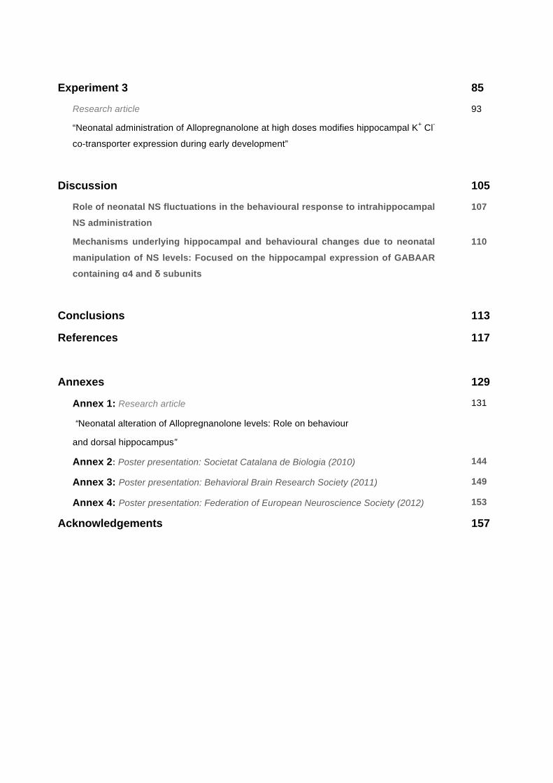
Table I: Ns mechanisms of action: Modulation of ionotropic receptors (Table adapted from Dubrovsky, 2005).
Neurosteroid Receptor Modulation
Progesterone
nAChR Negative (Valera et al., 1992)
Sigma 1 Negative (Monnet and Maurice, 2006)
5-HT3 Negative (Wetzel et al., 1998)
Allopregnanolone
GABAA Positive (Majewska et al., 1986)
nAChR Negative (Dazzi et al., 1996)
5-HT3 Negative (Wetzel et al., 1998)
PREGS
NMDA Positive (Wu et al., 1991)
GABAA Negative (Akk et al., 2001)
AMPA/Kainate Negative (Wu et al., 1991;Yaghoubi et
al., 1998)
nAChR Positive (Dubrovsky, 2005)
Sigma 1 Negative (Monnet and Maurice, 2006) 5-HT3 No effects (Wetzel et al., 1998)
1.3 Ns behavioural profile Similar to other GABAAR positive modulators such as benzodiazepines or barbiturates, Ns
that act as positive modulators of GABAARs have been demonstrated to exert an anxiolytic (Bitran
et al., 1993; 1995; 2000), hypnotic (Damianisch et al., 2001) and anticonvulsive effect (Landgren et
al., 1997). In fact, the role that Allopregnanolone plays on behaviour when is systemically injected,
has been well established. However, the implication of the brain structures that have a role in the
behavioural effects of Ns still needs to be clarified. In this sense, the amygdala has been
postulated as a relevant area. It has been hypothesised that this nucleus can modulate the

General overview
19
Introduction
anxiolytic effects of Ns, as microinjections of Allopregnanolone into the central nucleus of the
amygdala showed anxiolytic effects in the elevated plus maze (EPM) and conflict test (Akwa et al.,
1999). Another area postulated to be relevant regarding Ns effect on behaviour, is the
hippocampus. Previous studies demonstrated that systemic administration of finasteride (a 5α
reductase inhibitor) decreases hippocampal levels of Allopregnanolone and increases depressive
behaviour in pregnant rats (Frye and Walf, 2002; Frye and Walf, 2004b). In this line, increased
hippocampal Allopregnanolone levels have also been demonstrated to increases exploratory and
decreases anxiety-like behaviour (Frye and Rhodes, 2007). Furthermore, other studies also
showed that microinjection of pregnenolone into the dorsal hippocampus induced a decrease in
anxiety scores on the EPM (Bitran et al. 1999). However, other authors showed no effects of
hippocampal Allopregnanolone administration in the EPM or shock-probe burying test (Engin and
Treit, 2007), see Table II. Concerning learning and memory, Allopregnanolone administration has
been reported to exert a detrimental profile, similarly to other GABAAR positive modulators (Mayo
et al., 1993; Ladurelle et al., 2000; Matthews et al., 2002). Previous studies showed that
Allopregnanolone deteriorates spatial learning in the Morris test (Johansson et al., 2002; Matthews
et al., 2002; Turkmen et al., 2006), and also non-associative learning tasks in the "Y" maze
(Ladurelle et al., 2000), when administered systemic or intraventriculary. Furthermore, it has been
postulated that the increase in Allopregnanolone levels induced by ethanol administration, could be
participating in the detrimental effects of alcohol on learning (Silvers et al., 2003).
The promnesic effect of other Ns such as PREGS or DHEA has been well-documented
(Vallée et al., 1997; Darnaudéry et al., 2000; Johanson et al., 2002). For instance, improvement in
memory retention when PREGS was systemically administered (Isaacson et al., 1995) has been
demonstrated, but also a reversal of the memory impairment induced by alcohol administration
(Melchior and Ritzmann, 1996) or scopolamine (Meziane et al., 1996; Mathis et al., 1996; Vallée et
al., 2001) and in old cognitively impaired rats (Vallée et al., 1997). Moreover, PREGS promnesic
effects have also been described when injected into the amygdala (Flood et al., 1995), into the
hippocampus (Darnaudéry et al., 2000), intraventriculary (Flood et al., 1992) or into the nucleus
basalis magnocellularis (NBM) (Pallarès et al., 1998) in several learning tests. However, PREGS
effects on passive avoidance in a previous study carried out in our laboratory showed a detrimental
effect of PREGS in the passive avoidance when administered intrahippocampally but after the
application of environmental stress (Martin-Garcia and Pallarès, 2008). The promnesic effect of
PREGS has been postulated to take place through the potentiation of NMDA receptors located in
the pyramidal neurons of the hippocampus (Bowly, 1993). However, it has also been suggested
that PREGS enhancing profile could be done through the potentiation of the cholinergic neurons
(Pallarès et al., 1998; Darnaudéry et al., 2000). Table II show anxiety effects of Ns administration

General overview
20
Introduction
in several brain structures further experiments are detailed in the Annex 1.
Table II: Effects of Allopregnanolone, progesterone and pregnenolone administration on anxiety-like behaviour
Neurosteroid Administration Test Effect Ref.
Allopregnanolone
Amygdala EPM Anxiolytic Akwa et al., 1999
Amygdala Conflict test Anxiolytic Akwa et al., 1999
Hippocampus EPM No response Engin & Treit, 2007
Hippocampus
Ventricular
Shock-probe burying test
Y maze
No response
Detrimental
Engin & Treit, 2007 Ladurelle et al., 2000
Progesterone Amygdala EPM Anxiolytic Frye and Walf,
2004a Amygdala Open Field Anxiolytic Frye and Walf,
2004a
Pregnenolone Hippocampus EPM Anxiolytic Bitran et al., 1999
Ns and postnatal development
21
Introduction
2. Ns and postnatal development
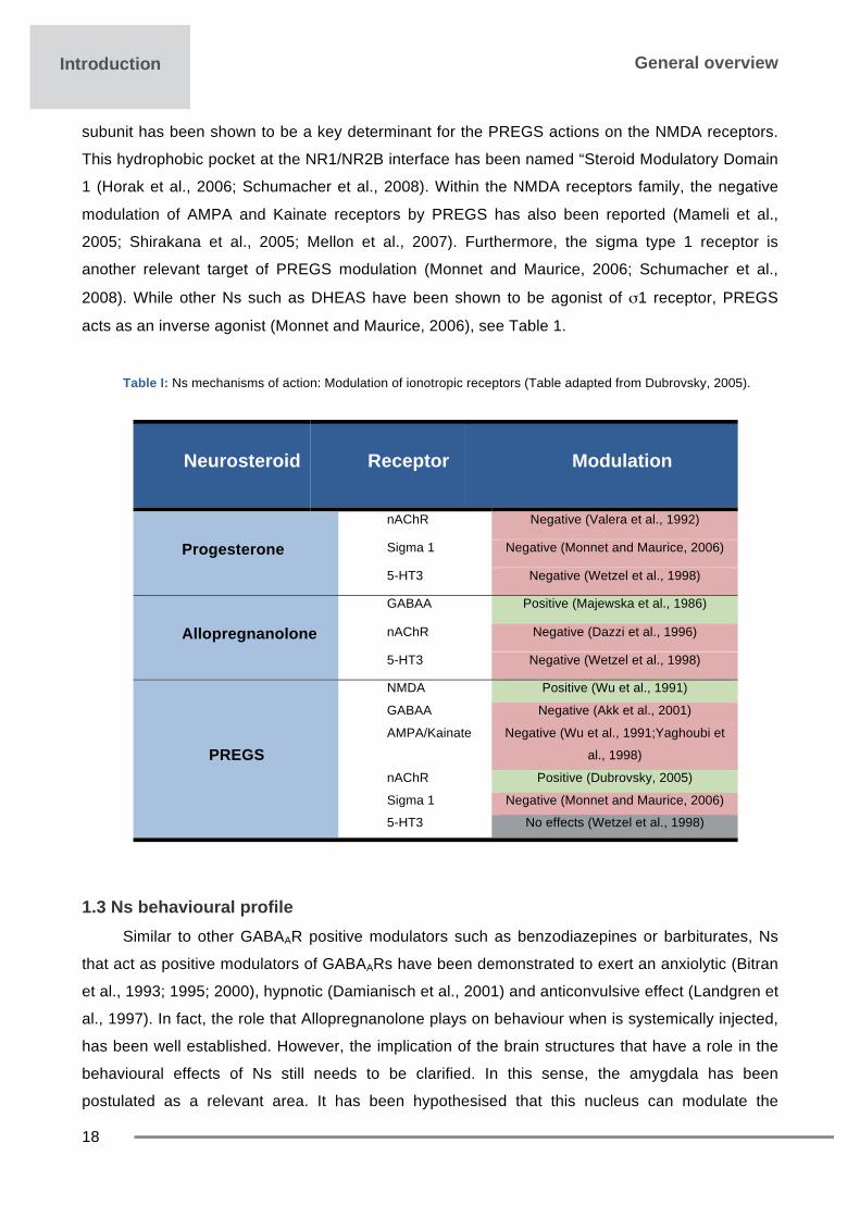
Ns synthesis has been demonstrated in early stages of foetal development of the rat brain
(Pomata et al., 2000). During the last pregnancy period Allopregnanolone levels highly increase
and decline prior to parturition in the forebrain of embryonic rats (Grobin and Morrow, 2001), see
Fig 4a. This increase in Allopregnanolone levels during pregnancy has been proposed to be part of
a protective mechanism against gestational stress. In this sense, it has been described that
Allopregnanolone induces central opioid inhibition of neuroendocrine stress responses during
pregnancy (Brunton et al., 2009; Hirst et al., 2009). Furthermore, Allopregnanolone has also been
described to be particularly relevant during developmental stages. Although Allopregnanolone is
present in foetal brain in similar levels to adults (Kellogs et al., 2006), its synthesis has been
described to fluctuate during the postnatal developmental period (Grobin and Morrow, 2001;
Grobin et al., 2003; Griffin et al., 2004). From the day of birth and in the two first weeks of life,
cortical Allopregnanolone levels show important fluctuations, as showed by an initial elevation in
the day of birth and a progressive decrease in the first week followed by a secondary elevation
during the second week, reaching maximum values between postnatal days 10-14 in rats (P10-
P14) (Grobin and Morrow, 2001; Grobin et al., 2003). The finding of the secondary peak in
Allopregnanolone present during at time of a remarkable change in GABAAR signalling (from
excitatory to inhibitory) suggests that GABAergic Ns modulation may participate in the normal
developmental of GABAergic neurotransmission (Grobin and Morrow, 2001), see Fig 4b.
Foetal development Postnatal development a b
Fig 4. Allopregnanolone levels during postnatal development. a) Allopregnanolone levels in the forebrain of embryonic rats fall prior to partiturion (P21). b) Fluctuations of cortical Alloprenganolone levels after birth (P0) and during postnatal development. Figure from Grobin and Morrow, 2001.

Ns and postnatal development
22
Introduction
2.1 Neonatal Ns levels and CNS maturation: Effects on hippocampal developmentIt has been documented that the alteration of the physiological levels of Ns in early neonatal
phases alters the maturation of certain cerebral structures such as the meso-cortical and meso-
striatal dopaminergic pathways (Muneoka et al., 2002; Muneoka and Takigawa, 2002), the
adenosinergic systems throughout A1 (Muneoka et al., 2002) or A2A receptors (Shirayama et al.,
2001) or the GABAergic thalamic-cortical system (Grobin et al., 2003; Gizerian et al., 2004).
Moreover, exogenous administration of Ns has also been described to alter hippocampal
maturation (Shirayama et al., 2005). Other studies also indicated that the NS
tetrahydrodeoxycorticosterone (THDOC) (that shares profile of action with Allopregnanolone as
GABAAR allosteric positive modulator) has important modulatory effects in hippocampal
GABAergic synapsis during development at concentrations that occur naturally in the brain
(Cooper et al., 1999). Neonatal administrations of pregnenolone (from postnatal days 3-7), the
main Allopregnanolone precursor by its conversion to progesterone, or the NS DHEA, increased
the expression of microtubule-associated protein 2 (MAP2) in the granule cell layer of dentate
gyrus and in the pyramidal cell layer of CA3 region at post-puberty (7 weeks of age) (Iwata et al.,
2005). MAP2 protein is a cytoskeleton member detected mainly in dendrites that affects the shape,
polarity and plasticity of neurons by controlling microtubule assembly. Thus, it has been proposed
that exogenous NS during the neonatal period can bind to MAP2 and directly affect its expression
and dendritic arborisation, and that this MAP2 increased expression might be an interesting
phenotype involving stress and motivation because CA3 region of the hippocampus is vulnerable
to stressful conditions, including elevated levels of glucocorticoids (Iwata et al., 2005). Other
studies have also shown post-pubertal alterations in the hippocampal expression of the synaptic
vesicle membrane-associated protein synapsin I, and also an increase in the number of
neuropeptide Y-positive cells, in animals that were administered neonatally with pregnenolone or
DHEA (Shirayama et al., 2005). Other changes such as an increase in the number and size of glial
fibrillary acidic protein (GFAP) immunoreactive astrocytes or an increase in the extension of
arborisation was seen in the overall hippocampus at both pre-puberty and post-puberty ages in
animals that were neonatally injected with pregnenolone or DHEA from P3 to P7 (Shirayama et al.,
2005). It is important to remark that some of the reported changes induced by gestational and
perinatal Ns administration such as glial cell abnormalities (Cotter et al., 2001; García-Segura and
Melcangi, 2006), changes in neuropeptide Y function (Redrobe et al., 2002) and alterations of
synaptic proteins in the hippocampus have been related to psychiatric diseases such as emotional
disorders, depression and schizophrenia (Vawter et al., 2002).
Regarding the effects of Allopregnanolone administration during early development stages,
in vitro studies have shown that this NS induces cytoarchitectural regression in cultured fetal
hippocampal neurons (Brinton, 1994). Other studies demonstrated that its administration in granule

Ns and postnatal development
23
Introduction
cells of the dentate gyrus during postnatal period increases GABAergic conductance in
hippocampal slices of rat pups (Mtchedlishvili et al., 2003). Studies carried out during late gestation
of foetal sheep also reported that Allopregnanolone influences the rates of cellular apoptosis and
proliferation in the hippocampus. In this way, finasteride treatment increased apoptosis in CA1 and
CA3 hippocampal regions as well as astrocytes proliferation (Yawno et al., 2009). As these effects
can be prevented by the co-administration of the Allopregnanolone analogous alfaxalone, it has
been proposed that Allopregnanolone (and homologs) provide protection to the foetal brain against
hypoxia and excitatory stress in late gestation and also have an important role in the modelling of
the brain throughout de last stages of gestation (Yawno et al., 2009). Besides the impact of
Allopregnanolone manipulation levels in the hippocampal maturation, other studies demonstrated
that maintenance of this NS levels is also relevant for the maturation of other brain areas. Indeed,
Grobin et al., (2003) showed an altered localization of cortical parvalbumin-positive interneurons of
adult rats that were neontally administered with Allopregnanolone (10mg/kg) (Grobin et al., 2003)
and a decrease in the number of total neurons in the medial dorsal thalamus (Gizerian et al.,
2004), further indicating that neonatal Ns have a relevant role during developmental stages and
participate in the foetal and postnatal development of the hippocampus and other brain structures.
2.2 Neonatal Ns and behaviour Manipulation of neonatal Ns levels throughout early development has been implicated in the
alteration of adolescent and adult behaviours (Martín-García et al., 2008; Darbra and Pallarès,
2009; 2010; 2011; 2012). In this sense, previous results of our laboratory demonstrated that
Allopregnanolone administration (10 mg/kg) at P5 increases novelty-directed locomotion in the
open field and decreases the anxiolytic-like profile of the benzodiazepine lorazepam in the EPM in
adult male rats (Darbra and Pallarès, 2009). This dose of Allopregnanolone was chosen as a
similar dose (8 mg/kg) in adult animals raises cortical Allopregnanolone levels to the range
observed with swim stress (Vallée et al., 2000). Moreover, the habituation of activity in the open
field test in adulthood was slowed down by the neonatal Allopregnanolone administration (at the
same dose) (Darbra and Pallarès, 2009). In this way, it has been shown that neonatal stress also
increased the locomotor activity and slowed down its intra-session habituation in the open field test
in adult male rats (Duvovicky et al., 1999), suggesting a possible relation between neonatal stress
and endogenous Allopregnanolone levels. Also, other authors have documented an increase in the
adult locomotor response to amphetamine as a consequence of neonatal administration of
Allopregnanolone (Gizerian et al., 2006). It has been reported that neonatal pregnenolone
administration (10 µg/g from P3 to P7), induced hyper-locomotion in rats in the open field at pre
and post-puberty, an increase that was more persistent in females than in males (Muneoka et al.,

Ns and postnatal development
24
Introduction
2002). The reported increase in novelty-directed locomotion seems indicate a possible reduction in
the environmental stress related to the novelty exposition.
On the other hand, neonatal finasteride administration (50 mg/kg, from P5 to P9) increases
emotional reactivity in situations of stress or conflict in the adolescent age, as reflected by the
reduction in exploration of a novelty situation (decreasing novelty-directed activity and holes
exploration in the Boissier test) (Darbra and Pallarès, 2010). Also, it has been documented an
anxiogenic-like profile of neonatal finasteride administration when the EPM was tested in adult
animals that were intrahippocampally infused with Allopregnanolone, PREGS or vehicle (Martín-
García et al., 2008). This anxiogenic profile could be related to a decrease of adult endogenous
Allopregnanolone levels induced by neonatal finasteride administration during neonatal stages, as
it has been reported by other authors (Paris et al., 2011). In agreement, termination of pseudo-
pregnancy state (characterized by an abrupt decrease in progesterone and Allopregnanolone
levels) induced an anxiogenic-like profile in the EPM (Bitran and Smith, 2005). Taken together, this
data point out that fluctuations of NS levels during this developmental period modify adult
behaviour resulting in a changes in anxiety-like behaviours.

GABAAR and development
25
Introduction
3. GABAAR and development
GABA is the major inhibitory neurotransmitter in the adult brain, but early in development
GABAAR actions are excitatory (reviewed in Ben-Ari et al., 2007). During brain development there
is a progressive shift in the pattern of network activity toward an adult form (from excitatory to
inhibitory) that is sustained by a sequence of gradual changes in voltage and transmitter gated
currents. Depolarizing GABA during development and the subsequent shift to inhibitory
transmission are widely accepted as key events in the proper development of neuronal networks
and brain structures (reviewed in Ben-Ari et al., 2007). Indeed, the progressive reduction of
intracellular chloride in neurons and the associated switch in GABA polarity has been confirmed in
several structures widespread along the CNS and in a wide range of animal species (from worms
to higher mammals) in vitro and in vivo studies (Obata et al., 1978; Ben-Ari et al., 1989, 1994;
Owens et al., 1996; Ben-Ari, 2002; Owens and Kriegstein, 2002; Tyzio et al., 2008; and reviewed
in Ben-Ari et al., 2007 and Blaesse et al., 2009). Furthermore, this change has been demonstrated
to participate in postnatal neurogenesis, neuronal migration, synaptogenesis and prunning (Groc et
al., 2002; Groc et al., 2003; Manet et al., 2005; Manet et al., 2006; Akermand and Cline, 2007),
and necessary to accomplish the formation of neuronal circuitry.
Activation of GABAARs during postnatal development produces membrane depolarization
sufficient (in some cases) to reach spike threshold and to generate sodium action potentials
(Dzhala and Staley, 2003; Sipila et al., 2006) or activation of voltage gated calcium (Ca2+)
channels (Leinekugel et al., 1995, 1997; Bray and Myenlieff, 2009). Furthermore, depolarization
induced by the activation of GABAARs have been reported to be sufficient to remove the voltage-
dependent magnesium blockade from NMDA channels operating in synergy with NMDA and
AMPA receptors in the developing circuit (Ben-Ari et al., 1997), see Fig 5. This synergistic action is
a key factor that enhances neuronal activity and facilitates the generation of synchronized patterns
that make the neurons to fire together, the so-called Giant depolarizing potentials (GDPs) (Ben-Ari
et a., 1989; Ben-Ari et al., 1997; Ben-Ari, 2002; Ben-Ari et al., 2007). GDPs are synchronized
events that engage large numbers of neurons to fire together (and remain together) (Leinekugel et
al., 1998; Ben-Ari et al., 2007; Dehorter et al., 2012) as a result of depolarizing actions of
GABAARs that occur only during developmental stages (Ben-Ari et al., 1989). This pattern of
activity is orchestrated by a subset of GABAergic hippocampo-septal interneurons described as
“Hub neuronal generators“ (Bonifazi et al., 2009). Although it has been extensively studied in the
hippocampus, it has also been confirmed in a wide range of other brain structures such as the
entorhinal cortex, the neocortex or the striatum, as a consequence of depolarizing actions of
GABAAR (Ben-Ari et al., 1989). Those synchronized events have been reported to occur between
the end of the first and the second postnatal week when GABAAR switches from excitatory to

GABAAR and development
26
Introduction
inhibitory in the hippocampus (reviewed in Ben-Ari et al., 2007). Developmental changes in GABA
signalling are determined by a reduction of intracellular chloride concentration. Several chloride co-
transporters control neuronal chloride homeostasis. Among them the NKCC1 (accumulate
chloride) and KCC2 (chloride extruder) play a pivotal role in GABAARs actions (Riviera et al., 1999;
Ganguly et al., 2001; Stein et al., 2004; Chudotvorova et al., 2005; Ikeda et al., 2005; Ben-Ari et
al., 2007; Bray and Myenlieff, 2009), see Fig 7. The KCC2 is the principal chloride extruder
expressed in adult neurons. Immature neurons maintain intracellular Cl− concentration at a high
level and exhibit a shunting or membrane depolarization upon activation of GABAARs due to the
increased expression of NKCC1 and the low expression of KCC2. After birth hippocampal KCC2 is
barely detectable and increases progressively until reaching an adult profile. At maturity, KCC2 is
up regulated whereas NKCC1 down-regulates and maintains low intracellular Cl− concentrations
resulting in GABAergic inhibitory responses (Payne et al., 2003; Ben-Ari et al., 2007), see Fig 6-7.
Several studies have provided a detailed description of embryonic and postnatal maturation in the
hippocampus (and other structures) (reviewed in Ben-Ari et al., 2007 and Dehorter et al., 2012). In
this sense, it has been reported that GABAARs remain excitatory until P6-P8 (Cherubini et al.,
1991; Riviera et al., 1999; Gubellinin et al., 2001), whereas other studies indicated that GABAARs
switch occur later (P8-P10) (Tyzio et al., 2006; Sipila et al., 2006). However, the intrinsic signalling
responsible for KCC2 up-regulation remains to be elucidated. Previous studies have reported the
relevance of increased GABAergic activity (when still is excitatory) as an autocrine way to increase
KCC2 expression rather than other signals (Khirug et al., 2010). However, the impact in the CNS
of early expression of the KCC2 is controversial. Some in vitro studies reported that induced
expression of KCC2 increases the number of functional synapses (Chudotvorova et al., 2005) and
is involved in the dendritic spine formation (Hong et a., 2007). However, other studies reported that
early expression of KCC2 stops neuronal migration and cortical connectivity in vitro (Bortone and
Polleux, 2009) and that KCC2 interacts with cytoskeleton proteins to promote spine development
(Li et al., 2007). Further experiments also demonstrated that reduction of KCC2 expression during
embryonic development alters morphological maturation of neonatal cortical neurons in vivo
(Cancedda et al., 2007).
Fig 5. Excitatory actions of GABAAR are capable to activate other receptors such as NMDA or AMPA: In the immature brain, GABAA-mediated excitation serves to depolarize the membrane, resulting in activation of the NMDA and AMPA channel and depolarization. (Figure extracted from Scott and Holmes, 2012).
GABAAR and development
27
Introduction
Na+ NKCC1K+
2CI–
A
CI–
Vm
ECI
CI–
GABAreceptor
GABA
Depolarization
KCC2
K+
CI–
B
Vm
ECI
CI–
GABA
Hyperpolarization
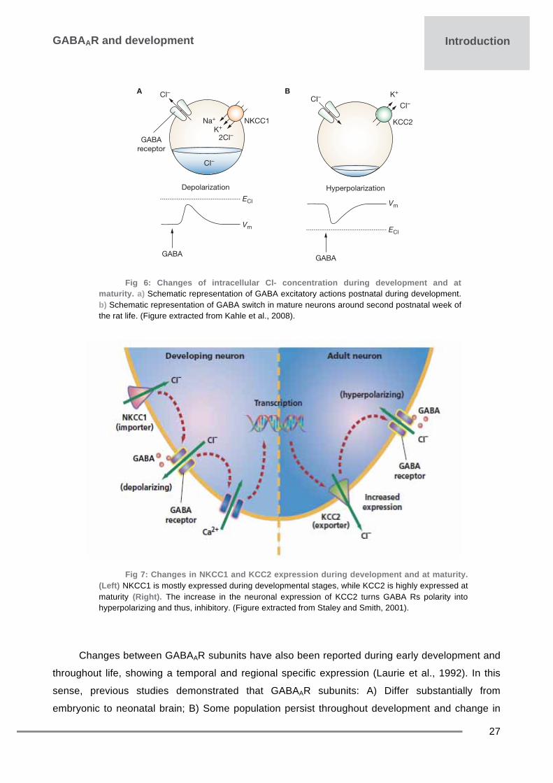
Fig 6: Changes of intracellular Cl- concentration during development and at
maturity. a) Schematic representation of GABA excitatory actions postnatal during development. b) Schematic representation of GABA switch in mature neurons around second postnatal week of the rat life. (Figure extracted from Kahle et al., 2008).
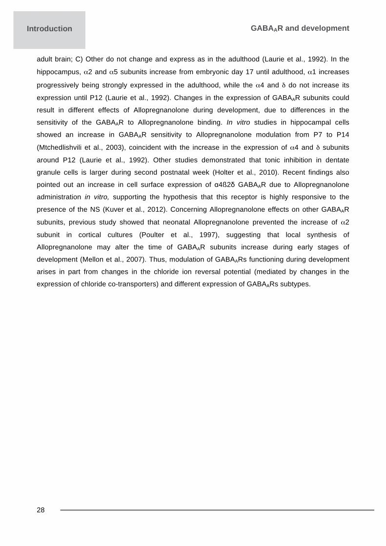
Fig 7: Changes in NKCC1 and KCC2 expression during development and at maturity. (Left) NKCC1 is mostly expressed during developmental stages, while KCC2 is highly expressed at maturity (Right). The increase in the neuronal expression of KCC2 turns GABA Rs polarity into hyperpolarizing and thus, inhibitory. (Figure extracted from Staley and Smith, 2001).
Changes between GABAAR subunits have also been reported during early development and
throughout life, showing a temporal and regional specific expression (Laurie et al., 1992). In this
sense, previous studies demonstrated that GABAAR subunits: A) Differ substantially from
embryonic to neonatal brain; B) Some population persist throughout development and change in
GABAAR and development
28
Introduction
adult brain; C) Other do not change and express as in the adulthood (Laurie et al., 1992). In the
hippocampus, α2 and α5 subunits increase from embryonic day 17 until adulthood, α1 increases
progressively being strongly expressed in the adulthood, while the α4 and δ do not increase its
expression until P12 (Laurie et al., 1992). Changes in the expression of GABAAR subunits could
result in different effects of Allopregnanolone during development, due to differences in the
sensitivity of the GABAAR to Allopregnanolone binding. In vitro studies in hippocampal cells
showed an increase in GABAAR sensitivity to Allopregnanolone modulation from P7 to P14
(Mtchedlishvili et al., 2003), coincident with the increase in the expression of α4 and δ subunits
around P12 (Laurie et al., 1992). Other studies demonstrated that tonic inhibition in dentate
granule cells is larger during second postnatal week (Holter et al., 2010). Recent findings also
pointed out an increase in cell surface expression of α4ß2δ GABAAR due to Allopregnanolone
administration in vitro, supporting the hypothesis that this receptor is highly responsive to the
presence of the NS (Kuver et al., 2012). Concerning Allopregnanolone effects on other GABAAR
subunits, previous study showed that neonatal Allopregnanolone prevented the increase of α2
subunit in cortical cultures (Poulter et al., 1997), suggesting that local synthesis of
Allopregnanolone may alter the time of GABAAR subunits increase during early stages of
development (Mellon et al., 2007). Thus, modulation of GABAARs functioning during development
arises in part from changes in the chloride ion reversal potential (mediated by changes in the
expression of chloride co-transporters) and different expression of GABAARs subtypes.

Objectives & hypothesis


Objectives
31
Objectives & hypothesis
Objectives and hypothesis The main objective of the present work is to assess the effects of neonatal manipulation of
NS levels on behavioural response to intrahippocampal Ns administration and the participation of
hippocampal GABAARs.
Specific objectives
1. To study the role of dorsal hippocampus in the Ns modulation on exploration,
anxiety-like behaviour and aversive learning.
2. To study the effects of neonatal manipulation of Ns levels in the modulation on exploration, anxiety and aversive learning in response to intrahippocampal administration of Ns.
3. To study the mechanisms underlying hippocampal and behavioural changes due to
neonatal manipulation of Ns levels by assessing:
- Exploration and anxiety-like behaviour in response to elevation of Allopregnanolone levels (progesterone administration)..
- Hippocampal GABAAR subunits: Focused on α4 and δ subunits
expression.
- Hippocampal KCC2 expression.

Hypothesis
32
Objectives & hypothesis
Hypothesis 1rts hypothesis: Hippocampus participates in the effects of Ns on exploratory, anxiety-like and aversive learning behaviour. 2nd hypothesis: Modulation of Ns levels during postnatal development affects hippocampal
maturation and behavioural response to intrahippocampal Ns administration.
3rd hypothesis: Manipulation of neonatal Ns levels alters hippocampal GABAARs containing
α4 and δ subunits.
4rd hypothesis: Neonatal manipulation of Ns can alter the hippocampal expression of
KCC2.
Research article
“Neurosteroids infusions into CA1 hippocampal region on exploration, anxiety-like behaviour and aversive learning”
Research article
“Alteration of neonatal Allopregnanolone levels affects exploration, anxiety, aversive learning and adult behavioural
response to intrahippocampal neurosteroids”
Poster presentation (Annex 2) “Effects of neurosteroids into CA1 hippocampal region on exploration, anxiety-like behaviour
and aversive learning” Societat Catalana de Biologia (2010)
Poster presentation (Annex 3) “Neonatal disturbed Allop levels affect adult performance of the passive avoidance and alter
the adult CA1 hippocampal response to neurosteroids” Behavioural Brain Research Society (2011)
)
Experiment 1


Results Overview
35
Experiment 1
In the present experiments we investigated the role of dorsal hippocampus in the modulatory
effects of Ns on exploratory, anxiety-like and aversive learning responses. For that purpose
animals were intrahippocampally administered with Allopregnanolone (2ug/0.5ul), PREGS
(5ng/0.5ul) or vehicle in each hippocampus (80-90 days old) and then tested in the Boissier
(exploratory behaviour), EPM test (anxiety-like behaviour) and passive avoidance (aversive
learning) in the adult age.
In addition, we tested whether manipulation of Ns during development changed exploratory,
anxiety-like and aversive learning responses to intrahippocampal administration of Ns in the
adulthood. For that purpose animals were administered with Allopregnanolone (20mg/kg),
finasteride (50mg/kg) or vehicle from P5 to P9. To control manipulation effects during early stages
a no handled group (NH) was also added to the experiment. In adult age, animals were
intrahippocampally administered with Allopregnanolone (2ug/0.5ul), PREGS (5ng/0.5ul) or vehicle
in each hippocampus and then tested in the Boissier (exploratory behaviour), EPM test (anxiety-
like behaviour) and passive avoidance (aversive learning) in the adult age.
An schematic representation of the experimental design can be observed in the following page.
Specific objectives -To study the role of dorsal CA1 hippocampus on exploration, anxiety and aversive learning
in response to Allopregnanolone or PREGS administration. -To study if neonatal Ns manipulation modify behavioural response to intrahippocampal
administration of Allopregnanolone or PREGS.


Experimental design Overview
37
Experiment 1
Fig 8: Experimental design
Experiment 1b: Alteration of neonatal Allopregnanolone levels affects exploration, anxiety, aversive learning and behavioural response to intrahippocampal
neurosteroids
Fig 9: Experimental design
Experiment 1a: Neurosteroids infusion into CA1 hippocampal region on exploration, anxiety-like behaviour and aversive learning


39
Experiment 1a: Hippocampus and Ns modulation of behaviour
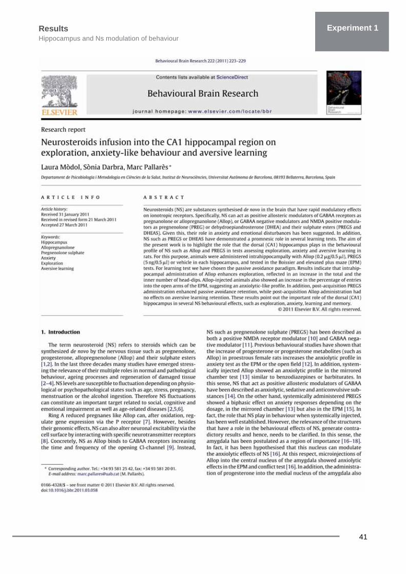
Research article Mòdol L, Darbra S, Pallarès M. Neurosteroids infusion into CA1 hippocampal region on
exploration, anxiety-like behaviour and aversive learning. Behav Brain Res 2011,

Results Hippocampus and Ns modulation of behaviour c
41
Experiment 1

Results Hippocampus and Ns modulation of behaviour
42
Experiment 1

Results Hippocampus and Ns modulation of behaviour c
43
Experiment 1

Results Hippocampus and Ns modulation of behaviour
44
Experiment 1

Results Hippocampus and Ns modulation of behaviour c
45
Experiment 1
Results Hippocampus and Ns modulation of behaviour
46
Experiment 1

Results Hippocampus and Ns modulation of behaviour c
47
Experiment 1


49
Experiment 1b: Neonatal Ns & behavioural response to intrahippocampal NS
Research article Mòdol L, Darbra S, Vallée M, Pallarès M. Alteration of neonatal Allopregnanolone levels
affects exploration, anxiety, aversive learning and adult behavioural response to intrahippocampal neurosteroids. Behav Brain Res 2013.

Results Neonatal Ns & behavioural response to intrahippocampal Ns
51
Experiment 1

Results Neonatal Ns & behavioural response to intrahippocampal Ns
52
Experiment 1

Results Neonatal Ns & behavioural response to intrahippocampal Ns
53
Experiment 1

Results Neonatal Ns & behavioural response to intrahippocampal Ns
54
Experiment 1
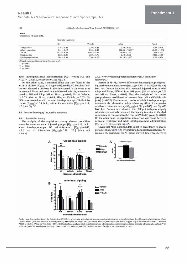
Results Neonatal Ns & behavioural response to intrahippocampal Ns
55
Experiment 1

Results Neonatal Ns & behavioural response to intrahippocampal Ns
56
Experiment 1

Results Neonatal Ns & behavioural response to intrahippocampal Ns
57
Experiment 1

Results Neonatal Ns & behavioural response to intrahippocampal Ns
58
Experiment 1
Results Neonatal Ns & behavioural response to intrahippocampal Ns
59
Experiment 1


Research article
“Neonatal finasteride administration alters hippocampal α4 and δ GABAAR subunits expression and behavioural responses to
progesterone in adult rats”
Poster presentation (Annex 4) “ Alteration of neonatal Allopregnanolone levels affects α4 and δ GABAAR subunits
expression and adult behavioural hippocampal response to neurosteroids”
Experiment 2

Results Overview
63
Experiment 2
In the present experiment we studied the changes in hippocampal α4 and δ GABAAR
subunits during early developmental stages (from P6 until P15) and in the adulthood, induced by
neonatal NS manipulation levels. For that purpose animals were neonatally administered as
described in the Experiment 1: with finasteride (50mg/kg), vehicle or saline from P5 to P9. A NH
group was also included.
In addition, we also tested whether changes in hippocampal α4 and δ GABAAR subunits
induced by neonatal NS administration were accompanied by an altered behavioural response to
increased Allopregnanolone levels (progesterone administration). For that purpose adult (80-90
days) animals were administrated with progesterone (25mg/kg) during 48h in order to increase
Allopregnanolone fluctuating levels and exploratory (Boissier) and anxiety-like behaviour (EPM)
was tested 20min after the last progesterone administration. An schematic representation of the
experimental design can be observed in the following page.
Specific objectives • To study hippocampal α4 and δ GABAAR subunits expression during early development as
a consequence of neonatal fluctuations of Ns levels. • To study whether neonatal manipulation of Ns levels modifies adult behavioural response
to increased Allopregnanolone levels (induced by progesterone administration). • To study hippocamapal α4 and δ GABAAR subunits expression in the adulthood as a
consequence of neonatal administration and adult progesterone administration.


Experimental design Overview
65
Experiment 2
Fig 10: Experimental design
Effects of neonatal manipulation of Ns and hippocampal expression of α4 and δ GABAAR subunits and
behavioural response to progesterone


67
Experiment 2: Neonatal finasteride, hippocampal GABAARs & behaviour
Research article Mòdol L, Casas C, Llidó A, Vallée M, Navarro X, Pallarès M, Darbra S. Neonatal finasteride
administration alters hippocampal α4 and δ GABAAR subunits expression and behavioural responses to progesterone in adult rats. Int J Neuropshychophamacology, 2014,


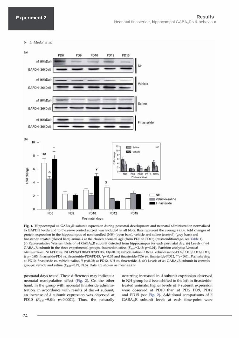
Results Neonatal finsteride, hippocampal GABAARs & behaviour
69
Experiment 2

Results Neonatal finasteride, hippocampal GABAARs & behaviour
70
Experiment 1 Experiment 2

Results Neonatal finsteride, hippocampal GABAARs & behaviour
71
Experiment 2
Results Neonatal finasteride, hippocampal GABAARs & behaviour
72
Experiment 1 Experiment 2

Results Neonatal finsteride, hippocampal GABAARs & behaviour
73
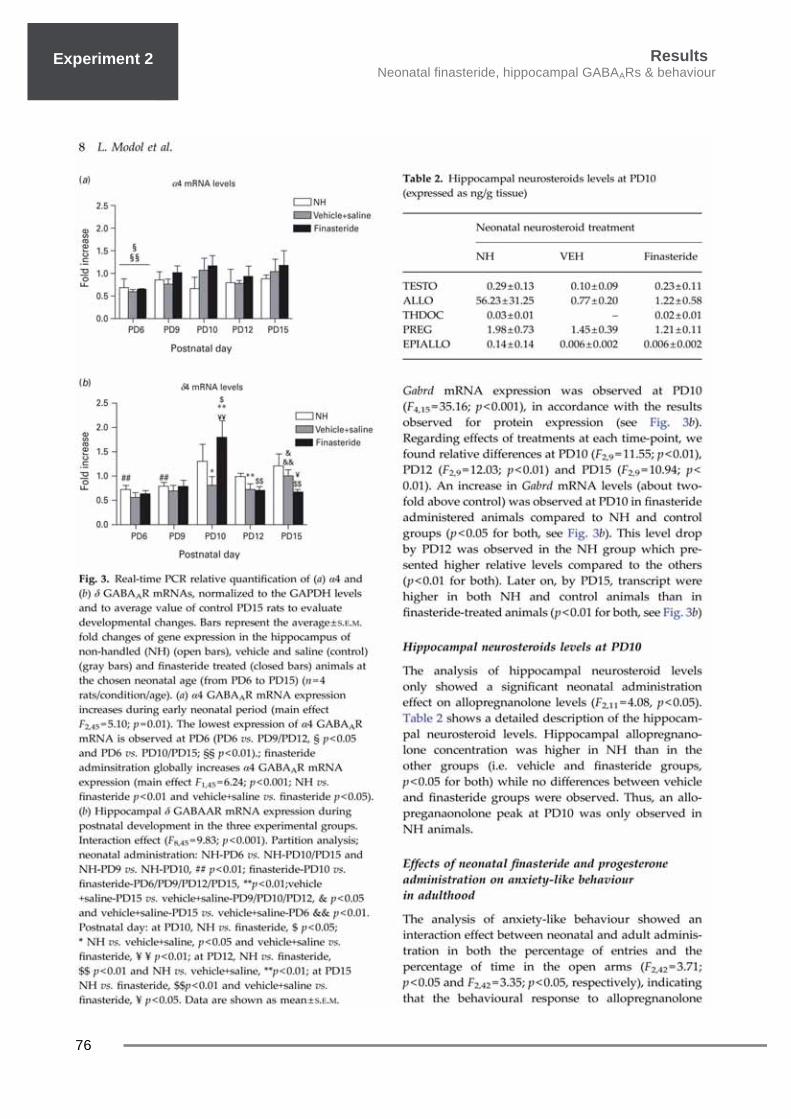
Experiment 2
Results Neonatal finasteride, hippocampal GABAARs & behaviour
74
Experiment 1 Experiment 2

Results Neonatal finsteride, hippocampal GABAARs & behaviour
75
Experiment 2
Results Neonatal finasteride, hippocampal GABAARs & behaviour
76
Experiment 1 Experiment 2

Results Neonatal finsteride, hippocampal GABAARs & behaviour
77
Experiment 2

Results Neonatal finasteride, hippocampal GABAARs & behaviour
78
Experiment 1 Experiment 2
Results Neonatal finsteride, hippocampal GABAARs & behaviour
79
Experiment 2

Results Neonatal finasteride, hippocampal GABAARs & behaviour
80
Experiment 1 Experiment 2

Results Neonatal finsteride, hippocampal GABAARs & behaviour
81
Experiment 2

Results Neonatal finasteride, hippocampal GABAARs & behaviour
82
Experiment 1 Experiment 2

Results Neonatal finsteride, hippocampal GABAARs & behaviour
83
Experiment 2


Research article “Neonatal allopregnanolone or finasteride administration modifies hippocampal K+ Cl- co-
transporter expression during early development in male rats”
Experiment 3


Results Overview
87
Experiment 3
In Experiment 3 we investigated the effects of neonatal manipulation of Ns levels on the
expression KCC2. During development GABAAR exerts depolarizing action instead of its inhibitory
profile at adult age. Cation-chloride cotransporters that modify intracellular chloride concentration
through changes in their expression mediate the polarity of GABAAR. Among them, KCC2 has
been described to change during developmental stages and related to the switch of the GABAAR
from inhibitory to excitatory. Thus, as results of Experiment 2 we observed that neonatal alteration
of Allopregnanolone levels modifies the expression of GABAARs, in the present experiment we
assessed the changes in KCC2. Animals were neonatally administered as described in Experiment
1: with Allopregnanolone (20mg/kg), finasteride (50mg/kg) or vehicle. An schematic representation
of the experimental design can be observed in the following page.
Specific objectives • To study the effects of the neonatal Allopregnanolone or finasteride administration in the
KCC2 expression during early stages of development.


Experimental design Overview
89
Experiment 3
Fig 11: Experimental design
Neonatal allopregnanolone or finasteride administration modifies hippocampal K+ Cl- co-transporter expression during early
development in male rats


91
Experiment 3: Neonatal Ns & hippocampal KCC2
Research article Mòdol L, Casas C, Navarro X, Llidó A, Pallarès M, Darbra S. Neonatal allopregnanolone or
finasteride administration modifies hippocampal K+ Cl- co-transporter expression during early development in male rats. Under review


Results Neonatal Ns & hippocampal KCC2
93
Experiment 3
Neonatal allopregnanolone or finasteride administration modifies
hippocampal K+ Cl- co-transporter expression during early development in male rats
Laura Mòdola, Caty Casasb, Anna Llidóa, Xavier Navarrob, Marc Pallarèsa and Sònia Darbraa*
a Group of Neurosteroids and Behaviour, Institut de Neurociències, Departament de
Psicobiologia i Metodologia de les Ciències de la Salut, Universitat Autònoma de Barcelona,
08193 Bellaterra, Barcelona, Spain
b Group of Neuroplasticity and Regeneration, Institut de Neurociències and Department of
Cell Biology, Physiology and Immunology, Universitat Autònoma de Barcelona, and Centro de
Investigación Biomédica en Red sobre Enfermedades Neurodegenerativas (CIBERNED), 08193
Bellaterra, Barcelona, Spain
*Corresponding author:
Sonia Darbra, Ph.D.
Departament de Psicobiologia i Metodologia de les Ciències de la Salut
Institut de Neurociències
Universitat Autònoma de Barcelona
08193 Bellaterra,
Barcelona
SPAIN
Phone: 0034.93.581.25.42
Fax: 0034.93.581.20.01
e-mail [email protected]
Results Neonatal Ns & hippocampal KCC2 expression
94
Experiment 3
ABSTRACT
The maintenance of levels of endogenous neurosteroids (NS) across early postnatal
development of the brain, particularly to the hippocampus, is crucial for their maturation.
Allopregnanolone (Allop) is a NS that exerts its effect mainly through the modulation of the GABAA
receptor (GABAAR). During early development, GABA, acting through GABAAR, that
predominantly produces depolarization shifts to hyperpolarization in mature neurons, around the
second postnatal week in rats. Several factors contribute to this change including the progressive
increase of the neuron-specific K+ /Cl- co-transporter 2 (KCC2) (a chloride exporter) levels. Thus,
we aimed to analyse whether a different profile of NS levels during development is critical and can
alter this natural progression of KCC2 stages. We administrated sustained Allop (20mg/kg) or
Finasteride (5α-reductase inhibitor, 50mg/kg) from the 5th postnatal day (PD5) to PD9 and
assessed changes in the hippocampal expression of KCC2 at transcript and protein levels as well
as its active phosphorylated state in male rats. Taken together data indicated that manipulation of
NS levels during early development influence KCC2 levels and point out the importance of
neonatal NS levels for the hippocampal development.
Keywords: neurosteroids, development, hippocampus, intra cellular chloride, GABAA
receptor, rat

Results Neonatal Ns & hippocampal KCC2
95
Experiment 3
Introduction
The maintenance of endogenous
neurosteroids (NS) levels has been
postulated to be of importance for the
maturation of the CNS and particularly for the
hippocampus (Mellon, 2007). Previous results
from our laboratory have shown the
relevance of neonatal NS levels (from
postnatal days 5 to 9, PD5 to PD9) for adult
behaviour such as anxiety, exploration and
sensorimotor gating evaluated by means of
the prepulse inhibition of the acoustic startle
response (Darbra and Pallarès 2010; 2012)
and for the behavioural response to
intrahippocampal NS administration (Darbra
et al, 2013b). Previous results from our
laboratory showed that alteration of neonatal
allopregnanolone (Allop) or pregnenolone
levels was capable to suppress the typical
anxiolytic effects provoked by
intrahippocampal Allop administration in the
adult (Mòdol et al., 2013). Besides, animals
which suffered subchronic increases in Allop
levels or pregnenolone and testosterone
levels during neonatal period, did not show
the improvement of the prepulse inhibition
response due to the intrahippocampal Allop
administration (Darbra et al., 2013a).
Although the mechanisms underlying these
alterations are still unexplained, we recently
reported data showing that neonatal NS
levels affected both neonatal and adult
hippocampal expression of the α4 and δ
gamma-aminobutyric acid A receptor
(GABAAR) subunits, which was accompanied
by an altered behavioural response to
progesterone administration in adulthood
(Mòdol et al., 2014).
During development, GABAAR activation
produces neuronal depolarization instead of
hyperpolarization which is characteristic of
the adult period. Depolarizing GABAAR
endows the system the necessary signalling
to accomplish postnatal neurogenesis,
neuronal migration, synaptogenesis and
prunning (Ben-Ari et al., 2007; Bortone and
Polleux, 2009). It is known that this
discrepancy between inmature and mature
neuronal behaviour is due to opposite sign of
Cl- intra and extracellular gradients. This
change in the sign of Cl- gradients occurs
around PD5–PD7, depending on the up
regulation of the neuron-specific K+ /Cl-
cotransporter 2 (KCC2) expression among
other signals (Ben-Ari et al., 2007). In
particular, the phosphorylation of the residue
S940 in the intracellular C-terminal domain of
KCC2, mediated by protein kinase C,
stabilizes KCC2 on the neuronal cell surface
and increases its co-transporter activity (Lee
et al., 2007; Lee et al., 2010). Thus, early
expression of KCC2 contribute to the shift of
GABA actions and impacts neuronal
maturation as well as the formation of
GABAergic synapses (Dehorter et al., 2012)
(for review see Fiumelli and Woodin, 2007).
Of note, Indeed, chronic GABAAR blockade
delayed both the GABA switch and the
developmental increase in the expression of
KCC2 (Ganguly et al., 2001; Leitch et al.,
2005).

Results Neonatal Ns & hippocampal KCC2 expression
96
Experiment 3
Taking into account the importance of NS for
the maturation of hippocampus , particularly
its GABAAR system, it is reasonable to think
that neonatal NS levels contribute to
developmental expression of KCC2. So, the
aim of the present study is to test whether the
neonatal Allop level alteration modifies KCC2
expression in the hippocampus. We
hypothesize that alteration of Allop levels is
capable to modify KCC2 expression pattern
due to its positive allosteric GABAAR
modulator profile. For that purpose, we have
altered neonatal Allop levels and we have
analysed KCC2 gene and protein expression
as well as the status of its active
phosphorylated form during postnatal period.
Methods
Animals and neonatal Allop levels alteration
One hundred and twenty-five male Wistar rats
were used and housed in a temperature-
controlled animal room (22–24 °C) on a 12-h
light/dark cycle. (Laboratori de Psicobiologia,
Universitat Autònoma de Barcelona). The male
breeders were separated from the females after
48h, pregnant females were closely watched
and on the day of birth (designed PD0) litters
were culled to 10 pups. Pups were
subcutaneously (s.c.) injected with: Allop (3α-
hydroxy-5α-pregnan-20-one; 20 mg/kg, n=33),
finasteride (a 5α-reductase inhibitor that
impedes the conversion from progesterone to
dihydroprogesterone; 50 mg/kg, n=28), vehicle
(10%-cyclodextrine as control, n=27) and saline
(n=24) once per day from PD5 to PD9). A non-
handled group (NH; n=30) was included. Drugs
were dissolved in 10% cyclodextrin ((2-
Hydroxypropyl)-β-cyclodextrin) in 0.9% NaCl.
The injection volume was 0.1 ml/10 g body
weight. All animals were obtained, housed, and
sacrificed in accordance with the protocol
approved by the Committee of the Universitat
Autònoma de Barcelona for Care and Use of
Experimental Animals and the Department of
Environment from Generalitat de Catalunya.
This protocol follows the guidelines approved by
the European Council Directive (2010/63/EU).
KCC2 expression during early development
KCC2 transcript and protein abundance was
determined using Real-time PCR and western
blot respectively Male rats were sacrificed by
decapitation at PD6 (n=27), PD9 (n=25), PD10
(n=22), PD12 (n=23), and PD15 (n=28). At
PD6 and PD9, animals were sacrificed 1 hour
after the injection. Their hippocampus was
dissected out, immediately frozen in dry ice,
and half of it was homogenized in 10 mM
HEPES (pH 7.4), 2% Triton X-100, 0.3 M KCL,
300 mM NaCl, 1 mM EDTA, protease inhibitor
cocktail (10 μl/ml, Sigma St. Louis, MO, USA)
and sodium orthovanadate (1 mM, Roche,
Basel, Switzerland) for protein extraction as
described in Mòdol et al 2014. Protein
concentration was measured by BCA protein
assay (Pierce, Rockford, IL) and equal
amounts (30 μg) were used for western
blotting. Membranes were blocked for 1 h in
TBST (100mM Tris, 0.9%. NaCl, 0.05%
Tween-20, pH 7.6) with 5 % BSA and primary
antibodies against KCC2 (rabbit anti-KCC2,
Results Neonatal Ns & hippocampal KCC2
97
Experiment 3
Millipore, 1/1000), phosphorylated KCC2
(rabbit anti-phospho-Ser940 KCC2,
Phosphosolutions, 1/1000) or glyceraldehyde-
3-phophate dehydrogenase (GAPDH, Sigma,
1:5000) were used for overnight incubation
(4°C). Horseradish peroxidase coupled
antibodies (Pierce) were used for secondary
incubation and blots were developed with the
ECL Plus detection kit (Millipore). Images were
analysed by band densitometry (Gene Tools
software, Gene Genome apparatus, Syngene,
Cambridge, UK). The same control animal was
used in each membrane performed to relativize
the results and GAPDH band was used as a
loading control. The other half of the
hippocampus was immerse in RLTβ buffer and
total RNA was obtained using the EasyRNA
extraction kit (Qiagen, Hilden, Germany)
following manufacturer instructions. Two
micrograms of RNA was reverse-transcribed
as described in Mòdol et al 2013b. Real-Time
PCR (iQ5, BioRad Foster City, CA, USA) using
Brilliant III Ultra-Fast SYBR® Green qPCR
master mix (Agilent Technologies, Santa Clara,
CA, USA) and the following primers: KCC2
(F,5’-CTTCACCCGAAACAATGTCACAGAG-
3’;R,5’-
CAGGGTGAAGTAGGAGGTCATATCAC-3’)
and Gapdh (F, 5’-
AGTTCAACGGCACAGTCAAG-3‘; R, 5’-
TACTCAGCACCAGCATCACC-3’). Three-four
samples were used per condition and each
sample was run in duplicate. The thermal
cycling conditions were: 50 ºC for 2 min, 95 ºC
for 10 min and 40 cycles of 95 ºC for 15 s, 60
ºC for 1 min. Fold change in gene expression
was estimated using the CT comparative
method (2 –DDCT) normalizing to Gapdh CT
values and relative to control samples at each
time point. Data was analyzed using two-way
analysis of variance (ANOVA) with neonatal
treatment (NEO, 5 levels:
NH/Saline/Veh/Finasteride/Allop) and postnatal
day (DAY, 5 levels) using STATISTICA
package (StatSoft, Tulsa, USA). In order to
control the possible β-cyclodextrine
administration effect on KCC2 expression, a
preliminary two way ANOVA was performed
with neonatal treatment (two levels:
Saline/VEH) and postnatal day (five levels:
PD6/PD9/PD10/PD12/PD15) as factors. Post
hoc polynomial contrasts were used when
necessary.
Results
In order to analyse the effects of the β-
cyclodextrine administration, an additional
ANOVA with NEO (2 levels, Saline and VEH)
and DAY was also performed. No differences
in KCC2 and pKCC2 protein and KCC2
mRNA between the neonatal vehicle (β-
cyclodextrin and saline) administered groups
were observed [F(1,42)=0.72; N.S; F(1,33)=0.01;
N.S; and F(1,35)=3. 35; N.S, respectively]. A
significant main effect of DAY [F(4,42)=11.72,
P< 0.001; F(4,33)=14.17, P< 0.001; and
F(4,35)=2.76, P<0.05 respectively] was also
found reflecting the expected progressive
increase in KCC3 expression. Because of
NEOX DAY interaction effect was not
observed, the normal KCC2 increase across

Results Neonatal Ns & hippocampal KCC2 expression
98
Experiment 3
the values was not affected by the
cyclodextrine. Thus, the neonatal NS
manipulation on hippocampal KCC2
expression could be unlikely attributable to
the use of cyclodextrine as vehicle. The analysis of the KCC2 mRNA levels
across early postnatal development showed a
significant NEO effect [F(4,107)=4.39;p<0.01]
and a significant DAY effect
[F(4,107)=3.98;p<0.01] while no significant
interaction effect NEO X DAY [F(16,107)=1.25;
NS] was observed. That is, a natural
progressive increase in KCC2 transcript
levels from PD6 onwards was observed in all
groups. NH animals, however, showed higher
KCC2 mRNA levels than the rest of the
groups (N-K; P<0.05 vs all). No significant
differences among others groups were
observed (see fig 1)
The analysis of the KCC2 protein abundance
across early postnatal development showed a
significant NEO effect
[F(4,118)=5.40;p<0.001], a significant DAY
effect [F(4,118)=12.99;P<0.0001] and a
significant interaction effect NEO X DAY
[F(16,118)=2,47;p<0.01]. In NH, VEH and
Saline animals, a significant effect of DAY
was observed [F(4,25)=5.71, p<0.005;
F(4,22)=1.61, p<0.01 and F(4,20)=6.24,
p<0.01, respectively]: A lineal increase of
Figure 1:Real-time PCR relative quantification of KCC2 mRNAs, normalized to the GAPDH levels and to average value of control PD15 rats to evaluate developmental changes (rats/condition/age, n=4-6). Globally, KCC2 mRNA levels increased in all experimental groups (polynomial contrast; t=--2.41 p<0.01). A neonatal administration effect was also observed [F(4, 107)=4.39;p<0.01]. Globally, KCC2 mRNA levels in NH animals were higher than the rest (N-K p<0.05 vs all) . Lines represent the average ± SEM fold changes of gene expression in the hippocampus of no handled (NH) (gray circle and gray line), vehicle (gray square and dashed gray line), saline (gray triangle and dashed gray line), Allop (black circle and black line) and finasteride treated (black squared and dashed black line) animals at the chosen neonatal age (from PD6 to PD15).Data are shown as mean ± SEM
Results Neonatal Ns & hippocampal KCC2
99
Experiment 3
protein levels along postnatal days P6 to P15
was observed in control groups (t=4.54,
p<0.001; t=4.82, p<0.001 and t=4.95,
p<0.001, respectively), indicating a
developmental increase in hippocampal
KCC2 levels (see Fig 2). In Allop treated
animals a DAY effect was also observed in
the hippocampal KCC2 expression
[F(4,28)=6.56, p<0.001], however, a
significant cubic trend was found by
polynomial post hoc contrast (t=-3.22,
p<0.01), indicating two inflection points,
which is characteristic of a cubic trend. That
is, an inverted U-shaped profile from PD6 to
PD10 and a progressive decrease from PD10
to PD15. Note that there is a significant cubic
trend, but not a significant linear or quadratic
trend. In contrast, results of neonatal
finasteride administered animals showed no
significant changes of the KCC2 protein
abundance during early postnatal
development [F(4,23)=1.52; N.S]. Thus, non-
lineal increase of protein levels along
postnatal days was observed in finasteride-
treated animals.
Expression of phosphorylated form of the
KCC2 (pKCC2) was also analysed. Results
also showed a significant NEO effect
[F(4,102)=9.02;p<0.001], DAY
[F(4,102)=14.00;p<0.001] and a significant
interaction between NEO X DAY
[F(16,102)=3.85;p<0.001]. Similar to what we
observed in the KCC2 results, a
developmental increase in the active
(phosphorylated) form of the KCC2 occurred
from PD6 to PD15 in control groups (NH,
VEH and Saline), and a significant cubic
trend was also found in samples from Allop-
treated animals while developmental lineal
increase in pKCC2 was not observed in
finasteride group (see fig 2A and 2B for
detailed post-hoc analysis).
Discussion
Results of the present study indicated that
alterations in the neonatal NS levels by
exogenous Allop administration or by
finasteride administration change the
developmental hippocampal expression and
protein abundance profile of the KCC2 during
neonatal period in vivo. We have observed that developmental KCC2
expression and levels can be modulated by
Allop administration to obtain a markedly
different profile from that of the natural
developmental increase observed in control
animals (NH and control groups) from PD6
onwards. Neonatal Allop administration
promoted an overexpression of KCC2 to
achieve supranatural protein levels around
PD10 which in turn can contribute to
feedback down-regulate its own gene
expression later on. However, an excess of
protein is not followed by a quick
phosphorylation of KCC2 which seems to be
different-regulated event that account
posteriorly, from PD12, but may depend on
protein availability at that time. Thus, a
precocious excess of KCC2 protein levels

Results Neonatal Ns & hippocampal KCC2 expression
100
Experiment 3
Figure 2: Hippocampal KCC2 abundance determined by protein semi-quantification during postnatal development and neonatal administration A). KCC2 protein abundance in the five experimental groups detected from hippocampus for each postnatal day (rats/condition/age, n=4-7). Interaction effect [F(12,105)=2,22;p<0.01]. A lineal increase of protein levels along postnatal days PD6 to PD15 was observed in all control groups (polynomial contrast; NH t=4.54, p<0.001; Vehicle t=4.82, p<0.001 and Saline t=4.95, p<0.001), while in Allop group a significant U-shaped profile from PD6 to PD10 and a progressive decrease from PD10 to PD15 (polynomial contrast; t=-3.22, p<0.01) was observed. No significant changes of the KCC2 abundance during early postnatal development were observed in finasteride group. B) Similar expression patterns were observed in the phosphorylated form of the KCC2 (pKCC2; rats/condition/age, n=4-7). Interaction effect [F(12,86)=3.67;p<0.01]. A developmental increase in pKCC2 occurred from PD6 to PD15 in all control groups (polynomial contrast; NH t=5.88, p<0.001; Vehicle t=4.51, p<0.001 and Saline t=3.93, p<0.001), a significant cubic trend was also found in Allop group (polynomial contrast; t=-3.07, p<0.01) while developmental lineal increase in pKCC2 was not observed in finasteride animals. Representative Western blots of KCC2 and pKCC2 accompanied the plots. KCC2 abundance normalized to GAPDH levels and to the same control subject was included in all blots. Lines represent the average ± SEM fold changes of protein abundance in the hippocampus of no handled (NH) (gray circle and gray line), vehicle (gray square and dashed gray line), saline (gray triangle and dashed gray line), Allop (black circle and black line) and finasteride treated (black squared and dashed black line) animals at the chosen neonatal age (from PD6 to PD15). Data are shown as mean ± SEM.

Results Neonatal Ns & hippocampal KCC2
101
Experiment 3
may not lead to an advanced disposal of its
functional form. Taking into account that Allop
exposure partially overlies the developmental
window when GABA’s depolarising action
becomes hyperpolarizing from PD7 to PD14
(Wang and Kriengstein, 2011), it is
conceivable that the increase of KCC2
expression was related to a prompted
elevation of intracellular chloride associated
with Allop-induced GABAAR activation.
Indeed, high concentrations of NS positive
GABAAR modulators, as those obtained in
the hippocampus by neonatal Allop treatment
(Darbra et al., 2013a), can directly gate the
GABAAR (Belelli and Lambert, 2005). This
functional up regulation is particularly notably
at PD6 at the beginning of Allop
administration (see fig 2B).
Interestingly, developmental up regulation of
KCC2 protein and KCC2 mRNA levels fail to
occur in finasteride-treated animals. Recently
reported data emphasize the role of
intracellular chloride concentration in
regulating the α3-α1 and δ GABAAR subunits
expression in vitro and therefore the decay
kinetics of GABAergic postsynaptic currents
and tonic inhibition (Succol et al., 2012).
Extra-synaptically localized δ contained
GABAAR receptors mediated persistent tonic
inhibition currents (Belelli and Lambert,
2005). It has been shown that the effects of
the neurosteroid Allop can reverse from
enhancing GABA-gated current to inhibiting
current at α4βδ GABAAR in a Cl- dependent
manner. The expression of these receptors
increased in the CA1 region of the
hippocampus at the onset of puberty, where
they generated an outward current (Shen et
al., 2007). In fact, Allop is capable to reverse
its classic effect of enhancing GABAAR-gated
current (hyperpolarizing response) only in
those GABAAR expressing α4 and δ
subunits, but not any other GABAAR (Shen et
al., 2007). An up regulation of the expression
of α4 and δ subunits is also observed in the
adult hippocampus when circulating levels of
Allop decrease (Allop withdrawal) (Shen et
al., 2007). In that sense, we have previously
reported that finasteride treatment increased
hippocampal levels of Allop, testosterone and
pregnenolone at PD9, i.e. at the end of
neonatal treatment (Darbra et al., 2013a),
and upregulated the expression of neonatal
and adult hippocampal α4 and δ subunits,
which was accompanied by an altered
behavioural response to progesterone
administration, even in adulthood (Mòdol et
al., 2014). Thus, the KCC2 down –regulation
observed in finasteride-treated animals could
be contributing to maintain a high level of
intracellular chloride concentration which may
lead to depolarizing actions observed at α4βδ
GABAAR receptors.
Accumulated evidence suggests that the
GABAergic system is associated with
neurodevelopmental psychiatric disorders
such as schizophrenia or depression (for
review see Hines et al., 2012). Disturbed
GABA neurotransmission in schizophrenia
might arise in part from alterations on the
nature of GABA neurotransmission, which

Results Neonatal Ns & hippocampal KCC2 expression
102
Experiment 3
can be hyperpolarizing, depolarizing, or
shunting depending on the flow of chloride
ions when GABAAR are activated (Bartos et
al., 2007). Recent studies implicate KCC2 in
both the genetic and neurodevelopmental
etiologies of schizophrenia (Hyde et al., 2011)
and changes in the KCC2 hippocampal and
cortical expression have been also recently
described in schizophrenia (Tao et al., 2012;
Arion and Lewis, 2011).
Extensive evidences indicate an activity-
dependent regulation of both the normal
developmental switch in GABAergic polarity
from excitation to inhibition and the efficacy of
GABAergic transmission through its action on
KCC2-mediated chloride homeostasis
(Fiumelli and Woodin, 2007). In this line, it
has been speculated that GABAAR subtypes
composition could be regulated by
intracellular chloride concentration in a
synaptic-specific fashion (Succol et al., 2013).
Thus, changes in the KCC2 hippocampal
expression during early stages could be
related with changes in the α4 and δ
GABAAR subunits expression in both
neonatal and adult hippocampus observed as
a consequence of neonatal NS levels
alterations (Mòdol et al, 2014).
ACKNOWLEDGMENTS This work was supported by a grant
from the Spanish Ministry of Economy and
Competitiveness (PSI2012-36646).
References
Arion D., Lewis D.A., 2011. Altered expressionof regulators of the cortical chloride transporters NKCC1 and KCC2 in schizophrenia. Arch Gen Psychiatry 68 (1), 21-31
Bartos M., Vida I., Jonas P., 2007. Synaptic mechanisms of synchronized gamma oscil- lations in inhibitory interneuron networks. Nat Rev Neurosci. 8(1):45-56.
Belelli D., Lambert, J.J.,2005. Neurosteroids: endogenous regulators of the GABA(A) receptor. Nat. Rev. Neurosci. 6(7), 565-575.
Ben-Ari, Y., Gaiarsa, J.L., Tyzio, R., Khazipov, R., 2007. GABA: A pioneer transmitter that exites immature neurons and generates primitive oscillations. Physiol Rev 87, 1215-1284.
Bortone, D., Polleux F., 2009. KCC2 expression promotes the termination of cortical interneuron mogration in a voltage-sensitive calcium-dependent manner. Neuron 62, 53-71.
Darbra, S., Pallarès, M., 2010. Alterations in neonatal neurosteroid affect exploration during adolescente and prepulse inhibition in the adulthood. Psychoneuroendocrinology 35(4), 525-35.
Darbra, S., Pallarès, M., 2012. Effects of early postnatal allopregnanolone administration on elevated plus maze anxiety scores in adult male wistar rats. Neuropsychobiology. 65(1), 20-27.
Darbra, S., Mòdol, L., Vallée, M., Pallarès. M., 2013 Neonatal neurosteroid levels are determinant in shaping adult prepulse inhibition response to hippocampal allopregnanolone in rats. Psychoneuroendocrinol.38(8),1397-1406.
Darbra, S., Mòdol, L., Llidó, A., Casas, C., Vallée, M., Pallarès, M., 2014. Neonatal allopregnanolone levels alteration: Effects on behavior and role of the hippocampus. Prog Neurobiol. 113, 95-105.
Dehorter, N., Vinay, L., Hammond, C., Ben-Ari, Y., 2012. Timing of developmental sequences in different brain structures: physiological and pathological implications. Eur J Neurosci.35(12),1846-1856.
Fiumelli, H., Woodin, A,, 2007. Role of activity-dependent regulation of neuronal chloride

Results Neonatal Ns & hippocampal KCC2
103
Experiment 3
homeostasis in development. Current opinion in neurobiology 17,81-86.
Ganguly, K., Schinder, A.F., Wong, S.T., Poo, M.M., 2001. GABA itself promotes the developmental switch of neuronal GABAergic responses from excitation to inhibition. Cell 105, 521–532.
Hines, R., Oavies, P., Moss, S. J., Maguire, J., 2012. Functional regulation of GABAA receptors in nervous system pathologies. Curr Opin Neurobiol, 22(3), 552-558.
yde, T.M., Lipska, B.K., Ali, T., Mathew, S.V., Law, A.J., Metitiri, O.E., Straub, R.E., Ye, T., Colantuoni, .C, Herman, M.M., Bigelow, L.B., Weinberger, D.R., Kleinman, J.E., 2011. Expression of GABAsignaling molecules KCC2, NKCC1,and GAD1 in cortical development and schizophrenia. J Neurosci 31:11088–11095.
Lee, H.H.,Walker, J.A.,Williams, J.R.,Goodier, R.J., Payne, J.A.,Moss, S.J., 2007. Direct protein kinase C-dependent phosphorylation regulates the cell surface stability and activity of the potassium chloride cotransporter KCC2. J. Biol. Chem. 282, 29777–29784.
Lee, H.H. C, Jurd, R., Moss, S.J., 2010. Tyrosine phosphorylation regulates the membrane trafficking of the potassium chloride co-transporter KCC2. Mol Cell Neurosci 45, 173–179.
Leitch, E., Coaker, J., Young, C., Mehta, V., Sernagor, E., 2005. GABA type-A activity controls its own developmental polarity switch in the maturing retina. J Neurosci 25: 4801–4805.
Mellon, S.H., 2007. Neurosteroid regulation of central nervous system development Pharmacol. Ther. 116 (1), 107–124.
Mòdol, L., Darbra, S., Vallée, M., Pallarès, M., 2013. Alteration of neonatal Allopregnanolone levels affects exploration, anxiety, aversive learning and adult behavioural response to intrahippocampal neurosteroids. Behav Brain Res. 241,96-104.
Mòdol, L., Casas, C., Navarro, X., Llidó, A., Vallée, M., Pallarès, M., Darbra, S., 2014. Neonatal finasteride administration alters hippocampal α4 and δ GABAAR subunits expression and behavioural responses to progesterone in adult rats. Int J Neuropsychopharmacol. 17(2):259-273
Shen, H., Gong, Q. H., Aoki, C., Yuan, M., Ruderman, Y., Dattilo, M., Williams, K., Smith, S.S., 2007. Reversal of neurosteroid effects at alpha4beta2delta GABAA receptors triggers anxiety at puberty. Nature neuroscience.10 (4),469-477.
Succol, F., Fiumelli, H., Benfenati, F., Cancedda, L., Barberis, A., 2012. Intracellular chloride concentration influences the GABAA receptor subunit composition. Nat. Commun. 3:738 doi: 10.1038/ncomms1744.
Tao, R., Li, C., Newburn, E.N., Ye, T., Lipska, B.K., Herman, M.M., Weinberger, D.R., Kleinman, J.E., Hyde, T.M., 2012. Transcript-specific associations of SLC12A5 (KCC2) in human prefrontal cortex with development, schizophrenia, and affective disorders. J Neurosci 32(15), 5216-5222.
Wang,. D.D., Kriegstein, A.R., 2011. Blocking early GABA depolarization with bumetanide results in permanent alterations in cortical circuits and sensorimotor gating deficits.
. 21(3), 574-587.

.

Discussion


Discussion Neonatal Ns and behavioural response to intrahippocampal Ns administration
107
Discussion
Discussion Role of neonatal Ns fluctuations in the behavioural response to
intrahippocampal Ns administration In the Experiment 1 we studied the effects of neonatal manipulation of Ns levels on the
behavioural response to intrahippocampal administration of Ns. For this purpose we first assessed
the implication of dorsal hippocampus in the Ns behavioural effects on exploration, anxiety and
aversive learning as the hippocampus has been implicated in the modulation of these behaviours
(Bitran et al., 1999; Zhang et al., 2002; Howland et al., 2004; Adams and Van den Buuse, 2011;
Mineur et al., 2013). Accordingly to its previously described GABAAR allosteric positive modulator
profile (Belelli and Lambert, 2005; Lambert et al., 2003, 2009), intrahippocampal Allopregnanolone
induced an increase in the exploratory behaviour in the Boissier test and exerted an anxiolytic-like
profile in the EPM (Experiment 1). Similar results have also been obtained with other Ns such as
pregnenolone into the dorsal hippocampus (Bitran et al., 1999). Also, we studied the role of the
hippocampus in the modulation of Ns effects in the passive avoidance learning, an aversive
learning with an important emotional component. In this paradigm, the animal must inhibit the
natural tendency to enter into a dark compartment in order to avoid an electric foot-shock. Thus,
passive avoidance is considered as a negative reinforced learning. Our results showed that
intrahippocampal PREGS administration increased the aversive learning (Experiment 1) in
accordance with the previously described promnesic profile of PREGS (Pallarès et al., 1998;
Vallée et al., 1997; Mayo et al., 2003). It has been suggested that PREGS promnesic profile can
be mediated by the potentiation of NMDA receptors in pyramidal neurons (Bowly, 1993), but also
through the potentiation of cholinergic neurons (Pallarès et al., 1998; Darnaudéry et al., 2000).
Thereby, these findings suggest that together with other postulated brain structures such as the
amygdala (Akwa et al., 1999; Engin and Treit, 2007) or the lateral septum (Bitran et al., 1999), the
dorsal hippocampus could be relevant for explaining the effects of Ns on exploratory, anxiety-like
or aversive learning behaviour.
It is well established that Ns participate in shaping and organising the CNS during
development, having influence in brain function and in a variety of behaviours (see Introduction).
Thus, it could be expected that if neonatal alteration of Ns affects the development of the
hippocampus, consequently the behavioural response to intrahippocampal Ns administration could
be also modified. For that purpose, we studied if neonatal manipulation of Ns levels alters
exploration, anxiety and aversive learning responses to intrahippocampal Ns administration. Our
results showed that neonatal Allopregnanolone administration abolished the anxiolytic profile
induced by hippocampal Allopregnanolone (Experiment 1). Moreover, results of aversive learning
response showed that the previously described PREGS promnesic profile when administered into
Discussion Neonatal Ns, hippocampal α4 and δ GABAAR subutnis & behaviour
108
Discussion
the hippocampus is not observed in animals neonatally administered with Allopregnanolone or
finasteride. Nevertheless, results of exploratory behaviour showed that neonatal manipulation of
Ns did not affect the behavioural response to intrahippocampal Allopregnanolone administration
(Experiment 1). These data indicate that neonatal manipulation of Ns modifies the
intrahippocampal Allopregnanolone anxiolytic and PREGS promnesic profile (Experiment 1). In this
line, previous studies of our laboratory have also demonstrated that neonatal alteration of Ns levels
abolish the decrease in locomotor activity induced by intrahippocampal Allopregnanolone
administration tested in the open field (Darbra and Pallarès, 2011). Thus, present results support
our hypothesis concerning that alteration of Ns levels during early developmental affects the
behavioural response to intrahippocampal Ns administration; and point out the role of neonatal Ns
for the hippocampal development.
Mechanisms underlying hippocampal and behavioural changes due to
neonatal manipulation of Ns levels: Focused on the hippocampal expression of
GABAAR containing α4 and δ subunits
The α4 and δ subunits combination has been described to be the most sensitive to Ns
(reviewed in Shen and Smith, 2009). Thus, given the obtained results in the Experiment 1, we
hypothesised that some of the hippocampal mechanisms underlying the behavioural alterations
that we observed due to neonatal alteration of Ns levels could be mediated through changes in
GABAAR containing α4 and δ subunits. Our results indicated that neonatal finasteride
administration increases hippocampal expression pattern of α4 and δ GABAAR subunits during
postnatal development (from P6 to P15) mainly at P10 (Experiment 2), thereby corroborating our
hypothesis concerning that neonatal manipulation of Ns levels modifies hippocampal α4 and δ
GABAAR subunits. Other authors reported that changes in the expression of α4 and δ GABAAR
subunits could be induced by the fluctuation of Allopregnanolone or its precursor progesterone
(Smith et al., 1998; Gulinello et al., 2001; Gulinello et al., 2002; Shen et al., 2005; Gangisetty and
Reddy, 2010). In this sense, we have also observed fluctuation of Ns levels induced by finasteride
administration (i.e. testosterone, pregnenolone, Allopregnanolone) (Darbra et al., 2013;
Experiment 2). Thus, the reported hippocampal changes observed in α4 and δ GABAAR subunits
expression might be related to the fluctuations on hippocampal Ns levels induced by the
administration of finasteride. Nevertheless, the functional mechanism by which finasteride
administration in neonatal pups could induce the observed increase in GABAAR subunits
expression remains to be elucidated. Indeed, neonatal activation of the GABAARs is an important
source of activity, necessary for the GABA-glutamate sequential maturation of interneurons and

Discussion Neonatal Ns, hippocampal α4 and δ GABAAR subutnis & behaviour
109
Discussion
pyramidal neurons in the hippocampus (Ben-Ari, 2002; Ben-Ari et al., 2007). Thereby, alterations
in physiological levels of GABA-positive NS during these periods can alter the physiological
maturation of GABAergic systems, also affecting other neurotransmitters and modifying the
expression of other GABAARs in the hippocampus.
Interestingly, neonatal finasteride administration increased the expression of hippocampal
α4 and δ GABAAR subunits not only during postnatal period but also in the adulthood (Experiment
2). Furthermore, our results showed that increased hippocampal α4 and δ GABAAR subunits
expression was also accompanied by an altered anxiety response to progesterone administration
in the adulthood (Experiment 2). A possible mechanism to explain the increase of anxiety scores
after progesterone administrations can be attributed to the ability of Allopregnanolone to reverse its
classic inhibitory effect of enhancing GABAAR-gated currents (hyperpolarizing response) only in
those GABAARs expressing α4 and δ subunits, but not any other GABAARs as showed by other
authors (Shen et al., 2007). However, the involvement of other mechanisms or other GABAAR
subunits cannot be discarded. Thus, our data indicate that changes in hippocampal α4 and δ
subunits of the GABAAR induced by finasteride administration are related to the altered anxiety-like
behaviour in response to progesterone administration in the adulthood. It is important to remark
that an increase in α4 and δ GABAAR subunits mRNA in adulthood has been related to epileptic
seizures (Brooks- Kayal et al., 1998; Cohen et al., 2003; Maguire et al., 2005), and that NS action
through δ subunit-containing GABAAR is required for the physiological response to stress and
stress-induced anxiety-like behaviour (Sarkar et al., 2011). In agreement with the above-presented
data, other studies demonstrated changes in cortical GABAAR subunits expression in adulthood as
a consequence of neonatal steroids administration (Calza et al., 2010). Taken together, results of
Experiment 2 suggest that neonatal Ns fluctuation alter the hippocampal expression of α4 and δ
subunits of the GABAAR. Concretely, finasteride administration induced changes in hippocampal
α4 and δ subunits related to the altered adult anxiety-like behaviour in response to progesterone
administration. Thus, present results indicate that early life NS manipulations are important for the
maturation of the GABAAR, and can lead to an altered adult system that responds differently to Ns
in the adulthood (Brooks-Kayal et al., 2001; Calza et al., 2010).

Discussion Neonatal Ns & changes in hippocampal KCC2 expression
110
Discussion
Mechanisms underlying hippocampal and behavioural changes due to neonatal manipulation of Ns levels: Changes in the hippocampal expression of KCC2
Our results of Experiment 3 indicated that neonatal alteration of Ns by both Allopregnanolone
or finasteride administration alters the developmental expression of KCC2 in the hippocampus. In
one hand, neonatal Allopregnanolone administration induced a marked up regulation of
hippocampal KCC2 at earlier stages in comparison with NH and Control groups, while neonatal
finasteride administration impaired the normal progressive up regulation of KCC2 from P6 until P15
(Experiment 3). Thus, the hypothesis that neonatal Ns fluctuation alters the hippocampal
expression of KCC2 during postnatal stages was verified.
Taking into account that Allopregnanolone exposure partially overlies the developmental
window when GABA is depolarising and that it became hyperpolarizing from P7 to P14 (Wang and
Kriengstein, 2011), it is conceivable that the KCC2 up regulation could be related to an elevation of
intracellular chloride associated with activation of GABAARs by Allopregnanolone administration.
As mentioned, NS that act as an allosteric positive modulators of GABAAR, can also directly gate
the GABAAR when administered at high concentrations (Belelli and Lambert, 2005). Other authors
showed that early KCC2 up regulation affects morphology of cortical neurons in vivo (Li et al.,
2007) and is sufficient to reduce interneuron migration by rendering GABAARs activation
hyperpolarizing (Li et al., 2007; Bortone and Polleux, 2009). Moreover, previous studies
demonstrated that neonatal Allopregnanolone administration to hippocampal cultures decreases
neurite length (Brinton, 1994). Thus, is possible that changes in the KCC2 expression induced by
the administration of NS during a relevant developmental window could affect structurally and
morphologically hippocampal interneurons. In this sense, other studies reported a decrease in
thalamic and cortical interneurons induced by Allopregnanolone administration at P5 (Gizerian et
al., 2004; Grobin et al., 2006). Changes in the timing pattern of KCC2 expression has also been
shown to alter the excitatory driving force of Cl-, shifting from excitatory to inhibitory signalling in
the hippocampus and other neural structures (Ben-Ari et al., 1989; Ikeda et al., 2005; Payne et al.,
2003; Ben-Ari et al., 2007; Khirug et al., 2010) related to an altered morphology and migration of
neurons (Li et al., 2007; Bortone and Polleux, 2009). Thereby, it is plausible that behavioural
alterations observed in the present work could also be related to the reported changes in the timing
pattern expression of hippocampal KCC2 induced by the alteration of neonatal NS levels. Indeed,
the relation between KCC2 alteration and adult behavioural disturbances is beginning to generate
an increasing interest in the study of the aetiology of some psychopathologies, such as
schizophrenia and affective disorders (Arion and Lewis, 2011; Hyde et al., 2011; Tao et al., 2012).
In this sense, extensive evidences indicate an activity-dependent regulation of both the normal
developmental switch in GABAergic polarity from excitation to inhibition and the efficacy of

Discussion Neonatal NS, hippocampal α4 and δ GABAAR subutnis & behaviour
111
Discussion
GABAergic transmission through its action on KCC2-mediated chloride homeostasis (Fiumelli and
Woodin, 2007). In this line, it has been speculated that GABAAR subtypes composition could also
be regulated by intracellular chloride concentration in a synaptic-specific fashion (Succol et al.,
2013).
In summary, present results demonstrate that Ns fluctuation during postnatal development
affect the hippocampal expression of KCC2. These changes could also be related with changes in
the α4 and δ GABAAR subunits expression in both neonatal and adult hippocampus observed as a
consequence of neonatal Ns levels alterations.


Conclusions


115
References Conclusions
Findings highlights
Experiment 1 A) Dorsal hippocampus is a relevant structure related to the Ns effects on exploration,
anxiety-like behaviour and aversive learning.
B) Neonatal Allopregnanolone administration alters the anxiolytic profile induced by
intrahippocampal Allopregnanolone administration
C) Alteration of neonatal Ns levels modify the promnesic profile induced by
intrahippocampal PREGS administration
Experiment 2 A) Neonatal finasteride administration increases hippocampal α4 and δ GABAAR subunits
expression during postnatal development and in the adulthood.
B) Neonatal finasteride increases anxiety-like behaviour in response to progesterone
administration accompanied by a decrease in hippocampal δ GABAAR subunit.
Experiment 3 A) Neonatal administration of Allopregnanolone induces an early up regulation of
hippocampal KCC2. B) Neonatal administration of finasteride prevented the hippocampal increase of KCC2
during postnatal development.


117
References

119
References
References
Agis-Balboa RC, Pinna G, Zhubu A, Maloku E, Veldic M, Costa E, Guidotti A. 2006. Characterization of brain neurons that express enzymes mediating neurosteroid biosynthesis. Proc Natl Acad Sci USA, 103(39):14602-7.
Akerman CJ, Cline HT. 2007. Refining the roles of GABAergic signalling during neural circuit formation. Trends Neurosci, 30(8): 382-9.
Akk G, Bracamonstes J, Steinbach JH. 2001. Pregnenolone sulfate block of GABA(A) receptors: mechanism and involvement of a residue in the M2 region of the alpha subunit. J Physiol, 532(3): 673-84.
Akwa Y, Purdy RH, Koob GF, and Britton KT. 1999. The amygdala mediates the anxiolytic-like effect of the neurosteroid allopregnanolone in rat. Behav Brain Res, 106: 119-125.
Arion D, Lewis DA. 2011. Altered expression of regulators of the cortical chloride transporters NKCC1 and KCC2 in schizophrenia. Arch Gen Psychiatry, 68(1): 21-31.
Barbaccia ML, Concas A, Serra M, Biggio G. 1998. Stress and neurosteroids in adult and aged rats. Exp Gerontol, 33(7–8):697–712.
Baulieu EE, Fluxe K, Gustafsson JA, Weterberg L. 1981. Steroid hormones in the brain: several mecahnism?In steroid hormone regulation of the brain. Oxford: Pergamon Press, 3-14.
Baulieu EE. 1998. Neurosteroids: A novel function of the brain. Psychoneuroendocrinology, 23(8): 963-87. Belelli D, Casula A, Ling A, Lambert JJ. 2002. The influence of subunit composition on the interaction of
neurosteroids with GABA(A) receptors. Neuropharmachol, 43: 651-661. Belelli D, Lambert JJ. 2005. Neurosteroids: endogenous regulators of the GABA(A) receptor, 6(7):565-75Belelli D, Harrison NL, Maguire J, Macdonald RL, Walker MC, Cope DW. 2009. Extrasynaptic GABAA
receptors: form, pharmacology, and function. J. Neurosci, 29(41):12757-63. Ben-Ari Y, Cherubini E, Corradetti R, Gaiarsa JL. 1989. Giant synaptic potentials in immature rat CA3
hippocampal neurons. J Physiol, 416: 303–325. Ben-Ari Y, Khazipov R, Leinekugel X, Caillard O, Gaiarsa JL. 1997. GABAA, NMDA and AMPA receptors: a
developmentally regulated 'ménage à trois'. Trends Neurosci, 20(11):523-9. Ben-Ari Y, Tseeb V, Raggozzino D, Khazipov R, Gaiarsa JL. 1994. Gamma-Aminobutyric acid (GABA): a
fast excitatory transmitter which may regulate the development of hippocampal neurones in early postnatal life. Prog Brain Res, 102:261-73.
Ben-Ari Y. 2002. Exitatory actions of GABA during development: The nature of nurture. Nat Rev Neurosci, 3: 728-39.
Ben-Ari Y, Gaiarsa JL, Tyzio R, Khazipov R. 2007. GABA: A pioneer transmitter that excites immature neurons and generates primitive oscillations. Physiol Rev, 87:1215–1284.
Biggio F, Gorini G; Caria S, Murru L, Mostallino M, Sanna E, Follesa P. 2006. Plastic neuronal changes in GABAA receptor gene expression induced by progesterone metabolites: In vitro molecular and functional studies. Pharmacol Biochem Behav, 84:545-554.
Bernardi F, Salvestroni C, Casarosa E, Nappi RE, Lanzone A, Luisi S, Puerdy RH, Petraglia F, Genazzani AR. 1998. Aging is associated with changes in allopregnanolone concentrationsin brain, endocrine glands and serum in male rats. Eur J Endocrinol, 138:316-21.
Bitran D, Purdy RH, Kellog CK. 1993. Anxiolytic effect of progesterone is associated with increases in cortical allopregnanolone and GABAA receptor function. Pharmacol Biochem Behav, 45(2):423-428.
Bitran D, Shiekh M, McLeod M. 1995. Anxiolytic effect of progesterone is mediated by the neurosteroid allopregnanolone at brain GABAA receptors. J Neuroendocrinol, 7(3):171-7.
Bitran D, Dugan M, Renda P, Ellis R, and Foley M. 1999. Anxiolytic effects of the neuroactive steroid pregnanolone (3 alpha-OH-5 beta-pregnan-20-one) after microinjection in the dorsal hippocampus and lateral septum. Brain Res, 850:217-224.
Bitran D, Foley M, Audette D, Leslie N, and Frye CA. 2000. Activation of peripheral mitochondrial benzodiazepine receptor in the hippocampus stimulates allopregnanolone synthesis and produces anxiolytic-like effects in the rat. Psychopharmacology, 151:64-71.

120
References
Bitran D, Smith SS. 2005. Termination of pseudopregnancy in the rat produces an anxiogenic-like response that is associated with an increase in benzodiazepine receptor binding density and a decrease in GABA-stimulated chloride influx in the hippocampus. Brain Res Bull, 64(6):511-8.
Blaesse P, Airaksinen MS, Rivera C, Kalia K. 2009. Cation-chloride cotransporters and neuronal function. Nueron, 61(6):820-38.
Boissier JR, Simon P. 1967. Automatisation du test de la planche à trous. Physiol Behav. 2:447–448 Bonifazi P, Goldin M, Picardo MA, Jorquera I, Cattani A, Bianconni G, Represa A, Ben-Ari Y, Cossart R.
2009. GABAergic hub neurons orchestrate synchrony in developing hippocampal networks. Science, 326(5958):1419-24.
Bortone D and Polleux F. 2009. KCC2 expression promotes the termination of cortical interneuron mogration in a voltage-sensitive calcium-dependent manner. Neuron, 62: 53-71.
Bowly, MR. 1993. Pregnenolone sulfate potentiation of NMDA receptor channels in hippocampal neurons. Mol Pharm 43:813-819.
Bray JG, Myblieff M. 2009. Influx of calcium through L-type calcium channels in early postnatal regulation of chloride transporters in the rat hippocampus. Dev Neurobiol, 89(13):885-96.
Brinton RD. 1994. The neurosteroid 3 alpha-hydroxy-5 alpha-pregnan-20-one induces cytoarchitectural regression in cultured fetal hippocampal neurons. J. Neurosci, 14(5 Pt 1):2763-74.
Brooks-Kayal AR, Shumate MD, Jin H, Rikhter TY, Coulter DA. 1998. Selective changes in single cell GABAA receptor subunit expression and function in temporal lobe epilepsy. Nat Med, 4:1166–1172.
Brooks-Kayal AR, Shumate MD, Jin H, Rikhter TY, Kelly ME, Coulter DA. 2001. Gamma-Aminobutyric acid(A) receptor subunit expression predicts functional changes in hippocampal dentate granule cells during postnatal development. J. Neurochem, 77(5):1266-78.
Brunton PJ, McKay AJ, Ochedalski T, Piastowska A, Rebas E, Lachowicz A, Russell JA. 2009. Central opioid inhibition of neuroendocrine stress responses in pregnancy in the rat is induced by the neurosteroid allopregnanolone. J. Neurosci, 29(20):6449-60.
Brunton PJ, Russell JA, Hirst JJ. 2014. Allopregnanolone in the brain: protecting pregnancy and birth outcomes. Prog Neurobiol 113:106-36.
Calza A, Sogliano C, Santoru F, Marra C, Angioni MM, Mostallino MC, Biggio G, Concas A. 2010. Neonatal exposure to estradiol in rats influences neuroactive steroids concentrations, GABAA receptor expression and behavioural sensitivity to anxiolytic drugs. J. Neurochem, 113:1285-1295.
Cancedda L, Fiumelli H, Chen K, Poo MM. 2007. Excitatory GABA action is essential for morphological maturation of cortical neurons in vivo. J Neurosci, 27:5224–5235.
Cherubini E, Gaı ̈arsa JL, Ben-Ari Y. 1991. GABA: an excitatory trans- mitter in early postnatal life. Trends Neurosci, 14:515–519.
Chisari M, Lawrence NE, Douglas FC, Mennerick S and Zorumski CF. 2010. The sticky issue of neurosteroids and GABAA receptors. Trends in Neuro, 33:299-306.
Chudotvorova I, Ivanov A, Rama S, Hubner CA, Pellegrino C, Ben-Ari Y, Medina I. 2005. Early expression of KCC2 in rat hippocampal cultures augments expression of functional GABA synapses. J Physiol, 566: 671–679.
Compagnone NA, Mellon SH. 2000. Neurosteroids: biosynthesis and function of these novel neuromodulators. Front Neuroendocrinol, 21(1):1-56.
Cooper EJ, Johnston GA, Edwards FA. 1999. Effects of a naturall occurring neurosteroid on GABAA IPSCs during development in rat hippocampal or cerebellar slices. J Physiol, 521(Pt 2):437–449.
Cotter, D.R., Pariante, C.M., Everall, I.P. 2001. Glial cell abnormalities in major psychiatric disorders: the evidence and implications. Brain Res Bull, 55(5):585–595.
Damianisch K, Rupprecht R, Lancel M. 2001. The influence of subchronic administration of the neurosteroid allopregnanolone on sleep in the rat. Neuropsychopharmacology, 25(4):576-94.
Darbra S and Pallarès M. 2009. Neonatal allopregnanolone increases novelty-directed locomotion and disrupts behavioural responses to GABA(A) receptor modulators in adulthood. Int J Dev Neurosci, 27:617-625.
Darbra S and Pallarès M. 2010. Alterations in neonatal neurosteroid affect exploration during adolescente and prepulse inhibition in the adulthood. Psychoneuroendocrinology, 35(4):525-35.
121
References
Darbra S, Pallarès M. 2011. Interaction between early postnatal neurosteroid manipulations and adult infusión of neurosteroid into CA1 hippocampal region on the open field behaviour. Behav Brain Res, 216(2):705-11.
Darbra S, Pallarès M. 2012. Effects of early postnatal allopregnanolone administration on elevated plus maze anxiety scores in adult male wistar rats. Neuropsychobiology, 65(1):20-7.
Darbra S, Mòdol L, Vallée M, Pallarès M. 2013. Neonatal neurosteroid levels are determinant in shaping adult prepulse inhibition response to hippocampal allopregnanolone in rats. Psychoneuroendocrinol, 38(8):1397-1406.
Darbra S, Mòdol L, Llidó A, Casas C, Vallée M, Pallarès M. 2014. Neonatal allopregnanolone levels alteration: Effects on behavior and role of the hippocampus. Prog Neurobiol, 113:95-105.
Darnaudéry M, Koehl, M, Piazza PV, Le Moal M, and Mayo, W. 2000. Pregnenolone sulfate increases hippocampal acetylcholine release and spatial recognition. Brain Res, 852:173-179.
Dehorter N, Vinay L, Hammond C, Ben-Ari Y. 2012. Timing of developmental sequences in different brain structures: physiological and pathological implications. Eur J Neurosci, 35(12),1846-1856.
Duvovicky M, Skultelvova I, Jezova D. 1999. Neonatal stress alters habituation of exploratory behavior in adult male but not female rats. Phamacol Biocehm Behav, 64(4):681-6.
Dubrovsky, BO. 2005. Steroids, neuroactive steroids and neurosteroids in psychopathology. Prog Neuropsychopharmacol Biol Psychiatry, 29:169-192.
Dzhala VI, Staley KJ. 2003. Excitatory actions of endogenously released GABA contribute to initiation of ictal epileptiform activity in the developing hippocampus. J Neurosci, 23:1840–1846.
Engin E, and Treit D. 2007. The role of hippocampus in anxiety: intracerebral infusion studies. Behav Pharmacol, 18:365-374.
Essrich C, Lorez M, Benson JA, Fritschy JM, Luscher B. 1998. Postsynaptic clustering of major GABAA receptor subtypes requires the γ2 subunit and gephyrin. Nat Neurosci,1(7):563–571.
Farrant M, Nusser Z. 2005. Variations on an inhibitory theme: phasic and tonic activation of GABA(A) receptors. Nat Rev Neurosci, 6(3):215-29.
Finn DA, Roberts AJ, Long S, Tanchuck M and Phillips TJ. 2003. Neurosteroid consumption has anxiolytic effects in mice. Pharmacol Biochem Behav, 76:451-462.
Fiumelli H, Woodin A. 2007. Role of activity-dependent regulation of neuronal chloride homeostasis in development. Current opinion in neurobiology, 17:81-86.
Flood JF, Morley JE and Roberts E. 1992. Memory-enhancing effects in male mice of pregnenolone and steroids metabolically derived from it. Proc Nat Acad Sci USA, 89(5):1567-71.
Flood JF, Morley JE and Roberts E. 1995. Pregnenolone sulfate enhances post-training memory processes when injected in very low doses into limbic system structures: the amygdala is by far the most sensitive. Proc Natl Acad Sci USA, 92:10806-10810.
Follesa P, Mancuso L, Biggio F, Cagetti E, Franco M, Trapani G, Biggio G. 2002. Changes in GABAA receptor gene expression induced by withdrawal of, but not by long-term exposure to, zaleplon or zolpidem. Neuropharmacology, 42:191–198.
Frye CA and Walf AA. 2002. Changes in progesterone metabolites in the hippocampus can modulate open field and forced swim test behavior of proestrous rats. Horm and Behav, 41:306-315.
Frye CA and Walf AA. 2004a. Estrogen and/or progesterone administered systemically or to the amygdala can have anxiety-, fear-, and pain-reducing effects in ovarectomized rats. Behav Neurosc, 118(2):306-313.
Frye CA and Walf AA. 2004b. Hippocampal 3α, 5α–THP may alter behavior of pregnant and lactating rats. Pharma, Biochem and Behav, 78:531-540.
Frye CA and Rhodes ME. 2007. Infusions of 3α, 5α–THP to the VTA enahnce exploratory, anti-anxiety, social, and sexual behavior and increase levels of 3α,5α-THP in midbrain, hippocampus, diencephalon, and cortex of female rats. Behav Brain Res, 187:88-99.
Frye CA. 2009. Neurosteroids' effects and mechanisms for social, cognitive, emotional, and physical functions. Psychoneuroendocrinology, 34(Suppl 1):S143-161.

122
References
Frye CA, Hirst JJ, Brunton PJ, Russell JA. 2011. Neurosteroids for a successful pregnancy. Stress, 14(1):1-5.
Frye CA, Paris JJ, Walf AA, Rusconi JC. 2012. Effects and Mechanisms of 3α,5α,-THP on Emotion, Motivation, and Reward Functions Involving Pregnane Xenobiotic Receptor. Front Neurosci, 19(5):136.
Gangisetty O and Reddy DS. 2010. Neurosteroids withdrawal regulates GABA-A receptor α4 subunit expression and seizure suceptibility by activation of progesterone receptor-independent early growth response factor-3 pathway. Neurocience, 170:865-880.
Ganguly K, Schinder AF, Wong ST, Poo MM. 2001. GABA itself promotes the developmental switch of neuronal GABAergic responses from excitation to inhibition. Cell, 105:521–532.
Garcia-Segura, LM, Melcangi RC. 2006. Steroids and glial cell function. Glia, 54(6): 485-498. Girdler SS, Straneva PA, Light KC, Perdersen CA, Morrow AL. 2001. Allopregnanolone levels and reactivity
to mental stress in premenstrual dysphoric disorder. Biol Psychiatry, 49(9):788-97. Gizerian SS, Morrow AL, Lieberman JA, Grobin AC. 2004. Neonatal neurosteroid administration alters
parvalbumin expression and neuron number in medial dorsal thalamus of adult rats. Brain Res, 1012:66 –74.
Gizerian SS, Moy SS, Lieberman JA, Grobin AC. 2006. Neonatal neurosteroid administration results in development-specific alterations in prepulse inhibition and locomotor activity: neurosteroids alter prepulse inhibition and locomotor activity. Psychopharmacology, 86:334–342.
Glykys J, Mody I. 2007. The main source of ambient GABA responsible for tonic inhibition in the mouse hippocampus. J Physiol, 582:1163–1178.
Griffin LD, Gong W, Verot L, Mellon SH. 2004. Niemann-Pick type C disease involves disrupted neurosteroidogenesis and responds to allopregnanolone. Nat Med, 10:704–711.
Grobin AC, Morrow AL. 2001. 3a-Hydroxy-5a-pregnan-20-one exposure reduces GABAA receptor a4 subunit mRNA levels. Eur J Pharmacol. 409(2):R1-2.
Grobin AC, Morrow AL. 2001. 3alpha-hydroxy-5alpha-pregnan-20-one levels and GABA(A) receptor-mediated 36 cl(-) flux across development in rat cerebral cortex. Brain Res Dev, 26 131 (1-2):31-9.
Grobin AC, Heenan EJ, Lieberman JA, Morrow AL. 2003. Perinatal neurosteroid levels influence GABAergic interneuron localization in adult rat prefrontal cortex. J Neurosci, 23:1832–1839.
Grobin AC, Gizerian S, Lieberman JA, Morrow AL. 2006. Perinatal allopregnanolone influences prefrontal cortex structure, connectivity and behavior in adult rats. Neuroscience 2006; 138:809–819.
Groc L, Petanjek Z, Gustafsson B, Ben-Ari Y, Hanse E, Khazipov R. 2002. In vivo blockade of neural activity alters dendritic develop- ment of neonatal CA1 pyramidal cells. Eur J Neurosci, 16:1931–1938.
Groc L, Petanjek Z, Gustafsson B, Ben-Ari Y, Khazipov R, Hanse E. Compensatory dendritic growth of CA1 pyramidal cell following growth impairment in the neonatal period. Eur J Neurosci 18: 1332–1336, 2003.
Gubellini P, Ben-Ari Y, Gaïarsa JL. 2001. Activity- and age-dependent GABAergic synaptic plasticity in the developing rat hippocampus. Eur J Neurosci, 14(12):1937-46.
Gulinello M, Gong QH, Li X, Smith SS. 2001. Short-term exposure to a neuroactive steroid increases α4 GABAA receptor subunit levels in association with increased anxiety. Brain Res, 910:55–66.
Gulinello M, Gong QH, Smith SS. 2002. Progesterone withdrawal increases the alpha4 subunit of the GABA(A) receptor in male rats in association with anxiety and altered pharmacology - a comparison with female rats. Neuropharmacology, 43(4):701-14.
Gulinello M, Smith SS. 2003. Anxiogenic effects of neurosteroid exposure: sex differences and altered GABAA receptor pharmacology in adult rats. J Pharmacol Exp Ther, 305(2):541-8.
Herd MB, Belelli D, Lambert JJ. 2007. Neurosteroid modulation of synaptic and extrasynaptic GABA(A) receptors. Pharmacol Ther, 116(1):20-34.
Hirst JJ, Walker DW, Yawno T, Palliser HK. 2009. Stress in pregnancy: a role for neuroactive steroids in protecting the fetal and neonatal brain. Dev Neurosci, 31(5):363-77.
Holter NI1, Zylla MM, Zuber N, Bruehl C, Draguhn A. 2010. Tonic GABAergic control of mouse dentate granule cells during postnatal development. Eur J Neurosci, 32(8):1300-9..
Hosie AM, Wilkins ME, da Silva HM, Smart TG. 2006. Endogenous neurosteroids regulate GABAA receptors through two discrete transmembrane sites. Nature, 444(7118):486-9.

123
References
Horak M, Vicek K, Petrovic M, Chodounska H, Vyklicky L jr. 2004. Molecular mechanism of pregnenolone sulfate action at NR1/NR2B receptors. J Neurosci, 24(46):10318-25.
Horn Z, Ringsted T, Blaesse P, Kaila K, Herlenius E. 2010. Premature expression of KCC2 in embrionic mice perturbs neural development by an ion transport-independe mechanism. Eur J Neurosci, 31(12):2142-55.
Hyde, T.M., Lipska, B.K., Ali, T., Mathew, S.V., Law, A.J., Metitiri, O.E., Straub, R.E., Ye, T., Colantuoni, .C, Herman, M.M., Bigelow, L.B., Weinberger, D.R., Kleinman, J.E. 2011. Expression of GABAsignaling molecules KCC2, NKCC1,and GAD1 in cortical development and schizophrenia. J Neurosci, 31:11088–11095.
Ikeda M, Toyoda H, Yamada J, Okabe A, Sato K, Hotta Y, Fukuda A. 2005. Differential development of cation-chloride co-transporters and Cl- homeostasis contributes to differential GABAergic actions between developing rat visual cortex and dorsal lateral geniculate nucleus. Brain Res, 984:149-159.
Iwata M, Muneoka KT, Shirayama Y, Yamamoto A, Kawahara R. 2005. A study of a dendritic marker, microtubule-associated protein 2 (MAP-2), in rats neonatally treated neurosteroids, pregnenolone and dehydroepiandrosterone (DHEA). Neurosci Letters, 386:145-149.
Jacob TC, Moss SJ, Jurd R. 2008. GABA(A) receptor trafficking and its role in the dynamic modulation of neuronal inhibition. Nat Rev Neurosci, 9(5):331-43.
Johansson IM, Birzniece V, Lindblad C, Olsson T and Backstrom T. 2002. Allopregnanolone inhibits learning in the Morris water maze. Brain Res, 934:125-131.
Kellogg W, Keniarski TP, Pleger GL, Frye CA. 2006. Region-, age-, and sex-specific effects of fetal diazepam exposure on the postnatal development of neurosteroids. Brain Res, 1067(1):115-25.
Khirug S, Ahmad F, Puskovjov M, Afzalov R, Kalia K, Blaesse P. 2010. A single seizure episode leads to rapid functional activation of KCC2 in the neonatal rat hippocampus. J Neurosci, 30(36):12028-35.
Kullmann DM, Ruiz A, Rsakov DM, Scott R, Seyamov A, Walker MC. 2005. Presynaptic, extrasynaptic and axonal GABAA receptors in the CNS: where and why? Prog Biophys Mol Biol, 97(1):33-46.
Kussius CL, Kaur N and Popescu GK. 2009. Pregnanolone sulfate promotes desensitization of activated NMDA receptors. J Neurosci, 29:6819-6827.
Kuver A, Shen S, Smith SS. 2012. Regulation of the surface expression of α4-β2-δ GABA-A receptors by high efficacy state. Brain Res, 1463:1-20.
Ladurelle N, Eychenne B, Denton D, Blair-West J, Schumacher M, Robel P and Baulieu E. 2000. Prolonged intracerebroventricular infusion of neurosteroids affects cognitive performances in the mouse. Brain Res, 858:371-379.
Lamba V, Yasuda K, Lamba JK, Assem M, Davila J, Strom S, Schuetz, EG. 2004. PXR (NR1I2): splice variants in human tissues, including brain, and identification of neurosteroids and nicotine as PXR activators. Toxicol Appl Pharmacol, 199(3):251-65.
Lambert JJ, Cooper MA, Simmons RD, Weir CJ, Belelli D. 2009. Neurosteroids: endogenous allosteric modulators of GABA(A) receptors. Psychoneuroendocrinology, 34(Suppl 1):S48-58
Langmade SJ, Gale SE, Froloy A, Mohri I, Suzuki K, Mellon SH, Walklev, SU, Cover DF, Schaffer JE, Ory DS. 2006. Pregnane X receptor (PXR) activation: a mechanism for neuroprotection in a mouse model of Niemann-Pick C disease. Poc Nat Acad Sci USA, 103(37):13807-12.
Laurie DJ, Wisden W, Seeburg PH. 1992. The distribution of thirteen GABA, receptor subunit mRNAs in the rat brain. III. Embryonic and postnatal development. J Neurosci, 12(11):4151-4172.
Leinekugel X, Tseeb V, Ben-Ari Y, Bregestovski P. 1995. Synaptic GABAA activation induces Ca2+ rise in pyramidal cells and interneurons from rat neonatal hippocampal slices. J Physiol, 487:319–329.
Leinekugel X, Medina I, Khalilov I, Ben-Ari Y, Khazipov R. 1997. Ca2+ oscillations mediated by the synergistic excitatory actions of GABAA and NMDA receptors in the neonatal hippocampus. Neuron, 18:243–255.
Leinekugel X, Khalilov I, Ben-Ari Y, Khazipov R. 1998. Giant depo- larizing potentials: the septal pole of the hippocampus paces the activity of the developing intact septohippocampal complex in vitro. J Neurosci, 18:6349 – 6357.

124
References
Li H, Khirug S, Cai C, Ludwig A, Blaesse P, Kolikova J, Afzalov R, Coleman SK, Lauri S, Airaksinen MS, Keinanen K, Khiroug L, Saarma M, Kaila K, Rivera C. 2007. KCC2 Interacts with the dendritic cytoskeleton to promote spine development. Neuron, 56:1019–1033.
Maguire JM, Stell BM, Rafizadeh M, Mody, I. 2005. Ovarian cycle–linked changes in GABAA receptors mediating tonic inhibition alter seizure susceptibility and anxiety. Nat Neurosci, 8(6):797-804.
Majewska MD, Harrison NL, Schwartz RD, Barker JL and Paul SM. 1986. Steroid hormone metabolites are barbiturate-like modulators of the GABA receptor. Science, 232:1004-1007.
Majewska MJ, Schwartz RD. 1987. Pregnenolone-sulfate: an endogenous antagonist of the gamma-aminobutyric acid receptor complex in brain? Brain Res, 404(1-2):355-60.
Mameli M, Carta M, Partridge LD, Valenzuela CF. 2005. Neurosteroid induced plasticity of immature synapses via retrograde modulation of presynaptic NMDA receptors. J Neurosci, 25:2285–2294.
Manent JB, Demarque M, Jorquera I, Pellegrino C, Ben-Ari Y, Aniksztejn L, Represa A. 2005. A noncanonical release of GABA and glutamate modulates neuronal migration. J Neurosci, 25:4755– 4765.
Manent JB, Jorquera I, Ben-Ari Y, Aniksztejn L, Represa A. 2006. Glutamate acting on AMPA but not NMDA receptors modulates the migration of hippocampal interneurons. J Neurosci, 26:5901–5909.
Martín-García E and Pallarès M. 2005. The neurosteroid pregnenolone sulfate neutralized the learning impairment induced by intrahippocampal nicotine in alcohol-drinking rats. Neuroscience, 139(4):1109-19.
Martín-García E, Darbra S, and Pallarès M. 2008. Neonatal finasteride induces anxiogenic-like profile and deteriorates passive avoidance in adulthood after intrahippocampal neurosteroid administration Neuroscience,154:1497-1505.
Martín-García E and Pallarès M. 2008. Apost-training intrahippocampal anxiogenic dose of neurosteroid pregnenolone sulphate impairs passive avoidance retention. Exp Brain Res, 191:123-131.
Marx CE, Shampine LJ, Duncan GE, VanDoren MJ, Grobin AC, Massing MW, Madison RD, Bradford DW, Butterfield MI, Lieberman JA, Morrow AL. 2006. Clozapine markedly elevates pregnenolone in rat hippocampus, cerebral cortex, and serum: candidate mechanism for superior efficacy? Pharmacol Biochem Behav, 84(4):598–608.
Marx CE, Keefe RS, Buchanan RW, Hamer RM, Kilts JD, Bradford DW, Strauss JL, Naylor JC, Payne VM, Lieberman JA, Savitz AJ, Leimone LA, Dunn L, Porcu P, Morrow AL, Shampine LJ. 2009. Proof-of-concept trial with the neurosteroid pregnenolone targeting cognitive and negative symptoms in schizophrenia. Neuropsychopharmacology, 34(8):1885–1903.
Marx CE, Bradford DW, Hamer RM, Naylor JC, Allen TB, Lieberman JA, Strauss JL, Kilts JD. 2011. Pregnenolone as a novel therapeutic candidate in schizophrenia: emerging preclinicla and clinical evidence. Neuroscience, 191:78-90.
Mathis C, Vogel B, Cagniard F, Criscuolo F and Ungerer A. 1996. The neurosteroid pregnenolone sulfate blocks deficits induced by a competitive NMDA antagonist in active avoidance and lever-press learning tasks in mice. Neuropharm, 35(8): 1057-1064.
Matthews DB, Morrow AL, Tokunaga S and McDaniel JR. 2002. Acute ethanol administration and acute allopregnanolone administration impair spatial memory in the Morris water task. Alcohol Clin Exp Res, 26:1747-1751.
Mayo W, Dellu F, Robel P, Cherkaoui J, Le Moal M, Baulieu EE and Simon H. 1993. Infusion of neurosteroids into the nucleus basalis magnocellularis affects cognitive processes in the rat. Brain Res, 607:324-328.
Melchior CL, Ritzmann RF. 1996. Neurosteroids block the memory-impairing effects of ethanol in mice. Pharmacol Biochem Behav, 53(1):51-6.
Mellon SH, Griffin LD, Compagnone NS. 2001. Biosynthesis and action of neurosteroids. Brain Res Brain Res Rev, 37(1-3):3-12.
Mellon SH, Griffin LD. 2002. Synthesis, regulation, and function of neurosteroids. Endocr Res, 28(4): 463. Mellon SH. 2007. Neurosteroid regulation of central nervous system development. Pharmacol Ther,
116:107-124.

125
References
Mensah-NyaganAG, Do-Rego JL, Beaujean D, Luu-The V, Pelletier G, Vaudry H. 1999. Neurosteroids: expression of steroidogenic enzymes and regulation of steroid biosynthesis in the central nervous system. Pharmacol Rev, 51(1):63-81.
Meziane H, Mathis C, Paul SM, Lingerer A. 1996. The neurosteroid pregnenolone sulfate reduces learning deficits induced by scopolamine and has promnestic effects in mice performing an appetitive learning task. Psychopharmacology (Berl), 126(4):323-30.
Mihalek RM, Baneriee PK, Korni ER, Quinlan JJ, Firestone LL, Mi ZP, Lagenaur C, Tretter V, Siegahrt W, Anagnostaras SG, Sage JR, Fanselow MS, Guidotti A, Spigelman I, Li Z, DeLorey TM, Olsen RW, Homanics GE. 1999. Attenuated sensitivity to neuroactive steroids in gamma-aminobutyrate type A receptor delta subunit knockout mice. Proc Nar Acad Sci USA, 96(22):12905-10.
Mtchedlishvili Z and Kapur J. 2003. A presynaptic action of neurosteroid pregnenolone sulfate on GABAergic synaptic transmission. Mol Pharmacol, 64:857-864.
Monnet FP, Maurice T. 2006. The sigma1 protein as a target for the non-genomic effects of neuro(active)steroids: molecular, physiological, and behavioral aspects. J Pharmacol Sci, 2: 93-118.
Muneoka KT, Shirayama Y, Minabe Y, Takigawa M. 2002. Effects of a neurosteroid, pregnenolone, during the neonatal period on adenosine A1 receptor, dopamine metabolites in the fronto-parietal cortex and behavioral response in the open field. Brain Res, 956(2):332-8.
Muneoka KT, Takigawa M. 2002. A neuroactive steroid, pregnenolone, alters the striatal dopaminergic tone before and after puberty. Neuroendocrinology, 75(5):289-95.
Obata K, Oide M, Tanaka H. 1978. Excitatory and inhibitory actions of GABA and glycine on embryonic chick spinal neurons in culture. Brain Res, 144(1):179-84.
Olsen RW, Sieghart W. 2009. GABAA receptors: subtypes provide diversity of function and pharmacology.Neuropharmacology, 56:141–148.
Owens DF, Boyce LH, Davis MB, Kriegstein AR. 1996. Excitatory GABA responses in embryonic and neonatal cortical slices demon- strated by gramicidin perforated-patch recordings and calcium imaging. J Neurosci, 16: 6414–6423.
Owens DF, Kriegstein AR. 2002. Is there more to GABA than synaptic inhibition? Nature Rev Neurosci, 3: 715–727.
Pallarès M, Darnaudéry M, Day J, Le Moal M and Mayo W. 1998. The neurosteroid pregnenolone sulfate infused into the nucleus basalis increases both acetylcholine release in the frontal cortex or amygdala and spatial memory. Neuroscience, 87: 551-558.
Papadopoulos V, Baraldi M, Guilarte TR, Knudsen TB, Lacapere JJ, Lindemann P, Norenberg MD, Nutt D, Weizman A, Zhang ,MR, Gayish M. 2006. Translocator protein (18 kDa): new nomenclature for the peripheral-type benzodiazepine receptor based on its structure and molecular function. Trends Pharmacol Sci, 27(8):402-9.
Paris JJ, Brunton PJ, Russell JA, Walf AA, Frye CA. 2011. Inhibition of 5 alpha-reductasse activity in late pregnancy decrease gestational lengh and fecundity and imapirs object memeroy and central progestrogen millieu of juvenile rat offspring. J. Neuroendocrinol, 23:1079-1090
Paul SM, Purdy RH. 1992. Neuroactive steroids. FASEB J, 6(6):2311-22. Payne JA, Rivera C, Voipio J., Kaila K. 2003. Cation-chloride co-transporters in neuronal communication,
development and trauma. Trends Neurosci, 26:199–206. Pomata PE, Colman-Lerner AA, Bargaño GJ, Eiszman ML. 2000. In vivo evidences of early neurosteroid
synthesis in the developing rat central nervous system and placenta. Brain Res Dev Brain Res, 120(1): 83-6.
Poulter MO, Ohannesian L, Larmet Y, Feltz P. 1997. Evidence that GABAA receptor subunit mRNA expression during development is regulated by GABAA receptor stimulation. J Neurochem, 68(2):631-9.
Rivera C., Voipio J., Payne J.A., Ruusuvuori E., Lahtinen H., Lamsa K., Pirvola U., Saarma M. and Kaila, K.. 1999. The K+/Cl- co-transporter KCC2 renders GABA hyperpolarizing during neuronal maturation. Nature, 397:251–255.
Reddy DS and Kulkarni SK. 1997. Differential anxiolytic effects of neurosteroids in the mirrored chamber behavior test in mice. Brain Res, 752:61-71.

126
References
Redrobe, J.P., Dumont, Y., Quirion, R. 2002. Neuropeptide Y (NPY) and depression: from animal studies to the human condition. Life Sci, 71(25):2921–2937.
Robel P, Baulieu EE. 1994. Neurosteroids biosynthesis and function. Trends Endocrinol Metab, 5(1):1-8. Rone MB, Liu J, Blonder J, Ye X, Veenstra TD, Young JC, Papadopoulos V. 2009. Targeting and insertion of
the cholesterol-binding translocator protein into the outer mitochondrial membrane. Biochemistry, 48(29):6909-20.
Rupprecht R, Hauser CA, Trang T, Holboer F. 1996. Neurosteroids: molecular mechanisms of action and psychopharmacological significance. J Steroid Biochem Mol BIOL, 58(1-6 Spec No):163-8.
Rupprecht R and Holsboer F. 1999. Neuroactive steroids: mechanism of action and neuropsychopharmacological perpectives. Trends in Neu, 22:410-416.
Rupprecht R, di Michele F, Hermann B, Strohle A, Lancel M, Romeo E and Holsboer F. 2001. Neuroactive steroids: molecular mechanisms of action and implications for neuropsychopharmacology. Brain Res Brain Res Rev, 37:59-67.
Rupprecht R. 2003. Neuroactive steroids: mechanisms of action and neuropsychopharmacological properties. Psychoneuroendocrinology, 28:139-168.
Semyanov A, Walker MC, Kullmann DM, Silver RA. 2004. Tonically active GABA A receptors: modulating gain and maintaining the tone Trends Neurosci, 27(5):262-9.
Sipila ST, Huttu K, Voipio J, Kaila K. 2006. Intrinsic bursting of immature CA3 pyramidal neurons and consequent giant depolariz- ing potentials are driven by a persistent Na current and terminated by a slow Ca-activated K current. Eur J Neurosci, 23:2330–2338.
Succol, F., Fiumelli, H., Benfenati, F., Cancedda, L., Barberis, A. 2012. Intracellular chloride concentration influences the GABAA receptor subunit composition. Nat Commun, 13(3):738.
Schumacher M, Liere P, Akwa Y, Rajkowski K, Griffiths W, Bodin K, Sjövall J, Baulieu EE. 2008. Pregnenolone sulfate in the brain: a controversial neurosteroid. Neurochem Int, 52(4-5):522-40
Shen H, Gong QH, Yuan M, Smith SS. 2005. Short-term steroid treatment increases δ GABAA receptor subunit expression in rat CA1 hippocampus: Pharmacological and behavioral effects. Neuropharmacology. 49:579-586.
Shen H, Gong QH, Aoki C, Yuan M, Ruderman Y, Dattilo M, Williams K, Smith SS. 2007. Reversal of neurosteroid effects at α4-β2-δ GABA-A receptors triggers anxiety at puberty. Nat Neurosci, 6:469–477.
Shen H, Smith SS. 2009. Plasticity of the α4βδ GABAA receptor. Biochem Soc Trans, 37:1378–138. Shirayama Y, Muneoka KT, Takigawa M, Minabe Y. 2001. Adenosine A2A, 5-HT1A and 5-HT7 receptor in
neonatally pregnenolone-treated rats. Neuroreport, 12(17):3773-6 Shirayama Y, Muneoka KT, Iwata M, Ishida H, Hazama G, Kawahara R. 2005. Pregnenolone and
dehydroepiandrosterone administration in neonatal rats alters the immunoreactivity of hippocampal synapsin I, neuropeptide Y and glial fibrillary acidic protein at post-puberty. Neuroscience,133:147–157.
Sierra A. 2004. Neurosteroids: the StAR protein in the brain. J Neuroendocrinol, 16(9):787:93. Silvers JM, Tokunaga S, Berry RB, White AM, Matthews DB. 2003. Impairments in spatial learning and
memory: ethanol, allopregnanolone, and the hippocampus. Brain Resea Rew, 43(3):275-284. Smith SS, Gong QH, Hsu FC, Markowitz RS, French-Mullen JMH, Li X. 1998. GABAA receptor α4 subunit
suppression prevents withdrawal properties of an endogenous steroid. Nature, 392:926−929. Smith SS, Shen H, Gong QH, Zhou X. 2007. Neurosteroid regulation of GABA(A) receptors: Focus on the
alpha4 and delta subunits. Pharmacol Ther, 116(1):58-76 Spigelman I, Li Z, Liang J, Cagetti E, Samzadeh S, Mihalek RM, Homanics GE, Olsen RW. 2003. Reduced
inhibition and sensitivity to neurosteroids in hippocampus of mice lacking the GABA A receptor δ subunit. J Neurophysiol, 90:903–910.
Staley KJ, Proctor WR. 1999. Modulation of mammalian dendritic GABA(A) receptor function by the kinetics of Cl- and HCO3- transport. J Physiol, 519(3):693-712.
Stein V, Hermans-Borgmeyer I, Jentsch TJ, Hubner CA. 2004. Expression of the KCl cotransporter KCC2 parallels neuronal maturation and the emergence of low intracellular chloride. J Comp Neurol, 468:57– 64.

127
References
Stell BM, Brickley SG, Tang CY, Farrant M, Mody I. 2003. Neuroactive steroids reduce neuronal excitability by selectively enhancing tonic inhibition mediated by δ subunit-containing GABA-A receptors. Proc Natl Acad Sci USA, 100:14439–14444.
Tao R, Li C, Newburn EN, Ye T, Lipska BK, Herman MM, Weinberger DR, Kleinman JE, Hyde TM. 2012. Transcript-specific associations of SLC12A5 (KCC2) in human prefrontal cortex with development, schizophrenia, and affective disorders. J Neurosci, 32(15):5216-5222.
Turkmen S, Lofgren M, Birzniece V, Backstrom T, Johansson IM. 2006. Tolerance development to Morris water maze test impairments induced by acute allopregnanolone. Neuroscience, 139(2):651-9.
Tyzio R, Cossart R, Khalilov I, Minlebaev M, Hubner CA, Represa A, Ben-Ari Y, Khazipov R. 2006. Maternal oxytocin triggers a transient inhibitory switch in GABA signaling in the fetal brain during delivery. Science, 314:1788–1792.
Tyzio R, Minlebaev M, Rheims S, Ivanov A,1 Jorquera I,Holmes GL, Zilberter Y, Ben-Ari Y, Khazipov R2008. Postnatal changes in somatic c-aminobutyric acid signalling in the rat hippocampus. Eur J Neurosci, 27:2515–2528.
Vallée M, Mayo W, Darnaudéry M, Corpechot C, Young J, Koehl M, Le Moal M, Baulieu EE, Robel P, and Simon H. 1997. Neurosteroids: Deficient cognitive performance in aged rats depends on low pregnenolone sulfate levels in the hippocampus. PNAS, 94(26):14865-14870.
Vallée M, Rivera JD, Koob GF, Purdy RH, Fitzgerald RL. 2000. Quantification of neurosteroids in rat plasma and brain following swim stress and Allopregnanolone adminstration using negative Chemicals ionization gas chromatography/mass spectrometry. Analytical Biochem, 287:153-166.
Vallée M, Shen W, Heinrichs SC, Zorumski CF, Covey DF, Koob GF and Purdy RH. 2001. Steroid structure and pharmacological properties determine the anti-amnesic effects of pregnenolone sulphate in the passive avoidance task in rats. Eur J Neurosci, 14:2003-2010.
Vawter MP, Thatcher L, Usen N, Hyde TM, Kleinman JE, Freed WJ. 2002. Reduction of synapsin in the hippocampus of patients with bipolar disorder and schizophrenia. Mol Psychiatry, 7:571-578
Wang D.D., Kriegstein, A.R. 2011. Blocking early GABA depolarization with bumetanide results in permanent alterations in cortical circuits and sensorimotor gating deficits. Cereb Cortex, 21(3):574-587.
Wang MD, Rahman M, Zhu D, Johansson IM, Backstrom T. 2007. 3Beta-hydroxysteroids and pregnenolone sulfate inhibit recombinant rat GABA(A) receptor through different channel property. Eur J Pharmacol, 557(2-3):124-31.
Wardell B, Marik PS, Piner D, Rutar T, Jorgensen EM, Bamber BA. 2006. Residues in the first transmembrane domain of the Caenorhabditis elegans GABA(A) receptor confer sensitivity to the neurosteroid pregnenolone sulfate. Br J Pharmacol, 148(2):162-72.
Yawno T, Hirst JJ, Castillo-Melendez M, Walker DW. 2009. Role of neurosteroids in regulating cell death and proliferation in the late gestation fetal brain. Neuroscience, 163(3):838-47.
Yu R, Follesa P, Ticku MK. 1996. Down-regulation of the GABA receptor subunits mRNA levels in mammalian cultured cortical neurons following chronic neurosteroid treatment. Brian Res Mol Brain Res, 41(1-2):163-8.
ZhengP. 2009. Neuroactive steroid regulation of neurotransmitter release in the CNS: action, mechanism and possible significance. Prog Neurobiol, 89(2):134-52.


Annex


131
Annex 1
Research article
Darbra S, Mòdol L, Llidó A, Casas C, Vallée M, Pallarès M. Neonata allopregnanolone levels alterations: Effects on behaviour and role of the hippocampus.
Prog Neurobiol, 2014

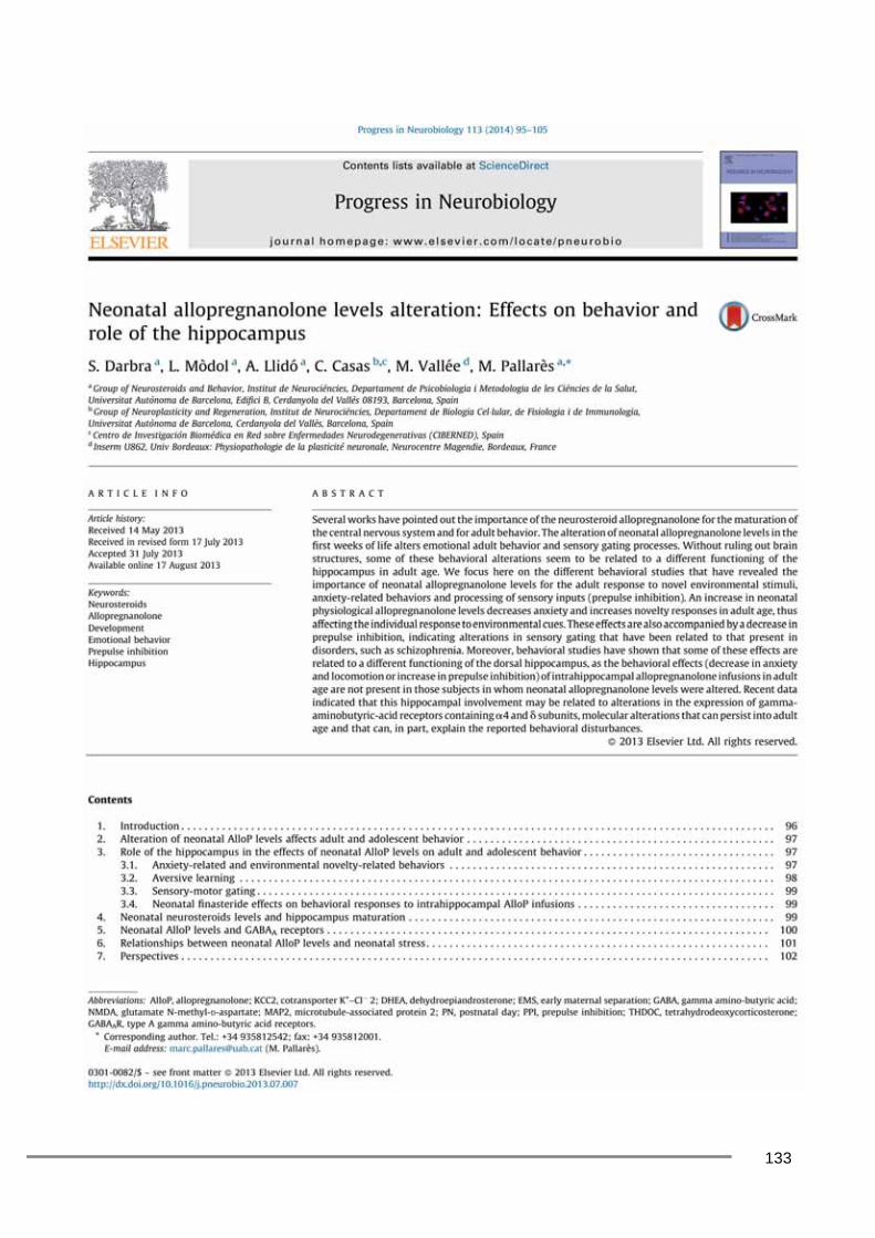
133

134

135
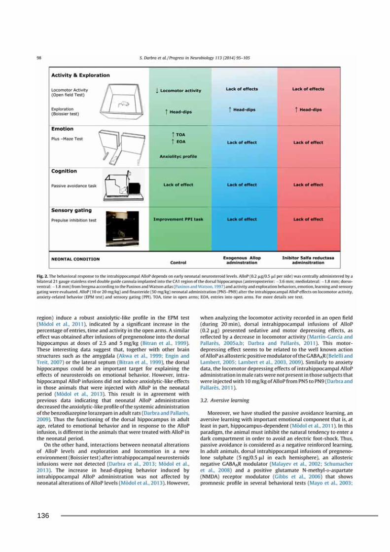
136
137

138

139

140

141
142

143


145
Annex 2
Poster presentation:
Societat Catalana de Biologia Barcelona (2010)

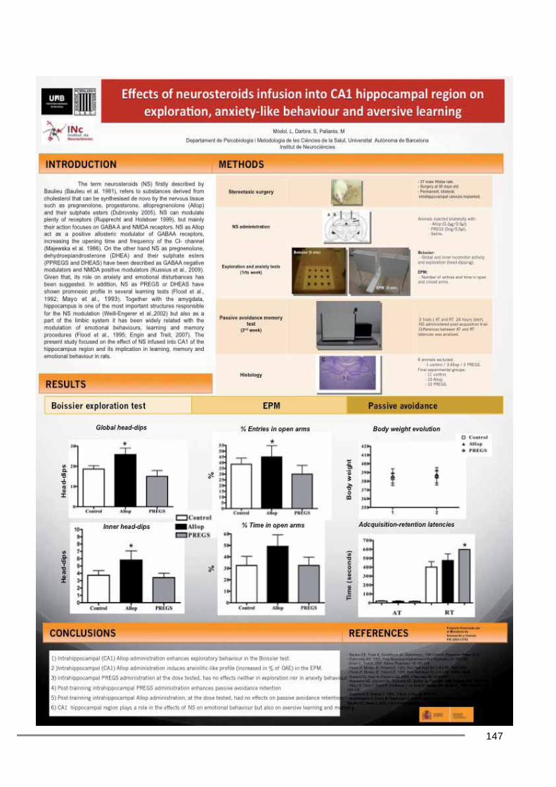
147


149
Annex 3
Poster presentation: Behavioural Brain Research Society
Sevilla (2011)


151


153
Annex 4
Poster presentation: Federation of European Neuroscience Society
Barcelona (2012)


155

157
Acknowledgements This thesis would not exist without the help of a lot of people, for this reason I would like to thank: Dr. Marc Pallarès and Dr. Sònia Darbra for turning me into a researcher and being my mentors during these years. All your suggestions made me a better scientist and a better person. Thanks for trusting, listening and fighting (for me and with me), and having the patience to made me learn. All the group of “Neurosteroids and Behaviour” for helping, encouraging and laughing with me. Dr. Xavier Navarro for taking me in his group and for all the support beyond research. Dr. Caty Casas for all the technical support, being there when I needed practical help and good suggestions.
Dr Monique Vallée for her collaborations assessing neurosteroids levels. All the group of Neuroplasticity and Regeneration for their help and to share scientific and non-scientific work.