Effects of Monomethylfumarate on Human Granulocytes · Because granulo cytes play an ... pellets by...
6
Effects of Monomethylfumarate on Human Granulocytes Peter H. Nibbering ,* Bing Thio,t Timo P.L. Zomerdijk,* Anja C. Bezemer,* Roderick L. Beijersbergen,* and Ralph van Furth* Departments of "Infectious Diseases and i"Dcrmato lo gy, Univcrsiry Hospital, Leiden, The Neth erlands Monomethylfumarate (MMF) is the most active metabolite of the new antipsoriasis drug Fumaderm. Because granulo- cytes play an important role in the pathophysiology of psoria- sis, the effects of this drug on the functional activities of these cells were investigated. MMF stimulated polarization and elastase release, and enhanced the intracellular killing ofbac- teria by granulocytes. This compound suppressed the for- myl-Met-Nle-Phe (FMLP) - stimulated respiratory burst in these cells. MMF and dimethyl fumarate but not its stereo- isomer dimethyl maleate, fumaric acid, or dimethylmalate stimulated polarization of and elastase release by granulo- cytes, indicating that methylated fumarate derivatives inter- act with granulocytes in a specific fashion. MMF did not P soriasis, a chronic inflammatory skin disease affecting up to 3% of the population, is characterized by epidermal hyperplasia and infiltration of lymphocytes and granu- locytes into the skin [l,2J. Treatment is based on inhibi- tion of the proliferation of epidermal cells and interfer- ence in the inflammatory process. In addition to the present therapeutic regimens a new systemic antipsoriasis drug, which con- sists of dimethyl fumarate (DMF) and monoethylfumarate (MEF), has been introduced with successful results [3 ,4]'* Monomethylfu- marate (MMF), which is formed in the circulation by hydrolysis of DMF, is believed to be the most potent metabolite of this drug. Granulocytes are retained at the epidermis by means of a number of factors [5 - 7]. The exact role of granulocytes in the pathophysiol- ogy of psoriasis is not known. IL-8 in combination with TNF-a, which is also present in psoriatic skin lesions [2,5], stimulates granu- locytes to produce reactive oxygen intermediates and proteolytic enzymes [8,9], which can damage epidermal cells and probably influence the growth and differentiation of keratinocytes. Because Manusc ript received September 17, 1992; accepted for publication Febru- ary 12, 1993. Reprint requests to: R. van Furth, Department of Infectious Diseases, Universiry Hospital, Building 1, C5-P, P.O. Box 9600, 2300 RC Leiden, The Netherlands. Abbreviations: db-cAMP, diburyryl-cyclic adenosine monophosphate; DMF, dimethylfumarate; DMM, dimethylmaleate; FA, fumaric acid; FITC, fluorescein isothiocyanate; FLPEP FITC, formyl-Nle-Leu-Phe-Nle- Tyr- Lys-FITC; FMLP, formyl-Met-Leu-Phe; FURA2/ AM, pentakis (acetoxy- methyl) ester of [1-[2-(5-carboxyoxazol-2-yl)-6-aminobenzofuran-5-oxy]- 2-(2' -amino-S' -methylphenoxy)-ethane-N ,N , N',N' -tetraacetic acid; hpf, high-power field; MEF, monoethylfumarate; MMF, monomethylfumarate; Tw, Tween 8. :j: Thio HB, van der SchroeR" JG , Nu gteren-Huying WH, Vermeer BJ: Long-term therapy with dimethylfumarate and monoethylfumarate (Fuma- derm) in psoriasis (in preparation). affect the binding of formyl-Nle-Leu-Phe-Nle- Tyr-Lys- fluorescein isothiocyanate to the FMLP receptor on granulo- cytes. This compound induced an incre ase in the intracellular Ca++ ([Ca++L) and cyclic adenosine monophosphate con- centration. The agonistic effects of MMF on granulocytes are thought to be mediated by the rise in the [Ca++] i and the antagonistic effects by the increase in the cyclic adenosine monophos- phate concentration. These effects of MMF on granulocytes may in part explain the beneficial action of methylated fuma- rate derivatives on psoriatic skin lesions. ] In.vest Dermatol 101:37-42,1993 granulocytes play an important role in the pathophysiology of psori- asis the beneficial effect of treatment with fumaric acid derivatives may be caused by its effects on granulocytes. The present study concerns the effects of MMF on granulocytes from healthy donors. MATERIALS AND METHODS Compounds Used to Stimulate Granulocytes Cells were in- cubated with the following compounds: MMF (Sigma Chemical Co., St. Louis, MO), dimethyl fumarate (DMF; Merck, Darmstadt, Germany), dimethylmaleate (DMM; Merck), fumaric acid (FA; Merck), and S- or R-dimethylmalate (S-DMM and R-DMM; Jans- se n Chimica, Geel, Belgium). In all experiments the pH of the stock solutions of these compounds was adjusted to about 7.2 with 0.1 N NaOH. At the concentrations and exposure time used in this study the stimuli did not affect ce ll viability, as determined by trypan blue exclusion and the lactate dehydrogenase release assay. For comparison, cells were stimulated with 10 to 100 nM for- myl-Met-Leu-Phe (FMLP) (Sigma) or 1 nM rlL-8 (a gift from Dr. I. Lindley, Department of Immunostimulation, Sandoz Forschung- sinstitut Gesellschaft H.B.H., Vienna, Austria). Because both FMLP and rlL-8 were dissolved in dimethylsulfoxide (S erva, Hei- delberg, Germany) and diluted in PBS, cells were also exposed to PBS with an equivalent concentration of dimethylsulfoxide. Phor- bol myristate acetate (PMA) and dibutyryl(db) -cyclic adenosine monophosphate (cAMP) were obtained from Sigma. Preparation of Human Leukocytes Buffy coats prepared from blood from healthy donors were diluted in PBS (pH 7.4) and then subjected to Ficoll-amidotrizoate (p = 1.077 gjml; Pharmacia Inc., UPf-sala, Sweden) density centrifugation at 440 X g for 20 min [10 . Granulocytes were purified from the Ficoll-Amidotrizoate pellets by dextran 1 X g sedimentation (Plasmasteril, Fresenius A.G., Bad Homberg, Germany) at 37°C for 10 min followed by removal of the remaining erythrocytes by a single hypotonic lysis. 0022-202X/93/S06.00 Copyright © 1993 by The Sociery for Investigative Dermatology, Inc. 37
Transcript of Effects of Monomethylfumarate on Human Granulocytes · Because granulo cytes play an ... pellets by...

Effects of Monomethylfumarate on Human Granulocytes
Peter H. Nibbering,* Bing Thio,t Timo P.L. Zomerdijk,* Anja C. Bezemer,* Roderick L. Beijersbergen,* and Ralph van Furth* Departments of "Infectious Diseases and i"Dcrmatology, Univcrsiry Hospital , Leiden, The Netherlands
Monomethylfumarate (MMF) is the most active metabolite of the new antipsoriasis drug Fumaderm. Because granulocytes play an important role in the pathophysiology of psoriasis, the effects of this drug on the functional activities of these cells were investigated. MMF stimulated polarization and elastase release, and enhanced the intracellular killing ofbacteria by granulocytes. This compound suppressed the formyl-Met-Nle-Phe (FMLP) - stimulated respiratory burst in these cells. MMF and dimethyl fumarate but not its stereoisomer dimethyl maleate, fumaric acid, or dimethylmalate stimulated polarization of and elastase release by granulocytes, indicating that methylated fumarate derivatives interact with granulocytes in a specific fashion. MMF did not
Psoriasis, a chronic inflammatory skin disease affecting up to 3% of the population, is characterized by epidermal hyperplasia and infiltration of lymphocytes and granulocytes into the skin [l,2J. Treatment is based on inhibition of the proliferation of epidermal cells and interfer
ence in the inflammatory process. In addition to the present therapeutic regimens a new systemic antipsoriasis drug, which consists of dimethyl fumarate (DMF) and monoethylfumarate (MEF) , has been introduced with successful results [3,4]'* Monomethylfumarate (MMF), which is formed in the circulation by hydrolysis of DMF, is believed to be the most potent metabolite of this drug. Granulocytes are retained at the epidermis by means of a number of factors [5 - 7]. The exact role of granulocytes in the pathophysiology of psoriasis is not known. IL-8 in combination with TNF-a, which is also present in psoriatic skin lesions [2,5], stimulates granulocytes to produce reactive oxygen intermediates and proteolytic enzymes [8,9], which can damage epidermal cells and probably influence the growth and differentiation of keratinocytes. Because
Manuscript received September 17, 1992; accepted for publication February 12, 1993.
Reprint requests to: R. van Furth, Department of Infectious Diseases, Universiry Hospital, Building 1, C5-P, P.O. Box 9600, 2300 RC Leiden, The Netherlands.
Abbreviations: db-cAMP, diburyryl-cyclic adenosine monophosphate; DMF, dimethylfumarate; DMM, dimethylmaleate; FA, fumaric acid; FITC, fluorescein isothiocyanate; FLPEP FITC, formyl-Nle-Leu-Phe-Nle-TyrLys-FITC; FMLP, formyl-Met-Leu-Phe; FURA2/ AM, pentakis (acetoxymethyl) ester of [1-[2-(5-carboxyoxazol-2-yl)-6-aminobenzofuran-5-oxy]-2-(2' -amino-S' -methylphenoxy)-ethane-N ,N ,N',N' -tetraacetic acid; hpf, high-power field; MEF, monoethylfumarate; MMF, monomethylfumarate; Tw, Tween 8.
:j: Thio HB, van der SchroeR" JG, Nugteren-Huying WH, Vermeer BJ: Long-term therapy with dimethylfumarate and monoethylfumarate (Fumaderm) in psoriasis (in preparation).
affect the binding of formyl-Nle-Leu-Phe-Nle-Tyr-Lysfluorescein isothiocyanate to the FMLP receptor on granulocytes. This compound induced an increase in the intracellular Ca++ ([Ca++L) and cyclic adenosine monophosphate concentration.
The agonistic effects of MMF on granulocytes are thought to be mediated by the rise in the [Ca++] i and the antagonistic effects by the increase in the cyclic adenosine monophosphate concentration. These effects of MMF on granulocytes may in part explain the beneficial action of methylated fumarate derivatives on psoriatic skin lesions. ] In.vest Dermatol 101:37-42,1993
granulocytes play an important role in the pathophysiology of psoriasis the beneficial effect of treatment with fumaric acid derivatives may be caused by its effects on granulocytes. The present study concerns the effects of MMF on granulocytes from healthy donors.
MATERIALS AND METHODS
Compounds Used to Stimulate Granulocytes Cells were incubated with the following compounds: MMF (Sigma Chemical Co., St. Louis, MO), dimethyl fumarate (DMF; Merck, Darmstadt, Germany), dimethylmaleate (DMM; Merck), fumaric acid (FA; Merck), and S- or R-dimethylmalate (S-DMM and R-DMM; Janssen Chimica, Geel, Belgium). In all experiments the pH of the stock solutions of these compounds was adjusted to about 7.2 with 0.1 N NaOH. At the concentrations and exposure time used in this study the stimuli did not affect cell viability, as determined by trypan blue exclusion and the lactate dehydrogenase release assay.
For comparison, cells were stimulated with 10 to 100 nM formyl-Met-Leu-Phe (FMLP) (Sigma) or 1 nM rlL-8 (a gift from Dr. I. Lindley, Department of Immunostimulation, Sandoz Forschungsinstitut Gesellschaft H.B.H., Vienna, Austria) . Because both FMLP and rlL-8 were dissolved in dimethylsulfoxide (Serva, Heidelberg, Germany) and diluted in PBS, cells were also exposed to PBS with an equivalent concentration of dimethylsulfoxide. Phorbol myristate acetate (PMA) and dibutyryl(db) -cyclic adenosine monophosphate (cAMP) were obtained from Sigma.
Preparation of Human Leukocytes Buffy coats prepared from blood from healthy donors were diluted in PBS (pH 7.4) and then subjected to Ficoll-amidotrizoate (p = 1.077 gjml; Pharmacia Inc., UPf-sala, Sweden) density centrifugation at 440 X g for 20 min [10 . Granulocytes were purified from the Ficoll-Amidotrizoate pellets by dextran 1 X g sedimentation (Plasmasteril, Fresenius A.G., Bad Homberg, Germany) at 37°C for 10 min followed by removal of the remaining erythrocytes by a single hypotonic lysis.
0022-202X/93/S06.00 Copyright © 1993 by The Sociery for Investigative Dermatology, Inc.
37

38 NIBBERING ET AL
The final cell suspensions consisted of 97 ± 2% granulocytes. The viability of the cell suspensions exceeded 95%, as determined by trypan blue dye exclusion.
Measurement of Polarization of Granulocytes The assay to determine polarization of granulocytes was performed as described [11] with minor modifications. Briefly, 400-fLl aliquots of a suspe.nsion of 1 X 10⁶ granulocytes/ml RPMI 1640 (Flow LaboratorIes Ltd., Irvine, UK) were incubated with 100 fLl of the stimulus diluted in RPMI 1640 at 3r C for 15 min. Incubation was stopped by the addition of 500 fLl 9% v /v formaldehyde in PBS; the cells were maintained in this fixative until light microscopical determination of the percentage of cells that were not spherical. Two different investigators each analyzed 200 cells from each preparation.
Chemotaxis Assay Chemotaxis of granulocytes was measured with the 48-well microchemotaxis chamber (Neuroprobe, Cabin John, MD) assay as described (12) . In short, chemotaxins 0: PBS were placed in the lower compartments. Two filters were adjusted between the lower and upper compartments. The lower filter had a pore width of 0.45 fLm and the upper filter of 8 fLm. Granulocytes were placed in the upper compartment and, after incubation for 1.5 h instead of 2.5 h at 37 °C, the filters were fixed, stained, and cleared with xylene. The number of granulocytes that had passed the upperfilter was determined by light microscopy (magnification X 400). The results are expressed as the number of cells per 10 high-power fields (hpf).
Measurement of Elastase Release by Granulocytes Granulocytes suspended in 137 mM NaCI, 2.7 mM KCI, 8.1 mM Na2HP04, 0.9 mM CaCI2 , 0.49 mM MgClz, 1.5 mM KHzP04 ,
and 2.5 mg bovine serum albumin (BSA)/ml at a concentration of 5 X 10⁶ cells/ml were incubated with the indicated stimuli at 37 °C for 30 min. The reaction was stopped by transferring the tubes to crushed ice. After centrifugation at 500 X g for 10 min, samples of the cell-free supernatants were assayed for elastase activity utili~ing substrate S2484 (Kabivitrum, Stockholm, Sweden) as descnbed [13J. The absorbance of the reaction product after 60 min was read at 405 nm using a Titertek multiscan (EFLAB, Helsinki, Finland). Granulocytes were incubated with 5 fLg cytochalasin E {Serva)/ml PBS under slow rotation (4 rpm) at 37°C for 5 min, washed, and suspended to a concentration of 5 X 10⁶ cells/ml.
Measurement ofIntracellular Killing of Mycobacterium fortuitum by Granulocytes Preparation ~f M. JortuJtul1l (ATCC 12790, American Type Culture CollectIOn, RockVille, MD) and the assay to determine the intracellular killing of this mycobacterium by granulocytes has been described [14]. Briefly,S X 106 granulocytes/ml Hanks' balanced salt solution (HBSS) (Oxoid Ltd., Basingstoke, UK) containing 0.01 % gelatin and 0.01 % Tween 80, further called HBSS-gel-Tw, and 5 X 10⁶ M. Jortuitum/ml HBSSgel-Tw were mixed and incubated in the presence of 10% v /v human serum at 37°C for 10 min. The supernatant contammg extracellular bacteria was discarded, the granulocytes washed, and then 5 X 10⁶ granulocytes containing bacteria were stimulated with 10% serum with or without 200 fLM MMF at 37°C. One and two hours after stimulation samples were taken and, after lysis of the granulocytes, the number of viable cell-associated bacteria was ?etermined microbiologically. As control, granulocytes contammg bacteria were incubated with MMF or no stimulus.
Measurement of the Oxygen Consumption by Granulocytes Oxygen consumption by granulocytes at rest and after stimulation was determined polarographically [15]. In short, 300 fLl of a suspension of 2.5 X 1 06leukocytes/ml RPMI 16~0 ~ere added .to a thermostated vessel (3rC), and the oxygen tenSIOn In the medIUm was measured with a Clark electrode. The rate of oxygen consumption was calculated from the slope of the oxygen tension trace over a period of 5 to 10 min. ~o discriminate between.oxygen c~nsum~tion by the NADPH OXidase complex and the mltochondnal respIration, cells were also stimulated in the presence of 1 mM NaN 3' an inhibitor of mitochondrial respiration.
THE JOURNAL OF INVESTIGATIVE DERMATOLOGY
Measurement of Superoxide Anion Production by Granu, locytes Superoxide anion (02-) production by cytochalasin E~ incubated granulocytes at rest and after stimulation was assessed by the superoxide dismutase-inhibitable reduction of ferricytochrom~ c (type IV; Sigma) as described [1 6] with minor modifications. Briefly, the stimulus was added to 1 X 10⁶ cells/ml PBS supplemented with 1.2 mM CaCI2 , 1 mM MgClz, 5 mM glucose. 20 mM Hepes, and 0.1 mM ferricytochrome c at 3rC and th~ absorbance at 550 nm was measured every 30 seconds during a 1 O-min period. The production of superoxide anion by the cells was calculated from a standard curve obtained with reduced cytochrome.
Measurement of the NADPH Oxidase Activity in a Cell-Free System NADPH oxidase activity was measured by means of a C lark oxygen electrode at 2rC as the rate of oxygen consumption by the cytosolic and membranous components of NADPH oxidase that were isolated from granulocytes as described [17]. Briefly, 360 plIO mM Hepes (pH 7.0) containing 0.17 M sucrose, 75 roM NaCI, 0.5 mM ethyleneglycol-bis-(-amino-ethylether)N,N' -tetraacetic acid (EGTA), 1 mM MgCI2 , 2 mM NaN3 • and 10 ILM GTPyS (Calbiochem Corp, La Jolla, CA) were mixed with 10111 membranous components and 10 fLl cytosolic components, each of which is the equivalent of 1 X 10⁶ granulocytes. Assembly of the NADPH oxidase complex was initiated by adding sodium dodecylsulfate at a final concentration of 100 fLM and closing the reaction vessel. After 3 min, NADPH atafinal concentration of250 pM was added and the oxygen consumption measured.
Determination of the Binding of FLPEP-FITC to Granulocytes by FACS Analysis The fluorescent probe formyl-NleLeu-Phe-Nle-Tyr-Lys -fluorescein isothiocyanate (FLPEP-FITC; Molecular Probes Inc., Eugene, OR) was used to determine binding to the receptor as described [19] . Briefly, granulocytes at a concentration of 2 X 1.06 cells/ml buffer (pH 7.4) were incubated with various concentrations of FLPEP-FITC in the dark at 4 0 C for 60 min and then the fluorescence of the cells was measured on a FACScan (Becton and Dickinson, Mountainville, CA) equipped with a 15 m W argon-ion laser. For assessment of the non-specific binding of this probe, cells were incubated with 1 fLM FMLP at 4 ° C for 15 min prior to addition ofFLPEP-FITC. The results expressed as mean fluorescence intensity values are in arbitrary units (AU).
Competition Binding Assay for cAMP At selected intervals after stimulation, 50 fLl of a suspension of 1 X 10⁶ granulocytes/ml were stimulated at 3rc for various intervals and then 450 fLl icecold I-propanol (Merck) were added to disrupt the cells. After sonication for 5 min this material was centrifuged at 7000 X g for 1 min and then the supernatant was lyophilized. The residue was dissolved in 100 fLl distilled water and the cAMP content of these samples was determined as described previously [19] . In short, cAMP in the samples was allowed to compete with 5',8-f3H]-cAMP (specific activity 40-60 Ci/mmol; Amersham International, Amersham, Bucks, UK) for binding to sites in the bovine microsomal preparation. The cAMP content of these samples was calculated from a standard curve constructed in parallel with unlabeled cAMP (Amer. sham). The cAMP concentration was calculated from this value and the mean cell volume of the granulocytes.
Measurement of the [Ca++]; in Granulocytes For measurement of the [Ca-] i, 2 X 10⁷ leukocytes/ml 20 mM Hepes supplemented with 138 mM NaCI, 6 mM ICCI, 1 mM MgS04' 1.1 m.M CaCI2 , 1 mM NaH2P04, 5.5 mM D-glucose, 0.1 mMEGTA, and 0.1 % BSA (Ca- buffer, pH 7.4) were incubated with 2 JlM of the acetoxymethyl ester of FURA-2 (FURA-2/ AM; Sigma) in the dark at 3rc for 30 min. Next. the cells were washed with Ca++ buffer and then suspended in either Ca- buffer or Ca++ buffer without CaCl2 but supplemented with 2 mM EGTA, further called Ca++· free buffer. Two millimeter aliquots of 5 X 10⁶ cells/ml Ca++ buffer or Ca++-free buffer were transferred to a quartz cuvette with magnetic stirring. The cell suspensions were excited alternately at 340, 360, and 380 nm (slit width 2.5 11m) and emission was

VOL. 101, NO.1 JULY 1993
100
80
~ 60 !!! Q; u
U
.~ 40
'" "0 a.
00 20
0
0 0 .2 2 20 200 500
MMF (pM)
Figure 1. Effect of various concentrations of MMF on the polarization of granulocytes. Granulocytes were incubated for 15 min with concentrations ofMMF ranging from 0.2.uM to 500 .uM and the percentage cells that were not spherical was assessed. Results are means ± SEM; 11 = 3.
recorded at 500 nm (slit width 5 nm) with a Perkin-Elmer 3000 spectrofluorometer (Perkin-Elmer, Ueberlingen, Germany) . The [Ca++] i was calculated from the ratio of the fluorescence excited at 340 nm and at 380 nm as described [20]. Calibration of the FURA-2 fluorescence was performed by lysis of the cells with 0.1 % wtjv Triton X-I00 (Sigma) in the presence of 1.1 mM CaClz and the subsequent addition of 10 mM EGT A.
Statistical Analysis The significance of the differences between the values found for cells exposed to stimuli and cells incubated with the vehicule was determined with the Mann-Whitney U test. p < 0.05 was considered significant.
RESULTS
Induction of the Polarization and Chemotaxis and Elastase Release by Granulocytes by Various Stimuli MMF induced polarization of granulocytes in a dose-dependent fashion; the maximum percentage polarized cells was obtained with 200 .aM MMF (Fig 1). DMF but not DMM, FA, S-DMM, or R-DMM stimulated the polarization of granulocytes (Table I). About 90% of the granulocytes had a polarized morphology after stimulation with FMLP (Table I).
The number of cells in the upper filter was slightly but significantly higher when MMF and DMF was present in the lower com-
EFFECTS OF MMF ON GRANULOCYTES 39
partment of the chambers than when no stimulus was applied (Table I). The effect on the number of cells in the filter did not depend on the concentration of MMF (range 2 nM to 2 mM; results not shown). These results indicate that MMF and DMF are hardly chemotactic for granulocytes; FMLP was a potent chemotaxin for these cells (Table I). Addition of MMF to the upper compartment of the n1.icrochemotaxis chamber did not affect this response to FMLP. Granulocytes incubated with MMF for 5 min responded to FMLP as well as cells incubated with PBS (results not shown). MMF but not DMF, DMM, FA, S-DMM, or R-DMM stimulated granulocytes to release elastase (Table I). Stimulation by MMF was considerably less than that by rIL-8 or FMLP (Table I). MMF-incubated granulocytes released significantly less elastase after rIL-8 or FMLP stimulation than PBS-incubated cells.
Intracellular Killing of M. fortuitum by Granulocytes The phagocytosis of M. fortuitul1I by granulocytes and the growth of this bacterium in HBSS - gel-Tw were not affected by MMF (results not shown). The intracellular killing of M. Jortllitum by granulocytes stimulated with serum was limited but addition of MMF enhanced the killing process significantly; the percentage of M. Jortuitum killed 2 h after stimulation with and without MMF was 60 ± 4.5% and 29 ± 3.5% (means ± SEM; n = 3), respectively. In the absence of serum, intracellular killing of M. Jortuitul1I by granulocytes did not occur and MMF had no effect. The cell-free medium, obtained after incubation for 2 h at 37"C of 5 X 10⁶ granulocytes and 5 X 10⁶ M. Jortuitul1I in 1 111.1 HBSS-gel-Tw containing 10% serum with MMF, did not affect the growth of M. Jortuitum, indicating that no extracellular killing of bacteria occurred (results not shown).
Stimulation of Oxygen Consumption and O 2 Production by Granulocytes with MMF and FMLP Oxygen consumption and O2 production by resting granulocytes and by granulocytes after stimulation with various concentrations of MMF, FMLP, or PMA are shown in Table II. Both MMF and FMLP stimulated oxygen consumption by these cells. Optimal concentrations for MMF and FMLP were 200.aM and 100 11.M, respectively. No additive effect of MMF on the FMLP-stimulated oxygen consumption by granulocytes was found (results not shown). In the presence of NaN3 , an inhibitor of mitochondrial respiration, the stimulatory effect of MMF on oxygen consumption was almost completely abolished whereas that of FMLP was hardly affected (Table II), indicating that MMF does not activate the NADPH oxidase complex. In agreement with this notion, MMF did not stimulate granulocytes to produce 0 2". This compound did not scavenge 02".
To find out whether incubation with MMF suppresses the FMLP-stimulated oxygen consumption, granulocytes were exposed to MMF in the presence ofNaN3 , or as control to PBS, prior
Table I. Effects of Various Stimuli on the Polarization and Chemotaxis of, Elastase Release by, and Increase in the [Ca++t in Granulocytes
Chemotaxis' Elastase Released [Ca++]i' Stimulus' Polarized Cellsb (%) (cell/tO hpf) (00405 nm) (nM)
None 18 ± 3f 60±6 0.30 ± 0.01 110 ± 6 MMF 58 ± 5g 91 ± 109 0.43 ± O.Ols 258 ± 14g OMF 40 ± 5: 113 ± 9: 0.30 ± 0.01 235 ± 8g OMM 18 ± 3 NO 0.27 ± 0.01 129 ± 9 FA 12 ± 2 NO 0.30 ± 0.02 119±1 S-OMM 19 ± 4 NO 0.34 ± 0.01 74 ± 10 R-OMM 12 ± 4 NO 0.31 ± 0.01 85 ± 4 rIL-8 NOb NO 1.61 ± O.llg 450 ± 26·~ FMLP 88 ± 3g 1129 ± 308g 2.10 ± 0.27: 618 ±38g
• Final concentration ofMMF, OMF, and OMM was 200 tIM; of FA, S-OMM, and R-OMM 1 mM; of rlL-8 1 nM. The final concentration ofFMLP in the polarization assay, the chemotaxis assay and the elastase release assay was 10 nM and for the measurement of the [Ca++]; it was 100 nM.
b Various stimuli were added to granulocytes and after 15 min the percentage of cells that were not spherical was assessed. n = 3. , Various stimuli were included in the lower compartment of the microchemotaxis chamber and after an incubation for 1.5 h at 37"C the number of cells in the upper filter was
determined light microscopically. The results expressed as the number of cells per 10 hpf. n = 3. , Various stimuli were incubated with granulocytes for 30 min; after the reaction was terminated, the cell-frec supcrnatant of this suspension was assayed for elastase activity. n = 3. l Various stimuli were added to FURA 2/AM-incubatcd granulocytes and the [Ca++]j was monitored. Data represent max iinuln [Ca++]j vaJucs. n = 4- 16. fResults are means ± SEM. t Indicates p < 0.05 for the difference between the values for stimulated and control cells. b NO, not done.

40 NIBBERING ET AL THE JOURNAL OF INVESTIGATIVE DERMATOLOGY
Table ll. Oxygen Consumption and Superoxide Anion Production by Granulocytes at Rest and After Stimulation with MMF, FMLP, and PMAa
Incubation Stimulusb Oxygen Consumption
(nmol/[5 X 106 cells X min) O2 Production
(I1mol/[l X 106 cells X 10 mjn))
None PMA MMF FMLP MMF FMLP FMLP
3 ± 2' (6) 25 ± 5J (6) 18 ± 6J (6) 16 ± Y (8)
6 ± 0.4 (7) 51±4J(5)
6 ± 0.4 (7) 34 ± 1J (7)
N[)f ND
2 ± 1 (6) 15±4J (3) 5±1'(3) 24 ± 0.4' (7)
• Oxygen consumption due to mitochondrial respiration was inhibited with 1 mM NaN,. Where indicated, granulocytes were incubated with MMF together with NaN, for 15 min before measurement of rhe FMLP-stimulated oxygen consumption and O. production.
• Final concentrations of200)JM MMF, 100 nM FMLP, 100 ng/m1 PMA, and 1 mM NaN, . , Results are means ± SEM, with the number of experiments in parentheses. , Indicates P < 0.05 for the difference between the values for stimulated and control cells. , Indicates p < 0.05 for the difference between the values for MMF-incubated and control cells. I ND, not done.
to stimulation with FMLP. The results showed that the respiratory burst in MMF-incubated cells was significantly lower than in control cells (Table II). Because it has been suggested that an increase in the cAMP concentration inhibits the oxidative metabolism of granulocytes [21,22], we incubated granulocytes with 0.1 mM db-cAMP for 5 min at 37°C before measurement of the FMLPstimulated 02" production. The results revealed that the FMLPstimulated production of 02" by db-cAMP-incubated granulocytes was 40 ± 9% (mean ± SEM) less (n = 6) than that by control granulocytes.
Effect ofMMF on the NADPH Oxidase Activity in the CellFree System To elucidate the mechanisms underlying the inhibition of the respiratory burst in MMF-incubated cells, we measured the effect of MMF on the NADPH oxidase activity in the cell-free system. For this purpose MMF, or as control PBS, was added to the mixture of cytosolic and membranous components of the NADPH oxidase compl.ex before the addition ofSDS. The results revealed no effect of MMF on the NADPH oxidase activity; the results of the addition of MMF and PBS were 8.4 ± 0.2 J.LM oxygen/min and 8.2 ± 0.6 JLM oxygen/min (means ± SEM; n = 5), respectively.
Effect ofMMF on the Binding ofFLPEP-FITC to Granulocytes To find out whether the MMF-mediated inhibition of the respiratory burst in granulocytes was due to down-regulation of the FMLP-receptor, the effect of MMF on binding of FLPEP-FITC to these cells was determined. The results revealed no effect of exposure of the cells to MMF for 15 min prior to the incubation with the fluorescent probe (Fig 2). Addition of MMF during the incubation with FLPEP-FITC was without effect as well (results not shown).
Changes in the cAMP Concentration in Granulocytes After Stimulation with MMF and FMLP Because an increase in the cAMP concentration in cells is involved in the negative feedback processes in cells [24], we investigated whether MMF induces a rise in the cAMP concentration in granulocytes. The cAMP concentration in resting granulocytes was 0.72 ± 0.19 J.LM (mean ± SEM; n = 4). Stimulation of granulocytes with MMF resulted in a transient increase in the cAMP concentration to a maximum value of 1.33 ± 0.14 J.LM (mean ± SEM; n = 4) 20 seconds after stimulation, which returned to resting values within 1 min after addition of the stimulus. FMLP induced a threefold increase in the cAMP concentration in granulocytes with a maximum value of 2.67 ± 0.60 J.LM (mean ± SEM; n = 4) 30 seconds after stimulation.
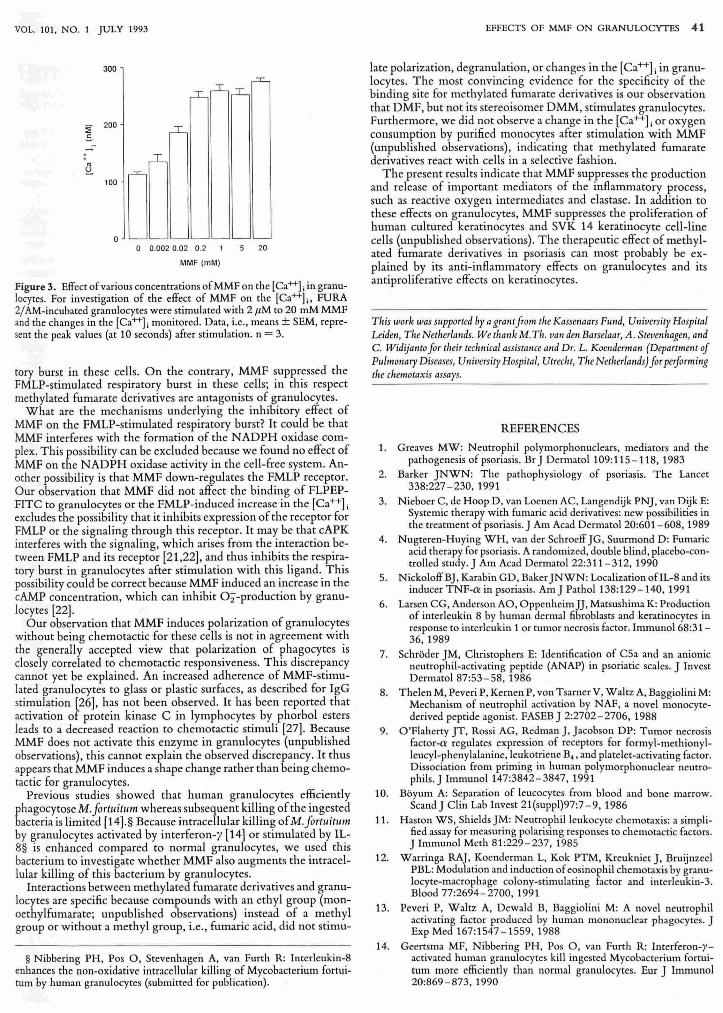
Changes in the [Ca++}; in Granulocytes Induced by Various Stimuli The [Ca++] i in resting granulocytes amounted to 110 ± 6 nM (mean ± SEM; Table I). Addition of2 f1.M to 20 mM MMF to granulocytes induced a dose-dependent increase in the [Ca++Ji; maximum values were found after stimulation with 200 JLM MMF (Fig. 3). The rise in the [Ca++L was already apparent within 5 seconds of the addition of MMF and the [Ca++] i gradually returned
to resting values within the next 2 - 3 min. The increase in the [Ca++] i after stimulation with MMF is due mainly to the release of Ca+t from intracellular stores. Similar kinetics for changes in the [Ca++]; were found for granulocytes stimulated with FMLP and rlL-8 (results not shown). Furthermore, the FMLP-induced increase in the [Ca++]; in MMF-incubated and PBS-incubated granulocytes did not differ (results not shown), indicating that MMF did not affect signaling in granulocytes.
To find out whether the effects of methylated fumarate derivatives on granulocytes depend on the presence of methyl groups, the changes in the [Ca++] i in granulocytes upon stimulation with DMF, DMM, FA, S-DMM, or R-DMM were assessed. The results reo vealed that of these compounds only DMF induced an increase in the [Ca++]; (Table I).
DISCUSSION
The main conclusion that can be drawn from the present results is that the methylated fumarate derivatives MMF and DMF modulate several functional activities of granulocytes. These fumarate derivatives are agonists of granulocytes because they stimulate the polarization and elastase release by granulocytes and enhance the intra· cell ular killing of bacteria by these cells. Some of these effects might be mediated by the rise in the [Ca+t] i in granulocytes stimulated by these compounds, since Ca++ -specific ionophores also stimulate the polarization and elastase release by granulocytes [23 - 25]. MMF and DMF are not chemotactic for granulocytes or stimulate the respira-
:: 'iii c: J!! .!:
'" " c:
'" " '" ~ 0
~
300
200
100
25 FLPEP (nM)
SO
Figure 2. Effect of MMF on the binding of FLPEP-FITC to granulocytes, Granulocytes were exposed to 200 liM MMF ~, PBS (0). or 1 liM FMLP (0) for 15 min before jncubation with variolls concentrations of FLPEP, FITe.' After a 60-min incubation with this probe, the fluorescence of th~ cells was measured with a FACScan f1owcytometer. The results are given fot one representative experiment out of three experiments.
VOL. 101, NO.1 JULY 1993
300
~ 200
.s -. . '" ~
u_~ 100
0 0 0.002 0.02 0.2 20
MMF (mM)
Figure 3. Effect of various concentrations of MMF on the [Ca++] i in granulocytes. For investigation of the effect of MMF on the [Ca++]., FURA 2/ AM-incubated granulocytes were stimulated with 2,uM to 20 mM MMF and the changes in the [Ca++]. monitored. Data, i.e., means ± SEM, represent the peak values (at 10 seconds) after stimulation. n = 3.
tory burst in these cells. On the contrary, MMF suppressed the FMLP-stimulated respiratory burst in these cells; in this respect methylated fumarate derivatives are antagonists of granulocytes.
What are the mechanisms underlying the inhibitory effect of MMF on the FMLP-stimulated respiratory burst? It could be that MMF interferes with the formation of the NADPH oxidase complex. This possibility can be excluded because we found no effect of MMF on the NADPH oxidase activity in the cell-free system. Another possibility is that MMF down-regulates the FMLP receptor. Our observation that MMF did not affect the binding of FLPEPFITC to granulocytes or the FMLP-induced increase in the [Ca++]. excludes the possibility that it inhibits expression of the receptor for FMLP or the signaling through this receptor. It may be that cAPK interferes with the signaling, which arises from the interaction berween FMLP and its receptor [21,22], and thus inhibits the respiratory burst in granulocytes after stimulation with this ligand. This possibility could be correct because MMF induced an increase in the cAMP concentration, which can inhibit 02-production by granulocytes [22].
Our observation that MMF induces polarization of granulocytes without being chemotactic for these cells is not in agreement with the generally accepted view that polarization of phagocytes is closely correlated to chemotactic responsiveness. This discrepancy cannot yet be explained. An increased adherence of MMF-stimulated granulocytes to glass or plastic surfaces, as described for IgG stimulation [26], has not been observed. It has been reported that activation of protein kinase C in lymphocytes by phorbol esters leads to a decreased reaction to chemotactic stimuli [27] . Because MMF does not activate this enzyme in granulocytes (unpublished observations), this cannot explain the observed discrepancy. It thus appears that MMF induces a shape change rather than being chemotactic for granulocytes.
Previous studies showed that human granulocytes efficiently phagocytose M.JortuitulII whereas subsequent killing of the ingested bacteria is limited [14] .§ Because intracellular killing ofM.JortuitulII by granulocytes activated by interferon-y [14] or stimulated by IL-8§ is enhanced compared to normal granulocytes, we used this bacterium to investigate whether MMF also augments the intracellular killing of this bacterium by granulocytes.
Interactions between methylated fumarate derivatives and granulocytes are specific because compounds with an ethyl group (monoethylfumarate; unpublished observations) instead of a methyl group or without a methyl group, i.e., fumaric acid, did not stimu-
§ Nibbering PH, Pos 0, Stevenhagen A, van Furth R: Interleukin-8 enhances the non-oxidative intracellular killing of Mycobacterium fortuitum by human granulocytes (submitted for publication).
EFFECTS OF MMF ON GRANULOCYTES 41
late polarization, degranulation, or changes in the [Ca++] i in granulocytes. The most convincing evidence for the specificity of the binding site for methylated fumarate derivatives is our observation that DMF, but not its stereoisomer DMM, stimulates granulocytes. Furthermore, we did not observe a change in the [Ca++] i or oxygen consumption by purified monocytes after stimulation with MMF (unpublished observations), indicating that methylated fumarate derivatives react with cells in a selective fashion .
The present results indicate that MMF suppresses the production and release of important mediators of the inflammatory process, such as reactive oxygen intermediates and elastase. In addition to these effects on granulocytes, MMF suppresses the proliferation of human cultured keratinocytes and SVK 14 keratinocyte cell-line cells (unpublished observations) . The therapeutic effect of methylated fumarate derivatives in psoriasis can most probably be explained by its anti-inflammatory effects on granulocytes and its anti proliferative effects on keratinocytes.
Tllis 1V0rk was 5Ilpported by a gratlt from the Kasswaars F,md, University Hospital Leidw, Tire Netlrerlands. We thank M. TIL IIa" dell Barselaar, A . Steve"hagen, atld C. Widijanto for tlreir ted'tlical assiSlarlCe a"d Dr. L. KOelldenllm, (Department of PlIlmo"ary Diseases, U"iversity Hospital, Utrec/,t, Tire Netl,erlm,ds) forperjormitlg tire chemotaxis assays.
REFERENCES
1. Greaves MW: Neutrophil polymorphonuclears, mediators and the pathogenesis of psoriasis. Br) Dermatol 109: 115 - 118, 1983
2. Barker )NWN: The pathophysiology of psoriasis. The Lancet 338:227 -230, 1991
3. Nieboer C, de Hoop D, van Loenen AC, Langendijk PN], van Dijk E: Systemic therapy with fumaric acid derivatives: new possibilities in the treatment of psoriasis. ) Am Acad DermatoI20:601-608, 1989
4. Nugteren-Huying WH, van der Schroeff )G, Suurmond D: Fumaric acid therapy for psoriasis. A randomized, double blind, placebo-controlled study.) Am Acad Dermatol 22:311- 312, 1990
5. Nickoloff B), Karabin GD, Baker )NWN: Localization ofIL-8 and its inducer TNF-a in psoriasis. Am) Pathol 138:129-140, 1991
6. Larsen CG, Anderson AO, OppenheimJ), Matsushima K: Production of interleukin 8 by human dermal fibroblasts and keratinocytes in response to interleukin 1 or tumor necrosis factor. ImmunoI68:31-36, 1989
7. Schroder )M, Christophers E: Identification of C5a and an anionic neutrophil-activating peptide (A NAP) in psoriatic scales. ) Invest Dermatol 87:53 - 58, 1986
8. Thelen M, Peveri P, Kernen P, von Tsarner V, Waltz A, Baggiolini M: Mechanism of neutrophil activation by NAF, a novel monocytederived peptide agonist. FASEB) 2:2702-2706, 1988
9. O'Flaherty ]T, Rossi AG, Redman], Jacobson DP: Tumor necrosis factor-a regulates expression of receptors for formyl-methionylleucyl-phenylalanine, leukotriene B. , and platelet-activating factor. Dissociation from priming in human polymorphonuclear neutrophils.) ImmunoI147:3842-3847, 1991
10. Boyum A: Separation of leucocytes from blood and bone marrow. Scand J Clin Lab Invest 21(suppl)97:7 -9,1986
11. Haston WS, Shields )M: Neutrophil leukocyte chemotaxis: a simplified assay for measuring polarising responses to chemotactic factors. ] Immunol Meth 81:229-237,1985
12. Warringa RA), Koenderman L, Kok PTM, Kreukniet), Bruijnzeel PBL: Modulation and induction of eosinophil chemotaxis by granulocyte-macrophage colony-stimulating factor and interleukin-3. Blood 77:2694-2700, 1991
13. Peveri P, Waltz A, Dewald B, Baggiolini M: A novel neutrophil activating factor produced by human mononuclear phagocytes. J Exp Med 167:1547 -1559, 1988
14. Geertsma MF, Nibbering PH, Pos 0, van Furth R: Interferon-/,activated human granulocytes kill ingested Mycobacterium fortuitum more efficiently than normal granulocytes. Eur J Immunol 20:869-873,1990
42 NIBBERING ET AL
15. Van Dissel JT, Olivier KN, Leijh PC), van Furth R: A reaction vessel for the measurement of oxygen consumption by smalJ numbers of cells in suspension. J Immunol Meth 92:271-280, 1986
16. Babior BM, Kipness RS, Cumette JT: Biological defense mechanisms. The production of superoxide, a porential bactericidal agenr.] Clio Invest 52:741-744, 1973
17. Bolscher BGJM, Denis SW, Verhoeven Aj , Roos 0 : The activity of one soluble component of the cell-free NADPH-02 oxidoreductase of human neutrophils depends on 5'-0-{3-thio)triphosphate. J Bioi Chern 265:15782-15787,1990
18. Sklar LA, Finney DA, Oades ZG, Jesaitis A], Painter RG, Cochrane CG: The dynamics of ligand-receptor interactions. J Bioi Chern 259:5661 - 5669, 1984
19. Zomerdijk TPL, Nibbering PH, Bezemer AC, LOwik CWGM, van Furth R: A new competition binding assay for determination of the cAMP content of human leukocytes. Biochem Biophys Res Commun 178:980 - 984, 1991
20. Grynkiewicz G, Poenie M, Tsicn RY: A new generation of Ca2+ indicators with greatly improved fluorescence properties. J Bioi Chern 260:3440-3450,1985
21 . Della Bianca V, De Togni P, Grzeskowiak M, Vicentini LM, DiVirgi-
THE JOURNAL OF INVESTIGATIVE DERMATOLOGY'
liD F: Cyclic AMP inhibition of phosphoinositide tUfllover ill humanneutrophils. Biochim Biophys Acta 886:441-447,1986
22. Mueller H, Sklar LA: Coupling of antagonistic signalling pathways ill modulation of neutrophil function. J Cell Biochem 40:287 -294, 1989
23. Boucek M, Snyderman R: Calcium influx requirement for humall neutrophil chemotaxis: inhibition by lanthanum chloride. Scienc~ 193:905-907,1977
24. Marcks PW, Maxfield FR: Transient increases in cytosolic free cal, cium appear to be required for migration of adherent human neutrophils. J Cell BioI 11 0:43 - 52, 1990
25. Lew DP, Wollheim C, Waldvogel FA, Pozzan T : Modulation ° cytosolic free calcium transients by changes in intracellular calciul11 buffering. Correlation with exocytosis and 02" production in human neutrophils. J CeJl BioI 99:1212-1220, 1984
26. Wilkinson pc. Effects of human IgG on locomotion of human neu, trophils related to IgG binding of a hydrophobic probe. Immunol 41:457-466,1980
27. Wilkinson PC, Lackie JM, Haston WS, Islam LN: Effects of phorboI. esters on shape and locomotion of human blood lymphocytes. J Cell Sci 90:645 - 655, 1988
ANNOUNCEMENT
The 4th Annual Meeting of the European Hair Research Society will take place in Stockholm on October 1-2, 1993 at the Ungholmen Conference Centre. Deadline for abstracts is July 1, 1993. The program of the meeting will tentatively cover the following areas:
Hair proteins - structure and function Basic research on hair and hair culture models Immunology and transgenic mice experiments Hormonal regulation The pilo-sebaceous unit Hair diseases Hair transplantation Environmental influences on hair and hair care
The Congress Chairman is Associate Prof. Bo Forslind, M.D., Ph.D. For information on the program, accommodation, etc. write to EHRS Congress Secretariat,
Ms. Jenny Bernstrom, Ms. Margareta Andersson, Ms. Inger Lindback, EHRS, Department of Medical Biophysics, Karolinska Institutet, Box 60400, S-104 01 Stockholm, Sweden, Tel, +46 (0) 8 7286790; Fax, +46 (0) 8 32 65 05.


![Translating Unconventional T Cells and Their Roles in ... · cytes in chronic lymphocytic leukemia (CLL) and granulo-cytes in chronic myeloid leukemia (CML) [2, 3]. The hallmark of](https://static.fdocuments.us/doc/165x107/60fe822ebb03945f18765114/translating-unconventional-t-cells-and-their-roles-in-cytes-in-chronic-lymphocytic.jpg)