Effects of astaxanthin onaxonal regeneration via …...on cerebral infarction. In this study,...
9
135 Abstract. – OBJECTIVE: To investigate the effect of astaxanthin on the neurological func- tion of the middle cerebral artery occlusion (MCAO) mice and its possible mechanism. MATERIALS AND METHODS: The male C57BL/6 mice were selected to establish the model of MCAO via electrocoagulation, and they were randomly divided into 4 groups: the sh- am operation group (Sham group), the cerebral ischemia model group (MCAO group), the astax- anthin intervention group (gavage with 30 mg/kg astaxanthin for 28 days, twice a day; Ast group), and astaxanthin + H89 group (Ast + H89 group). At 3, 7, 14, and 28 d after the operation, the Ro- tarod test and the balance beam footstep error test were performed. The brain tissues were tak- en for immunofluorescence to observe the ex- pression of the growth-associated protein 43 (GAP43) in the cortex around the infarction. The GAP43 protein and mRNA levels in the cortex around the infarction were detected via Western blotting, and the Reverse Transcription-Poly- merase Chain Reaction (RT-PCR), the levels of cyclic adenosine monophosphate (cAMP) and protein kinase A (PKA) in the bilateral cerebral cortex were detected via enzyme-linked immu- nosorbent assay (ELISA), and the PKAc and phosphorylated-cAMP-response element-bind- ing protein (p-CREB) levels in the bilateral cere- bral cortex were detected via Western blotting. Biotin dextran amine (BDA) was injected at 14 d after the operation, and the brain was taken at 28 d. The BDA-labeled neurons or axons were observed in the bilateral cortex via immunohis- tochemistry and immunofluorescence, and the colocalization of BDA and GAP43 in the cortex around the infarction was observed using dou- ble immunofluorescence staining. RESULTS: Compared with those in the MCAO group, the mean residence time in the Rotarod test was significantly increased, and the times of the footstep error on the balance beam were significantly reduced in the Ast group. In the Ast group, the expression of GAP43 in the cor- tex around the infarction, the GAP43 protein, and the mRNA levels were all significantly ele - vated. Immunofluorescence showed that in the Ast group, the number of the labeled neurons and axons in the bilateral cortex was slight- ly larger than that in the other groups, and the number of labeled axonal fibers in the isch- emic cortex was significantly increased. The colocalization area of BDA and GAP43 was ob- served, and it was found that the positive area in the Ast group was significantly larger than that in the MCAO group. The cAMP level was higher in the Ast group and Ast + H89 group at 7, 14, and 28 d after operation, while the PKA level was lower in the Ast + H89 group at 7 and 14 d after operation and higher in the Ast group at 7, 14, and 28 d after operation. The results of the Western blotting manifested that the PKAc and p-CREB levels were upregulated in the Ast group at 7, 14, and 28 d after the operation, and downregulated in the Ast + H89 group at 7, 14, and 28 d after the operation. CONCLUSIONS: Astaxanthin activates the cAMP/PKA/CREB signaling pathway by increas- ing the cAMP concentration in brain tissues, ul- timately promoting the axonal regeneration in the cerebral cortex and improving the motor function. Key Words: Astaxanthin, Cerebral Infarction, Axonal Regenera- tion, CAMP/PKA. Introduction Stroke is one of the most serious diseases threatening human health currently, whose fatal- ity rate ranks first in all the diseases in China 1 . Ischemic stroke is characterized by high morbid- ity, mortality, disability, and recurrence rates 2 . European Review for Medical and Pharmacological Sciences 2019; 23 (3 Suppl): 135-143 Y.-L. WANG, X.-L. ZHU, M.-H. SUN, Y.-K. DANG Department of Neurology, Tengzhou Central People’s Hospital, Tengzhou, China Corresponding Author: Yukun Dang, BM; e-mail: [email protected] Effects of astaxanthin onaxonal regeneration via cAMP/PKA signaling pathway in mice with focal cerebral infarction
Transcript of Effects of astaxanthin onaxonal regeneration via …...on cerebral infarction. In this study,...
135
Abstract. – OBJECTIVE: To investigate the effect of astaxanthin on the neurological func-tion of the middle cerebral artery occlusion (MCAO) mice and its possible mechanism.
MATERIALS AND METHODS: The male C57BL/6 mice were selected to establish the model of MCAO via electrocoagulation, and they were randomly divided into 4 groups: the sh-am operation group (Sham group), the cerebral ischemia model group (MCAO group), the astax-anthin intervention group (gavage with 30 mg/kg astaxanthin for 28 days, twice a day; Ast group), and astaxanthin + H89 group (Ast + H89 group). At 3, 7, 14, and 28 d after the operation, the Ro-tarod test and the balance beam footstep error test were performed. The brain tissues were tak-en for immunofluorescence to observe the ex-pression of the growth-associated protein 43 (GAP43) in the cortex around the infarction. The GAP43 protein and mRNA levels in the cortex around the infarction were detected via Western blotting, and the Reverse Transcription-Poly-merase Chain Reaction (RT-PCR), the levels of cyclic adenosine monophosphate (cAMP) and protein kinase A (PKA) in the bilateral cerebral cortex were detected via enzyme-linked immu-nosorbent assay (ELISA), and the PKAc and phosphorylated-cAMP-response element-bind-ing protein (p-CREB) levels in the bilateral cere-bral cortex were detected via Western blotting. Biotin dextran amine (BDA) was injected at 14 d after the operation, and the brain was taken at 28 d. The BDA-labeled neurons or axons were observed in the bilateral cortex via immunohis-tochemistry and immunofluorescence, and the colocalization of BDA and GAP43 in the cortex around the infarction was observed using dou-ble immunofluorescence staining.
RESULTS: Compared with those in the MCAO group, the mean residence time in the Rotarod test was significantly increased, and the times of the footstep error on the balance beam were significantly reduced in the Ast group. In the Ast group, the expression of GAP43 in the cor-
tex around the infarction, the GAP43 protein, and the mRNA levels were all significantly ele-vated. Immunofluorescence showed that in the Ast group, the number of the labeled neurons and axons in the bilateral cortex was slight-ly larger than that in the other groups, and the number of labeled axonal fibers in the isch-emic cortex was significantly increased. The colocalization area of BDA and GAP43 was ob-served, and it was found that the positive area in the Ast group was significantly larger than that in the MCAO group. The cAMP level was higher in the Ast group and Ast + H89 group at 7, 14, and 28 d after operation, while the PKA level was lower in the Ast + H89 group at 7 and 14 d after operation and higher in the Ast group at 7, 14, and 28 d after operation. The results of the Western blotting manifested that the PKAc and p-CREB levels were upregulated in the Ast group at 7, 14, and 28 d after the operation, and downregulated in the Ast + H89 group at 7, 14, and 28 d after the operation.
CONCLUSIONS: Astaxanthin activates the cAMP/PKA/CREB signaling pathway by increas-ing the cAMP concentration in brain tissues, ul-timately promoting the axonal regeneration in the cerebral cortex and improving the motor function.
Key Words:Astaxanthin, Cerebral Infarction, Axonal Regenera-
tion, CAMP/PKA.
Introduction
Stroke is one of the most serious diseases threatening human health currently, whose fatal-ity rate ranks first in all the diseases in China1. Ischemic stroke is characterized by high morbid-ity, mortality, disability, and recurrence rates2.
European Review for Medical and Pharmacological Sciences 2019; 23 (3 Suppl): 135-143
Y.-L. WANG, X.-L. ZHU, M.-H. SUN, Y.-K. DANG
Department of Neurology, Tengzhou Central People’s Hospital, Tengzhou, China
Corresponding Author: Yukun Dang, BM; e-mail: [email protected]
Effects of astaxanthin onaxonal regeneration via cAMP/PKA signaling pathway in mice with focal cerebral infarction
Y.-L. Wang, X.-L. Zhu, M.-H. Sun, Y.-K. Dang
136
The health and quality of life of stroke patients survived, especially disabled patients, are seri-ously affected. Studies3-5 have found that after ischemic stroke, the neurological function recov-ers to a certain degree, indicating that the brain tissues have certain self-repairing capability. It has been found in the animal experiments that the intervention measures, such as appropriate drugs or exercise after cerebral infarction can stimulate endogenous axonal regeneration in brain tissues, which can benefit the reconnection of neural net-work and compensate for neurological functions of some denervated regions6.
There are various influencing factors for axo-nal regeneration, and many complex intracellular and extracellular signal transduction mechanisms are involved. Currently, the cyclic adenosine mo-nophosphate (cAMP)/protein kinase A (PKA) signaling pathway is considered to be an im-portant pathway affecting axonal regeneration7,8. The cAMP keeps the vigorous neuronal growth by activating the PKA-mediated signaling path-way and can relieve the damage of the neuronal growth inhibitory factor to axonal growth cone by affecting the molecular effect caused by the downstream gene transcription, thereby promot-ing the axonal regeneration9,10.
Astaxanthin, widely distributed in nature, is the pigment of crustaceans, which possesses the broad-pharmacological activities, and its neuro-protective effect has attracted much attention of the researchers11,12. According to pharmacoki-netic study, astaxanthin can be localized on the surface of the lipid membrane or passes through the lipid membrane, and it can also pass through the blood-brain barrier of rodents, thus achiev-ing a better efficacy on the nervous system diseases. Some reports13,14 have found that astax-anthin can significantly inhibit the expression of interleukin-1α (IL-1α), IL-6, and tumor necrosis factor-α (TNF-α) in the brain and improve the lipopolysaccharide-induced neuroinflammatory response in the brain of mice. Moreover, astax-anthin, through simulating the neurotrophic fac-tors and promoting synaptic survival, can alle-viate the cortical damage volume, neuronal loss, and neural degeneration15,16. However, there are few investigations on the effect of astaxanthin on cerebral infarction. In this study, C57BL/6 mice were used as an object of study, the focal middle cerebral artery occlusion (fMCAO) mod-el was established via electrocoagulation, the restorative effect of astaxanthin on the neurolog-ical function of MCAO mice was observed, and
its possible mechanism and signaling pathway were investigated, so as to provide new ideas for the mechanisms of cerebral infarction and nerve repair.
Materials and Methods
Laboratory Animals and ModelsA total of 100 male C57BL/6 mice aged 8-12
weeks old weighing 25-30 g were purchased from the Shanghai SLAC Laboratory Animal Company (Shanghai, China). The mouse model of MCAO was established via electrocoagula-tion17: the mice were fasted for solids and liquids before the operation, and they were anesthetized via intraperitoneal injection of tribromoethanol (0.4 g/kg). After successful anesthesia, the mice were fixed in a supine position, a median incision was made on the neck, and the right common carotid artery (CCA) was exposed, separated, and permanently ligated. Then, the mice were fixed in a left lateral position, an incision was made along the line between external auditory canal and medial canthus, the skin was separated, the temporalis muscle was fixed on the right under a stereoscopic microscope, and the MCA was positioned under the skull. The skull was worn using the dental drill right above the MCA un-til the vessels were exposed, and the MCA was carefully burned using the single-pole electroco-agulator till coagulation. In the Sham operation group, the operations were the same as those in the operation group, but the CCA was not ligated, and the MCA was not coagulated. This study was approved by the Animal Ethics Committee of Tengzhou Central People’s Hospital.
Animal Grouping and Drug Administration
The mice were randomly divided into 4 groups: the sham operation group (Sham group, n=20), cerebral ischemia model group (MCAO group, n=20), astaxanthin intervention group (Ast group, n=20), and astaxanthin + H89 group (Ast + H89 group, n=20). Astaxanthin was prepared with olive oil for postoperative gavage (30 mg/kg), twice a day for 28 d. H89, a PKA inhibitor, was dissolved in ultrapure water (4 μg/μL) and inject-ed into the ventricle (2 μL) using the stereotaxic apparatus before modeling. Biotin dextran amine (BDA) was injected into the cortex at 14 d after the operation as follows: the mice were anesthe-
Astaxanthin promotes axonal regeneration
137
tized with 10% chloral hydrate and fixed on the stereotaxic apparatus in a prone position. Then, the anterior fontanel was exposed, and BDA was injected into the left motor-sensory cortex in two points (1 μL/point).
Rotarod TestThe Rotarod test was performed at 3, 7, 14, and
28 d after the operation. The mice were placed on the rotarod rotating at 4 rpm in a quiet envi-ronment, the speed was gradually increased from 4 rpm to 40 rpm, and the total test time was not more than 300 s. The test was terminated when the mice fell off the rotarod. The test was repeat-ed for 3 times, and an adaptive training was given for mice at 1 d before modeling.
Balance Beam Footstep Error TestThe test was performed at 3, 7, 14, and 28 d
after the operation. The mice were placed on a balance beam (L×W×H: 120 cm × 0.6 cm × 60 cm) with a platform set at one end. The mice walked through the balance beam, and they usu-ally turned 180°C and continued to walk to the other end once reaching the end of the balance beam. If the left hind limb or forelimb slid down from the balance beam, the footstep error was recorded once. The total times of footstep errors within 50 steps were recorded. The test was repeated 3 times, and an adaptive training was needed for mice before modeling.
Immunofluorescence StainingAt 7, 14, and 28 d after the operation, the mice
were anesthetized with chloral hydrate and in-fused with 4% paraformaldehyde into the heart. The brain was taken, and the brain tissues were stored in 4% paraformaldehyde solution at 4°C for 48 h, and then placed in 30% sucrose solution until they completely sank to the bottom. Then, the brain tissues were taken, sliced into 30 μm-thick sections using a freezing microtome, sealed with 10% donkey serum at room temperature for 1 h, and incubated with the primary antibody on a shaking table at 4°C overnight. After that, the sections were re-warmed for 1 h and incubated with the secondary antibody in a dark place at 37°C for 2 h. After the anti-fluorescence attenu-ating agent was added, the sections were sealed, and the images were acquired under a fluores-cence microscope.
Western BlottingAt 7, 14, and 28 d after the operation, the mice
were decollated, and the brain was taken. The total protein was extracted according to the total protein extraction kit (Beyotime, Shanghai, China), and the protein concentration was measured using the bicinchoninic acid (BCA) protein assay kit (Ab-cam, Cambridge, MA, USA). The discontinuous sodium dodecyl sulfate-polyacrylamide gel was prepared, and 50 μg total proteins in each group were loaded into the loading well for electrophore-sis. Then, the protein was transferred onto a poly-vinylidene difluoride (PVDF) membrane, sealed with 5% skim milk for 1 h, and incubated with the primary antibody diluted with Tris-Buffered Saline and Tween-20 (TBST) on a shaking table at 4°C overnight. After the protein was re-warmed on the next day, it was incubated again with the secondary antibody at room temperature for 2 h. After chemiluminescence image development, the protein was scanned using the far-infrared fluores-cence scanning imaging system.
Reverse Transcription-Polymerase Chain Reaction (RT-PCR)
The total RNA was extracted from 100 mg brain tissues on the infarct side, and the pu-rity and content of RNA were detected. Af-ter the total RNA was reversely transcribed at 42°C for 50 min, and the reverse transcriptase was inactivated at 95°C for 5 min, the PCR amplification was performed. GAP43: Forward: 5’-AGAAGGAGGGAGATGGCT-3’, Reverse: 5’-CTTGGAGGACGGGGAGTT-3’. glyceral-dehyde 3-phosphate dehydrogenase (GAPDH): Forward: 5’-CCTTCCGTGTTCCTACCC-3’, Re-verse: 5’-CCCAAGATGCCCTTCAGT-3’. After amplification, the data were quantitatively ana-lyzed using the 2-ΔΔCT method with GAPDH as an internal reference gene.
Diaminobenzidine (DAB) Immunohistochemistry
DAB was injected into the mice at 14 d after the operation, the mice were decollated, and the brain was taken at 28 d. Then, the brain was fixed and sliced into sections in the same way as above. The sections were incubated with the avidin-HRP (Horse Reddish Peroxidase) working solution for 4 h, followed by color development with DAB working solution for 15 min, dehydration with gradient alcohol, transparentization with xylene, and sealing. Finally, the images were acquired under an optical microscope.
Y.-L. Wang, X.-L. Zhu, M.-H. Sun, Y.-K. Dang
138
Enzyme-Linked Immunosorbent Assay (ELISA)
The mice were decollated, and the brain was taken at 3, 7, 14, and 28 d after the operation. The brain was immediately weighed and homog-enized in a pre-cooled 2 mL homogenate tube. The homogenate was collected and centrifuged using the low-temperature ultra-speed centrifuge at 4°C and 2000 rpm for 15 min, and the superna-tant was taken. The concentrations of cAMP and PKA were measured according to the instruc-tions of the ELISA kit (R&D Systems, Minneap-olis, MN, USA) taken from a refrigerator at 4°C.
Statistical AnalysisThe Statistical Product and Service Solutions
(SPSS) 19.0 software (IBM Corp., Armonk, NY, USA) was used for statistical analysis. The de-tection results were expressed as Mean ± SEM (Standard Error of Mean). The analysis of vari-ance (ANOVA) was performed for the data com-parison among groups, and the homogeneity test of variance was used. In the case of a signifi-cant difference in ANOVA, the Student-New-man-Keuls (SNK) test was further used for pair-wise comparison. The nonparametric rank test was adopted in the case of heterogeneity of vari-ance. p<0.05 suggested that the difference was statistically significant.
Results
Astaxanthin Promoted the Recovery of Motor Function of Mice After Ischemic Infarction
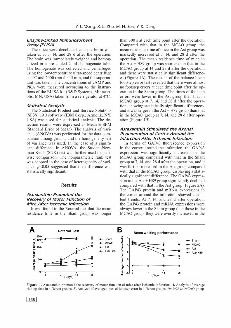
It was found in the Rotarod test that the mean residence time in the Sham group was longer
than 300 s at each time point after the operation. Compared with that in the MCAO group, the mean residence time of mice in the Ast group was markedly increased at 7, 14, and 28 d after the operation. The mean residence time of mice in the Ast + H89 group was shorter than that in the MCAO group at 14 and 28 d after the operation, and there were statistically significant differenc-es (Figure 1A). The results of the balance beam footstep error test revealed that there were almost no footstep errors at each time point after the op-eration in the Sham group. The times of footstep errors were fewer in the Ast group than that in MCAO group at 7, 14, and 28 d after the opera-tion, showing statistically significant differences, and it was larger in the Ast + H89 group than that in the MCAO group at 7, 14, and 28 d after oper-ation (Figure 1B).
Astaxanthin Stimulated the Axonal Regeneration of Cortex Around theInfarction After Ischemic Infarction
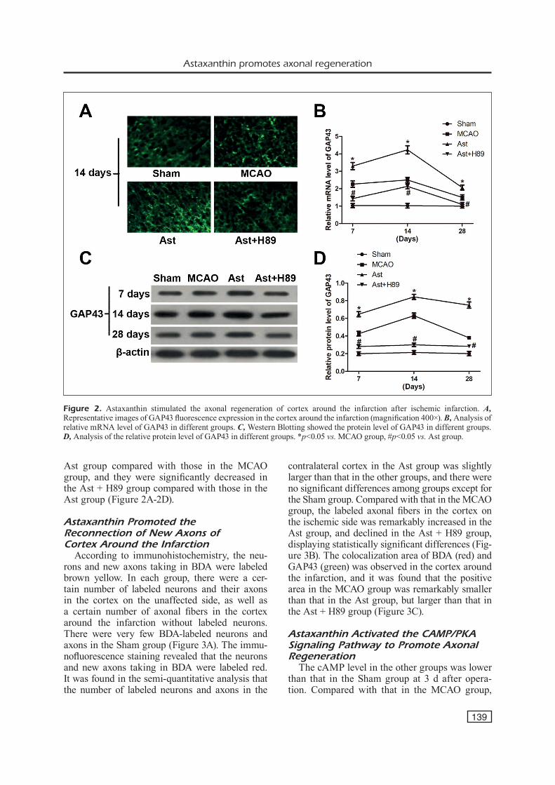
In terms of GAP43 fluorescence expression in the cortex around the infarction, the GAP43 expression was significantly increased in the MCAO group compared with that in the Sham group at 7, 14, and 28 d after the operation, and it was further increased in the Ast group compared with that in the MCAO group, displaying a statis-tically significant difference. The GAP43 expres-sion in the Ast + H89 group significantly declined compared with that in the Ast group (Figure 2A). The GAP43 protein and mRNA expressions in the cortex around the infarction showed consis-tent trends. At 7, 14, and 28 d after operation, the GAP43 protein and mRNA expressions were always lower in the Sham group than those in the MCAO group, they were overtly increased in the
Figure 1. Astaxanthin promoted the recovery of motor function of mice after ischemic infarction. A, Analysis of average ridding time in different groups. B, Analysis of average times of footstep error in different groups. *p<0.05 vs. MCAO group.
Astaxanthin promotes axonal regeneration
139
Ast group compared with those in the MCAO group, and they were significantly decreased in the Ast + H89 group compared with those in the Ast group (Figure 2A-2D).
Astaxanthin Promoted the Reconnection of New Axons of Cortex Around the Infarction
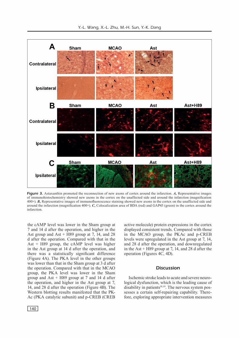
According to immunohistochemistry, the neu-rons and new axons taking in BDA were labeled brown yellow. In each group, there were a cer-tain number of labeled neurons and their axons in the cortex on the unaffected side, as well as a certain number of axonal fibers in the cortex around the infarction without labeled neurons. There were very few BDA-labeled neurons and axons in the Sham group (Figure 3A). The immu-nofluorescence staining revealed that the neurons and new axons taking in BDA were labeled red. It was found in the semi-quantitative analysis that the number of labeled neurons and axons in the
contralateral cortex in the Ast group was slightly larger than that in the other groups, and there were no significant differences among groups except for the Sham group. Compared with that in the MCAO group, the labeled axonal fibers in the cortex on the ischemic side was remarkably increased in the Ast group, and declined in the Ast + H89 group, displaying statistically significant differences (Fig-ure 3B). The colocalization area of BDA (red) and GAP43 (green) was observed in the cortex around the infarction, and it was found that the positive area in the MCAO group was remarkably smaller than that in the Ast group, but larger than that in the Ast + H89 group (Figure 3C).
Astaxanthin Activated the CAMP/PKA Signaling Pathway to Promote Axonal Regeneration
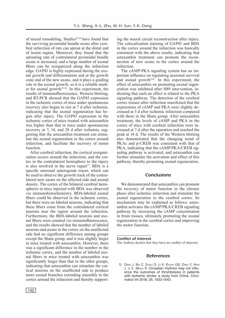
The cAMP level in the other groups was lower than that in the Sham group at 3 d after opera-tion. Compared with that in the MCAO group,
Figure 2. Astaxanthin stimulated the axonal regeneration of cortex around the infarction after ischemic infarction. A, Representative images of GAP43 fluorescence expression in the cortex around the infarction (magnification 400×). B, Analysis of relative mRNA level of GAP43 in different groups. C, Western Blotting showed the protein level of GAP43 in different groups. D, Analysis of the relative protein level of GAP43 in different groups. *p<0.05 vs. MCAO group, #p<0.05 vs. Ast group.
Y.-L. Wang, X.-L. Zhu, M.-H. Sun, Y.-K. Dang
140
the cAMP level was lower in the Sham group at 7 and 14 d after the operation, and higher in the Ast group and Ast + H89 group at 7, 14, and 28 d after the operation. Compared with that in the Ast + H89 group, the cAMP level was higher in the Ast group at 14 d after the operation, and there was a statistically significant difference (Figure 4A). The PKA level in the other groups was lower than that in the Sham group at 3 d after the operation. Compared with that in the MCAO group, the PKA level was lower in the Sham group and Ast + H89 group at 7 and 14 d after the operation, and higher in the Ast group at 7, 14, and 28 d after the operation (Figure 4B). The Western blotting results manifested that the PK-Ac (PKA catalytic subunit) and p-CREB (CREB
active molecule) protein expressions in the cortex displayed consistent trends. Compared with those in the MCAO group, the PKAc and p-CREB levels were upregulated in the Ast group at 7, 14, and 28 d after the operation, and downregulated in the Ast + H89 group at 7, 14, and 28 d after the operation (Figures 4C, 4D).
Discussion
Ischemic stroke leads to acute and severe neuro-logical dysfunction, which is the leading cause of disability in patients18,19. The nervous system pos-sesses a certain self-repairing capability. There-fore, exploring appropriate intervention measures
Figure 3. Astaxanthin promoted the reconnection of new axons of cortex around the infarction. A, Representative images of immunohistochemistry showed new axons in the cortex on the unaffected side and around the infarction (magnification 400×). B, Representative images of immunofluorescence staining showed new axons in the cortex on the unaffected side and around the infarction (magnification 400×). C, Colocalization area of BDA (red) and GAP43 (green) in the cortex around the infarction.

Astaxanthin promotes axonal regeneration
141
to stimulate the endogenous nerve repair has been a hot spot in medical research currently. Limb dyskinesia is one of the major clinical manifes-tations after stroke, and the partial recovery of limb motor function is an evident feature of nerve repair. The Rotarod test and the balance beam footstep error test are commonly-used behavioral tests to evaluate the motor function after brain in-jury20,21. The results of the two tests revealed that after astaxanthin treatment, the time of footstep errors and the residence time on rotarod of mice with cerebral ischemic infarction were superior
to those of mice under spontaneous recovery. The motor function of mice had spontaneous recovery after cerebral infarction, the recovery effect after astaxanthin treatment was better than that under spontaneous recovery, and the recovery of bal-ance and fine motor of mice was earlier than that of coordination and muscle strength.
The neural plasticity after ischemic infarction is a process of building a new structural con-nection between the tissues around the cerebral lesion and the lesion region. The axonal regen-eration is the anatomical basis and the key link
Figure 4. Astaxanthin activated the cAMP/PKA signaling pathway to promote axonal regeneration. A, Analysis of the cAMP level in different groups. B, Analysis of the PKA level in different groups. C, Western Blotting showed protein level of PKAc in different groups. D, Western Blotting showed protein level of p-CREB in different groups. *p<0.05 vs. MCAO group, #p<0.05 vs. Ast group.
Y.-L. Wang, X.-L. Zhu, M.-H. Sun, Y.-K. Dang
142
of neural remodeling. Studies22-24 have found that the surviving pyramidal bundle axons after cere-bral infarction of rats can sprout at the distal end of lesion region. Moreover, they found that the sprouting rate of contralateral pyramidal bundle axons is increased, and a large number of axonal fibers can be reorganized along the infarction edge. GAP43 is highly expressed during the axo-nal growth and differentiation and at the growth cone end of the new axons, and it plays a guiding role in the axonal growth, so it is a reliable mark-er for axonal growth25,26. In this experiment, the results of immunofluorescence, Western blotting, and RT-PCR showed that the GAP43 expression in the ischemic cortex of mice under spontaneous recovery also began to rise at 7 d after ischemia, indicating that the axonal regeneration has be-gun after injury. The GAP43 expression in the ischemic cortex of mice treated with astaxanthin was higher than that in mice under spontaneous recovery at 7, 14, and 28 d after ischemia, sug-gesting that the astaxanthin treatment can stimu-late the axonal regeneration in the cortex around infarction, and facilitate the recovery of motor function.
After cerebral infarction, the cortical reorgani-zation occurs around the infarction, and the cor-tex in the contralateral hemisphere to the injury is also involved in the nerve repair27. BDA is a specific neuronal anterograde tracer, which can be used to observe the growth track of the contra-lateral new axons on the affected side and axonal density. The cortex of the bilateral cerebral hemi-spheres in mice injected with BDA was observed via immunohistochemistry. BDA-labeled axonal fibers could be observed in the ischemic cortex, but there were no labeled neurons, indicating that these fibers come from the contralateral cortical neurons near the region around the infarction. Furthermore, the BDA-labeled neurons and axo-nal fibers were counted via immunofluorescence, and the results showed that the number of labeled neurons and axons in the cortex on the unaffected side had no significant difference among groups except the Sham group, and it was slightly larger in mice treated with astaxanthin. However, there was a significant difference in the number in the ischemic cortex, and the number of labeled axo-nal fibers in mice treated with astaxanthin was significantly larger than that in the other groups, indicating that astaxanthin can stimulate the cor-tical neurons on the unaffected side to produce more axonal branches extending smoothly to the cortex around the infarction and thereby support-
ing the neural circuit reconstruction after injury. The colocalization staining of GAP43 and BDA in the cortex around the infarction was basically consistent with the above results, indicating that astaxanthin treatment can promote the recon-nection of new axons in the cortex around the infarction.
The cAMP-PKA signaling system has an im-portant influence on regulating neuronal survival and axonal growth9,10. In this experiment, the effect of astaxanthin on promoting axonal regen-eration was inhibited after H89 intervention, in-dicating that such an effect is related to the PKA signaling pathway. The detection of the cerebral cortex tissues after infarction manifested that the expressions of cAMP and PKA were slightly de-creased at 3 d after ischemic infarction compared with those in the Sham group. After astaxanthin treatment, the levels of cAMP and PKA in the cortex of mice with cerebral infarction were in-creased at 7 d after the operation and reached the peak at 14 d. The results of the Western blotting also demonstrated that the changing trend of PKAc and p-CREB was consistent with that of PKA, indicating that the cAMP/PKA/CREB sig-naling pathway is activated, and astaxanthin can further stimulate the activation and effect of this pathway, thereby promoting axonal regeneration.
Conclusions
We demonstrated that astaxanthin can promote the recovery of motor function in the chronic phase after ischemic infarction and stimulate the axonal regeneration in the cerebral cortex. Its mechanism may be explained as follows: astax-anthin activates the cAMP/PKA/CREB signaling pathway by increasing the cAMP concentration in brain tissues, ultimately promoting the axonal regeneration in the cerebral cortex and improving the motor function.
Conflict of InterestThe Authors declare that they have no conflict of interests.
References
1) Ding J, Bai Z, Zhou D, Li X, RaJah gB, Ding Y, han J, Ji X, Meng R. Circadian rhythms may not influ-ence the outcomes of thrombolysis in patients with ischemic stroke: a study from China. Chro-nobiol Int 2018; 35: 1533-1542.
Astaxanthin promotes axonal regeneration
143
2) hao Z, Chang X, Zhou h, Lin S, Liu M. A cohort study of decompressive craniectomy for malig-nant middle cerebral artery infarction: areal-world experience in clinical practice. Medicine (Balti-more) 2015; 94: e1039.
3) PoRCaRi gS, BeSLow La, iChoRD Rn, LiCht DJ, KLein-Man Jt, JoRDan LC. Neurologic outcome predictors in pediatric intracerebral hemorrhage: a prospec-tive study. Stroke 2018; 49: 1755-1758.
4) aLLan PD, tZeng YC, gowing eK, CLaRKSon an, Fan JL. Dietary nitrate supplementation reduces low frequency blood pressure fluctuations in rats fol-lowing distal middle cerebral artery occlusion. J Appl Physiol (1985) 2018; 125: 862-869.
5) Liang Z, Chi YJ, Lin gQ, Luo Sh, Jiang QY, Chen YK. MiRNA-26a promotes angiogenesis in a rat mod-el of cerebral infarction via PI3K/AKT and MAPK/ERK pathway. Eur Rev Med Pharmacol Sci 2018; 22: 3485-3492.
6) Liu Z, ChoPP M. Astrocytes, therapeutic targets for neuroprotection and neurorestoration in ischemic stroke. Prog Neurobiol 2016; 144: 103-120.
7) RoDgeR J, goto h, Cui Q, Chen PB, haRveY aR. cAMP regulates axon outgrowth and guidance during optic nerve regeneration in goldfish. Mol Cell Neurosci 2005; 30: 452-464.
8) PaRK K, Luo JM, hiSheh S, haRveY aR, Cui Q. Cellular mechanisms associated with spontaneous and ciliary neurotrophic factor-cAMP-induced survival and axonal regeneration of adult retinal ganglion cells. J Neurosci 2004; 24: 10806-10815.
9) SiDDiQ MM, hanniLa SS. Looking downstream: the role of cyclic AMP-regulated genes in axonal re-generation. Front Mol Neurosci 2015; 8: 26.
10) BattY nJ, FenRiCh KK, FouaD K. The role of cAMP and its downstream targets in neurite growth in the adult nervous system. Neurosci Lett 2017; 652: 56-63.
11) YaMagiShi R, aihaRa M. Neuroprotective effect of astaxanthin against rat retinal ganglion cell death under various stresses that induce apoptosis and necrosis. Mol Vis 2014; 20: 1796-1805.
12) Lu YP, Liu SY, Sun h, wu XM, Li JJ, Zhu L. Neuro-protective effect of astaxanthin on H(2)O(2)-in-duced neurotoxicity in vitro and on focal cerebral ischemia in vivo. Brain Res 2010; 1360: 40-48.
13) Liu X, oSawa t. Astaxanthin protects neuronal cells against oxidative damage and is a potent candidate for brain food. Forum Nutr 2009; 61: 129-135.
14) Liu X, ShiBata t, hiSaKa S, oSawa t. Astaxanthin in-hibits reactive oxygen species-mediated cellular toxicity in dopaminergic SH-SY5Y cells via mito-chondria-targeted protective mechanism. Brain Res 2009; 1254: 18-27.
15) gRiMMig B, DaLY L, SuBBaRaYan M, huDSon C, wiLLiaM-Son R, naSh K, BiCKFoRD PC. Astaxanthin is neuro-
protective in an aged mouse model of Parkinson’s disease. Oncotarget 2018; 9: 10388-10401.
16) YooK JS, oKaMoto M, RaKwaL R, ShiBato J, Lee MC, MatSui t, Chang h, Cho JY, SoYa h. Astaxanthin supplementation enhances adult hippocampal neurogenesis and spatial memory in mice. Mol Nutr Food Res 2016; 60: 589-599.
17) BinghaM D, MaRtin SJ, MaCRae iM, CaRSweLL hv. Wa-termaze performance after middle cerebral artery occlusion in the rat: the role of sensorimotor ver-sus memory impairments. J Cereb Blood Flow Metab 2012; 32: 989-999.
18) eDwaRDS D, BiX gJ. Roles of integrins and extra-cellular matrix in stroke. Am J Physiol Cell Physi-ol 2019; 316: C252-C263.
19) ChitSaZ a, neJat a, nouRi R. Three-dimensional nu-merical simulations of aspiration process: evalua-tion of two penumbra aspiration catheters perfor-mance. Artif Organs 2018; 42: E406-E419.
20) wang R, Li J, Duan Y, tao Z, Zhao h, Luo Y. Effects of erythropoietin on gliogenesis during cerebral ischemic/reperfusion recovery in adult mice. Ag-ing Dis 2017; 8: 410-419.
21) Liang D, he XB, wang Z, Li C, gao BY, wu JF, Bai YL. Remote limb ischemic postconditioning pro-motes motor function recovery in a rat model of ischemic stroke via the up-regulation of endoge-nous tissue kallikrein. CNS Neurosci Ther 2018; 24: 519-527.
22) Yang X, Zhang JD, Duan L, Xiong hg, Jiang YP, Li-ang hC. Microglia activation mediated by toll-like receptor-4 impairs brain white matter tracts in rats. J Biomed Res 2018; 32: 136-144.
23) MeRino P, YePeS M. Urokinase-type plasminogen activator induces neurorepair in the ischemic brain. J Neurol Exp Neurosci 2018; 4: 24-29.
24) MeRino P, DiaZ a, YePeS M. Urokinase-type plas-minogen activator (uPA) and its receptor (uPAR) promote neurorepair in the ischemic brain. Re-ceptors Clin Investig 2017; 4. pii: e1552.
25) Singh B, KRiShnan a, MiCu i, KoShY K, Singh v, MaR-tineZ Ja, KoShY D, Xu F, ChanDRaSeKhaR a, DaLton C, SYeD n, StYS PK, ZoChoDne Dw. Peripheral neuron plasticity is enhanced by brief electrical stimula-tion and overrides attenuated regrowth in experi-mental diabetes. Neurobiol Dis 2015; 83: 134-151.
26) SaChDeva R, theiSen CC, ninan v, twiSS JL, houLé JD. Exercise dependent increase in axon regenera-tion into peripheral nerve grafts by propriospinal but not sensory neurons after spinal cord injury is associated with modulation of regeneration-asso-ciated genes. Exp Neurol 2016; 276: 72-82.
27) wiLLeMSe RB, hiLLeBRanD a, RonneR he, vanDeRtoP wP, StaM CJ. Magnetoencephalographic study of hand and foot sensorimotor organization in 325 consecutive patients evaluated for tumor or epi-lepsy surgery. Neuroimage Clin 2016; 10: 46-53.