
Effects of Amlodipine and its Withdrawal on the Testis of ...
13
The Egyptian Journal of Anatomy, Jan. 2018; 41(1):62-74 62 Original Article Effects of Amlodipine and its Withdrawal on the Testis of the Adult Albino Rat and the Protective Role of Omega-3 (A Light and an Electron Microscopic Study) Fatma Alzhraa Fouad Abdelbaky Allam 1 , Samah Mohammed Mahmoud Abozeid 1 and Eman Ismail Hasan 2 Anatomy 1 , Forensic Medicine and Clinical Toxicology Departments 2 Faculty of Medicine, Minia University ABSTRACT Background: The hypertension affects about one third of the adult persons all over the world. Amlodipine is a common drug used for treatment of this disease. Infertility is one of the side effects of amlodipine, but remains not completely explained . The antioxidant, anti-apoptotic and the anti-inflammatory effects of the omega-3 on many tissues including the testis had been documented. Aim of the work: To observe the effects of amlodipine exposure on the albino rat testis using a light and an electron microscopy. Also, to assess whether these effects were decreased by withdrawal of the drug or intake of omega-3. Material and Methods: Forty adult male albino rats were used in this study. They were divided into 4 groups, 10 rats each. Group (1), was the control. Group (2), Amlodipine treated group, the rats received 5 mg of amlodipine besylate dissolved in 333 ml of a normal saline daily orally for 2 months. Group (3), Amlodipine plus omega-3, the rats received 5 mg of amlodipine besylate dissolved in 333 ml of a normal saline daily orally plus omega-3 in a dose of 400 mg/kg b. w. daily orally for 2 months. Group (4), withdrawal group, the rats received 5 mg of amlodipine besylate dissolved in 333 ml of a normal saline daily orally for 2 months, then stop the drug and wait 3 months. All the animals of group 1, 2 and 3 were sacrificed after 2 months. The rats of group (4) sacrificed after 5 months. The rat testicular functions were assessed histo-pathologically by a light and an electron microscope and biochemically by free serum testosterone level measurement. Results: Regarding the testosterone level, amlodipine group had significantly lower level than the control and the withdrawal groups. The light microscopical examination of the testicular tissue of group (2) showed marked degenerative changes and arrested spermatogenesis in most seminiferous tubules, while these degenerative changes in group ( 3) appeared milder than that of the previous group. Most of the seminiferous tubules of group (4) retained its normal structure. Electro-microscopic examination of the group of amlodipine revealed several abnormalities such as multiple dilated mitochondria, multiple lipid droplets, and highly cytoplasmic vacuoles. The spermatids of the same group showed inverted acrosome and highly distorted ruptured plasma membrane. The fine structural features of group (3) had minimal vacuolations and lipid droplets. The withdrawal group revealed a nearly normal appearance of the testicular cells. Conclusion: A low dose of amlodipine causes testicular toxicity but an improvement was occurred after the stoppage of the drug. Also, omega-3 had an ameliorative role on the amlodipine testicular toxicity. Personal non-commercial use only. EJA copyright © 2018. All rights reserved DOI: 10.21608/EJANA.2019.32645 Received: 28 Feburary 2017, Accepted: 29 March 2017 Key Words: Amlodipine, omega-3, testosterone, testicular Corresponding Author: Fatma Alzhraa Fouad Abdelbaky Allam, Human Anatomy and Embryology Department, Faculty of Medicine, Minia University, Tel.: +20 1006204663, E-mail: [email protected] The Egyptian Journal of Anatomy, ISSN: 0013-2446, Vol. 41, No. 1
Transcript of Effects of Amlodipine and its Withdrawal on the Testis of ...

The Egyptian Journal of Anatomy, Jan. 2018; 41(1):62-74
62
Original Article
Effects of Amlodipine and its Withdrawal on the Testis of the Adult Albino Rat and the Protective Role of Omega-3 (A Light and an Electron Microscopic Study)
Fatma Alzhraa Fouad Abdelbaky Allam1, Samah Mohammed Mahmoud Abozeid1 and Eman Ismail Hasan2
Anatomy1, Forensic Medicine and Clinical Toxicology Departments2
Faculty of Medicine, Minia University
ABSTRACTBackground: The hypertension affects about one third of the adult persons all over the world. Amlodipine is a common drug used for treatment of this disease. Infertility is one of the side effects of amlodipine, but remains not completely explained . The antioxidant, anti-apoptotic and the anti-inflammatory effects of the omega-3 on many tissues including the testis had been documented.Aim of the work: To observe the effects of amlodipine exposure on the albino rat testis using a light and an electron microscopy. Also, to assess whether these effects were decreased by withdrawal of the drug or intake of omega-3. Material and Methods: Forty adult male albino rats were used in this study. They were divided into 4 groups, 10 rats each. Group (1), was the control. Group (2), Amlodipine treated group, the rats received 5 mg of amlodipine besylate dissolved in 333 ml of a normal saline daily orally for 2 months. Group (3), Amlodipine plus omega-3, the rats received 5 mg of amlodipine besylate dissolved in 333 ml of a normal saline daily orally plus omega-3 in a dose of 400 mg/kg b. w. daily orally for 2 months. Group (4), withdrawal group, the rats received 5 mg of amlodipine besylate dissolved in 333 ml of a normal saline daily orally for 2 months, then stop the drug and wait 3 months.All the animals of group 1, 2 and 3 were sacrificed after 2 months. The rats of group (4) sacrificed after 5 months. The rat testicular functions were assessed histo-pathologically by a light and an electron microscope and biochemically by free serum testosterone level measurement. Results: Regarding the testosterone level, amlodipine group had significantly lower level than the control and the withdrawal groups. The light microscopical examination of the testicular tissue of group (2) showed marked degenerative changes and arrested spermatogenesis in most seminiferous tubules, while these degenerative changes in group ( 3) appeared milder than that of the previous group. Most of the seminiferous tubules of group (4) retained its normal structure. Electro-microscopic examination of the group of amlodipine revealed several abnormalities such as multiple dilated mitochondria, multiple lipid droplets, and highly cytoplasmic vacuoles. The spermatids of the same group showed inverted acrosome and highly distorted ruptured plasma membrane. The fine structural features of group (3) had minimal vacuolations and lipid droplets. The withdrawal group revealed a nearly normal appearance of the testicular cells. Conclusion: A low dose of amlodipine causes testicular toxicity but an improvement was occurred after the stoppage of the drug. Also, omega-3 had an ameliorative role on the amlodipine testicular toxicity.
Personal non-commercial use only. EJA copyright © 2018. All rights reserved DOI: 10.21608/EJANA.2019.32645
Received: 28 Feburary 2017, Accepted: 29 March 2017
Key Words: Amlodipine, omega-3, testosterone, testicular
Corresponding Author: Fatma Alzhraa Fouad Abdelbaky Allam, Human Anatomy and Embryology Department, Faculty of Medicine, Minia University, Tel.: +20 1006204663, E-mail: [email protected] Egyptian Journal of Anatomy, ISSN: 0013-2446, Vol. 41, No. 1
63
Allam et al.
INTRODUCTION:
The testis is surrounded by a thick capsule of dense connective tissue, tunica albuginea, which thickened on the posterior surface to form a mediastinum testes from which a fibrous septa penetrate to divide it into about 250 pyramidal compartments called the seminiferous tubules (Standring, 2008).
The seminiferous tubules produce the male reproductive cells spermatozoa at a daily rate of about 20,000,000, the outer interstitial cells known as Leydig cells are specialized for the testosterone synthesis.
Each testes has about 250-1000 seminiferous tubules (Standring, 2008), which are lined with a complex stratified epithelium called the germinal epithelium which consists of two types of cells, Sertoli or supporting cells which are the site of the action of all hormonal effect governing the spermatogenesis, their action is supporting and nourishing the germ cells (Russell and Griswold, 2009).
The other cells that constitute the spermatogenic cells which are stacked in 4-8 layers (Trainer, 1987). They are:
1. The spermatogonia ,which of 2 types: type (A), undergoing mitotic division, and type (B), which gives rise to the primary spermatocyte . The type B cell has a rounded nucleus with clumped chromatin.
2. The primary and the secondary spermatocytes.
3. The spermatids, which will differentiate into the spermatozoa. The outer wall of each tubule is surrounded by a well-defined basal lamina and a fibrous connecting tissue consisting of several layers of a fibroblast, the layer next to it consists of a flattened myoid cell.
Hypertension ( HTN) is a sustained, non-physiologic rise in the blood pressure; clinically it is defined as having a systolic blood pressure (SBP) of 140 mm Hg or more; or having a diastolic blood pressure (DBP) of 90 mm Hg or more or both (Adams et al.2005).
Near one third of the adult persons all over the world have HTN (Karthick and Harisudha, 2014). The primary care of physicians and other health practitioners are the first to deal with the hypertensive persons. HTN increases the risk of many cardiovascular diseases as coronary heart
disease, congestive heart failure, stroke, and renal failure (Harrison, 2013).
Amlodipine is one of the calcium channel blockers (CCBs) (Karthick and Harisudha, 2014). CCBs reduce the blood pressure by blocking the entry of calcium ions through the L channels of the arterial smooth muscle cells. Amlodipine works by dilating arteries but also reduces the heart rate and contractility (ALLHAT, 2002).
Although amlodipine is now a commonly used drug for treatment for HTN, but its infertility side effect has been proved (Almeida et al.2000 and Yoshida, 2003).
The omega-3 fatty acid is a poly unsaturated fatty acid (Scorletti and Byrne, 2013.). The fish, the nut oils, and the plants are the dietary sources of omega-3 (Terry et al. 2001).
Many recent studies documented the antioxidant, anti-apoptotic and the anti-inflammatory effects of the omega-3 on many tissues including the testis that exposed to many extrinsic toxic agents (Uygur et al., .2014). Also, omega- 3 fatty acid plays an important role in the formation of an acrosome on the head of the sperm, the acrosome contains enzymes that break the outer layers of the ova, and so allow its fertilization by the sperm (Welsh, 2012).
The aim of this study was to determine the toxic effects of amlodipine on the testis of the male albino rat, and to assess whether these effects were reversible by intake of the omega-3 and\or withdrawal of the drug.
MATERIAL AND METHODS:
Animals:
A thirty adult male albino rats, weighing (180-200 gm) were used in the present study. The rats were obtained from the animal house of Human Anatomy and Embryology Department, Faculty of Medicine, El-Minia University. The rats were acclimatized for 7 days before the start of the experimental procedures.
Experimental Design:
After getting an ethical committee clearance, all rats were handled in accordance with the standard guide for the care and the use of laboratory animals. An oral administration of the drugs was done with the use of a 5 ml syringe and a long flexible feeding tube. Forty rats were divided into 4 groups:
64
EFFECTS OF AMLODIPINE AND ITS WITHDRAWAL ON THE TESTIS........
• Group 1 (10 rats); a control group: the rats received 4 ml of a normal saline daily for 2 months.
• Group 2 (10 rats); the amlodipine group: the rats received 5 mg of amlodipine besylate (NORVASC) which was dissolved in 333 ml of a normal saline daily for 2 months, each rat administered about a 0.9-1 ml of the solution (Akinlolu et al.2013).
• Group 3 (10 rats); the amlodipine plus omega-3 group: the rats received 5 mg of amlodipine besylate which was dissolved in 333 ml of a normal saline and the omega-3 in a daily dose of 400 mg\ kg b.w. ((Uygur et al.2014)) for 2 months.
All the rats of the group 1, 2 and 3 were sacrificed after 2 months.
• Group 4 (10 rats); the withdrawal group, the rats received 5 mg of amlodipine besylate which was dissolved in 333 ml of a normal saline for 2 months ,then the drug stopped for another 3 months, then the rats sacrificed.
Biochemical Study:
The serum Testosterone level:
The level of the free serum testosterone was measured by an enzyme- linked immunosorbant assay (ELISA) (McCann and Kirkish, 1985).
Histological Study:
The obtained testes from all groups were subjected to the following processing:
1- Light microscopic examination:
The specimens were fixed in phosphate buffered saline (PBS, pH 7.3) containing 3.7% formaldehyde for 1 an hour. The fixed samples were then washed with the water, and dehydrated in a graded ethanol: 70% ethanol for 1an hour, 80% ethanol for two times one an hour each, 95% ethanol for two times one an hour each, 100% ethanol for three times one an hour each, then cleared in a xylene for three times one an hour for the1sttime then 90 minutes twice and embedded in a wax at 60°C two times for two hours each. Finally, the samples were embedded in a paraffin wax in a labeled plastic cassette. The embedded samples were sectioned at a 5µm thickness, using a microtome. The sections were processed and stained with a Haematoxylin and eosin stain (Hx&E) (Bancroft and Gamble, 2002).
2- Electron microscopic examination:
The specimens were immediately washed
with 0.1 M. phosphate buffer and fixed in a 2.5% glutaraldehyde buffered in phosphate buffered at pH 7.4 for 2 hours and then post fixed in a 1% osmium tetraoxide in the same buffer for an another one an hour. They were processed to prepare the semi thin and the ultrathin sections. The one µm transverse semi-thin section was cut by the RMC ultratome, stained with the toluidine blue stain 1%, and examined by the light microscope.
The ultrathin sections were mounted on a copper grids stained with a 2% aqueous uranyl acetate for 20 minutes, and a lead citrate for an another 20 minutes, then examined and photographed using a JEOL electron microscope equipped with a camera in the electron microscope research laboratory of the histology and cell biology department, Faculty of science, Alexandria University.
RESULTS:
The Serum free testosterone level:
All the treated groups did not differ significantly in the serum free testosterone level from the control group, except the amlodipine- treated group which had significantly lower in the testosterone level than the control group (Table 1). However, the withdrawal group had significantly a higher testosterone level than the amlodipine -treated group (Table 2).
The Histopathological findings in the testis:
1) The light microscopic examination (Hx&E stain):
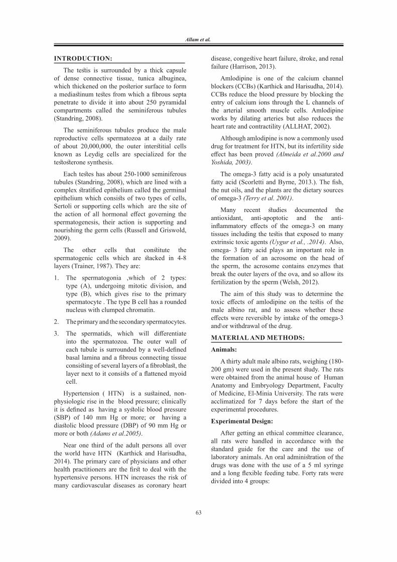
The testis of the rats of the control group (1), revealed the normal structural components of the testis which composed of the seminiferous tubules and the interstitial tissues. The seminiferous tubules were lined by regularly arranged rows of the spermatogenic cells at different stages of maturation (Fig. 1).
The seminiferous tubules of amlodipine group (2), showed a marked degenerative changes, an arrest of the spermatogenesis, at the level of the spermatids and sperms, appeared in all semineferous tubules. Also large areas of vacuolation and congestion appeared (Fig. 2).
In amlodipine plus omega-3 group (3), the degenerative changes were milder than that of the previous group. Most of the seminiferous tubules appeared with a number of sperms and spermatogenic cells, while congestion
65
Allam et al.
and vacuoles were decreased (Fig.3). Most of the seminiferous tubules of the withdrawal group (4), retained its normal structure, there was no disruption of the basement membrane, the seminiferous tubules showed a number of sperms and spermatogenic cells .Congestion and vacuolations were less (Fig. 4).
2) Semi-thin section examination (Toluidine blue stain):
The semithin sections of the testis of the control group (1), showed a normal seminiferous tubule with a different stages of spermatogonic cells and thin basement membrane. The interstitial cells had a pale stained nucleus and cytoplasm (Fig. 5a). A normal Sertoli, and spermatogenic cells (Fig 5b).
Regarding the semithin sections of the amlodipine-treated group, there was a marked degeneration in the form of a darkly stained cytoplasm and nuclei of the interstitial cells (Fig. 6a). Some tubules contained a marked areas of vacuolation within and in between the spermatogenic cells, and many distorted cells were noticed (Fig. 6b).
Fortunately, the amlodipine plus omega group showed a noticeable improvement in the spermatogenic cells, vacuolations were less, and the dark coloration of the interstitial cells were retained (Figs. 7a and 7b). The semithin sections of the testis of the withdrawal group showed a normal appearance of the seminiferous tubules (Figs. 8a and 8b).
3) The electron microscopic examination:
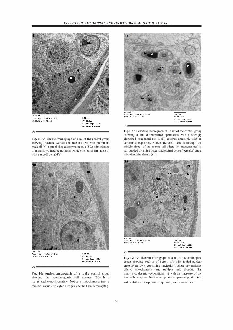
The seminiferous tubules of the control group were bounded by a spindle-shaped myoid cells that encircle the basal lamina, Sertoli cell showed a large indented nucleus with a prominent nucleoli (Fig. 9).
Spermatogonia were characterized by its rounded nucleus with a marginated heterochromatin (Fig. 10). Late differentiated spermatids appeared with strongly elongated condensed nuclei covered anteriorly with an acrosomal cap. The cross section through the middle pieces of the tail of the sperms showed the axoneme surrounded by a nine outer longitudinal dense fibers and a mitochondrial sheath (Fig. 11).
In amlodipine- treated group the seminiferous tubules revealed a several abnormal appearance such as, a multiple dilated mitochondria and lipid droplets, and a highly cytoplasmic vacuolation. Also, Sertoli cell appeared with a folded nuclear envelop, and an apoptotic spermatogonia found (Fig. 12). The spermatids showed an inverted acrosome towards the nuclear membrane and a highly distorted ruptured plasma membrane. Many dilated mitochondria , lipid droplets, and many areas of vacuolation appeared (Fig.13).The seminiferous tubules of omega plus amlodipine- treated group revealed that Sertoli cell appeared normal in shape but still had folded nuclear envelop, the vacuolated areas were less, but still the dilated mitochondria and the thick basal lamina found (Fig. 14).
The spermatids of the amlodipine plus omega-3 group appeared near to the normal shape (Fig.15). The seminiferous tubules of the withdrawal group revealed a nearly improvement of the nucleus of the spermatogonia cell, which appeared with a clustered marginated heterochromatine. Sertoli cell nucleus appeared normal in shape with a prominent nucleoli (Fig.16). Also the spermatids of the withdrawal group appeared normal in shape (Fig. 17).
Fig. 1:A photomicrograph of a section of the testis of a rat of the control group showing the seminiferous tubules lined by a regularly arranged rows of the spermatogenic cells, spermatogonia (SG), spermatocyte(ST), and sperms(SP). They are bounded with outer basal lamina (BL). Hx&E X200

66
EFFECTS OF AMLODIPINE AND ITS WITHDRAWAL ON THE TESTIS........
Fig. 2: A photomicrograph of a section of the testis of a rat of the amlodlpine group showing: An arrested spermatogenesis with loss of sperms (arrows), Also there are many areas of vacuolation (v), and congestion (c).Hx&E; X200
Fig. 3: A photomicrograph of a section of the testis of the amlodipine plus omega group showing:the seminiferous tubules appear with a number of sperms (sp) and spermatogenic cells: congestion(c), and vacuolations(v) are decreased. Hx&E; X200
Fig. 4:A photomicrograph of a section of the testis of a rat of the withdrawal group showing:the seminiferous tubules appear near to the normal shape, there is a number of sperms (sp) and spermatogenic cells appeared within the tubules.Congestion(c) and vacuolations (V) HX&E X200
Fig. 5a&5b: A photomicrograph of a semithin section of the testis of a rat of the control group: 5a) showing a normal seminiferous tubule with a different stages of spermatogonic cells(SG). The interstitial cells(I) have a pale stained nucleus and cytoplasm. 5b) Notice normal shape Sertoli cell(S), spermatocytes (SC), and sperms(SP). Toluidine blue; 5a X400 and 5b 1000

67
Allam et al.
Fig. 6a&6b: A photomicrograph of a semithin section of the testis of a rat ofamlodipine group showing: 6a) Interstitial cells(I) have a darkly stained cytoplasm and nuclei, there are many areas ofvacuolations (v) in between the spermatogenic cells with depletion of the sperms(arrows). 6b) distorted spermatogoniacells(SG) with many areas of vacolations in between the spermatogenic cells(v). Notice:Sertoli cell(S). Toluidine blue; 6a X 400and 6b X 1000
Fig. 7a&7b: A photomicrograph of a semithin section of the testis of a rat of amlidopine plus omega group :7aandb) showing apparently normalSertoli cell(S), spermatogenia (SG), spermatocytes(SC), spermatid(ST) , and sperms(sp). 7b) spermatid cells(ST) appear normal in shape with anacrosomal cap(arrow). Toluidine blue; X400and1000
Fig. 8a&8b: A photomicrograph of a semithin section of the testis of a rat of the withdrawal group showing:8aand 8b) A normally appeared seminiferous tubule with normal shaped Sertoli cell(S),spermatogonia (SG), spermatid(ST), and many sperm(SP). 8b) A spermatid cell appearswith an acrosomal cap(arrow). The interstitial cells (I) retain their pale stained nucleus and cytoplasm.Toluidine blue; 8a X400and8b X1000
68
EFFECTS OF AMLODIPINE AND ITS WITHDRAWAL ON THE TESTIS........
Fig. 9: An electron micrograph of a rat of the control group showing indented Sertoli cell nucleus (N) with prominent nucleoli (n), normal shaped spermatogonia (SG) with clumps of marginated heterochromatin. Notice the basal lamina (BL) with a myoid cell (MY).
Fig. 10: Anelectromicrograph of a ratthe control group showing the spermatogonia cell nucleus (N)with a marginatedheterochromatine. Notice a mitochondria (m), a minimal vacuolated cytoplasm (v), and the basal lamina(BL).
Fig.11: An electron micrograph of a rat of the control group showing a late differentiated spermatids with a strongly elongated condensed nuclei (N) covered anteriorly with an acrosomal cap (Ac). Notice the cross section through the middle pieces of the sperms tail where the axoneme (ax) is surrounded by a nine outer longitudinal dense fibers (Lf) and a mitochondrial sheath (mt).
Fig. 12: An electron micrograph of a rat of the amlodipine group showing nucleus of Sertoli (N) with folded nuclear envelop (arrow), containing nucleolus(n),there are multiple dilated mitochondria (m), multiple lipid droplets (L), many cytoplasmic vacuolations (v) with an increase of the intercellular space. Notice an apoptotic spermatogonia (SG) with a distorted shape and a ruptured plasma membrane.
69
Allam et al.
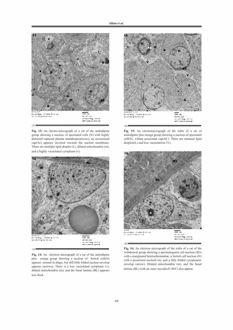
Fig. 13: An electro-micrograph of a rat of the amlodipine group showing a nucleus of spermatid cells (N) with highly distorted ruptured plasma membrane(arrows), an acrosomeal cap(Ac) appears inverted towards the nuclear membrane. There are multiple lipid droplet (L), dilated mitochondria (m), and a highly vacuolated cytoplasm (v).
Fig. 14: An electron micrograph of a rat of the amlodipine plus omega group showing a nucleus of Sertoli cell(N) appears normal in shape, but still little folded nuclear envelop appears (arrows). There is a less vacuolated cytoplasm (v), dilated mitochondria (m), and the basal lamina (BL) appears less thick.
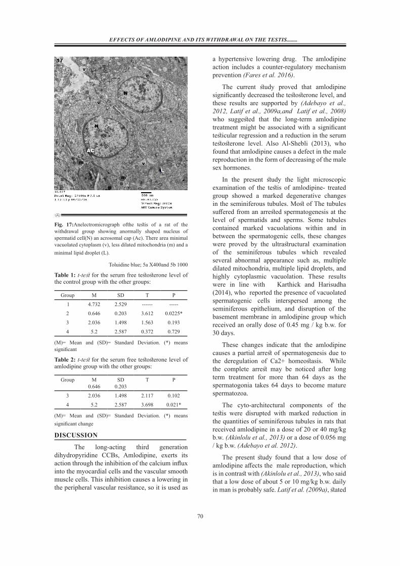
Fig. 15: An electromicrograph of the testis of a rat of amlodipine plus omega group showing a nucleus of spermatid cell(N), withan acrosomal cap(AC). There are minimal lipid droplets(L) and less vacuolations (V).
Fig.(15): An electromicrograph of the testis of a rat of amlodipine plus omega group showing a nucleus of spermatid cell(N), withan acrosomal cap(AC). There are minimal lipid droplets(L) and less vacuolations (V).
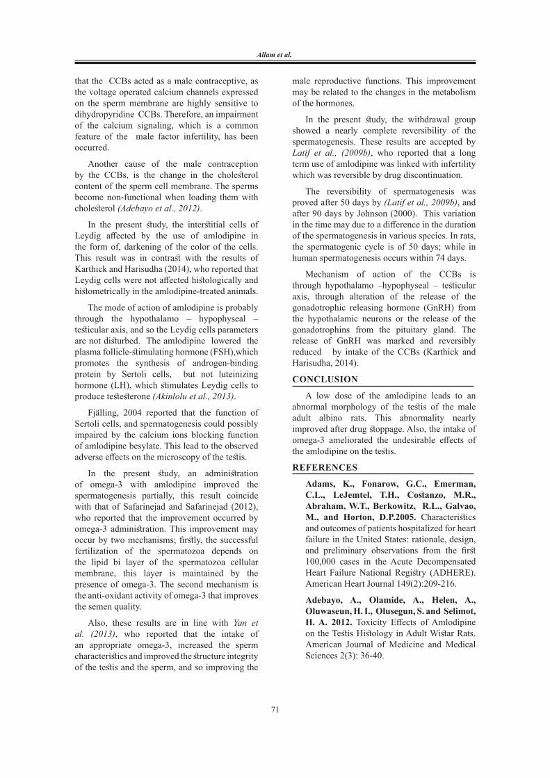
Fig. 16: An electron micrograph of the testis of a rat of the withdrawal group showing a spermatogonia cell nucleus (SG) with a marginated heterochromatine, a Sertoli cell nucleus (N) with a prominent nucleoli (n), and a little folded cytoplasmic envelop (arrow). Dilated mitochondria (m), and the basal lamina (BL) with an outer myoidcell (MY) also appear.
70
EFFECTS OF AMLODIPINE AND ITS WITHDRAWAL ON THE TESTIS........
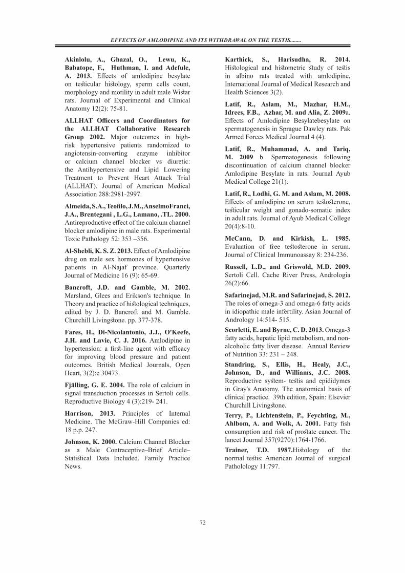
Fig. 17:Anelectromicrograph ofthe testis of a rat of the withdrawal group showing anormally shaped nucleus of spermatid cell(N) an acrosomal cap (Ac). There area minimal vacuolated cytoplasm (v), less dilated mitochondria (m) and a minimal lipid droplet (L).
Toluidine blue; 5a X400and 5b 1000
Table 1: t-test for the serum free testosterone level of the control group with the other groups:
Group M SD T P
1 4.732 2.529 ------ -----
2 0.646 0.203 3.612 0.0225*
3 2.036 1.498 1.563 0.193
4 5.2 2.587 0.372 0.729
(M)= Mean and (SD)= Standard Deviation. (*) means significant
Table 2: t-test for the serum free testosterone level of amlodipine group with the other groups:
Group M0.646
SD0.203
T P
3 2.036 1.498 2.117 0.102
4 5.2 2.587 3.698 0.021*
(M)= Mean and (SD)= Standard Deviation. (*) means significant change
DISCUSSION
The long-acting third generation dihydropyridine CCBs, Amlodipine, exerts its action through the inhibition of the calcium influx into the myocardial cells and the vascular smooth muscle cells. This inhibition causes a lowering in the peripheral vascular resistance, so it is used as
a hypertensive lowering drug. The amlodipine action includes a counter-regulatory mechanism prevention (Fares et al. 2016).
The current study proved that amlodipine significantly decreased the testosterone level, and these results are supported by (Adebayo et al., 2012, Latif et al., 2009a,and Latif et al., 2008) who suggested that the long-term amlodipine treatment might be associated with a significant testicular regression and a reduction in the serum testosterone level. Also Al-Shebli (2013), who found that amlodipine causes a defect in the male reproduction in the form of decreasing of the male sex hormones.
In the present study the light microscopic examination of the testis of amlodipine- treated group showed a marked degenerative changes in the seminiferous tubules. Most of The tubules suffered from an arrested spermatogenesis at the level of spermatids and sperms. Some tubules contained marked vacuolations within and in between the spermatogenic cells, these changes were proved by the ultrastructural examination of the seminiferous tubules which revealed several abnormal appearance such as, multiple dilated mitochondria, multiple lipid droplets, and highly cytoplasmic vacuolation. These results were in line with Karthick and Harisudha (2014), who reported the presence of vacuolated spermatogenic cells interspersed among the seminiferous epithelium, and disruption of the basement membrane in amlodipine group which received an orally dose of 0.45 mg / kg b.w. for 30 days.
These changes indicate that the amlodipine causes a partial arrest of spermatogenesis due to the deregulation of Ca2+ homeostasis. While the complete arrest may be noticed after long term treatment for more than 64 days as the spermatogonia takes 64 days to become mature spermatozoa.
The cyto-architectural components of the testis were disrupted with marked reduction in the quantities of seminiferous tubules in rats that received amlodipine in a dose of 20 or 40 mg/kg b.w. (Akinlolu et al., 2013) or a dose of 0.056 mg / kg b.w. (Adebayo et al. 2012).
The present study found that a low dose of amlodipine affects the male reproduction, which is in contrast with (Akinlolu et al., 2013), who said that a low dose of about 5 or 10 mg/kg b.w. daily in man is probably safe. Latif et al. (2009a), stated
71
Allam et al.
that the CCBs acted as a male contraceptive, as the voltage operated calcium channels expressed on the sperm membrane are highly sensitive to dihydropyridine CCBs. Therefore, an impairment of the calcium signaling, which is a common feature of the male factor infertility, has been occurred.
Another cause of the male contraception by the CCBs, is the change in the cholesterol content of the sperm cell membrane. The sperms become non-functional when loading them with cholesterol (Adebayo et al., 2012).
In the present study, the interstitial cells of Leydig affected by the use of amlodipine in the form of, darkening of the color of the cells. This result was in contrast with the results of Karthick and Harisudha (2014), who reported that Leydig cells were not affected histologically and histometrically in the amlodipine-treated animals.
The mode of action of amlodipine is probably through the hypothalamo – hypophyseal – testicular axis, and so the Leydig cells parameters are not disturbed. The amlodipine lowered the plasma follicle-stimulating hormone (FSH),which promotes the synthesis of androgen-binding protein by Sertoli cells, but not luteinizing hormone (LH), which stimulates Leydig cells to produce testesterone (Akinlolu et al., 2013).
Fjälling, 2004 reported that the function of Sertoli cells, and spermatogenesis could possibly impaired by the calcium ions blocking function of amlodipine besylate. This lead to the observed adverse effects on the microscopy of the testis.
In the present study, an administration of omega-3 with amlodipine improved the spermatogenesis partially, this result coincide with that of Safarinejad and Safarinejad (2012), who reported that the improvement occurred by omega-3 administration. This improvement may occur by two mechanisms; firstly, the successful fertilization of the spermatozoa depends on the lipid bi layer of the spermatozoa cellular membrane, this layer is maintained by the presence of omega-3. The second mechanism is the anti-oxidant activity of omega-3 that improves the semen quality.
Also, these results are in line with Yan et al. (2013), who reported that the intake of an appropriate omega-3, increased the sperm characteristics and improved the structure integrity of the testis and the sperm, and so improving the
male reproductive functions. This improvement may be related to the changes in the metabolism of the hormones.
In the present study, the withdrawal group showed a nearly complete reversibility of the spermatogenesis. These results are accepted by Latif et al., (2009b), who reported that a long term use of amlodipine was linked with infertility which was reversible by drug discontinuation.
The reversibility of spermatogenesis was proved after 50 days by (Latif et al., 2009b), and after 90 days by Johnson (2000). This variation in the time may due to a difference in the duration of the spermatogenesis in various species. In rats, the spermatogenic cycle is of 50 days; while in human spermatogenesis occurs within 74 days.
Mechanism of action of the CCBs is through hypothalamo –hypophyseal – testicular axis, through alteration of the release of the gonadotrophic releasing hormone (GnRH) from the hypothalamic neurons or the release of the gonadotrophins from the pituitary gland. The release of GnRH was marked and reversibly reduced by intake of the CCBs (Karthick and Harisudha, 2014).
CONCLUSION
A low dose of the amlodipine leads to an abnormal morphology of the testis of the male adult albino rats. This abnormality nearly improved after drug stoppage. Also, the intake of omega-3 ameliorated the undesirable effects of the amlodipine on the testis.
REFERENCES
Adams, K., Fonarow, G.C., Emerman, C.L., LeJemtel, T.H., Costanzo, M.R., Abraham, W.T., Berkowitz, R.L., Galvao, M., and Horton, D.P.2005. Characteristics and outcomes of patients hospitalized for heart failure in the United States: rationale, design, and preliminary observations from the first 100,000 cases in the Acute Decompensated Heart Failure National Registry (ADHERE). American Heart Journal 149(2):209-216.
Adebayo, A., Olamide, A., Helen, A., Oluwaseun, H. I., Olusegun, S. and Selimot, H. A. 2012. Toxicity Effects of Amlodipine on the Testis Histology in Adult Wistar Rats. American Journal of Medicine and Medical Sciences 2(3): 36-40.
72
EFFECTS OF AMLODIPINE AND ITS WITHDRAWAL ON THE TESTIS........
Akinlolu, A., Ghazal, O., Lewu, K., Babatope, F., Huthman, I. and Adefule, A. 2013. Effects of amlodipine besylate on testicular histology, sperm cells count, morphology and motility in adult male Wistar rats. Journal of Experimental and Clinical Anatomy 12(2): 75-81.
ALLHAT Officers and Coordinators for the ALLHAT Collaborative Research Group 2002. Major outcomes in high-risk hypertensive patients randomized to angiotensin-converting enzyme inhibitor or calcium channel blocker vs diuretic: the Antihypertensive and Lipid Lowering Treatment to Prevent Heart Attack Trial (ALLHAT). Journal of American Medical Association 288:2981-2997.
Almeida, S.A., Teofilo, J.M., AnselmoFranci, J.A., Brentegani , L.G., Lamano, .TL. 2000. Antireproductive effect of the calcium channel blocker amlodipine in male rats. Experimental Toxic Pathology 52: 353 –356.
Al-Shebli, K. S. Z. 2013. Effect of Amlodipine drug on male sex hormones of hypertensive patients in Al-Najaf province. Quarterly Journal of Medicine 16 (9): 65-69.
Bancroft, J.D. and Gamble, M. 2002. Marsland, Glees and Erikson's technique. In Theory and practice of histological techniques, edited by J. D. Bancroft and M. Gamble. Churchill Livingstone. pp. 377-378.
Fares, H., Di-Nicolantonio, J.J., O'Keefe, J.H. and Lavie, C. J. 2016. Amlodipine in hypertension: a first-line agent with efficacy for improving blood pressure and patient outcomes. British Medical Journals, Open Heart, 3(2):e 30473.
Fjälling, G. E. 2004. The role of calcium in signal transduction processes in Sertoli cells. Reproductive Biology 4 (3):219- 241.
Harrison, 2013. Principles of Internal Medicine. The McGraw-Hill Companies ed: 18 p.p. 247.
Johnson, K. 2000. Calcium Channel Blocker as a Male Contraceptive–Brief Article–Statistical Data Included. Family Practice News.
Karthick, S., Harisudha, R. 2014.Histological and histometric study of testis in albino rats treated with amlodipine, International Journal of Medical Research and Health Sciences 3(2).
Latif, R., Aslam, M., Mazhar, H.M., Idrees, F.B., Azhar, M. and Alia, Z. 2009a. Effects of Amlodipine Besylatebesylate on spermatogenesis in Sprague Dawley rats. Pak Armed Forces Medical Journal 4 (4).
Latif, R., Muhammad, A. and Tariq, M. 2009 b. Spermatogenesis following discontinuation of calcium channel blocker Amlodipine Besylate in rats. Journal Ayub Medical College 21(1).
Latif, R., Lodhi, G. M. and Aslam, M. 2008. Effects of amlodipine on serum testosterone, testicular weight and gonado-somatic index in adult rats. Journal of Ayub Medical College 20(4):8-10.
McCann, D. and Kirkish, L. 1985. Evaluation of free testosterone in serum. Journal of Clinical Immunoassay 8: 234-236.
Russell, L.D., and Griswold, M.D. 2009. Sertoli Cell. Cache River Press, Andrologia 26(2):66.
Safarinejad, M.R. and Safarinejad, S. 2012. The roles of omega-3 and omega-6 fatty acids in idiopathic male infertility. Asian Journal of Andrology 14:514- 515.Scorletti, E. and Byrne, C. D. 2013. Omega-3 fatty acids, hepatic lipid metabolism, and non-alcoholic fatty liver disease. Annual Review of Nutrition 33: 231 – 248. Standring, S., Ellis, H., Healy, J.C., Johnson, D., and Williams, J.C. 2008. Reproductive system- testis and epididymes in Gray's Anatomy. The anatomical basis of clinical practice. 39th edition, Spain: Elsevier Churchill Livingstone.Terry, P., Lichtenstein, P., Feychting, M., Ahlbom, A. and Wolk, A. 2001. Fatty fish consumption and risk of prostate cancer. The lancet Journal 357(9270):1764-1766.Trainer, T.D. 1987.Histology of the normal testis: American Journal of surgical Patholology 11:797.

73
Allam et al.
Uygur, R., Aktas, C., Tulubas, F., Uygur, E., Kanter, M., Erboga, M., Caglar, V., Topcu, B. and Ozen, O.A. 2014. Protective effects of fish omega-3 fatty acids on doxorubicin-induced testicular apoptosis and oxidative damage in rats. Andrologia 46(8):917- 926.
Welsh, J. 2012. Omega-3s Vital for Sperm Health. Live science.
Yan, L., Bai, X. L., Fang, Z. F., Che, L. Q., Xu, S. Y. and WuEmail, D. 2013. Effect of different dietary omega-3/omega-6 fatty acid ratios on reproduction in male rats. Lipids in Health and Disease 12: 33.
Yoshida, J. 2003. Amlodipine besylate. Europian Journal of Pharmacology 472:23–31.
74
EFFECTS OF AMLODIPINE AND ITS WITHDRAWAL ON THE TESTIS........
آثار الاملوديبين و الانسحاب على خصية الفأرالأبيض البالغ والدورالوقائ للاوميجا3-(دراسه باستخدام المجهرالضوئي والإلكتروني)
فاطمة الزهراء فؤاد عبدالباقي علام 1، سماح محمد محمود أبوزيد 1 وإيمان إسماعيل حسن 2
قسم التشريح الآدمى وعلم الاجنه 1، وقسم الطب الشرعي والسموم الاكلينيكيه 2 كلية الطب، جامعة المنيا
ملخص البحثالخلفيه: ارتفاع ضغط الدم يؤثر على حوالي ثلث الأشخاص البالغين في جميع أنحاء العالم. الاملوديبين من اشهر العقارات المستخدمه في هذا المرض ولكن ثبت ان من أثاره الجانبيه هو العقم ولكن لم يثبت بعد كيفيه حدوثه . وثقت العديد من الدراسات الحديثة الاثارالمضادة للأكسدة،و
المضادة للموت الخلوي المبرمج ومكافحة آثار التهابات للأوميغا3- على العديد من الأنسجة بما في ذلك في الخصية.
الهدف من البحث: دراسة تاثير التعرض للاملوديبين على خصيه الفأر الأبيض البالغ باستخدام المجهرالضوئي والإلكتروني ولتقييم ما إذا كانت هذه الآثار يقللها الانسحاب من الدواء أو تناول الأوميجا3-.
فئران: 10 مجموعه كل مجموعات 4 إلى قسمت وقد الدراسة. هذه في استخدم بالغ ذكر أبيض فأر أربعين البحث: وطرق المواد المجموعة(1)؛ فئران التحكم، المجموعة (2)؛ تلقي الفئران 5 ملغ من بزيلات الاملوديبين الذي تم حله في 333 مل ملح يوميا لمده شهرين. المجموعة(3 )؛ تلقي الفئران فيها 5 ملغمن بزيلات الاملوديبين الذي تم حله في 333 مل ملح بالإضافة إلى أوميجا3- بجرعة 400مل\ كجم يوميا لمده شهرين. المجموعة( 4)؛ الفريق الانسحاب: تلقي فيه الفئران 5 ملغ من بيزلالت الاملوديبين الذي تم حله في 333 مل من الملح يوميا لمدة شهرين، ثم توقف الدواء لمده 3 أشهراخري. تم التضحية بكل الحيوانات من المجموعة 1 و 2 و 3 بعد شهرين. فئران المجموعة ( 4) ضحى بهم بعد 3 أشه من توقف الدواء. تم تقييم وظائف الخصية للفئران ودراستهم هيستوباثولوجيا باستخدام المجهرالضوئي و الإلكتروني
وتحليل مستوى التستوستيرونً (هرمون الذكوره).
النتائج البحث: فيما يتعلق بمستوى التستوستيرون، كان لمجموعه الاملوديبين مستوى أقل كثيرا من مجموعه التحكم و الانسحاب. دراسة بينما ظهرت هذه المنوية في معظم خلايا الخصيه، الحيوانات أنسجة الخصية للمجموعة (2) أظهرت تغيرات مدمره ملحوظة، وانعدام الطبيعي. دراسه استعادت شكلها للمجموعة (4) الخصيه السابقة. معظم خلايا المجموعة اعتدالا من أكثر المجموعة (3) التغيرات في مجموعة الاملوديبين بواسطه المجهر الالكتروني اظهرت عدة تغيرات غير طبيعيه مثل تعدد الميتوكوندريا المتسعة ووجود متعدد لقطيرات الدهن وفراغات كثيره في سيتوبلازم الخليه. كما لوحظ انعكاس في الجسم الطرفي في مقدمه أرومه النطفه في نفس المجموعة وظهرت الخليه مشوهة جداً مع تمزق لغشاء البلازما لها. وكانت من سمات المجموعة (3) وجود قليل لقطيرات الدهن والفراغات بين الخلايا. وكشف
فريق سحب الدواء مظهر طبيعي تقريبا في خلايا الخصية.
الاستنتاج: تسبب جرعة منخفضة من الاملوديبين سمية الخصية ولكن التوقف عن تناول الدواء يحدث تحسنا ملحوظا. أيضا للاوميجا- 3 دور تحسيني لسميه الخصية بالاملوديبين.


![Development and Validation of Amlodipine Impurities in Amlodipine … · 2016-12-28 · tion of amlodipine alone or in combination with other drugs using HPLC, HPTLC, and LC-MS [2]-[13].](https://static.fdocuments.us/doc/165x107/5e358bf42f46e7726953fdf2/development-and-validation-of-amlodipine-impurities-in-amlodipine-2016-12-28-tion.jpg)
