Shc and Enigma Are Both Required for Mitogenic Signaling by Ret ...
Effect upon Mitogenic Stimulation of Calcium-dependent Phosphorylation of Cytoskeleton-associated 350,000- and 80,000-mol-wt Polypeptides in Quiescent 3Y1 Cells
CHIKAKO SATO, KIMIKO NISHIZAWA, TOKIKO NAKAYAMA, and TAKAAKI KOBAYASHI* Laboratory of Experimental Radiology, Aichi Cancer Center Research Institute, Chikusa-ku, Nagoya 464, and *Department of Biochemistry, Jikei University School of Medicine, Minato-ku, Tokyo, Japan
ABSTRACT Rabbit antiserum raised against highest molecular weight microtubule-associated protein (MAP-l) of brain immunoprecipitated 350,000-, 300,000-, and 80,000-mol-wt phos- phoproteins of rat embryo fibroblasts (3Y1-B). The 350,000-mol-wt protein was sensitive to heat as was brain MAP-l, but the 300,000- and 80,000-mol-wt proteins were not. These polypeptides were hardly phosphorylated in cells in the quiescent Go phase but were rapidly phosphorylated after addition of serum, epidermal growth factor, phorbol ester, insulin, or transferrin in the presence of calcium ions. All these agents also induced incorporation of [3H]- thymidine into DNA. These polypeptides were detected in isolated microtubules and cold- resistant filaments by immunoblotting. Since the 350,O00-mol-wt polypeptide was detected in the membrane, the cytoskeletons, and the nucleus, and has been suggested to function as a linker, its rapid phosphorylation might represent an early process in transduction of the signal of mitogenic stimulation to the nucleus.
Microtubule-associated protein- 1 (MAP- 1)1 is the highest mo- lecular weight protein (Mr 340,000-370,000) that co-poly- merizes with microtubules of the brain (1). This protein promotes the in vitro assembly of microtubules (2) and pro- jects from the surface of the microtubules in a periodic manner (3). Immunofluorescent staining of the cytoplasmic network and mitotic spindle (4, 5, 6, 7, 8) and the centrosome (7, 9) with antisera and monoclonal antibodies against MAP- 1 have suggested the association of the cross-reactive mole- cules with microtubules in the cell. Moreover, the high mo- lecular weight polypeptides have been co-purified both with microtubules and with intermediate filaments (10). These results suggest a dual role of the polypeptides in the cell as regulators of microtubule assembly and as linkers between microtubules and intermediate filaments (6, 10). In addition, the high molecular weight peptides were phosphorylated both
Abbreviations used in this paper. EGF, epidermal growth factor; FCS, fetal calf serum; MAP, microtubule-associated protein; TPA, 12-O-tetradecanoylphorbol 13-acetate.
in vitro and in the cell (1, 10, 11), and their amount in pheochromocytoma cells increased upon treatment with nerve growth factor (12). The presence of the high molecular weight phosphoprotein in the plasma membrane suggests that it also has a role in the interaction of membrane proteins and microtubules (l 1). Cyclic AMP-dependent phosphorylation of the brain 300,000-mol-wt protein, MAP-2, is known to cause a drastic change in its interaction with actin filaments in vitro (13, 14).
We assumed that cellular analogues of MAP-l act as linkers between receptors for growth factors and cytoskeletal com- ponents, and that their extent of phosphorylation regulates these interactions. We report the rapid Ca++-dependent phos- phorylations of 350,000- and 80,000-mol-wt polypeptides in response to various growth factors.
MATERIALS AND METHODS
Materials: The following materials were obtained from the people or manufacturers cited herein: 3Y1-B cells were a gift from Professor G. Kimura (Kyushu University); porcine brain microtubules were a girl from Dr. R.
THE JOURNAL OF CELL BIOLOGY • VOLUME 100 MARCH 1985 748-753 748 © The Rockefeller University Press - 0021-9525/85/03/0748/06 $1.00
on May 18, 2018jcb.rupress.org Downloaded from http://doi.org/10.1083/jcb.100.3.748Published Online: 1 March, 1985 | Supp Info:
Kuriyama (Wisconsin University); fetal calf serum (FCS) was obtained from Gibco Laboratories (Grand Island, NY); and phenylmethylsulfonyl fluoride was obtained from Boehringer Mannheim (Federal Republic of Germany). Leupeptin was obtained from Peptide Institute, Inc. (Minoo); and 12-O- tetradecanoylphorbol 13-acetate (TPA), transferrin, and hydrocortisone were obtained from Sigma Chemical Co. (St. Louis, MO). Epidermal growth factor (EGF) was obtained from Toyobo Co. (Osaka); insulin from Huka A.G. (Basel, Switzerland); EGTA from Nakari Chemicals Ltd. (Kyoto); PIPES from Dojin Chemical Inst. (Kumamoto); and GTP from Biochem. Indust. (Tokyo).
Cells and Culture Medium: We used clone 1-6 of 3YI-B cells derived from Fischer rat embryo fibroblasts. This cell line shows growth inhibition on cell contact or serum deprivation (15). We cultured the cells in Dulbecco's modified Eagle's medium (Gibco Laboratories, Grand Island, NY) supplemented with 10% or 1% FCS in a CO2-incubator.
Preparation of Antiserum: WepurifiedMAP-l fromfreshratbrain by the rapid method previously described (16). Briefly, we isolated microtubule proteins by two cycles of temperature-dependent assembly and disassembly (17), and then separated MAPs from tubulin by DEAE-cellulose column chromatography. We then fractionated the MAP preparations by high pressure liquid chromatography on TSKOGEL G4000SW. The fraction with the highest molecular weight contained predominantly MAP-l, as shown by SDS PAGE, and was used as the immunogen. Antiserum was raised in rabbits by 12 repeated injections of this MAP-l preparation intracutaneously into 100 sites each time every 2 wk.
Immunoblotting: We prepared brain microtubules, a whole cell ex- tract, and cytoskeletons as samples for immunoblotting. We purified brain microtubules from porcine brain by two cycles of reversible assembly (17). We dissolved the whole cell pellet with sonication in hot SDS sample buffer (2% SDS and 5% 2-mercaptoethanol in Tris-HC1 [pH 6.8], supplemented with 6 M urea) within 5 s to avoid proteolylic degradation. We isolated cytoskeletons from 3Y l-B cells by using Taxol (National Cancer Institute) (18). For this, we homogenized -2 x l0 s 3Y1-B cells in 2 vol of extraction medium (pH 6.8) with protease inhibitors (0. l M PIPES, l mM MgCl2, 2 mM EGTA, 4 mM 2- mercaptoethanol, 0.9 M glycerine, 1 mM phenylmethylsulfonyl fluoride and l0 ug/ml leupeptin, and centrifuged the cells at 50,000 g for 30 min. We incubated the supernatant with 20 #M Taxol and l mM GTP for l0 min at 37"C to allow the assembly of cytoskeletons. We then centrifuged the mixture at 50.000 g for 20 rain through a layer of 10% sucrose in extraction medium containing 20 uM Taxol and 1 mM GTP, We used the precipitate, of which tubulin and actin were major components, as whole cytoskeletons. To isolate microtubules from other cytoskeletons, we centrifuged the first 50,000 g super- natant at 136,000 g for 60 min to precipitate cold-resistant filaments. Then we polymerized the microtubule proteins in the supernatant with Taxol and GTP. We separated these samples by electrophoresis on a linear gradient (4-15%) polyacrylamide gel. We electrophoretica!ly transferred proteins to a nitrocellu- lose membrane, and stained them with immunoperoxidase as previously de- scribed (7).
Immunoprecipitation: We used an indirect immunoprecipitation method (19). Briefly, we lysed ~ 10 e 32P-labeled cells with 200 t~l of lysis buffer (1% Triton X-100, 0.5% sodium deoxycholate, 0.1% SDS, 0.5 M NaCl and 0.05 M Tris-HC1 [pH 8.0]) containing protease inhibitors, and clarified them by centrifugation at 14,000 rpm for 20 rain. The supernatant was reacted with 15 ~l of antiserum for 30 rain at room temperature; we then added 45 ~l of a 10% suspension of Staphylococcus aureus Cowan l to adsorb the immune complexes. After l0 rain at room temperature, the sample was washed three times with lysis buffer and processed for 4-15% SDS PAGE. We stained and dried the gel for autoradiography with Kodak X-Omat AR film. After devel- opment of the film, we counted the radioactivities of gel slices corresponding to the 350,000- and 80,000-mol-wt bands in toluene scintillator in a Beckman liquid scintillation counter.
RESULTS
Molecules with Immunoreactivity with the Antiserum
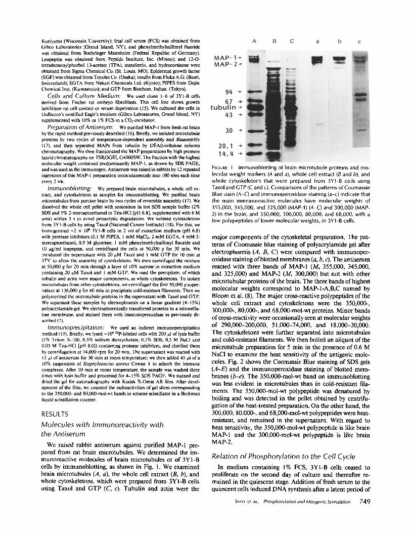
We raised rabbit antiserum against purified MAP-I pre- pared from rat brain microtubules. We determined the im- munoreactive molecules of brain microtubules or of 3Y1-B cells by immunoblotting, as shown in Fig. I. We examined brain microtubules (A, a), the whole cell extract (B, b), and whole cytoskeletons, which were prepared from 3YI-B cells using Taxol and GTP (C, c). Tubulin and actin were the
FIGURE 1 Immunoblotting of brain microtubule proteins and mo- lecular weight markers (A and a), whole cell extract (B and b), and whole cytoskeletons that were prepared from 3Y1-B cells using Taxol and GTP (C and c). Comparisons of the patterns of Coomassie Blue stain (A-C) and immunoperoxidase staining (a-c) indicate that the main immunoreactive molecules have molecular weights of 355,000, 345,000, and 325,000 (MAP-l) (A-C) and 300,000 (MAP- 2) in the brain, and 350,000, 300,000, 80,000, and 68,000, with a few polypeptides of lower molecular weights, in 3Y1-B cells.
major components of the cytoskeletal preparation. The pat- terns of Coomassie blue staining of polyacrylamide gel after electrophoresis (A, B, C) were compared with immunoper- oxidase staining of blotted membranes (a, b, c). The antiserum reacted with three bands of MAP-1 (Mr 355,000, 345,000, and 325,000) and MAP-2 (M¢ 300,000) but not with other microtubular proteins of the brain. The three bands of highest molecular weights correspond to MAP-1-A,B,C named by Bloom et al. (8). The major cross-reactive polypeptides of the whole cell extract and cytoskeletons were the 350,000-, 300,000-, 80,000-, and 68,000-mol-wt proteins. Minor bands of cross-reactivity were occasionally seen at molecular weights of 290,000-200,000, 51,000-74,000, and 18,000-30,000. The cytoskeletons were further separated into microtubules and cold-resistant filaments. We then boiled an aliquot of the microtubule preparation for 5 rain in the presence of 0.6 M NaCl to examine the heat sensitivity of the antigenic mole- cules. Fig. 2 shows the Coomassie Blue staining of SDS gels (A-E) and the immunoperoxidase staining of blotted mem- branes (b-e). The 350,000-mol-wt band on immunoblotting was less evident in microtubules than in cold-resistant fila- ments. The 350,000-mol-wt polypeptide was denatured by boiling and was detected in the pellet obtained by centrifu- gation of the heat-treated preparation. On the other hand, the 300,000, 80,000-, and 68,000-mol-wt polypeptides were heat- resistant, and remained in the supernatant. With regard to heat sensitivity, the 350,000-mol-wt polypeptide is like brain MAP-1 and the 300,000-mol-wt polypeptide is like brain MAP-2.
Relation of Phosphorylation to the Cell Cycle In medium containing 1% FCS, 3Y1-B cells ceased to
proliferate on the second day of culture and thereafter re- mained in the quiescent stage. Addition of fresh serum to the quiescent cells induced DNA synthesis after a latent period of
SATO ET AL. Phosphorylation and Mitogenic Stimulation 749
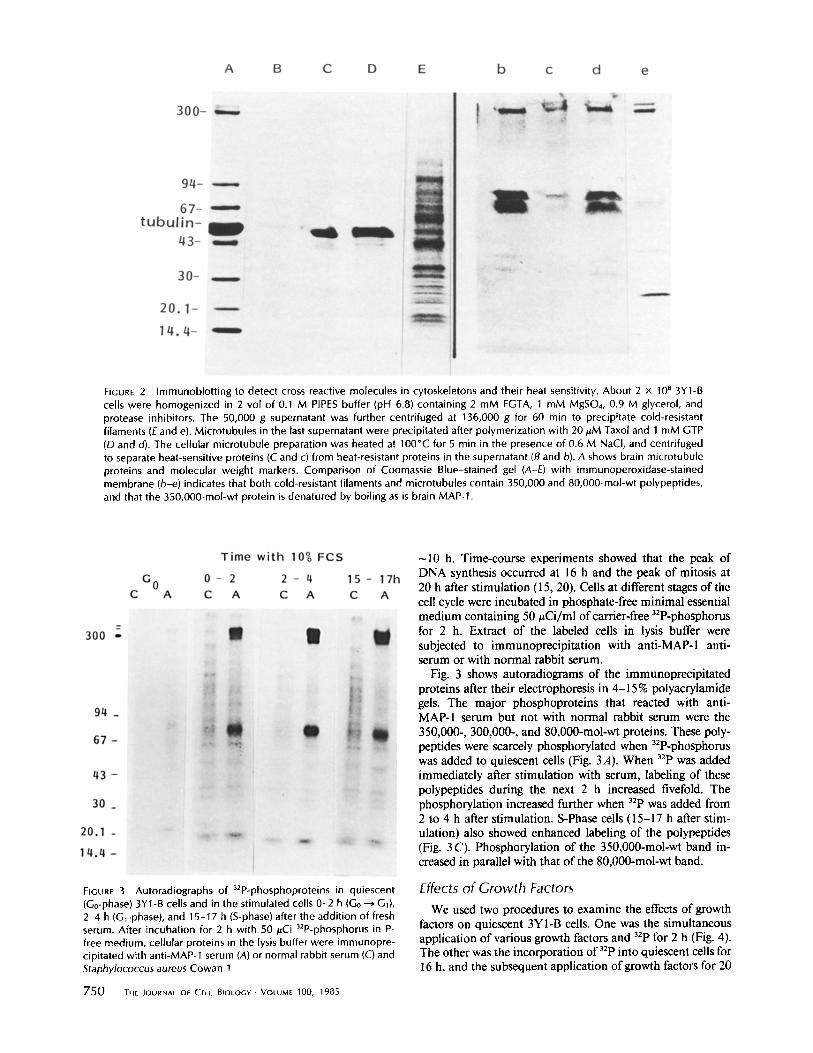
FIGURE 2 Immunoblotting to detect cross-reactive molecules in cytoskeletons and their heat sensitivity. About 2 x 10 B 3Y1-B cells were homogenized in 2 vol of 0.1 M PIPES buffer (pH 6.8) containing 2 mM EGTA, 1 mM MgSO4, 0.9 M glycerol, and protease inhibitors. The 50,000 g supernatant was further centrifuged at 136,000 g for 60 min to precipitate cold-resistant filaments (E and e). Microtubules in the last supernatant were precipitated after polymerization with 20 ~.M Taxol and 1 mM GTP (D and d). The cellular microtubule preparation was heated at 100"C for 5 rain in the presence of 0.6 M NaCI, and centrifuged to separate heat-sensitive proteins (C and c) from heat-resistant proteins in the supernatant (B and b). A shows brain microtubule proteins and molecular weight markers. Comparison of Coomassie Blue-stained gel (A-E) with immunoperoxidase-stained membrane (b-e) indicates that both cold-resistant filaments and microtubules contain 350,000 and 80,000-mol-wt polypeptides, and that the 350,000-mol-wt protein is denatured by boiling as is brain MAP-1.
FIGURE 3 Autoradiographs of a2P-phosphoproteins in quiescent (Go-phase) 3Y1-B cells and in the stimulated cells 0-2 h {G0 --* G1), 2-4 h (G~-phase), and 15-17 h (S-phase) after the addition of fresh serum. After incubation for 2 h with 50/~Ci 32P-phosphorus in P- free medium, cellular proteins in the lysis buffer were irnmunopre- cipitated with anti-MAP-1 serum (A) or normal rabbit serum (C) and Staphylococcus aureus Cowan 1.
750 THE JOURNAL OF CELL BIOLOGY . VOLUME 100, 1985
~10 h. Time-course experiments showed that the peak of DNA synthesis occurred at 16 h and the peak of mitosis at 20 h after stimulation (15, 20). Cells at different stages of the cell cycle were incubated in phosphate-free minimal essential medium containing 50 #Ci/ml of carrier-free 32P-phosphorus for 2 h. Extract of the labeled cells in lysis buffer were subjected to immunoprecipitation with anti-MAP-1 anti- serum or with normal rabbit serum.
Fig. 3 shows autoradiograms of the immunoprecipitated proteins after their electrophoresis in 4-15 % polyacrylamide gels. The major phosphoproteins that reacted with anti- MAP-1 serum but not with normal rabbit serum were the 350,000-, 300,000-, and 80,000-mol-wt proteins. These poly- peptides were scarcely phosphorylated when 32p-phosphorus was added to quiescent cells (Fig. 3A). When 32p was added immediately after stimulation with serum, labeling of these polypeptides during the next 2 h increased fivefold. The phosphorylation increased further when 32p was added from 2 to 4 h after stimulation. S-Phase cells (l 5-17 h after stim- ulation) also showed enhanced labeling of the polypeptides (Fig. 3 C). Phosphorylation of the 350,000-mol-wt band in- creased in parallel with that of the 80,000-mol-wt band.
Effects of Growth Factors
We used two procedures to examine the effects of growth factors on quiescent 3Y1-B cells. One was the simultaneous application of various growth factors and 32p for 2 h (Fig. 4). The other was the incorporation of 32P into quiescent cells for 16 h, and the subsequent application of growth factors for 20
F a c t o r s J I n c o r p o r a t i o n o f 32p i n t o 3 5 0 k ( x l 0 0 c p l 0 m ) , 3 Y 1 - B , G 0
Ca~ 'L~ ,2 3 z, S 6 7 8 9 , i , i I , h
C o n t r o l _+ ~ I FCS _ + ~ ~ i
I
E G F *- ~////////f/iJ///~'////ll/i/~/#'//////////l#'/////iA 1 I
T P A +- w//////Lf///E//f/~'/A [ :+L I ~ S u I i r~ ~//////////zT/P~I
T r a n s f e r r i n _ J
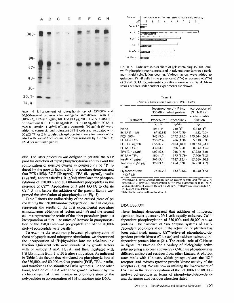
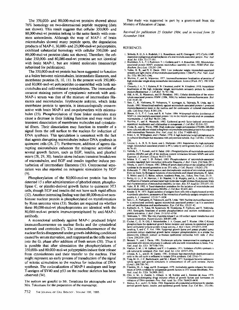
l FIGURE 5 Raclioactivities of slices of gels containin 8 350,O00-mol- wt 3ZP,pbospboproteins, measured in toluene scintillator in a Beck- man l iquid scintillation counter. Various factors were added to quiescent 3YI-B cells in the presence (Ca ++-) or absence (Ca+++) of 3 mM E(]TA. Experimental conditions were as for Fi B. 4. Mean values of three independent experiments are shown.
FIGURE 4 Enhancement of phosphorylation of 350,000- and 80,O00-mol-wt proteins after mitogenic stimulation. Fresh FCS (10%) (A), TPA (0.1 ug/ml) (B), TPA (0.1 txg/ml) + EGTA (3 raM) (C), no treatment (D), EGF (30 ng/ml) (E), EGF (30 ng/ml) + EGTA (3 mM) (F), insulin (1 #g/ml) (G), and transferrin (10 ~g/ml) (H) were added to serum-starved quiescent 3Y1-B cells and incubated with 50/~Ci 32p for 2 h. Labeled phosphoproteins were immunoprecipi- tared with anti-MAP-1 serum, and then resolved by 4-15% SDS PAGE for autoradiography.
rain. The latter procedure was designed to prelabel the ATP pool for detection of rapid phosphorylation and to avoid the complication of possible change in permeability of 32p in- duced by the growth factors. Both procedures demonstrated that FCS (10%), EGF (30 ng/ml), TPA (0.1 ~g/ml), insulin (1 ~g/ml), and transferrin (10/xg/ml) stimulated the phospho- rylations of 350,000- and 80,000-mol-wt polypeptides in the presence of Ca +÷. Application of 3 mM EGTA to chelate Ca ++ 5 min before the addition of the growth factors sup- pressed the stimulation of phosphorylation (Fig. 5).
Table I shows the radioactivity of the excised piece of gel containing the 350,000-mol-wt polypeptide. The first column represents the results of the first experimental procedure (simultaneous additions of factors and 32p) and the second column represents the results of the other procedure (previous incorporation of 32p). The ratios of increase in phosphoryla- tion of the 350,000-mol-wt polypeptide and of the 80,000- moi-wt polypeptide were parallel.
To examine the relationship between phosphorylation of these polypeptides and mitogenic stimulation, we determined the incorporation of [3H]thymidine into the acid-insoluble fraction. Quiescent cells were stimulated by growth factors with or without 3 mM EGTA, and then incubated with [3H]thymidine from 5 to 20 h after stimulation. As indicated in Table I, the factors that stimulated the phosphorylations of the 350,000- and 80,000-mol-wt proteins (EGF, TPA, insulin, and transferrin) also stimulated DNA synthesis. On the other hand, addition of EGTA with these growth factors or hydro- cortisone resulted in no increase in phosphorylation of the polypeptides or incorporation of [3H]thymidine into DNA.
TABLE I
Effects of Factors on Quiescent 3YI-B Cells
Treatment
Incorporation of 32p into Incorporation of 350,000-mol-wt protein [3H]TdR into
acid-insoluble Procedure 1 Procedure 2 fraction
cp lOre cp lOre cpm
None 105 (1)* 210 (1)* 5,740 (1)* EGTA (3 mM) 67 (0.63) 104 (0.50) 1,952 (0.34) FCS (10'/o) 945 (9.0) 2772 (13.2) 175,644 (30.6) EGTA + FCS 250 (2.4) 286 (1.36) 5,338 (0.93) EGF (30 ng/ml) 656 (6.2) 2104 (10.0) 138,334 (24.1) EGTA + EGF 430 {4.1) 506 (2.4) 8,012 (1.40) TPA (0.1 ~g/ml) 607 (5.8) 916 (4.4) 17,220 (3.0) EGTA + TPA 180 (1.7) 375 (1.79) 7,106 (1.23) Insulin (1 ~g/ml) 568 (5.4) 2612 (12.4) 62,566 (10.9) Transferrin (10/xg/ 329 (3.1) 1454 (6.9) 26,978 (4.7)
ml) Hydrocort isone 74 (0.70) 142 (0.68) 8,610 (1.5)
(10 -6 M)
Procedure 1, simultaneous application of growth factors and 32p for 2 h; procedure 2, previous incorporation of 32p into quiescent cells for 16 h, and application of growth factors for 20 min. [3H]TdR was incorporated 5- 20 h after stimulation.
* Ratio to the value without treatment.
D ISCUSSION
These findings demonstrated that addition of mitogenic agents to intact quiescent 3Y1 cells rapidly enhanced Ca ++- dependent phosphorylation of 350,000- and 80,000-mol-wt proteins. The existence of two enzyme systems of Ca ++- dependent phosphorylation in the activation of platelets has been established; namely, Ca++-activated phospholipid-de- pendent protein kinase (C-kinase) and calcium-calmodulin- dependent protein kinase (21). The crucial role of C-kinase in signal transduction for a variety of biologically active substances has also been shown (22). C-Kinase phosphorylates different amino acid residues from other kinases. A phorbol ester binds with C-kinase, which phosphorylates the EGF receptor, and reduces tyrosine protein kinase activity of the receptor (23, 24). We are now examining the involvement of C-kinase in the phosphorylations of the 350,000- and 80,000- moi-wt polypeptides in terms of phospholipid-dependency and the amino acid residues phosphorylated.
SATO ET AL. Phosphorylation and Mitogenic Stimulation 751
The 350,000- and 80,000-mol-wt proteins showed about 76% homology on two-dimensional peptide mapping (data not shown). This result suggests that cellular 350,000- and 80,000-mol-wt proteins belong to the same family with com- mon antecedents. Although the map of MAP-I of brain microtubules showed many peptide spots, the degradation products of MAP- l, 30,000- and 25,000-tool-wE polypeptides, exhibited substantial homology with cellular 350,000- and 80,000-mol-wt proteins (data not shown). Therefore, the cel- lular 350,000- and 80,000-mol-wt proteins are not identical with brain MAP-l, but are related molecules (manuscript submitted for publication).
The 350,000-mol-wt protein has been suggested to function as a linker between microtubules, intermediate filaments, and membrane proteins (6, I0, I l). In the present work 350,000- and 80,000-mol-wt polypeptides co-assembled with both mi- crotubules and cold-resistant cytoskeletons. The immunoflu- orescent staining pattern of cytoplasmic network with anti- MAP-l serum was that of the mixture of intermediate fila- ments and microtubules. Erythrocyte ankyrin, which links membrane protein to spectrin, is immunologically cross-re- active with brain MAP-1 and co-polymerizes with microtu- bules (25). Phosphorylation of these linker molecules may cause a decrease in their linking function and may result in transient dissociation of membrane molecules and cytoskele- tal fibers. Their dissociation may function in transfer of a signal from the cell surface to the nucleus for induction of DNA synthesis. This speculation is consistent with the fact that agents disrupting microtubules induce DNA synthesis in quiescent cells (26, 27). Furthermore, addition of agents dis- rupting microtubules enhances the mitogenic activities of several growth factors, such as EGF, insulin, and phorbol esters (28, 29, 30). Insulin alone induces transient breakdown of microtubules, and EGF and insulin together induce per- turbation of intermediate filaments (3 l). Centrosomal sepa- ration was also reported on mitogenic stimulation by EGF (9).
Phosphorylation of the 80,000-mol-wt protein has been detected 15 s after administration of phorbol esters, phospho- lipase C, or platelet-derived growth factor to quiescent 3T3 cells, though EGF and insulin did not have such rapid effects (32). Another interesting finding was that 80,000-mol-wt non- histone nuclear protein is phosphorylated on transformation by Rous sarcoma virus (33). Studies are required on whether these 80,000-mol-wt phosphoproteins are identical with the 80,000-mol-wt protein immunoprecipitated by anti-MAP-I antibody.
A monoclonai antibody against MAP-I produced bright immunofluoreseence on nuclear flecks and the cytoskeletal network and centrioles (7). The immunofluorescence of the nuclear flecks disappeared under growth-inhibiting conditions caused by serum starvation, and reappeared as the cells moved into the GI phase after addition of fresh serum (20). Thus it is possible that after stimulation the phosphorylations of 350,000- and 80,000-mol-wt polypeptides induce their release from cytoskeletons and their transfer to the nucleus. This might represent an early process oftransduction of the signal of mitotic stimulation to the nucleus for induction of DNA synthesis. The co-localization of MAP- 1 analogues and large T-antigen of SV40 and p53 on the nuclear skeleton has been observed (34).
The authors are grateful to Mr. Terashima for photographs and to Mrs. Tokumasu for the preparation of the manuscript.
752 THE JOURNAL OF CELL BIOLOGY • VOLUME 100, 1985
This study was supported in pan by a grant-in-aid from the Ministry of Education of Japan.
Received for publication 25 October 1984, and in revised form 20 November 1984.
REFERENCES
I. Sloboda, R. D., S. A. Rudolph, J. L. Rosenbaum, and R. Gteengard. t975. Cyclic AMP- dependent endogenous phoephorylation of a microtubule-assoeiated protein. Proc. Natl. Acad. Sci. USA. 72:177-181.
2. Kuznetsov, S. A., V. I. Rodionov, V. I. Gelfand, and V. A. Rosenblat. 1981. Microtubule- ~ t e d protein MAPI promotes microtubule assembly in vitro. FEBS (Fed. Eur. Biochem. So(:.) Left. 135:241-244.
3. Vallee, R. B., andeS. E. Davis. 1983. Low molecular weight microtubule-assooated proteins arc light chains of mierotubule-associated protein I (MAP l). Proc. Natl. Acad. Sci. USA. 80:1342-1346.
4. Sherline, P., and K. Schiavone. 1977. lmmunofluorescence localization of proteins of high molecular weight along intracellular microtubules. Science (Wash. DC). 198:1038- 1040.
5, Connolly, .L A., V. L Kalnins, P. W. Cleveland, and M. W. KJrschner. 1978. [ntmcellular localization of the high molecular weight microtubule accessory protein by indirect immunofloorescenc¢. J. Cell Biol. 76:781-786.
6. Hill, A,-M., R. Maunoury, and D. Pantaloni. 1981. Cellular distribution of the micro- tubule-associated proteins HMW (350k, 300k) by indirect immunofluorescence. Biol. Cell. 41:43-50.
7. Sato, C., K. Nishizawa, H. Nakamura, Y. Komagoe, K. Shimada, R. Ueda, and S. Suzuki, 1983. Monoclonal antibody against microtubule ~ a t e d protein-I produced immunofluorescent spots in the nucleus and the centrosome of cultured mammalian cells. Cell Struct Funct, 8:245-254.
8. Bloom, G. S., F. C. Luca, and R. B. Vallee, 1984. Widespread cellular distribution of MAP-IA (microtubule-associated protein IA) in the mitotic spindle and on interphase microtubules. ,L Cell Biol. 98:331-340,
9. Shefline, P., and R. Muscardo. 1982. Epidermal growth factor-induced centresomal separation: mechanism and relationship to mitogenesis. Z Cell Biol. 95:316-322.
I 0. Pytela, R., and G. Withe. 1980. High molecular weight polypeptides (270,000-340,000) from cultured cells are related to hog brain m i c r o t u b u l ~ i a t e d proteins but copurify with intermediate filaments. Proc. Natl. Acad. Sci. USA. 77:4808,.4812.
11. Klein, 1., M, Willingham, and I. Pastan. 1978. A high molecular weight phosphoprotein in cultured fibroblasts that associates with polymerized tubulin. Exp. Cell Res. 114:22% 238.
12. Greene, L, A., R. K. H. Liem, and L. Shelanski. 1983. Regulation of a high molecular weight microt ubule-assoeiated protein in PCI 2 cells by nerve growth factor..L Cell BiN. 96:76-83.
13. Nishida, E., T, Kuwaki, and H. Sakai. 1981. Phosphorylation of microtubule-assoeiated proteins (MAPs) and pH of the medium control interaction between MAPs and actin filaments. Z Biochem. (Tokyo). 90:575-578.
14. Selden, S. C., and T. D. Pollard. 1983. Phosphorylation of microtuhule-assoeiated proteins regulated their interaction with actin filaments..L Biol. Chem. 258:7068-707 I,
15. Okuda, A. and G. Kimura. 1982. Effects of serum deprivation on the initiation of DNA synthesis in the second generation in rat 3YI cells. J. Cell. Physiol. 110:262-270.
16, Kobayashi, T. 1982. Fractionation and electrophorctic analysis of microtubule proteins from rat brain. In Biological functions of microtubules and related structures. H. Sakai, H. Mohri, and G. G. Boris),, editors. Academic Press, Inc., Tokyo, New York. 23-31.
17. Borisy, G. G., J. M. Mareum, J. B. Olmsted, D. B. Murphy, and K. A. Johnson. 1975. Purification oftubulin and associated high molecular weight proteins from porcine brain and characterization of microtubule assembly in vitro. Ann. NYAcad ScL 253:107-132.
18. Vale'e, R. B. 1982. A Taxol-dependent procedure for the isolation of microtubules and microtubule-associated proteins (MAPs). J. Cell BioL 92:435-442.
19. Kessler, S. W. 1975. Rapid isolation of antigens from cells with a staphylococcal protein A-antlbody adsorbent: parameters of the interaction of antibody-antigen complexes with protein A. J. lmmunoL 115:1617-1624.
20. Sato, C., K. Nishizawa, H. Nakamura, and R. Ueda. 1984. Nuclear immunotluorescence by a monoclonal antibody against microtubule associated protein-I as it is assooated with cell proliferation and transformation. Exp. Cell Res. | 55:33~t2.
21. Kaibuchi, K., Y. Takai, M, Sawamara, M. Hoshijuma, T. Fujikura, and Y. Nishizuka. 1983. Synergistic functions of protein phosphorylation and calcium mobilization in platelct activation. J. BioL Chem. 257:6701-6704.
22. Nishizuka, Y. 1984. The role of protein kinase C in cell surface signal transduction and turnout promotion. Nature (Loud.). 308:693-698.
23. Cochet, C., G. N. Gill, J. Meisenhelder, J. A. Cooper, and T. Hunter. 1984. C-Kinase phosphorylates the epidermal growth factor receptor and reduces its epidermal growth factor.stimulated tyrosinc protein kinas¢ activity. J. Biol. Chem. 259:2553-2558.
24. lwashita, S. and C. F. Fox. 1984. Epidermal growth factor and potent phorbol tumor promoters induce epidermal growth factor receptor phosphorylation in a similar but distinctively different manner in human epidermoid carcinoma A431 cells. J. Biol. Chem. 259:2559-2567.
25. Bennett, V., and J. Davis. 1981. Erythrocyte ankyrin: immunoreactive analogues are associated with mitotic structures in cultured cells and with microtubules in brain. Proc. Natl. Acad. Sci. USA. 78:7550-7554.
26. Vasiliev, J. M., I. M. G¢lfand, and V. 1. Guelstein. 1971. Initiation of DNA synthesis in cell cultured by colcemid. Proc. Natl. Acad. Sci. USA. 68:977-979.
27. Crossin, K. L., and D. H. Carney. 1981. Evidence that microtubule depolymerization early in the cell cycle is sufficient to initiate ONA synthesis. Cell. 23:61-71.
28. Teng M. H., J. C. Bartholonew, and M. J. Bissell. 1977. Synergism between antimicro- tubule agents and growth stimulants in enhancement of cell cycle traverse. Nature ( Lond ). 268:738-741.
29. Friedkin, M., A. Leg& and E. Rozengurt. 1979. Antitubulin agents enhance the stimu- lation of DNA synthesis by polypeptide growth factors in 3T3 mouse fibroblasts. Proc. NatL Acad Sci. USA. 76:3909-3912,
30. Otto, A. M., A. Zumb¢, L. Gibson, A. M. Kubler, and L. Jimenez de Asua. 1979. Cytoskeleton-distrupting drugs enhance effects of growth factors and hormones on initiation of DNA synthesis. Pro(:. Natl. Acad Sci. USA. 76:6435-6438.
31. Bockus, B. J., and C. D. Stiles. 1984. Regulation of cytoskeletal architecture by platelet- derived growth factor, insulin, and epidermal growth factor. Exp. Cell Res., 153:186-
19% 32. Rozengurt, E., M. Rodriguez-Pena, and K. A. Smith. 1983. Phorbol esters, phosphotipase
C, and growth factors rapidly stimulate the phosphorylation of a Mf 80,000 protein in intact quiescenl 3T3 cells. Proc. Natl. Acad. Sci. USA. 80:7244-7248.
33. Blat, C., L. Haml, J. Villaudy, and A. Golde. 1983. Rous sarcoma virus-induced changes
in the pattern of phosphorylation of non-histone nuclear proteins. Exp. Cell Res. 145:305-314.
34-. Sato, C., K. Nishizawa. and N. Yamaguchi. 1984. Co-localization of SV40 T antigen and p53 with immunological analogues of microtubule-associated protein-I on the nuclear skeleton. Cell Struct. Funct. 9:305-309.
S^TO ET AL. Phosphorylation and Mitogenic Stimulation 753