Effect of (vesicular-) arbuscular mycorrhiza on survival and - eDiss
160
Effect of (vesicular-) arbuscular mycorrhiza on survival and post vitro development of micropropagated oil palms (Elaeis guineensis Jacq.) Dissertation zur Erlangung des Doktorgrades der Fakultät für Agrarwissenschaften der Georg-August-Universität Göttingen vorgelegt von Claudia Schultz geboren in Kassel Göttingen, im Oktober 2001
Transcript of Effect of (vesicular-) arbuscular mycorrhiza on survival and - eDiss
Effect of (vesicular-) arbuscular mycorrhiza on survival and post vitro
development of micropropagated oil palms (Elaeis guineensis Jacq.)
Dissertation
zur Erlangung des Doktorgrades
der Fakultät für Agrarwissenschaften
der Georg-August-Universität Göttingen
vorgelegt von
Claudia Schultz
geboren in Kassel
Göttingen, im Oktober 2001

D7 1. Referent: Prof. Dr. P. L. G. Vlek
2. Korefferent: Prof. H. Becker
Tag der mündlichen Prüfung: 22.11.2001
Table of contents 1. Introduction 1 2. Target/objective 3 3. State of the Art 5
3.1 The oil palm 5
3.2 Oil palm in Indonesia 6
3.3 Genetics and breeding of the oil palm 6
3.4 Micropropagation of the oil palm 7
3.5 The problem of acclimatization 9
3.6 The role of mycorrhizal symbiosis 10
3.7 Mycorrhizae and micropropagation 13
3.8 Working with mycorrhiza 14
4. Material and Methods 16
4.1 Location and climate of the experimental site 16
4.2 Plant material and maintenance 17
4.2.1 In vitro culture 17
4.2.2 Post vitro culture - transfer to greenhouse conditions 18
4.3 Soil preparation 19
4.4 Inoculum production 20
4.5 Design of the research 21
4.5.1 Experiment 1 21
4.5.2 Experiment 2 22
4.5.3 Experiment 3 23
4.6 Growth parameters 24
4.6.1 Survival and growth measurements 24
4.6.2 Plant analysis 25
4.6.2.1 Shoot analysis 25
4.6.2.2 Root analysis 25
4.7 Statistics 26
5. Results and Discussion 26 5.1 Experiment1 26
5.1.1 Survival rate 27
5.1.2 Date of mortality 28
5.1.3 Post vitro plant development 29
5.1.3.1 Shoot length 29
5.1.3.2 Relative growth rate 32
5.1.3.3 Number of leaves 33
5.1.4. Plant growth parameter after harvesting 35
5.1.4.1. Shoot and root fresh weight, root:shoot fresh weight ratio 35
5.1.4.2 Dry matter 37
5.1.4.3 Nutrient concentration and uptake 39
5.1.4.3.1 Phosphorus 39
5.1.4.3.2 Potassium 41
5.1.4.3.3 Nitrogen 42
5.1.5 (V)AMF infection rate 43
5.1.6 Assessment of plant development 45
5.1.7 Conclusions of Experiment 1 47
5.2 Experiment 2 48
5.2.1 Survival rate 49
5.2.2 Date of mortality 51
5.2.3 Post vitro plant development 52
5.2.3.1 Shoot length 53
5.2.3.2 Number of leaves 56
5.2.4. Plant growth parameter after harvesting 58
5.2.4.1. Shoot and root fresh weight, root:shoot fresh weight ratio 58
5.2.4.2 Dry matter 59
5.2.4.3 Nutrient concentration and uptake 62
5.2.4.3.1 Phosphorus 62
5.2.4.3.2 Potassium 66
5.2.4.3.3 Nitrogen 68
5.2.5 (V)AMF infection rate 69
5.2.6 Conclusions of Experiment 2 72
5.3. Experiment 3 73
5.3.1 Survival rate 73
5.3.2 Date of mortality 75
5.3.3 Post vitro plant development 76
5.3.3.1 Shoot length 77
5.3.3.2 Number of leaves 79
5.3.4. Plant growth parameter after harvesting 81
5.3.4.1. Shoot and root fresh weight, root:shoot fresh weight ratio 81
5.3.4.2 Dry matter 84
5.3.4.3 Nutrient concentration and uptake 87
5.3.4.3.1 Phosphorus 87
5.3.4.3.2 Potassium 89
5.3.4.3.3 Nitrogen 91
5.3.5 (V)AMF infection rate 93
5.3.6 Conclusions of Experiment 3 95
6. General discussion 96
6.1 Effect of (V)A mycorrhiza on the post vitro survival of micropropagated oil palms 97 6.2 Effect of (V)A mycorrhiza on post vitro plant development during the experimental period 99 6.3 Effect of (V)A mycorrhiza on the root development of micropropagated oil palm clones after 12 weeks of post vitro growth 101 6.4 Dry matter accumulation 102
6.5 Effect of (V)A mycorrhiza on nutrient concentration and uptake 104
6.5.1 Phosphorus 104
6.5.1 Potassium and Nitrogen 106
6.6 (V)AMF infectivity and effectivity 107
6.7 Specific effects of experimental factors and measures 109
6.7.1 Efficiency of the selected (V)AMF 109
6.7.2 Role of the applied P form 109
6.7.3 Effect of soil sterilization 110
6.7.4 Role of protection measures 100
7. Final conclusions and perspectives 110 8. Summary 113 9. Zusammenfassung 119 10. References 125 11. Annex 143 Acknowledgements Curriculum vitae
Abbreviations used ANOVA Analysis of Variance
BAY 'BAYFOLAN''
FO no fertilization
g gram
ha hectare
HA hydroxy-apatite (tricalcium phosphate, Ca5(PO4)3OH))
IOPRI 'Indonesian Oil Palm Research Institute'
K Potassium
LSD least significant difference
mg milligram
M mycorrhizal fungi,
mycorrhizal fungus
M1 Glomus manihot
M2 Enthrophospora columbiana
M3 Acaulospora mellea
M4 Acaulospora appendicula
M5 Acaulospora morowiae
M6 Acaulospora rehmii
M7 Gigaspora gigantea
M8 Glomus clarum
M9 Glomus occultum
M10 Glomus mosseae
M11 Scutellispora heterogama
M12 Scutellispora pellucida
N Nitrogen
NI non-inoculated
ns not significant
ppm part per million
P probatility
P Phosphorus
(V)AM (vesicular-) arbuscular mycorrhiza
(V)AMF (vesicular-) arbuscular mycorrhizal fungus,
(vesicular-) arbuscular mycorrhizal fungi

% per cent
°C degree celcius
< smaller than
1
1. Introduction
In the year 2000, about 89.4 million tons of vegetable oil and fats were produced worldwide
(FAO, 2000). Oils have an important place in human energy supply, 1 g of oil gives on
average 38 kJ energy. According to an estimation by the World Health Organisation
(WHO,1999) about one third of the world's population consumes less than 8 kg of oil and fats
per capita per year which is well below the 12 kg considered by the WHO as a minimum
requirement to ensure acceptable physical and mental growth in humans. From the present
level of 6 billion people the world population is expected to increase to 7.5 billion inhabitants
by 2025 (Lupien, 1999). As a consequence, consumption and demand for vegetable oils and
fats will increase. More than 90% of the oil plants are produced in the tropics and subtropics
which is the third place in world production in terms of value, after starch plants and fruit
(Rehm and Espig, 1993).
The oil palm Elaeis guineensis (Jacq.) is the oilplant species with the highest yield of
vegetable oil per hectare, i.e. 5-7 tons, which is over six times more than the oil yields from
commercial grown rapeseed (Mielke, 1991). The energy balance expressed in terms of energy
output-input ratio is wider for oil palm (1:10) than for other commercial grown oil crops like
rapeseed (1:3) (Wood and Corley, 1991). In 2000, the palm oil production amounted to 21.9
million tons and palm oil became the world's second most important vegetable oil after
soybean (23.2 million tons, FAO). Oil World (1999) predicts that palm oil will be the leading
oil in 2012 and by the year 2020, the world palm oil production is projected to hit 40 million
tons.
The Southeast Asian countries dominate the world production of palm oil and account for
nearly 79% of the world's oil palm fresh fruit bunch (FFB) yield. In 2000, Malaysia with 10.8
and Indonesia with 6.9 million tons (FAO, 2000) were the two leading producers covering
about 97% of the total Asian production. Oil palm continues to be Southeast Asia's most
rapidly expanding crop (FAO, 1999). There are several reasons for this rapid expansion.
Crude palm oil and palm kernel oil are adaptable vegetable oils and have a wide range of
markets in food and oleochemical industries. Due to the rapid increase in consumption of
dietary oils and fats in the developing economies of China and India, the world market prices
are strong, and have increased from 480 US$ in December 1996 to 680 US$ in May 1998
(CIFOR, 1998). This has encouraged investors to build up plantations on large areas of
suitable land found in the Malaysian peninsular and the Indonesian islands of Sumatra and
Kalimantan. Therefore, there is a huge demand for improved planting material for replanting
and for the expansion of oil palm plantations. In general, the oil palm in Indonesia is
multiplied by seeds of the Tenera-type, a cross-breeding between Dura x Pisifiera. The
2
disadvantage of propagation by seed is the heterogeneity of the plant material resulting in a
high variability of productivity, quality and disease resistance. On an average, the Tenera-
types produce an oil crop of 6 tons/ha/year. A major event in oil palm breeding was the
development of tissue culture techniques for vegetative propagation. High-yielding, so-called
elite palms of the Tenera varieties, which are able to produce more than 10 tons/ha, were
chosen as ortets for micropropagation. The produced clones showed a crop increase of 20-
30% up to 13 tons/ha, and a better uniformity than the seedlings.
The in vitro propagation has several advantages but some hindrances still occur. One of the
most critical problems is the transfer of in vitro plantlets to ex vitro conditions.
Micropropagated plants have been continuously exposed to a unique microenvironment that
has been selected to provide minimal stress and nearly optimal conditions for plant
multiplication. These conditions lead to a phenotype which is more fragile than greenhouse
grown plants due to different anatomical, morphological and physiological factors. After
transfer to ex vitro conditions the plantlets have to correct the above mentioned abnormalities
to become adapted to a natural environment. An acclimatization process before transfer to the
nursery is required to improve survival and growth of the plantlets. At this transitional stage,
plantlets were kept in the greenhouse under a semi-shaded plastic tunnel and misted twice
daily during the two first weeks to maintain a saturated air humidity. Despite these measures
to prevent plant mortality, 30-40 % of oil palm plantlets died during this phase (Ginting,
1996). The low percentage of surviving plantlet is a problem for expanding the commercial
planting of clonal oil palms.
Micropropagation is an excellent tool for rapid production of homogenous, genetically
improved plant material but it reduces or even eliminates the population of beneficial
microorganisms, such as mycorrhizal fungi, during the early stages of post in vitro
acclimatization. This phase is a critical step in the micropropagation cycle and the lack of
microorganisms frequently results in poor development of plants, so that their reintroduction
during plant production is important. Recent investigations on casava plantlets (Azcon-
Aguilar et al., 1997) and micropropagated fruit tree rootstocks (Monticelli et al., 2000) have
shown that early mycorrhizal inoculation reduced transplant shock during acclimatization,
and thus increases plant survival and establishment rates. Micropropagated Anona cherimola
mill. and wild cherry plantlets inoculated with mycorrhizal fungi showed enhanced plant
quality, nutrient uptake and plant growth (Azcon-Aguilar et al., 1997; Lovato et al., 1994).
The benefits of mycorrhiza for in vitro propagated plantlets have been reported for many
value crops such as grapes, apple, plum, pineapple, avocado, strawberry, raspberry, cherry,
ash and pear (Varma and Schüepp, 1995; Fortuna et al., 1996; Lovato et al., 1996; Azcon-
3
Aguilar and Barea, 1997). In contrast, very little research has been carried out on
economically important perennial species such as micropropagated oil palms (Blal et al.,
1987; Blal and Gianinazzi-Pearson, 1990). Therefore, there is a high potential for introducing
mycorrhizal fungi into the micropropagation system of this high valued plant.
The aim of this thesis was to combine the use of (vesicular-)arbuscular mycorrhizal fungi
((V)AMF) with the production of micropropagated oil palms. Several experiments were
carried out to determine whether (V)AMF could enhance transplanting success and plantlet
acclimatization as indicated by plant survival, plant growth and nutrient uptake.
2. Target /objective The research project described in the following was carried out within the main project
"Biotechnology Indonesia-Germany" (BTIG), which was started in 1988. Among several
other sub-projects, BTIG also included one project on the improvement of the oil palm
(Elaeis guineensis Jacq.) which is an agronomically important crop species in Indonesia. The
main aim was the application of modem techniques for improving conventional cultivation
systems of oil palm in Indonesia.
The research of this study was carried out in collaboration with the Indonesian
"Biotechnology Research Unit for Estate Crops" in Bogor and the "Indonesian Oil Palm
Research Institute" in Medan, and with the "Institute of Plant Breeding" and the "Institute of
Plant Production in the Tropics" of the University of Göttingen in Germany.
The "Indonesian Oil Palm Research Institute" (IOPRI) is the largest oil palm seed producer in
Indonesia. IOPRI startet research activities in oil palm tissue culture in 1986. Until 1996,
IOPRI had planted about 1.500 ha with clonal oil palms on various locations. Yield
advantages of clonal palms in a range of 30% have been demonstrated in field trials (Ginting
et al., 1996).
Tissue culture of palms is time- and labour intensive. The total time required between the
sampling of the mother palm and the hardening of the plantlets is 18 month in average (Duval
et al., 1995). Because of the high production costs the selling price of clonal plantlets could
not be lowered to less than five times the price of selected seeds (2-3 US$).
One of the acute problems of oil palms multiplied by tissue culture is the survival rate of the
young plantlets during the post vitro growth. In this sensitive stage, after micropropagation
under sterile controlled conditions and before transplanting to the nursery, plantlets are
subject to severe environmental stress, due to poor root and shoot growth and reduced
cuticular wax formation.
4
During 1992 and 1993 IOPRI has produced 56.401 and 76.612 plantlets, only 63% and 58%
of these plantlets were sold, respectively. This phenomenon caused by high percentage of
mortality (37-42%) during acclimatization process and after transferring plantlets to the pre-
nursery. The low percentage of plantlet survival is a problem for expanding the commercial
planting of clonal oil palms. The technology used for micropropagated plants does not take
into consideration the existence of the symbiosis of mycorrhizae and other related plant
growth promoting microorganisms like rhizobacteria. The media used are devoid of
mycorrhizal propagules, and therefore the plants obtained from this sytems are non-
mycorrhizal. These non-mycorrhizal plants obtained from the micropropagation process
eventually become mycorrhizal when they are planted to the field soil.
Their omission during micropropagation frequently results in poor development of plants so
that their reintroduction during plant production is important. Most natural field soils and non-
sterile nursery soils contain indigenous mycorrhizal fungi. The roots of oil palm were
colonized by (vesicular-) arbuscular mycorrhizal fungi under natural conditions (Nadarajah
1980; Blal and Gianinazzi-Pearson 1989). A survey of the soil of oil palm plantations in
Malaysia revealed species of all six genera of the Glomales (Nadarajah 1980).
Where the indigenous fungi are ineffective, inoculation with effective mycorrhizal fungi may
increase plant growth. A decision to introduce mycorrhizal fungi under such conditions
depends on the effectiveness of these indigenous fungi, i.e. on their ability to benefit plant
growth in comparison with possible introduced fungi.
The usefulness potential of (V)AMF for agricultural production systems, in particular for the
recovery and growth of high-value micropropagated plants, has been shown for several
temperate species (Morandi et al., 1979; Pons et al., 1983; Gianinazzi et al., 1986; Branzanti
et al., 1990; Ravolanirina et al., 1990). In 1993, the "Biotechnology Research Institute for
Estate Crops" in Bogor, Indonesia started a greenhouse experiment with the aim to find out
how an inoculation with different (V)AM-fungal strains of in vitro multiplied oil palm clones
would improve the ingestion, and thus the rise of plant growth. The results of Widiastuti and
Tahardi (1993) showed that (V)AMF inoculation increased plant growth and nutrient uptake,
specially the P uptake in the shoots of inoculated plantlets increased by 37-44%. The authors
concluded that (V)AM seems to enhance the survival and development of the plantlets during
the acclimatization phase.
Based on these results the research described in this study was carried out. One of the main
aims of this research work was to introduce and establish the mycorrhizal inoculation in an
existing micropropagation process and make it possible for practical users.
5
3. State of the Art 3.1 The oil palm The oil palm belongs to the family Palmaceae and the genus Elaeis. There are two important
species in the genus Elaeis, E. guineensis (Jacq.), which is African in origin and E. olifeira,
which is South American in origin. This research was done in Indonesia, on Elaeis
guineensis. The natural habitat of the oil palm is in the humid tropics, 10' on both side of the
equator and in altitudes below 500 m. The optimum temperature lies between 22° and 32°C
and rainfall requirement is high (2500-3500 mm year-1).
The plant is a monocotyledon of the order Spadiciflorae with a single growing point from
which fronds emerge in a regular sequence at a rate of 20-26 fronds per year. It is
monoecious, which enforces cross-pollination since it produces separate male and female
interflorescences from axillary buds in alternate cycles (Hartley, 1988). The fruits (drupes) are
situated on secondary branches and consist of an orange-colored mesocarp which contains
'palm oil', a hard lignified shell (endocarp) and a white kernel containing 'kernel oil'. The
number of fruits per bunch varies from 50 to 100 in young palms and up to 1000-3000 in
older ones. Individual bunches weight from less than 1 kg to 20-50 kg. The economic life
span of an oil palm plantation is about 20-25 years, beyond which tree height makes the
harvesting procedure uneconomic.
The main product is palm oil, which is obtained from the pulp (mesocarp) of the fruit and
palm kernel oil, which is extracted from the endosperm of the kernel, considered as a by-
product. Both types of oil have different physical and chemical characteristics so that they are
used in different applications. Palm oil mainly contains the saturated palmitic acid and a high
quantity of the unsaturated oleic acid, giving it a higher unsaturated acid content than palm
kernel or coconut oil. Approximately 90% of palm oil, which is commercially fractionated
into olein and stearin, is used in food products. Its physical properties as a semi-solid
vegetable oil make it particularly suitable for margarines, bakery shortenings and some
confectionery fats. The remaining 10% are used as oleochemicals. Recent investigations
suggest a potential use of palm oil fatty acid esters as a fuel for combustion engines similar to
diesel fuel (Hardon et al., 1997). The oil palm remains a formidable competitor with other
vegetable oil crops in terms of yield per hectare. The oil yield from properly maintained oil
palms is over ten times larger than oil yields from commercial grown soybean (Mielke, 1991).
The energy balance expressed by the ratio of energy output to input is wider for the oil palm
(1:10) than other oil crops like rapeseed (1:3) (Wood and Corley, 1991).
6
3.2 Oil palm in Indonesia The Southeast Asian countries dominate the world production of palm oil. In the year 2000
18.3 millon tons of palm oil were produced in South East Asia, which is nearly 84 % of the
world's palm oil production (FAO, 2000). Malaysia with 10.8 and Indonesia with 6.9 million
tons (FAO, 2000) were the two leading producers, with about 97% of total Asian production.
Indonesia is one of the lowest-cost producers of vegetable oil in the world (Larson, 1996). In
1998, the production costs for 1 ton palm oil in this country were about 150 US$, in Malaysia
250 US$ (FEER, 1998). Palm oil industries are considered to be important since they are
labor-intensive and able to provide employment opportunities for Indonesia's growing
population. In 1997, Indonesia's oil palm industry employed more than 2 million people
(Arafin and Susila, 1998). With a population of 200 million - which is still rising - Indonesia
has a big domestic market for oil and fats. The prolific growth of the oil palm sub-sector has
brought important economic benefits. In 1997, 2.9 million tons of palm oil were exported
bringing in earnings estimated at 1.4 billion US$ which accounted for 31 % percent of
Indonesia's agricultural export in 1997 (CIFOR, 1998). The main import countries of
Indonesian palm oil exports were the Netherlands, Germany and Italy (Oil World, 1999).
The oil palm production has been one of the most dynamic of Indonesia's sub-sectors and its
development has attracted much attention in recent years. It is regarded as an important cause
of the conversion of Indonesia's natural forest to non-forest uses. From 1967 to 1997, the oil
palm sub-sector increased 20-fold in regard to the planted area (from 106.000 ha to 2.516.000
ha), and crude palm oil production increased 12% annually (Casson, 1999). Most of the oil
palm plantation area is concentrated in Sumatra and West Kalimantan. Government plans
drawn up before the economic crash in 1997 called for the plantation area to reach 5.5 million
ha by 2000 (Cohen and Hiebert, 1997), and for Indonesia to be the world's biggest palm oil
producer by 2005 (McBeth, 1997).
3.3 Genetic and breeding of oil palm The migration of the oil palm from its original growth area toward South America took place
from the 16th century onwards. In 1848, a few individuals were introduced to Asia from
Reunion via Amsterdam. They were planted as ornamental palms at the Bogor Botanical
Garden in Indonesia. The last of the original trees died in 1993 but offspring of these mother
plants can be found all over South East Asia. These palms have been the base material for the
first plantations at the beginning of the 20th century.
7
Breeding and selection started around 1920, when the oil palm was initially developed as a
plantation crop in Sumatra, Malaysia and Zaire. The first commercial plantings in Indonesia
were established between 1911 and 1920 on Sumatra's east coast. This population is referred
to as 'Deli-dura' characterised by a high production potential. This was followed by selection
for high number of bunches and single bunch weight. Until the early 1960's the dura palms,
with characteristic thick shells, were the main source of planting material. A mayor
contribution was the research of the genetics of three naturally occurring botanical fruit types,
Dura characterized by a thick shell, Tenera with a thin shell and Pisifera, without any shell at
all and a high-sterility rate among female trees. A significant effect in increasing oil yield was
the discovery by Beirnaert and Vanderweyen (1941) that the inheritance of shell thickness is
controlled by a single gene. The homozygous dominant, thick-shelled Dura (sh+sh+) was
crossed with the homozygous recessive shelless Pisifera (sh-sh-), and a 100% heterozygous
thin shelled Tenera (sh+sh-) was produced. Tenera fruits have relatively more oil bearing
mesocarp (60-90% per fruit weight) than Duras (20-65% per fruit weight). From 1962 to
1988, the oil yield increased from an average of 5 to 9,6 t/ha/y (Lee, 1991). The population of
‘Deli-Dura’ is most commonly used around the world as a female line for the production of
seeds (Nampoothiri, 1998).
The oil palm is an outcrossing species, hence individual palms are highly heterozygous and
vary widely in yield, oil quality and disease resistance. In Indonesia, under highly favourable
environmental conditions the average oil production per palm per annum was 44.3 kg (6.3
tons per hectare) but the individual production varied between 21 kg and 77 kg or between 3
and 11 tons per hectare per annum (Noiret et al., 1985). The sexual breeding cycle in oil palm
is very long, as a result of the performance and oil yield of the progeny it can be predicted
only after 15-20 years. The variation in vigor and productivity is often observable only many
years after field planting. The generation time in a breeding program of oil palm is about 8
years and makes breeding for new lines a very long-term project. Clonal oil palms offer the
potential for greater productivity because it is possible to establish uniform tree stands
comprising identical clones of a limited number of highly productive oil palms.
3.4 Micropropagation of oil palm Research on oil palm tissue culture was started in 1970 by UNILEVER, England and CIRAD-
CP in France. Vegetative propagated oil palms were reported for the first time by Jones in
1974 and Corley in 1976. They used root tips as the source of tissue (explant). Rabechauld
and Martin (1976) reported the use of young leaf tissue as explant (Durand et al., 1989). In the
1980's, research and development of oil palm micropropagation expanded to South East Asia.
8
The "Indonesian Oil Palm Research Institute" (IOPRI) started research on activities in oil
palm tissue culture in 1986 by adopting the technology of the French Institute de Recherches
et pour les Huiles et Oleagineux (IRHO). In the mid of 1986 the tissue culture laboratory of
IOPRI at Marihat, North Sumatra, produced the first oil palm clone (Lubis et al., 1996). The
propagated clones were selected from the best Tenera crosses in terms of oil yield and other
related characters, and until now more than 400 ortet have been cultured. From 1985 to the
end of 1994 more than 300.000 plants derived from 168 clones which had been distributed to
estates, covering an area of ca. 1.532 ha (Ginting et al., 1993). The results of field trials from
10 locations of clones produced by IOPRI showed that the yield (FFB) of clones at the
average was 27.8% higher than that of commercial tenera seedlings (Lubis et al., 1993). Since
the clonal plants are more uniform in flowering and setting fruits, the clonal plants can be
harvested earlier (24-28 month) compared to seedling (30-34 month) (Lubis et al., 1996).
Oil palm tissue culture has become commercially important. An oil palm has an economic
productive life of over 20 years. The price of crude palm oil fluctuated in the period 1995-
1998 between U$ 500 and U$ 700 per ton (World Bank). Oil palm clones show a 20-30%
increased yield compared to seedling material (Lubis et al., 1993) providing an added benefit
per palm of U$ 75-U$ 100 during the economic life span.
The oil palm as a monocotyledon is growing from one primary shoot meristem and has no
obvious morphological base for vegetative propagation. Cloning of oil palm is performed by
somatic embryogenesis on calli of leaf origin according the method of CIRAD-CP in France
which was developed by Pennetier et al. (1981). The source of explant are young leaves from
selected mature ortet palms which are still enclosed by leaf bases and inflorescence within the
spathes. They can be sampled without destroying the mother palm because the apex is not
damaged (for more details see chapter: Material and methods). The main criteria for selecting
the 10-15 years old ortets are oil yield >7.5 t/ha/y or fresh fruit bunch (FFB) yield >30 t/ha/y
and a moderate growth rate (Ginting et al., 1993). After field testing the best performing
clones will further be multiplied and distributed to the consumers (Duval et al., 1995).
Tissue culture of palms is in general a time-consuming and very labour intensive procedure.
The total time necessary between the sampling of the mother palm and the hardening of the
first batch of plantlets is 18 month in average (Duval et al, 1995). This has a negative impact
on production costs. The selling price of clonal plantlets (US$ 2-3) cannot be lowered to less
than five times the price of selected seeds. The success of micropropagation depends on the
ability to transfer plantlets out of culture on a large scale at low cost, and with a high survival
rate (Preece and Sutter, 1991; Debergh, 1991).
9
One of the acute problems is that plantlets derived from tissue culture have been observed to
be less vigorous than those obtained by seeds. Before plantlets can be transferred to the
nursery it is necessary to include an acclimatization process to improve the survival and
growth of the plantlets. In the case of micropropagated oil palm the percentage of mortality is
about 30% during this phase and further 10% of the plantlets do not survive the transfer to the
pre-nursery.
3.5 The problem of acclimatization Several problems among which the transfer of in vitro plantlets to ex vitro conditions is the
most critical limit the widespread use of micropropagation. For many species losses from 50
to 90% of in vitro propagated plantlets have occurred when transferring them to the soil
(Sutter, 1985; Ziv, 1986).
The micropropagated plants have been continuously exposed to a unique microenvironment
that had been selected to provide minimal stress and nearly optimal conditions for plant
multiplication. Plantlets grow heterotrophic, and develop within culture vessels under
conditions which are characterized by a saturated atmosphere, relative low light intensity
(photosynthetic photon flux) averaging 12-70 µmol m-2s-1; relatively high and constant
temperature (20-28°C), low rates of gas exchange between the containers and high
concentrations of carbohydrate and exogenous growth regulators in the medium. These
factors often induce physiological, anatomical and morphological abnormalities which
interfere with the acclimatization subsequent to transplanting resulting in low survival rates ex
vitro (Ziv, 1991; Puthur et al., 1998).
In vitro plantlets are invariably diminutive and much smaller than greenhouse grown plants
(Donelly and Tisdall, 1993). The percentage of water content is increased and the dry matter
accumulation per unit area is reduced compared to greenhouse grown plants (Brainerd and
Fuchigami, 1981; Donelly and Vidaver, 1984). This is reflected in fragile organs with reduced
mechanical support tissue and thin cell walls. Relatively low light levels and a saturated
internal atmosphere promote leaves in vitro that anatomically resemble shade leaves (Brainerd
and Fuchigami, 1981; Lee and Wetzstein, 1988; Marin and Gella, 1988) and hydrophytic
plant leaves (Grout and Aston, 1978). They often have reduced or absent epicuticular or
cuticular wax, which can lack the characteristic crystalline structure or differ in chemical
composition from the control plants (Grout, 1975; Grout and Aston, 1977; Sutter, 1984, 1985;
Short et al., 1987). In vitro stomata have slow response times and due to an impaired function
they do not close in response to stimuli such as darkness, abscisic acid application or when
exposed to high levels of carbon dioxide (Brainerd and Fuchigami, 1982; Wardle and Short,
10
1983; Wetzstein and Sommer, 1983; Ziv et al., 1987). In vitro plants principally require sugar
as a carbon source (Conner and Thomas, 1982), and the CO2 uptake capability is low
(Donelly and Vidaver, 1984). Consequently when transplanted out of the culture vessel,
plantlets suffer from severe environmental stresses and substantial losses may occur (Van
Huylenbroeck and Debergh, 1996).
Difficulties in transplanting tissue cultured plantlets to soil are well documented (Puthur et al.,
1998; Subhan et al., 1998). They appear to be a direct result of the culture-induced phenotype
which reflects adaptation to in vitro conditions but have a harmful effect when plantlets are
transferred to the greenhouse or field where the relative humidity tends to be less than 100%,
the ambient light levels are much higher than in culture, the temperatures are fluctuating and
the substrate has a much higher water potential. The transfer of plantlets cultivated in vitro is
one of the most important steps in the structural and physiological adaptation during the
preparation of plantlets. During this time, plantlets must increase their absorption of water and
minerals and their photosynthetic rate (Grattapaglia and Machado, 1990). To promote ex vitro
survival and physiological competence, especially to protect them against water stress and
encourage autotrophy, a transitional environment is usually supplied for the acclimatization
interval, ranging from one to several weeks (Brainerd and Fuchigami, 1982; Grout and
Millam, 1985; Fabbri et al., 1986). In this transitional environment the relative humidity is
kept in the range of 70-100% via tending and the light level is regulated by shading against
direct sunlight. Gradually, as the plantlets acclimatize the relative humidity can be decreased
and the light levels can be increased towards ambient conditions. The stage of acclimatization
is the beginning of the autotrophic existence of the plant, including the initiation of the
physiological processes necessary for survival.
A number of studies have been published to improve the survival and first growth of shootlets
at transplant time. These research works were performed mostly under greenhouse conditions
and have focused on controlling environmental conditions. The methods were based on CO2
enrichment (Kozai and Iwanami, 1988; Laforge et al., 1990) increased light intensity and the
use of mist or fog, plastic tunnels and application of fungizides, but with only limited success.
Manipulating of acclimatization condition prior to or upon transplanting usually reduced
losses (Donnelly et al., 1985; Desjardins et al., 1988; Preece and Sutter, 1991; Dejardins,
1995; Van Huylenbroeck and Debergh, 1996), however, at additional costs to the producer.
3.6 The role of mycorrhizal symbiosis The ability of the root systems to establish beneficial symbiotic relationships with soil
microorganisms represents one of the most successful strategies that land plants have
11
developed to cope with abiotic and biotic stresses imposed during the colonization of
terrestrial ecosystems (Allen, 1996). These beneficial components of soil biota include
mycorrhizae, which are mutualistic associations occurring between the roots of most plant
species and certain groups of fungi. The term 'mycorrhiza', created by Frank (1885), refers to
an association between fungi (mycor) and root (rhiza) as a mutualistic symbiosis where the
host plant receives mineral nutrients via fungal mycelium (mycotrophism) while the
heterotrophic fungus obtains carbon compounds from the photosynthesis of the host (Mukerji,
1996).
Seven types of mycorrhizal associations are namely known, (vesicular-) arbuscular
mycorrhiza, ectomycorrhiza, ectenmycorrhiza, arbutoid-, monotropoid-, ericoid- and
orchidoid mycorrhiza. Types of mycorrhizas are categorized on the basis of taxonomic groups
of fungi and plants involved and the alteration in the morphogenesis of fungi and roots, which
occurs during the development of the new structure that is mycorrhiza (Harley and Smith,
1987).
The (vesicular-) arbuscular mycorrhiza or (V)AM are the most intensively studied types of
mycorrhizae because they are present in most agricultural and natural ecosystems and play an
important role in plant growth, health and productivity (Harley and Smith, 1983; Gianinazzi
et al. 1990; Lovato et al., 1995). There are only a few genera belonging to Cruciferae;
Chenopodiaceae and Cyperaceae where they are not found due to the presence of gluco-
sinolates and their hydrolysis products isothiocyanates in and around the roots (Glenn et al.,
1988), which are toxic to the growth of fungi.
(V)AM fungi belong to the class Zygomycotina, order Glomales (Walker, 1992; Morton et
al., 1995) whose origin has been dated 353-452 million years ago (Simon et al., 1993). About
150 species of the genera Acaulospora, Enthrophospora, Gigaspora, Glomus, Sclerocystis
and Scutellospora have up to now been recognized as forming symbiotic associations with
plants. These fungi form morphologically distinct resting spores in the soil and can be
multiplied in the presence of a hostplant. Some of these spores can be surface sterilized and
used to produce new spores in axenic seedlings or root organ culture (Mosse and Hepper,
1975; Arnaud, et al., 1996). The close relationship of (V)AM fungi with their host plants is
mirrored by their obligatory biotrophic status. In absence of a host, their growth is limited to a
relatively short time (20-30 days) after which many modifications in fungal morphology point
to a cessation of hyphal growth. Presence of the root allows development of vegetative
mycelium, which, under favorable conditions, can colonize 60-90% of the length of the root
system (Becard and Piche, 1989; Bonfante and Bianciotto, 1995).
12
Mycorrhizal colonization begins with the hyphae that arise from soil-borne propagules, large
resting spores of the (V)AMF or mycorrhizal root fragments. The fungal hyphae penetrate the
root between the epidermal cells and form an appressorium in the first cell layers. This stage
marks the autotrophic growth of the fungus. The colonizing hyphae pass through the inter-
cellular spaces and then enter the root tissues spreading between and through cells of the
cortical root layers. Once the hyphae have reached the inner cortex they grow into the cells
and form tree-like structures called 'arbuscules'. These branched hyphae are closely
surrounded by the intact host plasmalemma and represent a large surface of cellular contact
between both symbionts. These facilitates the exchange of metabolites between host and
fungus. The arbuscules are probably the main transfer site of mineral nutrients, mainly
phosphorus, from the fungus to the plant and of carbon compounds to the fungus (Smith and
Gianinazzi-Pearson, 1988; Smith and Smith, 1990; Bonfante and Bianciotto, 1995). As
internal colonization spreads, the extraradical hyphae ramify, and grow along the root surface
forming more penetration points. They also grow outwards into the surrounding soil, thus
developing an extensive tri-dimensional network of mycelium which interfaces with soil
particles. Smith and Gianinazzi-Pearson (1988) indicate that the length of the external hyphae
growing in soil associated with mycorrhizal roots reaches an average of 1m cm-1 root, but
values of up to 10-14 m cm-1 root have also been recorded. These mycelial network can
extend several centimeters outwards from the root surface, bridging over the zone of nutrient
depletion around roots to absorb low-mobile ions from the bulk soil (mineral nutrients). In
return the plant provides the fungus with sugars, amino acids and vitamins essential for its
growth (Harley and Smith, 1983).
The colonized plant is better nourished and better adapted to its environment. It obtains
increased protection against environmental stresses (Sylvia and Williams, 1992), cold
(Charest et al., 1993; Paradis et al., 1995), salinity (Davis and Young, 1985) and pollution
(Leyval et al., 1994; Shetty et al., 1995). In addition, the symbiosis tends to reduce the
incidence of root diseases and minimizes the harmful effect of certain pathogenic agents (St-
Arnaud et al., 1995). In agriculture, the increased uptake of soil minerals by colonized plants
means that it is possible to consider reducing substantially the application of fertilizers and
pesticides, and at the same time obtain equivalent or even higher crop yields (Abbott and
Robson, 1992).
The fact that colonized plants are better able to obtain their nourishment from the soil and
resist environmental stresses gives fungal symbionts a biofertilizing and crop protecting role.
Maximum benefits will only be obtained from inoculation with efficient mycorrhizal fungi
and careful selection of compatible host/fungus/substrate combinations. The performance of
13
micropropagated plants may be greatly improved by ensuring a suitable mycorrhizal
establishment at outplanting. In particular woody plants which are difficult to root in vitro
have been shown to improve their survival rate and quality when inoculated with (V)AM
fungi.
3.7 Mycorrhizae and micropropagation The technique of micropropagation eliminates all microorganisms, including mycorrhizal
fungi, which are naturally an integral part of the plant, assuring satisfactory growth and
development in microbial-rich and nutrient-poor environments. The post vitro acclimatization
is a critical step in the micropropagation cycle and the lack of microorganisms can affect
survival and growth of in vitro produced plantlets. Previous studies have shown that
inoculation with endomycorrhizal fungi at the time of transplanting the micropropagated
plantlets from axenic to ex vitro conditions significantly improves survival and growth due to
improved absorption of nutrients and water, and to increased stress tolerance (Azcon-Aguilar
et al., 1997; Jaizme-Vega et al., 1997). Therefore, there is a high potential for introducing
(V)AMF into the micropropagation system of high valued plants.
Experiments of Vestberg (1992) and Verma and Adholeya (1996) and showed an improved
vegetative growth of micropropagated strawberry after mycorrhizal inoculation. Significant
increases in leaf area have been reported of three micropropageted pineapple varieties
inoculated with five different (V)AM fungi (Guillemin et al., 1994). Blocking of shoot apical
growth after transplanting Allium porrum (Berta at al., 1990), apple and plum rootstocks
(Fortuna, et al., 1992) and Leucaena leucocephala (Naqvi and Mukerji, 1998) could be
prevented by (V)AMF inoculation. Mycorrhizal fungal inoculation has also been found to
influence root morphology of micropropagated grapevine (Schellenbaum et al., 1991).
Inoculation with Glomus mosseae induced a more branched root system into micropropagated
plum rootstock (Giovanetti et al., 1996). Endomycorrhizal inoculation is also reported to have
a positive influence on the homogeneity of micropropagated apple (Branzanti et al., 1992) and
shortened their culture time a quarter to one third compared to uninoculated plants
(Uosukkainen and Vestberg, 1994).
The application of (V)AM fungi also offers the opportunity to reduce fertilizer inputs (mainly
of phosphorus) as well as the limit use of pesticides (Hooker et al., 1994). Williams et al.
(1992) found that fertilizer inputs can be reduced in mycorrhizal micropropagated strawberry
to levels considerably lower than those used in commercial practice and yet plant
development was equivalent to that of non-mycorrhizal plants receiving full fertilizer inputs.
Increase in phosphate concentration and content has been recorded in micropropagated
14
Anthyllis after mycorrhizal inoculation (Salamanca et al., 1992). In Gerbera jamesonii
plantlets mycorrhizal inoculation with Glomus etunicatum increased P, K, Zn and Cu uptake
besides increasing the dry weight of plants. (V)AMF inoculation also advanced flowering by
16 days as compared with the control (Chiluan et al., 1994). Improvement of survival rate has
been observed in micropropagated avocado (Azcón-Aguilar et al., 1994), Anthyllis
(Salamanca et al., 1992), jackfruit (Sivaprasad et al., 1995) and in Leucaena leucocephala
(Naqvi and Mukerji, 1998). (V)AM inoculation reduced the acclimatization period by about 8
weeks in Anthyllis cystisoides (Salamanca et al., 1992). There are also reports on the effect of
(V)AM fungal inoculation on the plant tolerance to fungal pathogenes like Phytophthora
(Vestberg et al., 1994; Guillemin at al., 1994).
Mycorrhiza are also known to occur in several palm genera including Elaeis (Blal et al. 1990)
after artificial inoculation in the nursery. Blal et al. (1990) reported that micropropagated oil
palms increased the coefficient of fertilizer utilization four to five fold after mycorrhization,
in particular with rock phosphate. (V)AMF from oil palm plantations were described by
Nadarajah (1993) and a beneficial effect of mycorrhizal inoculation on oil palm seedlings has
been reported by Loh (1980).
Experiments of Widiastuti and Tahardi (1993) showed that (V)AMF inoculation increased
plant growth and nutrient uptake of oil palm plantlets. Especially the P uptake in the shoots of
inoculated plantlets increased by 37-44%. The authors concluded that (V)AMF seemed to
enhance the survival and development of the plantlets during the acclimatization phase.
3.8 Working with mycorrhiza Micropropagation involves two main stages, an in vitro and an ex vitro and mycorrhizal
inoculation can be done at either of these two stages. The rooting of micropropagated plants is
commonly done in agar medium supplemented with auxins. Extensive studies have been done
on the germination of (V)AM fungal spores and early stages of growth of mycelium. Results
indicate that most mycorrhizal fungal spores require low nutrient content media for rapid
germination. High concentration of nutrients particularly phosphorus has a inhibitory effect
on spore germination and growth (Green et al., 1976). (V)AM fungal spore inoculation in
vitro at the root initiation stage has been succesfully obtained by Becard and Fortin (1988),
Diop et al. (1992), Verma and Adholeya (1995, 1996) to establish (vesicular-) arbuscular
mycorrhizal symbiosis. At this stage for each plant and fungus combination appropriate
medium for the growth of both the micropropagated plant and (V)AM fungus is required.
Pons et al. (1983) achieved in vitro inoculation of Prunus avium using Gigaspora margarita.
Ravolanirinana et al. (1989) also reported a functional mycorrhizal symbiosis in Vitis vinifera
15
in the rooting medium with surface sterilized spores of Gigaspora margarita, Glomus
caledonium and Glomus mosseae. Elmeskaoui et al. (1995) obtained an in vitro system for
culturing Glomus intraradices with 4 Ri T-DNA-transformed carrot roots or non-transformed
tomato roots which were used as a potential active source of inoculum for colonization of
micropropagated strawberry plantlets after root induction in growth chambers. Colonized
plantlets were reported to have more extensive root system and better shoot growth than
control plants. In vitro inoculation of micropropagated plants with (V)AM fungal spores is
possible but it is a cumbersome process which involves technical expertise. This technique
therefore is not practically feasible on commercial scale and is limited to research studies
only.
The inoculation at the ex vitro stage can be achieved either at the beginning or at the end of
the acclimatization stage, before the start of the hardening phase. A number of references are
available on ex vitro inoculation studies of micropropagated plants like strawberry (Vestberg
et al., 1972), grapevine (Ravolanirina et al., 1989), avocado (Azcon-Aguilar et al., 1992;
Vidal et al., 1992), kiwifruit (Schubert et al., 1992), apple (Branzanti et al., 1992;
Vosukainen, 1992), plum (Fortuna et al., 1992), pineapple (Guillemin et al., 1992, 1994,
1995), jackfruit (Sivaprasad et al., 1995) and banana (Naqvi and Mukerji, 1998). Different
workers have inoculated micropropagated plants at various stages. In Pistacia integenima,
(V)AM inoculation was done after the acclimatization stage (Schubert and Martinelli, 1988).
Vidal et al. (1992) inoculated avocado both at the onset of the acclimatization stage and at the
beginning of the hardening phase. Their results revealed that although (V)AM symbiosis
could be established at both stages but the inoculation at the beginning of the hardening stage
showed better results. The greatest effect of (V)AM fungi on plant growth of Annona
cherimola plants was observed when they were inoculated after the acclimatisation period,
before starting the hardening under greenhouse conditions (Azcón-Aguilar et al., 1994).
The substrate used for ex vitro growth of micropropagated plants are important for growth of
plants and development of (V)AM symbiosis. The most common substrates used in plant
tissue culture are peat based without soil and synthetic substrates like perlite or vermiculite.
Studies have revealed that different gowth substrates have varying effects on development of
(V)AM fungi. In investigations of Vidal et al. (1992) a soil-sand mix was found better for
(V)AM fungal colonization of micropropagated avocado plants as compared to a peat-sand
mix. Schubert et al. (1990) reported that (V)AM colonized roots of micropropagated
grapevine in peat-based media but significant growth response was noted when soil was
added to the substrate.
16
The inoculum can be used in different forms like mixture of soil-based inoculum and
mycorrhizal roots (Naqvi and Mukerji, 1998; Schubert et al., 1990), mycorrhizal root only or
surface sterilized mycorrhizal roots (Ponton et al., 1990).
4. Materials and Methods 4.1 Location and climate of the experimental site All experiments of this study were conducted in the greenhouse of the biotechnological
research unit of the “Indonesian Oil Palm Research Institute” (IOPRI) which is located in
Marihat, on North Sumatra, southern of Medan. (Figure 1).
The climate of Indonesia is predominantly tropical and is characterized by two tropical
seasons, which vary with the equatorial air circulation (The Walker Circulation) and the
meridian air circulation (The Hardley Circulation). The displacement of the latter follows the
north-south movement of the sun and its relative position form the earth, in particular from
the continents of Asia and Australia, at certain periods of the year. These factors contribute to
the displacement and instensity of the Inter-Tropical Convergence Zone (ITCZ) which is an
equatorial trough of low pressure that produces rain. Thus, the west and east monsoons, or the
rainy and dry seasons, are a prevalent feature of the tropical climate.
Figure 1: Map of Indonesia (World Fact Book, 2001), location ( ) of the experimental site.
The East Monsoon lasts from June to September and is characterized by dry weather,
especially towards the east. The West Monsoon, which runs from December to March brings
17
heavy rainfall especially towards the west. Even outside the West Monsoon period, the
rainfall is generally high (1.100-3.400 mm/year) in the western islands ie Sumatra and
Kalimantan. The area's relative humidity ranges between 70 and 90%.
Due to the large number of islands and mountains in the country, average temperatures may
be classified as follows: coastal plains: 28°C inland and mountain areas: 26°C higher
mountain areas: 23°C, varying with the altitude.
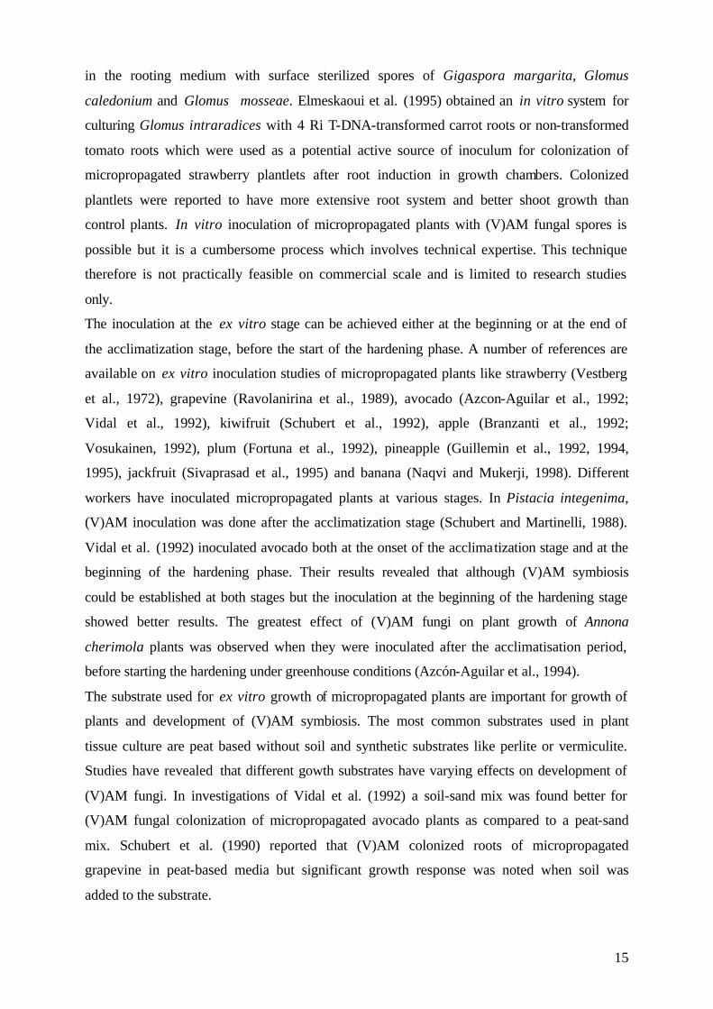
4.2 Plant material and maintenance 4.2.1 In vitro culture Micropropageted oil palm plantlets (Elaeis guineensis Jacq.) were obtained from the bio-
technological research unit of the Indonesian Oil Palm Research Institute (IOPRI). Cloning of
oil palm was performed by somatic embryogenesis (Figure 2) on calli of leaf origin according
the method of CIRAD-CP in France which was developed by Pennetier et al. (1981).
Figure 2: Cultural steps of somatic embryogenesis of oil palm.
The source of explant were young leaves from selected mature ortet palms which were still
enclosed by leaf bases and inflorescence within the spathes. The outer surface of leaf bases or
spathes were decontaminated and cut into 1cm2 fragments. The explants were cultured on
modified culture media (Murashige and Skoog, 1964), supplemented with growth regulators
18
to promote callus induction. Exact media formulation was not available because of intense
commercial interest in oil palm tissue culture. The explants were kept in the dark at a
temperature around 26ºC. After 4-6 months callusses were observed and then recultured into
media with auxins or cytokinins or both in combination to promote embryogenic process. The
embryoids were formed after 4 to 12 months and transferred to embryoid poliferation media
to initiate secondary embryogenesis. The embryoids germinated into shoots in the same media
and when they are large enough (≥5cm) they were transferred into rooting media,
incorporated with auxins or hormon-free media. Embryoid poliferation, germination and
rooting were done under fluorescent light at 27ºC ±2ºC temperature. Time requirement from
taking ortet up to obtaining plantlets is around 12-18 months. The obtained clones which were
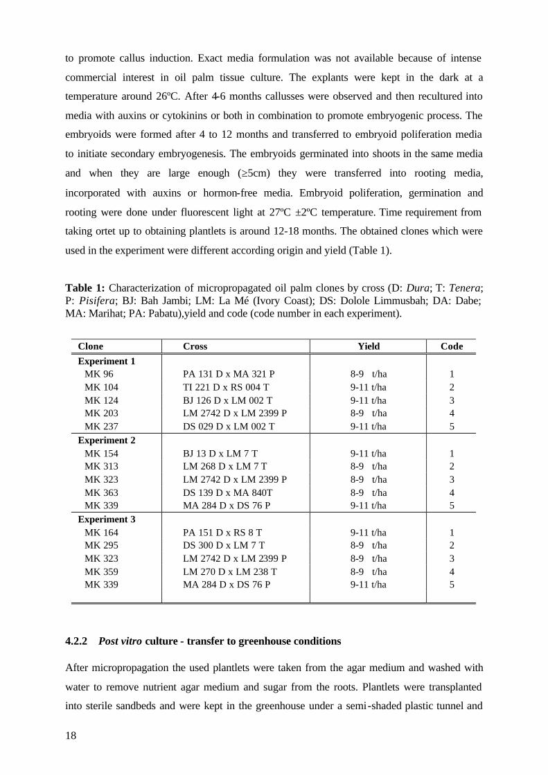
used in the experiment were different according origin and yield (Table 1).
Table 1: Characterization of micropropagated oil palm clones by cross (D: Dura; T: Tenera; P: Pisifera; BJ: Bah Jambi; LM: La Mé (Ivory Coast); DS: Dolole Limmusbah; DA: Dabe; MA: Marihat; PA: Pabatu),yield and code (code number in each experiment).
Clone Cross Yield Code Experiment 1
MK 96 PA 131 D x MA 321 P 8-9 t/ha 1 MK 104 TI 221 D x RS 004 T 9-11 t/ha 2 MK 124 BJ 126 D x LM 002 T 9-11 t/ha 3 MK 203 LM 2742 D x LM 2399 P 8-9 t/ha 4 MK 237 DS 029 D x LM 002 T 9-11 t/ha 5
Experiment 2 MK 154 BJ 13 D x LM 7 T 9-11 t/ha 1 MK 313 LM 268 D x LM 7 T 8-9 t/ha 2 MK 323 LM 2742 D x LM 2399 P 8-9 t/ha 3 MK 363 DS 139 D x MA 840T 8-9 t/ha 4 MK 339 MA 284 D x DS 76 P 9-11 t/ha 5
Experiment 3 MK 164 PA 151 D x RS 8 T 9-11 t/ha 1 MK 295 DS 300 D x LM 7 T 8-9 t/ha 2 MK 323 LM 2742 D x LM 2399 P 8-9 t/ha 3 MK 359 LM 270 D x LM 238 T 8-9 t/ha 4 MK 339 MA 284 D x DS 76 P 9-11 t/ha 5
4.2.2 Post vitro culture - transfer to greenhouse conditions After micropropagation the used plantlets were taken from the agar medium and washed with
water to remove nutrient agar medium and sugar from the roots. Plantlets were transplanted
into sterile sandbeds and were kept in the greenhouse under a semi-shaded plastic tunnel and
19
were misted twice daily during the two first weeks to maintain a saturated air humidity. The
relative humidity was 90-100% in the first week after transplanting and was gradually
reduced to 60-70% during the following week. This phase lasted 10-14 days. Such treatments
were of great importance for the survival of the plantlets, as it significantly reduces water
losses and transpiration demand and avoids photodegradation of chlorophyll by excessive
light (Debergh, 1991). Shading (50-90%) during the first four weeks of acclimatization is
necessary for micropropagated oil palm as for other species ( Preece and Sutter, 1991).
This study was carried out when plantlets were transplanted into polybags containing 200 g of
the soil-sand mixture. All polybags were arranged randomly on greenhouse benches and
maintained under natural light/dark conditions. The greenhouse was covered by glass and the
side walls were made of wire. These conditions creates an nearly natural environment were
plants were sheltered against direct sunlight and rainfall. The temparature ranged from max.
34°C during the day and min. 21°C during the night. Humidity varied between 40 and 70 %
during the experimental period. The plantlets were watered as required.
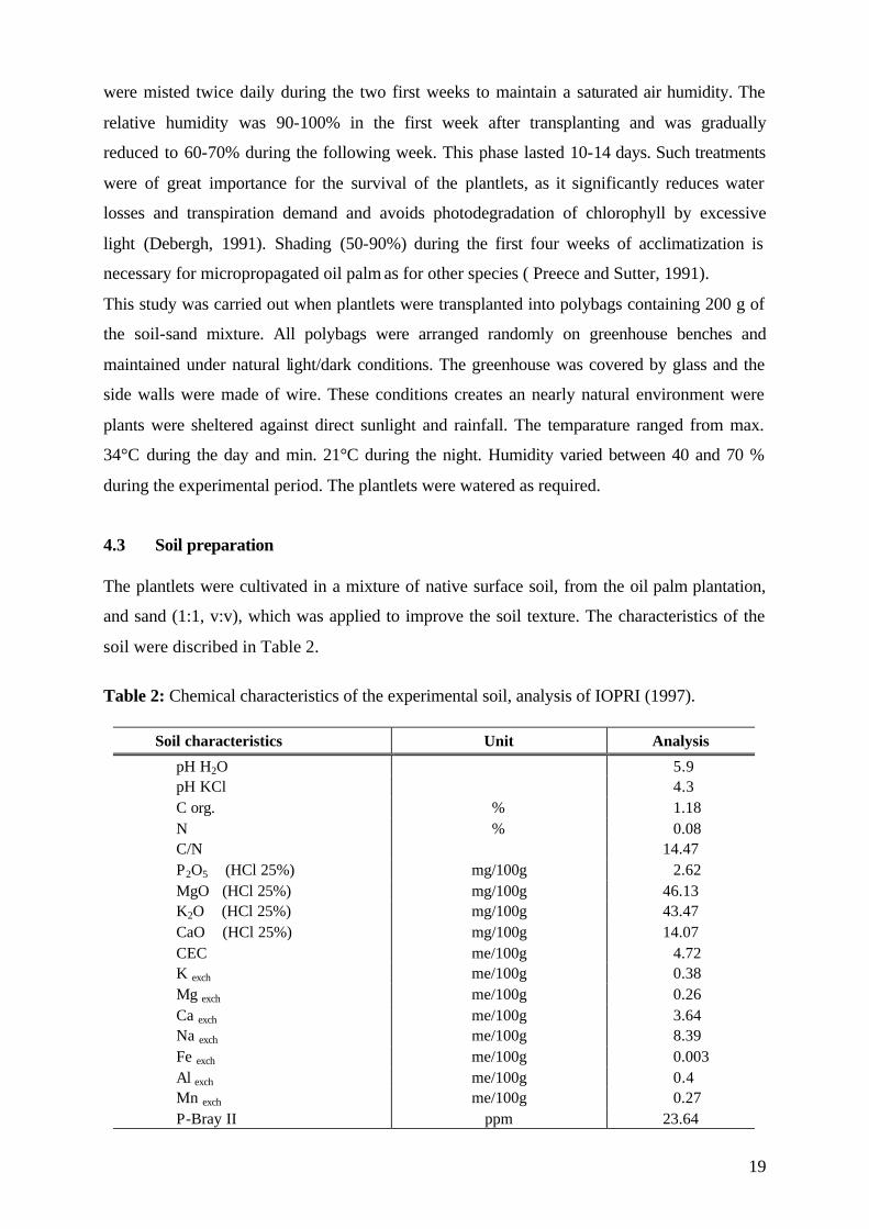
4.3 Soil preparation The plantlets were cultivated in a mixture of native surface soil, from the oil palm plantation,
and sand (1:1, v:v), which was applied to improve the soil texture. The characteristics of the
soil were discribed in Table 2.
Table 2: Chemical characteristics of the experimental soil, analysis of IOPRI (1997).
Soil characteristics Unit Analysis
pH H2O 5.9 pH KCl 4.3 C org. % 1.18 N % 0.08 C/N 14.47 P2O5 (HCl 25%) mg/100g 2.62 MgO (HCl 25%) mg/100g 46.13 K2O (HCl 25%) mg/100g 43.47 CaO (HCl 25%) mg/100g 14.07 CEC me/100g 4.72 K exch me/100g 0.38 Mg exch me/100g 0.26 Ca exch me/100g 3.64 Na exch me/100g 8.39 Fe exch me/100g 0.003 Al exch me/100g 0.4 Mn exch me/100g 0.27 P-Bray II ppm 23.64
20
Prior to potting the substrate was sieved (2 cm) to remove bigger soil particels and plant
material. The substrate in Experiment 1 and 2 was non-sterilized. Experiment 3 included
treatments with sterilized and non-sterilized soil. Soil sterilization was done by heating, at
95°C for three days, to kill native endophytes.
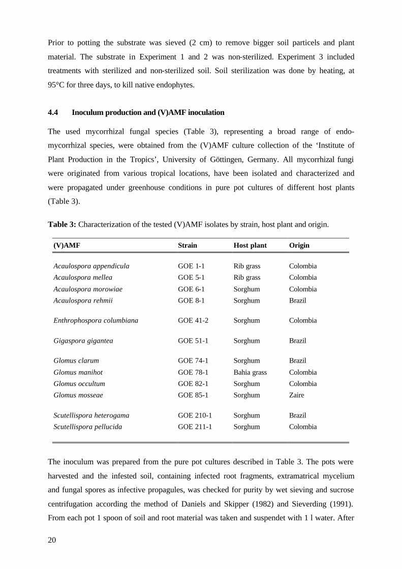
4.4 Inoculum production and (V)AMF inoculation The used mycorrhizal fungal species (Table 3), representing a broad range of endo-
mycorrhizal species, were obtained from the (V)AMF culture collection of the ‘Institute of
Plant Production in the Tropics’, University of Göttingen, Germany. All mycorrhizal fungi
were originated from various tropical locations, have been isolated and characterized and
were propagated under greenhouse conditions in pure pot cultures of different host plants
(Table 3).
Table 3: Characterization of the tested (V)AMF isolates by strain, host plant and origin.
(V)AMF Strain Host plant Origin
Acaulospora appendicula GOE 1-1 Rib grass Colombia
Acaulospora mellea GOE 5-1 Rib grass Colombia
Acaulospora morowiae GOE 6-1 Sorghum Colombia
Acaulospora rehmii GOE 8-1 Sorghum Brazil
Enthrophospora columbiana GOE 41-2 Sorghum Colombia
Gigaspora gigantea GOE 51-1 Sorghum Brazil
Glomus clarum GOE 74-1 Sorghum Brazil
Glomus manihot GOE 78-1 Bahia grass Colombia
Glomus occultum GOE 82-1 Sorghum Colombia
Glomus mosseae GOE 85-1 Sorghum Zaire
Scutellispora heterogama GOE 210-1 Sorghum Brazil
Scutellispora pellucida GOE 211-1 Sorghum Colombia
The inoculum was prepared from the pure pot cultures described in Table 3. The pots were
harvested and the infested soil, containing infected root fragments, extramatrical mycelium
and fungal spores as infective propagules, was checked for purity by wet sieving and sucrose
centrifugation according the method of Daniels and Skipper (1982) and Sieverding (1991).
From each pot 1 spoon of soil and root material was taken and suspendet with 1 l water. After
21
heavier soil particles settled the suspension was decandet over a series of soil sieves. The
mesh size of the sieves used were chosen according to spore size of the (V)AMF species. The
first sieve with the biggest mesh size (355 µm) separates the roots from soil and organic
matter. The obtained roots were carefully transferred with water to a petri dish and kept
separately for staining.
The fungal spores passed throught the medium sieve (125 µm) and remain on the finest sieve
(45µm) from where they were transferred with max. 50 ml water to a 100 ml centrifuge tube.
30-40 ml of a sucrose solution (1000 g sugar/700ml water) was injected into the bottom of the
tube, so a gradient was established in the centrifuge tube. The samples were centrifuged for 2
minutes at 2000 revolutions per min. During this process the soil particles settled and the
spores remain on the surface of the sugar gradient. The spores were separated in a 45µm
sieve, rinsed with water to remove the sugar solution and were carefully transferred to a petri
dish. The spores were observed under a stereomicroscope (x 40 magnification) and were
separated into individual groups according to morphological features (size, colour and surface
characteristics) counted and checked for purity.
The remained roots were stained with 0,05 % trypan blue in lactic acid (modified from
Phillips and Haymann, 1970), to detect mycorrhizal infection. The percentage of mycorrhizal
colonisation was assessed using the grid-line intersect method (Giovanetti and Mosse, 1980).
After the pots were checked for purity isolates which were used for inoculum production were
selected. The inoculum was produced by cutting roots into segments of 2 cm length and
mixed with rhizosphere soil, containing spores and hyphae. The inoculum potentials of the
different fungal species was balanced by using a quantity of 10 g inoculum (fresh weight), 5%
of the polybag substrate (200 g). Inoculation was done when plantlets were transferred from
sandbed to soil. The inoculum was placed in the planting hole closed to the root system. The
non-inoculated (NI) control plantlets received the same amount of autoclaved inoculum to
provide similar physical and chemical conditions in all experiments.
4.5 Design of the research 4.5.1 Experiment 1: Screening Micropropagated plants were transplanted to polybags containing 200 g of the soil-sand
mixture (Table 2). Plantlets were inoculated with 12 different mycorrhizal fungal strains
(Table 3). The non-inoculated (NI) control plantlets received the same amount of inoculum,
but autoclaved. The plantlets received no fertilisation in this experiment.
22
The five different oil palm clones (Table 1) were equally distributed over the treatments. Each
treatment consisted of 12 replications and each replication consisted of one plant per polybag.
The polybags were arranged randomly on the greenhouse bench. Three months after the
plants were transferred to the polybags the harvest was carried out on the remaining plants.
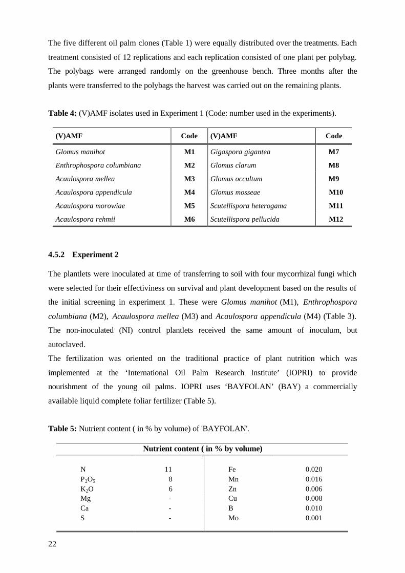
Table 4: (V)AMF isolates used in Experiment 1 (Code: number used in the experiments).
(V)AMF Code (V)AMF Code
Glomus manihot M1 Gigaspora gigantea M7
Enthrophospora columbiana M2 Glomus clarum M8
Acaulospora mellea M3 Glomus occultum M9
Acaulospora appendicula M4 Glomus mosseae M10
Acaulospora morowiae M5 Scutellispora heterogama M11
Acaulospora rehmii M6 Scutellispora pellucida M12
4.5.2 Experiment 2 The plantlets were inoculated at time of transferring to soil with four mycorrhizal fungi which
were selected for their effectiviness on survival and plant development based on the results of
the initial screening in experiment 1. These were Glomus manihot (M1), Enthrophospora
columbiana (M2), Acaulospora mellea (M3) and Acaulospora appendicula (M4) (Table 3).
The non-inoculated (NI) control plantlets received the same amount of inoculum, but
autoclaved.
The fertilization was oriented on the traditional practice of plant nutrition which was
implemented at the ‘International Oil Palm Research Institute’ (IOPRI) to provide
nourishment of the young oil palms. IOPRI uses ‘BAYFOLAN’ (BAY) a commercially
available liquid complete foliar fertilizer (Table 5).
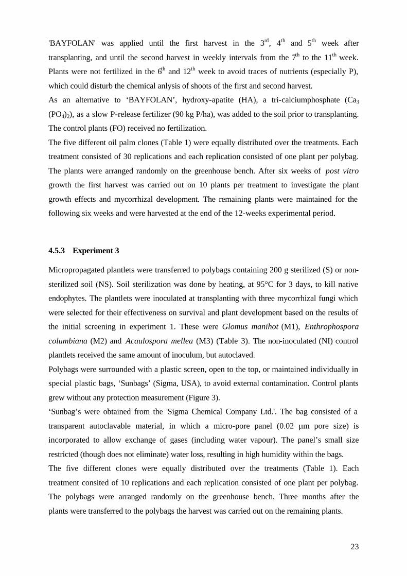
Table 5: Nutrient content ( in % by volume) of 'BAYFOLAN'.
Nutrient content ( in % by volume) N 11 Fe 0.020 P2O5 8 Mn 0.016 K2O 6 Zn 0.006 Mg - Cu 0.008 Ca - B 0.010 S - Mo 0.001
23
'BAYFOLAN' was applied until the first harvest in the 3rd, 4th and 5th week after
transplanting, and until the second harvest in weekly intervals from the 7th to the 11th week.
Plants were not fertilized in the 6th and 12th week to avoid traces of nutrients (especially P),
which could disturb the chemical anlysis of shoots of the first and second harvest.
As an alternative to ‘BAYFOLAN’, hydroxy-apatite (HA), a tri-calciumphosphate (Ca3
(PO4)2), as a slow P-release fertilizer (90 kg P/ha), was added to the soil prior to transplanting.
The control plants (FO) received no fertilization.
The five different oil palm clones (Table 1) were equally distributed over the treatments. Each
treatment consisted of 30 replications and each replication consisted of one plant per polybag.
The plants were arranged randomly on the greenhouse bench. After six weeks of post vitro
growth the first harvest was carried out on 10 plants per treatment to investigate the plant
growth effects and mycorrhizal development. The remaining plants were maintained for the
following six weeks and were harvested at the end of the 12-weeks experimental period.
4.5.3 Experiment 3 Micropropagated plantlets were transferred to polybags containing 200 g sterilized (S) or non-
sterilized soil (NS). Soil sterilization was done by heating, at 95°C for 3 days, to kill native
endophytes. The plantlets were inoculated at transplanting with three mycorrhizal fungi which
were selected for their effectiveness on survival and plant development based on the results of
the initial screening in experiment 1. These were Glomus manihot (M1), Enthrophospora
columbiana (M2) and Acaulospora mellea (M3) (Table 3). The non-inoculated (NI) control
plantlets received the same amount of inoculum, but autoclaved.
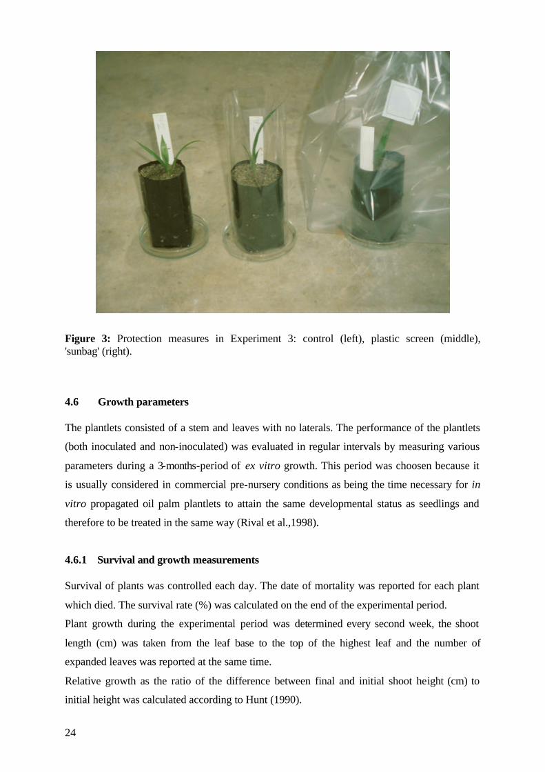
Polybags were surrounded with a plastic screen, open to the top, or maintained individually in
special plastic bags, ‘Sunbags’ (Sigma, USA), to avoid external contamination. Control plants
grew without any protection measurement (Figure 3).
‘Sunbag’s were obtained from the 'Sigma Chemical Company Ltd.'. The bag consisted of a
transparent autoclavable material, in which a micro-pore panel (0.02 µm pore size) is
incorporated to allow exchange of gases (including water vapour). The panel’s small size
restricted (though does not eliminate) water loss, resulting in high humidity within the bags.
The five different clones were equally distributed over the treatments (Table 1). Each
treatment consited of 10 replications and each replication consisted of one plant per polybag.
The polybags were arranged randomly on the greenhouse bench. Three months after the
plants were transferred to the polybags the harvest was carried out on the remaining plants.
24
Figure 3: Protection measures in Experiment 3: control (left), plastic screen (middle), 'sunbag' (right).
4.6 Growth parameters The plantlets consisted of a stem and leaves with no laterals. The performance of the plantlets
(both inoculated and non-inoculated) was evaluated in regular intervals by measuring various
parameters during a 3-months-period of ex vitro growth. This period was choosen because it
is usually considered in commercial pre-nursery conditions as being the time necessary for in
vitro propagated oil palm plantlets to attain the same developmental status as seedlings and
therefore to be treated in the same way (Rival et al.,1998).
4.6.1 Survival and growth measurements Survival of plants was controlled each day. The date of mortality was reported for each plant
which died. The survival rate (%) was calculated on the end of the experimental period.
Plant growth during the experimental period was determined every second week, the shoot
length (cm) was taken from the leaf base to the top of the highest leaf and the number of
expanded leaves was reported at the same time.
Relative growth as the ratio of the difference between final and initial shoot height (cm) to
initial height was calculated according to Hunt (1990).
25
The relative growth rate (RGR cm day-1) of shoots was calculated between two-weeks
intervals, and over the whole experimental period as average values by using the formula:
RGR = (ln TSL2-ln TSL1 )/( t2-t1)
(where ln= natural logarithm; TSL2= total shoot lenght at final measurement; TSL1= total
shoot length at initial measurement; t2-t1= days between measurements)
4.6.2 Plant analysis 4.6.2.1 Shoot analysis At each harvest shoot fresh weights (g), shoot height (cm) and number of leaves were
recorded. Shoots were separated from the roots and washed with distilled water and dried to
constant weight at 65°C for 42 h. Dry weights were taken and shoots were ground before
taking out sub-samples for nutrient analysis, in a plant mill to pass a 20-mesh screen. Three
macro-elements were determined in foliar tissue at harvest .
Samples of the dried ground plant material (0.5-0.55 g) were wet digested with a mixture of
Hydrochloric-perchloric-nitric-acid (Juo, 1982). Total phosphorus content of the digest was
determined colorimetrically with the Vanado-Molybdate method on the Technicon Model II
Autoanalyzer (Pulse Instrumentation Lts, Sakatoon, Canada). The reference material was a
dry powdered corn bran (RM8433, NIST).
Concentrations of potassium in foliar tissue was measured by a atom absorption spectrometer
(PU 9200 x, Phillips).
The total N and C content was determined directly on a ground subsample with a Carlo Erba
NA 1500 gas chromatograph.
4.6.2.2 Root analysis At each harvest root fresh weights (g) were recorded. The roots were separeted from the
shoots, rinsed, blotted dry and their fresh weights were recorded. Until staining for
histochemical studies the fresh roots were stored in a solution of ethanol (99%) and acetic
acid (60%) (3:1).
The preserved roots were rinsed in tap water and cleared in 10% KOH at about 90°C for 1h.
Dark roots were bleached in hydrogen peroxide for 20 Min. All roots were acidified with HCl
and stained with 0,05% trypan blue in lactic acid (modified from Phillips and Haymann,
1970). Individual roots were examined at x40-100 magnification for the presence of
arbuscules, coils, vesicles, internal hyphae and external hyphae of (V)AM fungi. Mycorrhizal
colonization was estimated using the grid-line intersect method (Giovanetti and Mosse, 1980).
26
Samples in which root colonization was difficult to quantify due to low amount of root
material were mounted on a slide for more detailed observation using a compound
microscope (Dodd and Jeffries 1986).
4.7 Statistics The experiments were set up according to randomized complete block designs. However,
missing data occurred due to loss of plantlets in most experiments. Therefore, results were
analysed with the General Linear Model routine of SYSTAT software as Completely
Randomized Designs and clones were used as covariate to control random variation. In the case of variables which were expressed as percentages, data were transformed by the
arcsin square-root procedure prior to ANOVA to ensure homogeneity of variances (Sachs,
1992).
For all characteristics studied the statistical significance of differences between means were
determined using the least significant difference (LSD), (Steel and Torrie) or Tukey-test at
P=0.05.
5. Results and Discussion 5.1 Experiment 1 Screening of 12 different mycorrhizal fungal species for their effectiveness on post vitro development of micropropagated oil palms The first step in any inoculation program is to obtain isolates which are both infective, able to
penetrate and spread in the roots, and effective, able to enhance growth and stress tolerance of
the host. Individual isolates of mycorrhizal fungi vary widely in these properties, so screening
trials are important to select isolates that will perform successfully. For future application of
endomycorrhizal inoculation in the existing micropropagation process of oil palms, screening
under actual cropping conditions is best because indigenous mycorrhizal fungi, pathogens,
and soil chemical and physical properties will influence the results. The first experiment of
this study was conducted as an initial screening. Twelve different mycorrhizal isolates (Table
3), representing a broad range of endomycorrhizal fungi, were tested for their effectiveness on
survival and development of micropropagated oil palms during twelve weeks of post vitro
growth.
27
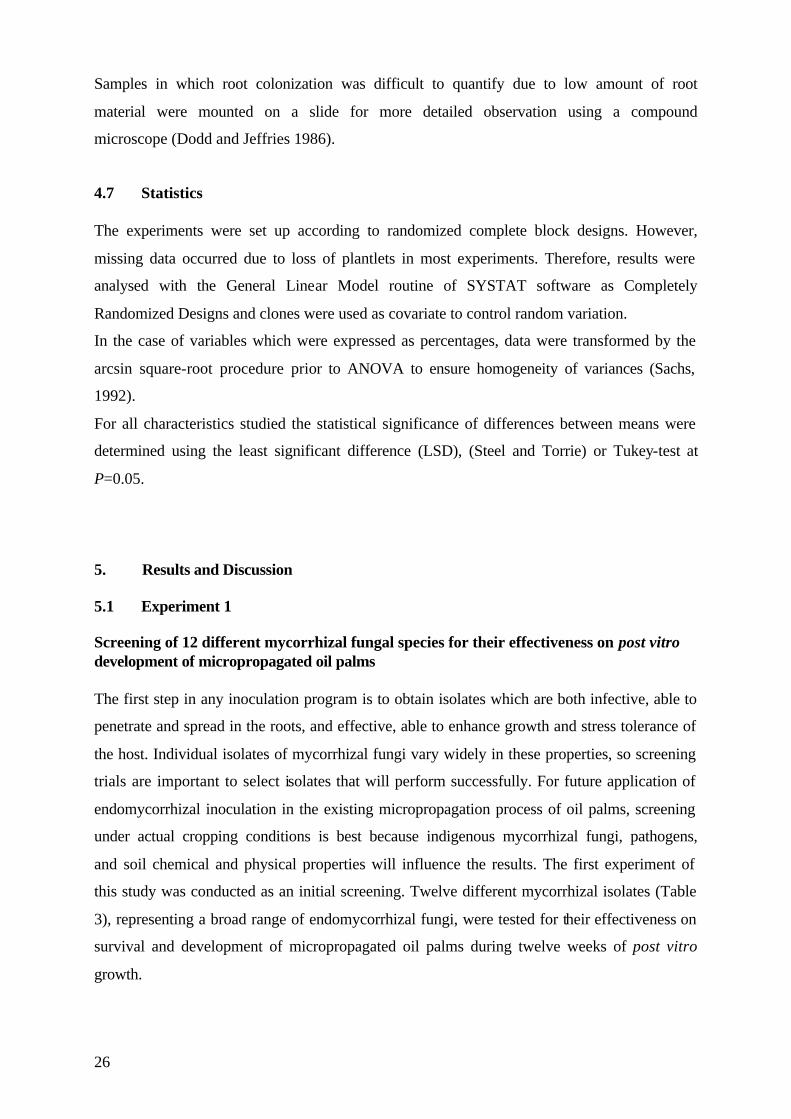
5.1.1 Survival rate Mycorrhizal inoculation at the transplanting stage affected the percentage of survival during
the three-month period significantly (Table 6). The effect of the clones and the interactions of
the two main factors (inoculation, clones) studied were not significant regarding plant
survival rate during this plant stage.
Table 6: ANOVA of main factors and their interaction on the survival rate (%) of micropropagated oil palms after a three-month period of post vitro growth. ANOVA DF MQ P
Inoculation 12 3974.28 0.002 Clone 4 1247.32 0.488 Inoculation x Clone 48 761.64 0.994
Error 131 1444.93
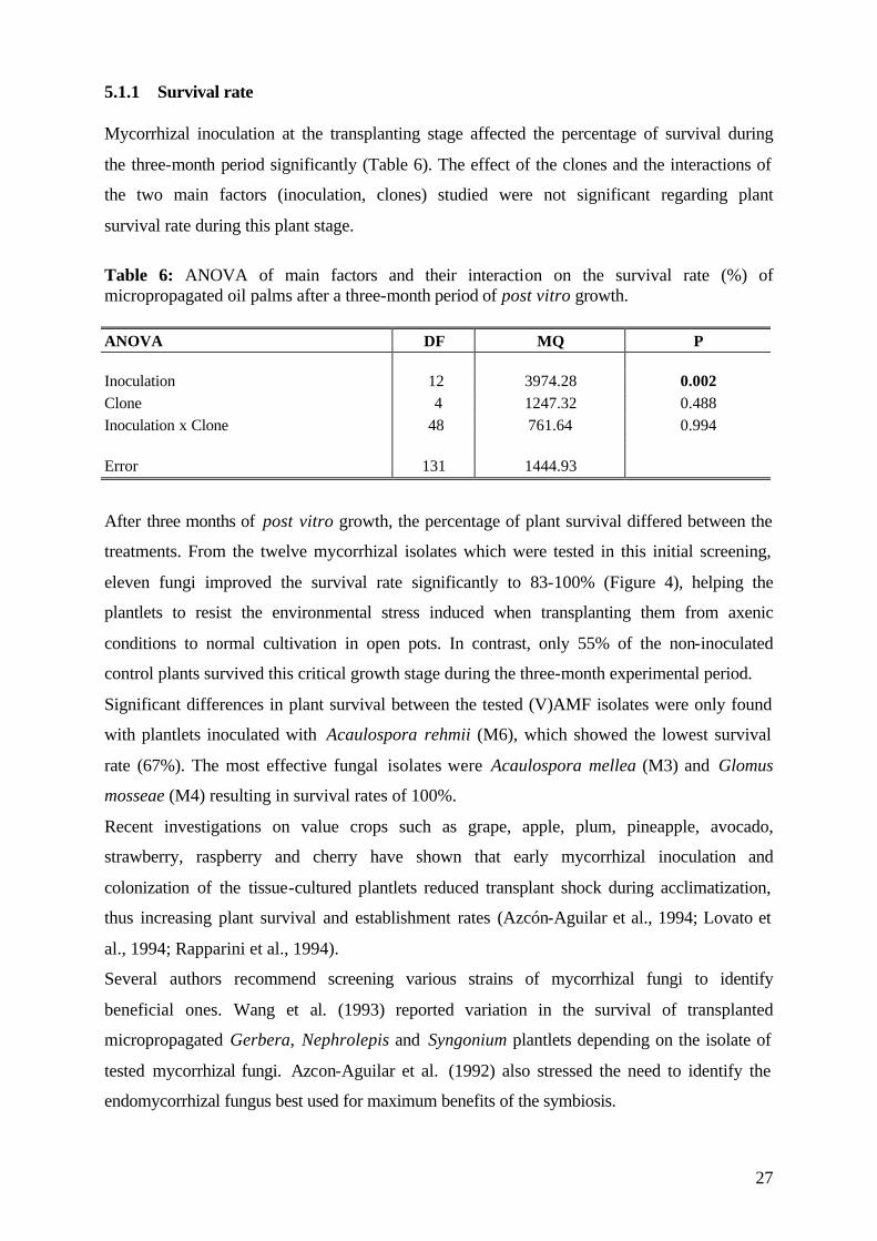
After three months of post vitro growth, the percentage of plant survival differed between the
treatments. From the twelve mycorrhizal isolates which were tested in this initial screening,
eleven fungi improved the survival rate significantly to 83-100% (Figure 4), helping the
plantlets to resist the environmental stress induced when transplanting them from axenic
conditions to normal cultivation in open pots. In contrast, only 55% of the non-inoculated
control plants survived this critical growth stage during the three-month experimental period.
Significant differences in plant survival between the tested (V)AMF isolates were only found
with plantlets inoculated with Acaulospora rehmii (M6), which showed the lowest survival
rate (67%). The most effective fungal isolates were Acaulospora mellea (M3) and Glomus
mosseae (M4) resulting in survival rates of 100%.
Recent investigations on value crops such as grape, apple, plum, pineapple, avocado,
strawberry, raspberry and cherry have shown that early mycorrhizal inoculation and
colonization of the tissue-cultured plantlets reduced transplant shock during acclimatization,
thus increasing plant survival and establishment rates (Azcón-Aguilar et al., 1994; Lovato et
al., 1994; Rapparini et al., 1994).
Several authors recommend screening various strains of mycorrhizal fungi to identify
beneficial ones. Wang et al. (1993) reported variation in the survival of transplanted
micropropagated Gerbera, Nephrolepis and Syngonium plantlets depending on the isolate of
tested mycorrhizal fungi. Azcon-Aguilar et al. (1992) also stressed the need to identify the
endomycorrhizal fungus best used for maximum benefits of the symbiosis.
28
0
10
20
30
40
50
60
70
80
90
100
NI M1 M2 M3 M4 M5 M6 M7 M8 M9 M10 M11 M12
c
ab
ab
a aab
bc
ab
ab
ab
ab
ab absu
rviv
alra
te(%
)
Figure 4: Survival rate (%) of micropropagated oil palm plantlets after 3 months of post vitro growth, non-inoculated (NI) or inoculated with 12 mycorrhizal fungi (M1-M12).Vertical bars represent standard errors of the mean. Columns with different letters are significantly different at P=0.05 as indicated by LSD-test.
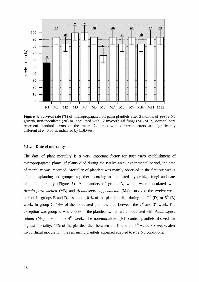
5.1.2 Date of mortality The date of plant mortality is a very important factor for post vitro establishment of
micropropagated plants. If plants died during the twelve-week experimental period, the date
of mortality was recorded. Mortality of plantlets was mainly observed in the first six weeks
after transplanting and grouped together according to inoculated mycorrhizal fungi and date
of plant mortality (Figure 5). All plantlets of group A, which were inoculated with
Acaulospora mellea (M3) and Acaulospora appendicula (M4), survived the twelve-week
period. In groups B and D, less than 10 % of the plantlets died during the 2nd (D) or 5th (B)
week. In group C, 14% of the inoculated plantlets died between the 2nd and 3rd week. The
exception was group E, where 33% of the plantlets, which were inoculated with Acaulospora
rehmii (M6), died in the 4th week. The non-inoculated (NI) control plantlets showed the
highest mortality; 45% of the plantlets died between the 1st and the 5th week. Six weeks after
mycorrhizal inoculation, the remaining plantlets appeared adapted to ex vitro conditions.
29
0
5
10
15
20
25
30
35
40
45
1 2 3 4 5 6 7 8 9 10 11 12
NI (control)A (M3, M4)B (M1, M5, M7, M9)C (M2, M8, M10)D (M11, M12)E (M6)
mor
talit
yra
te(%
)
weeks after inoculation Figure 5: Mortality rate (%) of oil palm clones during 12 weeks of post vitro stage, non-inoculated (NI) or inoculated with 12 different mycorrhizal fungi (M1-M12).
5.1.3 Post vitro plant development
The effect of mycorrhizal inoculation on the plant development of oil palm plantlets during
post vitro growth was characterized by different growth measurements. During twelve weeks
of post vitro growth, the shoot length was measured and the number of leaves counted in two-
week intervals from the day of transplanting to harvesting.
5.1.3.1 Shoot length Mycorrhizal inoculation had significantly affected shoot length 6 weeks after mycorrhizal
inoculation and the effect persisted up to the 12th week when the plants were harvested (Table
7). The used clones showed no significant differences in shoot length and there was no
significant interaction between the two factors.
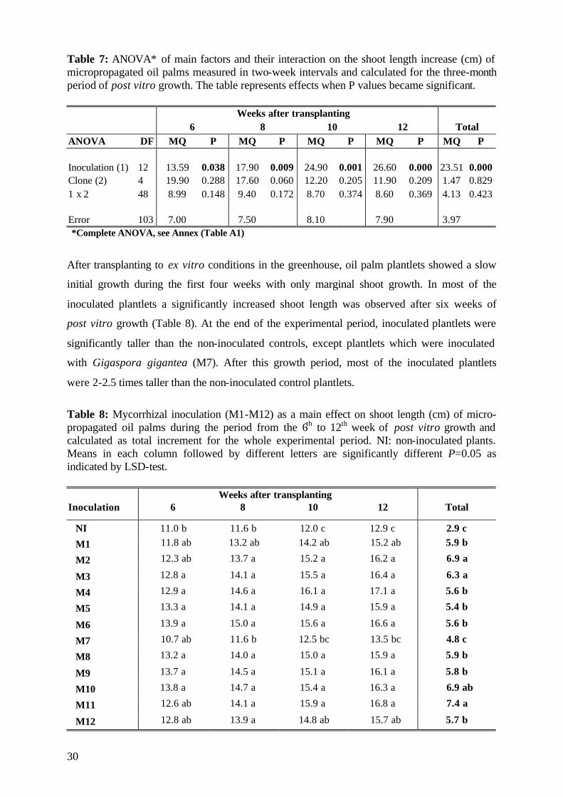
30
Table 7: ANOVA* of main factors and their interaction on the shoot length increase (cm) of micropropagated oil palms measured in two-week intervals and calculated for the three-month period of post vitro growth. The table represents effects when P values became significant. Weeks after transplanting 6 8 10 12 Total ANOVA DF MQ P MQ P MQ P MQ P MQ P Inoculation (1) 12 13.59 0.038 17.90 0.009 24.90 0.001 26.60 0.000 23.51 0.000 Clone (2) 4 19.90 0.288 17.60 0.060 12.20 0.205 11.90 0.209 1.47 0.829 1 x 2 48 8.99 0.148 9.40 0.172 8.70 0.374 8.60 0.369 4.13 0.423 Error 103 7.00 7.50 8.10 7.90 3.97 *Complete ANOVA, see Annex (Table A1)
After transplanting to ex vitro conditions in the greenhouse, oil palm plantlets showed a slow
initial growth during the first four weeks with only marginal shoot growth. In most of the
inoculated plantlets a significantly increased shoot length was observed after six weeks of
post vitro growth (Table 8). At the end of the experimental period, inoculated plantlets were
significantly taller than the non-inoculated controls, except plantlets which were inoculated
with Gigaspora gigantea (M7). After this growth period, most of the inoculated plantlets
were 2-2.5 times taller than the non-inoculated control plantlets.
Table 8: Mycorrhizal inoculation (M1-M12) as a main effect on shoot length (cm) of micro-propagated oil palms during the period from the 6th to 12th week of post vitro growth and calculated as total increment for the whole experimental period. NI: non-inoculated plants. Means in each column followed by different letters are significantly different P=0.05 as indicated by LSD-test. Weeks after transplanting Inoculation 6 8 10 12 Total
NI 11.0 b 11.6 b 12.0 c 12.9 c 2.9 c
M1 11.8 ab 13.2 ab 14.2 ab 15.2 ab 5.9 b
M2 12.3 ab 13.7 a 15.2 a 16.2 a 6.9 a
M3 12.8 a 14.1 a 15.5 a 16.4 a 6.3 a
M4 12.9 a 14.6 a 16.1 a 17.1 a 5.6 b
M5 13.3 a 14.1 a 14.9 a 15.9 a 5.4 b
M6 13.9 a 15.0 a 15.6 a 16.6 a 5.6 b
M7 10.7 ab 11.6 b 12.5 bc 13.5 bc 4.8 c
M8 13.2 a 14.0 a 15.0 a 15.9 a 5.9 b
M9 13.7 a 14.5 a 15.1 a 16.1 a 5.8 b
M10 13.8 a 14.7 a 15.4 a 16.3 a 6.9 ab
M11 12.6 ab 14.1 a 15.9 a 16.8 a 7.4 a
M12 12.8 ab 13.9 a 14.8 ab 15.7 ab 5.7 b
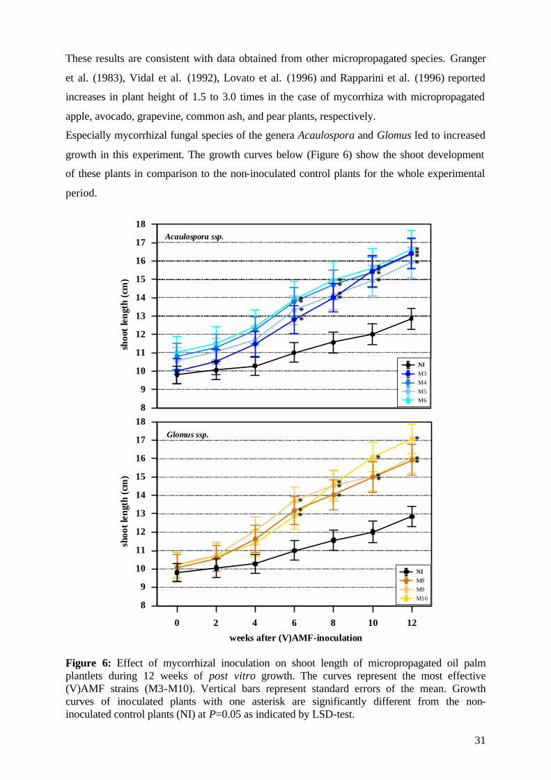
31
These results are consistent with data obtained from other micropropagated species. Granger
et al. (1983), Vidal et al. (1992), Lovato et al. (1996) and Rapparini et al. (1996) reported
increases in plant height of 1.5 to 3.0 times in the case of mycorrhiza with micropropagated
apple, avocado, grapevine, common ash, and pear plants, respectively.
Especially mycorrhizal fungal species of the genera Acaulospora and Glomus led to increased
growth in this experiment. The growth curves below (Figure 6) show the shoot development
of these plants in comparison to the non-inoculated control plants for the whole experimental
period.
8
9
10
11
12
13
14
15
16
17
18
NIM3M4M5M6
weeks after (V)AMF-inoculation
shoo
tlen
gth
(cm
)sh
oot
leng
th(c
m)
**** *
**
***
* *
****
8
9
10
11
12
13
14
15
16
17
18
0 2 4 6 8 10 12
NIM8M9M10
*** *
****
* **
*
Acaulospora ssp.
Glomus ssp.
Figure 6: Effect of mycorrhizal inoculation on shoot length of micropropagated oil palm plantlets during 12 weeks of post vitro growth. The curves represent the most effective (V)AMF strains (M3-M10). Vertical bars represent standard errors of the mean. Growth curves of inoculated plants with one asterisk are significantly different from the non-inoculated control plants (NI) at P=0.05 as indicated by LSD-test.
32
At the beginning of the experiment, all plants were uniform in size with a shoot length of 10
±1.2 cm (Figure 6). Significantly different shoot lengths were first measured six weeks after
transplanting in plantlets inoculated with Acaulospora mellea (M3), Acaulospora.
appendicula (M4), Acaulospora morowiae (M5), Acaulospora rehmii (M6), Glomus clarum
(M8), Glomus occultum (M9) and Glomus mosseae (M10).
5.1.3.2 Relative growth rate The relative growth rate (RGR cm day-1) of total shoot length (cm) was monitored every
second week. Plantlets were not homogenous in their morphological structure at the time of
transplanting. Mycorrhizal inoculation significantly affected both, the RGR of shoots at 8, 10
and 12 weeks after inoculation, and the average RGR calculated over the total experimental
period (Table 9). The clones and the interactions of the two main factors studied had no
significant effect on the RGR during this growth period.
Table 9: ANOVA* of main factors and their interaction on the relative growth rate (RGR cm day-1) of micropropagated oil palms measured in two-week intervals during a three-month period of post vitro growth and calculated as average values. The table represents effects when P values became significant. Weeks after transplanting 8 10 12 Average ANOVA DF MQ P MQ P MQ P MQ P Inoculation (1) 12 0.000 0.035 0.000 0.033 0.000 0.000 0.000 0.004 Clone (2) 4 0.000 0.922 0.000 0.586 0.000 0.179 0.000 0.087 1 x 2 48 0.000 0.368 0.000 0.759 0.000 0.365 0.000 0.558 Error 103 0.000 0.000 0.000 0.000 *Complete ANOVA, see Annex (Table A2)
After transplanting ex vitro, oil palm plantlets generally showed a slow initial growth during
the first two weeks (Table 10). In most of the inoculated plants the RGR was higher after 4
weeks. Significant differences compared to the control plants of the RGR were observed 8
weeks after transplanting in plantlets which were inoculated with Glomus manihot (M1),
Enthrophospora columbiana (M2), Acaulospora appendicula (M4), and Scutellispora
heterogama (M11). The growth-promoting effect continued in most of the inoculated plantlets
till the end of the experimental period. From the 10th to 12th week, all inoculated plantlets
showed a significantly higher shoot growth compared to the non-inoculated (NI) control
plants. The average relative growth calculated over the whole experimental period confirmed
the growth improvement through mycorrhizal inoculation. The most effective mycorrhizal
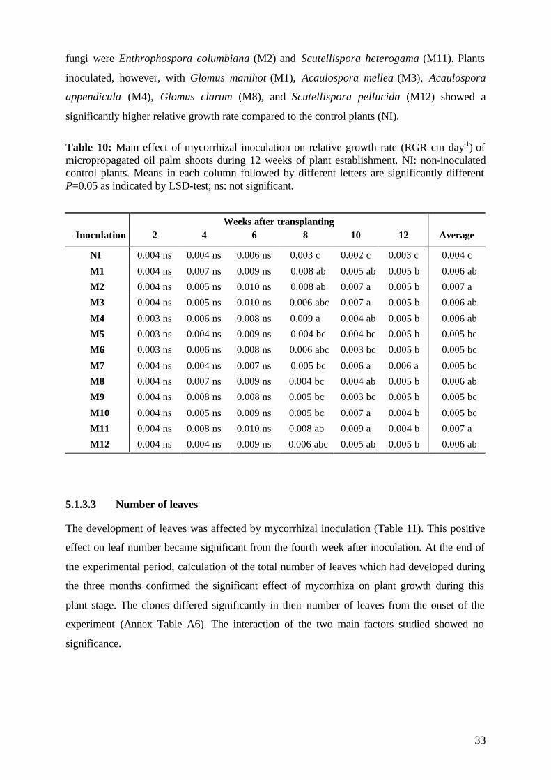
33
fungi were Enthrophospora columbiana (M2) and Scutellispora heterogama (M11). Plants
inoculated, however, with Glomus manihot (M1), Acaulospora mellea (M3), Acaulospora
appendicula (M4), Glomus clarum (M8), and Scutellispora pellucida (M12) showed a
significantly higher relative growth rate compared to the control plants (NI).
Table 10: Main effect of mycorrhizal inoculation on relative growth rate (RGR cm day-1) of micropropagated oil palm shoots during 12 weeks of plant establishment. NI: non-inoculated control plants. Means in each column followed by different letters are significantly different P=0.05 as indicated by LSD-test; ns: not significant.
Weeks after transplanting Inoculation 2 4 6 8 10 12 Average
NI 0.004 ns 0.004 ns 0.006 ns 0.003 c 0.002 c 0.003 c 0.004 c
M1 0.004 ns 0.007 ns 0.009 ns 0.008 ab 0.005 ab 0.005 b 0.006 ab
M2 0.004 ns 0.005 ns 0.010 ns 0.008 ab 0.007 a 0.005 b 0.007 a
M3 0.004 ns 0.005 ns 0.010 ns 0.006 abc 0.007 a 0.005 b 0.006 ab
M4 0.003 ns 0.006 ns 0.008 ns 0.009 a 0.004 ab 0.005 b 0.006 ab
M5 0.003 ns 0.004 ns 0.009 ns 0.004 bc 0.004 bc 0.005 b 0.005 bc
M6 0.003 ns 0.006 ns 0.008 ns 0.006 abc 0.003 bc 0.005 b 0.005 bc
M7 0.004 ns 0.004 ns 0.007 ns 0.005 bc 0.006 a 0.006 a 0.005 bc
M8 0.004 ns 0.007 ns 0.009 ns 0.004 bc 0.004 ab 0.005 b 0.006 ab
M9 0.004 ns 0.008 ns 0.008 ns 0.005 bc 0.003 bc 0.005 b 0.005 bc
M10 0.004 ns 0.005 ns 0.009 ns 0.005 bc 0.007 a 0.004 b 0.005 bc
M11 0.004 ns 0.008 ns 0.010 ns 0.008 ab 0.009 a 0.004 b 0.007 a
M12 0.004 ns 0.004 ns 0.009 ns 0.006 abc 0.005 ab 0.005 b 0.006 ab
5.1.3.3 Number of leaves The development of leaves was affected by mycorrhizal inoculation (Table 11). This positive
effect on leaf number became significant from the fourth week after inoculation. At the end of
the experimental period, calculation of the total number of leaves which had developed during
the three months confirmed the significant effect of mycorrhiza on plant growth during this
plant stage. The clones differed significantly in their number of leaves from the onset of the
experiment (Annex Table A6). The interaction of the two main factors studied showed no
significance.
34
Table 11: ANOVA* of main factors and their interaction on the leaf number of micropropagated oil palms in two-week intervals and calculated for the three-month period of post vitro growth. The table represents effects when P values became significant.
Weeks after transplanting ANOVA DF 0 2 4 6 8 10 12 Total Inoculation (1) 12 0.683 0.623 0.000 0.002 0.001 0.006 0.001 0.000 Clone (2) 4 0.017 0.004 0.002 0.008 0.005 0.002 0.001 0.013 1 x 2 48 0.305 0.133 0.243 0.130 0.273 0.309 0.541 0.602 * Complete ANOVA, see Annex (Table A3)
At the time of transplanting, all plantlets had generally the same number of leaves (Table 12).
Emergence of the first new leaves, developed ex vitro, could be observed 2 weeks after
transplanting in some of the inoculated plants, but these effects were statistically not
significant. Most of the inoculated plantlets developed the first new leaves 4 weeks after
transplanting in contrast to the non-inoculated plants (NI) where the first new leaves were
formed after 6 weeks. This significant effect on leaf development due to mycorrhizal
inoculation persists up to the end of the experimental period and is reflected in the total
increase in number of leaves developed during the measured 12 weeks of post vitro growth
(Table 12).
Table 12: Effect of mycorrhizal inoculation (M1-M12) on leaf development of micropropagated oil palm plantlets during 12 weeks of post vitro growth. The total number of leaves developed during the experimental period was calculated and tested for significant difference. NI: non-inoculated control plants. Values with different letters are significantly different at P=0.05 as indicated by LSD-test; ns: not significant.
Weeks after transplanting Inoculation 0 2 4 6 8 10 12 Total NI 3.3 ns 3.3 ns 3.5 d 4.0 c 4.3 c 4.9 b 5.3 b 2.0 b
M1 3.0 ns 3.2 ns 3.8 cd 4.3 bc 4.4 c 4.8 b 5.3 b 2.2 b
M2 3.3 ns 3.3 ns 3.9 bcd 4.2 bc 4.6 bc 5.2 b 5.9 ab 2.7 a
M3 3.3 ns 3.3 ns 4.2 bc 4.4 bc 4.7 bc 5.2 b 5.7 b 2.4 b
M4 3.4 ns 3.4 ns 4.8 a 5.1 a 5.4 a 6.0 a 6.6 a 3.2 a
M5 3.3 ns 3.4 ns 4.0 cd 4.5 bc 4.5 bc 5.0 b 5.5 b 2.2 b
M6 3.4 ns 3.4 ns 4.6 a 5.0 a 5.0 b 5.7 ab 6.6 a 3.2 a
M7 3.4 ns 3.4 ns 4.0 bcd 4.4 bc 4.7 bc 5.1 b 5.6 b 2.2 b
M8 3.4 ns 3.4 ns 4.3 ab 4.9 ab 5.2 a 5.9 a 6.3 a 3.2 a
M9 3.2 ns 3.4 ns 4.0 bcd 4.7 ab 4.9 bc 5.6 ab 6.1 ab 2.9 a
M10 3.5 ns 3.5 ns 4.7 a 5.2 a 5.7 a 6.1 a 6.7 a 3.1 a
M11 3.5 ns 3.7 ns 4.5 ab 4.7 ab 5.0 b 5.5 ab 5.9 ab 2.4 b
M12 3.3 ns 3.4 ns 4.4 ab 4.7 ab 5.0 b 5.7 ab 6.3 a 3.1 a
35
The most effective (V)AMF species on leaf development were Enthrophospora columbiana
(M2), Acaulospora appendicula (M4), Acaulospora rehmii (M6), Glomus clarum (M8),
Glomus occultum (M9), Glomus mosseae (M10), and Scutellispora pellucida (M12) (Table
12).
At the end of the experiment, the growth promoting effect of mycorrhiza resulted in a greater
total shoot length, accompanied by a higher number of expanded leaves in most of the
inoculated treatments compared to the non-inoculated plants (Table 12). These results are
consistent with data obtained from other micropropagated species. Fortuna et al. (1995)
observed these growth-promoting effects during the post vitro acclimatization phase of
micropropagated plum and apple plants which were inoculated with different mycorrhizal
fungi. Subhan et al. (1998) reports that, at any given time during a 16-week experimental
period, the ex vitro growth in terms of shoot height and number of leaves of inoculated
Sesbania sesban plantlets was superior to that of non-inoculated control plants.
5.1.4 Plant growth parameters after harvesting 5.1.4.1 Shoot and root fresh weight, root:shoot fresh weight ratio The shoot and root fresh weights and the root:shoot fresh weight (R:S) ratios were strongly
affected by mycorrhizal inoculation (Table 13). The used clones showed significant
differences only in the root fresh weights. There was no significant interaction between
mycorrhizal inoculation and clones.
Table 13: ANOVA of main factors and their interaction on the shoot and root fresh weight (g) and root:shoot fresh weight (R:S) ratio of micropropagated oil palms after three months of post vitro growth. Shoot fresh weight Root fresh weight R:S ratio ANOVA DF MQ P MQ P MQ P Inoculation (1) 12 4.13 0.000 0.14 0.002 0.009 0.003 Clone (2) 4 0.95 0.068 0.16 0.013 0.006 0.133 1 x 2 48 0.59 0.086 0.06 0.178 0.003 0.406 Error 99 0.42 0.049 0.003
After twelve weeks of post vitro growth, all tested (V)AMF significantly increased shoot
fresh weights compared to the non-inoculated (NI) control plants (Table 14). Plantlets
inoculated with Acaulospora appendicula (M4) produced more than two times higher shoot
fresh weights than that of the non-inoculated control plants. Also, the root systems of
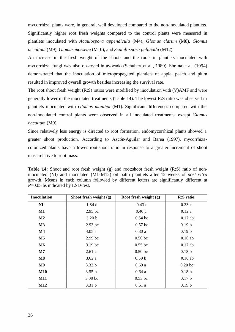
36
mycorrhizal plants were, in general, well developed compared to the non-inoculated plantlets.
Significantly higher root fresh weights compared to the control plants were measured in
plantlets inoculated with Acaulospora appendicula (M4), Glomus clarum (M8), Glomus
occultum (M9), Glomus mosseae (M10), and Scutellispora pellucida (M12).
An increase in the fresh weight of the shoots and the roots in plantlets inoculated with
mycorrhizal fungi was also observed in avocado (Schubert et al., 1989). Sbrana et al. (1994)
demonstrated that the inoculation of micropropagated plantlets of apple, peach and plum
resulted in improved overall growth besides increasing the survival rate.
The root:shoot fresh weight (R:S) ratios were modified by inoculation with (V)AMF and were
generally lower in the inoculated treatments (Table 14). The lowest R:S ratio was observed in
plantlets inoculated with Glomus manihot (M1). Significant differences compared with the
non-inoculated control plants were observed in all inoculated treatments, except Glomus
occultum (M9).
Since relatively less energy is directed to root formation, endomycorrhizal plants showed a
greater shoot production. According to Azcón-Aguilar and Barea (1997), mycorrhiza-
colonized plants have a lower root:shoot ratio in response to a greater increment of shoot
mass relative to root mass.
Table 14: Shoot and root fresh weight (g) and root:shoot fresh weight (R:S) ratio of non-inoculated (NI) and inoculated (M1-M12) oil palm plantlets after 12 weeks of post vitro growth. Means in each column followed by different letters are significantly different at P=0.05 as indicated by LSD-test.
Inoculation Shoot fresh weight (g) Root fresh weight (g) R:S ratio
NI 1.84 d 0.43 c 0.23 c
M1 2.95 bc 0.40 c 0.12 a
M2 3.20 b 0.54 bc 0.17 ab
M3 2.93 bc 0.57 bc 0.19 b
M4 4.05 a 0.80 a 0.19 b
M5 2.99 bc 0.50 bc 0.16 ab
M6 3.19 bc 0.55 bc 0.17 ab
M7 2.61 c 0.50 bc 0.18 b
M8 3.62 a 0.59 b 0.16 ab
M9 3.32 b 0.69 a 0.20 bc
M10 3.55 b 0.64 a 0.18 b
M11 3.08 bc 0.53 bc 0.17 b
M12 3.31 b 0.61 a 0.19 b
37
At the end of the experimental period the inoculated plants were better developed than the
non-inoculated control plants (Figure 7).
Figure 7: Development of inoculated (A) and non-inoculated (B) oil palm plantlets after 3 month of post vitro growth
5.1.4.2 Dry matter The mycorrhizal fungi had a highly significant effect on shoot dry weights of the oil palm
plantlets (Table 16). The clones and the interactions of the two main factors showed no
significant differences in dry matter accumulation at this growth stage.
Table 16: ANOVA of main factors and their interaction on the shoot dry weight (g) of micropropagated oil palms after a three-month period of post vitro growth. ANOVA DF MQ P
Inoculation 12 0.083 0.000 Clone 4 0.031 0.251 Inoculation x Clone 48 0.028 0.171
Error 100 0.023
Results obtained in this experiment showed that most of the tested mycorrhizal fungi were
effective in promoting plant growth, with little difference in the effectiveness of the (V)AMF
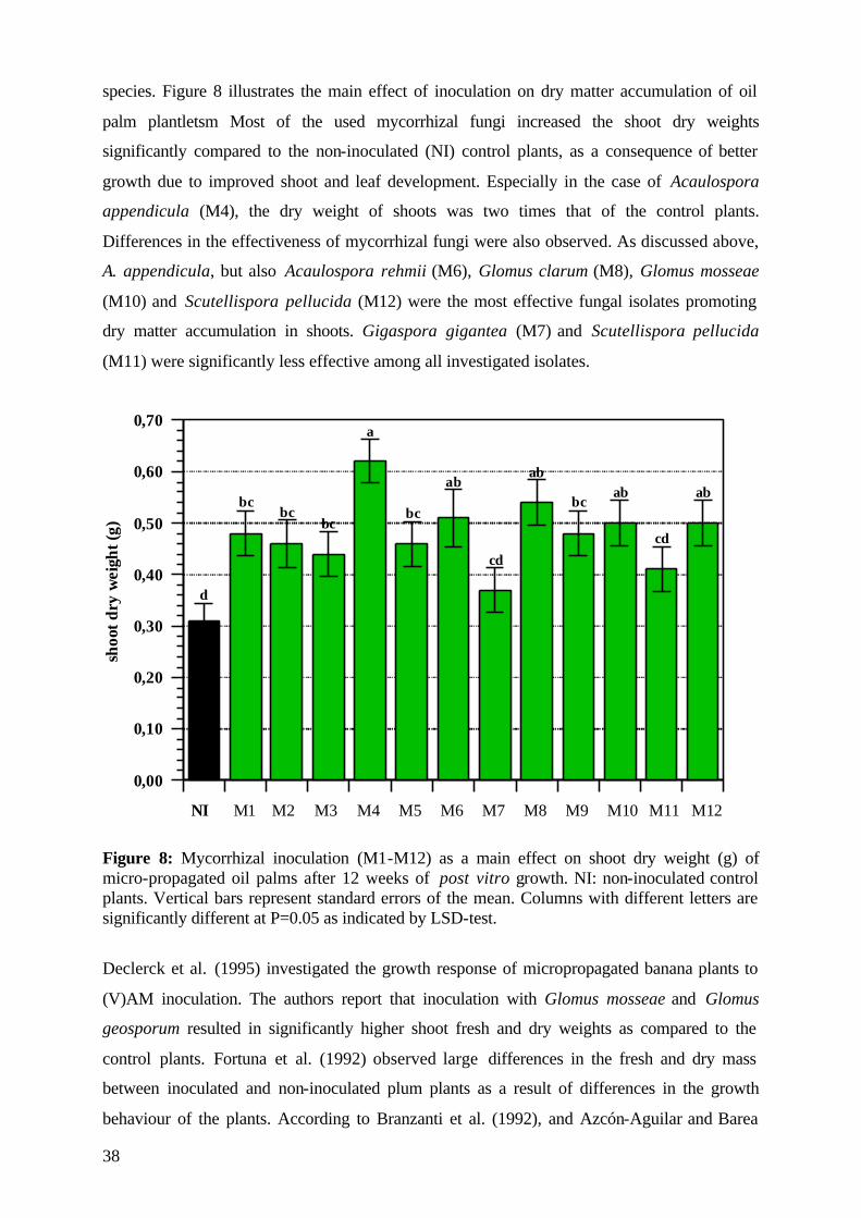
38
species. Figure 8 illustrates the main effect of inoculation on dry matter accumulation of oil
palm plantletsm Most of the used mycorrhizal fungi increased the shoot dry weights
significantly compared to the non-inoculated (NI) control plants, as a consequence of better
growth due to improved shoot and leaf development. Especially in the case of Acaulospora
appendicula (M4), the dry weight of shoots was two times that of the control plants.
Differences in the effectiveness of mycorrhizal fungi were also observed. As discussed above,
A. appendicula, but also Acaulospora rehmii (M6), Glomus clarum (M8), Glomus mosseae
(M10) and Scutellispora pellucida (M12) were the most effective fungal isolates promoting
dry matter accumulation in shoots. Gigaspora gigantea (M7) and Scutellispora pellucida
(M11) were significantly less effective among all investigated isolates.
0,00
0,10
0,20
0,30
0,40
0,50
0,60
0,70
NI M1 M2 M3 M4 M5 M6 M7 M8 M9 M10 M11 M12
shoo
tdry
wei
ght(
g)
d
bcbc
bc
a
bc
ab
cd
ab
bcab
cd
ab
Figure 8: Mycorrhizal inoculation (M1-M12) as a main effect on shoot dry weight (g) of micro-propagated oil palms after 12 weeks of post vitro growth. NI: non-inoculated control plants. Vertical bars represent standard errors of the mean. Columns with different letters are significantly different at P=0.05 as indicated by LSD-test.
Declerck et al. (1995) investigated the growth response of micropropagated banana plants to
(V)AM inoculation. The authors report that inoculation with Glomus mosseae and Glomus
geosporum resulted in significantly higher shoot fresh and dry weights as compared to the
control plants. Fortuna et al. (1992) observed large differences in the fresh and dry mass
between inoculated and non-inoculated plum plants as a result of differences in the growth
behaviour of the plants. According to Branzanti et al. (1992), and Azcón-Aguilar and Barea
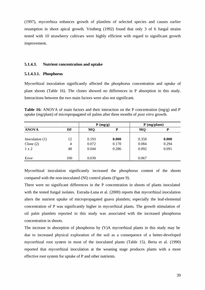
39
(1997), mycorrhiza enhances growth of plantlets of selected species and causes earlier
resumption in shoot apical growth. Vestberg (1992) found that only 3 of 6 fungal strains
tested with 10 strawberry cultivars were highly efficient with regard to significant growth
improvement.
5.1.4.3. Nutrient concentration and uptake 5.1.4.3.1. Phosphorus Mycorrhizal inoculation significantly affected the phosphorus concentration and uptake of
plant shoots (Table 16). The clones showed no differences in P absorption in this study.
Interactions between the two main factors were also not significant.
Table 16: ANOVA of main factors and their interaction on the P concentration (mg/g) and P uptake (mg/plant) of micropropagated oil palms after three months of post vitro growth.
P (mg/g) P (mg/plant) ANOVA DF MQ P MQ P Inoculation (1) 12 0.193 0.000 0.358 0.000 Clone (2) 4 0.072 0.170 0.084 0.294 1 x 2 48 0.044 0.286 0.092 0.091 Error 100 0.039 0.067
Mycorrhizal inoculation significantly increased the phosphorus content of the shoots
compared with the non-inoculated (NI) control plants (Figure 9).
There were no significant differences in the P concentration in shoots of plants inoculated
with the tested fungal isolates. Estrada-Luna et al. (2000) reports that mycorrhizal inoculation
alters the nutrient uptake of micropropagated guava plantlets; especially the leaf-elemental
concentration of P was significantly higher in mycorrhizal plants. The growth stimulation of
oil palm plantlets reported in this study was associated with the increased phosphorus
concentration in shoots.
The increase in absorption of phosphorus by (V)A mycorrhizal plants in this study may be
due to increased physical exploration of the soil as a consequence of a better-developed
mycorrhizal root system in most of the inoculated plants (Table 15). Berta et al. (1990)
reported that mycorrhizal inoculation at the weaning stage produces plants with a more
effective root system for uptake of P and other nutrients.
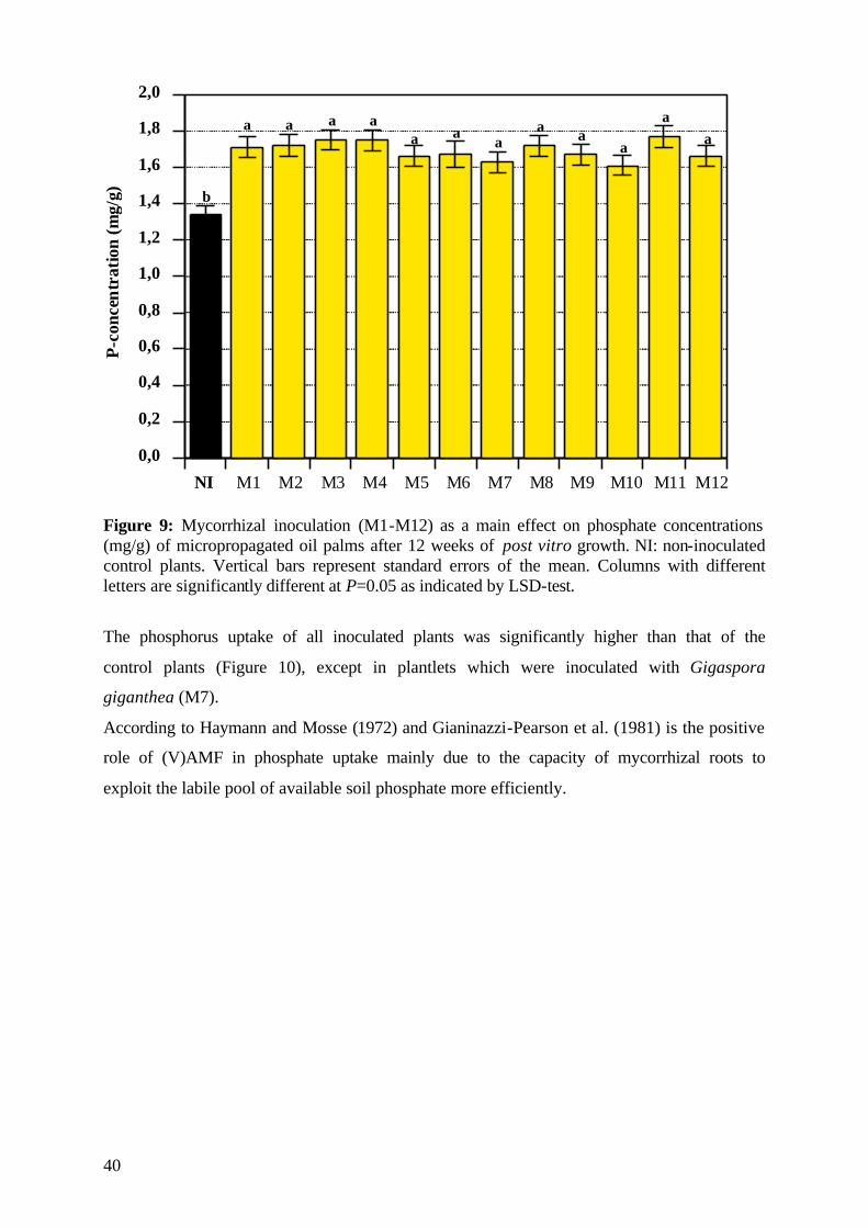
40
0,0
0,2
0,4
0,6
0,8
1,0
1,2
1,4
1,6
1,8
2,0
NI M1 M2 M3 M4 M5 M6 M7 M8 M9 M10 M11 M12
b
a a a aa a
aa a
a
aa
P-co
ncen
trat
ion
(mg/
g)
Figure 9: Mycorrhizal inoculation (M1-M12) as a main effect on phosphate concentrations (mg/g) of micropropagated oil palms after 12 weeks of post vitro growth. NI: non-inoculated control plants. Vertical bars represent standard errors of the mean. Columns with different letters are significantly different at P=0.05 as indicated by LSD-test.
The phosphorus uptake of all inoculated plants was significantly higher than that of the
control plants (Figure 10), except in plantlets which were inoculated with Gigaspora
giganthea (M7).
According to Haymann and Mosse (1972) and Gianinazzi-Pearson et al. (1981) is the positive
role of (V)AMF in phosphate uptake mainly due to the capacity of mycorrhizal roots to
exploit the labile pool of available soil phosphate more efficiently.
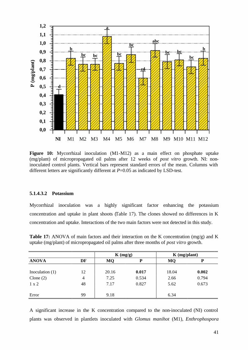
41
0,0
0,1
0,2
0,3
0,4
0,5
0,6
0,7
0,8
0,9
1,0
1,1
1,2
NI M1 M2 M3 M4 M5 M6 M7 M8 M9 M10 M11 M12
P(m
g/pl
ant)
d
bbc bc
a
bc
bc
cd
abc
bc bcbc
b
Figure 10: Mycorrhizal inoculation (M1-M12) as a main effect on phosphate uptake (mg/plant) of micropropagated oil palms after 12 weeks of post vitro growth. NI: non-inoculated control plants. Vertical bars represent standard errors of the mean. Columns with different letters are significantly different at P=0.05 as indicated by LSD-test.
5.1.4.3.2 Potassium Mycorrhizal inoculation was a highly significant factor enhancing the potassium
concentration and uptake in plant shoots (Table 17). The clones showed no differences in K
concentration and uptake. Interactions of the two main factors were not detected in this study.
Table 17: ANOVA of main factors and their interaction on the K concentration (mg/g) and K uptake (mg/plant) of micropropagated oil palms after three months of post vitro growth.
K (mg/g) K (mg/plant) ANOVA DF MQ P MQ P Inoculation (1) 12 20.16 0.017 18.04 0.002 Clone (2) 4 7.25 0.534 2.66 0.794 1 x 2 48 7.17 0.827 5.62 0.673 Error 99 9.18 6.34
A significant increase in the K concentration compared to the non-inoculated (NI) control
plants was observed in plantlets inoculated with Glomus manihot (M1), Enthrophospora
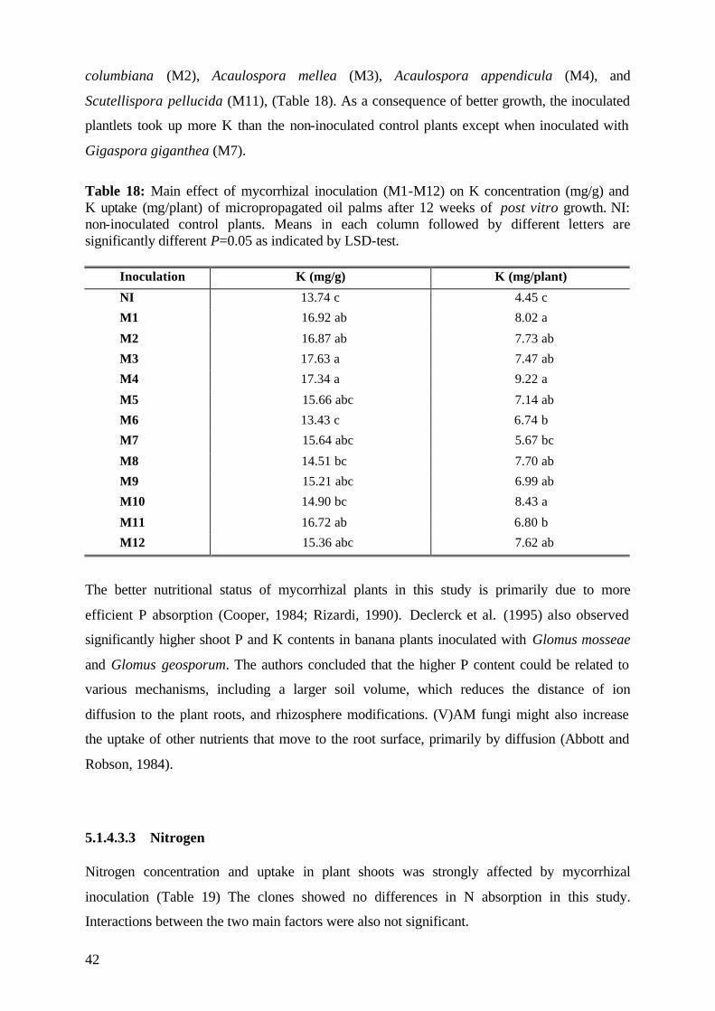
42
columbiana (M2), Acaulospora mellea (M3), Acaulospora appendicula (M4), and
Scutellispora pellucida (M11), (Table 18). As a consequence of better growth, the inoculated
plantlets took up more K than the non-inoculated control plants except when inoculated with
Gigaspora giganthea (M7).
Table 18: Main effect of mycorrhizal inoculation (M1-M12) on K concentration (mg/g) and K uptake (mg/plant) of micropropagated oil palms after 12 weeks of post vitro growth. NI: non-inoculated control plants. Means in each column followed by different letters are significantly different P=0.05 as indicated by LSD-test.
Inoculation K (mg/g) K (mg/plant)
NI 13.74 c 4.45 c
M1 16.92 ab 8.02 a
M2 16.87 ab 7.73 ab
M3 17.63 a 7.47 ab
M4 17.34 a 9.22 a
M5 15.66 abc 7.14 ab
M6 13.43 c 6.74 b
M7 15.64 abc 5.67 bc
M8 14.51 bc 7.70 ab
M9 15.21 abc 6.99 ab
M10 14.90 bc 8.43 a
M11 16.72 ab 6.80 b
M12 15.36 abc 7.62 ab
The better nutritional status of mycorrhizal plants in this study is primarily due to more
efficient P absorption (Cooper, 1984; Rizardi, 1990). Declerck et al. (1995) also observed
significantly higher shoot P and K contents in banana plants inoculated with Glomus mosseae
and Glomus geosporum. The authors concluded that the higher P content could be related to
various mechanisms, including a larger soil volume, which reduces the distance of ion
diffusion to the plant roots, and rhizosphere modifications. (V)AM fungi might also increase
the uptake of other nutrients that move to the root surface, primarily by diffusion (Abbott and
Robson, 1984).
5.1.4.3.3 Nitrogen Nitrogen concentration and uptake in plant shoots was strongly affected by mycorrhizal
inoculation (Table 19) The clones showed no differences in N absorption in this study.
Interactions between the two main factors were also not significant.
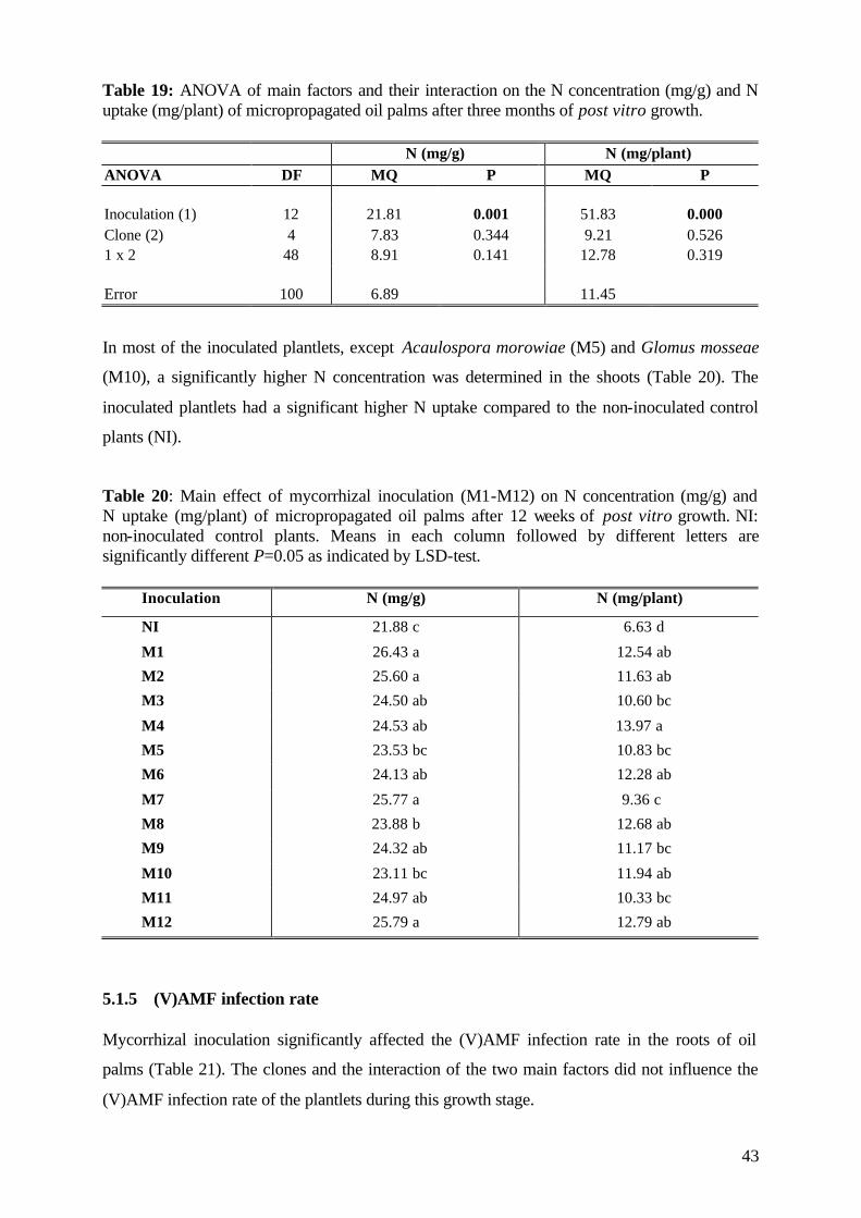
43
Table 19: ANOVA of main factors and their interaction on the N concentration (mg/g) and N uptake (mg/plant) of micropropagated oil palms after three months of post vitro growth.
N (mg/g) N (mg/plant) ANOVA DF MQ P MQ P Inoculation (1) 12 21.81 0.001 51.83 0.000 Clone (2) 4 7.83 0.344 9.21 0.526 1 x 2 48 8.91 0.141 12.78 0.319 Error 100 6.89 11.45
In most of the inoculated plantlets, except Acaulospora morowiae (M5) and Glomus mosseae
(M10), a significantly higher N concentration was determined in the shoots (Table 20). The
inoculated plantlets had a significant higher N uptake compared to the non-inoculated control
plants (NI).
Table 20: Main effect of mycorrhizal inoculation (M1-M12) on N concentration (mg/g) and N uptake (mg/plant) of micropropagated oil palms after 12 weeks of post vitro growth. NI: non-inoculated control plants. Means in each column followed by different letters are significantly different P=0.05 as indicated by LSD-test.
Inoculation N (mg/g) N (mg/plant)
NI 21.88 c 6.63 d
M1 26.43 a 12.54 ab
M2 25.60 a 11.63 ab
M3 24.50 ab 10.60 bc
M4 24.53 ab 13.97 a
M5 23.53 bc 10.83 bc
M6 24.13 ab 12.28 ab
M7 25.77 a 9.36 c
M8 23.88 b 12.68 ab
M9 24.32 ab 11.17 bc
M10 23.11 bc 11.94 ab
M11 24.97 ab 10.33 bc
M12 25.79 a 12.79 ab
5.1.5 (V)AMF infection rate Mycorrhizal inoculation significantly affected the (V)AMF infection rate in the roots of oil
palms (Table 21). The clones and the interaction of the two main factors did not influence the
(V)AMF infection rate of the plantlets during this growth stage.
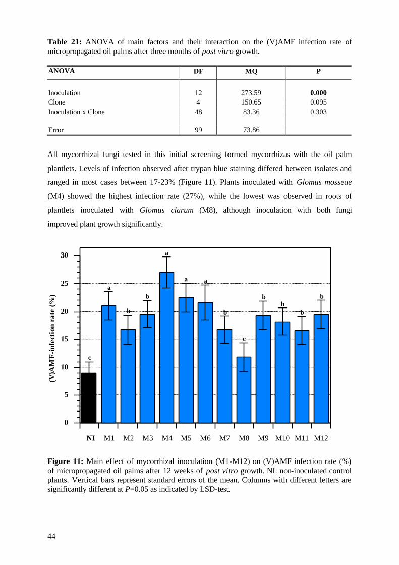
44
Table 21: ANOVA of main factors and their interaction on the (V)AMF infection rate of micropropagated oil palms after three months of post vitro growth. ANOVA DF MQ P
Inoculation 12 273.59 0.000 Clone 4 150.65 0.095 Inoculation x Clone 48 83.36 0.303
Error 99 73.86
All mycorrhizal fungi tested in this initial screening formed mycorrhizas with the oil palm
plantlets. Levels of infection observed after trypan blue staining differed between isolates and
ranged in most cases between 17-23% (Figure 11). Plants inoculated with Glomus mosseae
(M4) showed the highest infection rate (27%), while the lowest was observed in roots of
plantlets inoculated with Glomus clarum (M8), although inoculation with both fungi
improved plant growth significantly.
0
5
10
15
20
25
30
NI M1 M2 M3 M4 M5 M6 M7 M8 M9 M10 M11 M12
(V)A
MF-
infe
ctio
nra
te(%
)
c
a
b
b
a
a a
b
c
bb
b
b
Figure 11: Main effect of mycorrhizal inoculation (M1-M12) on (V)AMF infection rate (%) of micropropagated oil palms after 12 weeks of post vitro growth. NI: non-inoculated control plants. Vertical bars represent standard errors of the mean. Columns with different letters are significantly different at P=0.05 as indicated by LSD-test.
45
Pinochet et al. (1997) reported that the percentage of mycorrhizal colonization of Glomus
interadices was relatively low at 27%, although plant growth response in mycorrhizal
treatments was significantly higher compared with the non-mycorrhizal treatments for all
growth characteristics (plant height, number of leaves and fresh and dry weights).
This study demonstrates that (V)AM fungi can establish a symbiotic association with the oil
palm clones and that an early endomycorrhizal inoculation ensures maximum survival and
growth of the young oil palms after they have been transferred to the soil. Chavez and
Ferrera-Cerrato (1990) measured the response of 4 micropropagated strawberry cultivars to
root colonization by (V)AMF. They found that plant yields differed significantly depending
on the (V)AMF species tested and that yield was unrelated to percentage of root colonization.
Some plant cultivar/fungus combinations resulted in improved growth while others did not.
The weak mycorrhizal formation (9%) in the roots of control plants (NI) grown in the non-
sterilized soil (Figure 11) in this study indicated the natural levels of infection, which occur in
plant roots under ‘normal’ nursery conditions. The low effectiveness of these fungi on plant
survival and development was evident in this experiment. These results indicate that the early
inoculation of micropropagated oil palms with more effective (V)AM fungi greatly improves
the tolerance to the stress situation after transplanting to post vitro conditions.
5.1.6 Assessment of plant development At the end of the experimental period, the remaining plants were visually classified as of
‘good quality’, which is an important factor for successful marketing of oil palm plantlets.
The young plants were classified according to a scheme which is generally used at the
‘Indonesian Oil Palm Research Institute’ (IOPRI) when plantlets reach this growth stage and
are to be sold to the planters or transferred to the nursery for further cultivation. This
assessment is based on the three categories, which are presented in Figure 12: (Class A: Best
quality, plantlets are ready for selling; Class B: Good quality, plantlets are expected for future
selling (after some weeks); Class C: Plantlets will be discarded, due to stunted or abnormal
growth).
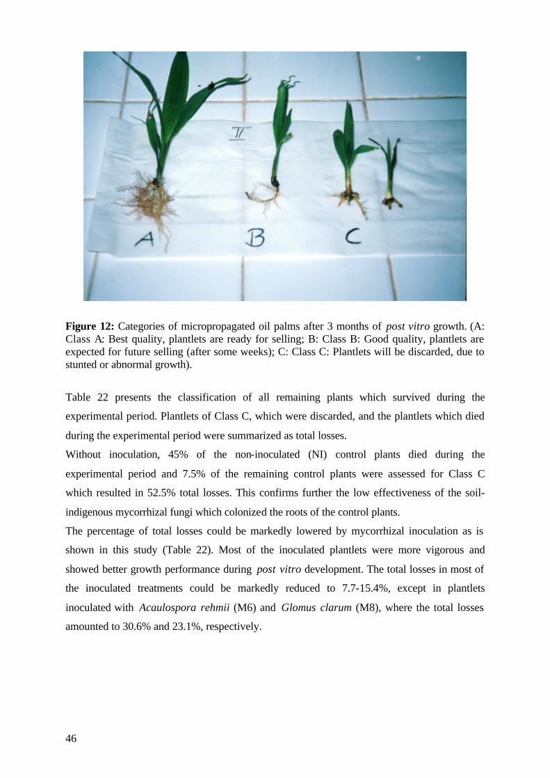
46
Figure 12: Categories of micropropagated oil palms after 3 months of post vitro growth. (A: Class A: Best quality, plantlets are ready for selling; B: Class B: Good quality, plantlets are expected for future selling (after some weeks); C: Class C: Plantlets will be discarded, due to stunted or abnormal growth).
Table 22 presents the classification of all remaining plants which survived during the
experimental period. Plantlets of Class C, which were discarded, and the plantlets which died
during the experimental period were summarized as total losses.
Without inoculation, 45% of the non-inoculated (NI) control plants died during the
experimental period and 7.5% of the remaining control plants were assessed for Class C
which resulted in 52.5% total losses. This confirms further the low effectiveness of the soil-
indigenous mycorrhizal fungi which colonized the roots of the control plants.
The percentage of total losses could be markedly lowered by mycorrhizal inoculation as is
shown in this study (Table 22). Most of the inoculated plantlets were more vigorous and
showed better growth performance during post vitro development. The total losses in most of
the inoculated treatments could be markedly reduced to 7.7-15.4%, except in plantlets
inoculated with Acaulospora rehmii (M6) and Glomus clarum (M8), where the total losses
amounted to 30.6% and 23.1%, respectively.
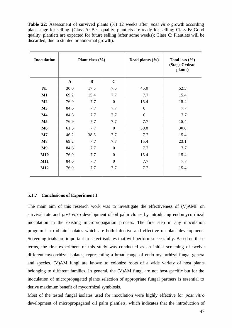
47
Table 22: Assessment of survived plants (%) 12 weeks after post vitro growth according plant stage for selling. (Class A: Best quality, plantlets are ready for selling; Class B: Good quality, plantlets are expected for future selling (after some weeks); Class C: Plantlets will be discarded, due to stunted or abnormal growth).
Inoculation
Plant class (%)
Dead plants (%)
Total loss (%) (Stage C+dead
plants)
A B C
NI 30.0 17.5 7.5 45.0 52.5
M1 69.2 15.4 7.7 7.7 15.4
M2 76.9 7.7 0 15.4 15.4
M3 84.6 7.7 7.7 0 7.7
M4 84.6 7.7 7.7 0 7.7
M5 76.9 7.7 7.7 7.7 15.4
M6 61.5 7.7 0 30.8 30.8
M7 46.2 38.5 7.7 7.7 15.4
M8 69.2 7.7 7.7 15.4 23.1
M9 84.6 7.7 0 7.7 7.7
M10 76.9 7.7 0 15.4 15.4
M11 84.6 7.7 0 7.7 7.7
M12 76.9 7.7 7.7 7.7 15.4
5.1.7 Conclusions of Experiment 1 The main aim of this research work was to investigate the effectiveness of (V)AMF on
survival rate and post vitro development of oil palm clones by introducing endomycorrhizal
inoculation in the existing micropropagation process. The first step in any inoculation
program is to obtain isolates which are both infective and effective on plant development.
Screening trials are important to select isolates that will perform successfully. Based on these
terms, the first experiment of this study was conducted as an initial screening of twelve
different mycorrhizal isolates, representing a broad range of endo-mycorrhizal fungal genera
and species. (V)AM fungi are known to colonize roots of a wide variety of host plants
belonging to different families. In general, the (V)AM fungi are not host-specific but for the
inoculation of micropropagated plants selection of appropriate fungal partners is essential to
derive maximum benefit of mycorrhizal symbiosis.
Most of the tested fungal isolates used for inoculation were highly effective for post vitro
development of micropropagated oil palm plantlets, which indicates that the introduction of
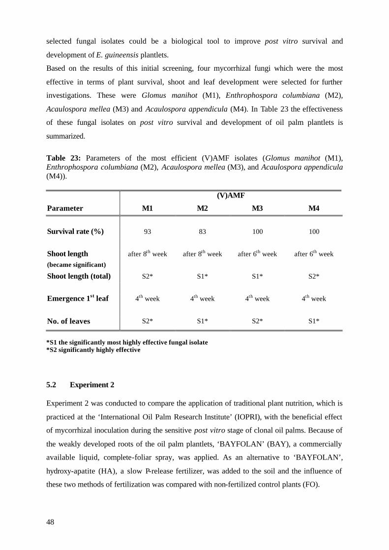
48
selected fungal isolates could be a biological tool to improve post vitro survival and
development of E. guineensis plantlets.
Based on the results of this initial screening, four mycorrhizal fungi which were the most
effective in terms of plant survival, shoot and leaf development were selected for further
investigations. These were Glomus manihot (M1), Enthrophospora columbiana (M2),
Acaulospora mellea (M3) and Acaulospora appendicula (M4). In Table 23 the effectiveness
of these fungal isolates on post vitro survival and development of oil palm plantlets is
summarized.
Table 23: Parameters of the most efficient (V)AMF isolates (Glomus manihot (M1), Enthrophospora columbiana (M2), Acaulospora mellea (M3), and Acaulospora appendicula (M4)). (V)AMF
Parameter M1 M2 M3 M4
Survival rate (%) 93 83 100 100
Shoot length after 8th week after 8th week after 6th week after 6th week (became significant)
Shoot length (total) S2* S1* S1* S2*
Emergence 1st leaf 4th week 4th week 4th week 4th week
No. of leaves S2* S1* S2* S1*
*S1 the significantly most highly effective fungal isolate *S2 significantly highly effective 5.2 Experiment 2 Experiment 2 was conducted to compare the application of traditional plant nutrition, which is
practiced at the ‘International Oil Palm Research Institute’ (IOPRI), with the beneficial effect
of mycorrhizal inoculation during the sensitive post vitro stage of clonal oil palms. Because of
the weakly developed roots of the oil palm plantlets, ‘BAYFOLAN’ (BAY), a commercially
available liquid, complete-foliar spray, was applied. As an alternative to ‘BAYFOLAN’,
hydroxy-apatite (HA), a slow P-release fertilizer, was added to the soil and the influence of
these two methods of fertilization was compared with non-fertilized control plants (FO).
49
Based on the results of the initial screening in Experiment 1, plantlets were inoculated with
four endomycorrhizal fungi which were selected for their effectiveness on plant survival and
development. These were Glomus manihot (M1), Enthrophospora columbiana (M2),
Acaulospora mellea (M3), and Acaulospora appendicula (M4).
In this experiment, plants were harvested at two different growth stages to investigate the
effect of mycorrhizal inoculation and fertilization on the post vitro plant development. The
first harvest was carried out 6 weeks and the second harvest 12 weeks after transplanting.
5.2.1 Survival rate Inoculation with the selected (V)AMF at the transplanting stage was highly effective in
securing plant survival during the three-month growth period (Table 24) and confirmed the
results of the first experiment. Both mycorrhizal inoculation and fertilization as main factors,
but also the interaction between these two factors, affected plant survival significantly. The
clones as a main factor and the interactions of the three factors studied were not significant.
Table 24: ANOVA of main effects and their interactions on the survival rate (%) of micropropagated oil palms after a three-month period of post vitro growth. ANOVA DF MQ P Inoculation 4 14716.67 0.000 Fertilization 2 4933.33 0.017 Clone 4 2883.33 0.149 Inoculation x Fertilization 8 2516.67 0.035 Inoculation x Clone 16 320.83 0.998 Fertilization x Clone 8 433.34 0.938 Inoculation x Fertilization x Clone 32 620.83 0.985 Error 225 1188.89
The interactions between the two main factors inoculation and fertilization regarding plant
survival during three months of post vitro growth are represented in Figure 13. The lowest
percentage of plant survival (45%) was observed in the non-inoculated (NI) control plants
which were either non-fertilized (FO) or in soil fertilized with hydroxy-apatite (HA). The
survival rate was significantly improved by mycorrhizal inoculation with either of the tested
(V)AMF: up to 75-95% in the non-fertilized, and up to 85-100% in the HA-fertilized plants,
respectively.
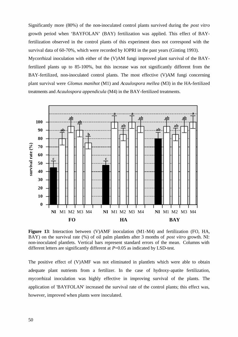
50
Significantly more (80%) of the non-inoculated control plants survived during the post vitro
growth period when ‘BAYFOLAN’ (BAY) fertilization was applied. This effect of BAY-
fertilization observed in the control plants of this experiment does not correspond with the
survival data of 60-70%, which were recorded by IOPRI in the past years (Ginting 1993).
Mycorrhizal inoculation with either of the (V)AM fungi improved plant survival of the BAY-
fertilized plants up to 85-100%, but this increase was not significantly different from the
BAY-fertilized, non-inoculated control plants. The most effective (V)AM fungi concerning
plant survival were Glomus manihot (M1) and Acaulospora mellea (M3) in the HA-fertilized
treatments and Acaulospora appendicula (M4) in the BAY-fertilized treatments.
0
10
20
30
40
50
60
70
80
90
100
NI M1 M2 M3 M4 NI M1 M2 M3 M4 NI M1 M2 M3 M4
FO HA BAY
surv
ival
rate
(%)
c c
abab
abab
b
a
ab
aab ab
ab
aba
Figure 13: Interaction between (V)AMF inoculation (M1-M4) and fertilization (FO, HA, BAY) on the survival rate (%) of oil palm plantlets after 3 months of post vitro growth. NI: non-inoculated plantlets. Vertical bars represent standard errors of the mean. Columns with different letters are significantly different at P=0.05 as indicated by LSD-test.
The positive effect of (V)AMF was not eliminated in plantlets which were able to obtain
adequate plant nutrients from a fertilizer. In the case of hydroxy-apatite fertilization,
mycorrhizal inoculation was highly effective in improving survival of the plants. The
application of 'BAYFOLAN' increased the survival rate of the control plants; this effect was,
however, improved when plants were inoculated.
51
These results underscore the importance of an early inoculation and establishment of the
symbiosis with mycorrhizal fungi for post vitro growth of the oil palm plantlets, even when
these plants were fertilized with the complete soluble foliar ‘BAYFOLAN’.
5.2.2 Date of mortality The curves in Figure 14 represent the date of plant mortality during the 12-week experimental
period, influenced by different (V)AM fungal isolates and fertilization treatments. The highest
mortality rates (55%) were observed in the non-inoculated control plants (NI), either non-
fertilized (FO) or soil fertilized with hydroxy-apatite (HA). Plant mortality was reduced to 5-
25% by the mycorrhizal inoculation in the non-fertilized treatments. The lowest mortality
rates in this experiment were observed in the inoculated plantlets fertilized with HA. None of
the plantlets inoculated with Glomus manihot (M1) and Acaulospora mellea (M3),
respectively, died in this treatment. Two weeks after inoculation, 5% and 15% of the plantlets
inoculated with Acaulospora appendicula (M4) and Enthrophospora columbiana (M2),
respectively, died. No further mortality of the inoculated plants in this treatment was observed
after the second week of post vitro growth.
Receiving 'BAYFOLAN', 20% of the non-inoculated control plants died up to the 6th week.
All inoculated plantlets in this treatment showed reduced mortality rates. Especially
inoculation with Acaulospora appendicula (M4,) but also with Glomus manihot (M1) and
Acaulospora mellea (M3), resulted in reduced plant mortality.
As discussed in Experiment 1, plant mortality was only observed during the first six weeks
after transplanting. After this growth period, plantlets became more adapted to post vitro
conditions. These results were also confirmed in this experiment, especially for the non-
inoculated (NI) control plants, independent of fertilization. In contrast, the date of mortality of
all inoculated plants in this experiment was limited to the first three weeks after plants were
transferred to polybags. During this period, (V)AM infection was initiated and fungi
colonized the plants. Several studies on the early stages of post vitro acclimatization have
reported this phase to be a critical step in the micropropagation cycle, at which the lack of
beneficial microorganisms can adversely affect survival and growth of the micropropagated
plantlets (Pons et al., 1983).
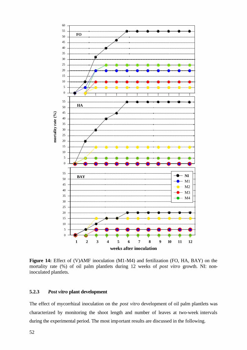
52
0
5
10
15
20
25
30
35
40
45
50
55
60
0
5
10
15
20
25
30
35
40
45
50
55
0
5
10
15
20
25
30
35
40
45
50
55
1 2 3 4 5 6 7 8 9 10 11 12
NIM1M2M3M4
FO
HA
BAY
mor
talit
yra
te(%
)
weeks after inoculation Figure 14: Effect of (V)AMF inoculation (M1-M4) and fertilization (FO, HA, BAY) on the mortality rate (%) of oil palm plantlets during 12 weeks of post vitro growth. NI: non-inoculated plantlets.
5.2.3 Post vitro plant development The effect of mycorrhizal inoculation on the post vitro development of oil palm plantlets was
characterized by monitoring the shoot length and number of leaves at two-week intervals
during the experimental period. The most important results are discussed in the following.
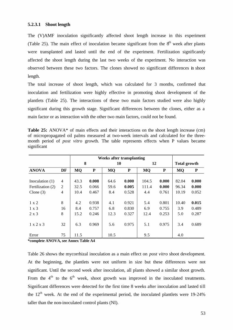
53
5.2.3.1 Shoot length The (V)AMF inoculation significantly affected shoot length increase in this experiment
(Table 25). The main effect of inoculation became significant from the 8th week after plants
were transplanted and lasted until the end of the experiment. Fertilization significantly
affected the shoot length during the last two weeks of the experiment. No interaction was
observed between these two factors. The clones showed no significant differences in shoot
length.
The total increase of shoot length, which was calculated for 3 months, confirmed that
inoculation and fertilization were highly effective in promoting shoot development of the
plantlets (Table 25). The interactions of these two main factors studied were also highly
significant during this growth stage. Significant differences between the clones, either as a
main factor or as interaction with the other two main factors, could not be found.
Table 25: ANOVA* of main effects and their interactions on the shoot length increase (cm) of micropropagated oil palms measured at two-week intervals and calculated for the three-month period of post vitro growth. The table represents effects when P values became significant
Weeks after transplanting 8 10 12 Total growth
ANOVA DF MQ P MQ P MQ P MQ P Inoculation (1) 4 43.3 0.008 64.6 0.000 104.5 0.000 82.04 0.000 Fertilization (2) 2 32.5 0.066 59.6 0.005 111.4 0.000 96.34 0.000 Clone (3) 4 10.4 0.467 8.4 0.528 4.4 0.761 10.19 0.052 1 x 2 8 4.2 0.938 4.1 0.921 5.4 0.801 10.40 0.015 1 x 3 16 8.4 0.757 6.8 0.830 6.9 0.755 3.9 0.489 2 x 3 8 15.2 0.246 12.3 0.327 12.4 0.253 5.0 0.287 1 x 2 x 3 32 6.3 0.969 5.6 0.975 5.1 0.975 3.4 0.689 Error 75 11.5 10.5 9.5 4.0
*complete ANOVA, see Annex Table A4
Table 26 shows the mycorrhizal inoculation as a main effect on post vitro shoot development.
At the beginning, the plantlets were not uniform in size but these differences were not
significant. Until the second week after inoculation, all plants showed a similar shoot growth.
From the 4th to the 6th week, shoot growth was improved in the inoculated treatments.
Significant differences were detected for the first time 8 weeks after inoculation and lasted till
the 12th week. At the end of the experimental period, the inoculated plantlets were 19-24%
taller than the non-inoculated control plants (NI).
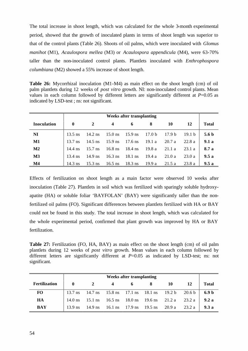
54
The total increase in shoot length, which was calculated for the whole 3-month experimental
period, showed that the growth of inoculated plants in terms of shoot length was superior to
that of the control plants (Table 26). Shoots of oil palms, which were inoculated with Glomus
manihot (M1), Acaulospora mellea (M3) or Acaulospora appendicula (M4), were 63-70%
taller than the non-inoculated control plants. Plantlets inoculated with Enthrophospora
columbiana (M2) showed a 55% increase of shoot length.
Table 26: Mycorrhizal inoculation (M1-M4) as main effect on the shoot length (cm) of oil palm plantlets during 12 weeks of post vitro growth. NI: non-inoculated control plants. Mean values in each column followed by different letters are significantly different at P=0.05 as indicated by LSD-test ; ns: not significant.
Weeks after transplanting
Inoculation 0 2 4 6 8 10 12 Total
NI 13.5 ns 14.2 ns 15.0 ns 15.9 ns 17.0 b 17.9 b 19.1 b 5.6 b
M1 13.7 ns 14.5 ns 15.9 ns 17.6 ns 19.1 a 20.7 a 22.8 a 9.1 a
M2 14.4 ns 15.7 ns 16.8 ns 18.4 ns 19.8 a 21.1 a 23.1 a 8.7 a
M3 13.4 ns 14.9 ns 16.3 ns 18.1 ns 19.4 a 21.0 a 23.0 a 9.5 a
M4 14.3 ns 15.3 ns 16.5 ns 18.3 ns 19.9 a 21.5 a 23.8 a 9.5 a
Effects of fertilization on shoot length as a main factor were observed 10 weeks after
inoculation (Table 27). Plantlets in soil which was fertilized with sparingly soluble hydroxy-
apatite (HA) or soluble foliar ‘BAYFOLAN’ (BAY) were significantly taller than the non-
fertilized oil palms (FO). Significant differences between plantlets fertilized with HA or BAY
could not be found in this study. The total increase in shoot length, which was calculated for
the whole experimental period, confirmed that plant growth was improved by HA or BAY
fertilization.
Table 27: Fertilization (FO, HA, BAY) as main effect on the shoot length (cm) of oil palm plantlets during 12 weeks of post vitro growth. Mean values in each column followed by different letters are significantly different at P=0.05 as indicated by LSD-test; ns: not significant.
Weeks after transplanting
Fertilization 0 2 4 6 8 10 12 Total
FO 13.7 ns 14.7 ns 15.8 ns 17.1 ns 18.1 ns 19.2 b 20.6 b 6.9 b
HA 14.0 ns 15.1 ns 16.5 ns 18.0 ns 19.6 ns 21.2 a 23.2 a 9.2 a
BAY 13.9 ns 14.9 ns 16.1 ns 17.9 ns 19.5 ns 20.9 a 23.2 a 9.3 a
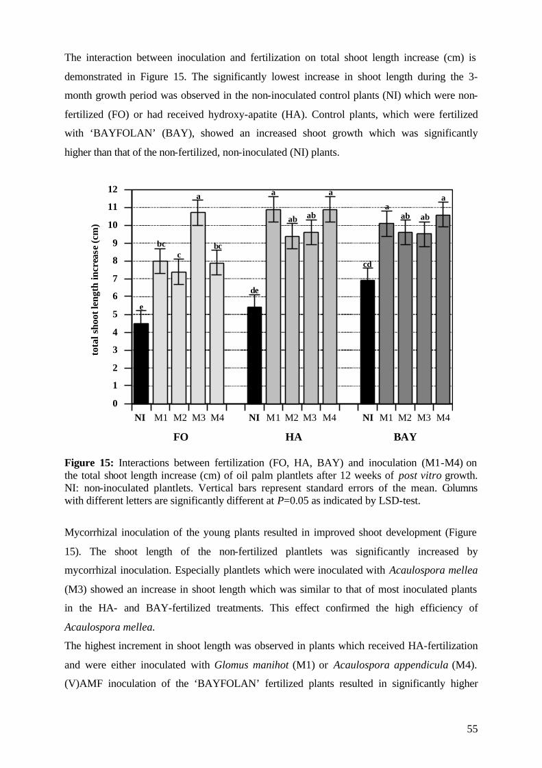
55
The interaction between inoculation and fertilization on total shoot length increase (cm) is
demonstrated in Figure 15. The significantly lowest increase in shoot length during the 3-
month growth period was observed in the non-inoculated control plants (NI) which were non-
fertilized (FO) or had received hydroxy-apatite (HA). Control plants, which were fertilized
with ‘BAYFOLAN’ (BAY), showed an increased shoot growth which was significantly
higher than that of the non-fertilized, non-inoculated (NI) plants.
0
1
2
3
4
5
6
7
8
9
10
11
12
NI M1 M2 M3 M4 NI M1 M2 M3 M4 NI M1 M2 M3 M4
tota
lsho
otle
ngth
incr
ease
(cm
)
FO HA BAY
e
bcc
a
bc
de
a
ab ab
a
cd
aab ab
a
Figure 15: Interactions between fertilization (FO, HA, BAY) and inoculation (M1-M4) on the total shoot length increase (cm) of oil palm plantlets after 12 weeks of post vitro growth. NI: non-inoculated plantlets. Vertical bars represent standard errors of the mean. Columns with different letters are significantly different at P=0.05 as indicated by LSD-test.
Mycorrhizal inoculation of the young plants resulted in improved shoot development (Figure
15). The shoot length of the non-fertilized plantlets was significantly increased by
mycorrhizal inoculation. Especially plantlets which were inoculated with Acaulospora mellea
(M3) showed an increase in shoot length which was similar to that of most inoculated plants
in the HA- and BAY-fertilized treatments. This effect confirmed the high efficiency of
Acaulospora mellea.
The highest increment in shoot length was observed in plants which received HA-fertilization
and were either inoculated with Glomus manihot (M1) or Acaulospora appendicula (M4).
(V)AMF inoculation of the ‘BAYFOLAN’ fertilized plants resulted in significantly higher
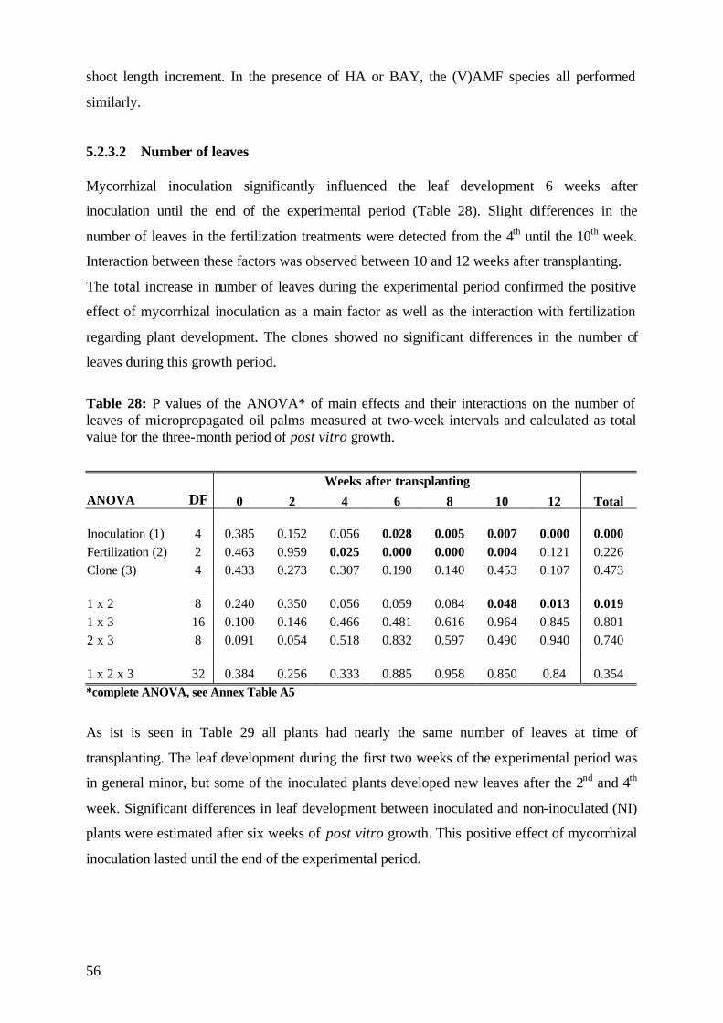
56
shoot length increment. In the presence of HA or BAY, the (V)AMF species all performed
similarly.
5.2.3.2 Number of leaves Mycorrhizal inoculation significantly influenced the leaf development 6 weeks after
inoculation until the end of the experimental period (Table 28). Slight differences in the
number of leaves in the fertilization treatments were detected from the 4th until the 10th week.
Interaction between these factors was observed between 10 and 12 weeks after transplanting.
The total increase in number of leaves during the experimental period confirmed the positive
effect of mycorrhizal inoculation as a main factor as well as the interaction with fertilization
regarding plant development. The clones showed no significant differences in the number of
leaves during this growth period.
Table 28: P values of the ANOVA* of main effects and their interactions on the number of leaves of micropropagated oil palms measured at two-week intervals and calculated as total value for the three-month period of post vitro growth.
Weeks after transplanting ANOVA DF 0 2 4 6 8 10 12 Total Inoculation (1) 4 0.385 0.152 0.056 0.028 0.005 0.007 0.000 0.000 Fertilization (2) 2 0.463 0.959 0.025 0.000 0.000 0.004 0.121 0.226 Clone (3) 4 0.433 0.273 0.307 0.190 0.140 0.453 0.107 0.473 1 x 2 8 0.240 0.350 0.056 0.059 0.084 0.048 0.013 0.019 1 x 3 16 0.100 0.146 0.466 0.481 0.616 0.964 0.845 0.801 2 x 3 8 0.091 0.054 0.518 0.832 0.597 0.490 0.940 0.740 1 x 2 x 3 32 0.384 0.256 0.333 0.885 0.958 0.850 0.84 0.354 *complete ANOVA, see Annex Table A5
As ist is seen in Table 29 all plants had nearly the same number of leaves at time of
transplanting. The leaf development during the first two weeks of the experimental period was
in general minor, but some of the inoculated plants developed new leaves after the 2nd and 4th
week. Significant differences in leaf development between inoculated and non-inoculated (NI)
plants were estimated after six weeks of post vitro growth. This positive effect of mycorrhizal
inoculation lasted until the end of the experimental period.
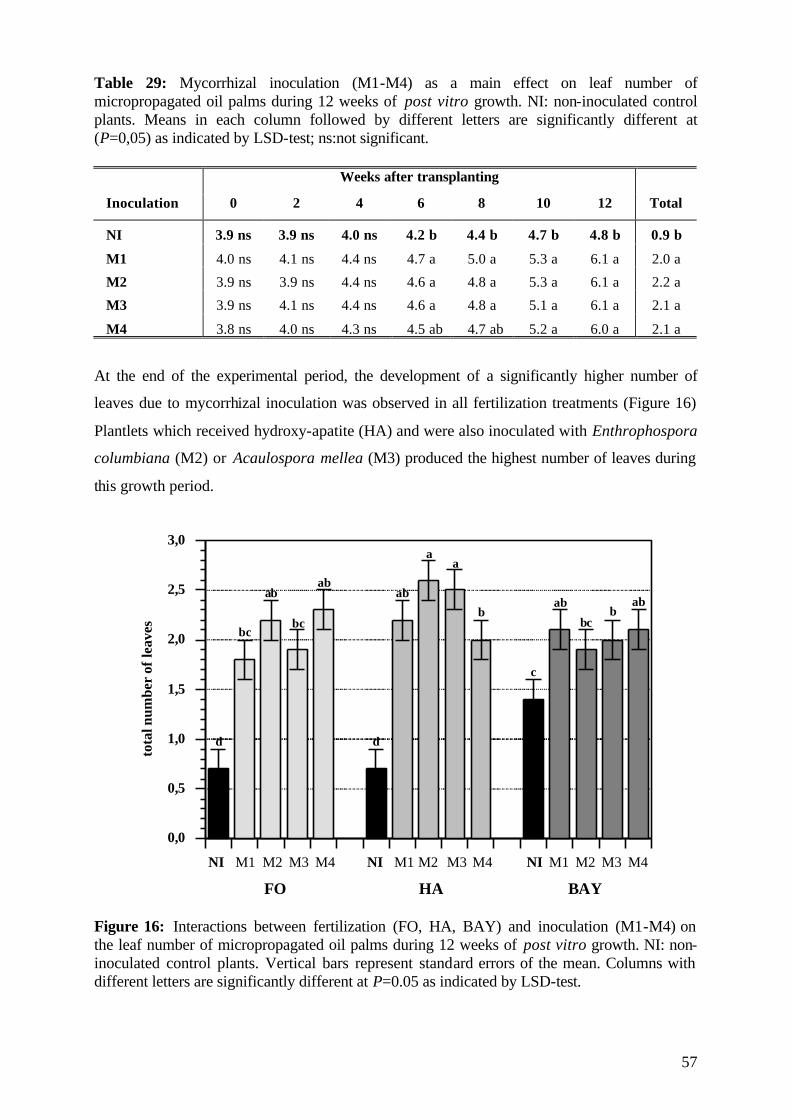
57
Table 29: Mycorrhizal inoculation (M1-M4) as a main effect on leaf number of micropropagated oil palms during 12 weeks of post vitro growth. NI: non-inoculated control plants. Means in each column followed by different letters are significantly different at (P=0,05) as indicated by LSD-test; ns:not significant.
Weeks after transplanting
Inoculation 0 2 4 6 8 10 12 Total
NI 3.9 ns 3.9 ns 4.0 ns 4.2 b 4.4 b 4.7 b 4.8 b 0.9 b
M1 4.0 ns 4.1 ns 4.4 ns 4.7 a 5.0 a 5.3 a 6.1 a 2.0 a
M2 3.9 ns 3.9 ns 4.4 ns 4.6 a 4.8 a 5.3 a 6.1 a 2.2 a
M3 3.9 ns 4.1 ns 4.4 ns 4.6 a 4.8 a 5.1 a 6.1 a 2.1 a
M4 3.8 ns 4.0 ns 4.3 ns 4.5 ab 4.7 ab 5.2 a 6.0 a 2.1 a
At the end of the experimental period, the development of a significantly higher number of
leaves due to mycorrhizal inoculation was observed in all fertilization treatments (Figure 16)
Plantlets which received hydroxy-apatite (HA) and were also inoculated with Enthrophospora
columbiana (M2) or Acaulospora mellea (M3) produced the highest number of leaves during
this growth period.
0,0
0,5
1,0
1,5
2,0
2,5
3,0
NI M1 M2 M3 M4 NI M1 M2 M3 M4 NI M1 M2 M3 M4
tota
lnum
ber
ofle
aves
d
bc
ab
bc
ab
d
c
ab
aa
bab
bcb
ab
FO HA BAY Figure 16: Interactions between fertilization (FO, HA, BAY) and inoculation (M1-M4) on the leaf number of micropropagated oil palms during 12 weeks of post vitro growth. NI: non-inoculated control plants. Vertical bars represent standard errors of the mean. Columns with different letters are significantly different at P=0.05 as indicated by LSD-test.
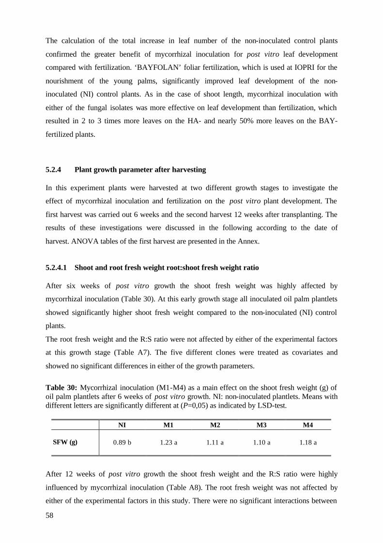
58
The calculation of the total increase in leaf number of the non-inoculated control plants
confirmed the greater benefit of mycorrhizal inoculation for post vitro leaf development
compared with fertilization. ‘BAYFOLAN’ foliar fertilization, which is used at IOPRI for the
nourishment of the young palms, significantly improved leaf development of the non-
inoculated (NI) control plants. As in the case of shoot length, mycorrhizal inoculation with
either of the fungal isolates was more effective on leaf development than fertilization, which
resulted in 2 to 3 times more leaves on the HA- and nearly 50% more leaves on the BAY-
fertilized plants.
5.2.4 Plant growth parameter after harvesting In this experiment plants were harvested at two different growth stages to investigate the
effect of mycorrhizal inoculation and fertilization on the post vitro plant development. The
first harvest was carried out 6 weeks and the second harvest 12 weeks after transplanting. The
results of these investigations were discussed in the following according to the date of
harvest. ANOVA tables of the first harvest are presented in the Annex.
5.2.4.1 Shoot and root fresh weight root:shoot fresh weight ratio After six weeks of post vitro growth the shoot fresh weight was highly affected by
mycorrhizal inoculation (Table 30). At this early growth stage all inoculated oil palm plantlets
showed significantly higher shoot fresh weight compared to the non-inoculated (NI) control
plants.
The root fresh weight and the R:S ratio were not affected by either of the experimental factors
at this growth stage (Table A7). The five different clones were treated as covariates and
showed no significant differences in either of the growth parameters.
Table 30: Mycorrhizal inoculation (M1-M4) as a main effect on the shoot fresh weight (g) of oil palm plantlets after 6 weeks of post vitro growth. NI: non-inoculated plantlets. Means with different letters are significantly different at (P=0,05) as indicated by LSD-test.
NI M1 M2 M3 M4 SFW (g) 0.89 b 1.23 a 1.11 a 1.10 a 1.18 a
After 12 weeks of post vitro growth the shoot fresh weight and the R:S ratio were highly
influenced by mycorrhizal inoculation (Table A8). The root fresh weight was not affected by
either of the experimental factors in this study. There were no significant interactions between
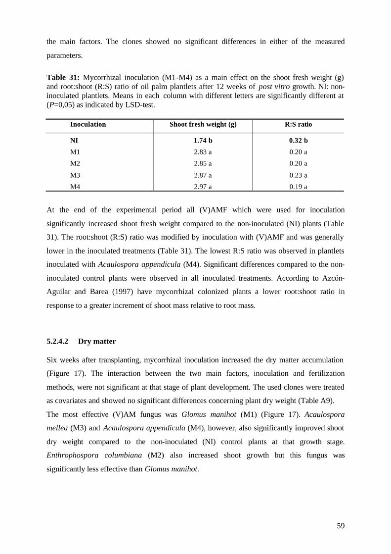
59
the main factors. The clones showed no significant differences in either of the measured
parameters.
Table 31: Mycorrhizal inoculation (M1-M4) as a main effect on the shoot fresh weight (g) and root:shoot (R:S) ratio of oil palm plantlets after 12 weeks of post vitro growth. NI: non-inoculated plantlets. Means in each column with different letters are significantly different at (P=0,05) as indicated by LSD-test.
Inoculation Shoot fresh weight (g) R:S ratio
NI 1.74 b 0.32 b
M1 2.83 a 0.20 a
M2 2.85 a 0.20 a
M3 2.87 a 0.23 a
M4 2.97 a 0.19 a
At the end of the experimental period all (V)AMF which were used for inoculation
significantly increased shoot fresh weight compared to the non-inoculated (NI) plants (Table
31). The root:shoot (R:S) ratio was modified by inoculation with (V)AMF and was generally
lower in the inoculated treatments (Table 31). The lowest R:S ratio was observed in plantlets
inoculated with Acaulospora appendicula (M4). Significant differences compared to the non-
inoculated control plants were observed in all inoculated treatments. According to Azcón-
Aguilar and Barea (1997) have mycorrhizal colonized plants a lower root:shoot ratio in
response to a greater increment of shoot mass relative to root mass.
5.2.4.2 Dry matter Six weeks after transplanting, mycorrhizal inoculation increased the dry matter accumulation
(Figure 17). The interaction between the two main factors, inoculation and fertilization
methods, were not significant at that stage of plant development. The used clones were treated
as covariates and showed no significant differences concerning plant dry weight (Table A9).
The most effective (V)AM fungus was Glomus manihot (M1) (Figure 17). Acaulospora
mellea (M3) and Acaulospora appendicula (M4), however, also significantly improved shoot
dry weight compared to the non-inoculated (NI) control plants at that growth stage.
Enthrophospora columbiana (M2) also increased shoot growth but this fungus was
significantly less effective than Glomus manihot.
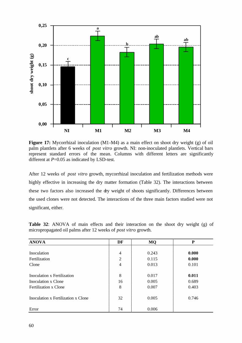
60
0,00
0,05
0,10
0,15
0,20
0,25
NI M1 M2 M3 M4
shoo
tdr
yw
eigh
t(g
)a
b
c
abab
Figure 17: Mycorrhizal inoculation (M1-M4) as a main effect on shoot dry weight (g) of oil palm plantlets after 6 weeks of post vitro growth. NI: non-inoculated plantlets. Vertical bars represent standard errors of the mean. Columns with different letters are significantly different at P=0.05 as indicated by LSD-test.
After 12 weeks of post vitro growth, mycorrhizal inoculation and fertilization methods were
highly effective in increasing the dry matter formation (Table 32). The interactions between
these two factors also increased the dry weight of shoots significantly. Differences between
the used clones were not detected. The interactions of the three main factors studied were not
significant, either.
Table 32: ANOVA of main effects and their interaction on the shoot dry weight (g) of micropropagated oil palms after 12 weeks of post vitro growth. ANOVA DF MQ P Inoculation 4 0.243 0.000 Fertilization 2 0.115 0.000 Clone 4 0.013 0.101 Inoculation x Fertilization 8 0.017 0.011 Inoculation x Clone 16 0.005 0.689 Fertilization x Clone 8 0.007 0.403 Inoculation x Fertilization x Clone 32 0.005 0.746 Error 74 0.006
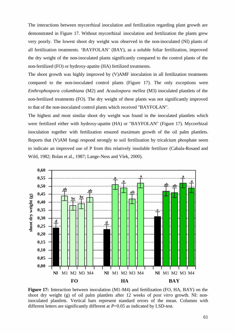
61
The interactions between mycorrhizal inoculation and fertilization regarding plant growth are
demonstrated in Figure 17. Without mycorrhizal inoculation and fertilization the plants grew
very poorly. The lowest shoot dry weight was observed in the non-inoculated (NI) plants of
all fertilization treatments. ‘BAYFOLAN’ (BAY), as a soluble foliar fertilization, improved
the dry weight of the non-inoculated plants significantly compared to the control plants of the
non-fertilized (FO) or hydroxy-apatite (HA) fertilized treatments.
The shoot growth was highly improved by (V)AMF inoculation in all fertilization treatments
compared to the non-inoculated control plants (Figure 17). The only exceptions were
Enthrophospora columbiana (M2) and Acaulospora mellea (M3) inoculated plantlets of the
non-fertilized treatments (FO). The dry weight of these plants was not significantly improved
to that of the non-inoculated control plants which received "BAYFOLAN".
The highest and most similar shoot dry weight was found in the inoculated plantlets which
were fertilized either with hydroxy-apatite (HA) or "BAYFOLAN" (Figure 17). Mycorrhizal
inoculation together with fertilization ensured maximum growth of the oil palm plantlets.
Reports that (V)AM fungi respond strongly to soil fertilization by tricalcium phosphate seem
to indicate an improved use of P from this relatively insoluble fertilizer (Cabala-Rosand and
Wild, 1982; Bolan et al., 1987; Lange-Ness and Vlek, 2000).
0,00
0,05
0,10
0,15
0,20
0,25
0,30
0,35
0,40
0,45
0,50
0,55
0,60
NI M1 M2 M3 M4 NI M1 M2 M3 M4 NI M1 M2 M3 M4
FO HA BAY
shoo
tdry
wei
ght(
g)
d d
c
ab
bc bc
ab
aa
ab
a
ab ab
aa
Figure 17: Interaction between inoculation (M1-M4) and fertilization (FO, HA, BAY) on the shoot dry weight (g) of oil palm plantlets after 12 weeks of post vitro growth. NI: non-inoculated plantlets. Vertical bars represent standard errors of the mean. Columns with different letters are significantly different at P=0.05 as indicated by LSD-test.
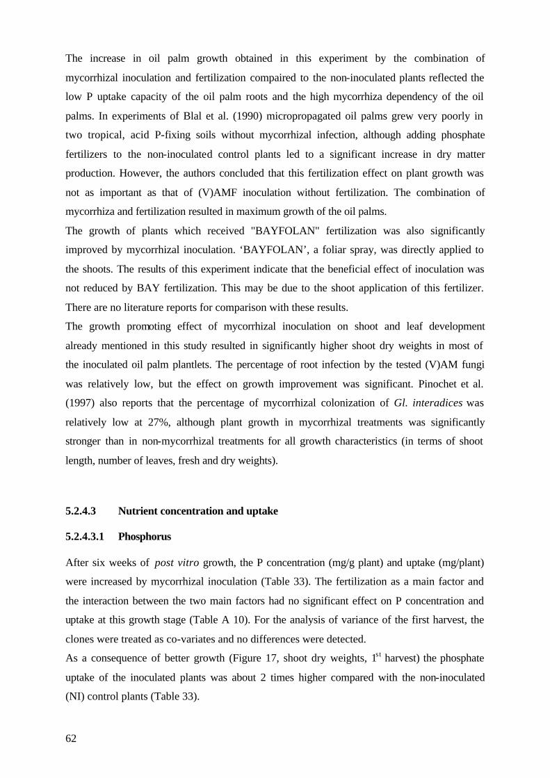
62
The increase in oil palm growth obtained in this experiment by the combination of
mycorrhizal inoculation and fertilization compaired to the non-inoculated plants reflected the
low P uptake capacity of the oil palm roots and the high mycorrhiza dependency of the oil
palms. In experiments of Blal et al. (1990) micropropagated oil palms grew very poorly in
two tropical, acid P-fixing soils without mycorrhizal infection, although adding phosphate
fertilizers to the non-inoculated control plants led to a significant increase in dry matter
production. However, the authors concluded that this fertilization effect on plant growth was
not as important as that of (V)AMF inoculation without fertilization. The combination of
mycorrhiza and fertilization resulted in maximum growth of the oil palms.
The growth of plants which received "BAYFOLAN" fertilization was also significantly
improved by mycorrhizal inoculation. ‘BAYFOLAN’, a foliar spray, was directly applied to
the shoots. The results of this experiment indicate that the beneficial effect of inoculation was
not reduced by BAY fertilization. This may be due to the shoot application of this fertilizer.
There are no literature reports for comparison with these results.
The growth promoting effect of mycorrhizal inoculation on shoot and leaf development
already mentioned in this study resulted in significantly higher shoot dry weights in most of
the inoculated oil palm plantlets. The percentage of root infection by the tested (V)AM fungi
was relatively low, but the effect on growth improvement was significant. Pinochet et al.
(1997) also reports that the percentage of mycorrhizal colonization of Gl. interadices was
relatively low at 27%, although plant growth in mycorrhizal treatments was significantly
stronger than in non-mycorrhizal treatments for all growth characteristics (in terms of shoot
length, number of leaves, fresh and dry weights).
5.2.4.3 Nutrient concentration and uptake 5.2.4.3.1 Phosphorus After six weeks of post vitro growth, the P concentration (mg/g plant) and uptake (mg/plant)
were increased by mycorrhizal inoculation (Table 33). The fertilization as a main factor and
the interaction between the two main factors had no significant effect on P concentration and
uptake at this growth stage (Table A 10). For the analysis of variance of the first harvest, the
clones were treated as co-variates and no differences were detected.
As a consequence of better growth (Figure 17, shoot dry weights, 1st harvest) the phosphate
uptake of the inoculated plants was about 2 times higher compared with the non-inoculated
(NI) control plants (Table 33).
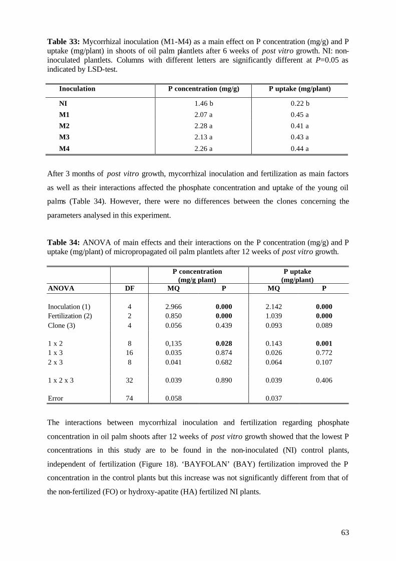
63
Table 33: Mycorrhizal inoculation (M1-M4) as a main effect on P concentration (mg/g) and P uptake (mg/plant) in shoots of oil palm plantlets after 6 weeks of post vitro growth. NI: non-inoculated plantlets. Columns with different letters are significantly different at P=0.05 as indicated by LSD-test.
Inoculation P concentration (mg/g) P uptake (mg/plant)
NI 1.46 b 0.22 b
M1 2.07 a 0.45 a
M2 2.28 a 0.41 a
M3 2.13 a 0.43 a
M4 2.26 a 0.44 a
After 3 months of post vitro growth, mycorrhizal inoculation and fertilization as main factors
as well as their interactions affected the phosphate concentration and uptake of the young oil
palms (Table 34). However, there were no differences between the clones concerning the
parameters analysed in this experiment.
Table 34: ANOVA of main effects and their interactions on the P concentration (mg/g) and P uptake (mg/plant) of micropropagated oil palm plantlets after 12 weeks of post vitro growth.
P concentration (mg/g plant)
P uptake (mg/plant)
ANOVA DF MQ P MQ P Inoculation (1) 4 2.966 0.000 2.142 0.000 Fertilization (2) 2 0.850 0.000 1.039 0.000 Clone (3) 4 0.056 0.439 0.093 0.089 1 x 2 8 0,135 0.028 0.143 0.001 1 x 3 16 0.035 0.874 0.026 0.772 2 x 3 8 0.041 0.682 0.064 0.107 1 x 2 x 3 32 0.039 0.890 0.039 0.406 Error 74 0.058 0.037
The interactions between mycorrhizal inoculation and fertilization regarding phosphate
concentration in oil palm shoots after 12 weeks of post vitro growth showed that the lowest P
concentrations in this study are to be found in the non-inoculated (NI) control plants,
independent of fertilization (Figure 18). ‘BAYFOLAN’ (BAY) fertilization improved the P
concentration in the control plants but this increase was not significantly different from that of
the non-fertilized (FO) or hydroxy-apatite (HA) fertilized NI plants.
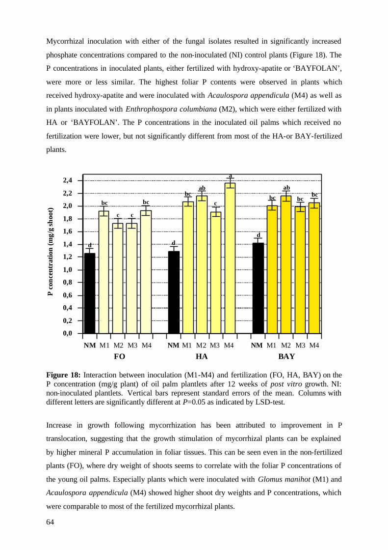
64
Mycorrhizal inoculation with either of the fungal isolates resulted in significantly increased
phosphate concentrations compared to the non-inoculated (NI) control plants (Figure 18). The
P concentrations in inoculated plants, either fertilized with hydroxy-apatite or ‘BAYFOLAN’,
were more or less similar. The highest foliar P contents were observed in plants which
received hydroxy-apatite and were inoculated with Acaulospora appendicula (M4) as well as
in plants inoculated with Enthrophospora columbiana (M2), which were either fertilized with
HA or ‘BAYFOLAN’. The P concentrations in the inoculated oil palms which received no
fertilization were lower, but not significantly different from most of the HA-or BAY-fertilized
plants.
0,0
0,2
0,4
0,6
0,8
1,0
1,2
1,4
1,6
1,8
2,0
2,2
2,4
NM M1 M2 M3 M4 NM M1 M2 M3 M4 NM M1 M2 M3 M4
Pco
ncen
trat
ion
(mg/
gsh
oot)
FO HA BAY
d
bc
c c
bc
d
bcab
c
a
d
bc
ab
bcbc
Figure 18: Interaction between inoculation (M1-M4) and fertilization (FO, HA, BAY) on the P concentration (mg/g plant) of oil palm plantlets after 12 weeks of post vitro growth. NI: non-inoculated plantlets. Vertical bars represent standard errors of the mean. Columns with different letters are significantly different at P=0.05 as indicated by LSD-test.
Increase in growth following mycorrhization has been attributed to improvement in P
translocation, suggesting that the growth stimulation of mycorrhizal plants can be explained
by higher mineral P accumulation in foliar tissues. This can be seen even in the non-fertilized
plants (FO), where dry weight of shoots seems to correlate with the foliar P concentrations of
the young oil palms. Especially plants which were inoculated with Glomus manihot (M1) and
Acaulospora appendicula (M4) showed higher shoot dry weights and P concentrations, which
were comparable to most of the fertilized mycorrhizal plants.
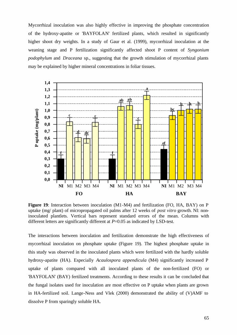
65
Mycorrhizal inoculation was also highly effective in improving the phosphate concentration
of the hydroxy-apatite or 'BAYFOLAN' fertilized plants, which resulted in significantly
higher shoot dry weights. In a study of Gaur et al. (1999), mycorrhizal inoculation at the
weaning stage and P fertilization significantly affected shoot P content of Syngonium
podophylum and Draceana sp., suggesting that the growth stimulation of mycorrhizal plants
may be explained by higher mineral concentrations in foliar tissues.
0,0
0,1
0,2
0,30,4
0,5
0,6
0,7
0,8
0,9
1,0
1,1
1,2
1,3
1,4
NI M1 M2 M3 M4 NI M1 M2 M3 M4 NI M1 M2 M3 M4
Pup
take
(mg/
plan
t)
FO HA BAY
f
c
d de
c
f
ab ab
c
a
ef
bcb b b
Figure 19: Interaction between inoculation (M1-M4) and fertilization (FO, HA, BAY) on P uptake (mg/ plant) of micropropagated oil palms after 12 weeks of post vitro growth. NI: non-inoculated plantlets. Vertical bars represent standard errors of the mean. Columns with different letters are significantly different at P=0.05 as indicated by LSD-test.
The interactions between inoculation and fertilization demonstrate the high effectiveness of
mycorrhizal inoculation on phosphate uptake (Figure 19). The highest phosphate uptake in
this study was observed in the inoculated plants which were fertilized with the hardly soluble
hydroxy-apatite (HA). Especially Acaulospora appendicula (M4) significantly increased P
uptake of plants compared with all inoculated plants of the non-fertilized (FO) or
'BAYFOLAN' (BAY) fertilized treatments. According to these results it can be concluded that
the fungal isolates used for inoculation are most effective on P uptake when plants are grown
in HA-fertilized soil. Lange-Ness and Vlek (2000) demonstrated the ability of (V)AMF to
dissolve P from sparingly soluble HA.
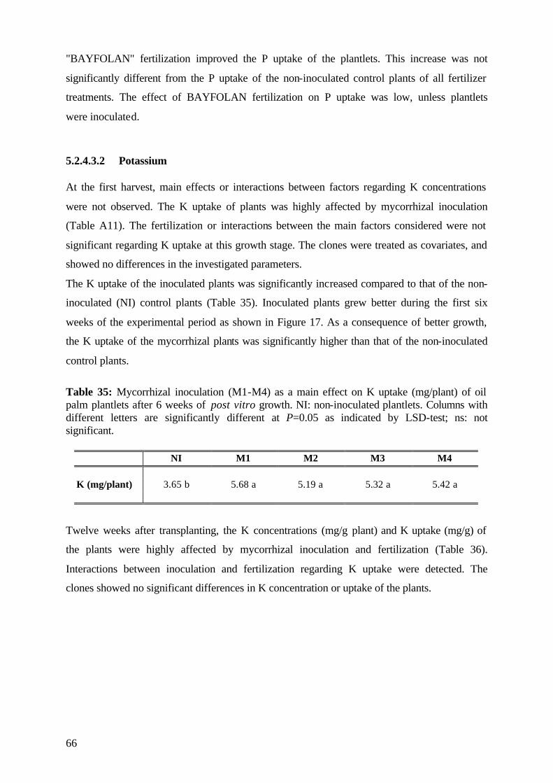
66
"BAYFOLAN" fertilization improved the P uptake of the plantlets. This increase was not
significantly different from the P uptake of the non-inoculated control plants of all fertilizer
treatments. The effect of BAYFOLAN fertilization on P uptake was low, unless plantlets
were inoculated.
5.2.4.3.2 Potassium At the first harvest, main effects or interactions between factors regarding K concentrations
were not observed. The K uptake of plants was highly affected by mycorrhizal inoculation
(Table A11). The fertilization or interactions between the main factors considered were not
significant regarding K uptake at this growth stage. The clones were treated as covariates, and
showed no differences in the investigated parameters.
The K uptake of the inoculated plants was significantly increased compared to that of the non-
inoculated (NI) control plants (Table 35). Inoculated plants grew better during the first six
weeks of the experimental period as shown in Figure 17. As a consequence of better growth,
the K uptake of the mycorrhizal plants was significantly higher than that of the non-inoculated
control plants.
Table 35: Mycorrhizal inoculation (M1-M4) as a main effect on K uptake (mg/plant) of oil palm plantlets after 6 weeks of post vitro growth. NI: non-inoculated plantlets. Columns with different letters are significantly different at P=0.05 as indicated by LSD-test; ns: not significant.
NI M1 M2 M3 M4 K (mg/plant) 3.65 b 5.68 a 5.19 a 5.32 a 5.42 a
Twelve weeks after transplanting, the K concentrations (mg/g plant) and K uptake (mg/g) of
the plants were highly affected by mycorrhizal inoculation and fertilization (Table 36).
Interactions between inoculation and fertilization regarding K uptake were detected. The
clones showed no significant differences in K concentration or uptake of the plants.
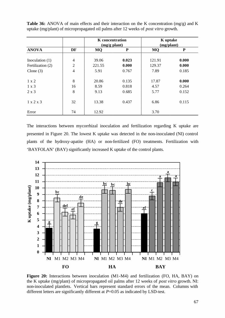
67
Table 36: ANOVA of main effects and their interaction on the K concentration (mg/g) and K uptake (mg/plant) of micropropagated oil palms after 12 weeks of post vitro growth.
K concentration (mg/g plant)
K uptake (mg/plant)
ANOVA DF MQ P MQ P Inoculation (1) 4 39.06 0.023 121.91 0.000 Fertilization (2) 2 221.55 0.000 129.37 0.000 Clone (3) 4 5.91 0.767 7.89 0.185 1 x 2 8 20.86 0.135 17.87 0.000 1 x 3 16 8.59 0.818 4.57 0.264 2 x 3 8 9.13 0.685 5.77 0.152 1 x 2 x 3 32 13.38 0.437 6.86 0.115 Error 74 12.92 3.70
The interactions between mycorrhizal inoculation and fertilization regarding K uptake are
presented in Figure 20. The lowest K uptake was detected in the non-inoculated (NI) control
plants of the hydroxy-apatite (HA) or non-fertilized (FO) treatments. Fertilization with
‘BAYFOLAN’ (BAY) significantly increased K uptake of the control plants.
01
234
5678
91011
121314
NI M1 M2 M3 M4 NI M1 M2 M3 M4 NI M1 M2 M3 M4
Kup
take
(mg/
plan
t)
FO HA BAY
g
bc
defef
de
g
bc bc
de
bc
ef
c
aa
a
Figure 20: Interactions between inoculation (M1-M4) and fertilization (FO, HA, BAY) on the K uptake (mg/plant) of micropropagated oil palms after 12 weeks of post vitro growth. NI: non-inoculated plantlets. Vertical bars represent standard errors of the mean. Columns with different letters are significantly different at P=0.05 as indicated by LSD-test.
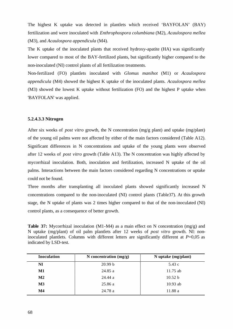
68
The highest K uptake was detected in plantlets which received ‘BAYFOLAN’ (BAY)
fertilization and were inoculated with Enthrophospora columbiana (M2), Acaulospora mellea
(M3), and Acaulospora appendicula (M4).
The K uptake of the inoculated plants that received hydroxy-apatite (HA) was significantly
lower compared to most of the BAY-fertilized plants, but significantly higher compared to the
non-inoculated (NI) control plants of all fertilization treatments.
Non-fertilized (FO) plantlets inoculated with Glomus manihot (M1) or Acaulospora
appendicula (M4) showed the highest K uptake of the inoculated plants. Acaulospora mellea
(M3) showed the lowest K uptake without fertilization (FO) and the highest P uptake when
'BAYFOLAN' was applied.
5.2.4.3.3 Nitrogen After six weeks of post vitro growth, the N concentration (mg/g plant) and uptake (mg/plant)
of the young oil palms were not affected by either of the main factors considered (Table A12).
Significant differences in N concentrations and uptake of the young plants were observed
after 12 weeks of post vitro growth (Table A13). The N concentration was highly affected by
mycorrhizal inoculation. Both, inoculation and fertilization, increased N uptake of the oil
palms. Interactions between the main factors considered regarding N concentrations or uptake
could not be found.
Three months after transplanting all inoculated plants showed significantly increased N
concentrations compared to the non-inoculated (NI) control plants (Table37). At this growth
stage, the N uptake of plants was 2 times higher compared to that of the non-inoculated (NI)
control plants, as a consequence of better growth.
Table 37: Mycorrhizal inoculation (M1-M4) as a main effect on N concentration (mg/g) and N uptake (mg/plant) of oil palm plantlets after 12 weeks of post vitro growth. NI: non-inoculated plantlets. Columns with different letters are significantly different at P=0,05 as indicated by LSD-test.
Inoculation N concentration (mg/g) N uptake (mg/plant)
NI 20.99 b 5.43 c
M1 24.85 a 11.75 ab
M2 24.44 a 10.52 b
M3 25.86 a 10.93 ab
M4 24.78 a 11.88 a
69
The N uptake of ‘BAYFOLAN’ fertilized plants was significantly higher than that of the HA
or non-fertilized (PO) plants at this growth stage (Table 38). A significantly higher uptake of
N was also detected in plants which received hydroxy-apatite compared to the non-fertilized
plants, as a consequence of a better growth of the BAY- and HA-fertilized plants mainly due
to a better nourishment.
Table 38: Fertilization (FO, HA, BAY) as a main effect on N uptake (mg/plant) of oil palm plantlets after 12 weeks of post vitro growth. Columns with different letters are significantly different at P=0,05 as indicated by LSD-test; ns: not significant. FO HA BAY
N (mg/plant) 8.59 c 10.24 b 11.46 a
5.2.5 (V)AMF infection The percentage of (V)AMF root colonization after six weeks of post vitro growth was greatly
increased by mycorrhizal inoculation (Figure 20). Main effects of fertilization or interaction
between the two main factors considered were not observed (Table A14). The clones, which
were analysed as a co-variate, showed no differences in their (V)AMF infection rate.
Acaulospora mellea (M3) inoculated plants showed the highest infection rate (23%), followed
by Acaulospora appendicula (M4) (18%) and Glomus manihot (M1) (17%) inoculated plants
(Figure 20). The lowest infection (10%) of the inoculated plants was formed by
Enthrophospora columbiana (M2). Differences in the ability of (V)AMF to colonize the root
system are well known and may have played a part in the lower percentage of root
colonization observed in E. columbiana (M2) inoculated plants six weeks after inoculation.
Mycorrhizal infection in the non-inoculated (NI) control plants, due to the soil indigenous
mycorrhizal fungi of the non-sterilized soil, was not observed at that growth stage.
These results showed that the oil palms were infected early, during the first six weeks after
inoculation, by the used (V)AMF. Despite the relatively low percentage of (V)AMF infection,
these colonisations were highly effective in promoting P concentrations (Table 33) of the
inoculated clones, which resulted in improved plant development (Figure 17). Differences in
the ability of (V)AMF to colonize the root system are well known and may have played a part
in the lower percentage of colonization observed after the first harvest in roots of plants
inoculated with E. columbiana (M2).
70
-5
0
5
10
15
20
25
30
NI M1 M2 M3 M4
(V)A
MF
-infe
ctio
n(%
)
c
ab
bc
a
ab
Figure 20: Mycorrhizal inoculation (M1-M4) as a main effect on the (V)AMF-infection rate (%) of micropropagated oil palms after 6 weeks of post vitro growth. NI: non-inoculated plantlets. Vertical bars represent standard errors of the mean. Columns with different letters are significantly different at P=0,05 as indicated by LSD-test.
The results confirm the beneficial effect of mycorrhizal inoculation with effective fungal
isolates at the early post vitro stage of the micropropagated oil palms. Little is known about
the early stages of (V)AM infection in plants. It has been demonstrated by Ravolanirina et al.
(1989) and Gianinazzi et al. (1990) that the early root colonization is very important during
the delicate acclimatization phase .
Table 39: ANOVA of main effects and their interaction on the (V)AMF infection rate (%) of micropropagated oil palms after 12 weeks of post vitro growth. ANOVA DF MQ P Inoculation 4 1914.06 0.007 Fertilization 2 260.63 0.592 Clone 4 1449.17 0.126 Inoculation x Fertilization 8 290.23 0.786 Inoculation x Clone 16 323.82 0.828 Fertilization x Clone 8 310.99 0.751 Inoculation x Fertilization x Clone 32 298.59 0.943 Error 75 497.43
71
After twelve weeks of post vitro growth, the percentage of (V)AMF root colonization was
greatly enhanced by mycorrhizal inoculation (Figure 21). There was no significant effect of
fertilization and its interaction with inoculation (Table 39). The clones, which were analysed
as a co-variate, showed no differences in their (V)AMF infection rate.
The percentage of infection in roots of the inoculated plants varied between 28-32% and was
significantly higher than that of the non-inoculated (NI) plants (Figure 21). Compared to the
mycorrhizal colonization of plant roots at the first harvest, significant differences between
fungal isolates were not observed. In contrast to the first harvest, mycorrhizal fungi colonized
the non-inoculated plants after 12 weeks of post vitro growth (Figure 21). The percentage of
infection was low (12%), and was due to indigenous mycorrhizal fungi which were present in
the native, non-sterilized soil.
0
5
10
15
20
25
30
35
40
NI M1 M2 M3 M4
(V)A
MF
-infe
ctio
nra
te(%
)
b
a
a aa
Figure 21: Mycorrhizal inoculation (M1-M4) as a main effect on the (V)AMF-infection rate (%) of micropropagated oil palms after 12 weeks of post vitro growth. NI: non-inoculated plantlets. Vertical bars represent standard errors of the mean. Columns with different letters are significantly different at P=0,05 as indicated by LSD-test.
These results show that indigenous mycorrhizal fungi colonized the plant roots but that the
(V)AMF isolates used for inoculation were more effective and able to compete with the native
fungi. The used isolates infected the roots earlier and to a greater extent than the indigenous
fungi. According to Koomen et al. (1987) are indigenous fungi not always the most effective
ones as regards either mycorrhizal development or mycorrhizal growth response. In
experiments of Vosatka et al. (2000), it was shown that even inoculation in non-sterile field
72
soil could be of a great advantage for further development of mycorrhizal symbiosis despite
the presence of native mycorrhizal fungi in the field.
5.2.6 Conclusions Experiment 2 The objective of Experiment 2 was to compare the application of traditional plant nutrition
with "BAYFOLAN", which is practiced at the "International Oil Palm Research Institute"
(IOPRI), with the effect of mycorrhizal inoculation during the sensitive post vitro stage of
clonal oil palms. "BAYFOLAN", a foliar spray, is usually applied at IOPRI to supply the oil
palms with nutrients via the shoot, as the root system of the plantlets is weakly developed at
the time of transplanting. When "BAYFOLAN" fertilization was applied, the percentage of
plant survival obtained in this experiment was high (80%). This observation does not
correspond with the survival data of 60-70%, which were recorded by IOPRI during former
years. It seems that, under experimental conditions, it was possible to achieve a higher
efficiency with "BAYFOLAN" than under commercial conditions. The combination of foliar
fertilization and mycorrhizal inoculation resulted in an improved survival rate reaching 85-
100%, which was similar to that of hydroxy-apatite fertilized, inoculated plants.
The beneficial effect by the combination of fertilization and inoculation was also found for
the post vitro development of the sensitive plants. The growth of non-fertilized or hydroxy-
apatite fertilized plants was low and not much improved if plants received "BAYFOLAN".
Both, fertilization and (V)AMF inoculation, greatly increased dry matter accumulation of the
plants.
The increase in oil palm growth obtained by the addition of fertilizer and (V)AMF inoculation
reflects the low uptake capacity of oil palm roots and the high mycorrhiza dependency of the
plantlets in the initial growth phase. The highest P uptake was observed in the inoculated
plants which were fertilized with the hardly soluble hydroxy-apatite. According to these
results, it can be concluded that the fungal isolates used for inoculation were most effective on
P uptake when plants were grown in hydroxy-apatite fertilized soil.
Plantlets were grown in a growth substrate which was of low pH, a fact which frequently
occurs in most tropical soils. Under such conditions inoculation with the tested (V)AMF
isolates was an important factor in facilitating plant recovery of P from these fertilizer.
The response to "BAYFOLAN", a complete foliar nutrient containing macro- and
micronutrients, was expected to be higher. The use of the cheaper hydroxy-apatite as a P
73
fertilizer in combination with (V)AMF inoculation for improving the efficiency of the
symbiosis, offers an alternative to the more expensive "BAYFOLAN" or soluble P fertilizers.
Inoculated plantlets which were harvested after six weeks of post vitro growth showed that all
(V)AM fungal isolates significantly increased the foliar phosphate of the inoculated plants. At
that early growth stage, the P uptake of mycorrhizal plants was 2 times higher compared to
the non-inoculated control plants. A significantly increased nutrient uptake was only found
for phosphate, but not for potassium or nitrogen. These results indicate that phosphate is
probably the most limiting growth factor during the early post vitro stage of the oil palm
clones. Addition of fertilizers does not greatly improve the nutrient status of propagated plants
in their early growing stage because of their relatively small root systems and inability to take
up nutrients efficiently.
5.3 Experiment 3 Experiment 3 was conducted to examine the effects of mycorrhizal inoculation, protection
measurements and soil sterilization on the post vitro development of clonal oil palms. The
plantlets were inoculated with one of three (V)A mycorrhizal fungi which were selected for
their effectiveness on survival and plant development based on the results of the screening in
Experiment 1. These were Glomus manihot (M1), Enthrophospora columbiana (M2) and
Acaulospora mellea (M3). Plantlets were transferred either to sterilized or non-sterilized soil
and were cultivated under ‘normal’ pre-nursery conditions or with protection against
pathogenes by a plastic screen open to the top or covered by special plastic bags (‘sunbag’).
5.3.1 Survival rate
The survival rate of the oil palm plantlets in Experiment 3 was mainly influenced by
mycorrhizal inoculation and soil sterilization (Table 40). These two treatments as main effects
as well as interactions affected plant survival significantly during the three-month
experimental period. The interactions were not significant. The clones were analysed as
covariates and showed no significant differences regarding plant survival.
74
Table 40: ANOVA of main factors and their interactions on the survival rate (%) of micropropagated oil palms after three months of post vitro growth. ANOVA DF MQ P Inoculation 3 32500.00 0.000 Protection 2 291.67 0.823 Sterilization 1 24000.00 0.000 Inoculation x Protection 6 291.68 0.978 Inoculation x Sterilization 3 4111.11 0.044 Protection x Sterilization 2 375.00 0.779 Inoculation x Protection x Sterilization 6 152.78 0.996 Clone 1 20.83 0.906 Error 215 1497.58
The interactions between mycorrhizal inoculation and soil sterilization on plant survival are
demonstrated in Figure 22. The lowest survival rates (40%) were detected in the non-
inoculated (NI) control plants which grew either in sterilized or non-sterilized soil. Unless
accompanied by mycorrhizal inoculation the effect of soil sterilization on plant survival was
minor.
The survival rate of inoculated plantlets grown in non-sterilized soil was significantly
improved compared to the control plants. Glomus manihot (M1) was the most effective fungi,
increasing plant survival to 83%, followed by Acaulospora mellea (M3) with 73%. The
lowest plant survival rate (60%) was detected with Enthrophospora columbiana (M2).
The (V)AMF isolates showed a higher effectiveness when they were applied to sterilized soil.
Survival of the oil palm plantlets significantly increased to 97-100%. Significant differences
were specially seen in Enthrophospora columbiana (M2) and Acaulospora mellea (M3)
inoculated plantlets.
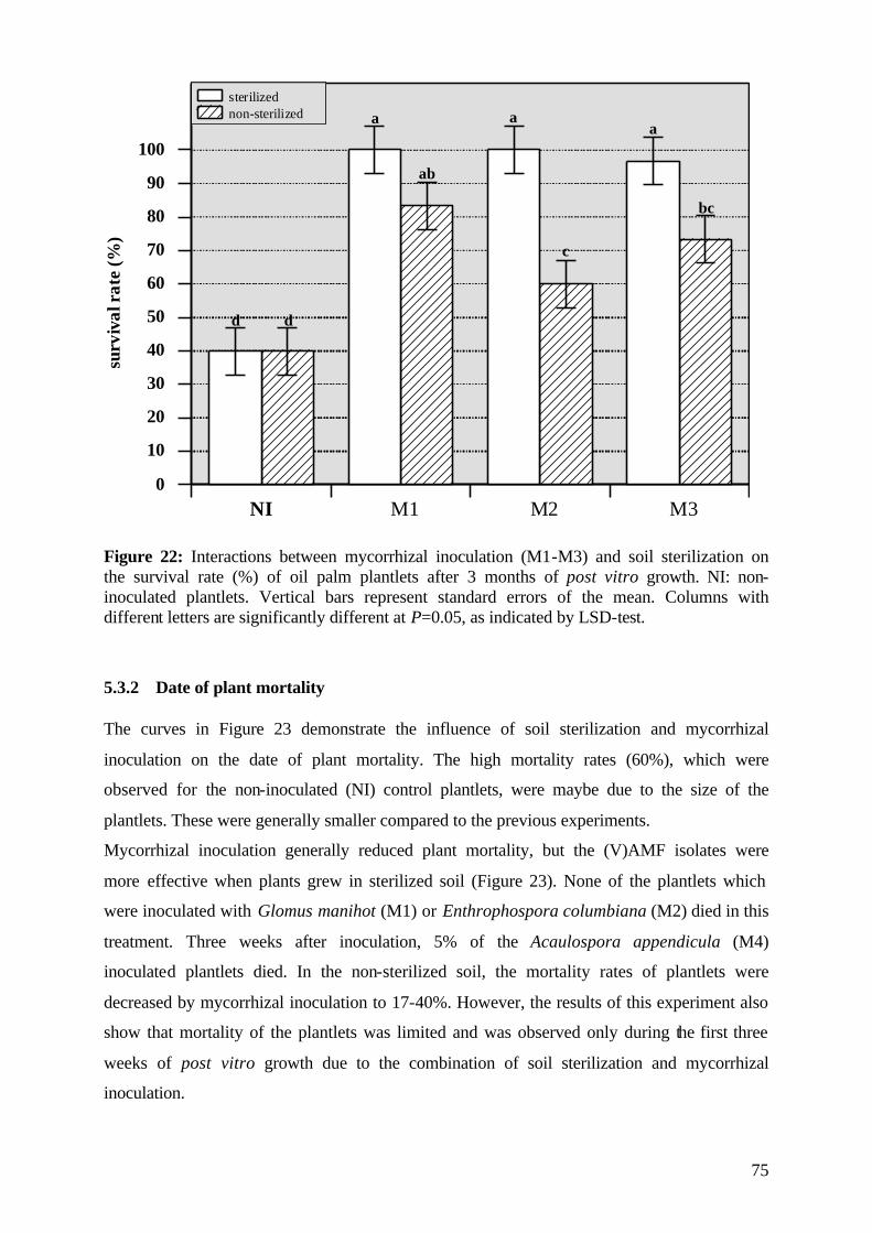
75
0
10
20
30
40
50
60
70
80
90
100
NI M1 M2 M3
sterilizednon-sterilized
surv
ival
rate
(%)
d d
a
ab
a
c
a
bc
Figure 22: Interactions between mycorrhizal inoculation (M1-M3) and soil sterilization on the survival rate (%) of oil palm plantlets after 3 months of post vitro growth. NI: non-inoculated plantlets. Vertical bars represent standard errors of the mean. Columns with different letters are significantly different at P=0.05, as indicated by LSD-test.
5.3.2 Date of plant mortality The curves in Figure 23 demonstrate the influence of soil sterilization and mycorrhizal
inoculation on the date of plant mortality. The high mortality rates (60%), which were
observed for the non-inoculated (NI) control plantlets, were maybe due to the size of the
plantlets. These were generally smaller compared to the previous experiments.
Mycorrhizal inoculation generally reduced plant mortality, but the (V)AMF isolates were
more effective when plants grew in sterilized soil (Figure 23). None of the plantlets which
were inoculated with Glomus manihot (M1) or Enthrophospora columbiana (M2) died in this
treatment. Three weeks after inoculation, 5% of the Acaulospora appendicula (M4)
inoculated plantlets died. In the non-sterilized soil, the mortality rates of plantlets were
decreased by mycorrhizal inoculation to 17-40%. However, the results of this experiment also
show that mortality of the plantlets was limited and was observed only during the first three
weeks of post vitro growth due to the combination of soil sterilization and mycorrhizal
inoculation.
76
0
10
20
30
40
50
60
NIM1M2M3
0
10
20
30
40
50
60
1 2 3 4 5 6 7 8 9 10 11 12
mor
talit
yra
te(%
)m
orta
lity
rate
(%)
weeks after inoculation
sterilized soil
non-sterilized soil
Figure 23: Effect of mycorrhizal inoculation (M1-M3) and soil sterilization on the mortality rate (%) of oil palm plantlets during 12 weeks of post vitro growth. NI: non inoculated plantlets.
5.3.3 Post vitro plant development
The effect of mycorrhizal inoculation, soil sterilization and protection measurements on the
post vitro development of oil palm plantlets was characterized by monitoring the shoot length
and number of leaves in two-weeks intervals during the experimental period. The most
important results are discussed as follows.
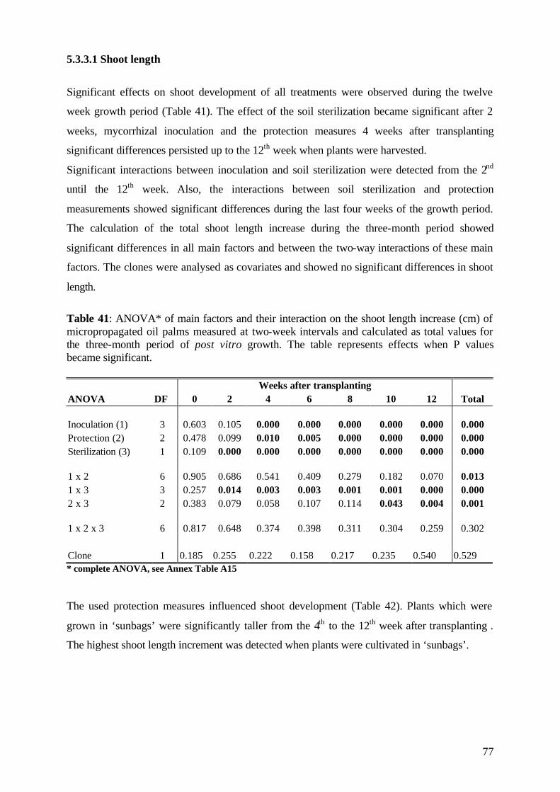
77
5.3.3.1 Shoot length
Significant effects on shoot development of all treatments were observed during the twelve
week growth period (Table 41). The effect of the soil sterilization became significant after 2
weeks, mycorrhizal inoculation and the protection measures 4 weeks after transplanting
significant differences persisted up to the 12th week when plants were harvested.
Significant interactions between inoculation and soil sterilization were detected from the 2nd
until the 12th week. Also, the interactions between soil sterilization and protection
measurements showed significant differences during the last four weeks of the growth period.
The calculation of the total shoot length increase during the three-month period showed
significant differences in all main factors and between the two-way interactions of these main
factors. The clones were analysed as covariates and showed no significant differences in shoot
length.
Table 41: ANOVA* of main factors and their interaction on the shoot length increase (cm) of micropropagated oil palms measured at two-week intervals and calculated as total values for the three-month period of post vitro growth. The table represents effects when P values became significant.
Weeks after transplanting ANOVA DF 0 2 4 6 8 10 12 Total Inoculation (1) 3 0.603 0.105 0.000 0.000 0.000 0.000 0.000 0.000 Protection (2) 2 0.478 0.099 0.010 0.005 0.000 0.000 0.000 0.000 Sterilization (3) 1 0.109 0.000 0.000 0.000 0.000 0.000 0.000 0.000 1 x 2 6 0.905 0.686 0.541 0.409 0.279 0.182 0.070 0.013 1 x 3 3 0.257 0.014 0.003 0.003 0.001 0.001 0.000 0.000 2 x 3 2 0.383 0.079 0.058 0.107 0.114 0.043 0.004 0.001 1 x 2 x 3 6 0.817 0.648 0.374 0.398 0.311 0.304 0.259 0.302 Clone 1 0.185 0.255 0.222 0.158 0.217 0.235 0.540 0.529 * complete ANOVA, see Annex Table A15
The used protection measures influenced shoot development (Table 42). Plants which were
grown in ‘sunbags’ were significantly taller from the 4th to the 12th week after transplanting .
The highest shoot length increment was detected when plants were cultivated in ‘sunbags’.
78
Table 42: Protection measures as main effects on the shoot length increase (cm) of micropropagated oil palms measured at two-week intervals and calculated as total values for the three-month period of post vitro growth. NI: non-inoculated plantlets. Means within collumns of one date, with different letters, are significantly different at P=0.05, as indicated by Tukey-test; ns: not significant.
Weeks after transplanting
Protection 0 2 4 6 8 10 12 Total
Control 6.5 ns 6.5 ns 6.8 b 7.2 b 7.7 b 8.2 b 8.5 c 2.0 c
Plastic screen 6.6 ns 6.9 ns 7.4 ab 7.8 b 8.3 b 8.9 b 9.6 b 3.0 b
‘Sunbag’ 6.8 ns 6.9 ns 7.6 a 8.2 a 9.0 a 9.8 a 10.5 a 3.8 a
The interaction between mycorrhizal inoculation and soil sterilization (Table 43) showed that
(V)AMF inoculation had a greater effect on plant development when plants were grown in
sterilized soil. All fungal isolates used for inoculation significantly improved shoot length two
weeks after transplanting. The beneficial effect of (V)AMF lasted until the end of the
experimental period. During the three months of post vitro growth, the shoot length increment
of all inoculated plants was four times higher than that of the non-inoculated (NI) control
plants.
Table 43: Interaction between mycorrhizal inoculation (M1-M3) and soil sterilization on the shoot length increase (cm) of micropropagated oil palms, measured at two-week intervals and calculated for the three-month period of post vitro growth. NI: non-inoculated plantlets. Means within collumns of one date, with different letters, are significantly different at P=0.05, as indicated by Tukey-test. Weeks after transplanting
0 2 4 6 8 10 12 Total
Sterilized soil NI 6.4 ns 6.5 b 6.8 b 6.9 b 7.1 c 7.3 c 7.6 c 1.2 cd M1 6.9 ns 7.4 a 8.2 a 9.0 a 9.8 a 10.8 a 11.7 a 4.7 a M2 7.1 ns 7.6 a 8.3 a 9.2 a 10.0 a 10.9 a 11.6 a 4.5 a M3 6.9 ns 7.5 a 8.2 a 9.1 a 10.1 a 11.0 a 11.9 a 4.9 a Non-sterilized soil NI 6.6 ns 6.7 ab 6.7 b 6.7 b 6.8 c 7.0 c 7.4 c 0.8 d M1 6.6 ns 6.8 ab 7.5 ab 7.9 ab 8.3 b 8.9 b 9.4 b 2.8 b M2 6.3 ns 6.3 b 6.4 b 6.7 b 7.3 bc 7.8 bc 8.3 bc 1.9 bcd M3 6.3 ns 6.5 b 6.5 b 6.6 b 7.3 bc 7.9 bc 8.4 bc 2.1 bc
Inoculation was also effective when plantlets were growing in non-sterilized soil (Table 43).
From the second until the 6th week after transplanting, Enthrophospora columbiana (M2) and
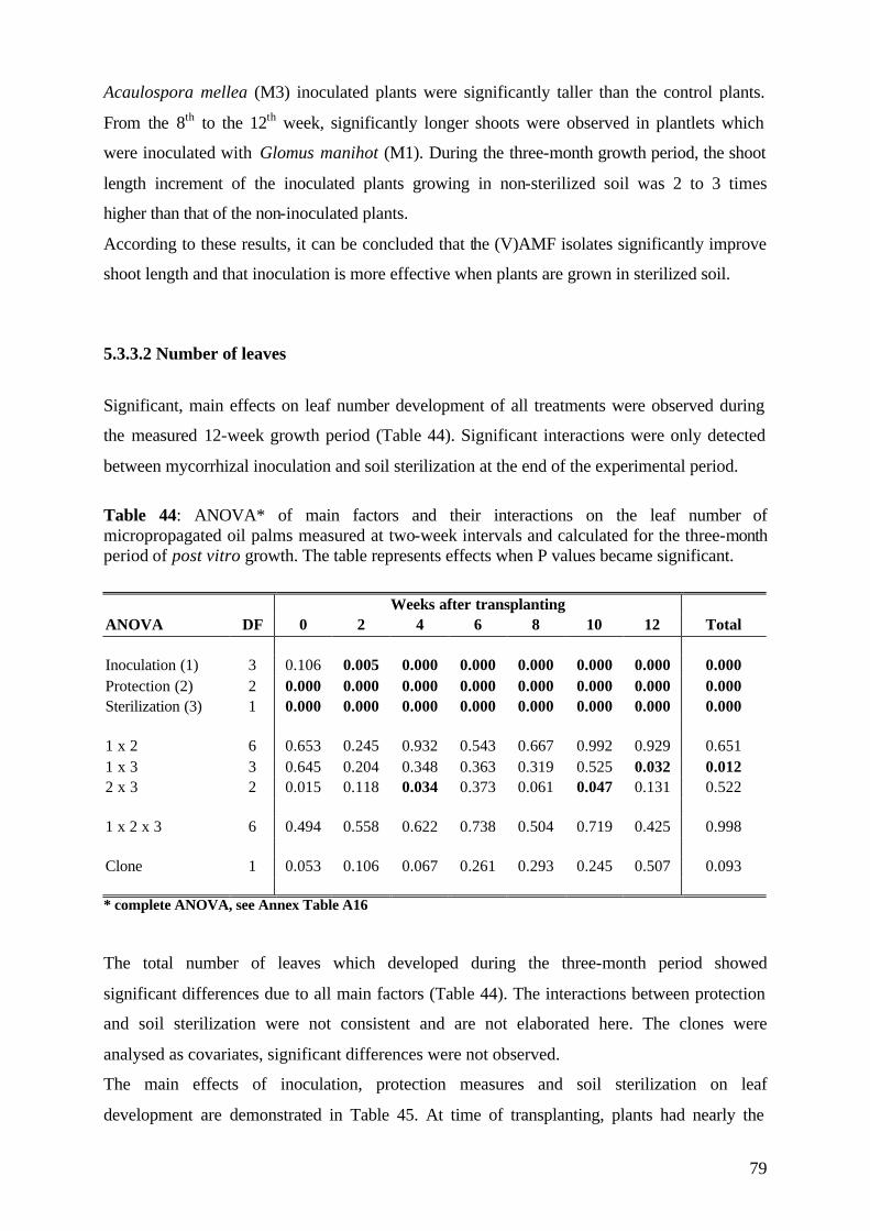
79
Acaulospora mellea (M3) inoculated plants were significantly taller than the control plants.
From the 8th to the 12th week, significantly longer shoots were observed in plantlets which
were inoculated with Glomus manihot (M1). During the three-month growth period, the shoot
length increment of the inoculated plants growing in non-sterilized soil was 2 to 3 times
higher than that of the non-inoculated plants.
According to these results, it can be concluded that the (V)AMF isolates significantly improve
shoot length and that inoculation is more effective when plants are grown in sterilized soil.
5.3.3.2 Number of leaves
Significant, main effects on leaf number development of all treatments were observed during
the measured 12-week growth period (Table 44). Significant interactions were only detected
between mycorrhizal inoculation and soil sterilization at the end of the experimental period.
Table 44: ANOVA* of main factors and their interactions on the leaf number of micropropagated oil palms measured at two-week intervals and calculated for the three-month period of post vitro growth. The table represents effects when P values became significant.
Weeks after transplanting ANOVA DF 0 2 4 6 8 10 12 Total Inoculation (1) 3 0.106 0.005 0.000 0.000 0.000 0.000 0.000 0.000 Protection (2) 2 0.000 0.000 0.000 0.000 0.000 0.000 0.000 0.000 Sterilization (3) 1 0.000 0.000 0.000 0.000 0.000 0.000 0.000 0.000 1 x 2 6 0.653 0.245 0.932 0.543 0.667 0.992 0.929 0.651 1 x 3 3 0.645 0.204 0.348 0.363 0.319 0.525 0.032 0.012 2 x 3 2 0.015 0.118 0.034 0.373 0.061 0.047 0.131 0.522 1 x 2 x 3 6 0.494 0.558 0.622 0.738 0.504 0.719 0.425 0.998 Clone 1 0.053 0.106 0.067 0.261 0.293 0.245 0.507 0.093 * complete ANOVA, see Annex Table A16
The total number of leaves which developed during the three-month period showed
significant differences due to all main factors (Table 44). The interactions between protection
and soil sterilization were not consistent and are not elaborated here. The clones were
analysed as covariates, significant differences were not observed.
The main effects of inoculation, protection measures and soil sterilization on leaf
development are demonstrated in Table 45. At time of transplanting, plants had nearly the
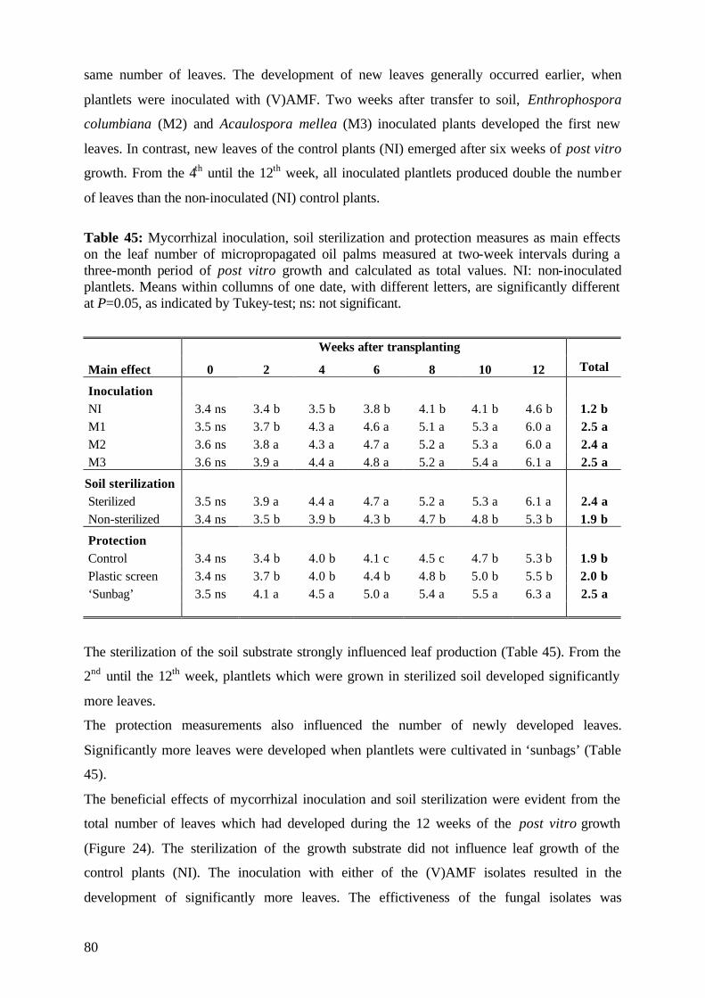
80
same number of leaves. The development of new leaves generally occurred earlier, when
plantlets were inoculated with (V)AMF. Two weeks after transfer to soil, Enthrophospora
columbiana (M2) and Acaulospora mellea (M3) inoculated plants developed the first new
leaves. In contrast, new leaves of the control plants (NI) emerged after six weeks of post vitro
growth. From the 4th until the 12th week, all inoculated plantlets produced double the number
of leaves than the non-inoculated (NI) control plants.
Table 45: Mycorrhizal inoculation, soil sterilization and protection measures as main effects on the leaf number of micropropagated oil palms measured at two-week intervals during a three-month period of post vitro growth and calculated as total values. NI: non-inoculated plantlets. Means within collumns of one date, with different letters, are significantly different at P=0.05, as indicated by Tukey-test; ns: not significant.
Weeks after transplanting
Main effect 0 2 4 6 8 10 12 Total
Inoculation NI 3.4 ns 3.4 b 3.5 b 3.8 b 4.1 b 4.1 b 4.6 b 1.2 b M1 3.5 ns 3.7 b 4.3 a 4.6 a 5.1 a 5.3 a 6.0 a 2.5 a M2 3.6 ns 3.8 a 4.3 a 4.7 a 5.2 a 5.3 a 6.0 a 2.4 a M3 3.6 ns 3.9 a 4.4 a 4.8 a 5.2 a 5.4 a 6.1 a 2.5 a
Soil sterilization Sterilized 3.5 ns 3.9 a 4.4 a 4.7 a 5.2 a 5.3 a 6.1 a 2.4 a Non-sterilized 3.4 ns 3.5 b 3.9 b 4.3 b 4.7 b 4.8 b 5.3 b 1.9 b
Protection Control 3.4 ns 3.4 b 4.0 b 4.1 c 4.5 c 4.7 b 5.3 b 1.9 b Plastic screen 3.4 ns 3.7 b 4.0 b 4.4 b 4.8 b 5.0 b 5.5 b 2.0 b ‘Sunbag’ 3.5 ns 4.1 a 4.5 a 5.0 a 5.4 a 5.5 a 6.3 a 2.5 a
The sterilization of the soil substrate strongly influenced leaf production (Table 45). From the
2nd until the 12th week, plantlets which were grown in sterilized soil developed significantly
more leaves.
The protection measurements also influenced the number of newly developed leaves.
Significantly more leaves were developed when plantlets were cultivated in ‘sunbags’ (Table
45).
The beneficial effects of mycorrhizal inoculation and soil sterilization were evident from the
total number of leaves which had developed during the 12 weeks of the post vitro growth
(Figure 24). The sterilization of the growth substrate did not influence leaf growth of the
control plants (NI). The inoculation with either of the (V)AMF isolates resulted in the
development of significantly more leaves. The effictiveness of the fungal isolates was
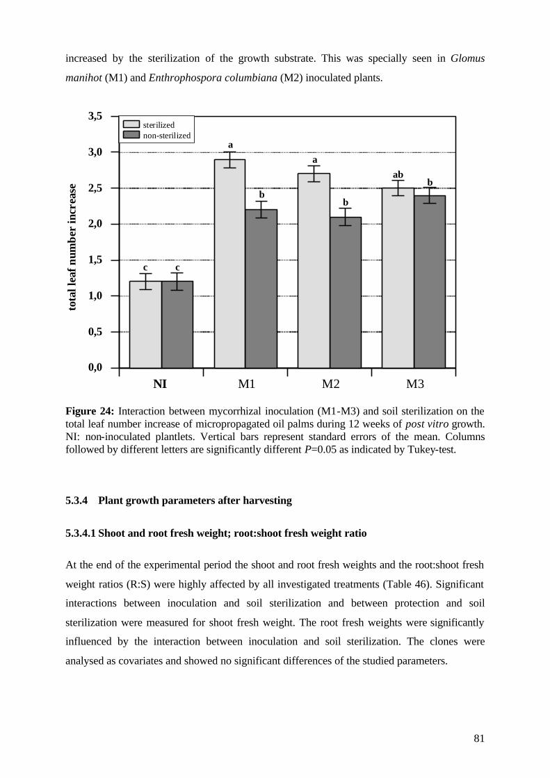
81
increased by the sterilization of the growth substrate. This was specially seen in Glomus
manihot (M1) and Enthrophospora columbiana (M2) inoculated plants.
0,0
0,5
1,0
1,5
2,0
2,5
3,0
3,5
NI M1 M2 M3
sterilizednon-sterilized
tota
llea
fnum
ber
incr
ease
a
b
a
b
abb
c c
Figure 24: Interaction between mycorrhizal inoculation (M1-M3) and soil sterilization on the total leaf number increase of micropropagated oil palms during 12 weeks of post vitro growth. NI: non-inoculated plantlets. Vertical bars represent standard errors of the mean. Columns followed by different letters are significantly different P=0.05 as indicated by Tukey-test.
5.3.4 Plant growth parameters after harvesting
5.3.4.1 Shoot and root fresh weight; root:shoot fresh weight ratio
At the end of the experimental period the shoot and root fresh weights and the root:shoot fresh
weight ratios (R:S) were highly affected by all investigated treatments (Table 46). Significant
interactions between inoculation and soil sterilization and between protection and soil
sterilization were measured for shoot fresh weight. The root fresh weights were significantly
influenced by the interaction between inoculation and soil sterilization. The clones were
analysed as covariates and showed no significant differences of the studied parameters.
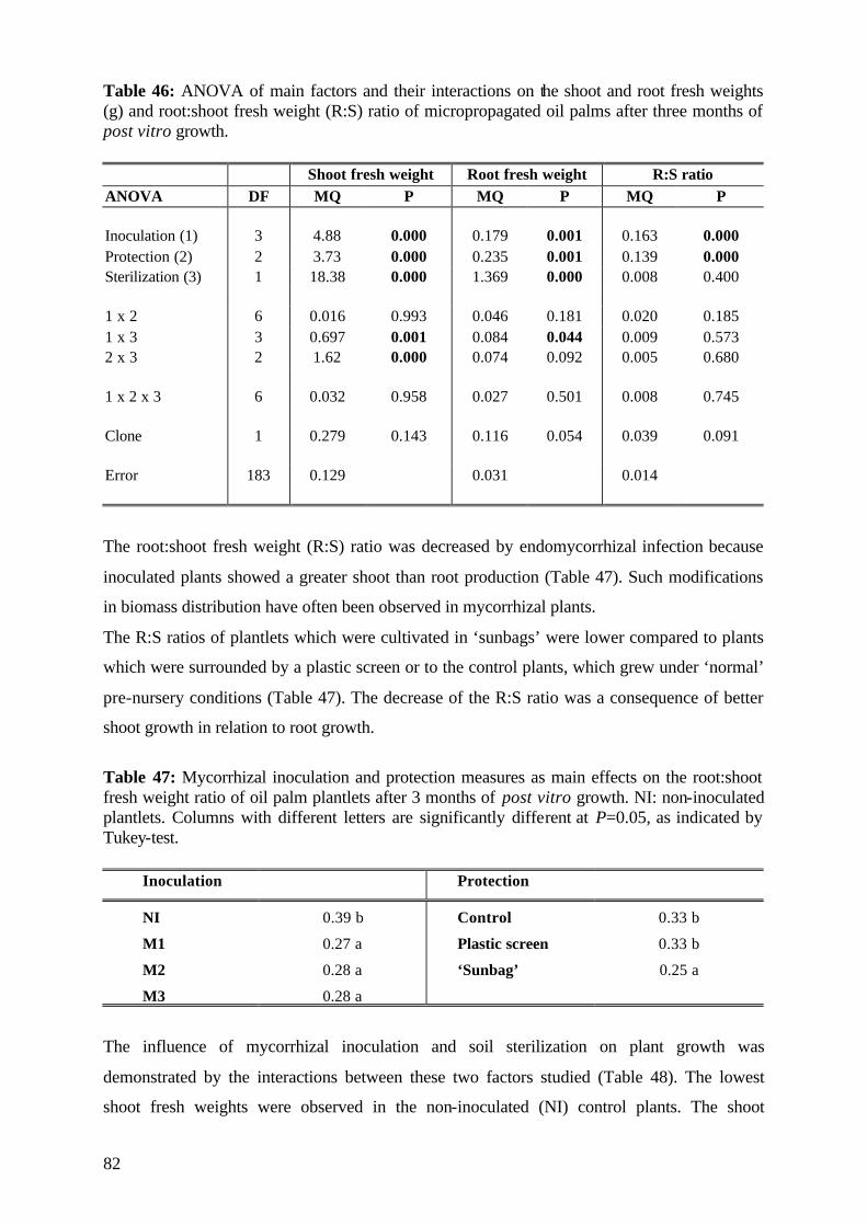
82
Table 46: ANOVA of main factors and their interactions on the shoot and root fresh weights (g) and root:shoot fresh weight (R:S) ratio of micropropagated oil palms after three months of post vitro growth.
Shoot fresh weight Root fresh weight R:S ratio ANOVA DF MQ P MQ P MQ P Inoculation (1) 3 4.88 0.000 0.179 0.001 0.163 0.000 Protection (2) 2 3.73 0.000 0.235 0.001 0.139 0.000 Sterilization (3) 1 18.38 0.000 1.369 0.000 0.008 0.400 1 x 2 6 0.016 0.993 0.046 0.181 0.020 0.185 1 x 3 3 0.697 0.001 0.084 0.044 0.009 0.573 2 x 3 2 1.62 0.000 0.074 0.092 0.005 0.680 1 x 2 x 3 6 0.032 0.958 0.027 0.501 0.008 0.745 Clone 1 0.279 0.143 0.116 0.054 0.039 0.091 Error 183 0.129 0.031 0.014
The root:shoot fresh weight (R:S) ratio was decreased by endomycorrhizal infection because
inoculated plants showed a greater shoot than root production (Table 47). Such modifications
in biomass distribution have often been observed in mycorrhizal plants.
The R:S ratios of plantlets which were cultivated in ‘sunbags’ were lower compared to plants
which were surrounded by a plastic screen or to the control plants, which grew under ‘normal’
pre-nursery conditions (Table 47). The decrease of the R:S ratio was a consequence of better
shoot growth in relation to root growth.
Table 47: Mycorrhizal inoculation and protection measures as main effects on the root:shoot fresh weight ratio of oil palm plantlets after 3 months of post vitro growth. NI: non-inoculated plantlets. Columns with different letters are significantly different at P=0.05, as indicated by Tukey-test.
Inoculation Protection
NI 0.39 b Control 0.33 b
M1 0.27 a Plastic screen 0.33 b
M2 0.28 a ‘Sunbag’ 0.25 a
M3 0.28 a
The influence of mycorrhizal inoculation and soil sterilization on plant growth was
demonstrated by the interactions between these two factors studied (Table 48). The lowest
shoot fresh weights were observed in the non-inoculated (NI) control plants. The shoot
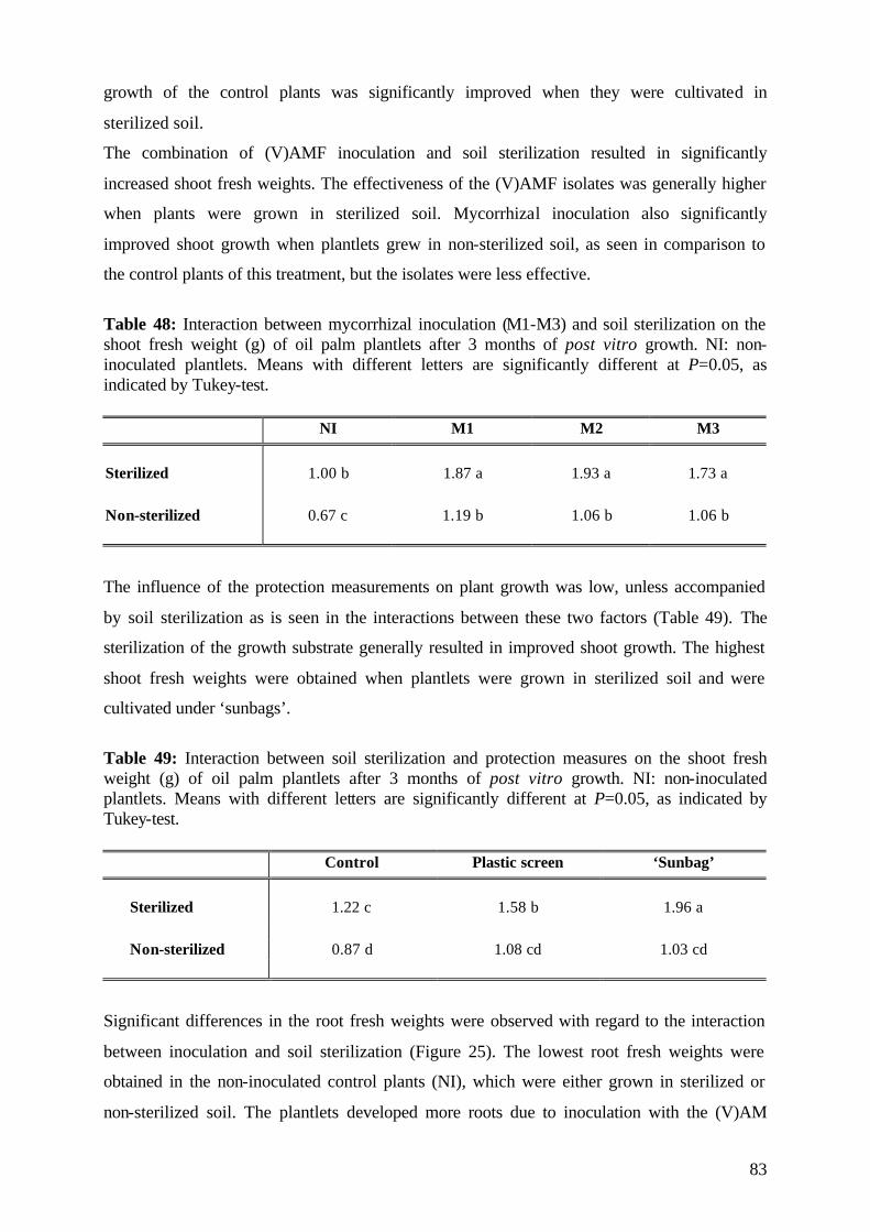
83
growth of the control plants was significantly improved when they were cultivated in
sterilized soil.
The combination of (V)AMF inoculation and soil sterilization resulted in significantly
increased shoot fresh weights. The effectiveness of the (V)AMF isolates was generally higher
when plants were grown in sterilized soil. Mycorrhizal inoculation also significantly
improved shoot growth when plantlets grew in non-sterilized soil, as seen in comparison to
the control plants of this treatment, but the isolates were less effective.
Table 48: Interaction between mycorrhizal inoculation (M1-M3) and soil sterilization on the shoot fresh weight (g) of oil palm plantlets after 3 months of post vitro growth. NI: non-inoculated plantlets. Means with different letters are significantly different at P=0.05, as indicated by Tukey-test. NI M1 M2 M3
Sterilized 1.00 b 1.87 a 1.93 a 1.73 a Non-sterilized 0.67 c 1.19 b 1.06 b 1.06 b
The influence of the protection measurements on plant growth was low, unless accompanied
by soil sterilization as is seen in the interactions between these two factors (Table 49). The
sterilization of the growth substrate generally resulted in improved shoot growth. The highest
shoot fresh weights were obtained when plantlets were grown in sterilized soil and were
cultivated under ‘sunbags’.
Table 49: Interaction between soil sterilization and protection measures on the shoot fresh weight (g) of oil palm plantlets after 3 months of post vitro growth. NI: non-inoculated plantlets. Means with different letters are significantly different at P=0.05, as indicated by Tukey-test. Control Plastic screen ‘Sunbag’
Sterilized 1.22 c 1.58 b 1.96 a Non-sterilized 0.87 d 1.08 cd 1.03 cd
Significant differences in the root fresh weights were observed with regard to the interaction
between inoculation and soil sterilization (Figure 25). The lowest root fresh weights were
obtained in the non-inoculated control plants (NI), which were either grown in sterilized or
non-sterilized soil. The plantlets developed more roots due to inoculation with the (V)AM
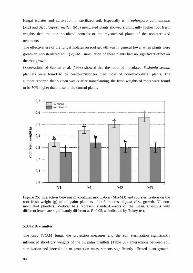
84
fungal isolates and cultivation in sterilized soil. Especially Enthrophospora columbinana
(M2) and Acaulospora mellea (M3) inoculated plants showed significantly higher root fresh
weights than the non-inoculated controls or the mycorrhizal plants of the non-sterilized
treatments.
The effectiveness of the fungal isolates on root growth was in general lower when plants were
grown in non-sterilized soil. (V)AMF inoculation of these plants had no significant effect on
the root growth.
Observations of Subhan et al. (1998) showed that the roots of inoculated Sesbania sesban
plantlets were found to be healthier/stronger than those of non-mycorrhizal plants. The
authors reported that sixteen weeks after transplanting, the fresh weights of roots were found
to be 50% higher than those of the control plants.
0,0
0,1
0,2
0,3
0,4
0,5
0,6
0,7
NI M1 M2 M3
sterilizednon-sterilized
bc
c
ab
bc
a
bc
a
c
root
fres
hw
eigh
t(g)
Figure 25: Interaction between mycorrhizal inoculation (M1-M3) and soil sterilization on the root fresh weight (g) of oil palm plantlets after 3 months of post vitro growth. NI: non-inoculated plantlets. Vertical bars represent standard errors of the mean. Columns with different letters are significantly different at P=0.05, as indicated by Tukey-test.
5.3.4.2 Dry matter The used (V)AM fungi, the protection measures and the soil sterilization significantly
influenced shoot dry weights of the oil palm plantlets (Table 50). Interactions between soil
sterilization and inoculation or protection measurements significantly affected plant growth.
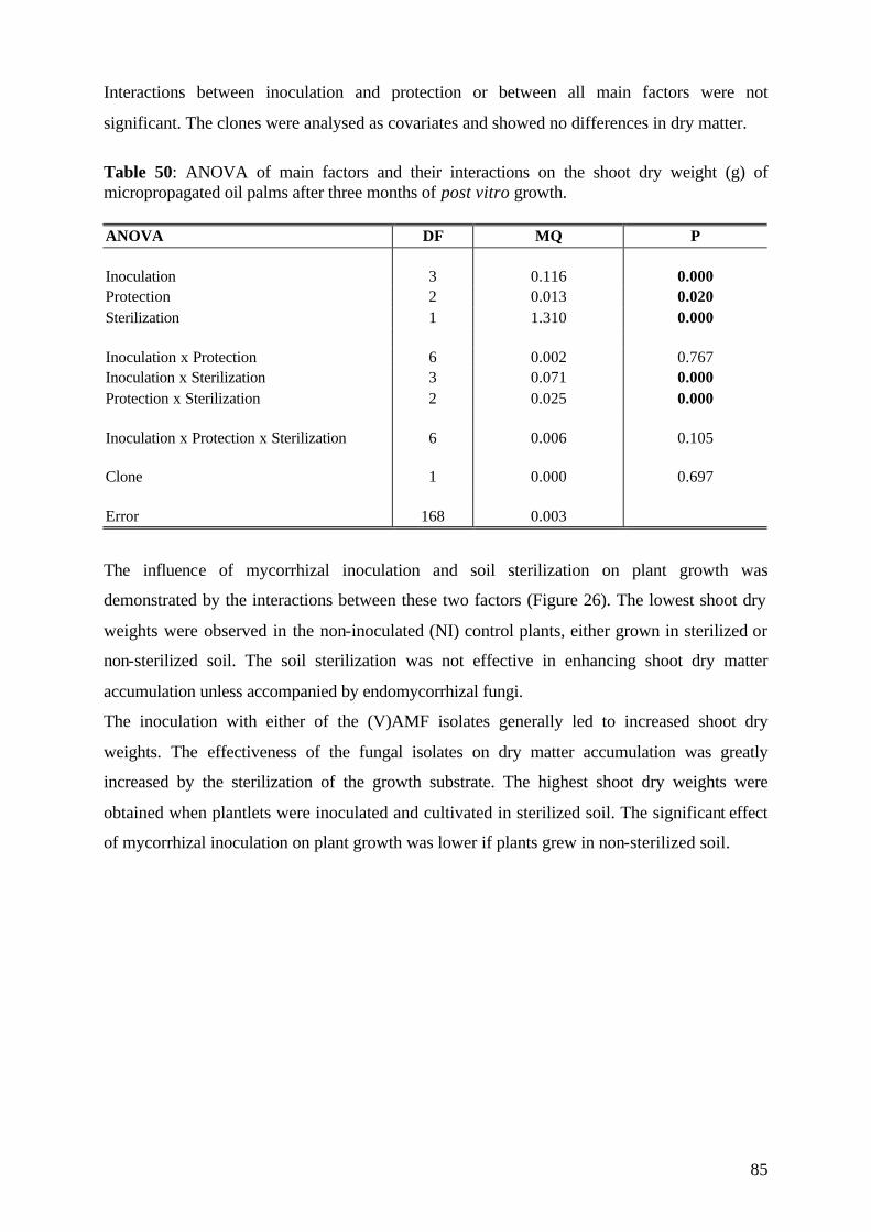
85
Interactions between inoculation and protection or between all main factors were not
significant. The clones were analysed as covariates and showed no differences in dry matter.
Table 50: ANOVA of main factors and their interactions on the shoot dry weight (g) of micropropagated oil palms after three months of post vitro growth. ANOVA DF MQ P Inoculation 3 0.116 0.000 Protection 2 0.013 0.020 Sterilization 1 1.310 0.000 Inoculation x Protection 6 0.002 0.767 Inoculation x Sterilization 3 0.071 0.000 Protection x Sterilization 2 0.025 0.000 Inoculation x Protection x Sterilization 6 0.006 0.105 Clone 1 0.000 0.697 Error 168 0.003
The influence of mycorrhizal inoculation and soil sterilization on plant growth was
demonstrated by the interactions between these two factors (Figure 26). The lowest shoot dry
weights were observed in the non-inoculated (NI) control plants, either grown in sterilized or
non-sterilized soil. The soil sterilization was not effective in enhancing shoot dry matter
accumulation unless accompanied by endomycorrhizal fungi.
The inoculation with either of the (V)AMF isolates generally led to increased shoot dry
weights. The effectiveness of the fungal isolates on dry matter accumulation was greatly
increased by the sterilization of the growth substrate. The highest shoot dry weights were
obtained when plantlets were inoculated and cultivated in sterilized soil. The significant effect
of mycorrhizal inoculation on plant growth was lower if plants grew in non-sterilized soil.
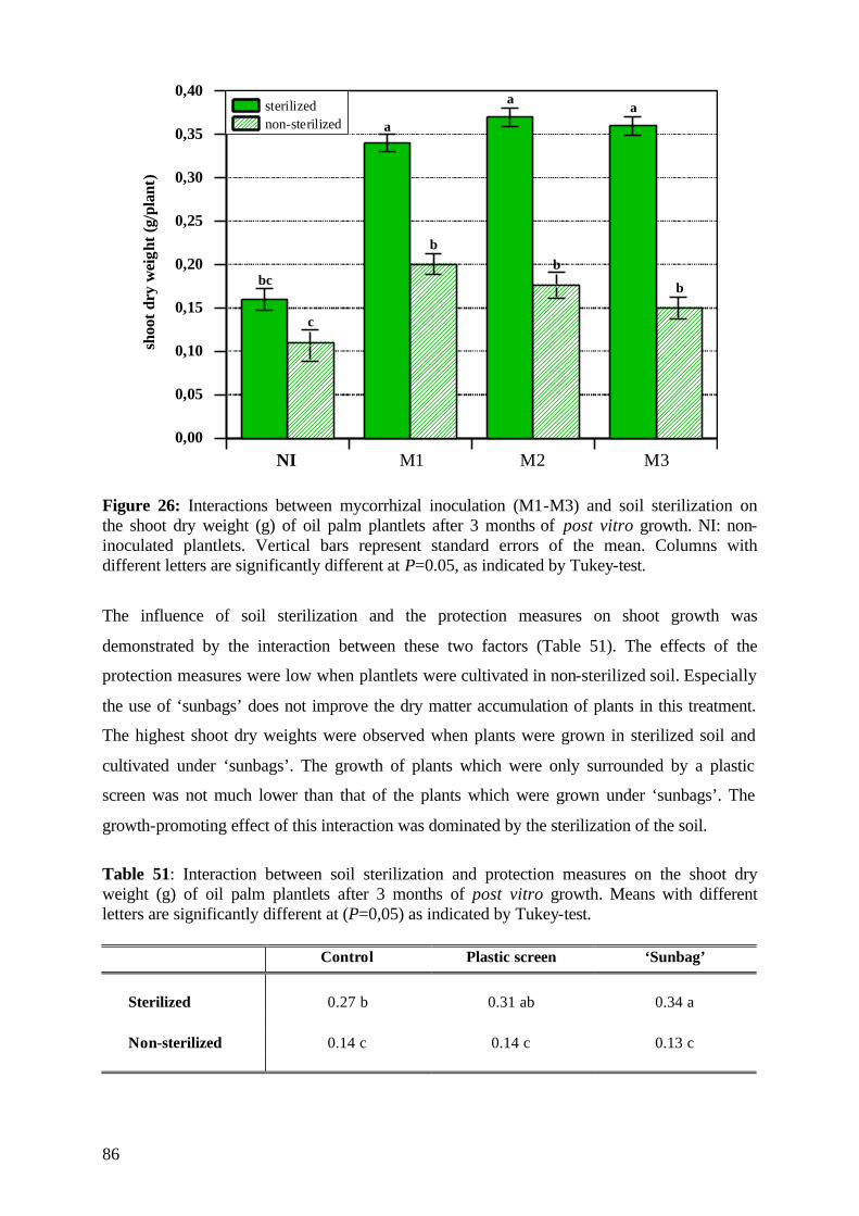
86
0,00
0,05
0,10
0,15
0,20
0,25
0,30
0,35
0,40
NI M1 M2 M3
sterilizednon-sterilized
shoo
tdry
wei
ght(
g/pl
ant)
bc
a
aa
bb
b
c
Figure 26: Interactions between mycorrhizal inoculation (M1-M3) and soil sterilization on the shoot dry weight (g) of oil palm plantlets after 3 months of post vitro growth. NI: non-inoculated plantlets. Vertical bars represent standard errors of the mean. Columns with different letters are significantly different at P=0.05, as indicated by Tukey-test.
The influence of soil sterilization and the protection measures on shoot growth was
demonstrated by the interaction between these two factors (Table 51). The effects of the
protection measures were low when plantlets were cultivated in non-sterilized soil. Especially
the use of ‘sunbags’ does not improve the dry matter accumulation of plants in this treatment.
The highest shoot dry weights were observed when plants were grown in sterilized soil and
cultivated under ‘sunbags’. The growth of plants which were only surrounded by a plastic
screen was not much lower than that of the plants which were grown under ‘sunbags’. The
growth-promoting effect of this interaction was dominated by the sterilization of the soil.
Table 51: Interaction between soil sterilization and protection measures on the shoot dry weight (g) of oil palm plantlets after 3 months of post vitro growth. Means with different letters are significantly different at (P=0,05) as indicated by Tukey-test. Control Plastic screen ‘Sunbag’
Sterilized 0.27 b 0.31 ab 0.34 a Non-sterilized 0.14 c 0.14 c 0.13 c
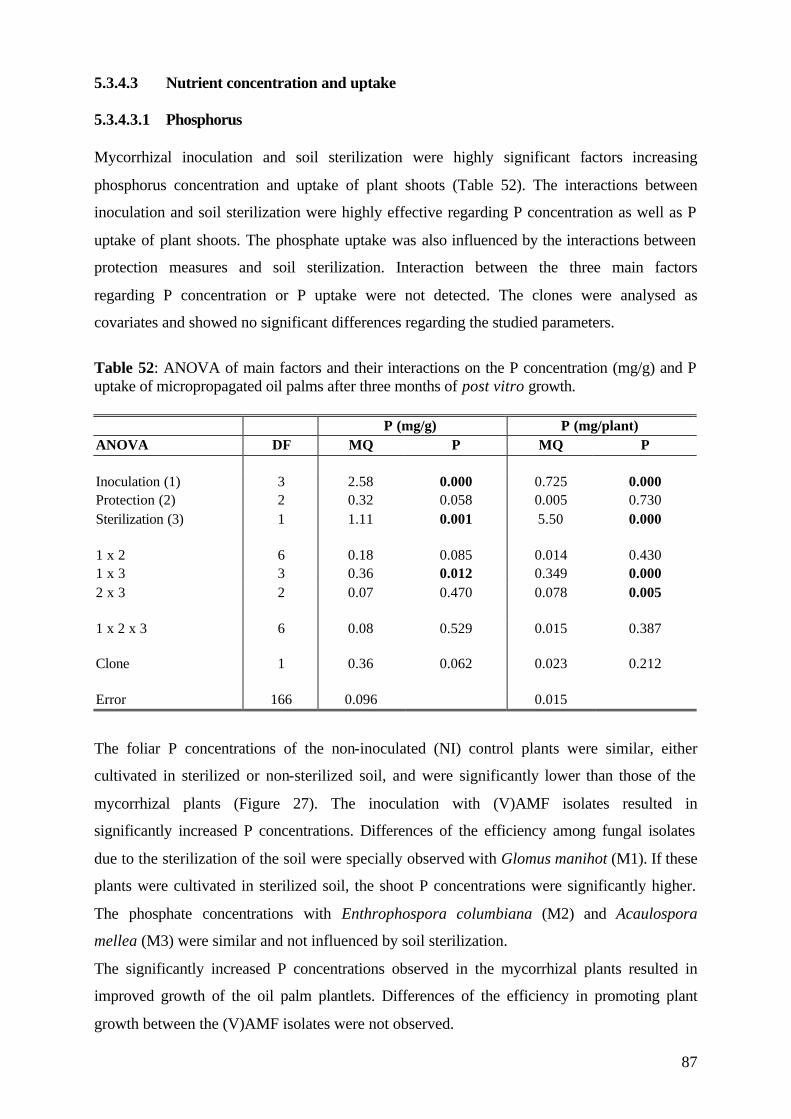
87
5.3.4.3 Nutrient concentration and uptake 5.3.4.3.1 Phosphorus Mycorrhizal inoculation and soil sterilization were highly significant factors increasing
phosphorus concentration and uptake of plant shoots (Table 52). The interactions between
inoculation and soil sterilization were highly effective regarding P concentration as well as P
uptake of plant shoots. The phosphate uptake was also influenced by the interactions between
protection measures and soil sterilization. Interaction between the three main factors
regarding P concentration or P uptake were not detected. The clones were analysed as
covariates and showed no significant differences regarding the studied parameters.
Table 52: ANOVA of main factors and their interactions on the P concentration (mg/g) and P uptake of micropropagated oil palms after three months of post vitro growth. P (mg/g) P (mg/plant) ANOVA DF MQ P MQ P Inoculation (1) 3 2.58 0.000 0.725 0.000 Protection (2) 2 0.32 0.058 0.005 0.730 Sterilization (3) 1 1.11 0.001 5.50 0.000 1 x 2 6 0.18 0.085 0.014 0.430 1 x 3 3 0.36 0.012 0.349 0.000 2 x 3 2 0.07 0.470 0.078 0.005 1 x 2 x 3 6 0.08 0.529 0.015 0.387 Clone 1 0.36 0.062 0.023 0.212 Error 166 0.096 0.015
The foliar P concentrations of the non-inoculated (NI) control plants were similar, either
cultivated in sterilized or non-sterilized soil, and were significantly lower than those of the
mycorrhizal plants (Figure 27). The inoculation with (V)AMF isolates resulted in
significantly increased P concentrations. Differences of the efficiency among fungal isolates
due to the sterilization of the soil were specially observed with Glomus manihot (M1). If these
plants were cultivated in sterilized soil, the shoot P concentrations were significantly higher.
The phosphate concentrations with Enthrophospora columbiana (M2) and Acaulospora
mellea (M3) were similar and not influenced by soil sterilization.
The significantly increased P concentrations observed in the mycorrhizal plants resulted in
improved growth of the oil palm plantlets. Differences of the efficiency in promoting plant
growth between the (V)AMF isolates were not observed.
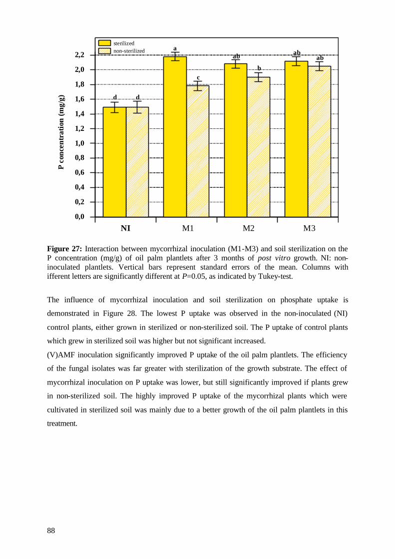
88
0,0
0,2
0,4
0,6
0,8
1,0
1,2
1,4
1,6
1,8
2,0
2,2
NI M1 M2 M3
sterilizednon-sterilized
Pco
ncen
trat
ion
(mg/
g) d d
a
c
ab
b
abab
Figure 27: Interaction between mycorrhizal inoculation (M1-M3) and soil sterilization on the P concentration (mg/g) of oil palm plantlets after 3 months of post vitro growth. NI: non-inoculated plantlets. Vertical bars represent standard errors of the mean. Columns with ifferent letters are significantly different at P=0.05, as indicated by Tukey-test.
The influence of mycorrhizal inoculation and soil sterilization on phosphate uptake is
demonstrated in Figure 28. The lowest P uptake was observed in the non-inoculated (NI)
control plants, either grown in sterilized or non-sterilized soil. The P uptake of control plants
which grew in sterilized soil was higher but not significant increased.
(V)AMF inoculation significantly improved P uptake of the oil palm plantlets. The efficiency
of the fungal isolates was far greater with sterilization of the growth substrate. The effect of
mycorrhizal inoculation on P uptake was lower, but still significantly improved if plants grew
in non-sterilized soil. The highly improved P uptake of the mycorrhizal plants which were
cultivated in sterilized soil was mainly due to a better growth of the oil palm plantlets in this
treatment.
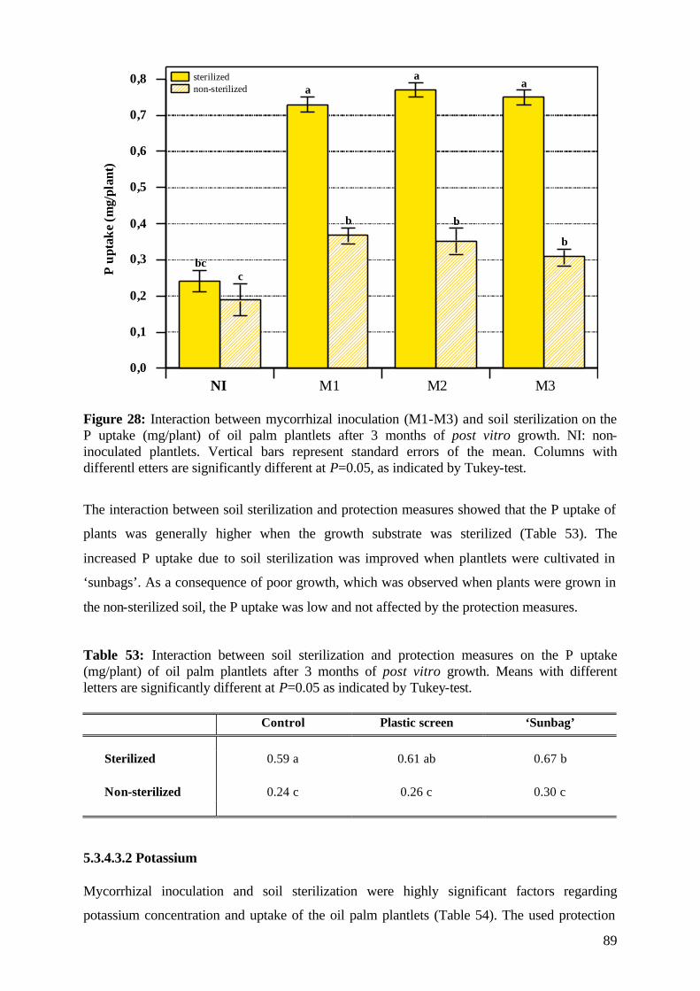
89
0,0
0,1
0,2
0,3
0,4
0,5
0,6
0,7
0,8
NI M1 M2 M3
sterilizednon-sterilized
Pup
take
(mg/
plan
t)a
aa
bc
b b
b
c
Figure 28: Interaction between mycorrhizal inoculation (M1-M3) and soil sterilization on the P uptake (mg/plant) of oil palm plantlets after 3 months of post vitro growth. NI: non-inoculated plantlets. Vertical bars represent standard errors of the mean. Columns with differentl etters are significantly different at P=0.05, as indicated by Tukey-test.
The interaction between soil sterilization and protection measures showed that the P uptake of
plants was generally higher when the growth substrate was sterilized (Table 53). The
increased P uptake due to soil sterilization was improved when plantlets were cultivated in
‘sunbags’. As a consequence of poor growth, which was observed when plants were grown in
the non-sterilized soil, the P uptake was low and not affected by the protection measures.
Table 53: Interaction between soil sterilization and protection measures on the P uptake (mg/plant) of oil palm plantlets after 3 months of post vitro growth. Means with different letters are significantly different at P=0.05 as indicated by Tukey-test. Control Plastic screen ‘Sunbag’
Sterilized 0.59 a 0.61 ab 0.67 b Non-sterilized 0.24 c 0.26 c 0.30 c
5.3.4.3.2 Potassium Mycorrhizal inoculation and soil sterilization were highly significant factors regarding
potassium concentration and uptake of the oil palm plantlets (Table 54). The used protection
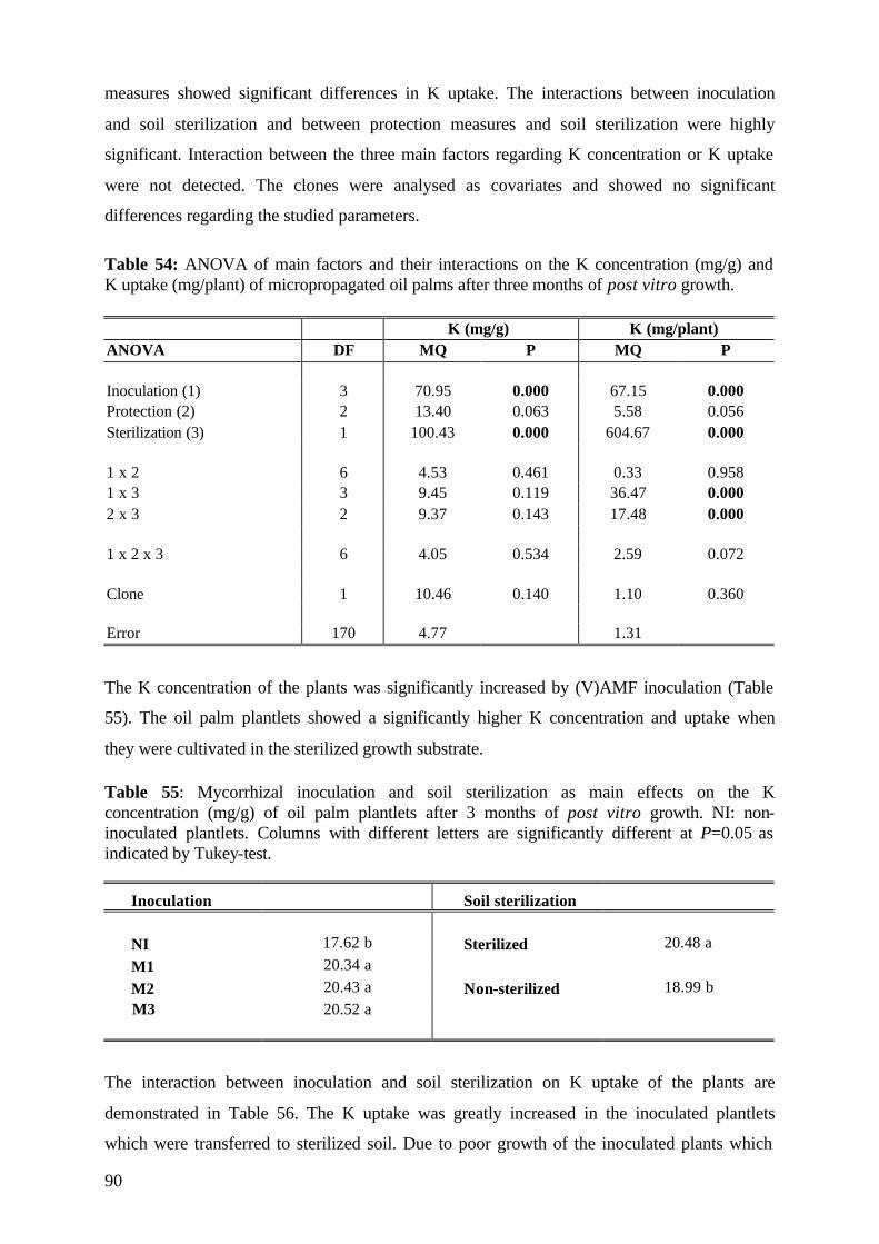
90
measures showed significant differences in K uptake. The interactions between inoculation
and soil sterilization and between protection measures and soil sterilization were highly
significant. Interaction between the three main factors regarding K concentration or K uptake
were not detected. The clones were analysed as covariates and showed no significant
differences regarding the studied parameters.
Table 54: ANOVA of main factors and their interactions on the K concentration (mg/g) and K uptake (mg/plant) of micropropagated oil palms after three months of post vitro growth. K (mg/g) K (mg/plant) ANOVA DF MQ P MQ P Inoculation (1) 3 70.95 0.000 67.15 0.000 Protection (2) 2 13.40 0.063 5.58 0.056 Sterilization (3) 1 100.43 0.000 604.67 0.000 1 x 2 6 4.53 0.461 0.33 0.958 1 x 3 3 9.45 0.119 36.47 0.000 2 x 3 2 9.37 0.143 17.48 0.000 1 x 2 x 3 6 4.05 0.534 2.59 0.072 Clone 1 10.46 0.140 1.10 0.360 Error 170 4.77 1.31
The K concentration of the plants was significantly increased by (V)AMF inoculation (Table
55). The oil palm plantlets showed a significantly higher K concentration and uptake when
they were cultivated in the sterilized growth substrate.
Table 55: Mycorrhizal inoculation and soil sterilization as main effects on the K concentration (mg/g) of oil palm plantlets after 3 months of post vitro growth. NI: non-inoculated plantlets. Columns with different letters are significantly different at P=0.05 as indicated by Tukey-test.
Inoculation Soil sterilization NI 17.62 b Sterilized 20.48 a M1 20.34 a M2 20.43 a Non-sterilized 18.99 b
M3 20.52 a
The interaction between inoculation and soil sterilization on K uptake of the plants are
demonstrated in Table 56. The K uptake was greatly increased in the inoculated plantlets
which were transferred to sterilized soil. Due to poor growth of the inoculated plants which
91
were cultivated in non-sterilized soil, the potassium uptake in these treatments was
significantly lower, and, with the exception of Glomus manihot (M1), similar to that of the
non-inoculated (NI) control plants.
Table 56: Interaction between mycorrhizal inoculation (M1-M3) and soil sterilization on the K uptake (mg/plant) of oil palm clones after 3 months of post vitro growth. NI: non-inoculated plantlets. Means with different letters are significantly different at P=0.05, as indicated by Tukey-test. NI M1 M2 M3
Sterilized 2.94 bc 7.34 a 7.88 a 7.62 a Non-sterilized 2.18 c 3.39 b 3.06 bc 2.46 bc
The effects of soil sterilization and protection measures on K uptake are demonstrated in
Table 57. The highest K uptake was detected in plants which were cultivated in sterilized soil.
This effect was improved when plantlets were grown in ‘sunbags’. The P uptake of plants
which were transferred to non-sterilized soil was low due to poor shoot growth and was not
improved by either of the protection measures.
Table 57: Interactions between soil sterilization and protection measures on the K uptake (mg/plant) of oil palm clones after 3 months of post vitro growth. Means with different letters are significantly different at P=0,05 as indicated by Tukey-test. Control Plastic screen ‘Sunbag’
Sterilized 5.59 c 6.44 b 7.30 a Non-sterilized 3.04 d 2.72 d 2.56 d
5.3.4.3.2 Nitrogen Mycorrhizal inoculation and soil sterilization were highly significant main factors regarding
nitrogen concentration and uptake of plants (Table 57). However, the interactions between
inoculation and soil sterilization and between protection measures and soil sterilization were
highly significant regarding N uptake. Interactions between the three main factors as regards
N concentration or N uptake were not detected. The clones were analysed as covariates and
showed no significant differences with regard to the studied parameters.
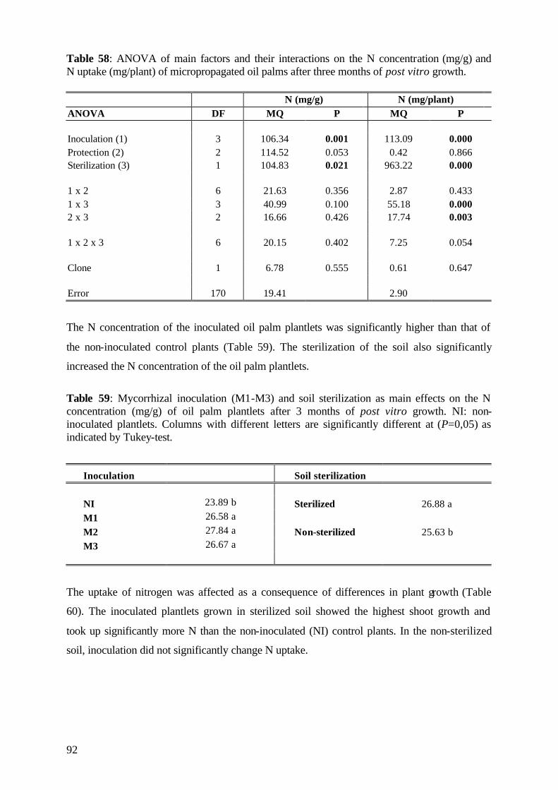
92
Table 58: ANOVA of main factors and their interactions on the N concentration (mg/g) and N uptake (mg/plant) of micropropagated oil palms after three months of post vitro growth. N (mg/g) N (mg/plant) ANOVA DF MQ P MQ P Inoculation (1) 3 106.34 0.001 113.09 0.000 Protection (2) 2 114.52 0.053 0.42 0.866 Sterilization (3) 1 104.83 0.021 963.22 0.000 1 x 2 6 21.63 0.356 2.87 0.433 1 x 3 3 40.99 0.100 55.18 0.000 2 x 3 2 16.66 0.426 17.74 0.003 1 x 2 x 3 6 20.15 0.402 7.25 0.054 Clone 1 6.78 0.555 0.61 0.647 Error 170 19.41 2.90
The N concentration of the inoculated oil palm plantlets was significantly higher than that of
the non-inoculated control plants (Table 59). The sterilization of the soil also significantly
increased the N concentration of the oil palm plantlets.
Table 59: Mycorrhizal inoculation (M1-M3) and soil sterilization as main effects on the N concentration (mg/g) of oil palm plantlets after 3 months of post vitro growth. NI: non-inoculated plantlets. Columns with different letters are significantly different at (P=0,05) as indicated by Tukey-test.
Inoculation Soil sterilization
NI 23.89 b Sterilized 26.88 a M1 26.58 a M2 27.84 a Non-sterilized 25.63 b M3 26.67 a
The uptake of nitrogen was affected as a consequence of differences in plant growth (Table
60). The inoculated plantlets grown in sterilized soil showed the highest shoot growth and
took up significantly more N than the non-inoculated (NI) control plants. In the non-sterilized
soil, inoculation did not significantly change N uptake.
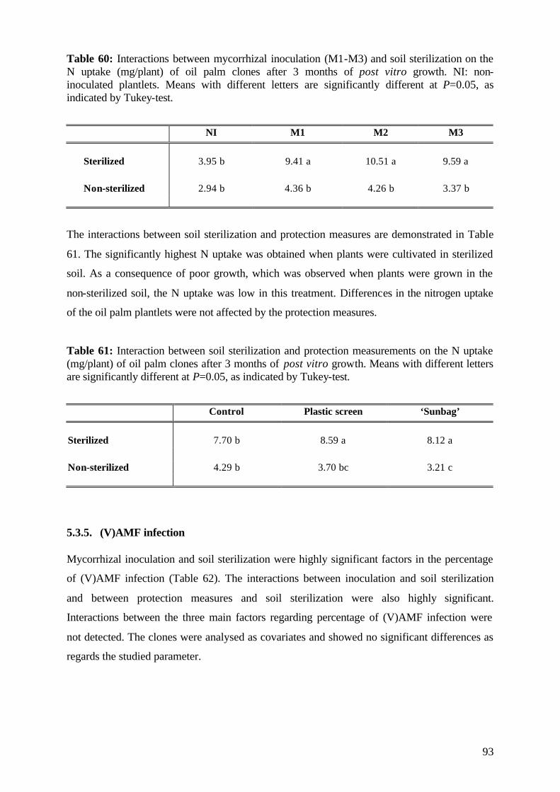
93
Table 60: Interactions between mycorrhizal inoculation (M1-M3) and soil sterilization on the N uptake (mg/plant) of oil palm clones after 3 months of post vitro growth. NI: non-inoculated plantlets. Means with different letters are significantly different at P=0.05, as indicated by Tukey-test.
NI M1 M2 M3
Sterilized 3.95 b 9.41 a 10.51 a 9.59 a Non-sterilized 2.94 b 4.36 b 4.26 b 3.37 b
The interactions between soil sterilization and protection measures are demonstrated in Table
61. The significantly highest N uptake was obtained when plants were cultivated in sterilized
soil. As a consequence of poor growth, which was observed when plants were grown in the
non-sterilized soil, the N uptake was low in this treatment. Differences in the nitrogen uptake
of the oil palm plantlets were not affected by the protection measures.
Table 61: Interaction between soil sterilization and protection measurements on the N uptake (mg/plant) of oil palm clones after 3 months of post vitro growth. Means with different letters are significantly different at P=0.05, as indicated by Tukey-test.
Control Plastic screen ‘Sunbag’
Sterilized 7.70 b 8.59 a 8.12 a Non-sterilized 4.29 b 3.70 bc 3.21 c 5.3.5. (V)AMF infection Mycorrhizal inoculation and soil sterilization were highly significant factors in the percentage
of (V)AMF infection (Table 62). The interactions between inoculation and soil sterilization
and between protection measures and soil sterilization were also highly significant.
Interactions between the three main factors regarding percentage of (V)AMF infection were
not detected. The clones were analysed as covariates and showed no significant differences as
regards the studied parameter.
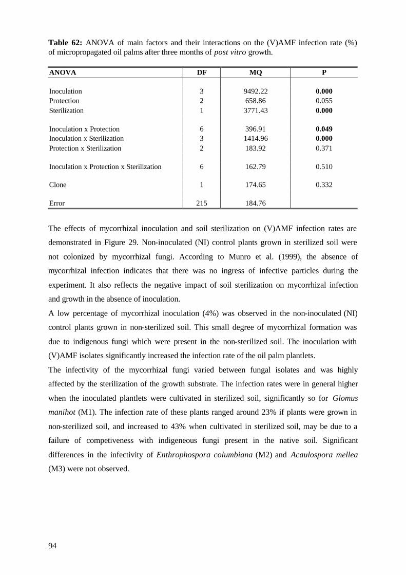
94
Table 62: ANOVA of main factors and their interactions on the (V)AMF infection rate (%) of micropropagated oil palms after three months of post vitro growth. ANOVA DF MQ P Inoculation 3 9492.22 0.000 Protection 2 658.86 0.055 Sterilization 1 3771.43 0.000 Inoculation x Protection 6 396.91 0.049 Inoculation x Sterilization 3 1414.96 0.000 Protection x Sterilization 2 183.92 0.371 Inoculation x Protection x Sterilization 6 162.79 0.510 Clone 1 174.65 0.332 Error 215 184.76
The effects of mycorrhizal inoculation and soil sterilization on (V)AMF infection rates are
demonstrated in Figure 29. Non-inoculated (NI) control plants grown in sterilized soil were
not colonized by mycorrhizal fungi. According to Munro et al. (1999), the absence of
mycorrhizal infection indicates that there was no ingress of infective particles during the
experiment. It also reflects the negative impact of soil sterilization on mycorrhizal infection
and growth in the absence of inoculation.
A low percentage of mycorrhizal inoculation (4%) was observed in the non-inoculated (NI)
control plants grown in non-sterilized soil. This small degree of mycorrhizal formation was
due to indigenous fungi which were present in the non-sterilized soil. The inoculation with
(V)AMF isolates significantly increased the infection rate of the oil palm plantlets.
The infectivity of the mycorrhizal fungi varied between fungal isolates and was highly
affected by the sterilization of the growth substrate. The infection rates were in general higher
when the inoculated plantlets were cultivated in sterilized soil, significantly so for Glomus
manihot (M1). The infection rate of these plants ranged around 23% if plants were grown in
non-sterilized soil, and increased to 43% when cultivated in sterilized soil, may be due to a
failure of competiveness with indigeneous fungi present in the native soil. Significant
differences in the infectivity of Enthrophospora columbiana (M2) and Acaulospora mellea
(M3) were not observed.
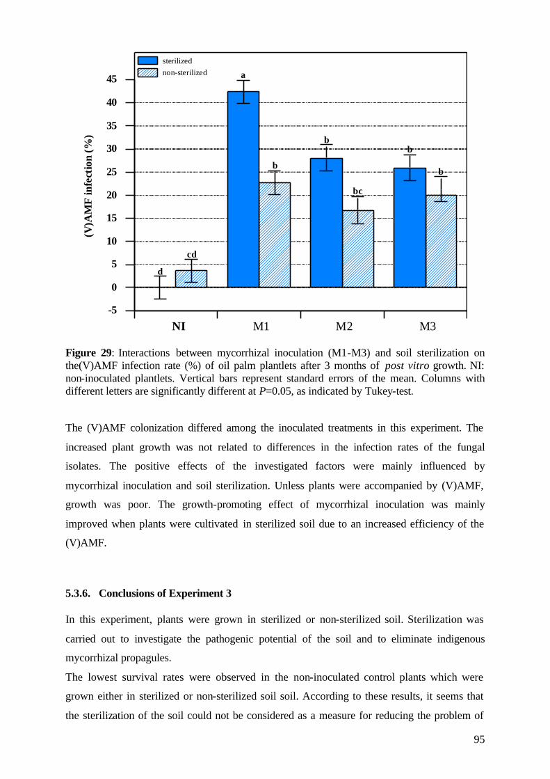
95
-5
0
5
10
15
20
25
30
35
40
45
NI M1 M2 M3
sterilizednon-sterilized
(V)A
MF
infe
ctio
n(%
)a
d
cd
b
bb
b
bc
Figure 29: Interactions between mycorrhizal inoculation (M1-M3) and soil sterilization on the(V)AMF infection rate (%) of oil palm plantlets after 3 months of post vitro growth. NI: non-inoculated plantlets. Vertical bars represent standard errors of the mean. Columns with different letters are significantly different at P=0.05, as indicated by Tukey-test. The (V)AMF colonization differed among the inoculated treatments in this experiment. The
increased plant growth was not related to differences in the infection rates of the fungal
isolates. The positive effects of the investigated factors were mainly influenced by
mycorrhizal inoculation and soil sterilization. Unless plants were accompanied by (V)AMF,
growth was poor. The growth-promoting effect of mycorrhizal inoculation was mainly
improved when plants were cultivated in sterilized soil due to an increased efficiency of the
(V)AMF.
5.3.6. Conclusions of Experiment 3 In this experiment, plants were grown in sterilized or non-sterilized soil. Sterilization was
carried out to investigate the pathogenic potential of the soil and to eliminate indigenous
mycorrhizal propagules.
The lowest survival rates were observed in the non-inoculated control plants which were
grown either in sterilized or non-sterilized soil soil. According to these results, it seems that
the sterilization of the soil could not be considered as a measure for reducing the problem of
96
plant mortality due to pathogens or other reasons. The effect of soil sterilization alone was
low, unless plants were inoculated with the (V)AMF isolates. Due to the effectiveness of the
endomycorrhizal fungi, the combination of these two treatments was the most effective in
promoting plant survival and development, especially when the plantlets were not well
developed at the time of transfer to the soil.
Heating or partial sterilization of soil alters the biological and chemical properties of the soil
in addition to eliminating microorganisms. It can be concluded that soil sterilization leads to a
combined effect of mineralization and mobilization of nutrients, thus promoting plant growth,
and failure of competition with soil own microorganisms.
The plant growth promoting effect through cultivation in ‘sunbags’ was probably due to the
development of a special microclimate in this semi-closed system. These conditions were
more similar to in vitro conditions and more favourable for plant development than ‘normal’
pre-nursery conditions. It is doubtful if this protection makes sense, because the main aim of
the post vitro growth period in the greenhouse is the hardening of the sensitive
micropropagated plantlets. The question is still open whether the later adaptation of the
plantlets to normal conditions will be successful if plants are grown in this protected
environment. Moreover, the cost of ‘sunbags’ is not low and would be additional to the
production costs. From the viewpoint of commercialisation and according to the results
obtained in this experiment, the implementation of these measures cannot be recommended.
6. General discussion Over the past few years the use of in vitro micropropagation techniques for multiplication of a
wide range of plantation crops, ornamentals, fruits, vegetables and forest trees, has increased
rapidly. One of the main objectives of such a technique is to obtain a large number of
identical plants from a selected mother plant. For this purpose, tissue-culture techniques are
being developed rapidly. However, little attention has been given to acclimatisation and
adaptation of in vitro propagated plants to greenhouse and/or field.
The high mortality and poor development of micropropagated oil palms experienced at the
"Indonesian Oil Palm Research Institute" (IOPRI) after plantlets were transferred to ex vitro
conditions is a major problem for expanding clonal oil palm production commercially. It
could be possible to improve plant survival and development after transplanting by
introducing a beneficial microbial population into the growth substrate.
Mycorrhizal fungi are known to have beneficial effects on growth and development of many
micropropagated species (Ponders, 1984; Douds and Chaney, 1986; Lovato et al., 1994, 1996;
97
Azcón-Aguilar and Barea, 1996, 1997) through improvement of mineral nutrition, induction
of root system modifications (Berta et al., 1995) and increased resistance to pathogens
(Guillemin et al., 1992; Lindermann, 1994). Their elimination during micropropagation
frequently results in poor development of plants so that their reintroduction during plant
production is important. Based on these facts, the research described in this study was carried
out. One of the main aims of this research work was to introduce and establish the
mycorrhizal inoculation in an existing micropropagation process with a view to future
practical applications. A further objective of this research was to determine whether
(vesicular-) arbuscular mycorrhiza could enhance transplanting success and plantlet
acclimatisation as indicated by the main plant-growth parameters such as survival rate, plant
growth properties, and nutrient uptake.
6.1 Effect of (V)A mycorrhiza on the post vitro survival of micropropagated oil palms The transfer of in vitro plantlets to ex vitro conditions is one of the most critical steps in the
micropropagation process. High mortality rates are recorded for certain micropropagated plant
species upon transfer to ex vitro conditions due to the plantlets’ limited ability to resist the
transplant stress. At IOPRI, the percentage of mortality of micropropagated oil palms is high,
30-40% of the oil palms dying after being transferring ex vitro to the pre-nursery. The main
aim of this study was to investigate whether the introduction of (V)AM fungi in the
micropropagation system of oil palms can reduce mortality and improve the tolerance of the
sensitive plants to the transplanting stress.
The survival of micropropagated oil palms was remarkably improved with mycorrhizal
inoculation in all experiments of this study. From the twelve mycorrhizal isolates tested in the
initial screening of Experiment 1, eleven fungi were able to improve the survival rate
significantly to 83-100%, helping the plantlets to resist the environmental stress induced
through transplanting from axenic conditions to normal cultivation in open pots. In contrast,
only 55% of the non-inoculated control plants survived during the three-month experimental
period. Inoculation with (V)AM fungi appears to be critical for the survival of
micropropagated oil palms. Several investigations have shown that early mycorrhizal
inoculation and colonisation of tissue-cultured plantlets of other plant species reduce
transplant shock during acclimatisation, and increase plant survival and establishment rates
(Varma and Schüepp, 1995; Fortuna et al., 1996; Lovato et al., 1996; Azc?n-Aguilar and
Barea, 1997). Azc?n-Aguilar et al. (1997) reported that the survival rate of micropropagated
cassava plants, inoculated with Glomus deserticola at the beginning of the acclimatisation
stage, was significantly improved by mycorrhizal inoculation. Subhan et al. (1998) used the
98
(vesicular-) arbuscular mycorrhizal fungus Glomus fasciculatum for inoculation of
micropropagated Sesbania sesban plantlets. All of the inoculated plants survived
transplanting in contrast to the non-inoculated control plants, where only 30% of the plants
survived.
In the second and third experiment, four and three out of the twelve tested (V)AMF isolates,
respectively, were selected and further investigations were carried out concerning their
effectiveness on plant survival.
Experiment 2 investigated the effect of (V)AMF on the survival of oil palm plantlets in
combination with different fertilisation methods. The question here was whether the (V)AMF
effect described above could also be achieved in plantlets that were able to obtain adequate
plant nutrients provided by fertilisation. The lowest percentage of plant survival (45%) was
observed in the non-inoculated control plants growing either in non-fertilised soil or in soil
fertilised with poorly soluble hydroxy-apatite (HA). These low survival rates were
significantly improved by mycorrhizal inoculation with the tested (V)AMF to 75-95% in the
non-fertilised, and up to 85-100% in the HA-fertilised plants, respectively.
Significantly more (80%) of the non-inoculated control plants survived during the post vitro
growth period when ‘BAYFOLAN’, a completely soluble foliar fertiliser, was applied;
(V)AMF inoculation, however, further improved plant survival of these plants to 85-100%.
This effect of the ‘BAYFOLAN’ fertilisation observed in the control plants of this experiment
does not correspond with the survival data of 60-70% recorded by IOPRI in the past years
(Ginting 1996). These results underscore the importance of an early inoculation and the
establishment of the symbiosis with mycorrhizal fungi for post vitro growth of the oil palm
plantlets, even when they are fertilised with a foliar spray fertiliser.
Mycorrhizal infection often reduces susceptibility or increases tolerance of roots to soil-borne
pathogens like fungi or nematodes (Powell and Bagyaraj, 1984; Cordier et al., 1996). In the
third experiment, the role of pathogens, which are supposed to be the main cause of plant
mortality (Ginting, personal communication), was investigated. Plants were transplanted to
sterilised or non-sterilised soil and grew with special protection measures to prevent
pathogenic contamination. The lowest survival rates (40%) were detected in the non-
inoculated plants which grew either in sterilised or non-sterilised soil. These results show that
soil sterilisation has no effect on plant survival, suggesting a lack of soil-borne pathogens.
(V)AMF inoculation highly increased plant survival in both soils, and the efficiency of the
fungal isolates was enhanced by soil sterilisation. If the inoculated plants were grown in non-
sterilised soil, survival improved to 60-83%, which increased to 97-100% when the soil was
sterilised. The positive effect of soil sterilisation on the efficiency of the (V)AMF was
99
possibly due to the decrease of indigenous mycorrhizal fungi which are normally present in
native soil and would have competed with the inoculated (V)AMF.
It was observed in this study, that the first six weeks after transplanting were the most critical
for the survival of the sensitive oil palm plantlets. Most of the plants died during this period.
In Experiment 2, plants were harvested twice, at six and twelve weeks after inoculation and
transplanting, to investigate the effect of mycorrhiza during the early stages of post vitro
development. The results show that during this period (V)AMF infection was initiated and
fungi colonised the host, so that the reduced mortality in (V)AMF inoculated plants was
possibly due to the early mycorrhizal colonisation of the plants.
Several other studies on the early stages of post vitro acclimatisation have reported this phase
to be critical in the micropropagation cycle, during which the lack of beneficial
microorganisms can adversely affect survival and growth of the micropropagated plantlets
(Pons et al., 1983). In contrast, in a study of Wang et al. (1993), (V)AMF infection was not
recorded in the first 4 weeks following inoculation of S. podophyllum, Gerbera or
Nephroleptis sp. The authors concluded that mycorrhizal fungi do not affect survival of
micropropagated plants.
In this study, the post vitro inoculation with endomycorrhizal isolates increased the survival
percentage markedly, a result which is in line with previous studies which show that
endomycorrhizal inoculation at the time of transplanting from axenic to in vivo conditions
significantly improve survival and growth of micropropagated plantlets (Ravolanirina et al.,
1989; Branzanti et al., 1992; Sbrana et al., 1994). Different explanations have been proposed
for this phenomenon. (V)AM fungi were suggested to promote renewed shoot apical growth
of micropropagated plants (Fortuna, 1998). Inoculation at the weaning stage produces plants
with a more effective root system for uptake of P and other nutrients (Bertha et al., 1990) as
well as making them more resistant to transplant stress (Cordier et al., 1996).
6.2 Effect of (V)A mycorrhiza on post vitro plant development during the experimental period The vitality and post vitro development of the oil palms during the experimental period was
characterised by the shoot length and number of leaves, which were measured at two-week
intervals. The increase in shoot length and emergence of new leaves are signs of adaptation to
ex vitro conditions.
After transplanting, initial development of the oil palm plantlets during the first weeks was
generally slow with only marginal shoot growth. Micropropagated plants frequently show
blocked apical growth just after transplanting, described as ‘transplant shock’. Renewed
100
growth occurs after different periods, depending on nutritional and environmental conditions
as, for example, reported for peach plants (Morini and Concetti, 1984).
The inoculation of the plantlets with (V)AMF isolates initiated an earlier shoot growth
compared to the non-inoculated plants. This effect was observed in all experiments. In
experiments carried out by Fortuna et al. (1992), plum plants showed a similar behaviour, but
with mycorrhizal inoculation most plants showed renewed apical growth during the first
month after transplanting, whilst non-inoculated controls did not actively grow and apices
remained blocked up to the end of the 3-month experimental period. The authors concluded
that probably additional effects other than nutrition, such as hormone balance modifications,
are produced by the symbiosis. This was also suggested by Allen et al. (1980, 1982).
The beneficial effect of inoculation on shoot growth resulted in 2-3 times taller shoots
compared to the non-inoculated control plants. This observation is consistent with data on
other micropropagated species. Granger et al. (1983), Vidal et al. (1992), Lovato et al. and
Rapparini et al. (1996) report increases in plant height of 1.5 to 3.0 times when mycorrhizae
were applied to micropropagated apple, avocado, grapevine, common ash, and pear plants.
The results of Experiment 2 indicate that mycorrhizal inoculation is more effective on shoot
growth than fertilisation. The shoot length of the non-fertilised plantlets was significantly
increased by mycorrhizal inoculation. The highest increment in shoot length was observed in
most of the inoculated plants growing in soil fertilised with hydroxy-apatite. (V)AMF
inoculation of the ‘BAYFOLAN’ fertilised plants resulted in significantly higher shoot
lengths.
Experiment 3 confirmed the beneficial effect of mycorrhizal inoculation on shoot growth
especially when the plantlets were cultivated in sterilised soil.
At the end of each investigated post vitro period in this study, plants with mycorrhizal
inoculation developed stronger and more vigorous shoots than plants without. Root infection
by mycorrhizal fungi can enhance the growth of plantlets of selected species as reported by
Branzanti et al. (1992) and Azcón-Aguilar and Barea (1997), and cause earlier resumption of
shoot apical growth in apple (Fortuna et al., 1992, 1996) and plum rootstock (Azcón-Aguilar
et al. 1994). In an investigation of Berta et al. (1995), it was demonstrated that the presence of
this association avoids blocking of shoot apical growth at transplanting of Prunus cerasifera,
a fact which is of great importance. In experiments of Cordier et al. (1996), blocked apical
shoot growth was also frequent in control plants after transplanting, while this did not occur in
the mycorrhizal Prunus avium plants. This has also been observed for other woody plants like
micropropagated fruit rootstocks (Fortuna et al., 1992; Sbrana et al., 1994). The influence of
(V)AM on shoot apical growth of these micropropagated fruit trees was reported; however,
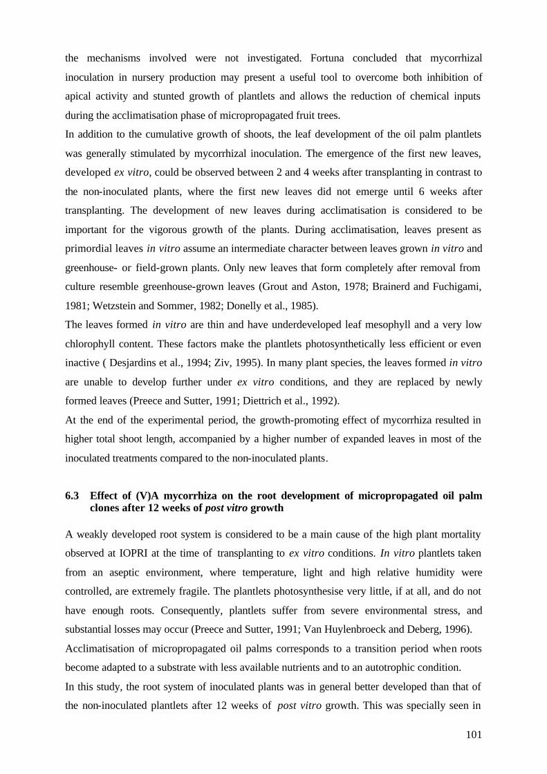
101
the mechanisms involved were not investigated. Fortuna concluded that mycorrhizal
inoculation in nursery production may present a useful tool to overcome both inhibition of
apical activity and stunted growth of plantlets and allows the reduction of chemical inputs
during the acclimatisation phase of micropropagated fruit trees.
In addition to the cumulative growth of shoots, the leaf development of the oil palm plantlets
was generally stimulated by mycorrhizal inoculation. The emergence of the first new leaves,
developed ex vitro, could be observed between 2 and 4 weeks after transplanting in contrast to
the non-inoculated plants, where the first new leaves did not emerge until 6 weeks after
transplanting. The development of new leaves during acclimatisation is considered to be
important for the vigorous growth of the plants. During acclimatisation, leaves present as
primordial leaves in vitro assume an intermediate character between leaves grown in vitro and
greenhouse- or field-grown plants. Only new leaves that form completely after removal from
culture resemble greenhouse-grown leaves (Grout and Aston, 1978; Brainerd and Fuchigami,
1981; Wetzstein and Sommer, 1982; Donelly et al., 1985).
The leaves formed in vitro are thin and have underdeveloped leaf mesophyll and a very low
chlorophyll content. These factors make the plantlets photosynthetically less efficient or even
inactive ( Desjardins et al., 1994; Ziv, 1995). In many plant species, the leaves formed in vitro
are unable to develop further under ex vitro conditions, and they are replaced by newly
formed leaves (Preece and Sutter, 1991; Diettrich et al., 1992).
At the end of the experimental period, the growth-promoting effect of mycorrhiza resulted in
higher total shoot length, accompanied by a higher number of expanded leaves in most of the
inoculated treatments compared to the non-inoculated plants.
6.3 Effect of (V)A mycorrhiza on the root development of micropropagated oil palm clones after 12 weeks of post vitro growth
A weakly developed root system is considered to be a main cause of the high plant mortality
observed at IOPRI at the time of transplanting to ex vitro conditions. In vitro plantlets taken
from an aseptic environment, where temperature, light and high relative humidity were
controlled, are extremely fragile. The plantlets photosynthesise very little, if at all, and do not
have enough roots. Consequently, plantlets suffer from severe environmental stress, and
substantial losses may occur (Preece and Sutter, 1991; Van Huylenbroeck and Deberg, 1996).
Acclimatisation of micropropagated oil palms corresponds to a transition period when roots
become adapted to a substrate with less available nutrients and to an autotrophic condition.
In this study, the root system of inoculated plants was in general better developed than that of
the non-inoculated plantlets after 12 weeks of post vitro growth. This was specially seen in
102
Experiment 1 and 3, where significantly higher root fresh weights were estimated in the
inoculated plants.
Improvement of rooting and root function due to mycorrhizal inoculation have also been
observed in several micropropagated species (Schellenbaum et al., 1991), along with greater
tolerance of environmental stress (Varma and Schüepp, 1995). There is increased evidence
that the role of mycorrhizal fungi in enhancing the survival rates of micropropagated plantlets
is based on the development of a stronger root system and enhancement in root functions
(Elmeskaoui et al., 1995). Berta et al. (1990) reported that mycorrhizal inoculation at the
weaning stage produces plants with a more effective root system for uptake of P and other
nutrients. At this stage the presence of mycorrhizas increases the availability of limiting
nutrients such as phosphorus and nitrogen by facilitating their absorption.
In this study, the symbiosis with mycorrhizal fungi enhanced both root and shoot growth and
resulted in significantly lower root:shoot fresh weight ratios for mycorrhizal plants than for
control plants. This is in line with results obtained by Azcón-Aguilar and Barea (1997). Plants
forming mycorrhizae tend to have a lower root:shoot (R:S) ratio, which means a greater
biomass efficiency, since less energy is directed to root formation. Using micropropagated
pineapple, Guillemin et al. (1991) were even able to demonstrate the existence of a negative
correlation between the mycorrhizal effect on root and shoot development. According to the
authors, the higher the root:shoot ratio, the less efficient the system for shoot production. This
mycorrhizal effect on root development is partly due to architectural changes in the root
system, as has been demonstrated in particular for micropropagated woody species such as
grapevine (Schellenbaum et al., 1991) and Prunus (Berta et al., 1994). These studies show
that mycorrhiza formation changes root topology from a herringbone pattern to a more
dichotomous pattern, the latter being more efficient for scavenging of the soil nutrients.
Pacovisky et al. (1986) reported that P-deficient plants lacking (V)AM symbiosis tend to have
a high root:shoot ratio, which is usually associated with nutrient-stressed plants.
6.4 Dry matter accumulation The role of mycorrhizae in improving the growth and productivity of seed-grown plants as a
natural symbiont has been well established (Srivastava et al., 1996). During the last few years,
the mycorrhizal technology has been used in a number of micropropagated plant species in
order to improve plant growth (Varma and Schuepp, 1994; Puthur et al., 1998; Subhan et al.,
1998).
The growth-promoting effect of mycorrhizal inoculation on shoot and leaf development
already mentioned in this study resulted in significantly higher shoot dry weights in most of

103
the inoculated oil palm plantlets. Sbrana et al. (1994) demonstrated that the inoculation of
micropropagated plantlets of apple, peach and plum resulted in improved overall growth
besides increasing the survival rate.
The screening of twelve (V)AMF species showed that ten mycorrhizal fungi were highly
effective in promoting plant growth, even when plantlets received no fertilisation. The most
effective (V)AMF isolates were selected and further investigated in their effect on plant
development. Experiment 2 investigated whether the growth-promoting effect of (V)AMF
could be achieved in plantlets that were able to obtain adequate plant nutrients by fertilisation.
The shoot growth was generally improved by (V)AMF inoculation compared to that of non-
inoculated control plants. The highest shoot dry weight was found in the inoculated plantlets
which were fertilised either with hydroxy-apatite or ‘BAYFOLAN’. Significant differences in
the shoot dry weight of the inoculated plants in both fertilisation treatments could not be
found in this experiment. The application of both, mycorrhizal inoculation and fertilisation,
ensured maximum growth of the oil palm plantlets.
The effect of soil fertilisation with slowly soluble hydroxy-apatite on plant growth in
Experiment 2 was low unless accompanied by mycorrhizal inoculation. Plantlets were grown
in a growth substrate which was of low pH, which is typical for most tropical soils. Under
such conditions inoculation with the tested (V)AMF isolates was an important factor in
facilitating plant recovery of P from these fertiliser and therefore fertiliser efficiency (Lange-
Ness and Vlek, 2000).
Blal et al. (1990) compared the influence of (vesicular-) arbuscular mycorrhizae on the
efficiency of triple superphosphate and rock phosphate fertilisers in two tropical, acid, P-
fixing soils from the Ivory Coast in which available P was labelled with 32P. Both soils were
planted with micropropagated oil palms. Both fertilisers were equally available to the plants
but the growth responses to the fertiliser applications were low unless accompanied by
(V)AM inoculation. The oil palms grew very poorly in both soils without mycorrhizal
infection, although adding phosphate fertilisers to the non-inoculated control plants led to a
significant increase in dry matter production.
The increase in oil palm growth obtained in this experiment by the combination of
mycorrhizal inoculation and fertilisation compared to the non-inoculated plants reflected the
low P-uptake capacity of the oil palm roots and the high mycorrhiza dependency of the oil
palms.
The growth-promoting effect of (V)AMF observed in this study was also confirmed in
Experiment 3. The effectiveness of the (V)AMF isolates was highly improved if the oil palms
were grown in sterilised soil.
104
In this study, there were large differences in growth between inoculated and non-inoculated
plants, confirming the importance of mycorrhizal inoculation for micropropagated oil palms,
as reported for micropropagated fruit trees (Branzanti et al., 1994) and cassava plantlets
(Azcón-Aguilar and Barea, 1997). The shoot dry weight was higher in mycorrhizal oil palm
plantlets as a consequence of the better nutritional status and active growth of inoculated
plantlets.
In investigations of Azcón-Aguilar et al. (1994), Lovato et al. (1994) and Rapparini et al.
(1994), it was reported that micropropagated plantlets which were inoculated with
mycorrhizal fungi had enhanced plant quality, nutrient uptake and plant growth. The
inoculation of plants with (V)AM fungi increases the growth of plants by several fold, and
generally most of the growth enhancement effects observed in root colonisation with (V)AM
fungi are caused by increased P absorption (Bagyaraj and Varma, 1995).
6.5 Effect of (V)A mycorrhiza on nutrient concentration and uptake of oil palm
plantlets 6.5.1 Phosphorus In this study, the inoculation with (V)AMF isolates significantly increased phosphorus
concentration and uptake of the oil palm plantlets compared to the non-inoculated controls.
The role of (V)AM fungi in uptake of nutrients, particularly P, is well documented (Bolan,
1991; Marschner and Dell, 1994; Smith et al., 1998). Estrada-Luna et al. (2000) reported that
mycorrhizal inoculation altered the nutrient uptake of micropropagated guava plantlets,
especially the leaf elemental concentration of P was significantly higher in mycorrhizal
plants. It was seen in Experiment 1 and 3 that inoculated plants showed increased P uptake
even when they were not fertilised.
The growth stimulation of mycorrhizal plants in this study may be explained by higher
mineral P concentrations in foliar tissues. Koch and Johnson (1984) and Fitter (1988) reported
that net photosynthesis of mycorrhizal plants can increase as a result of improved plant
nutritional status. Plants with an optimal P concentration should be more vigorous with high
net photosynthesis (A) and stomatal conductance (gs) than plants with limiting P (Radin and
Eidenbock, 1986). Low P can also reduce the concentration and activity of RubisCO, and
slow down the regeneration of intermediate substances in the Calvin cycle (Lauer et al. 1989).
The interaction between inoculation and fertilisation in Experiment 2 demonstrated the high
effectiveness of mycorrhizal inoculation on P uptake. The highest P uptake in this experiment
was observed in the inoculated plants which were fertilised with the slowly soluble hydroxy-
105
apatite (HA). Especially Acaulospora appendicula significantly increased P uptake of plants
compared with all inoculated plants of the non-fertilised or 'BAYFOLAN' fertilised
treatments. Mycorrhizal fungi often have access to forms of nutrients which are not directly
available to plants (Lange-Ness and Vlek, 2000), as well as being able to acquire nutrients
which are spatially separated from roots. However, it is now evident that these associations
have a greater benefit when phosphorus is present in less-soluble forms (Bolan 1991, Tarafdar
& Marschner 1994, Kahiluoto & Vestberg 1998, Lange-Ness and Vlek, 2000).
The results of Experiment 2 also indicate that mycorrhizal inoculation has a greater effect on
P uptake of shoots than fertilisation. A comparison between all non-inoculated control plants
shows that the fertilisation with the complete foliar ‘BAYFOLAN’, which is practised at
IOPRI, or fertilisation with hydroxy-apatite, does not significantly increase the P uptake of
the plants, unless accompanied by (V)AMF. The combination of both, inoculation and
fertilisation, resulted in a highly improved P uptake by the plants.
According to the results of this study, the first six weeks after transplanting to soil seem to be
the most critical for the survival and growth of the sensitive oil palm plantlets. Inoculated
plantlets of Experiment 2 harvested after six weeks of post vitro growth showed significantly
increased P concentration and uptake. At this early growth stage, the P uptake of mycorrhizal
plants was 2 times higher compared to the non-inoculated control plants. A significantly
increased nutrient concentration at that growth stage was only found for phosphorus, but not
for potassium or nitrogen. These results indicate that P seems to be the most limiting growth
factor during the early post vitro stage of the oil palm clones. Addition of fertilisers does not
greatly improve the nutrient status of propagated plants in their early growing stage because
of their relatively small root systems and inability to take up nutrients efficiently.
In Experiment 3, the beneficial effect of (V)AMF inoculation on P uptake of the plants was
confirmed. The effectiveness of the fungal isolates was far greater with soil sterilisation. The
effect of inoculation on P uptake was lower, but still significantly improved if plants grew in
non-sterilised soil. The higher effectiveness of the (V)AMF isolates may be due to the
mobilisation of nutrients, particularly P, and/or the decreased competition between native and
introduced mycorrhizal fungi, as a result of the soil sterilisation.
The mycorrhizal dependency of oil palm plantlets on P uptake during the critical post vitro
phase was illustrated in this study. The improvement of plant nutrition, especially phosphate,
is a physiological factor that can be used to evaluate the functioning of the endomycorrhizal
symbiosis.
According to a wide spectrum of research work documented in literature, phosphorus is
recognised as being the most important plant-growth limiting factor which can be supplied by
106
mycorrhizal associations, because of the many abiotic and biotic factors which can restrict its
mobility in soils (Harley & Smith 1983, Hayman 1983, Marschner 1986, Bolan 1991, etc.).
6.5.2 Potassium and Nitrogen (V)AM fungi might also increase the uptake of other nutrients that move to the root surface
primarily by diffusion (Abbott and Robson, 1984). This could explain the higher K uptake of
the inoculated oil palms in this study.
Nevertheless, no direct mycorrhizal effect on K uptake has been demonstrated to date (Harley
and Smith, 1993). Considering the importance of P and K during the early growth stages of
micropropagated plants, the better growth response in mycorrhizal plants was probably due to
the increased uptake of these two nutrients.
Matos et al. (1996) investigated the effect of endomycorrhizal inoculation on the growth of
micropropagated pineapple plants. After 12 months of growth, the mycorrhizal plants showed
significantly greater dry matter accumulation of leaves than the controls. It was also observed
that in the mycorrhizal treatments, the P and K concentrations in the leaves were higher.
These data indicate that mycorrhizal plants showed a greater vigour and a better nutritional
status when compared to the non-mycorrhizal plants. The better nutritional status of
mycorrhizal plants is primarily due to the improved plant growth as a result of increased P
uptake (Cooper, 1984; Rizardi, 1990).
Declerck et al., (1995) investigated the growth response of micropropagated banana plants to
(V)AM inoculation. Inoculation with Glomus mosseae and Glomus geosporum resulted in
significantly higher shoot fresh and dry weights as compared to the control plants. Shoot P
and K contents were also significantly higher in inoculated plants. The authors concluded that
the higher P content could be related to various mechanisms, including the larger soil volume
which reduces the distance of ion diffusion to the plant roots, and rhizosphere modifications
(Bolan, 1991). (V)AM fungi might also increase the uptake of other nutrients that move to the
root surface, primarily by diffusion (Abbott and Robson, 1984).
Improvement of the nitrogen status of the host is one of the ecologically significant
consequences of mycorrhizal associations. (V)AM fungal hyphae have the ability to extract
nitrogen and transport it from the soil to the plant due to the increased absorption surface
provided by the mycorrhizal fungi. Nitrogen exists in many forms, namely free nitrogen,
nitrate, nitrite, ammonium ions and organic nitrogen. Ammonium constitutes a significant
proportion of the inorganic nitrogen pool at a low pH level. Ammonium is less mobile in soil
because it is fixed in lattice of clay minerals and absorbed to negatively charged clay
107
minerals. (V)A mycorrhiza hyphae transport this immobile ammonium to the plant roots
(Bowen, 1987).
This study shows that inoculation with efficient (V)AMF isolates leads to an improved plant
nutritional status which is probably the cause of improved plant development and
consequently reduces plant mortality. Several authors report that (V)AM fungal colonisation
of horticultural plants improves growth by increasing uptake of P and other minerals
(Gianinazzi and Gianinazzi-Pearson, 1986; Sieverding, 1991; Pearson and Jakobsen, 1993),
thus increasing transplanting uniformity which resulted in reduced plant mortality (Biermann
and Linderman, 1983) and injury (Menge et al., 1978).
6.6 (V)AMF infectivity and effectiveness In (VA)MF symbiosis, the ability of a fungal species to infect a host plant rapidly plays an
important role in determining the success of the relationship. The main parameters, which can
be utilised to measure this type of host affinity, are fungal infectivity and the growth-
promoting ability of (V)AM fungi.
All mycorrhizal fungi tested in the initial screening of Experiment 1 had been shown to form
mycorrhizas with the oil palm plantlets. Levels of infection observed after trypan blue
staining differed between isolates and ranged in most cases between 17-23%. Plants
inoculated with Glomus mosseae (M4) showed the highest infection rate (27%) while the
lowest infection rate was observed in roots of plantlets inoculated with Glomus clarum (M8),
although inoculation with both fungi improved plant growth significantly. This would suggest
that Gl. clarum is a highly effective symbiont in this mycorrhiza-host association, even when
root infection is weak. Despite the low percentage of root infection by the tested (V)AM
fungi, the effectiveness on plant survival and on growth improvement was significant in this
study. Pinochet et al. (1997) also reported that the percentage of mycorrhizal colonisation of
Gl. interadices was relatively low at 27%, although plant growth in mycorrhizal treatments
was significantly better than in non-mycorrhizal treatments for all growth characteristics (in
terms of shoot length, number of leaves, fresh and dry weights).
Investigations of Lovato et al. (1992) showed that the growth of micropropagated grapevine
plants was improved due to inoculation with different commercial arbuscular
endomycorrhizal fungal inoculates. The authors reported that the infection levels were low
(30%) and concluded that the intensity of infection is a relative parameter (Abbot and
Robson, 1978). According to Douds et al. (1998), the physiological response of a plant is the
result of the interactions between environment, plant, and fungus genotype. Increases in the
growth rate are not always related to colonisation (Guillemin et al., 1992), but may be
108
influenced by other factors such as extension of the external mycelium and transport of
nutrients from the soil to the host.
In Experiment 2, the oil palms were infected early, during the first six weeks after inoculation,
by the used (V)AMF. Despite the relatively low percentage of (V)AMF infection (9-23%),
which varied between the fungal isolates, the colonisation was highly effective in promoting P
concentration of the inoculated clones at that early growth stage, which resulted in improved
plant development. Differences in the ability of (V)AMF to colonise the root system are well
known and may have played a part in the lower percentage of root colonisation observed in E.
columbiana (M2) inoculated plants after the first harvest.
Little is known about the early stages of (V)AM infection in micropropagated plants, through
early root colonisation has been demonstrated to be very important during the delicate
acclimatisation phase (Ravolanirina et al., 1990; Gianinazzi et al., 1990).
The infectivity of the mycorrhizal fungi was highly affected by the sterilisation of the growth
substrate as could be seen in Experiment 3. The assessment of the root colonisation at this
plant stage showed that the soil sterilisation influenced the degree to which the fungi
colonised the root. In the sterilised soil, colonisation of the roots was 27-42%, while in the
non-sterilised soil the colonisation was only 17-23%. The sterilisation of the soil reduced the
indigenous fungal population of the soil, an effect that led to a decreased competition between
the native and the introduced fungi.
Non-inoculated control plants of Experiment 3 grown in sterilised soil were not infected by
mycorrhizal fungi. According to Munro et al. (1999) the absence of mycorrhizal infection
indicates that there was no ingress of infective propagules during the experiment; this is also
indicative of the negative impacts of soil sterilisation on mycorrhizal infection and growth in
the absence of inoculation.
Weak mycorrhizal formation in the non-inoculated control plants, which occurred in all
experiments after twelve weeks of post vitro growth, was due to indigenous fungi which were
present in the native, non-sterilised soil. These colonisations indicated the natural levels of
infection, which occur in plant roots under ‘normal’ nursery conditions. The low effectiveness
of these fungi on plant survival and development was evident in this study. The (V)AMF
isolates were able to compete with the native fungi and infected the roots earlier (see 1st
harvest) and to a higher extent than the indigenous fungi, showing that they were more
effective in the development of a well functioning symbiosis.
In experiments of Vosatka et al. (2000), it was shown, as has already been stated, that the
indigenous (V)AMF are not always the most effective fungi as regards either mycorrhizal
109
development or mycorrhizal growth response tested on strawberry plants (Koomen et al.,
1988).
It can be concluded that in soils containing infective but inefficient mycorrhizal fungi, growth
of micropropagated oil palms can be greatly improved by inoculation with introduced fungal
isolates. The results of this study indicate that although mycorrhizal fungi are present in the
soil, and that the roots are eventually colonised with the native endophytes, the early
inoculation of micropropagated oil palms with selected more effective (V)AM fungi greatly
improves the resistance of the plants to the stress situation after transplanting to post vitro
conditions.
6.7 Specific effects of experimental factors and measures
6.7.1 Efficiency of the selected (V)AMF The results of this study confirmed the different efficiencies of (V)AMF isolates to be
utilized. The selected species were highly efficient for improving the survival and
development of micropropagated oil palms during the sensitive post vitro period.
6.7.2 Role of the applied P form In Experiment 2 two methods of fertilization and their effect on plant survival and
development were compared. Hydroxy-apatite a slowly soluble P fertilizer was applied to the
soil. "BAYFOLAN", a foliar spray, traditionally used by IOPRI to supply the plantlets with
nutrients via shoot, due to weakly developed roots.
"BAYFOLAN", which was directly applied on the plant shoots does not interfere with the
colonization process of mycorrhizal fungi which can be observed when soil fertilizers were
applied. Nevertheless, there are no comparable results in the literature yet.
It is to be recommended that fertilizing the soil, specially with hardly soluble P forms, like
hydroxy-apatite, was highly efficient when combined with (V)AM inoculation.
Similar results have been obtained with other several fertilizers in the production of
commercially micropropagated strawberry in the United Kingdom and in Finland. These
studies confirm that fertilizer inputs can be reduced with mycorrhizal inoculation of
micropropagated plants, whilst maintaining high production levels.
110
6.7.3 Effect of soil sterilization Some treatments with sterilized soil were involved in Experiment 3 to investigate the role of
its pathogenic potential and to eliminate indigenous (V)AM propagules. It could not be found
that the sterilization of the soil could be a measure to reduce the mortality of oil palm
plantlets. However, soil sterilization increased the infectivity and the effectiveness of the used
(V)AMF isolates. The soil sterilization gave these introduced isolates the advantage of no
competition with indigenous microorganisms, including (V)AMF, of the non-sterilized
experimental soil.
On the other side, heating or partial sterilization of soil causes physical, biological and
chemical changes (Wolf and Skipper, 1994). It is to be exspected that the soil sterilization
leds to a combined effect of mineralisation and mobilization of nutrients, such as P.
6.7.4 Role of protection measures The use of protection measures in order to prevent pathogenic contamination did not
influence plant survival. The cultivation in ‘sunbags’, positively influenced plant growth
compared to control plants which were grown under ‘normal’ pre-nursery conditions. The
plant growth promoting effect by the cultivation in ‘sunbags’ was probably due to the
development of a special micro-climate by this semi-closed system. These conditions were
more similar to in vitro conditions. Nevertheless, this protective measure cannot be
recommended due to additional costs, beside the sensitive reaction of the plantlets to be
exspected after shifting to the nursery.
7. Final conclusions and perspectives This study contributed to the idea that the application of the mycorrhizal biotechnology can
benefit the production of micropropagated plantlets of target crops. Micropropagation is a
technique of increasing importance for the mass multiplication of quality and disease free
plants of many commercial crops. The technique focuses on the major problem of a low
survival rate of these plants and their poor growth performance.
The results of this study confirm that the biotechnology of using selected (V)AMF isolates to
improve the survival and post vitro development of oil palm plantlets appears to be a
promising way to optimize the production of micropropagated oil palms.
The increase in oil palm growth achieved through the combination of mycorrhizal inoculation
and fertilization reflects the low P uptake capacity of the oil palm roots and the high
111
mycorrhiza dependency of the oil palm plantlets in the weaning stage. The mechanisms of
enhancement of survival and growth of the clonal oil palms seems to be due to the ability of
the mycorrhizal fungi in increasing plant nutrient uptake. Inoculation with (V)AM fungi
appears to became essential for the survival and growth of micropropagated oil palms.
The results of this study indicate that phosphate is probably the most limiting growth factor
especially during the early post vitro stage of the oil palm clones and illustrates the
mycorrhizal dependency of oil palm plantlets for taking up P. Addition of fertilizers does not
greatly improve the nutrient status of propagated plants in their early growing stage because
of their relatively small root systems and consequently inability to take up nutrients
efficiently. The improvement of plant nutrition, especially with phosphate, is a physiological
indicator that can be used to evaluate the functioning of the endomycorrhizal symbiosis.
Regarding future application of (V)AMF inoculation in the micropropagation process of oil
palms, an important criterion of this study was the inclusion of practical conditions, where
plants grow in native soil. The performance of 'normal' nursery plants (non-inoculated, grown
in unsterile soil), which are likely to develop some mycorrhizal infection naturally, was
compared with (V)AMF inoculated plants. The results of this study show that the inoculation
of micropropagated oil palms grown in 'normal', non-sterilized nursery soil with effective
(V)AMF can be advantageous to plant survival and development, resulting in reduction of
plant losses which occur during the acclimatization and hardening stage at IOPRI.
It can be concluded from this study that in soils containing infective but inefficient
endomycorrhizal fungal populations, growth of micropropagated oil palms can be
considerably improved by inoculation with effective (V)AM isolates. Weak mycorrhizal
formation in the non-inoculated control plants, which occurred in all experiments after twelve
weeks of post vitro growth, were due to indigenous fungi which were present in the native,
non-sterilized soil. This colonization indicate the natural low levels of infection, which occur
in plant roots under ‘normal’ nursery conditions. The low effectiveness of these fungi on plant
survival and development was evident in this study. The introduced (V)AMF isolates were
able to compete with the native fungi and infected the roots earlier and to a higher extent than
the indigenous fungi, showing that they were more effective in the development of a well
functioning symbiosis.
The benefits of inoculation are likely to extend beyond the nursery, as young oil palms which
are sturdier survive and grow better after outplanting. Further studies are necessary to ensure
that these beneficial mycorrhizal effects are maintained, and that root colonization by the
introduced fungi persists after transferring to the nursery or outplanting into field conditions.
112
Such investigations are presently hampered by the difficulty in being able to distinguish
between different (V)AM fungi within the roots, and they emphasize the need to develop
suitable immunological or nucleic acid probes to monitor control-inoculated efficient fungi
after outplanting to non-sterilized soils.
Particularly for oil palm clones, the proposed methodology appears transferable, easy to
handle with simple inputs and feasible for the ecophysiological conditions in the tropical
areas where the oil palm is a key crop. The implementation of the method adapted for
developing mycorrhizal association between roots of micropropagated oil palm plantlets and
(V)AMF isolates during the present investigation is simple and most convenient to use.
Laying crude inoculum immediately below/around the roots of the tissue-culture-raised
plantlets at the time of transferring to ex vitro conditions is sufficient to establish a beneficial
symbiotic association. This methodology is a direct single step process, being easy and
suitable for commercial applications.
Commercially available inocula or company-produced inoculum can be considered as an
inoculum source for implementation in the micropropagation process of oil palms. The latter
can be produced in the company's own propagation culture. Both must be tested for
effectiveness and could be used as single strain or mixed inoculum.
Pathogenic fungi are considered to be a main cause of the high mortality which is observed
after oil palm plantlets were transferred to soil (Ginting, personal communication). In this
study a direct connection between plant mortality and pathogenic fungi was not observed. The
plant mortality was significantly reduced by the introduction of efficient (V)AM fungi, which
resulted in an improved nutritional status of the oil palm plantlets. Plantlets which are
mycorrhizal showed a higher vitality to resist pathogenes.
The presence of mycorrhizal fungi in the roots seems to induces some kind of resistance
mechanism. The processes involved in this increased resistance are not clear, but one
possibility is a permanent activation of plant defences by the mycorrhizal fungus.
In other studies, the effectiveness of mycorrhizal fungi was dependent on the plant cultivar or
clone, a typical feature in using mycorrhizal fungi for micropropagated plants. Widiastuti and
Tahardi (1993) reported differences in plant survival between oil palm clones during an
experimental period of seven months, with no interaction between the oil palm genotype and
the tested (V)AMF isolates. The lack of interaction is not unusual as it might be due to the
relatively low host specificity of the (V)AMF species (Smith et al., 1992). Differences
between the used clones were not detected in this study, may be due to the higher number of
clones used and the low number of replicates within each clone, or the shorter period of this
113
experiment. The relatively high number of clones used in the experiments could not be
avoided due to the low number of available plantlets in total. As a consequence, the obtained
clones were equally distributed over the treatments. For future research the number of clones
with higher replicates must be taken into consideration.
In the recent years the interest on mycorrhiza has increased, partly due to economic benefits
because most of the economically important plants in agriculture, horticulture and forestry
have been found to be mycorrhizal. The savings of energy and chemical inputs due to
significantly shorter production cycles in micropropagation and the increases in survival and
uniformity of produced plants as a result of inoculation are a major incentive to introduce
(V)AMF in such techniques. Considering this benefits in the future the biotechnology of
(V)AMF inoculation will be an integral measure of most micropropagated systems. A
carefully selected and produced (V)AMF inoculum based on relevant research is urgent
needed.
In the short term, it is necessary to develop management practices taking into account the
establishment, functioning and benefits of the mycorrhizas. Parameters such as substrate
composition, forms and rates of fertilizers or other chemical products, as well as schedules for
weaning and outplanting, may be optimized by the combination with these biotechnologies.
A perspective for the future should be the development of integral biotechnologies in which
not only mycorrhizal fungi, but also other organisms capable of promoting plant growth or
protection, such as symbiontic or associative bacteria, plant-growth promoting rhizo-bacteria
(PGPR), pathogen antagonists, or hypovirulent strains of pathogens, should be incorporated
into the substrates for micropropagated oil palms.
8. Summary This study was implemented within the framework of the main project "Biotechnology
Indonesia-Germany" (BTIG), which started in 1988. Among several other sub-projects, BTIG
also included one project on the improvement of the oil palm (Elaeis guineensis Jacq.), which
is an agronomically important crop species in Indonesia. The main aim was the application of
modern techniques for improving conventional oil palm cultivation systems in Indonesia.
The ‘Indonesian Oil Palm Research Institute’ (IOPRI) is the largest oil palm seed producer in
Indonesia. IOPRI started research activities in oil palm tissue culture in 1986. During 1992
and 1993 IOPRI produced 56.401 and 76.612 plantlets, only 63% and 58% of these plantlets
were sold, respectively. This phenomenon was due to the high percentage of mortality (37-
114
42%) during the acclimatization process and after transferring the plantlets to the pre-nursery.
The low percentage of plantlet survival still is a problem for expanding the commercial
planting of clonal oil palms.
The technology used for micropropagated plants does not take into consideration the
existence of a mutualistic symbiosis of mycorrhizae and other related plant growth promoting
microorganisms like rhizobacteria. Their omission during micropropagation frequently results
in poor development of plants so that their re-introduction during plant production is
important. Most natural field soils and non-sterile nursery soils contain indigenous
mycorrhizal fungi. Under natural conditions, the roots of oil palm are colonized by (vesicular-
) arbuscular mycorrhizal fungi (Nadarajah 1993; Blal and Gianinazzi-Pearson 1989). A
decision to introduce mycorrhizal fungi under such conditions depends on the effectiveness of
these indigenous fungi, i.e. on their ability to support plant growth in comparison with
introduced fungi.
The objective of this thesis was to combine the use of (vesicular-) arbuscular mycorrhizal
fungi ((V)AMF) with the production of micropropagated oil palms with a view to future
practical applications. From 1996-1998, several experiments were carried out to determine
whether introduced (V)AMF could enhance transplanting success and plantlet acclimatization
as indicated by plant survival, plant growth and nutrient uptake.
The research for this study was carried out in collaboration with the Indonesian
"Biotechnology Research Unit for Estate Crops" in Bogor and the "Indonesian Oil Palm
Research Institute" in Medan, and with the "Institute of Plant Breeding" and the "Institute of
Plant Production in the Tropics" of the University of Göttingen in Germany.
All experiments were conducted in the greenhouse of the biotechnological research unit of
IOPRI, located in Marihat, North Sumatra. Micropropagated oil palm plantlets (E. guineensis
Jacq.) of different origins were obtained from IOPRI. Cloning of oil palm was performed by
somatic embryogenesis on calli of leaf origin according to the method of CIRAD-CP in
France developed by Pennetier et al. (1981).
Micropropagated oil palm plantlets were transplanted into polybags containing a soil-sand
mixture. The experimental soil originated from the oil palm plantation and was not sterilized
in Experiments 1 and 2. Experiment 3 included treatments with sterilized and non-sterilized
soil (95°C), to kill native endophytes. The pH (KCl) of the soil was 4.3; P2O5 (total) was 2.62
mg/100 g, and Bray-II P was 23.6 ppm.
Mycorrhizal inoculation was done at the time of transplanting. Plantlets were not inoculated
(control plants), or inoculated with 10 g of soil inoculum below the plant roots. The inoculum
115
consisted of rhizospheric soil from pot cultures containing heavily clonized root fragments
with many internal spores. The used mycorrhizal fungal species were obtained from the
(V)AMF culture collection of the "Institute of Plant Production in the Tropics", University of
Göttingen, Germany. All mycorrhizal fungi originated from various tropical locations, were
isolated and characterized, and propagated under greenhouse conditions in pure pot cultures
of different host plants.
All polybags were arranged randomly on greenhouse benches and maintained under natural
conditions with a temperature of max. 34°C during the day and min. 21°C during the night.
The greenhouse was covered by glass and the side walls were made of wire. These conditions
created an almost natural environment where plants were sheltered from direct sunlight and
rainfall. Humidity varied between 40 and 70 % during the experimental period. The oil palm
plantlets were watered as required, no fertilization was applied, except in Experiment 2.
Each experimental period lasted 3 months. During this period, survival of plants was
controlled each day. If plants died, the date of mortality was recorded. Plant growth was
determined every second week by measuring the shoot length and number of leaves. After
harvesting, the plant roots were stained and examined for (V)AMF infection. Shoots were
dried and analysed for biomass accumulation and nutrient uptake.
The first experiment of this study was conducted as an initial screening. Twelve different
mycorrhizal isolates, representing a broad range of endomycorrhizal fungi, were tested for
their effectiveness in improving the survival and development of micropropagated oil palms
during twelve weeks of post vitro growth. From twelve mycorrhizal isolates which were
introduced, eleven fungi were able to improve the survival rate significantly to 83-100%,
helping the plantlets to resist the environmental stress induced at transplanting from axenic
conditions to normal cultivation in open pots. In contrast, only 55% of the non-inoculated
control plants survived during the three-month experimental period. The used isolates were
also highly effective in promoting plant growth. The shoot dry weights of plantlets which
were inoculated with these fungal isolates significantly increased compared to the non-
inoculated plants. Furthermore, inoculated plants showed a significantly increased P uptake.
All mycorrhizal fungi tested in the initial screening had been shown to form mycorrhizas with
the oil palm plantlets. Levels of infection observed after trypan blue staining were relatively
low and differed between isolates and ranged in most cases between 17-27%. Despite the low
percentage of root infection by the tested (V)AM fungi the effectiveness on plant survival and
on growth improvement was significant in this study.
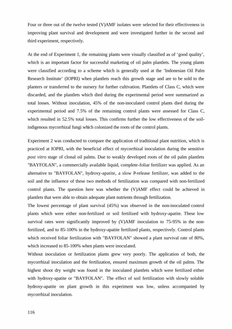
116
Four or three out of the twelve tested (V)AMF isolates were selected for their effectiveness in
improving plant survival and development and were investigated further in the second and
third experiment, respectively.
At the end of Experiment 1, the remaining plants were visually classified as of ‘good quality’,
which is an important factor for successful marketing of oil palm plantlets. The young plants
were classified according to a scheme which is generally used at the ‘Indonesian Oil Palm
Research Institute’ (IOPRI) when plantlets reach this growth stage and are to be sold to the
planters or transferred to the nursery for further cultivation. Plantlets of Class C, which were
discarded, and the plantlets which died during the experimental period were summarized as
total losses. Without inoculation, 45% of the non-inoculated control plants died during the
experimental period and 7.5% of the remaining control plants were assessed for Class C,
which resulted in 52.5% total losses. This confirms further the low effectiveness of the soil-
indigenous mycorrhizal fungi which colonized the roots of the control plants.
Experiment 2 was conducted to compare the application of traditional plant nutrition, which is
practiced at IOPRI, with the beneficial effect of mycorrhizal inoculation during the sensitive
post vitro stage of clonal oil palms. Due to weakly developed roots of the oil palm plantlets
"BAYFOLAN", a commercially available liquid, complete-foliar fertilizer was applied. As an
alternative to "BAYFOLAN", hydroxy-apatite, a slow P-release fertilizer, was added to the
soil and the influence of these two methods of fertilization was compared with non-fertilized
control plants. The question here was whether the (V)AMF effect could be achieved in
plantlets that were able to obtain adequate plant nutrients through fertilization.
The lowest percentage of plant survival (45%) was observed in the non-inoculated control
plants which were either non-fertilized or soil fertilized with hydroxy-apatite. These low
survival rates were significantly improved by (V)AMF inoculation to 75-95% in the non-
fertilized, and to 85-100% in the hydroxy-apatite fertilized plants, respectively. Control plants
which received foliar fertilization with "BAYFOLAN" showed a plant survival rate of 80%,
which increased to 85-100% when plants were inoculated.
Without inoculation or fertilization plants grew very poorly. The application of both, the
mycorrhizal inoculation and the fertilization, ensured maximum growth of the oil palms. The
highest shoot dry weight was found in the inoculated plantlets which were fertilized either
with hydroxy-apatite or "BAYFOLAN". The effect of soil fertilization with slowly soluble
hydroxy-apatite on plant growth in this experiment was low, unless accompanied by
mycorrhizal inoculation.
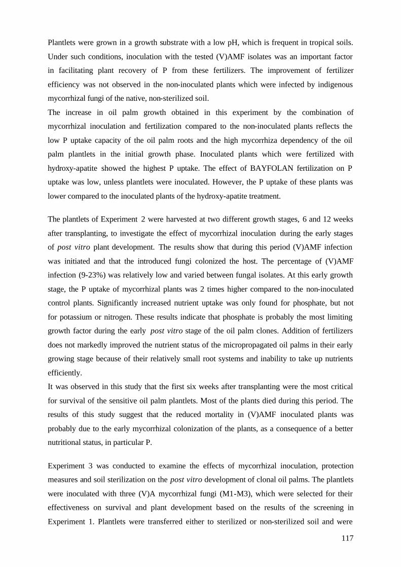
117
Plantlets were grown in a growth substrate with a low pH, which is frequent in tropical soils.
Under such conditions, inoculation with the tested (V)AMF isolates was an important factor
in facilitating plant recovery of P from these fertilizers. The improvement of fertilizer
efficiency was not observed in the non-inoculated plants which were infected by indigenous
mycorrhizal fungi of the native, non-sterilized soil.
The increase in oil palm growth obtained in this experiment by the combination of
mycorrhizal inoculation and fertilization compared to the non-inoculated plants reflects the
low P uptake capacity of the oil palm roots and the high mycorrhiza dependency of the oil
palm plantlets in the initial growth phase. Inoculated plants which were fertilized with
hydroxy-apatite showed the highest P uptake. The effect of BAYFOLAN fertilization on P
uptake was low, unless plantlets were inoculated. However, the P uptake of these plants was
lower compared to the inoculated plants of the hydroxy-apatite treatment.
The plantlets of Experiment 2 were harvested at two different growth stages, 6 and 12 weeks
after transplanting, to investigate the effect of mycorrhizal inoculation during the early stages
of post vitro plant development. The results show that during this period (V)AMF infection
was initiated and that the introduced fungi colonized the host. The percentage of (V)AMF
infection (9-23%) was relatively low and varied between fungal isolates. At this early growth
stage, the P uptake of mycorrhizal plants was 2 times higher compared to the non-inoculated
control plants. Significantly increased nutrient uptake was only found for phosphate, but not
for potassium or nitrogen. These results indicate that phosphate is probably the most limiting
growth factor during the early post vitro stage of the oil palm clones. Addition of fertilizers
does not markedly improved the nutrient status of the micropropagated oil palms in their early
growing stage because of their relatively small root systems and inability to take up nutrients
efficiently.
It was observed in this study that the first six weeks after transplanting were the most critical
for survival of the sensitive oil palm plantlets. Most of the plants died during this period. The
results of this study suggest that the reduced mortality in (V)AMF inoculated plants was
probably due to the early mycorrhizal colonization of the plants, as a consequence of a better
nutritional status, in particular P.
Experiment 3 was conducted to examine the effects of mycorrhizal inoculation, protection
measures and soil sterilization on the post vitro development of clonal oil palms. The plantlets
were inoculated with three (V)A mycorrhizal fungi (M1-M3), which were selected for their
effectiveness on survival and plant development based on the results of the screening in
Experiment 1. Plantlets were transferred either to sterilized or non-sterilized soil and were
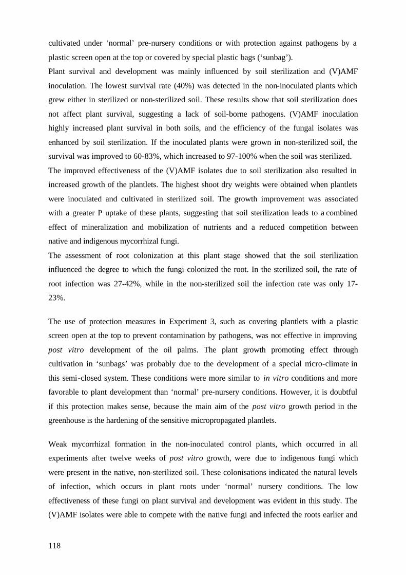
118
cultivated under ‘normal’ pre-nursery conditions or with protection against pathogens by a
plastic screen open at the top or covered by special plastic bags (‘sunbag’).
Plant survival and development was mainly influenced by soil sterilization and (V)AMF
inoculation. The lowest survival rate (40%) was detected in the non-inoculated plants which
grew either in sterilized or non-sterilized soil. These results show that soil sterilization does
not affect plant survival, suggesting a lack of soil-borne pathogens. (V)AMF inoculation
highly increased plant survival in both soils, and the efficiency of the fungal isolates was
enhanced by soil sterilization. If the inoculated plants were grown in non-sterilized soil, the
survival was improved to 60-83%, which increased to 97-100% when the soil was sterilized.
The improved effectiveness of the (V)AMF isolates due to soil sterilization also resulted in
increased growth of the plantlets. The highest shoot dry weights were obtained when plantlets
were inoculated and cultivated in sterilized soil. The growth improvement was associated
with a greater P uptake of these plants, suggesting that soil sterilization leads to a combined
effect of mineralization and mobilization of nutrients and a reduced competition between
native and indigenous mycorrhizal fungi.
The assessment of root colonization at this plant stage showed that the soil sterilization
influenced the degree to which the fungi colonized the root. In the sterilized soil, the rate of
root infection was 27-42%, while in the non-sterilized soil the infection rate was only 17-
23%.
The use of protection measures in Experiment 3, such as covering plantlets with a plastic
screen open at the top to prevent contamination by pathogens, was not effective in improving
post vitro development of the oil palms. The plant growth promoting effect through
cultivation in ‘sunbags’ was probably due to the development of a special micro-climate in
this semi-closed system. These conditions were more similar to in vitro conditions and more
favorable to plant development than ‘normal’ pre-nursery conditions. However, it is doubtful
if this protection makes sense, because the main aim of the post vitro growth period in the
greenhouse is the hardening of the sensitive micropropagated plantlets.
Weak mycorrhizal formation in the non-inoculated control plants, which occurred in all
experiments after twelve weeks of post vitro growth, were due to indigenous fungi which
were present in the native, non-sterilized soil. These colonisations indicated the natural levels
of infection, which occurs in plant roots under ‘normal’ nursery conditions. The low
effectiveness of these fungi on plant survival and development was evident in this study. The
(V)AMF isolates were able to compete with the native fungi and infected the roots earlier and
119
to a higher extent than the indigenous fungi, showing that they were more effective in the
development of a well functioning symbiosis.
To summarize, the results of this study confirm that the biotechnology of using selected
(V)AMF isolates to improve the survival and post vitro development of oil palm plantlets
appears to be a promising way to optimize the production of micropropagated oil palms.
9. Zusammenfassung Die vorliegende Arbeit wurde im Rahmen des Projektes "Biotechnology Indonesien-
Deutschland" (BTIG) durchgeführt. Durch Anwendung moderner Biotechnologie im Bereich
der Zell- und Gewebekultur sollte versucht werden, Lösungen für akute Probleme des
Ölpalmenanbaus in Indonesien zu finden. Die Ölpalme (Elaeis guineensis Jacq.) zählt in
Indonesien zu den wichtigsten landwirtschaftlich genutzten Kulturpflanzen.
Das "Indonesian Oil Palm Research Institute" (IOPRI) ist einer der größten Ölpalm-Saatgut
Erzeuger Indonesiens. 1986 begann IOPRI mit der Vermehrung der Ölpalme durch
Gewebekultur. Von 1992-1993 produzierte IOPRI 56.401 bzw. 76.612 Ölpalmklone, wovon
jedoch nur 63% bzw. 58% marktfähig waren und verkauft werden konnten. Der Grund dafür
lag in der hohen Sterblichkeitsrate der empfindlichen Jungpflanzen, die ein großes Problem
für die Kommerzialisierung von Ölpalmklonen darstellt.
In vitro vermehrte Pflanzen werden unter sterilen, künstlichen Bedingungen vermehrt. Bevor
die empfindlichen Jungpflanzen weiterkultiviert werden können, müssen sie in einem
Akklimatisierungsprozess, den natürlichen Umweltbedingungen schrittweise angepasst
werden.
Durch die sterilen Anzuchtbedingungen während der in vitro Vermehrung werden
Symbiotische Mikroorganismen, wie Mikorrhizapilze und Rhizobien, die unter natürlichen
Bedingungen im Boden vorkommen, ausgeschlossen. Die meisten natürliche Böden, sowie
nicht sterilisierte Anzuchtböden, enthalten natürliche Mykorrhizapilzpopulationen. Aus
vorangegangenen Untersuchungen ist bekannt, daß die Ölpalmen unter natürlichen
Bedingungen von Endomykorrhizapilzen besiedelt werden (Nadarajah, 1993; Blal und
Gianinazzi-Pearson, 1989). Ihre Induzierung unter diesen Umständen hängt von der
Effektivität der Mykorrhizapilze, d.h. von ihrer Fähigkeit die Entwicklung der Pflanzen zu
verbessern, ab.
120
Die vorliegende Arbeit untersuchte, ob eine Einführung (vesikulär-) arbuskulärer Mykorrhiza-
Pilz-Isolate in den Akklimatisierungsprozeß der Ölpalmen zu einer Verbesserung der
Überlebensraten und Entwicklung der empfindlichen Jungpflanzen führen kann. Im Hinblick
auf eine spätere Einbindung dieses Verfahrens in die Akklimatisierungsphase wurden praxis-
relevante Bedingungen bei der Planung und Durchführung der Versuche berücksichtigt.
Die Untersuchungen wurden in Zusammenarbeit mit der "Biotechnology Research Unit for
Estate Crops" in Bogor und dem "Indonesian Oil Palm Research Institute" in Medan, sowie
dem "Institut für Pflanzenzüchtung" und dem "Institut für Pflanzenproduktion in den Tropen
und Subtropen", an der Universität Göttingen, geplant. Alle Versuche wurden von 1996-1998
im Gewächshaus der Biotechnologischen Forschungsabteilung von IOPRI in Marihat,
Nordsumatra, durchgeführt.
Als Versuchspflanzen wurden in Gewebekultur vermehrte Ölpalmenklone, verschiedener
Herkünfte verwendet, die von IOPRI zur Verfügung gestellt worden waren. Zum Zeitpunkt
des Umpflanzens wurden die Ölpalmen in eine Boden-Sand Mischung gepflanzt, die
üblicherweise am IOPRI verwendet wird. Der ursprünglich aus Ölpalmenplantagen
stammende Versuchsboden wurde in Versuch 1 und 2 nicht sterilisiert. In Versuch 3 wurde
neben der oben genannten Bodenmischung sterilisierter Boden verwendet. Der Boden wurde
für 4 Tage auf 95°erhitzt, um natürlich vorkommende Endomykorrhizapilze abzutöten. Der
pH-Wert (KCl) des Bodens betrug 4.3; P2O5 (Gesamt) 2.62/mg/100g und Bray-II P betrug
23.6 ppm.
Die Pflanzen wurden zum Zeitpunkt des Umpflanzens mit den verwendeten (V)AM-Isolaten
inokuliert. Dazu wurde 10g des Inokulums direkt unter die Wurzeln abgelegt. Das Inokulum
bestand aus Wurzel und Bodenmaterial von Topfkulturen. Die verwendeten Mykorrhizapilze
stammten aus der Vermehrungskultur des Institutes für Pflanzenbau in den Tropen und
Subtropen der Universität Göttingen. Alle Mykorrhizapilze stammten ursprünglich aus den
Tropen und wurden in Reinkultur unter Gewächshausbedingungen vermehrt.
Die inokulierten Ölpalmen wurden während eines Versuchszeitraumes von drei Monaten im
Gewächshaus weiterkultiviert. Das Gewächshaus war mit Glas abgedeckt um die Pflanzen vor
Regen und direkter Sonneneinstahlung zu schützen. Die Seitenwände bestanden aus Draht,
wodurch nahezu natürliche Wachstumsbedingungen herrschten. Die Temperatur betrug
maximal 34°C während des Tages, und minimal 21°C während der Nacht. Die relative
Luftfeuchtigkeit schwankte zwischen 40 und 70%. Die Pflanzen wurden nach Bedarf
gegossen und, ausser in Versuch 2, nicht gedüngt.
121
Die Versuchsdauer betrug jeweils 12 Wochen. Während dieser Zeit wurde das Überleben der
Pflanzen täglich kontrolliert. Das Datum abgestorbener Pflanzen wurde festgehalten. Das
Wachstum der Pflanzen wurde in zweiwöchigen Abständen durch die Sproßlänge und die
Anzahl der Blätter ermittelt. Am Ende der Versuchsperiode wurden die Pflanzen geerntet. Die
Wurzeln wurden angefärbt, um die Infektionsraten der Mykorrhizapilze zu ermitteln. Die
Sprosse wurden getrocknet, gewogen und anschließend wurde ihr Nährstoffgehalt bestimmt.
Im ersten Versuch der vorliegenden Arbeit wurde ein breites Spektrum von
Endomykorrhizapilz Isolaten in einem sogenannten "Screening" auf ihre Wirksamkeit und
Anwendbarkeit untersucht. Von den zwölf untersuchten (V)AM-Isolaten führte die
Inokulation mit elf Isolaten zu einer Steigerung der Überlebensrate auf 83-100%. Im
Gegensatz dazu überlebten nur 55% der nicht inokulierten Ölpalmen den dreimonatigen
Versuchszeitraum. Die verwendeten Isolate wirkten sich ebenfalls sehr positiv auf die
Entwicklung der Jungpflanzen aus. Die Sproßtrockengewichte der inokulierten Pflanzen
wurden gegenüber den Kontrollpflanzen signifikant erhöht. Darüber hinaus wurde eine
signifikant erhöhte Phosphat-Aufnahme festgestellt.
Alle untersuchten Mykorrhizapilze infizierten die Wurzeln der Ölpalmen. Die Infektionsraten
waren relativ gering und variierten je nach Pilzart zwischen 17-22%. Trotz der geringen
Infektionsraten zeigten die untersuchten (V)AM-Pilze eine hohe Effizienz in der
Verbesserung der Überlebensrate und der Pflanzenentwicklung. Vier, beziehungsweise drei,
der effizientesten (V)AM-Isolate wurden ausgewählt und in den folgenden Versuchen weiter
untersucht.
Am Ende der Untersuchungsperiode wurden die überlebenden Pflanzen in Versuch 1 visuell
nach Qualitätsmerkmalen klassifiziert, einem wichtigen Faktor für die erfolgreiche
Vermarktung der jungen Ölpalmen. Die Pflanzen wurden nach einem Schema bewertet, das
normalerweise von IOPRI zu diesem Zeitpunkt verwendet wird, da die Pflanzen in diesem
Entwicklungsstadium entweder an Pflanzer verkauft, oder in der Baumschule weiterkultiviert
werden. Pflanzen der Klasse C, die nach Abschluß dieser dreimonatigen Phase nicht
ausreichend entwickelt sind, werden ausgesondert. Diese Pflanzen, sowie die während des
Untersuchungszeitraumes abgestorbenen Pflanzen, wurden zusammen als Totalverluste
bezeichnet. Ohne Inokulation starben während dieser Zeit 45% der Kontrollpflanzen ab, und
7,5% der überlebenden Pflanzen wurden mit Klasse C bewertet, woraus sich 52,5%
Totalverluste ergaben.
Versuch 2 verglich traditionell am IOPRI durchgeführte Düngungsmethoden mit dem Effekt
einer Mykorrhizapilz-Inokulation. "BAYFOLAN", ein handelsüblicher Blattdünger, wird
122
wegen des schlecht entwickelten Wurzelsystems der Ölpalmklone verwendet. Als Altenative
zu "BAYFOLAN" wurde Hydroxylapatit, ein schwerlöslicher Phosphatdünger verwendet.
Diese beiden Düngungsmethoden wurden mit nicht gedüngten Pflanzen verglichen.
Diese Untersuchung wurden durchgeführt, um festzustellen, ob die positive Wirkung der
(V)AM-Inokulation erreicht werden konnte, wenn Pflanzen gedüngt worden waren.
Die geringste Überlebensrate (45%) wurde in den nicht inokulierten, ungedüngten oder mit
Hydroxylapatit gedüngten Pflanzen festgestellt. Durch Inokulation wurden diese geringen
Überlebensraten auf 75-95% in den ungedüngten, und auf 85-100% in den Hydroxylapatit
gedüngten Pflanzen gesteigert. Nicht inokulierte Kontrollpflanzen, die mit "BAYFOLAN"
gedüngt wurden zeigten eine Überlebensrate von 80%, die auf 85-100% anstieg, wenn die
Pflanzen inokuliert waren.
Ohne Inokulation und ohne Düngung wuchsen die Pflanzen nur schwach. Die Kombination
von (V)AM-Inokulation und Düngung, entweder mit Hydroxylapatit oder "BAYFOLAN",
führte zu den besten Wachstumsraten.
Der Effekt einer Hydroxylapatit Düngung war gering, wenn die Pflanzen nicht inokuliert
waren. Der verwendete Versuchsboden hatte einen geringen pH-Wert, was in tropischen
Böden häufig vorkommt. Unter diesen Bedingungen war die Anwendung effizienter (V)AM-
Pilze ein wichtiger Faktor für die Nährstoffversorgung der Pflanzen.
Die Verbesserung des Pflanzenwachstums bei der Anwendung von Inokulation und Düngung
spiegelte die geringe P-Aufnahme-Kapazität der Ölpalmwurzeln in dieser frühen
Wachstumsphase wieder. Ohne Inokulation war der Effect der "BAYFOLAN"-Düngung auf
die P-Aufnahme der Pflanzen gering.
Die Pflanzen in Versuch 2 wurden an zwei Terminen geerntet, sechs bzw. zwölf Wochen
nach dem Umpflanzen, um die Wirkung einer Mykorrhiza-Beimpfung auf die frühe post vitro
Entwicklung der Pflanzen zu untersuchen. Die Ergebnisse zeigten, dass die Pflanzen bereits
zu diesem Zeitpunkt durch die (V)AM-Isolate infiziert waren. Die Infektionsraten waren
verhältnismäßig gering und schwankten je nach Pilzart zwischen 9% und 23%. Die P-
Aufnahme der inokulierten Pflanzen war in diesem Wachstumsstadium zweimal höher als die
der Kontrollpflanzen. Eine signifikant erhöhte Nährstoffaufnahme wurde nur bei Phosphat
gefunden, nicht jedoch für Kalium oder Stickstoff. Diese Ergebnisse deuten darauf hin, daß P
wahrscheinlich der am meisten begrenzende Wachstumsfaktor in diesem frühen post vitro
Stadium der Pflanzen war. Die Nährstoffversorgung der Pflanzen wurde, bedingt durch ein
schwach ausgebildetes Wurzelsystem, durch Düngung nicht verbessert.
123
Die meisten Pflanzen starben in den ersten sechs Wochen nach dem Umpflanzen. Dieser
Zeitraum stellt die kritischste Phase für das Überleben der empfindlichen Pflanzen dar. Die
Ergebnisse der vorliegenden Arbeit deuten darauf hin, daß die reduzierte Sterblichkeit der
(V)AM inokulierten Pflanzen auf einer frühen Besiedlung und einem verbesserten P-Status
der Pflanzen beruhte.
Versuch 3 untersuchte die Wirkung der (V)AM-Inokulation auf den Einfluß pathogener
Schaderreger, die bei der Sterblichkeit der Pflanzen eine große Rolle spielen. Die Pflanzen
wurden mit drei selektierten Mykorrhizapilzen beimpft und in sterilisierten oder nicht
sterilisierten Boden gepflanzt. Die Pflanzen wurden unter 'normalen' Gewächshaus-
bedingungen kultiviert, oder waren durch eine nach oben hin offenen Plastikfolie vor
Kontamination durch Spritzwasser geschützt. Ein Teil der Pflanzen wuchs in speziellen
Tüten, sogenannten 'sunbag's, die einen Gasaustausch erlauben, aber die Pflanzen nach außen
hin nahezu isolierten.
Der Effekt der Bodensterilisation wirkte sich nicht positiv auf das Überleben der Pflanzen
aus. 40% der nicht inokulierten Pflanzen starben, unabhangig von der Sterilisation des
Bodens. Die Inokulation der Pflanzen erhöhte die Überlebensraten der Pflanzen in beiden
Böden. Die Effizienz der Mykorrhizapilze wurde durch die Sterilisation des Bodens stark
beeinflußt. In nicht sterilisiertem Boden zeigten die inokulierten Pflanzen eine Überlebensrate
von 60-83%, die in sterilisiertem Boden auf 97-100% anstieg. Die gesteigerte Effiziens der
(V)AM-Pilze führte auch zu einem verbesserten Wachstum der Pflanzen. Die höchsten
Sproßtrockengewichte wurden erreicht, wenn die inokulierten Pflanzen in sterilisiertem
Boden kultiviert wurden. Die Wachstumsverbesserung korrelierte mit einer verbesserten P-
Aufnahme der Pflanzen. Dieser Effekt läßt vermuten, daß die Sterilisation des Bodens zu
einer Mobilisierung von Nährstoffen führte, sowie zu einer Reduzierung der Konkurrenz
zwischen natürlichen und induzierten Mykorrhiza-Pilzen.
Die Wurzeluntersuchungen zeigten, das die Bodensterilisation auch die Infektionsrate der
(V)AM beeinflußte. In sterilisiertem Boden war die Infektionsrate höher (27-42%), als in
nicht sterilisiertem Boden (17-23%).
Die Anwendung von speziellen Schutzmaßnahmen in Versuch 3, die zur Abschirmung der
Pflanzen dienen sollte, wirkte sich nicht auf die Entwicklung der Pflanzen aus. Die Förderung
des Pflanzenwachstum, die bei der Kultivierung in 'sunbags' festgestellt wurde, resultierte
wahrscheinlich aus dem speziellen Mikroklima in diesem fast geschlossenen System. Diese
Bedingungen waren vergleichbar mit in vitro Bedingungen und waren günstiger für die
Entwicklung der Pflanzen. Es ist jedoch fraglich, ob diese Art der Kultivierung sinnvoll ist, da
124
das Hauptziel der untersuchten post vitro Periode in der Abhärtung der Pflanzen an natürliche
Bedingungen liegt.
Die schwache Mykorrhiza Entwicklung, die zum Ende der jeweiligen Versuchsperiode in den
nicht inokulierten Kontrollpflanzen auftrat, war eine Folge der Infektion durch die natürlichen
Mykorrhizapilzpopulation, die in dem nicht sterilisierten Versuchsboden vorkamen. Diese
Infektion ähnelte der, die unter natürlichen Bedingungen im Gewächshaus vorkommen kann.
Die geringe Wirkung dieser Pilze auf das Überleben und die Entwicklung der Ölpalmen
wurde in dieser Untersuchung sehr deutlich. Die Inokulation der Pflanzen mit den
verwendeten (V)AM-Isolaten führte zu einer früheren and stärkeren Infektion der Wurzeln
und war effizienter in der Entwicklung einer gut fuktionierenden Symbiose.
Zusammenfassend bestätigen die Ergebnisse der vorliegenden Untersuchungen, daß die
biotechnologische Anwendung selektierter (V)AM-Isolate eine vielversprechende Perspektive
für eine verbesserte Produktion der in Gewebekultur vermehrten Ölpalmen darstellt.
125
10. References Abbott, L.K. and Robson, A.D. (1978)."Growth of subterranean clover in relation to the formation of endomycorrhizas by introduced and indigenous fungi in a field soil." New Phytologist 81: 575-587. Abbott, L.K. and Robson, A.D. (1981). "Infectivity and effectiveness of five endomycorrhizal fungi: competition with indigenous fungi in field soils." Australian Journal of Agricultural Research 32: 621-630. Abbott, L.K. and Robson, A.D. (1981). "Infectivity and effectiveness of vesicular arbuscular mycorrhizal fungi: effect of inoculum type." Australian Journal of Agricultural Research 32: 631-639. Abbott, L.K. and Robson, A.D. (1982). "Infectivity of vesicular-arbuscular mycorrhizal fungi in agricultural soils." Australian Journal of Agricultural Research 33: 1049. Abbott, L.K. and Robson, A.D. (1984). "The Effect of Mycorrhizae on Plant Growth," in VA mycorrhiza, edited by C. Ll. Powell and D. J. Bagyaraj (Boca Raton, Florida: CRC Press, Inc., 113-30. Abbott, L.K. and Robson, A.D. (1992). "Factors influencing the occurrence of vesicular-arbuscular mycorrhizas." Agriculture, Ecosystems and Environment 35: 121-150. Ahuja, M.R. (Ed.) (1993). Micropropagation of woody plants. Kluwer Academic Publishers, Dordrecht, The Netherlands. Allen, E.B. and Allen, M.F. (1980). "Natural re-establishment of vesicular-arbuscular mycorrhizae following stripmine reclamation in Wyoming." Journal of Applied Ecology 17. 137-148. Allen, M.F., Sexton, J.C., Moore, T.S., and Christensen, M. (1981). "Influence of phosphate source on vesicular-arbuscular mycorrhizae of Bouteloua gracilis." New Phytologist 87: 687-694. Allen, E.B., Allen M.F., Helm D.J., Trappe J.M. Molina R., Rincon E. (1995). Patterns and regulation of mycorrhizal plant and fungal biodiversity. Plant and Soil 170, 47-62. Allen, M.F. (1996). The ecology of arbuscular mycorrhizas: a look back into the 20th century and a peek into the 21th . Mycological Research 100 (7), 769-782. Arifin, S. and Susila, R. (1999). Development and prospect of palm oil industry in Indpnesia. IRD Journal Vol. 20, No. 2:25-32. Azcón-Aguilar, C., Barcelo, A., Vidal, M.T., and Delavina, G. (1992). "Further Studies on the influence of Mycorrhizae on Growth and Development of Micropropagated Avocado Plants." Agronomie 12: 837-840. Azcón–Aguilar, C. und Barea J.M. (1997). Applying mycorrhiza biotechnology to horticulture: significance and potentials. Scientia Horticulturae 68, 1-24.
126
Azcón-Aguilar, C., Padilla, I.G., Encina, C.L., Azcon, R., and Barea, J.M. (1997). "Arbuscular mycorrhizal inoculation enhances plant growth and changes root system morphology in micropropagated Annona cherimola mill." Agronomie 16, no. 10: 647-652. Azcón-Aguilar, C., Cantos, M., Troncoso, A., and Barea, J.M. (1997). "Beneficial effect of arbuscular mycorrhizas on acclimatization of micropropagated cassava plantlets." Scientia Horticulturae 72, no. 1: 63-71. Azcón-Aguilar, C. and Barea, J.M. (1997). "Arbuscular mycorrhizas and biological control of soil-borne plant pathogens -An overview of the mechanisms involved." Mycorrhiza 6, no. 6: 457-464. Azcón-Aguilar, C., Cantos, M., Troncoso, A., and Barea, J.M. (1997). "Beneficial effect of arbuscular mycorrhizas on acclimatization of micropropagated cassava plantlets." Scientia Horticulturae 72, no. 1: 63-71. Azcón-Aguilar, C., Barea, J.M. (1997). Arbuscular mycorrhizas and biological control of soil-borne plant pathogenes. An overview of the mechanisms involved. Mycorrhiza, 6, 457-464. Barea J.M., Calvet C., Estaún, V., und Camprubi, A. (1996). Biological control as a key component in sustainable agriculture. Plant and Soil 185, 171-172. Barea, J.M., Azcón-Aguilar, C., and Azcon, R. (1997). "Interactions between mycorrhizal fungi and rhizosphere microorganisms within the context of sustainable soil-plant systems," in Multitrophic Interactions in Terrestrial Systems, 65-77. Barea, J.M., Andrade, G., Bianciotto, V., Dowling, D., Lohrke, S., Bonfante, P., O'Gara, F., and Azcón-Aguilar, C. (1998). Impact on arbuscular mycorrhiza formation of Pseudomonas strains used as inoculants for biocontrol of soil-borne fungal plant pathogens." Applied and Environmental Microbiology 64, no. 6: 2304-2307. Basiron Y., Thiagajaran T., (1998). Asean: Oils and fats center of the world. AOCS Symposium on "Trends Global Oilseed Utilization and Processing", Chicago, Illinois, 1998. Bécard, G. and Fortin, J.A. (1988). "Early events of vesicular-arbuscular mycorrhiza formation on Ri T-DNA transformed roots." New Phytologist 108: 211-218. Bécard G., Piche Y., (1989). New aspects in the acquisition of biotrophic status by a vesicular-arbuscular mycorrhizal fungus Gigaspora margarita. New Phytologist 112: 77-83. Beirnaert, A. & Vanderweyen, R. (1941). Contribution a l'étude génétique et biométriques des variétés d' Elaeis guineensis Jacqin. Publ. Inst. Nat. Etude agron. Congo Belge, Sér. Sci., 27:101. Berta, G., Fusconi, A., Trotta, A., and Scannerini, S. (1990). "Morphogeneic modifications induced by the mycorrhizal fungus Glomus strain E3 in the root system of Allium porrum L." New Phytologist 114: 207-215. Berta, G., Trotta, A., Fusconi, A. Hooker, J.E., Munro, M., Atkinson, D., Giovanetti, M., Morini, S., Fortuna, P., Tisserant, B., Gianinazzi-Pearson, V., Gianinazzi, S. (1995).
127
Arbuscular mycorrhizal induced changes to plant growth and root system morphology in Prunus cerasifera. Tree Physiology, 15, 281-293. Bianciotto, V., Barbiero, G., and Bonfante, P. (1995). "Analysis of the cell cycle in an arbuscular mycorrhizal fungus by flow cytometry and bromodeoxyuridine labelling." Protoplasma 188, no. 3-4: 161-169. Blal, B., Gianinazzi-Pearson, V. (1989). Interest of mycorrhiza for the production of micropropagated oil palm clones. Agricultural Ecosystems Environment 29: 39-43. Blal, B., Morel, C., Gianinazzi-Pearson, V., Fardeau, J.C., and Gianinazzi, S. (1990). "Influence of vesicular-arbuscular mycorrhizae on phosphate fertilizer efficiency in two tropical acid soils planted with micropropagated oil palm (Elaeis guineensis Jacq.)." Biology and Fertility of Soils 9: 43-48. Bolan, N.S. (1991). A critical review on the role of mycorrhizal fungi in the uptake of phosphorus by plants. Plant and Soil 134, 189-207. Bonfante, P. (1995). "Arbuscular mycorrhizae: Biotechnological applications: An environmental sustainable biological agent - Foreword." Critical Reviews in Biotechnology 15, no. 3-4: U2-U3. Bowen, G.D. (1987). "The biology and physiology of infection and its development," in Ecophysiology of VA Mycorrhizal Plants, edited by G. Safir (Boca Raton, Florida: CRC Press, Inc., 27-57. Brainerd, K. E. nad Fuchigami, L.H. (1981). Acclimazization of aseptically cultured apple plants to low relative humidity. J Amer Soc Hort Sci 106:515-518. Brundrett, M.C. (1991). "Mycorrhizas in natural ecosystems," in Advances in Ecological Research vol. 21, edited by M. Begon, A. H. Fitter, and A. MacFadyen (New York: Academic Press, 171-213. Budi, S.W., Cordier, C., Trouvelot, A., Gianinazzi-Pearson, V., Gianinazzi, S., Blal, B., and Lemoine, M.C. (1998). "Arbuscular mycorrhiza as a way of promoting sustainable growth of micropropagated plants." Symposium On Plant Biotechnology As A Tool For The Exploitation Of Mountain Lands, no. 457: 71-77. Casson, A. (1999). The Hesitant Boom: Indonesia's Oil Palm Subsector in an Era of Economic Crisis and Political Change. Program on the underlying causes of deforestation. Bogor, Indonesia: Center for International Forestry Research (CIFOR). Charest, C., Dalpé, Y., and Brown, A. (1993). "The Effect of Vesicular-Arbuscular Mycorrhizae and Chilling on 2 Hybrids of Zea mays L." Mycorrhiza 4: 89-92. Chavez, M.G. and Ferrera-Cerrato, R. (1990). "Effect of vesicular-arbuscular mycorrhizae on tissue culture- derived plantlets of strawberry." Horticultural Science 25: 903-905. CIFOR: Center for International Forestry Research, Bogor, Indonesia. Online, CIFOR Homepage, http://www.cgiar.org/cifor.
128
Conner, A.J, and Thomas, M.B. (1982). Re-establishing plants from tissue culture-a review. Proc Inter Pplant Prop Coc 31: 342-357. Cooper, K.M. (1984). "Physiology of VA Mycorrhizal Associations," In: VA Mycorrhiza, edited by C. Ll. Powell and D. J. Bagyaraj (Boca Raton, Florida: CRC Press, Inc., 155-86. Cordier, C. S., Gianinazzi, and Gianinazzi-Pearson, V. (1996). Colonisation patterns of root tissues by Phytophthora nicotianae var parasitica related to reduced disease in mycorrhizal tomato. Plant and Soil 185, 223-232. Cordier, C., Trouvelot, A., Gianinazzi, S., and Gianinazzi-Pearson, V. (1997). "Arbuscular mycorrhiza technology applied to micropropagated Prunus avium and to protection against Phytophthora cinnamomi." Agronomie 16, no. 10: 679-688. Corley, R.H.V. (1976). Oil Palm Research. Developments in Crop Science. Elsevier Scientific Publishing Company. Corley, R.H.V., Barret, J.N., and Jones, L.H. (1977). Vegetative propagation of oil palm via tissue culture. Oil Palm News 22: 2-7. Corley, R.H.V., Wooi, K.C., and Wong, C.Y. (1979). Progress with vegetative propagation of oil palm. Planter 55: 377-380. Corley, R.H.V. (1982). Clonal planting material for the oil palm industry. Planter 55: 515-528. Corley, R.H.V. (1991). Fifteen years experience with oil palm clones - a review of progres. PORIM International Palm Oil Conference, 1991, Kuala Lumpur. Daniels, B.A. and Skipper, H.D. (1982). "Methods for the recovery and quantitative estimation of propagules from soil," In: Methods and principles of mycorrhizal research, edited by N. C. Schenck St. Paul: The American Phytopathological Society, 29-35. Davis, E.A. and Young, J.L. (1985). "Endomycorrhizal colonization of glasshouse-grown wheat as influenced by fertilizer salts when banded or soil-mixed." Canadian Journal of Botany 63: 1196-1203. Deberg, P.C. and Zimmermann, R.H. (ed) (1991). Micropropagation Technology and Application. Kluwer Academic Publishers, Dordrecht, Boston, London. Deberg, P. (1988). Improving mass propagation of in vitro plantlets. In: Horticulture in High Technology Era. Symp. Tokyo, Japan. May 10-11, 47-57. Declerck, S., Plenchette, C., and Strullu, D.G. (1995). "Mycorrhizal dependency of banana (Musa acuminata, AAA group) cultivar." Plant and Soil 176, no. 1: 183-187. Declerck, S., Strullu, D.G., and Plenchette, C. (1996). "In vitro mass-production of the arbuscular mycorrhizal fungus, Glomus versiforme, associated with Ri T-DNA transformed carrot roots." Mycological Research 100, no. Part 10: 1237-1242.
129
Dehne, H.W. (1982). Interaction between vesicular-arbuscular mycorrhizal fungi and plant pathogenes. Phytopathology 72: 1115-1119. Desjardins, Y. (1995). Factors affecting CO2 fixation in striving to optimize photoautotrophy in micropropagated plants. Plant Tiss Cult and Biotechnol, 1:13-25. Desjardins, Y., Laforge, F., Lussier, C., Gosselin, A. (1988). Effect of CO2 enrichement and high photosynthetic photon flux on the development of autotrophy and growth of tissue cultured strawberry, raspberry, and asparagus plants. Acta Hort, 230:45-53. Dhawan, V. and Bhojwani, S.S. (1987). Hardening in vitro and morpho-physiological changes in the leaves during acclimatization of micropropagated plants of Leucaena leucophalia L. De Wit. Plant Sci 53:65-72. Diop, T.A., Bécard, G., and Piche, Y. (1992) "Long-Term In Vitro Culture of an Endomycorrhizal Fungus, Gigaspora margarita, on Ri T-DNA Transformed Roots of Carrot." Symbiosis 12: 249-259. Dodd, J.C. and Jeffries, P. (1986). "Early development of vesicular-arbuscular mycorrhizas in autumn-sown cereals." Soil Biology and Biochemistry 18: 149-154. Dodd, J.C., Gianinazzi-Pearson, V., Rosendahl, S., and Walker, C. (1994). "European Bank of Glomales - An essential tool for efficient international and interdisciplinary collaboration," in Impact of Arbuscular Mycorrhizas on Sustainable Agriculture and Natural Ecosystems, edited by J. C. Dodd et al. (Basel, Switzerland: Birkhauser Press, 41-46. Douds D.D., Chaney W.R., (1986). The effect of high nutrition addition upon seasonal patterns of mycorrhizal development, host growth, root phosphorus and carbohydrate content in Fraxinus pennsylvanica March. New Phytol. 103: 91-106. Douds, D.D., Galvez, L., Bécard, G., and Kapulnik, Y. (1998). "Regulation of arbuscular mycorrhizal development by plant host and fungus species in alfalfa." New Phytologist 138, no. 1: 27-35. Douds, D.D. and Millner, P. (1999). "Biodiversity of arbuscular mycorrhizal fungi in agroecosystems." Agriculture, Ecosystems and Environment 74, no. 1-3: 77-93. Donelly, D.J. and Vidaver, W.E. (1984). Leaf anatomy of red raspberry transferred from culture to soil. J Amer Soc Hort Sci 109: 172-176. Donelly, D.J., Vidaver, W.E. and Lee, K.Y. (1985). The anatomy of tissue cultured raspberry prior to and after transfer to soil. Plant Cell Tissue Organ Culture 4: 43-50. Durand Gasselin, T.. Le Guen, Konan, K, Duval, Y. (1989). Oil palm (Elaeis guineensi Jacq.) plantations in Cote d'Ivoire, obtained through in vitro culture, first results. In: NIFOR (Ed.) PROC INTER CONF on Palms and Palm Products, Benin. Durand Gasselin, T.. Le Guen, Konan, K, Duval, Y. (1990). Oil palm productions in Ivory Coast obtained through in vitro culture. Oleagineux 43: 323-328.
130
Duval, Y., Durand Gasselin, T., Konan, K. and Pannetier, C. (1988). Multiplication vegetative di palmier a huile (Elaeis guineensi Jacq.) par culture in vitro. Strategie et resultats (1) Oleagineux 43: 39-49. Duval, Y. Aberlenc, F. and Touchet, B. (1995). ISOPB International Symposium on Recent Development in Oil Palm Tissue Culture and Biotechnology, 1993, Kuala Lumpur, Malaysia, pp 38-47. Duval, Y. Amblard, P. Rival A., Konan E., Gogor, S., and Durand-Gasselin, T. (1997). Progress in oil palm tissue culture and clonal performance in Indonesia and Cote d'Ivoire. Planters. Elmeskaoui, A., Damont, J.P., Poulin, M.J., Piche, Y., and Desjardins, Y. (1995). "A tripartite culture system for endomycorrhizal inoculation of micropropagated strawberry plantlets in vitro." Mycorrhiza 5, no. 5: 313-319. Estaún, V., Calvet, C., Camprubi, A., and Pinochet, J. (1999). "Long-term effects of nursery starter substrate and AM inoculation of micropropagated peach x almond hybrid rootstock GF677." Agronomie 19, no. 6: 483-489. Estrada-Luna, A.A., Davies, F.T., and Egilla, J.N. (2000). "Mycorrhizal fungi enhancement of growth and gas exchange of micropropagated guava plantlets (Psidium guajava L.) during ex vitro acclimatization and plant establishment." Mycorrhiza 10, no. 1: 1-8. Fabbri, A, Sutter, E., and Dunston, S.K. (1986). Anatomical changes in persistent leaves of tissue cultured strawberry plants after removal from culture. Scientia Hort 28:331-337. FAO: (Food and Agriculture Organization of the United Nations). Online, FAO Homepage: http://www.fao.org; FAOSTAT, statistical database. Fitter, A.H. (1991). "Costs and benefits of mycorrhizas implications for functioning under natural conditions." Experinetia 47: 350-355. Fitter, A.H. (1988). "Water relations of red clover Trifolium pratense L. as affected by VA mycorrhizal infection and phosphorus supply before and during drought." Journal of Experimental Botany 39: 595-603. Fortuna, P., Citernesi, S., Morini, S., Giovannetti, M., and Loreti, F. (1992). "Infectivity and Effectiveness of Different Species of Arbuscular Mycorrhizal Fungi in Micropropagated Plants of Mr S 2/5 Plum Rootstock." Agronomie 12: 825-829. Fortuna, P., Citernesi, A.S., Morini, S., Vitagliano, C., and Giovannetti, M. (1996). "Influence of arbuscular mycorrhizae and phosphate fertilization on shoot apical growth of micropropagated apple and plum rootstocks." Tree Physiology 16, no. 9: 757-763. Fortuna, P., Morini, S., and Giovannetti, M. (1998). "Effects of arbuscular mycorrhizal fungi on in vivo root initiation and development of micropropagated plum shoots." J Hortic Sci Biotechnol 73, no. 1: 19-28.
131
Frank, A. B. (1885). Über die Wurzelsymbiose beruhende Ernährung gewisser Bäume durch unterirdische Pilze. (On the nutrition of certain trees based on root symbiosis of underground fungi). Ber. Deutsche Botanische Gesellschaft. 3: 128-145. Fuchigami, L.H., Cheng, T.Y. and Soeldner, A. (1981). Abaxial transpiration and water loss in aseptically cultured plum. J Amer Soc Hort Sci 106; 519-522. Gaur, A. and Adholeya, A. (1999). "Mycorrhizal effects on the acclimatization, survival, growth and chlorophyll of micropropagated Syngonium and Draceana inoculated at weaning and hardening stages." Mycorrhiza 9, no. 4: 215-219. Gerdemann and Nicolson, T.H. (1963 ). "Spores of mycorrhizal endogone extracted from soil by wet-sieving and decanting." Trans. Br. Mycol. Soc. 46: 235-244 Ginaninazzi, S.; Ginaninazzi -Pearson, V.; Trouvelot, A. (1990a): "Potentialities and procedures for the use of endomycorrhizas with emphasis on high value crops." In: WHIPS, J.M. und LUMSDEN, B. (Hrsg.): Biotechnology of fungi for improving plant growth. Cambridge University Press , Cambridge , 41-54. Ginaninazzi, S.; Ginaninazzi-Pearson, V.; Trouvelot, A. (1990b): "Role and use of mycorrhizas in horticultural crop production. 23rd I.H.C. Plenary Lectures." Intern. Soc. for Hort. Sci., 25-30. Ginaninazzi, S.; Schüepp, H. ; Masson, J.P. (1995 ): Arbuscular mycorrhizas in sustainable soil-plant systems. EC , Brüssel. Gianinazzi-Pearson, V. (1981). "Ultrastructure and ultracytochemical features of Glomus tenuis mycorrhiza." New Phytologist 88: 633-639. Ginaninazzi-Pearson, V.; and Ginaninazzi, S. (1986 ): "Mycorrhizae - a potential for better use of phosphate fertilizer." Fert. Agric. 92 , 1-10. Gianinazzi-Pearson, V. and Gianinazzi, S. (1989). "Phosphorus metabolism in mycorrhizas," in Nitrogen, phosphorus and sulfur utilization by fungi, 1 ed., edited by L. Boddy, R. Marchant, and D. J. Read (New York: Cambridge University Press), 227-41. Ginting, G., Lubis, A.U., Subronto, Fatmawati (1993). Management of oil palm tissue culture production for semi-commercial scale. In: Current status of Agricultural Biotechnology in Indonesia. Giovannetti, M. and Mosse, B. (1980). "An evaluation of techniques for measuring vesicular arbuscular mycorrhizal infection in roots." New Phytologist 84: 489-500. Glenn, M.G., Chew, F.S., and Williams, P.H. (1988). "Influence of glucosinolate content of Brassica (Cruciferae) roots on growth of vesicular-arbuscular mycorrhizal fungi." New Phytologist 110: 217-225. Green, N.E., Graham, S.O., and Schenck, N.C. (1976). "The influence of pH on the germination of vesicular-arbuscular mycorrhizal spores." Mycologia 68: 929-934.
132
Grout, B.W.W. (1975). Wax development on leaf surfaces of Brassica oleraceae var. Currawong regenerated from meristem culture. Plant Sci Lett 5: 401-405. Grout, B.W.W.and Ashton, M.J. (1977). Transplanting of cauliflower plants regenerated from meristem culture. I. Water loss and water transfer related to changes in leaf wax and xylem regeneration. Hort Res 17:1-7. Grout, B.W.W.and Millam, S. (1985). Photosynthetic development of micropropagated strawberry plantlets following transplanting. Ann Bot 55: 129-131. Guillemin, J.P., Gianinazzi, S., Trouvelot, A. (1992). Screening of arbuscular-mycorrhizal fungi for establishment of micropropagated pineapple plants. Agronomie 12: 831-836. Guillemin, J.P., Gianinazzi, S., Gianinazzi-Pearson, V., and Marchal, J. (1994). "Contribution of arbuscular mycorrhizas to biological protection of micropropagated pineapple (Ananas comosus (L.) Merr) against Phytophthora cinnamomi Rands." Agricultural Science in Finland 3: 241-251. Hardon, J.J. (1984). The oil palm: Progress through plant breeding in Malaysia. Span 27: 59-61. Hardon, J.J., Corley, R.H.V., and Lee, C.H. (1987). Breeding and selecting the oil palm. In: Abbott, A.J. and Atkin, R.K. (Eds.) Improving vegetatively propagated crops. Pp. 63-81. Academic Press, London. Hamilton, R. J. (1995). Development in oils and fats. Chapman&Hall, 1995. Harley, J.L. (1971). Mycorrhiza. Oxford, UK. Harley, J.L., Smith, S.E. (1983). Mycorrhizal Symbiosis. Academic Press Inc, London. Harley, J.L., Harley, E.L. (1987) A check list of mycorrhiza in the British Flora. Supplement to: The New Phytologist 105 (2), Academic Press London. Hartley, C.W.S. (1988). The Oil Palm. Tropical Agriculture Series. Longman. Hayman, D.D. and Mosse, B. (1972). "Plant growth responses to vesicular-arbuscular mycorrhiza.III. Increased uptake of labile P from soil." New Phytologist 71: 41-47. Hooker, J.E., Gianinazzi, S., Vestberg, M., Barea, J.M., and Atkinson, D. (1994). "The application of arbuscular mycorrhizal fungi to micropropagation systems: an opportunity to reduce chemical inputs." Agricultural Science in Finland 3: 227-232. Ibrahim A. (1999). The international trade in palm oil: Prospects and challenges. PORIM, International Palm Oil Congress, 1999. Ikram, A., Mahmud, A.M., Ghani, M.N., Ibrahim, M.T., und Zainal, A.B. (1992). Field nursery inoculation of Hevea brasiliensis Muell. Arg. Seedling rootstock with vesicular-arbuscular mycorrhizal (VAM) fungi. Plant and Soil 145: 231-236.
133
Jalani, B.S., Chea, S.C., Rajanaidu, N. and Darus, A. (1997). Improvement of Palm Oil through Breeding and Biotechnology. JAOCS, Vol.74, no. 11:1451-1455. Jakarta Post (English language Indonesian daily newspaper). 12/6/1998.
Jaquemard, J.C. (1998). Oil Palm. Macmillan Education LTD, London. Jaizme-Vega M.C., und Azcón, R. (1995). Response of some tropical and subtropical cultures to endomycorrhizal fungi. Mycorrhiza 5: 213-217. Jaizme-Vega, M.C., Tenoury, P., Pinochet, J., and Jaumot, M. (1997). "Interactions between the root-knot nematode Meloidogyne incognita and Glomus mosseae in banana." Plant and Soil 196, no. 1: 27-35. Jeffries, P. and Dodd, J.C. (1991). "The use of mycorrhizal inoculants in forestry and agriculture," in Handbook of applied mycology. Vol I: Soil and Plants, edited by D. K. Arora et al. (New York: Marcel Dekker, Inc.), 155-85. Jones, L.H. (1974). Propagation of clonal oil palms by tissue culture. Oil Palm News 17: 1-8. Planter 50: 374-381. Jones, L.H. anf Hughes, W.H. (1989). Oil palm (Elaeis guineensis Jacq.) In: Bajaj YPS (Ed.) Biotechnology in Agriculture and Forestry. 5, Tree II Springer-Verlag, Berlin, Heidelberg, New York. Pp. 176-202. Kahiluoto, H. and Vestberg, M. (1998). "The effect of arbuscular mycorrhiza on biomass production and phosphorus uptake from sparingly soluble sources by leek (Allium porrum L.) in Finnish field soils." Biological Agriculture & Horticulture 16, no. 1: 65-85. Koch, K.E. and Johnson, C.R. (1984). "Photosynthate partitioning in split-root citrus seedlings with mycorrhizal and nonmycorrhizal root systems." Plant Physiology 75: 26-30. Koide, R.T. und Schneider, R.P. (1992.). Regulation of the vesicular-arbuscular mycorrhizal symbiosis. Annual Review of Plant Physiology and Plant Molecular Biology 43: 557-581. Koomen, I., Grace, C., and Hayman, D.S. (1988). "Effectiveness of single and multiple mycorrhizal inocula on growth of clover and strawberry plants at two soil pHs." Soil Biology and Biochemistry 19: 539-544. Kozai, T. (1988). High technology in protected cultivation - from environment control enginneering point of view. In: Horticulture in High Technology Era. Special Lecture. Tokyo. pp. 1-43. Kozai, T. (1991). Autotrophic micropropagation, pp 313-343. In: Bajaj, Y.P.S (Ed.) Biotechnology in Agriculture and Forestry, Vol. 17. Springer Verlag Berlin, Heidelberg, New York, pp.528. Lange-Ness, R.L. and Vlek, P.L.G. (2000). Mechanism of Calcium and Phosphate Release from Hxdroxy-Apatite by Mycorrhizal Hyphae. Soil Sci. Soc. Am. J. 64:949-955.
134
Larson, D.F. (1996). Indonesian's oil palm subsector. Policy Research Working Paper No. 1654; International Economics Department, Commodity Policy and Analysis Unit, Word Bank, Washington, DC: Lee, N., Wetzstein H.Y. and Sommer H-E. (1988). Quantum flux density effects on the anatomy and surface morphology of in vitro and in vivo developed sweetgum leaves. Amer Soc Hort Sci 113: 167-171. Li, X.-L., George, E., Marschner, H., and Zhang, J.L. (1997). "Phosphorus acquisition from compacted soil by hyphae of a mycorrhizal fungus associated with red clover (Trifolium pratense)." Canadian Journal of Botany 75, no. 5: 723-729. Lindermann, L.C. (1994). Role of VAM fungi in biocontrol. In Mycorrhizae and Plant Health. The American Phytopathological Society Press, St Paul, MN, USA; 1-27 Lovato, P., Guillemin, J.P., and Gianinazzi, S. (1992). "Application of commerical arbuscular endomycorrhizal fungal inoculants to the establishment of micropropagated grapevine rootstock and pineapple plants." Agronomie 12: 873-880. Lovato, P.E., Hammat, N.H., Gianinazzi-Pearson, V., Gianinazzi, S. (1994). Mycorrhization of micropropagated mature wild cherry (Prunus avium L.) and common ash (Fraxinus excelsior L.). Agriculture Science in Finland 3: 297-303. Lovato, P. E., Hammatt, N., Gianinazzi-Pearson, V., and Gianinazzi, S. (1994). "Mycorrhization of micropropagated mature wild cherry (Prunus avium L.) and common ash (Fraxinus excelsior L.)." Agricultural Science in Finland 3: 297-302. Lovato, P.E., Gianinazzi-Pearson, V., Trouvelot A., Gianinazzi, S. (1996). The state of art of mycorrhizas and micropropagation. Advanced Horticulture Science 10: 46-52. Lubis, A. U., Ginting, G. Fatmawati (1993). Yield and vegetative characteristics of oil palm clone planting material. Reprint A9; 1993 PIPOC PORIM Int. Palm Oil Congress 'update and vision', 20-25 September 1993; Kuala Lumpur. Lubis, A. U., Ginting, G. Subronto and Fatmawati (1993). Early performance of oil palm clones produced by IOPRI. In: Current Status of Agricultural Biotechnology in Indonesia. IOPRI. Lubis, A. U. and Daswir (1996). Progress in oil palm industry in the last ten years in Indonesia. Proceedings of the 1996 PORIM International Oil Palm Congress, Kuala Lumpur, Malaysia. Palm Oil Research Institute of Malaysia, Kuala Lumpur, pp. 1-16. Lupien, J.R. and Menza, V. (1999). Assessing prospects for improving food security and nutrition. UNDP, 1999; World Bank, 1997. Marin, J.A. and Gella, R. (1987). Acclimatization of micropropagated cherry rootstock 'Masto de Montana' (Prunus cerasus L.). Acta Hort 212: 603-609. Marschner, H. und Dell, B. (1994 ). Nutrient uptake in mycorrhizal symbiosis. Plant Soil 159: 89-102.
135
Marschner, H. (1995 ). Mineral nutrition of higher plants. Academic Press, Harcourt Brace & Co, London. Martins, A., Casimiro, A., and Pais, M.S. (1997). "Influence of mycorrhization on physiological parameters of micropropagated Castanea sativa Mill. plants." Mycorrhiza 7, no. 3: 161-165. Matsubara, Y., Karikomi, T., Ikuta, M., Hori, H., Ishikawa, S., Harada, T. (1996). Effect of vesicular-arbuscular mycorrhizal fungi inoculation on growth of apple (Malus ssp.) seedlings. J. Japanese Soc. Hort. Sci. 65, 297-302. Matos, B. and Da Silva, R. (1996). Effect of inoculation by arbuscular mycorrhizal fungi on the growth of micropropagated pineapple plants. Fruits, Vol. 51:155-119. McBeth, J. (1997). El Nino gets blamed. Far Eastern Economic Review, October 9, 1997, 80-91. Menge, J.A. (1983). Utilisation of vesicular-arbuscular mycorrhizal fungi in agriculture. Can. J. Botany 61: 1015-1024. Menge, J.A. (1984). "Inoculum production," in VA Mycorrhiza, edited by C. Ll. Powell and D. J. Bagyaraj (Boca Raton, Florida: CRC Press, Inc.), 187-203. Mengel, K. und Krikby, E.A. (1987). Principles in Plant Nutrition. International Potash Institute, Worblaufen-Bern, Switzerland. Mielke, S. (1991). Economic prospects for oilseeds, oils and fats towards the 21st century. In: Basiron, Y, and Ibrahim, A. (Ed.) The proceedings of the 1991 PORIM International Palm Oil Conference. Module iv: Promotion and Marketing. September 9-14, 1991, Kuala Lumpur. Palm Oil Research Institute of Malaysia, Kuala Lumpur, pp. 1-16. Morandi, D., Branzanti, B., and Gianinazzi-Pearson, V. (1992). "Effect of some plant flavonoids on in vitro behaviour of an arbuscular mycorrhizal fungus." Agronomie 12: 811-816. Morton, J.B. (1993). Problems and solutions for integration of glomalean taxonomy, systematic biology and the study of endomycorrhizal phenomena. Mycorrhiza 2: 97-109. Morton, J.B. (1995). "Taxonomic and phylogenetic divergence among five Scutellospora species based on comparativedevelopmental sequences." Mycologia 87, no. 1: 127-137. Mosse, B., Hayman, D.S., and Arnold, D.J. (1973). "Plant growth responses to vesicular-arbuscular mycorrhiza. V. Phosphate uptake by three plant species from P deficient soils labelled with 32P." New Phytologist 72: 809-815. Mosse, B. (1975). Specificity in VA mycorrhizas. In: Sanders, F.E., Mosse, B. und P.B. Tinker (hrdg.): Endomycorrhizas, 469-484, Academic Press, London. Mosse, B. and Hepper, C.M. (1975). "Vesicular-arbuscular mycorrhizal infections in root organ cultures." Physiological Plant Pathology 5: 215-223.

136
Mosse, B. (1988). "Mycorrhiza in a sustainable agriculture," In: The role of microorganisms in a sustainable agriculture, edited by J. M. Lopez-Real and R. D. Hodges (Great Britian: A.B. Academic Publishers, 1986), 105-23. Mosse, B. and Hayman, D.S. (1989). "Mycorrhizae in agricultural plants,". Möllers, C., und Schultz, C. (1997). Biotechnological applications for oil palm improvement. In: Proceedings of the BTIG workshop on Oil Palm Improvement through Biotechnology. Bogor, Indonesien. Munro, R.C., Wilson, J., Jefwa, J. and Mbuthia, K.W. (1999). A low-cost method of mycorrhizal inoculation improves growth of Acacia tortilis seedlings in the nursery. Forest Ecol and Management 113:51-56. Murashige, T. and Skoog, F. (1962). A revised medium for rapid growth and bioassays with tobacco tissue cultures. Physiol Plant 15: 473-497. Nadarajah, P. and Nawawi, A. (1993). Mycorrhizal status of epiphytes in malaysian oil palm plantations. Mycorrhiza 4: 21-25. Nampoothiri, K.U.K. (1998). Oil palm breeding. Journal of plantation crops 26: 1-12. Naqvi, N.S. and Mukerji, K.G. (1998). "Mycorrhization of micropropagated Leucaena leucocephala (Lam.) de Wit."Symbiosis 24, no. 1: 103-113. Naqvi, N.S. and Mukerji, K.G. (2000). "Mycorrhizal technology in plant micropropagation system," ;217-33. Nelson, C.E., und G.R. Safir (1982). Increased drought tolerance of mycorrhizal onion plants caused by improved phosphorus nutrition. Planta 154: 407-413. Noiret, J.M. (1981). Application of in vitro culture to improvement and production of clonal material of the oil palm. Oleagineux 36:123-126. Noiret, J.M., Gascon, J.P. and Pannetier, C. (1985). La production de palmier a huiele par culture In vitro. Oleagineux 543: 365-372. Nowak, J. (1998). "Benefits of in vitro "biotization" of plant tissue cultures with microbial inoculants." In Vitro Cellular & Developmental Biology-Plant 34, no. 2: 122-130. Oil World (1999). Oil World 2020: Supply, Demand and Prices from 176 through 2020. Ista Mielke GmbH Hamburg, Germany Paradis, R., Dalpé, Y., and Charest, C. (1995). "The combined effect of arbuscular mycorrhizas and short- term cold exposure on wheat." New Phytologist 129, no. 4: 637-642. Pearson, J.N. and Jakobsen, I. (1993). "Symbiotic exchange of carbon and phosphorus between cucumber and three arbuscular mycorrhizal fungi." New Phytologist 124: 481-488.
137
Pearson, J.N. and Jakobsen, I. (1993). "The relative contribution of hyphae and roots to phosphorus uptake by arbuscular mycorrhizal plants, measured by dual labelling with 32P and 33P." New Phytologist 124: 489-494. Pennetier, C., Arhuis, P. and Lievoux, D. (1981). Neoformation of young Elaeis guineensis plantlets from primary callus obtained from leaf fragments cultured in vitro. Oleagineux 36: 119-122. Phillips, J.M. and Hayman, D.S. (1970). "Improved procedures for clearing roots and staining parasitic and vesicular-arbuscular mycorrhizal fungi for rapid assessment of infection." Transactions of the British Mycological Society 55: 158-161. Pinochet, J., Calvet, C., Camprubí und Fernández, C. (1996). Interactions between migratory endoparasitic nematodes and arbuscular mycorrhizal fungi in perennial crops: A review. Plant and Soil 185, 183-190. Pinochet, J., Fernandez, C., Jaizme, M.D., and Tenoury, P. (1997). "Micropropagated banana infected with Meloidogyne javanica responds to Glomus intraradices and phosphorus." Hortscience 32, no. 1: 101-103. Ponders, F. Jr. (1984). Growth of mycorrhizal development of potted white ash and black walnut fertilized by two methods. Can J Botany 62, 509-512. Pons, F., Gianinazzi-Pearson, V., Gianinazzi, S., and Navatel, J.C. (1983). "Studies of VA mycorrhizae in vitro: Mycorrhizal synthesis of axenically propogated wild cherry (Prunus avium L.) plants." Plant and Soil 71: 217-221. Ponton, F., Piche, Y., Parent, S., and Caron, M. (1990). "Use of vesicular-arbuscular mycorrhizae in Boston fern production: II. Evaluation of four inocula." Horticultural Science 25: 416-419. Powell, C.Ll. and Bagyaraj, D.J. (1984). VA mycorrhiza. (Boca Raton, FL: CRC Press, Inc., 1984). Preece, J.E. and Sutter, E.G. (1991). Acclimatization of micropropagated plants to the greenhouse and field. In: Debergh, P.C. and Zimmermann, R.H. (ed.), Micropropagation, pp. 71-93. Kluwer Academic Publishers (1991). Puthur, J.T., Prasad, K.V.S.K., Sharmila, P. und Saradhi, P.P. (1998). Vesicular-arbuscular mycorrhizal fungi improves establishment of micropropagated Leuceana leucocephala plantlets. Plant Cell Tissue Organ Culture 53 (1), 41-47. Rabéchauld, H. and and Martin , J.P. (1976). Multiplication vegetative du palmier a huile (Elaeis guineensis Jacq.) a l'aide de cultures da tussue foliar. CR Acad Sci Paris Ser 283: 1733-1737. Rapparini, F., Baraldi, R., Bertazza, G., Branzanti, B., and Predieri, S. (1994). "Vesicular-arbuscular mycorrhizal inoculation of micropropagated fruit trees." Journal of Horticultural.Science 69, no. 6: 1101-1109.
138
Ravolanirina, F., Gianinazzi, S., Trouvelot, A., and Carre, M. (1990). "Production of endomycorrhizal explants of microprogated grapevine rootstocks." Agriculture, Ecosystems and Environment 29: 323-327. Read, D., Lewis, D., Fitter, A., und Alexander, I. (eds.) (1992). Mycorrhizas in Ecosystems. CAB International, Wallingford, UK. Rehm, S. and Espig, G. (1993). Oil plants. In: The cultivated plants of the tropics and subtropics. Weikersheim: Margraf, 1991. pp 77-125. Rival, A. Berlenc, A. Morcillo, F. Tregear, J. Verdeil, J.L. and Duval, Y. (1997). Scaling-up in vitro clonal propagation through somatic embryogenesis: the case of oil palm (Elaeis guineensis Jacq.). In: Plant Tissue Culture and Biotechnology, Vol.3 No.2: pp 74-83. Salamanca, C.P., Herrera, M.A., and Barea, J.M. (1992). "Mycorrhizal Inoculation of Micropropagated Woody Legumes Used in Revegetation Programmes for Desertified Mediterranean Ecosystems." Agronomie 12: 869-872. Sbrana, C., Giovannetti, M., and Vitagliano, C. (1994). "The effect of mycorrhizal infection on survival and growth renewal of micropropagated fruit rootstocks." Mycorrhiza 5, no. 2: 153-156. Schellenbaum, L., Müller, J., Boller, T., Wiemken, A., and Schuepp, H. (1998). "Effects of drought on non-mycorrhizal and mycorrhizal maize: changes in the pools of non-structural carbohydrates, in the activities of invertase and trehalase, and in the pools of amino acids and imino acids." New Phytologist 138, no. 1: 59-66. Schenck, N.C. (1991). Methods and Principles of Mycorrhizal Research. ASP Press, The American Phytopathological Society , St. Paul. Schenck, N.C. und Perez, Y. (1987). Manual for the identification of VA-mycorrhizal fungi. INVAM, Univ. of Florida. Schubert, A. and Martinelli, A. (1983). "Effect of vesicular-arbuscular mycorrhizae on growth of in vitro propagated Pistacia integerrima." Acta Horticulturae 227: 441-443. Schubert, A., Bodrino, C., and Gribaudo, I. (1992). "Vesicular-Arbuscular Mycorrhizal Inoculation of Kiwifruit (Actinidia deliciosa) Micropropagated Plants." Agronomie 12: 847-850. Schubert, A. and Lubraco, G. (2000). "Mycorrhizal inoculation enhances growth and nutrient uptake of micropropagated apple rootstocks during weaning in commercial substrates of high nutrient availability." Applied Soil Ecology 15, no. 2: 113-118. Schultz, C., Ginting, G., Moawad, A., Möllers, C., Pamin, K., Subronto, Tahardi, J.S., Vlek, P.L.G. (1997). The role of (vesicular-) arbuscular mycorrhiza in the weaning stage of micropropagated oil palms. In: Proceedings of the BTIG workshop on Oil Palm Improvement through Biotechnology. Bogor, Indonesien.
139
Schultz, C., Ginting, G., Moawad, A.M., und Vlek, P.L.G. (1999). Verbesserung der Überlebensrate in vitro vermehrter Ölpalmen in der Akklimatisierungsphase durch (V)A-Mykorrhizapilze. In: Backhaus, G.F., und Feldmann, F. (1999). Arbuskuläre Mykorrhiza in der Pflanzenproduktion: Praxisbeispiele und Perspektiven. Mitteilungen aus der Biologischen Bundesanstalt für Land- und Forstwirtschaft, Berlin-Dahlem. Sharmila, P., Puthur, J.T., and Saradhi, P.P. (2000). "Vesicular arbuscular mycorrhizal fungi improves establishment of micropropagated plants", 235-50. Shetty, K.G., Hetrick, B.A.D., and Schwab, A.P. (1995). "Effects of mycorrhizae and fertilizer amendments on zinc toleranceof plants." Environmental Pollution 88, no. 3: 307-314. Short, K.C., Warburton, J. and Roberts, A.V. (1987). In vitro hardening of cultured cauliflower and chrysanthenum plantlets to humidity. Acta Hort 212: 329-334. Sieverding, E. (1991). Vesicular-Arbuscular Managemant in Tropical Agrosystems. Deutsche Gesellschaft für Technische Zusammenarbeit (GTZ): Eschborn, Germany. Simon, L., Bousquet, J., Levesque, R.C., und Lalonde, M. (1993). Origin and diversification of endomycorrhizal fungi and coincidence with vascular land plants. Nature 363, 67-69. Smith, S.E. and Gianinazzi-Pearson, V. (1990). "Phosphate uptake and arbuscular activity in mycorrhizal Allium cepa L.: effects of photon irradiance and phosphate nutrition." Aust.J.Plant Physiol. 17: 177-188. Smith, S.E. and Smith, F.A. (1991). "Structure and function of the interfaces in biotrophic symbioses as they relate to nutrient transport." New Phytologist 114: 1-38. Smith, S.E., und Read, D.J. (1997). Mycorrhizal Symbiosis. 2nd ed. Academic Press, London. Srivastava, D., Kapoor, R., Srivastava, S.K. and Mkerji, K.G. (1996). Vesicular-arbuscular mycorrhiza - an overview. In: Concepts of mycorrhizal research, K.G. Mukerji, ed., Kluwer Academic Publishers, Dordrecht, Netherlands, pp.1-39. St-Arnaud, M., Hamel, C., Vimard, B., Caron, M., and Fortin, J.A. (1995). "Altered growth of Fusarium oxysporum f.sp.chrysanthemi in an in vitro dual culture system with the vesicular arbuscular mycorrhizal fungus Glomus intraradices growing on Daucus carota transformed roots." Mycorrhiza 5, no. 6: 431-438. St.John, T.V. and Hunt, H.W. (1983). "Statistical treatment of vesicular-arbuscular mycorrhizal infection data." Plant and Soil 73: 307-313. Subhan, S., Sharmila, P., Pardha Saradhi, P. (1998). Glomus fasciculatum alleviates transplantation shock of micropropagated Sesbania sesban. Plant Cell Reports 17, 268-272. Subramanian, K.S., Charest, C., Dwyer, L.M., Hamilton, R.I. (1995). Arbuscular mycorrhizas and water relation in maize under drought stress. Mycorrhiza in crop plants. Canadian journal of Botany 51, 2487-2493.
140
Sunderlein, W. D. and Ida Aju Pradnja Resosudarno (1996). Rates and causes of deforestation in Indonesia: towards the resolution of an ambiguitie. Occasional Papare No. 9, Center for International Forestry Research (CIFOR), Bogor, Indonesia. Sutter, E. (1984). Chemical compositions of epicuticular wax of cabbage plants grown in vitro. Can J Bot 62: 74-77. Sutter, E.G. (1985). Morphological, physical and chemical characteristics of epicuticular wax on ornamental plants regenerated in vitro. Ann Bot 55: 321-329. Srivastava, D. and Mukerji, K.G. (1995). "Field response of mycorrhizal and nonmycorrhizal Medicago sativa var local in the F1 generation." Mycorrhiza 5, no. 3: 219-221. Sunderlin, W.D. (1999). The effects of economic crisis and political change on Indonesia's forest sector, 1997-99. CIFOR, Bogor, Indonesia. Sylvia, D.M. and Williams, S.E. (1992). "Vesicular-arbuscular mycorrhizae and environmental stress," in Mycorrhizae in Sustainable Agriculture, edited by R. G. Linderman and G. J. Bethlenfalvay (Madison, WI: ASA Special Publication no 54: 101-24). Tarafdar, J.C. and Marschner, H. (1994). "Efficiency of VAM hyphae in utilisation of organic phosphorus by wheat plants." Soil Science and Plant Nutrition 40, no. 4: 593-600. Tarafdar, J.C. and Marschner, H. (1994). "Phosphatase activity in the rhizosphere and hyphosphere of VA mycorrhizal wheat supplied with inorganic and organic phosphorus." Soil Biology and Biochemistry 26: 387-395. Tarafdar, J.C. and Marschner, H. (1995). "Dual inoculation with Aspergillus fumigatus and Glomus mosseae enhances biomass production and nutrient uptake in wheat (Triticum aestivum L) supplied with organic phosphorus as Na-phytate." Plant and Soil 173: no. 1: 97-102. Trotta, A., Varese, G.C., Gnavi, E., Fusconi,A., Sampo, S., and Berta, B. (1996). Interactions between the soilborne root pathogen Phytophtora nicotianae var parasitica and the arbuscular mycorrhizal fungus Glomus mosseae in tomato plants. Plant and Soil 185: 199-209. Uosukainen, M. and Vestberg, M. (1994). "Effect of inoculation with arbuscular mycorrhizas on rooting, weaning and subsequent growth of micropropagated Malus (L.) Moench." Agricultural Science in Finland 3: 269-280. Vaast, P., und Zasoski, R.J., (1992). Effects of VA-mycorrhiza and nutrient sources on rhozisphere soil characteristics, growth amd nutrient acquisition of coffee seedlings (Coffea arabica L.). Plant and Soil 147, 31-39. Vaast, P., Zasoski, R.J., and Bledsoe, C.S. (1997). "Effects of vesicular-arbuscular mycorrhizal inoculation at different soil P availabilities on growth and nutrient uptake of in vitro propagated coffee (Coffea arabica l) plants." Mycorrhiza 6, no. 6: 493-497. Van Huylenbroeck, J.M., Debergh, P.C. (1996). Physiological aspects of micropropagated plantlets. Plant Tiss. Cult. And Biotechnol., 23:136-141.
141
Varma, A. and Schüepp, H. (1994). "Infectivity and Effectiveness of Glomus intraradices on Micropropagated Plants."Mycorrhiza 5, no. 1: 29-37. Varma, A. and Schüepp, H. (1994). "Positive influence of arbuscular mycorrhizal fungus on in vitro raised hortensia plantlets." Angewandte Botanik 68, no. 3-4: 108-115. Varma, A. and Schuepp, H. (1995). "Mycorrhization of the commercially important micropropagated plants." Critical Reviews in Biotechnology 15, no. 3-4: 313-328. Vestberg, M. (1992). "Arbuscular Mycorrhizal Inoculation of Micropropagated Strawberry and Field Observations in Finland." Agronomie 12: 865-867. Vidal, M.T., Azcon-G.de Aguilar, C., Barea, J.M., and Pliegoalfaro, F. (1992). "Mycorrhizal Inoculation Enhances Growth and Development of Micropropagated Plants of Avocado." Hortscience 27: 785-787. Vosátka, M. and Gryndler, M. (2000). "Response of micropropagated potatoes transplanted to peat media to post-vitro inoculation with arbuscular mycorrhizal fungi and soil bacteria." Applied Soil Ecology 15, no. 2: 145-152. Wang, H., Parent, S., Gosselin, A., and Desjardins, Y. (1993). "Vesicular-Arbuscular Mycorrhizal Peat-Based Substrates Enhance Symbiosis Establishment and Growth of Three Micropropagated Species." Journal of the American Society of Horiticultural Science: 896-901. Walker, C. (1992). Sytematics and taxonomy of the arbuscular mycorrhizal fungi (Glomales)- a possible way forward. Agronomy 12, 443-897. Wardle, K., Dobbs, E.B. and Short, K.C. (1983). In vitro acclimatization of aseptically cultured plantlets to humidity. J Amer Soc Hort Sci 108: 386-389. Weissenhorn, I., Glashoff, A., Leyval, C., and Berthelin, J. (1994). "Differential tolerance to Cd and Zn of arbuscular mycorrhizal (AM) fungal spores isolated from heavy metal- polluted and unpolluted soils." Plant and Soil 167, no. 2: 189-196. Widiastuti, H., und Tahardi, J.S. (1993). Effect of vesicular-arbuscular mycorrhizal inoculation on the growth and nutrient uptake of micropropagated oil palm. Menara Perkebunan 61, (3), 56-60. Williams, S.C.K., Vestberg, M., Uosukainen, M., Dodd, J.C., and Jeffries, P. (1992). "Effects of fertilizers and arbuscular mycorrhizal fungi on the post vitro growth of micropropagated strawberry." Agronomie 12: 851-857. Wooi, K.C. (1990). Oil palm (E. guineensis Jacq.): Tissue culture and micropropagation. In: Biotechnology in Agriculture and Forestry10: Legumes and Oilseed Crops I Vol.I; Bajaj, Y.P.S. (Ed.), Springer-Verlag Berlin, Heidelberg, New York, 1990: 570-591. Wong, G. Tan, C.C., Soh, A.C. (1997). Large scale propagation of oil palm clones- expiriences todate. Hort. Biotech. In Vitro Cult. and Breeding. (Eds. A. Altma, M. Ziv), Acta Hort 444: 649-658.
142
World Bank (1998). Commodity markets and the developing countries: A World Bank quaterly; Washington DC; 1998; 6: 1-17. Wood T. (1992 ): "VA Mycorrhizal fungi: Challenges for commercialisation." Handb. Appl. Mycol. 4 , 823-847 Wood, B.J. and Corley, R.H.V. (1991). The energy bylance of oil palm cultivation. In: Basiron, Y, Sukaimi, J., Chang, K.C., Cheah, S.C., Henson, I.E., Kamaruddin, N., Paranjothy, K., Rajanaidu, n., Dolmat, T.H.T. and Araffin, D. (Ed.) The proceedings of the 1991 PORIM International Palm Oil Conference. Kuala Lumpur. Palm Oil Research Institute of Malaysia, Kuala Lumpur, pp. 130-143.. Wood, T., und Cummings, B. (1992). Biotechnology and the future of VAM commer-cialisation. In: M. J. Allen (ed): Mycorrhizal Functioning. Chapman & Hall, New York, 468-487. Yano-Melo, A.M., Saggin, O.J., Lima, J.M., Melo, N.F., and Maia, L.C. (1999). "Effect of arbuscular mycorrhizal fungi on the acclimatization of micropropagated banana plantlets." Mycorrhiza 9, no. 2: 119-123. Zakaria, Z.Z., (1996). VA mycorrhiza and its potential on oil palm. ISOPA Newsletter: 4, 5-6. Ziv, M., Schwartz, A. and Fleminger, D. (1987). Malfunctioning stomata in vitreous leaves of carnation (Dianthus caryophyllus) plants propagated in vitro, implications of hardening. Plant Sci 52: 127-134. Ziv, M. (1991). Vitrification: morphological and physical disorders of in vitro plants. In: Deberg, P.C. and Zimmermann, R.H. (ed) (1991). Micropropagation Technology and Application. Kluwer Academic Publishers, Dordrecht, Boston
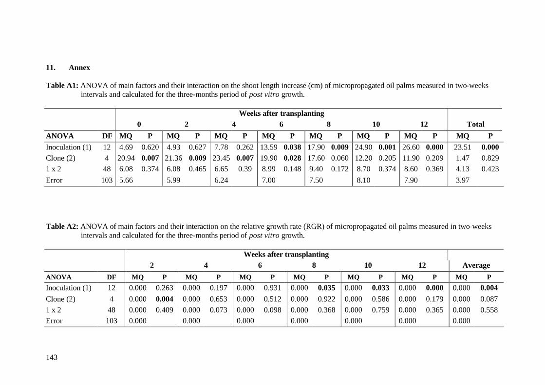
143
11. Annex Table A1: ANOVA of main factors and their interaction on the shoot length increase (cm) of micropropagated oil palms measured in two-weeks intervals and calculated for the three-months period of post vitro growth.
Weeks after transplanting 0 2 4 6 8 10 12 Total
ANOVA DF MQ P MQ P MQ P MQ P MQ P MQ P MQ P MQ P Inoculation (1) 12 4.69 0.620 4.93 0.627 7.78 0.262 13.59 0.038 17.90 0.009 24.90 0.001 26.60 0.000 23.51 0.000 Clone (2) 4 20.94 0.007 21.36 0.009 23.45 0.007 19.90 0.028 17.60 0.060 12.20 0.205 11.90 0.209 1.47 0.829 1 x 2 48 6.08 0.374 6.08 0.465 6.65 0.39 8.99 0.148 9.40 0.172 8.70 0.374 8.60 0.369 4.13 0.423 Error 103 5.66 5.99 6.24 7.00 7.50 8.10 7.90 3.97 Table A2: ANOVA of main factors and their interaction on the relative growth rate (RGR) of micropropagated oil palms measured in two-weeks intervals and calculated for the three-months period of post vitro growth.
Weeks after transplanting 2 4 6 8 10 12 Average
ANOVA DF MQ P MQ P MQ P MQ P MQ P MQ P MQ P
Inoculation (1) 12 0.000 0.263 0.000 0.197 0.000 0.931 0.000 0.035 0.000 0.033 0.000 0.000 0.000 0.004 Clone (2) 4 0.000 0.004 0.000 0.653 0.000 0.512 0.000 0.922 0.000 0.586 0.000 0.179 0.000 0.087 1 x 2 48 0.000 0.409 0.000 0.073 0.000 0.098 0.000 0.368 0.000 0.759 0.000 0.365 0.000 0.558 Error 103 0.000 0.000 0.000 0.000 0.000 0.000 0.000
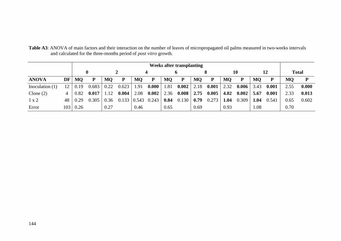
144
Table A3: ANOVA of main factors and their interaction on the number of leaves of micropropagated oil palms measured in two-weeks intervals and calculated for the three-months period of post vitro growth.
Weeks after transplanting 0 2 4 6 8 10 12 Total
ANOVA DF MQ P MQ P MQ P MQ P MQ P MQ P MQ P MQ P Inoculation (1) 12 0.19 0.683 0.22 0.623 1.91 0.000 1.81 0.002 2.18 0.001 2.32 0.006 3.43 0.001 2.55 0.000 Clone (2) 4 0.82 0.017 1.12 0.004 2.08 0.002 2.36 0.008 2.75 0.005 4.02 0.002 5.67 0.001 2.33 0.013 1 x 2 48 0.29 0.305 0.36 0.133 0.543 0.243 0.84 0.130 0.79 0.273 1.04 0.309 1.04 0.541 0.65 0.602 Error 103 0.26 0.27 0.46 0.65 0.69 0.93 1.08 0.70
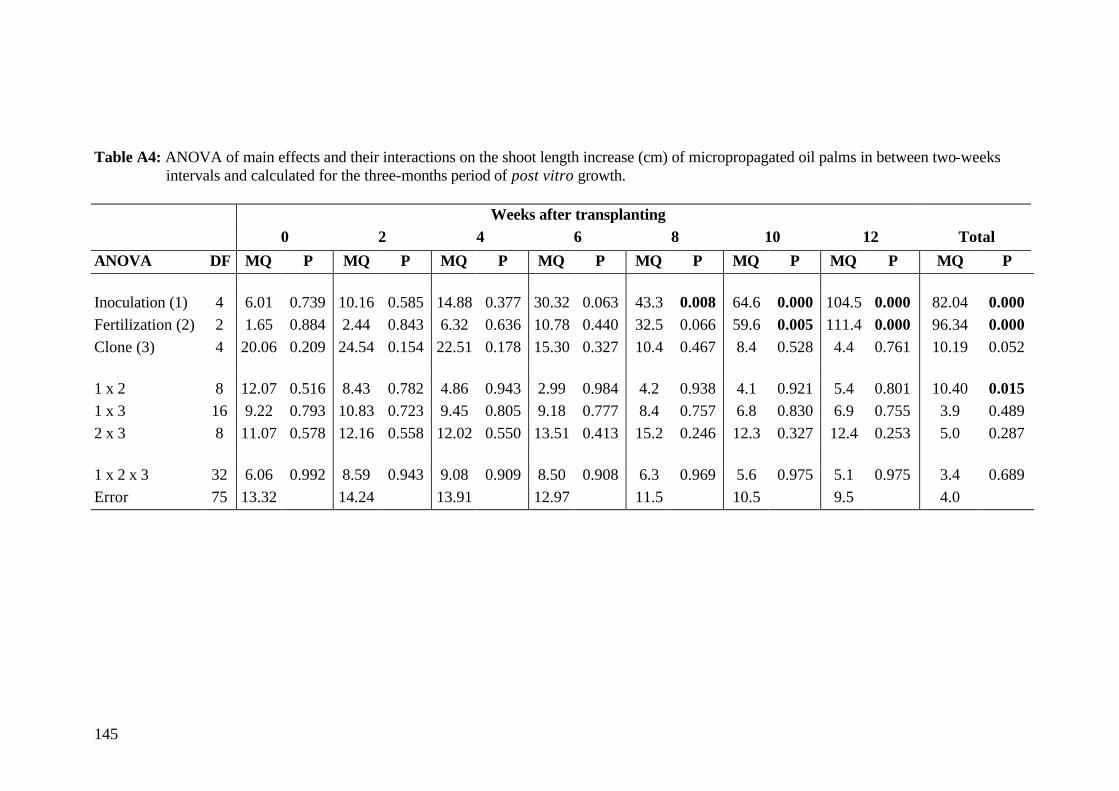
145
Table A4: ANOVA of main effects and their interactions on the shoot length increase (cm) of micropropagated oil palms in between two-weeks intervals and calculated for the three-months period of post vitro growth.
Weeks after transplanting 0 2 4 6 8 10 12 Total
ANOVA DF MQ P MQ P MQ P MQ P MQ P MQ P MQ P MQ P Inoculation (1) 4 6.01 0.739 10.16 0.585 14.88 0.377 30.32 0.063 43.3 0.008 64.6 0.000 104.5 0.000 82.04 0.000 Fertilization (2) 2 1.65 0.884 2.44 0.843 6.32 0.636 10.78 0.440 32.5 0.066 59.6 0.005 111.4 0.000 96.34 0.000 Clone (3) 4 20.06 0.209 24.54 0.154 22.51 0.178 15.30 0.327 10.4 0.467 8.4 0.528 4.4 0.761 10.19 0.052 1 x 2 8 12.07 0.516 8.43 0.782 4.86 0.943 2.99 0.984 4.2 0.938 4.1 0.921 5.4 0.801 10.40 0.015 1 x 3 16 9.22 0.793 10.83 0.723 9.45 0.805 9.18 0.777 8.4 0.757 6.8 0.830 6.9 0.755 3.9 0.489 2 x 3 8 11.07 0.578 12.16 0.558 12.02 0.550 13.51 0.413 15.2 0.246 12.3 0.327 12.4 0.253 5.0 0.287 1 x 2 x 3 32 6.06 0.992 8.59 0.943 9.08 0.909 8.50 0.908 6.3 0.969 5.6 0.975 5.1 0.975 3.4 0.689 Error 75 13.32 14.24 13.91 12.97 11.5 10.5 9.5 4.0
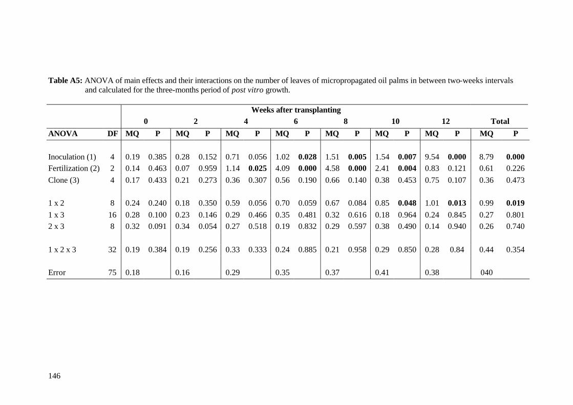
146
Table A5: ANOVA of main effects and their interactions on the number of leaves of micropropagated oil palms in between two-weeks intervals and calculated for the three-months period of post vitro growth.
Weeks after transplanting 0 2 4 6 8 10 12 Total
ANOVA DF MQ P MQ P MQ P MQ P MQ P MQ P MQ P MQ P Inoculation (1) 4 0.19 0.385 0.28 0.152 0.71 0.056 1.02 0.028 1.51 0.005 1.54 0.007 9.54 0.000 8.79 0.000 Fertilization (2) 2 0.14 0.463 0.07 0.959 1.14 0.025 4.09 0.000 4.58 0.000 2.41 0.004 0.83 0.121 0.61 0.226 Clone (3) 4 0.17 0.433 0.21 0.273 0.36 0.307 0.56 0.190 0.66 0.140 0.38 0.453 0.75 0.107 0.36 0.473 1 x 2 8 0.24 0.240 0.18 0.350 0.59 0.056 0.70 0.059 0.67 0.084 0.85 0.048 1.01 0.013 0.99 0.019 1 x 3 16 0.28 0.100 0.23 0.146 0.29 0.466 0.35 0.481 0.32 0.616 0.18 0.964 0.24 0.845 0.27 0.801 2 x 3 8 0.32 0.091 0.34 0.054 0.27 0.518 0.19 0.832 0.29 0.597 0.38 0.490 0.14 0.940 0.26 0.740 1 x 2 x 3 32 0.19 0.384 0.19 0.256 0.33 0.333 0.24 0.885 0.21 0.958 0.29 0.850 0.28 0.84 0.44 0.354 Error 75 0.18 0.16 0.29 0.35 0.37 0.41 0.38 040
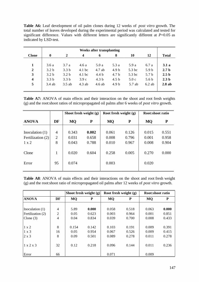
147
Table A6: Leaf development of oil palm clones during 12 weeks of post vitro growth. The total number of leaves developed during the experimental period was calculated and tested for significant difference. Values with different letters are significantly different at P=0.05 as indicated by LSD-test.
Weeks after transplanting Clone 0 2 4 6 8 10 12 Total
1 3.6 a 3.7 a 4.6 a 5.0 a 5.3 a 5.9 a 6.7 a 3.1 a 2 3.2 b 3.3 b 4.1 bc 4.7 ab 4.9 b 5.3 bc 5.9 b 2.7 b 3 3.2 b 3.2 b 4.1 bc 4.4 b 4.7 b 5.3 bc 5.7 b 2.5 b 4 3.3 b 3.3 b 3.9 c 4.3 b 4.5 b 5.0 c 5.6 b 2.3 b 5 3.4 ab 3.5 ab 4.3 ab 4.6 ab 4.9 b 5.7 ab 6.2 ab 2.8 ab
Table A7: ANOVA of main effects and their interaction on the shoot and root fresh weights (g) and the root:shoot ratios of micropropagated oil palms after 6 weeks of post vitro growth.
Shoot fresh weight (g) Root fresh weight (g) Root:shoot ratio
ANOVA DF MQ P MQ P MQ P
Inoculation (1) 4 0.343 0.002 0.061 0.126 0.015 0.551 Fertilization (2) 2 0.031 0.658 0.008 0.796 0.001 0.958 1 x 2 8 0.043 0.788 0.010 0.967 0.008 0.904
Clone 1 0.020 0.604 0.258 0.005 0.270 0.000
Error 95 0.074 0.003 0.020
Table A8: ANOVA of main effects and their interactions on the shoot and root fresh weight (g) and the root:shoot ratio of micropropagated oil palms after 12 weeks of post vitro growth.
Shoot fresh weight (g) Root fresh weight (g) Root:shoot ratio
ANOVA DF MQ P MQ P MQ P
Inoculation (1) 4 5.89 0.000 0.058 0.518 0.063 0.000 Fertilization (2) 2 0.05 0.623 0.003 0.964 0.001 0.851 Clone (3) 4 0.04 0.834 0.039 0.700 0.008 0.433
1 x 2 8 0.154 0.142 0.103 0.191 0.009 0.391 1 x 3 16 0.05 0.954 0.067 0.526 0.009 0.415 2 x 3 8 0.09 0.501 0.089 0.278 0.011 0.278
1 x 2 x 3 32 0.12 0.218 0.096 0.144 0.011 0.236
Error 66 0.071 0.009
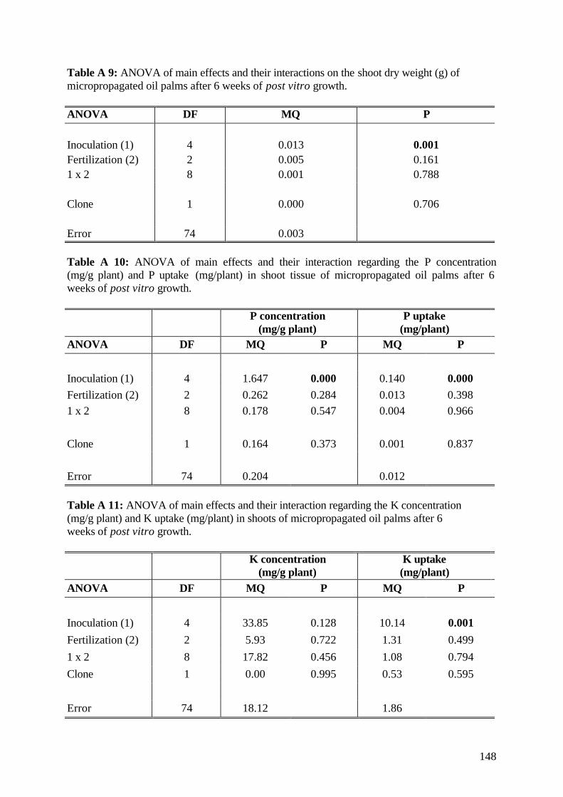
148
Table A 9: ANOVA of main effects and their interactions on the shoot dry weight (g) of micropropagated oil palms after 6 weeks of post vitro growth. ANOVA DF MQ P Inoculation (1) 4 0.013 0.001 Fertilization (2) 2 0.005 0.161 1 x 2 8 0.001 0.788 Clone 1 0.000 0.706 Error 74 0.003 Table A 10: ANOVA of main effects and their interaction regarding the P concentration (mg/g plant) and P uptake (mg/plant) in shoot tissue of micropropagated oil palms after 6 weeks of post vitro growth.
P concentration (mg/g plant) P uptake
(mg/plant)
ANOVA DF MQ P MQ P Inoculation (1) 4 1.647 0.000 0.140 0.000 Fertilization (2) 2 0.262 0.284 0.013 0.398 1 x 2 8 0.178 0.547 0.004 0.966 Clone 1 0.164 0.373 0.001 0.837 Error 74 0.204 0.012 Table A 11: ANOVA of main effects and their interaction regarding the K concentration (mg/g plant) and K uptake (mg/plant) in shoots of micropropagated oil palms after 6 weeks of post vitro growth.
K concentration (mg/g plant) K uptake
(mg/plant)
ANOVA DF MQ P MQ P
Inoculation (1) 4 33.85 0.128 10.14 0.001 Fertilization (2) 2 5.93 0.722 1.31 0.499 1 x 2 8 17.82 0.456 1.08 0.794 Clone 1 0.00 0.995 0.53 0.595 Error 74 18.12 1.86
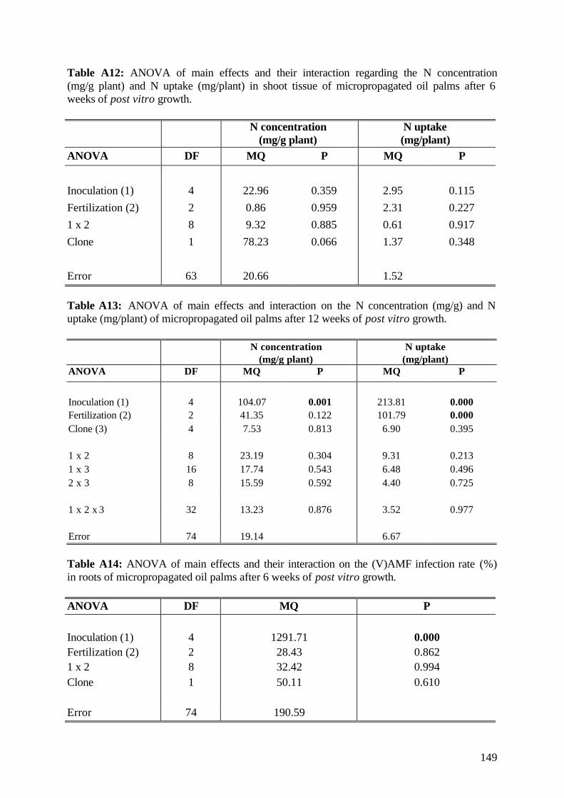
149
Table A12: ANOVA of main effects and their interaction regarding the N concentration (mg/g plant) and N uptake (mg/plant) in shoot tissue of micropropagated oil palms after 6 weeks of post vitro growth.
N concentration (mg/g plant) N uptake
(mg/plant)
ANOVA DF MQ P MQ P
Inoculation (1) 4 22.96 0.359 2.95 0.115 Fertilization (2) 2 0.86 0.959 2.31 0.227 1 x 2 8 9.32 0.885 0.61 0.917 Clone 1 78.23 0.066 1.37 0.348 Error 63 20.66 1.52 Table A13: ANOVA of main effects and interaction on the N concentration (mg/g) and N uptake (mg/plant) of micropropagated oil palms after 12 weeks of post vitro growth.
N concentration (mg/g plant)
N uptake (mg/plant)
ANOVA DF MQ P MQ P
Inoculation (1) 4 104.07 0.001 213.81 0.000 Fertilization (2) 2 41.35 0.122 101.79 0.000 Clone (3) 4 7.53 0.813 6.90 0.395 1 x 2 8 23.19 0.304 9.31 0.213 1 x 3 16 17.74 0.543 6.48 0.496 2 x 3 8 15.59 0.592 4.40 0.725 1 x 2 x 3 32 13.23 0.876 3.52 0.977 Error 74 19.14 6.67 Table A14: ANOVA of main effects and their interaction on the (V)AMF infection rate (%) in roots of micropropagated oil palms after 6 weeks of post vitro growth. ANOVA DF MQ P Inoculation (1) 4 1291.71 0.000 Fertilization (2) 2 28.43 0.862 1 x 2 8 32.42 0.994 Clone 1 50.11 0.610 Error 74 190.59
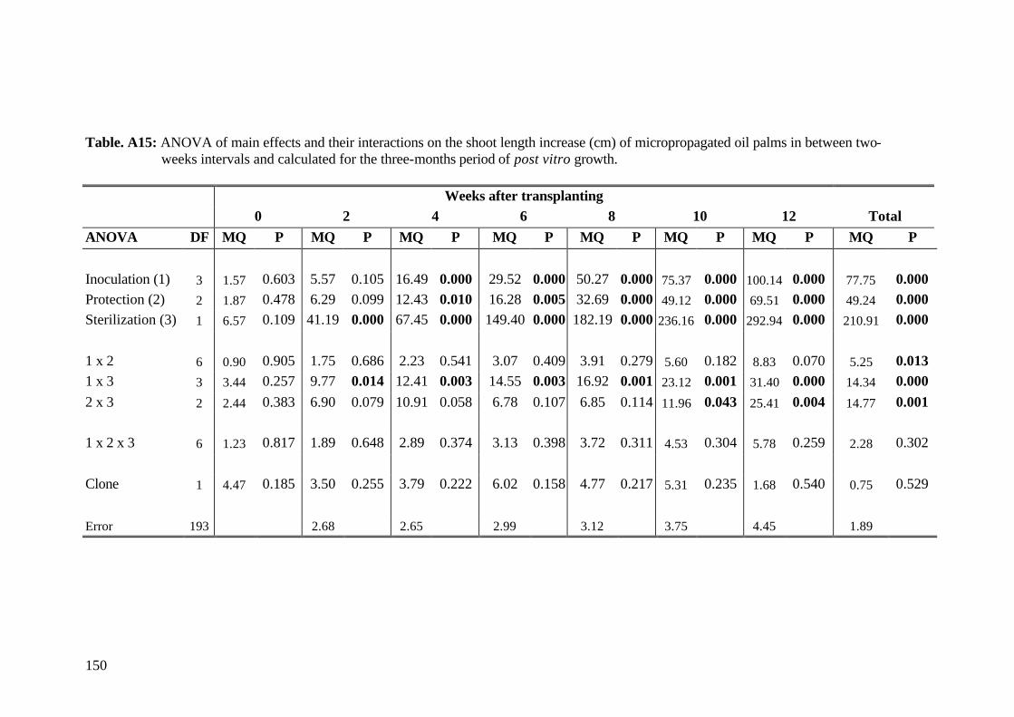
150
Table. A15: ANOVA of main effects and their interactions on the shoot length increase (cm) of micropropagated oil palms in between two- weeks intervals and calculated for the three-months period of post vitro growth.
Weeks after transplanting 0 2 4 6 8 10 12 Total
ANOVA DF MQ P MQ P MQ P MQ P MQ P MQ P MQ P MQ P
Inoculation (1) 3 1.57 0.603 5.57 0.105 16.49 0.000 29.52 0.000 50.27 0.000 75.37 0.000 100.14 0.000 77.75 0.000 Protection (2) 2 1.87 0.478 6.29 0.099 12.43 0.010 16.28 0.005 32.69 0.000 49.12 0.000 69.51 0.000 49.24 0.000 Sterilization (3) 1 6.57 0.109 41.19 0.000 67.45 0.000 149.40 0.000 182.19 0.000 236.16 0.000 292.94 0.000 210.91 0.000 1 x 2 6 0.90 0.905 1.75 0.686 2.23 0.541 3.07 0.409 3.91 0.279 5.60 0.182 8.83 0.070 5.25 0.013 1 x 3 3 3.44 0.257 9.77 0.014 12.41 0.003 14.55 0.003 16.92 0.001 23.12 0.001 31.40 0.000 14.34 0.000 2 x 3 2 2.44 0.383 6.90 0.079 10.91 0.058 6.78 0.107 6.85 0.114 11.96 0.043 25.41 0.004 14.77 0.001 1 x 2 x 3 6 1.23 0.817 1.89 0.648 2.89 0.374 3.13 0.398 3.72 0.311 4.53 0.304 5.78 0.259 2.28 0.302 Clone 1 4.47 0.185 3.50 0.255 3.79 0.222 6.02 0.158 4.77 0.217 5.31 0.235 1.68 0.540 0.75 0.529
Error 193 2.68 2.65 2.99 3.12 3.75 4.45 1.89
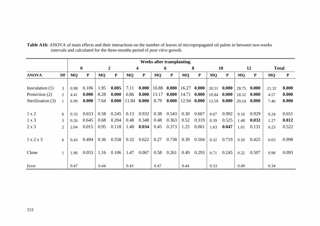
151
Table A16: ANOVA of main effects and their interactions on the number of leaves of micropropagated oil palms in between two-weeks intervals and calculated for the three-months period of post vitro growth.
Weeks after transplanting 0 2 4 6 8 10 12 Total
ANOVA DF MQ P MQ P MQ P MQ P MQ P MQ P MQ P MQ P
Inoculation (1) 3 0.98 0.106 1.95 0.005 7.11 0.000 10.88 0.000 16.27 0.000 20.51 0.000 28.75 0.000 21.32 0.000 Protection (2) 2 4.41 0.000 8.28 0.000 6.86 0.000 13.17 0.000 14.71 0.000 10.84 0.000 18.32 0.000 4.57 0.000 Sterilization (3) 1 6.99 0.000 7.64 0.000 11.84 0.000 8.79 0.000 12.94 0.000 13.59 0.000 29.54 0.000 7.46 0.000 1 x 2 6 0.33 0.653 0.58 0.245 0.13 0.932 0.38 0.543 0.30 0.667 0.07 0.992 0.16 0.929 0.24 0.651 1 x 3 3 0.26 0.645 0.68 0.204 0.48 0.348 0.48 0.363 0.52 0.319 0.39 0.525 1.48 0.032 1.27 0.012 2 x 3 2 2.04 0.015 0.95 0.118 1.48 0.034 0.45 0.373 1.25 0.061 1.63 0.047 1.01 0.131 0.23 0.522 1 x 2 x 3 6 0.43 0.494 0.36 0.558 0.32 0.622 0.27 0.738 0.39 0.504 0.32 0.719 0.50 0.425 0.03 0.998 Clone 1 1.96 0.053 1.16 0.106 1.47 0.067 0.58 0.261 0.49 0.293 0.71 0.245 0.22 0.507 0.98 0.093
Error 0.47 0.44 0.43 0.47 0.44 0.53 0.49 0.34

Acknowledgments My sincere thanks go to Prof. Dr. P.L.G. Vlek for the initiation of this study, stimulating
and valuable criticism and support.
I also like to thank Prof. H. Becker from the Institute of Agronomy and Plant Breeding,
University of Göttingen, for taking over the co-referee.
I would like to express my thanks to Dr. A.M. Moawad for his helpful and constructive
suggestions, critical comments and assistance during this study.
I would like to thank Dr. Ronald Kühne, without his statistical advises, good ideas and critical
comments the data analysis would not be accomplished that fast.
I thank all my colleagues and staff at the Institute of Agronomy in the Tropics, Göttingen for
supporting me and sharing my good and bad tempers.
I would like to thank Dr. C. Moellers for the valuable comments and support with necessary
information during the initial period of this study.
I would like to express my sincere thanks to Dr. J. Tahardi, Subronto and Gale Ginting for
good discussions, helpful suggestions and supporting me during my experimental work in
Indonesia.
I would like to express my sincere thanks to IOPRI for providing technical assistance and
research infrastructure.
Special thanks to my family, particularly my grandma for her patience and supporting me in
all the time.
I also like to thank my friends for the constant presence and moral support.
Finally I thank the BMFT for partially financing this study within the frame of the project
'Biotechnology Indonesia Germany' (BTIG). The financial support of my study by the
Institute of Agronomy in the Tropics, University of Göttingen and the Center for
Development Research (ZEF), University of Bonn, are gratefully acknowledged.
Curriculum vitae
Personal data
Name: Claudia Schultz
Date of Birth: April 10, 1964
Place of Birth: Kassel, Germany
Professional Experiences
since 1993 Research Associate at the Institute of Agronomy in the
Tropics, Georg-August University, Göttingen, Germany
Head: Prof. Dr. P.L.G. Vlek until March 1998
March 1996-November 1998 Doctoral student at the Institute of Plant Breeding,
Georg-August University, Göttingen, Germany
Head: Prof. H. Becker
October 1996-February 1998: on-station experiments at
the 'International Oil Palm Research Institute', Medan,
Indonesia
April 1994-December 1995 Post-graduate Program of Studies in Agricultural Science
in the Tropics and Subtropics, Faculty of Agriculture,
University of Göttingen, Germany;
including 6 month experimental work at ICRISAT, India
June-December 1995 Grant DAAD for the research project: "Influence of
drought stress on pearl millet (Pennisetum glaucum L.)
genotypes in India"
Education June 1993 M.Sc. Agricultural Sciences (Dipl.-Ing. Agrar)
1986-1993 University of Göttingen; Studies of Agricultural Sciences
1985-1986 One-year practice period at an agricultural research
station