Effect of oscillating fluid flow stimulation on osteocyte mRNA expression
5
Effect of oscillating fluid flow stimulation on osteocyte mRNA expression Jason Li a,b , Emily Rose b , Daniel Frances a , Yu Sun a,b , Lidan You a,b,n a Department of Mechanical and Industrial Engineering, University of Toronto, Toronto, ON, Canada b Institute of Biomaterials and Biomedical Engineering, University of Toronto, Toronto, ON, Canada article info Article history: Accepted 31 October 2011 Keywords: Osteocyte Mechanotransduction Fluid flow Bone remodeling abstract Structural adaptation of the bone tissue is mediated by loading-induced interstitial fluid flow within the bone microstructure. Within this framework, osteocytes fulfill the central mechanotransductive role in the bone remodeling process. While osteocytes have been demonstrated to be exquisitely sensitive to various forms of fluid flow stimulus in vitro, the effect of different oscillating fluid flow (OFF) parameters on osteocyte activity has yet to be systematically characterized. In this study, we investigate the effect of three OFF parameters on osteocyte activity in vitro and hypothesize that COX-2, RANKL, and OPG mRNA expression in osteocytes are sensitive to the OFF parameters: peak shear stress amplitude (0.5 Pa, 1 Pa, 2 Pa, and 5 Pa), oscillating frequency (0.5 Hz, 1 Hz, and 2 Hz), and total flow duration (1 h, 2 h, and 4 h). Our findings demonstrate that COX-2 mRNA levels are elevated in osteocytes subjected to higher peak shear stress amplitudes and longer flow durations, while RANKL/OPG mRNA levels decreased to a minimum threshold in response to higher peak shear stress amplitudes, faster oscillating frequencies, and longer flow durations. These findings suggest that dynamic fluid flow with higher peak shear stress amplitudes, faster oscillating frequencies, and longer loading durations provide the best conditions for promoting bone formation. & 2011 Elsevier Ltd. All rights reserved. 1. Introduction Bone is a dynamic tissue capable of loading-induced structural adaptation through the process of bone remodeling. While this concept is well accepted, the underlying mechanisms by which it is accomplished remain poorly understood. Mounting evidence suggest that (a) tissue-level strains are translated to cellular-level mechanical stimuli in the form of interstitial fluid flow within the lacunar–canalicular system, a fluid-filled network of intercon- nected pores permeating throughout the bone tissue (Wang et al., 2004; Fritton and Weinbaum, 2009; Kwon and Frangos, 2010; Price et al., 2011) and (b) osteocytes, terminally differentiated osteoblastic cells that reside within the lacunar–canalicular sys- tem, are able to sense and respond to mechanical stimulation (Cowin et al., 1995; Klein-Nulend et al., 1995; Jacobs et al., 1998; You et al., 2001; Han et al., 2004; Ponik et al., 2007; Huo et al., 2010; Rath et al., 2010; Cheung et al., 2011). In vitro mechanical stimulation of osteocytes has been demon- strated to regulate the production of several soluble signaling molecules known to influence osteoblast and osteoclast activity including prostaglandin E 2 (PGE 2 ), receptor activator of nuclear factor kappa B (NF-kB) ligand (RANKL), and osteoprotegerin (OPG) (Ajubi et al., 1999; Tan et al., 2007; You et al., 2008; Kamel et al., 2010; Kitase et al., 2010). PGE 2 acts on osteoblasts in a paracrine fashion to promote increased bone formation while RANKL, through complexation with RANK (receptor activator of nuclear factor kappa B) receptors found on the surface of osteoclast precursors, stimulates pre-osteoclast commitment to the osteoclastic phenotype such that the total amount of bone resorption is increased. OPG acts as a decoy receptor that competes with RANK for the binding of RANKL. The relative abundance of RANKL to OPG (RANKL/OPG) is indicative of the amount of bone resorption (Hofbauer et al., 1999; Nagai and Sato, 1999; Kim et al., 2006). At the gene transcription level, physical stimulation of osteocytes has been shown to simultaneously decrease the RANKL/OPG mRNA ratio and increase the cycloox- ygenase-2 (COX-2) mRNA expression. COX-2 is an essential enzyme in the synthesis of PGE 2 and its abundance can be directly correlated to PGE 2 release since PGE 2 is synthesized and released as needed rather than being stored by the cell. Taken together, osteocytes exposed to physiological level of mechanical stimuli produce and secrete soluble signaling molecules that regulate osteoblast and osteoclast activity in a paracrine fashion with the net effect of increasing bone formation. The degree of cellular response elicited by osteocytes sub- jected to fluid flow stimulation remains largely unexplored and has been suggested to be a function of several dynamic fluid flow parameters including the applied peak shear stress amplitude, dynamic flow frequency, flow duration, and the number of loading cycles (Reich et al., 1990; Williams et al., 1994; Jacobs et al., 1998; Bacabac et al., 2004). Of these four parameters, the Contents lists available at SciVerse ScienceDirect journal homepage: www.elsevier.com/locate/jbiomech www.JBiomech.com Journal of Biomechanics 0021-9290/$ - see front matter & 2011 Elsevier Ltd. All rights reserved. doi:10.1016/j.jbiomech.2011.10.037 n Corresponding author at: Department of Mechanical and Industrial Engineering, University of Toronto, Toronto, ON, Canada. Tel.: þ1 416 978 5736; fax: þ1 416 978 7753. E-mail address: [email protected] (L. You). Journal of Biomechanics 45 (2012) 247–251
Transcript of Effect of oscillating fluid flow stimulation on osteocyte mRNA expression
Journal of Biomechanics 45 (2012) 247–251
Contents lists available at SciVerse ScienceDirect
journal homepage: www.elsevier.com/locate/jbiomech
Journal of Biomechanics
0021-92
doi:10.1
n Corr
Univers
fax: þ1
E-m
www.JBiomech.com
Effect of oscillating fluid flow stimulation on osteocyte mRNA expression
Jason Li a,b, Emily Rose b, Daniel Frances a, Yu Sun a,b, Lidan You a,b,n
a Department of Mechanical and Industrial Engineering, University of Toronto, Toronto, ON, Canadab Institute of Biomaterials and Biomedical Engineering, University of Toronto, Toronto, ON, Canada
a r t i c l e i n f o
Article history:
Accepted 31 October 2011Structural adaptation of the bone tissue is mediated by loading-induced interstitial fluid flow within the bone
microstructure. Within this framework, osteocytes fulfill the central mechanotransductive role in the bone
Keywords:
Osteocyte
Mechanotransduction
Fluid flow
Bone remodeling
90/$ - see front matter & 2011 Elsevier Ltd. A
016/j.jbiomech.2011.10.037
esponding author at: Department of Mechanic
ity of Toronto, Toronto, ON, Canada. Tel.: þ1 4
416 978 7753.
ail address: [email protected] (L. You
a b s t r a c t
remodeling process. While osteocytes have been demonstrated to be exquisitely sensitive to various forms of
fluid flow stimulus in vitro, the effect of different oscillating fluid flow (OFF) parameters on osteocyte activity
has yet to be systematically characterized. In this study, we investigate the effect of three OFF parameters on
osteocyte activity in vitro and hypothesize that COX-2, RANKL, and OPG mRNA expression in osteocytes are
sensitive to the OFF parameters: peak shear stress amplitude (0.5 Pa, 1 Pa, 2 Pa, and 5 Pa), oscillating
frequency (0.5 Hz, 1 Hz, and 2 Hz), and total flow duration (1 h, 2 h, and 4 h). Our findings demonstrate that
COX-2 mRNA levels are elevated in osteocytes subjected to higher peak shear stress amplitudes and longer
flow durations, while RANKL/OPG mRNA levels decreased to a minimum threshold in response to higher
peak shear stress amplitudes, faster oscillating frequencies, and longer flow durations. These findings suggest
that dynamic fluid flow with higher peak shear stress amplitudes, faster oscillating frequencies, and longer
loading durations provide the best conditions for promoting bone formation.
& 2011 Elsevier Ltd. All rights reserved.
1. Introduction
Bone is a dynamic tissue capable of loading-induced structuraladaptation through the process of bone remodeling. While thisconcept is well accepted, the underlying mechanisms by which itis accomplished remain poorly understood. Mounting evidencesuggest that (a) tissue-level strains are translated to cellular-levelmechanical stimuli in the form of interstitial fluid flow within thelacunar–canalicular system, a fluid-filled network of intercon-nected pores permeating throughout the bone tissue (Wang et al.,2004; Fritton and Weinbaum, 2009; Kwon and Frangos, 2010;Price et al., 2011) and (b) osteocytes, terminally differentiatedosteoblastic cells that reside within the lacunar–canalicular sys-tem, are able to sense and respond to mechanical stimulation(Cowin et al., 1995; Klein-Nulend et al., 1995; Jacobs et al., 1998;You et al., 2001; Han et al., 2004; Ponik et al., 2007; Huo et al.,2010; Rath et al., 2010; Cheung et al., 2011).
In vitro mechanical stimulation of osteocytes has been demon-strated to regulate the production of several soluble signalingmolecules known to influence osteoblast and osteoclast activityincluding prostaglandin E2 (PGE2), receptor activator of nuclearfactor kappa B (NF-kB) ligand (RANKL), and osteoprotegerin(OPG) (Ajubi et al., 1999; Tan et al., 2007; You et al., 2008;
ll rights reserved.
al and Industrial Engineering,
16 978 5736;
).
Kamel et al., 2010; Kitase et al., 2010). PGE2 acts on osteoblasts ina paracrine fashion to promote increased bone formation whileRANKL, through complexation with RANK (receptor activator ofnuclear factor kappa B) receptors found on the surface ofosteoclast precursors, stimulates pre-osteoclast commitment tothe osteoclastic phenotype such that the total amount of boneresorption is increased. OPG acts as a decoy receptor thatcompetes with RANK for the binding of RANKL. The relativeabundance of RANKL to OPG (RANKL/OPG) is indicative of theamount of bone resorption (Hofbauer et al., 1999; Nagai and Sato,1999; Kim et al., 2006). At the gene transcription level, physicalstimulation of osteocytes has been shown to simultaneouslydecrease the RANKL/OPG mRNA ratio and increase the cycloox-ygenase-2 (COX-2) mRNA expression. COX-2 is an essentialenzyme in the synthesis of PGE2 and its abundance can be directlycorrelated to PGE2 release since PGE2 is synthesized and releasedas needed rather than being stored by the cell. Taken together,osteocytes exposed to physiological level of mechanical stimuliproduce and secrete soluble signaling molecules that regulateosteoblast and osteoclast activity in a paracrine fashion with thenet effect of increasing bone formation.
The degree of cellular response elicited by osteocytes sub-jected to fluid flow stimulation remains largely unexplored andhas been suggested to be a function of several dynamic fluid flowparameters including the applied peak shear stress amplitude,dynamic flow frequency, flow duration, and the number ofloading cycles (Reich et al., 1990; Williams et al., 1994; Jacobset al., 1998; Bacabac et al., 2004). Of these four parameters, the
J. Li et al. / Journal of Biomechanics 45 (2012) 247–251248
first three are independent parameters, while the number of loadingcycles is dependent on both the total flow duration and the dynamicflow frequency. The relationship between these flow parametersand osteocyte activity is further complicated by the observation thatosteocytes are additionally sensitive to the fluid flow stimulusprofile (e.g., steady, pulsating, and oscillating) (Jacobs et al., 1998;Mullender et al., 2006; Malone et al., 2007; Ponik et al., 2007). Theseobservations illustrate that osteocytes are exquisitely sensitive todifferent loading conditions and loading profiles. A thorough under-standing of loading-induced bone adaptation and the etiology ofsome bone diseases, such as osteoporosis, requires the systematiccharacterization of osteocyte biochemical response under differentfluid flow stimulus conditions. To date, however, there have beenno systematic investigations examining the effect of differentoscillating fluid flow (OFF) conditions on osteocyte activity. Hereinwe study the effect of OFF-induced peak shear stress amplitude,oscillating frequency, and total flow duration on osteocyte mRNAexpression levels. We limit our study to the OFF profile because thisflow profile is considered to be the most relevant in describing thein vivo loading environment experienced by osteocytes (Jacobs et al.,1998). Specifically, we investigate the effects of these conditions onthe mRNA expression of RANKL, OPG, and COX-2, as the expressionlevels of these genes have been previously reported to be indicativeof load-induced bone remodeling. We hypothesize that osteocytesdifferentially respond to the different OFF parameters includingshear stress amplitudes, oscillating frequencies, and flow durations.
2. Materials and methods
2.1. Cell culture
MLO-Y4 osteocyte-like cells (gift from Dr. Lynda Bonewald, University of
Missouri-Kansas City, Kansas City, MO, USA) were cultured on rat tail collagen
I (BD Biosciences, Bedford, MA) coated polystyrene surfaces with a-Modified
Eagle’s Medium (a-MEM, GIBCO/Invitrogen, Carlsbad, CA) supplemented with
2.5% fetal bovine serum (FBS, Hyclone, Logan, UT), 2.5% calf serum (CS, Hyclone),
and 1% penicillin and streptomycin (PS, GIBCO/Invitrogen). Cultured cells were
maintained at 37 1C in a 5% CO2 atmosphere and grown to 70% confluence.
2.2. Fluid flow stimulation
MLO-Y4 cells were seeded at 10,500 cells/cm2 on rat tail type I collagen coated
glass slides (75 mm�38 mm�1 mm; Corning), starved for 12 h in a-MEM
supplemented with 0.2% FBS and 1% PS, and placed in parallel plate flow
chambers. A previously established flow system was used to apply OFF in this
study (Kim et al., 2006; You et al., 2008; Cheung et al., 2011). In brief, flow was
driven by a Hamilton glass syringe, which was mounted on and driven by an
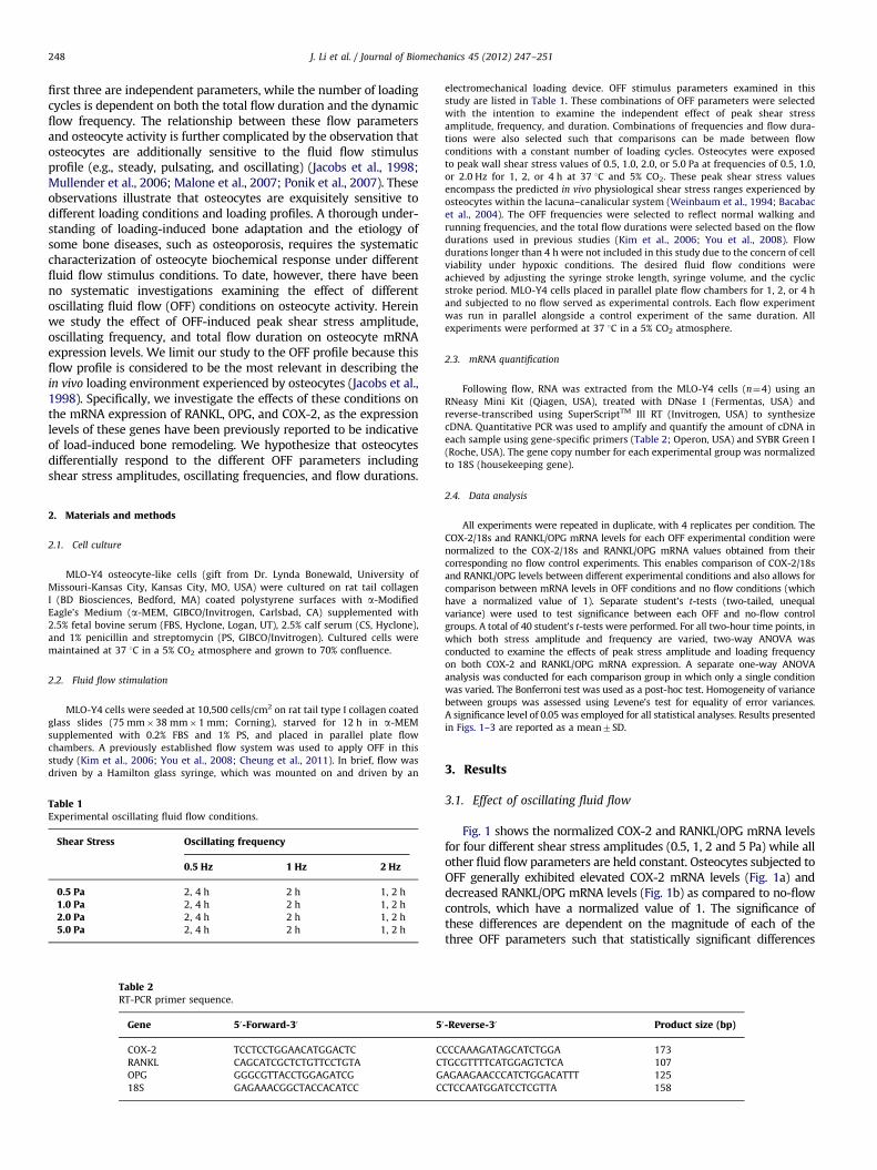
Table 1Experimental oscillating fluid flow conditions.
Shear Stress Oscillating frequency
0.5 Hz 1 Hz 2 Hz
0.5 Pa 2, 4 h 2 h 1, 2 h
1.0 Pa 2, 4 h 2 h 1, 2 h
2.0 Pa 2, 4 h 2 h 1, 2 h
5.0 Pa 2, 4 h 2 h 1, 2 h
Table 2RT-PCR primer sequence.
Gene 50-Forward-30 5
COX-2 TCCTCCTGGAACATGGACTC C
RANKL CAGCATCGCTCTGTTCCTGTA C
OPG GGGCGTTACCTGGAGATCG G
18S GAGAAACGGCTACCACATCC C
electromechanical loading device. OFF stimulus parameters examined in this
study are listed in Table 1. These combinations of OFF parameters were selected
with the intention to examine the independent effect of peak shear stress
amplitude, frequency, and duration. Combinations of frequencies and flow dura-
tions were also selected such that comparisons can be made between flow
conditions with a constant number of loading cycles. Osteocytes were exposed
to peak wall shear stress values of 0.5, 1.0, 2.0, or 5.0 Pa at frequencies of 0.5, 1.0,
or 2.0 Hz for 1, 2, or 4 h at 37 1C and 5% CO2. These peak shear stress values
encompass the predicted in vivo physiological shear stress ranges experienced by
osteocytes within the lacuna–canalicular system (Weinbaum et al., 1994; Bacabac
et al., 2004). The OFF frequencies were selected to reflect normal walking and
running frequencies, and the total flow durations were selected based on the flow
durations used in previous studies (Kim et al., 2006; You et al., 2008). Flow
durations longer than 4 h were not included in this study due to the concern of cell
viability under hypoxic conditions. The desired fluid flow conditions were
achieved by adjusting the syringe stroke length, syringe volume, and the cyclic
stroke period. MLO-Y4 cells placed in parallel plate flow chambers for 1, 2, or 4 h
and subjected to no flow served as experimental controls. Each flow experiment
was run in parallel alongside a control experiment of the same duration. All
experiments were performed at 37 1C in a 5% CO2 atmosphere.
2.3. mRNA quantification
Following flow, RNA was extracted from the MLO-Y4 cells (n¼4) using an
RNeasy Mini Kit (Qiagen, USA), treated with DNase I (Fermentas, USA) and
reverse-transcribed using SuperScriptTM III RT (Invitrogen, USA) to synthesize
cDNA. Quantitative PCR was used to amplify and quantify the amount of cDNA in
each sample using gene-specific primers (Table 2; Operon, USA) and SYBR Green I
(Roche, USA). The gene copy number for each experimental group was normalized
to 18S (housekeeping gene).
2.4. Data analysis
All experiments were repeated in duplicate, with 4 replicates per condition. The
COX-2/18s and RANKL/OPG mRNA levels for each OFF experimental condition were
normalized to the COX-2/18s and RANKL/OPG mRNA values obtained from their
corresponding no flow control experiments. This enables comparison of COX-2/18s
and RANKL/OPG levels between different experimental conditions and also allows for
comparison between mRNA levels in OFF conditions and no flow conditions (which
have a normalized value of 1). Separate student’s t-tests (two-tailed, unequal
variance) were used to test significance between each OFF and no-flow control
groups. A total of 40 student’s t-tests were performed. For all two-hour time points, in
which both stress amplitude and frequency are varied, two-way ANOVA was
conducted to examine the effects of peak stress amplitude and loading frequency
on both COX-2 and RANKL/OPG mRNA expression. A separate one-way ANOVA
analysis was conducted for each comparison group in which only a single condition
was varied. The Bonferroni test was used as a post-hoc test. Homogeneity of variance
between groups was assessed using Levene’s test for equality of error variances.
A significance level of 0.05 was employed for all statistical analyses. Results presented
in Figs. 1–3 are reported as a mean7SD.
3. Results
3.1. Effect of oscillating fluid flow
Fig. 1 shows the normalized COX-2 and RANKL/OPG mRNA levelsfor four different shear stress amplitudes (0.5, 1, 2 and 5 Pa) while allother fluid flow parameters are held constant. Osteocytes subjected toOFF generally exhibited elevated COX-2 mRNA levels (Fig. 1a) anddecreased RANKL/OPG mRNA levels (Fig. 1b) as compared to no-flowcontrols, which have a normalized value of 1. The significance ofthese differences are dependent on the magnitude of each of thethree OFF parameters such that statistically significant differences
0-Reverse-30 Product size (bp)
CCCAAAGATAGCATCTGGA 173
TGCGTTTTCATGGAGTCTCA 107
AGAAGAACCCATCTGGACATTT 125
CTCCAATGGATCCTCGTTA 158
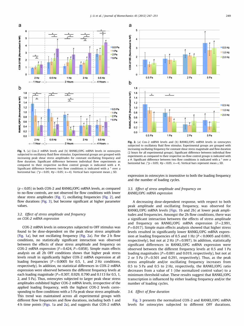
Fig. 1. (a) Cox-2 mRNA levels and (b) RANKL/OPG mRNA levels in osteocytes
subjected to oscillatory fluid flow stimulus. Experimental groups are grouped with
increasing peak shear stress amplitudes for constant oscillating frequency and
flow duration. Significant difference between individual flow experiments as
compared to their respective no-flow control groups is indicated with a #.
Significant difference between two flow conditions is indicated with a * over a
horizontal bar. (*po0.05; #po0.05; n¼4). Vertical bars represent mean7SD.
Fig. 2. (a) Cox-2 mRNA levels and (b) RANKL/OPG mRNA levels in osteocytes
subjected to oscillatory fluid flow stimulus. Experimental groups are grouped with
increasing oscillating frequency for constant shear stress magnitude and flow duration
(2 hours for all experimental groups). Significant difference between individual flow
experiments as compared to their respective no-flow control groups is indicated with
a #. Significant difference between two flow conditions is indicated with a * over a
horizontal bar. (*po0.05; #po0.05; n¼4). Vertical bars represent mean7SD.
J. Li et al. / Journal of Biomechanics 45 (2012) 247–251 249
(po0.05) in both COX-2 and RANKL/OPG mRNA levels, as comparedto no-flow controls, are not observed for flow conditions with lowershear stress amplitudes (Fig. 1), oscillating frequencies (Fig. 2), andflow durations (Fig. 3), but become significant at higher parametervalues.
3.2. Effect of stress amplitude and frequency
on COX-2 mRNA expression
COX-2 mRNA levels in osteocytes subjected to OFF stimulus wasfound to be dose-dependent on the peak shear stress amplitude(Fig. 1a), but not oscillating frequency (Fig. 2a). For the 2 h flowconditions, no statistically significant interaction was observedbetween the effects of shear stress amplitude and frequency onCOX-2 mRNA expression (F¼0.424, P¼0.858). Simple main effectsanalysis on all 2h OFF conditions shows that higher peak stresslevels result in significantly higher COX-2 mRNA expression at allloading frequencies (Po0.0005 for 0.5, 1, and 2 Hz conditions,respectively). In addition, no statistical differences in COX-2 mRNAexpression were observed between the different frequency levels ateach loading magnitude (P¼0.307, 0.929, 0.790 and 0.113 for 0.5, 1,2, and 5 Pa). Thus, osteocytes subjected to larger peak shear stressamplitudes exhibited higher COX-2 mRNA levels, irrespective of theapplied loading frequency, with the highest COX-2 levels corre-sponding to flow conditions with a 5 Pa peak shear stress amplitude.This trend was maintained across all experimental groups withdifferent flow frequencies and flow durations, including both 1 and4 h time points (Figs. 1a and 2a), and suggests that COX-2 mRNA
expression in osteocytes is insensitive to both the loading frequencyand the number of loading cycles.
3.3. Effect of stress amplitude and frequency on
RANKL/OPG mRNA expression
A decreasing dose-dependent response, with respect to bothpeak amplitude and oscillating frequency, was observed forRANKL/OPG mRNA levels (Figs. 1b and 2b) at lower peak ampli-tudes and frequencies. Amongst the 2h flow conditions, there wasa significant interaction between the effects of stress amplitudeand frequency on RANKL/OPG mRNA expression (F¼2.991,P¼0.017). Simple main effects analysis showed that higher stresslevels resulted in significantly lower RANKL/OPG mRNA expres-sion at loading frequencies of 0.5 and 1 Hz (Po0.0005 and 0.001,respectively), but not at 2 Hz (P¼0.997). In addition, statisticallysignificant differences in RANKL/OPG mRNA expression wereobserved between the different frequency levels at 0.5 and 1 Paloading magnitudes (P¼0.001 and 0.019, respectively), but not at2 or 5 Pa (P¼0.501 and 0.291, respectively). Thus, as the peakstress amplitude and/or oscillating frequency increases from0.5 to 5 Pa and 0.5 to 2 Hz, respectively, the RANKL/OPG ratiodecreases from a value of 1 (the normalized control value) to aminimum threshold value. These results suggest that RANKL/OPGtranscription is influenced by either loading frequency and/or thenumber of loading cycles.
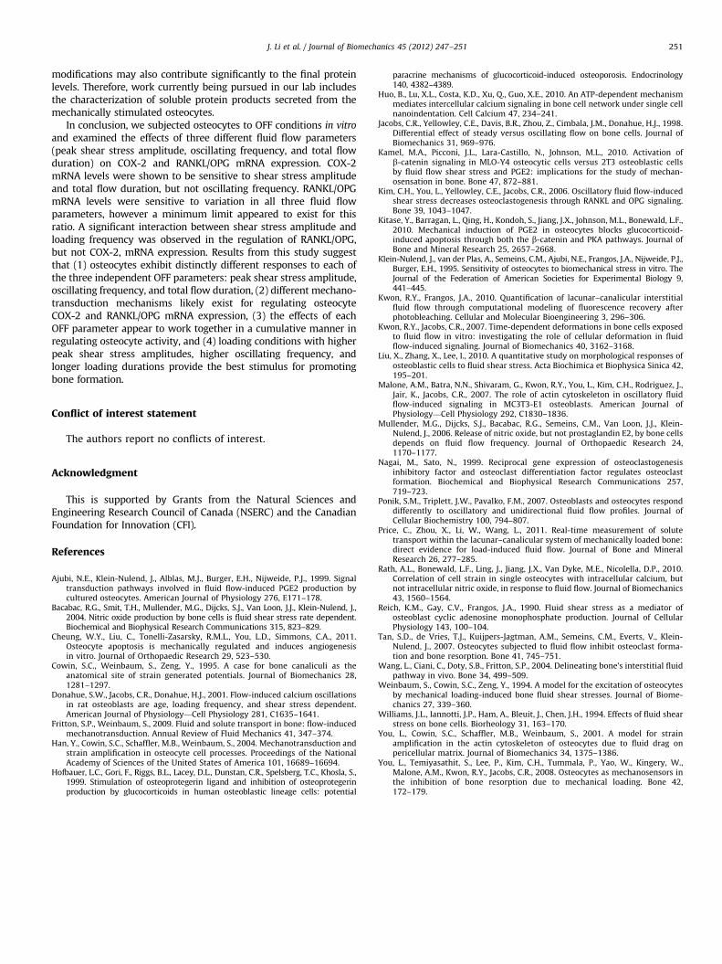
3.4. Effect of flow duration
Fig. 3 presents the normalized COX-2 and RANKL/OPG mRNAlevels for osteocytes subjected to different OFF durations.
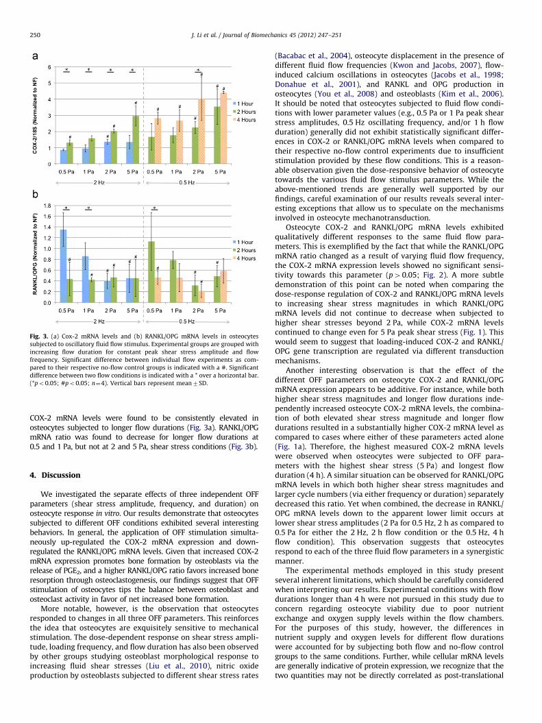
Fig. 3. (a) Cox-2 mRNA levels and (b) RANKL/OPG mRNA levels in osteocytes
subjected to oscillatory fluid flow stimulus. Experimental groups are grouped with
increasing flow duration for constant peak shear stress amplitude and flow
frequency. Significant difference between individual flow experiments as com-
pared to their respective no-flow control groups is indicated with a #. Significant
difference between two flow conditions is indicated with a * over a horizontal bar.
(*po0.05; #po0.05; n¼4). Vertical bars represent mean7SD.
J. Li et al. / Journal of Biomechanics 45 (2012) 247–251250
COX-2 mRNA levels were found to be consistently elevated inosteocytes subjected to longer flow durations (Fig. 3a). RANKL/OPGmRNA ratio was found to decrease for longer flow durations at0.5 and 1 Pa, but not at 2 and 5 Pa, shear stress conditions (Fig. 3b).
4. Discussion
We investigated the separate effects of three independent OFFparameters (shear stress amplitude, frequency, and duration) onosteocyte response in vitro. Our results demonstrate that osteocytessubjected to different OFF conditions exhibited several interestingbehaviors. In general, the application of OFF stimulation simulta-neously up-regulated the COX-2 mRNA expression and down-regulated the RANKL/OPG mRNA levels. Given that increased COX-2mRNA expression promotes bone formation by osteoblasts via therelease of PGE2, and a higher RANKL/OPG ratio favors increased boneresorption through osteoclastogenesis, our findings suggest that OFFstimulation of osteocytes tips the balance between osteoblast andosteoclast activity in favor of net increased bone formation.
More notable, however, is the observation that osteocytesresponded to changes in all three OFF parameters. This reinforcesthe idea that osteocytes are exquisitely sensitive to mechanicalstimulation. The dose-dependent response on shear stress ampli-tude, loading frequency, and flow duration has also been observedby other groups studying osteoblast morphological response toincreasing fluid shear stresses (Liu et al., 2010), nitric oxideproduction by osteoblasts subjected to different shear stress rates
(Bacabac et al., 2004), osteocyte displacement in the presence ofdifferent fluid flow frequencies (Kwon and Jacobs, 2007), flow-induced calcium oscillations in osteocytes (Jacobs et al., 1998;Donahue et al., 2001), and RANKL and OPG production inosteocytes (You et al., 2008) and osteoblasts (Kim et al., 2006).It should be noted that osteocytes subjected to fluid flow condi-tions with lower parameter values (e.g., 0.5 Pa or 1 Pa peak shearstress amplitudes, 0.5 Hz oscillating frequency, and/or 1 h flowduration) generally did not exhibit statistically significant differ-ences in COX-2 or RANKL/OPG mRNA levels when compared totheir respective no-flow control experiments due to insufficientstimulation provided by these flow conditions. This is a reason-able observation given the dose-responsive behavior of osteocytetowards the various fluid flow stimulus parameters. While theabove-mentioned trends are generally well supported by ourfindings, careful examination of our results reveals several inter-esting exceptions that allow us to speculate on the mechanismsinvolved in osteocyte mechanotransduction.
Osteocyte COX-2 and RANKL/OPG mRNA levels exhibitedqualitatively different responses to the same fluid flow para-meters. This is exemplified by the fact that while the RANKL/OPGmRNA ratio changed as a result of varying fluid flow frequency,the COX-2 mRNA expression levels showed no significant sensi-tivity towards this parameter (p40.05; Fig. 2). A more subtledemonstration of this point can be noted when comparing thedose-response regulation of COX-2 and RANKL/OPG mRNA levelsto increasing shear stress magnitudes in which RANKL/OPGmRNA levels did not continue to decrease when subjected tohigher shear stresses beyond 2 Pa, while COX-2 mRNA levelscontinued to change even for 5 Pa peak shear stress (Fig. 1). Thiswould seem to suggest that loading-induced COX-2 and RANKL/OPG gene transcription are regulated via different transductionmechanisms.
Another interesting observation is that the effect of thedifferent OFF parameters on osteocyte COX-2 and RANKL/OPGmRNA expression appears to be additive. For instance, while bothhigher shear stress magnitudes and longer flow durations inde-pendently increased osteocyte COX-2 mRNA levels, the combina-tion of both elevated shear stress magnitude and longer flowdurations resulted in a substantially higher COX-2 mRNA level ascompared to cases where either of these parameters acted alone(Fig. 1a). Therefore, the highest measured COX-2 mRNA levelswere observed when osteocytes were subjected to OFF para-meters with the highest shear stress (5 Pa) and longest flowduration (4 h). A similar situation can be observed for RANKL/OPGmRNA levels in which both higher shear stress magnitudes andlarger cycle numbers (via either frequency or duration) separatelydecreased this ratio. Yet when combined, the decrease in RANKL/OPG mRNA levels down to the apparent lower limit occurs atlower shear stress amplitudes (2 Pa for 0.5 Hz, 2 h as compared to0.5 Pa for either the 2 Hz, 2 h flow condition or the 0.5 Hz, 4 hflow condition). This observation suggests that osteocytesrespond to each of the three fluid flow parameters in a synergisticmanner.
The experimental methods employed in this study presentseveral inherent limitations, which should be carefully consideredwhen interpreting our results. Experimental conditions with flowdurations longer than 4 h were not pursued in this study due toconcern regarding osteocyte viability due to poor nutrientexchange and oxygen supply levels within the flow chambers.For the purposes of this study, however, the differences innutrient supply and oxygen levels for different flow durationswere accounted for by subjecting both flow and no-flow controlgroups to the same conditions. Further, while cellular mRNA levelsare generally indicative of protein expression, we recognize that thetwo quantities may not be directly correlated as post-translational
J. Li et al. / Journal of Biomechanics 45 (2012) 247–251 251
modifications may also contribute significantly to the final proteinlevels. Therefore, work currently being pursued in our lab includesthe characterization of soluble protein products secreted from themechanically stimulated osteocytes.
In conclusion, we subjected osteocytes to OFF conditions in vitro
and examined the effects of three different fluid flow parameters(peak shear stress amplitude, oscillating frequency, and total flowduration) on COX-2 and RANKL/OPG mRNA expression. COX-2mRNA levels were shown to be sensitive to shear stress amplitudeand total flow duration, but not oscillating frequency. RANKL/OPGmRNA levels were sensitive to variation in all three fluid flowparameters, however a minimum limit appeared to exist for thisratio. A significant interaction between shear stress amplitude andloading frequency was observed in the regulation of RANKL/OPG,but not COX-2, mRNA expression. Results from this study suggestthat (1) osteocytes exhibit distinctly different responses to each ofthe three independent OFF parameters: peak shear stress amplitude,oscillating frequency, and total flow duration, (2) different mechano-transduction mechanisms likely exist for regulating osteocyteCOX-2 and RANKL/OPG mRNA expression, (3) the effects of eachOFF parameter appear to work together in a cumulative manner inregulating osteocyte activity, and (4) loading conditions with higherpeak shear stress amplitudes, higher oscillating frequency, andlonger loading durations provide the best stimulus for promotingbone formation.
Conflict of interest statement
The authors report no conflicts of interest.
Acknowledgment
This is supported by Grants from the Natural Sciences andEngineering Research Council of Canada (NSERC) and the CanadianFoundation for Innovation (CFI).
References
Ajubi, N.E., Klein-Nulend, J., Alblas, M.J., Burger, E.H., Nijweide, P.J., 1999. Signaltransduction pathways involved in fluid flow-induced PGE2 production bycultured osteocytes. American Journal of Physiology 276, E171–178.
Bacabac, R.G., Smit, T.H., Mullender, M.G., Dijcks, S.J., Van Loon, J.J., Klein-Nulend, J.,2004. Nitric oxide production by bone cells is fluid shear stress rate dependent.Biochemical and Biophysical Research Communications 315, 823–829.
Cheung, W.Y., Liu, C., Tonelli-Zasarsky, R.M.L., You, L.D., Simmons, C.A., 2011.Osteocyte apoptosis is mechanically regulated and induces angiogenesisin vitro. Journal of Orthopaedic Research 29, 523–530.
Cowin, S.C., Weinbaum, S., Zeng, Y., 1995. A case for bone canaliculi as theanatomical site of strain generated potentials. Journal of Biomechanics 28,1281–1297.
Donahue, S.W., Jacobs, C.R., Donahue, H.J., 2001. Flow-induced calcium oscillationsin rat osteoblasts are age, loading frequency, and shear stress dependent.American Journal of Physiology—Cell Physiology 281, C1635–1641.
Fritton, S.P., Weinbaum, S., 2009. Fluid and solute transport in bone: flow-inducedmechanotransduction. Annual Review of Fluid Mechanics 41, 347–374.
Han, Y., Cowin, S.C., Schaffler, M.B., Weinbaum, S., 2004. Mechanotransduction andstrain amplification in osteocyte cell processes. Proceedings of the NationalAcademy of Sciences of the United States of America 101, 16689–16694.
Hofbauer, L.C., Gori, F., Riggs, B.L., Lacey, D.L., Dunstan, C.R., Spelsberg, T.C., Khosla, S.,1999. Stimulation of osteoprotegerin ligand and inhibition of osteoprotegerinproduction by glucocorticoids in human osteoblastic lineage cells: potential
paracrine mechanisms of glucocorticoid-induced osteoporosis. Endocrinology140, 4382–4389.
Huo, B., Lu, X.L., Costa, K.D., Xu, Q., Guo, X.E., 2010. An ATP-dependent mechanismmediates intercellular calcium signaling in bone cell network under single cellnanoindentation. Cell Calcium 47, 234–241.
Jacobs, C.R., Yellowley, C.E., Davis, B.R., Zhou, Z., Cimbala, J.M., Donahue, H.J., 1998.Differential effect of steady versus oscillating flow on bone cells. Journal ofBiomechanics 31, 969–976.
Kamel, M.A., Picconi, J.L., Lara-Castillo, N., Johnson, M.L., 2010. Activation ofb-catenin signaling in MLO-Y4 osteocytic cells versus 2T3 osteoblastic cellsby fluid flow shear stress and PGE2: implications for the study of mechan-osensation in bone. Bone 47, 872–881.
Kim, C.H., You, L., Yellowley, C.E., Jacobs, C.R., 2006. Oscillatory fluid flow-inducedshear stress decreases osteoclastogenesis through RANKL and OPG signaling.Bone 39, 1043–1047.
Kitase, Y., Barragan, L., Qing, H., Kondoh, S., Jiang, J.X., Johnson, M.L., Bonewald, L.F.,2010. Mechanical induction of PGE2 in osteocytes blocks glucocorticoid-induced apoptosis through both the b-catenin and PKA pathways. Journal ofBone and Mineral Research 25, 2657–2668.
Klein-Nulend, J., van der Plas, A., Semeins, C.M., Ajubi, N.E., Frangos, J.A., Nijweide, P.J.,Burger, E.H., 1995. Sensitivity of osteocytes to biomechanical stress in vitro. TheJournal of the Federation of American Societies for Experimental Biology 9,441–445.
Kwon, R.Y., Frangos, J.A., 2010. Quantification of lacunar–canalicular interstitialfluid flow through computational modeling of fluorescence recovery afterphotobleaching. Cellular and Molecular Bioengineering 3, 296–306.
Kwon, R.Y., Jacobs, C.R., 2007. Time-dependent deformations in bone cells exposedto fluid flow in vitro: investigating the role of cellular deformation in fluidflow-induced signaling. Journal of Biomechanics 40, 3162–3168.
Liu, X., Zhang, X., Lee, I., 2010. A quantitative study on morphological responses ofosteoblastic cells to fluid shear stress. Acta Biochimica et Biophysica Sinica 42,195–201.
Malone, A.M., Batra, N.N., Shivaram, G., Kwon, R.Y., You, L., Kim, C.H., Rodriguez, J.,Jair, K., Jacobs, C.R., 2007. The role of actin cytoskeleton in oscillatory fluidflow-induced signaling in MC3T3-E1 osteoblasts. American Journal ofPhysiology—Cell Physiology 292, C1830–1836.
Mullender, M.G., Dijcks, S.J., Bacabac, R.G., Semeins, C.M., Van Loon, J.J., Klein-Nulend, J., 2006. Release of nitric oxide, but not prostaglandin E2, by bone cellsdepends on fluid flow frequency. Journal of Orthopaedic Research 24,1170–1177.
Nagai, M., Sato, N., 1999. Reciprocal gene expression of osteoclastogenesisinhibitory factor and osteoclast differentiation factor regulates osteoclastformation. Biochemical and Biophysical Research Communications 257,719–723.
Ponik, S.M., Triplett, J.W., Pavalko, F.M., 2007. Osteoblasts and osteocytes responddifferently to oscillatory and unidirectional fluid flow profiles. Journal ofCellular Biochemistry 100, 794–807.
Price, C., Zhou, X., Li, W., Wang, L., 2011. Real-time measurement of solutetransport within the lacunar–canalicular system of mechanically loaded bone:direct evidence for load-induced fluid flow. Journal of Bone and MineralResearch 26, 277–285.
Rath, A.L., Bonewald, L.F., Ling, J., Jiang, J.X., Van Dyke, M.E., Nicolella, D.P., 2010.Correlation of cell strain in single osteocytes with intracellular calcium, butnot intracellular nitric oxide, in response to fluid flow. Journal of Biomechanics43, 1560–1564.
Reich, K.M., Gay, C.V., Frangos, J.A., 1990. Fluid shear stress as a mediator ofosteoblast cyclic adenosine monophosphate production. Journal of CellularPhysiology 143, 100–104.
Tan, S.D., de Vries, T.J., Kuijpers-Jagtman, A.M., Semeins, C.M., Everts, V., Klein-Nulend, J., 2007. Osteocytes subjected to fluid flow inhibit osteoclast forma-tion and bone resorption. Bone 41, 745–751.
Wang, L., Ciani, C., Doty, S.B., Fritton, S.P., 2004. Delineating bone’s interstitial fluidpathway in vivo. Bone 34, 499–509.
Weinbaum, S., Cowin, S.C., Zeng, Y., 1994. A model for the excitation of osteocytesby mechanical loading-induced bone fluid shear stresses. Journal of Biome-chanics 27, 339–360.
Williams, J.L., Iannotti, J.P., Ham, A., Bleuit, J., Chen, J.H., 1994. Effects of fluid shearstress on bone cells. Biorheology 31, 163–170.
You, L., Cowin, S.C., Schaffler, M.B., Weinbaum, S., 2001. A model for strainamplification in the actin cytoskeleton of osteocytes due to fluid drag onpericellular matrix. Journal of Biomechanics 34, 1375–1386.
You, L., Temiyasathit, S., Lee, P., Kim, C.H., Tummala, P., Yao, W., Kingery, W.,Malone, A.M., Kwon, R.Y., Jacobs, C.R., 2008. Osteocytes as mechanosensors inthe inhibition of bone resorption due to mechanical loading. Bone 42,172–179.