Effect of nanocellulose isolation techniques on the ... · the systematic study of the effect of...
11
1. Introduction Owing to environmentally friendly attributes, good mechanical properties, low density, biodegradabil- ity and abundant availability of renewable resources, the production of nanocellulose and their applica- tion in composites materials has gained increasing attention in recent times [1–2]. Nanocelluloses are recognized to be more effective in reinforcing poly- mers, due to the interaction between the nano-sized elements that form a percolated network connected by hydrogen bonding [3]. Two different types of nanocellulose can be isolated from a cellulosic source: nanocrystals and nanofibrils. Nanocrystals have a perfect crystalline structure and high modu- lus, close to the theoretical modulus of cellulose; nanofibrils are fibrillar units containing both amor- phous and crystalline regions and have the ability to create entangled networks [4]. Different properties of these two types of nanocellulose will result in varying reinforcement of nanocomposites. In order to utilize nanocellulose as a reinforcing phase to form nanocomposites, the strong hydrogen bonding between cellulose crystals must be sepa- rated and dispersed well in the polymer matrices [5]. Extensive research has been reported to extract nanocellulose from different sources [6–10]. Typi- cal processes involve mechanical and chemical treatments. The chemical ways, mainly by strong 794 Effect of nanocellulose isolation techniques on the formation of reinforced poly(vinyl alcohol) nanocomposite films Y. M. Zhou 1 , S. Y. Fu 1* , L. M. Zheng 2 , H. Y. Zhan 1 1 State Key Laboratory of Pulp and Paper Engineering, College of Light Industry and Food Science, South China University of Technology, Guangzhou, 510640 Guangdong, China 2 School of Chemistry and Chemical Engineering, South China University of Technology, Guangzhou, 510640 Guangdong, China Received 19 February 2012; accepted in revised form 1 May 2012 Abstract. Three techniques including acid hydrolysis (AH), 2,2,6,6-tetramethylpiperidine-1-oxyl radical (TEMPO)-medi- ated oxidation (TMO) and ultrasonication (US) were introduced to isolate nanocellulose from microcrystalline cellulose, in order to reinforce poly(vinyl alcohol) (PVA) films. Important differences were noticed in fiber quality of nanocellulose and film properties of PVA nanocomposite films. The TMO treatment was more efficient in nanocellulose isolation with higher aspect ratio, surface charge (–47 mV) and yields (37%). While AH treatment resulted in higher crystallinity index (88.1%) and better size dispersion. The fracture surface, thermal behavior and mechanical properties of the PVA nanocomposite films were investigated by means of scanning electron microscopy (SEM), differential scanning calorimetry (DSC), ther- mogravimetric analysis (TGA) and tensile testing. The results showed that both the TMO-derived and AH-derived nanocel- lulose could be dispersed homogeneously in the PVA matrices. AH/PVA films had higher elongation at break (51.59% at 6wt% nanocellulose loading) as compared with TMO/PVA, while TMO/PVA films shown superior tensile modulus and strength with increments of 21.5% and 10.2% at 6wt% nanocellulose loading. The thermal behavior of the PVA nanocom- posite films was higher improved with TMO-derived nanofibrils addition. Keywords: nanocomposites, isolation techniques, cellulose, reinforcements eXPRESS Polymer Letters Vol.6, No.10 (2012) 794–804 Available online at www.expresspolymlett.com DOI: 10.3144/expresspolymlett.2012.85 * Corresponding author, e-mail: [email protected] © BME-PT
Transcript of Effect of nanocellulose isolation techniques on the ... · the systematic study of the effect of...

1. IntroductionOwing to environmentally friendly attributes, goodmechanical properties, low density, biodegradabil-ity and abundant availability of renewable resources,the production of nanocellulose and their applica-tion in composites materials has gained increasingattention in recent times [1–2]. Nanocelluloses arerecognized to be more effective in reinforcing poly-mers, due to the interaction between the nano-sizedelements that form a percolated network connectedby hydrogen bonding [3]. Two different types ofnano cellulose can be isolated from a cellulosicsource: nanocrystals and nanofibrils. Nanocrystalshave a perfect crystalline structure and high modu-
lus, close to the theoretical modulus of cellulose;nanofibrils are fibrillar units containing both amor-phous and crystalline regions and have the ability tocreate entangled networks [4]. Different propertiesof these two types of nanocellulose will result invarying reinforcement of nanocomposites.In order to utilize nanocellulose as a reinforcingphase to form nanocomposites, the strong hydrogenbonding between cellulose crystals must be sepa-rated and dispersed well in the polymer matrices[5]. Extensive research has been reported to extractnanocellulose from different sources [6–10]. Typi-cal processes involve mechanical and chemicaltreatments. The chemical ways, mainly by strong
794
Effect of nanocellulose isolation techniques on the formationof reinforced poly(vinyl alcohol) nanocomposite filmsY. M. Zhou1, S. Y. Fu1*, L. M. Zheng2, H. Y. Zhan1
1State Key Laboratory of Pulp and Paper Engineering, College of Light Industry and Food Science, South ChinaUniversity of Technology, Guangzhou, 510640 Guangdong, China
2School of Chemistry and Chemical Engineering, South China University of Technology, Guangzhou, 510640Guangdong, China
Received 19 February 2012; accepted in revised form 1 May 2012
Abstract. Three techniques including acid hydrolysis (AH), 2,2,6,6-tetramethylpiperidine-1-oxyl radical (TEMPO)-medi-ated oxidation (TMO) and ultrasonication (US) were introduced to isolate nanocellulose from microcrystalline cellulose, inorder to reinforce poly(vinyl alcohol) (PVA) films. Important differences were noticed in fiber quality of nanocellulose andfilm properties of PVA nanocomposite films. The TMO treatment was more efficient in nanocellulose isolation with higheraspect ratio, surface charge (–47 mV) and yields (37%). While AH treatment resulted in higher crystallinity index (88.1%)and better size dispersion. The fracture surface, thermal behavior and mechanical properties of the PVA nanocompositefilms were investigated by means of scanning electron microscopy (SEM), differential scanning calorimetry (DSC), ther-mogravimetric analysis (TGA) and tensile testing. The results showed that both the TMO-derived and AH-derived nanocel-lulose could be dispersed homogeneously in the PVA matrices. AH/PVA films had higher elongation at break (51.59% at6 wt% nanocellulose loading) as compared with TMO/PVA, while TMO/PVA films shown superior tensile modulus andstrength with increments of 21.5% and 10.2% at 6wt% nanocellulose loading. The thermal behavior of the PVA nanocom-posite films was higher improved with TMO-derived nanofibrils addition.
Keywords: nanocomposites, isolation techniques, cellulose, reinforcements
eXPRESS Polymer Letters Vol.6, No.10 (2012) 794–804Available online at www.expresspolymlett.comDOI: 10.3144/expresspolymlett.2012.85
*Corresponding author, e-mail: [email protected]© BME-PT

acid hydrolysis, can remove the amorphous regionsof cellulose fibers and produce cellulose nanocrys-tals [11–12]. While for mechanical methods, whichinclude high intensity ultrasonication [13], highpressure refiner [14] or grinder treatment [15], themain product generated is not a single fiber and hasbeen referred as nanofibrils. However, these twotechniques of extracting nanocellulose from plantsare time consuming and very costly [1]. It involveshigh consumption of energy for processes as mechan-ical treatments [16], which can cause dramaticdecrease in both the yield and fibril length down to100–150 nm and also introduces damage to theenvironment, as in the case of chemical treatments[17]. Current research has been focused on findingenvironmental conservation, high efficiency and lowcosts methods to isolate nanocellulose. Recently,individualized cellulose nanofibrils have beenobtained using 2,2,6,6-tetramethylpiperidine-1-oxylradical (TEMPO)-mediated oxidation for regioselec-tive conversion of the cellulose primary hydroxylgroups to aldehydes and carboxylate ones. The mildreaction condition (room temperature and alkales-cent medium), the characteristic of little fiber mor-phological change and the resultant diverse surfacefunctionalities (carboxyl, aldehyde, and hydroxyl)lend the TEMPO-mediated oxidation technique sig-nificant potential in the fields of composites rein-forcement [18–19].Currently, very few references are available aboutthe systematic study of the effect of nanocelluloseisolation techniques on the quality of nanocelluloseand its performance in reinforced nanocomposites.The main goal of this work is to employ three dif-ferent techniques including acid hydrolysis (AH),TEMPO-mediated Oxidation (TMO) and ultrasoni-cation (US) to isolate nanocellulose from micro-crystalline cellulose (MCC) and to evaluate thequality of nanocellulose and the reinforcing abilityof these nanocellulose in PVA matrices.
2. Experiment2.1. MaterialsMicrocrystalline cellulose (MCC) with a meandiameter size of 20 µm, purchased from XuanyuanMachinery (Shandong, China) was used as raw mate-rial. Poly(vinyl alcohol) (PVA, 99% hydrolyzed,Mw 85 000~124 000) was used as matrices. Sulfuricacid (98 wt%) was used for the acid hydrolysis of
MCC. 2,2,6,6-tetramethylpiperidine-1-oxyl radical(TEMPO), sodium bromide (NaBr) and 6% sodiumhypochlorite (NaClO) solution were used for theTEMPO-mediated oxidation of MCC. All chemi-cals were of laboratory grade (Sigma-Aldrich,China) and used without further purification.
2.2. Nanocellulose isolation2.2.1. Acid hydrolysis (AH)About 5 g MCC was mixed with 45 mL sulfuric acid(64 wt%), the mixture was hydrolyzed at 45ºC for120 min with continuous stirring (500 rpm). Thehydrolysis was quenched by adding 500 mL waterto the reaction mixture and then the slurry waswashed with distilled water for 20 min at 5000 rpm,using repeated centrifugation. The supernatant wasremoved from the sediment and replaced by newdistilled water and mixed, the centrifugation stepcontinued until the pH of the supernatant became 1.The last wash was conducted using dialysis withdistilled water until the wash water maintained aconstant pH of 7. The resultant suspension wasstored at 4°C before further analysis or treatments.
2.2.2. TEMPO-mediated oxidation (TMO)About 5 g MCC was suspended in 500 mL distilledwater containing TEMPO (0.080 g, 0.5 mmol) andNaBr (0.5 g, 5 mmol). The TEMPO-mediated oxi-dation was started by adding 6% NaClO solution(25.0 mmol) with continuous stirring (500 rpm) atroom temperature. The pH was maintained at 10 byadding 0.5 M NaOH using a pH stat until no NaOHconsumption was observed. The TEMPO-oxidizedcellulose thus obtained was then ultrasonicated for20 min, using an ultrasonic homogenizer (KBS-1200, China) with an output power of 1200 W. Theslurry was washed with distilled water by repeatedcentrifugation (5000 rpm, 20 min) and then cen-trifuged at 12 000 rpm to separate large particles.After that, the samples were dialyzed against dis-tilled water until the pH reached 7. The resultant sus-pension was stored at 4°C before further analysis ortreatments.
2.2.3. Ultrasonication (US)About 5 g MCC was dispersed in 500 mL distilledwater under continuous stirring for a whole day,then ultrasonicated for 60 min using an ultrasonichomogenizer (KBS-1200, China) with an output
Zhou et al. – eXPRESS Polymer Letters Vol.6, No.10 (2012) 794–804
795

power of 1200 W, equipped with a sonication probeof 20 mm. In order to avoid overheating, the beakerwith the cellulose suspension was put in an ice bathwith controlled temperature. Two hours after theultrasonication process ended, nanocellulose wasobtained from the water suspension by decantingthe supernatant into other vessels. The resultant sus-pension with pH of 7 was stored at 4°C before fur-ther analysis or treatments.
2.3. Nanocomposite films preparationPVA water solution (10 wt%) and nanocellulosesuspension (2 wt%, relative to the PVA mass) weremixed under continuous stirring (500 rpm) at 80ºCfor 3 hours, then dispersed by ultrasonic treatment(KBS-1200, China) for about 2 min with 50% powerlevel. Films were cast onto a PTFE plate with con-trolled leveling, the mixture in the plate weredegassed in a vacuum desiccator, and then evapo-rated at 25°C and relative humidity of 30% untilfilms were formed, the films were heat treated in anoven at 80ºC for more than 12 h. Films with threelevels of nanocellulose loading (2, 6 and 10 wt%)were manufactured. The thicknesses of the filmswere controlled to be approximately 150 µm.
2.4. Characterization2.4.1. Morphology and crystallinity analysisMorphology of the nanocellulose and fractured sur-face of the PVA nanocomposite films were exam-ined using scanning electron microscopy (Nova,NanoSEM 430, FEI Company) with an acceleratingvoltage of 15 kV. For nanocellulose observation, adroplet of the nanocellulose suspensions (0.1 wt%)was put on a glass grid and dried under vacuumbefore SEM analysis. The dried nanocellulose andthe fractured surface of the films (after tensile tests)were coated with gold on an ion sputter coater, var-ious magnification levels were used to obtain images.More than three images were taken and chosen toobserve the morphology of all samples.Wide-angle X-ray diffraction (WXRD) data wascollected using a X-ray diffractometer (Rigaku D/max-III A, Japan) equipped with Cu K! radiation (" =0.1541 nm) at the operating voltage and current of45 kV and 100 mA, respectively. Diffractogramswere collected in a 2! range of 4~50° at a rate of1°/min with a resolution of 0.05°.
2.4.2. Wet particle size and surface chargeanalysis
Nanocellulose particle size analysis was conductedby dynamic light scattering (DLS) using a ZetasizerNanoZS instrument (Malvern, UK), under the fol-lowing conditions: dispersant water, material refrac-tive index 1.47, dispersion refractive index 1.33,viscosity 0.8872 cP, temperature 25°C and generalcalculation model for irregular particles. Three meas-urements of 10 s each were taken and the averagingwas done.The zeta-potential (estimated as surface charge)tests of the nanocellulose particles were conductedwith the Zetasizer NanoZS Instrument (Malvern,UK). Experiments were performed in a cuvette con-sisting of 4 ml 0.1 wt% nanocellulose suspension,solutions were all adjusted at pH values of 7.
2.4.3. Thermal propertiesThe differential scanning calorimetry (DSC) of PVAnanocomposite films was performed on a DSC Q200(TA Instruments, USA) from 25 to 300°C at a heat-ing rate of 10°C/min under nitrogen flow. Approxi-mately 8 mg samples were used. The thermogravi-metric analysis (TGA) were performed using a TGAQ500 (TA Instruments, USA). About 5~10 mg sam-ples were heated from 30 to 600°C with a heatingrate of 10°C/min under a nitrogen atmosphere. Theweight change was recorded as a function of theheating temperature.
2.4.4. Mechanical propertiesThe mechanical tests were performed using a test-ing machine (Instron 5567, USA) with a crossheadspeed of 5 mm/min. The crosshead extensions wereused as specimen deformations. The films were cutto dog bone shapes with width of 15 mm for the nar-row portion and total length of 50 mm (gauge lengthwas 25 mm). The thickness of the films was calcu-lated before the test. The values of tensile modulus,tensile strength and elongation at break of the sam-ples were evaluated and reported as the average val-ues of five measurements of each composition. Priorto testing, films were kept in a humidity chamberdesiccator with a 50% relative humidity (RH) and25°C for 5 days (according to the ASTM D1708standard [20]).
Zhou et al. – eXPRESS Polymer Letters Vol.6, No.10 (2012) 794–804
796
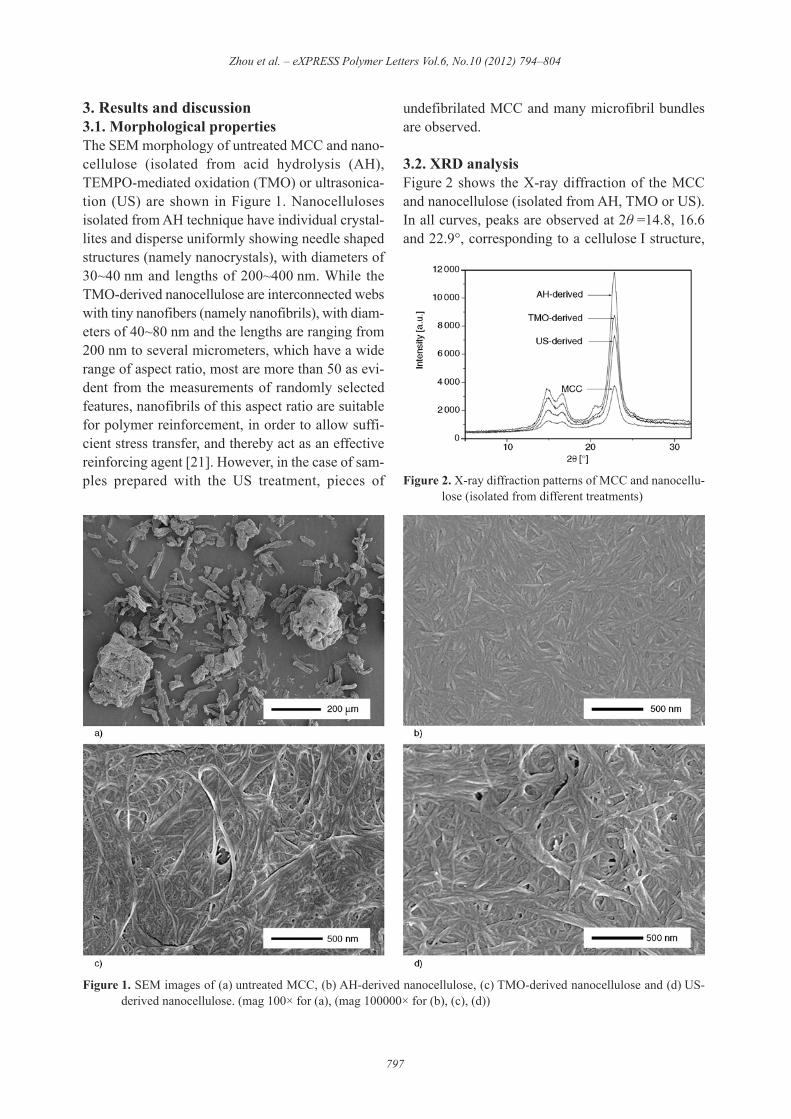
3. Results and discussion3.1. Morphological propertiesThe SEM morphology of untreated MCC and nano -cellulose (isolated from acid hydrolysis (AH),TEMPO-mediated oxidation (TMO) or ultrasonica-tion (US) are shown in Figure 1. Nanocellulosesisolated from AH technique have individual crystal-lites and disperse uniformly showing needle shapedstructures (namely nanocrystals), with diameters of30~40 nm and lengths of 200~400 nm. While theTMO-derived nanocellulose are interconnected webswith tiny nanofibers (namely nanofibrils), with diam-eters of 40~80 nm and the lengths are ranging from200 nm to several micrometers, which have a widerange of aspect ratio, most are more than 50 as evi-dent from the measurements of randomly selectedfeatures, nanofibrils of this aspect ratio are suitablefor polymer reinforcement, in order to allow suffi-cient stress transfer, and thereby act as an effectivereinforcing agent [21]. However, in the case of sam-ples prepared with the US treatment, pieces of
undefibrilated MCC and many microfibril bundlesare observed.
3.2. XRD analysisFigure 2 shows the X-ray diffraction of the MCCand nanocellulose (isolated from AH, TMO or US).In all curves, peaks are observed at 2! =14.8, 16.6and 22.9°, corresponding to a cellulose I structure,
Zhou et al. – eXPRESS Polymer Letters Vol.6, No.10 (2012) 794–804
797
Figure 1. SEM images of (a) untreated MCC, (b) AH-derived nanocellulose, (c) TMO-derived nanocellulose and (d) US-derived nanocellulose. (mag 100# for (a), (mag 100000# for (b), (c), (d))
Figure 2. X-ray diffraction patterns of MCC and nanocellu-lose (isolated from different treatments)

which means that all the three techniques have noeffect on the crystal form of the native cellulose.The intensity of the peaks is higher for all thenanocellulose samples, showing that nanocellulosesamples are more crystalline than MCC. The crys-tallinity index (Xc) of the cellulose can be calculatedusing the Equation (1) [22]:
(1)
where Icrystalline is the intensity of the peak at 2!about 22.9°representing crystalline material andIamorphous is the intensity of the peak at 2! about 18°representing amorphous material in cellulosic fibers.The values of the crystallinity index obtained areshown in Table 1. The cellulose Xc is of the orderAH-derived$>$TMO-derived$>$US-derived$>$MCC.The maximum Xc (88.1%) was obtained when AHprocess was carried out for the treatment of MCC,due to the removal of the majority of amorphousregions during the harsh process and resulted inneedle shaped individual crystallites. On the otherhand, there is an increase of diffraction intensities inthe crystalline peak around 2! = 20.5° of AH-derivednanocellulose, which may be attributed to the higher-ordered region of cellulose chains. For TMO andUS treatments, the nanocellulose Xc values are alsohigh (86.4 and 86.5%, respectively), it has beenreported that TMO process produces no change incrystallinity of cellulose even at a high oxidationlevel of 10 mmol NaClO/g cellulose [23]. The Xcincrease of the TMO-derived nanocellulose in thisstudy may be attributed to the partial removal ofamorphous regions, due to the harsh ultrasonicationtreatment of oxidized samples.
3.3. Yields analysisThe yield results of the nanocellulose (isolated fromAH, TMO or US) are shown in Table 1. In cellulosicplant fibers, cellulose is present in an amorphousstate, but also associates to crystalline domainsthrough both inter-molecular and intra-molecular
hydrogen bonding [24]. AH is a well-known harshprocess conducted to the disintegration of amor-phous regions and degradation of crystalline partsgenerating a low yield of 28.6%. This kind of pro-cedure affects the total integrity of fibers. While forUS method, the mechanical treatment alone cannotbe effective to separate the strong hydrogen bond-ing of native fibers. This is why the yield of US-derived nanocellulose is as low as 12.7% and piecesof undefibrilated MCC still remained. While TMOis a method of combination of chemical and mechan-ical treatments, the mild reaction condition of roomtemperature, alkalescent medium and characteristicof regioselective oxidation [18–19] maintain par-tially amorphous regions left in the axial directionof the starting material, which result in a higheryield of 37.4%, as well as higher aspect ratio of thefinal TMO-derived nanofibrils.
3.4. Surface charge analysisZeta potential (estimated as surface charge) can bemeasured by tracking the moving rate of negativelyor positively charged particles across an electricfield. Usually a value less than –15m V representsthe onset of agglomeration. Values greater than –30 mV generally signifies that there is sufficientmutual repulsion which results in colloidal stability[25]. The zeta potential data of the nanocellulose sus-pension (isolated from AH, TMO or US) are shownin Table 1, attributed to the esterification of cellulosehydroxyl groups to sulfonate groups during the AHprocess and the regioselective conversion of thecellulose primary hydroxyl groups to carboxyl onesduring the TMO process, the AH and TMO-derivednanocellulose possess high negative charge of –38.2and –46.5 mV, respectively. For TMO-derived nano -fibrils, the highest surface charge imparts electrostaticrepulsive forces to the system, preventing the bind-ing between nanofibrils-nanofibrils, and thus homo-geneous nanocellulose suspension is obtained. Theuniform dispersion of nanocellulose is critical toimprove the mechanical properties of the final nano -
Xc 5Icrystalline 2 Iamorphous
Icrystalline~100Xc 5
Icrystalline 2 Iamorphous
Icrystalline~100
Zhou et al. – eXPRESS Polymer Letters Vol.6, No.10 (2012) 794–804
798
Table 1. Parameters of MCC and nanocellulose (isolated from different treatments)
Results expressed as mean±standard deviation
Nanocellulose MCC AH-derived TMO-derived US-derivedXc [%] 79.5±2.0 88.1±1.8 86.4±2.0 86.5±2.1Yield [%] – 28.6±8.0 37.4±8.3 12.7±3.2Zeta [mV] –8.3±3.4 –38.2±3.4 –46.5±3.4 –23.1±3.4Particle size [nm] ~µm 115±35 210±54 623±93

composite products, promoting the actual formationof hydrogen bonding between the PVA and nano -cellulose (nanocrystals or nanofibrils), which alsoleads to a higher efficiency of the stress transferfrom the matrices to the fibers. In contrast, the USgenerated nanocellulose has only a weak charge of–23.1 mV originated from its inherent hydroxylgroups, the formation of larger insoluble precipi-tates was revealed by SEM image (Figure 1).
3.5. Size dispersion analysisDynamic light scattering (DLS) analysis has beenemployed to find the statistical distribution of theparticles present in nanocellulose. After measuringmillions of particles, an average particle size of thenanocellulose (isolated from AH, TMO or US) werefound to be 115, 210 and 623 nm (Table 1), respec-tively. Larger particle size than those determined forsamples in SEM analysis were obtained in DLSmeasurements, because of the rapid aggregation ofnanocellulose in water suspension. The size disper-sion of the different nanocellulose particles areshown in Figure 3. Because of the concentrated acid(64 wt% H2SO4) hydrolysis used to harshly destroythe majority of cellulose hydrogen bonding, the AHreaction is homogeneous and more complete, thatresulted in a narrower nanocellulose size distribu-tion, besides, the strong surface charge (–38.2 mV)also prevents the nanocellulose particles fromagglomerating. The TMO-derived nanocelluloseparticles display a relatively poor size dispersion,mainly due to the mild reaction condition comparedto the AH treatment. For US treatment, the lack ofsurface charge (–23.1 mV) induced continuingagglomerate of insoluble nanocellulose particles,resulted in a wide size dispersion.
3.6. Thermal propertiesThermal characterization of neat PVA and PVAnanocomposite films was carried out using DSCand TGA measurements. From the analysis of DSCtraces (Figure 4), the glass rubber transition temper-ature (Tg), the melting temperature (Tm), heat of
Zhou et al. – eXPRESS Polymer Letters Vol.6, No.10 (2012) 794–804
799
Figure 3. Size dispersion of (a) AH-derived nanocellulose,(b) TMO-derived nanocellulose, (c) US-derivednanocellulose
Figure 4. DSC curves of (a) neat PVA, (b) MCC/PVA,(c) AH/PVA, (d) TMO/PVA and (e) US/PVAfilms (6 wt% filler loading)

fusion (%Hm) and degree of crystallinity (Xc) wereevaluated and compared. The resulting experimen-tal data are listed in Table 2. The glass transition tem-perature (Tg) of PVA nanocomposites is increasedwith the addition of AH-derived and TMO-derivednanocellulose. The strong hydrogen bonding for-mation between the PVA matrices and nanocellu-lose is expected to restrict the segmental mobility ofpolymer chains and thereby increase Tg. The fea-tures of Tm, %Hm, and Xc are also enhanced as com-pared to neat PVA films. The increase in Xc is possi-bly due to the nucleating effect of the nano-sizedfibers. This enhancement of the Xc of the PVAmatrices probably results, at least partially, in theimprovement of the mechanical properties for TMO/PVA films as reported later. On the contrary, thethermal behavior is not obviously enhanced forUS/PVA films and even tends to decrease uponMCC addition. It means that the presence of largeagglomerate and microfibril bundles induce sterichindrance effects restricting the growth of crys-talline PVA regions. It results in both a lower melt-ing point and lower degree of crystallinity.The thermal stability of the neat PVA and PVAnanocomposite films examined by TGA are shownin Figure 5. All samples show major weight loss in
the range of 30~550°C. For PVA nanocompositefilms, a slight increase of the major degradationtemperatures (T10°C, T50°C and Td) (Table 2) isobserved, in the sequence of TMO/PVA$>$AH/PVA>$US/PVA$>$MCC/PVA$>$neat PVA, thus furtherconfirming the enhanced thermal stability due to astrong hydrogen bonding between the TMO-derivednanocellulose and the PVA matrices. The reasonmaybe that despite the regioselective oxidation ofthe cellulose primary hydroxyl groups to carboxy-late ones in TMO treatments, the total amount ofthe strong hydrogen bonding in film formation isnot affected. In contrast, the hydrogen bondingbetween the AH-derived nanocellulose and PVAmatrices is decreased, due to the esterification ofhydroxyl groups to sulfonate groups during the AHprocess. The US/PVA nanocomposite films haverelatively lower degradation temperature. Becausethe specific surface area of US-derived nanocellu-lose is not as high as the AH-derived or TMO-derived ones, resulting in fewer hydroxyl groups onthe nanocellulose surface, besides, the easy forma-tion of larger insoluble precipitates will also accel-erate this process, these two factors resulted inlower thermal stability of the final composites.
3.7. SEM and mechanical propertiesThe fractured surface after tensile tests of neat PVAand PVA nanocomposite films were examined usingSEM as shown in Figure 6. As compared to the neatPVA films, the morphology of the AH/PVA andTMO/PVA films can be easily identified. The nano -cellulose appears as white dots, these white dotscould correspond to the nanocrystals (AH-derived)or nanofibrils (TMO-derived). No large aggregatesand a homogeneous distribution of the nanocellu-lose in the PVA matrix are observed in both AH/PVAand TMO/PVA films, implying good adhesionbetween fillers and matrix. This should be attrib-uted to the formation of a rigid hydrogen-bonded
Zhou et al. – eXPRESS Polymer Letters Vol.6, No.10 (2012) 794–804
800
Table 2. TGA data of neat PVA and PVA nanocomposite films (6 wt% filler loading)Samples Neat PVA MCC/PVA AH/PVA TMO/PVA US/PVA
Tg [°C] 70.3 68.0 75.1 76.9 71.2Tm [°C] 215.9 214.1 218.8 219.1 214.5%Hm [J/g] 81.1 76.8 88.5 84.9 80.6Xc [%] 54.1 54.5 62.8 60.2 57.1T10 [°C] 239.3 239.2 252.8 254.9 245.8T50 [°C] 283.4 304.5 306.8 314.9 300.4Td [°C] 270.4 271.1 277.2 287.8 264.0
Figure 5. TGA curves of neat PVA and PVA nanocompositefilms (6 wt% filler loading)
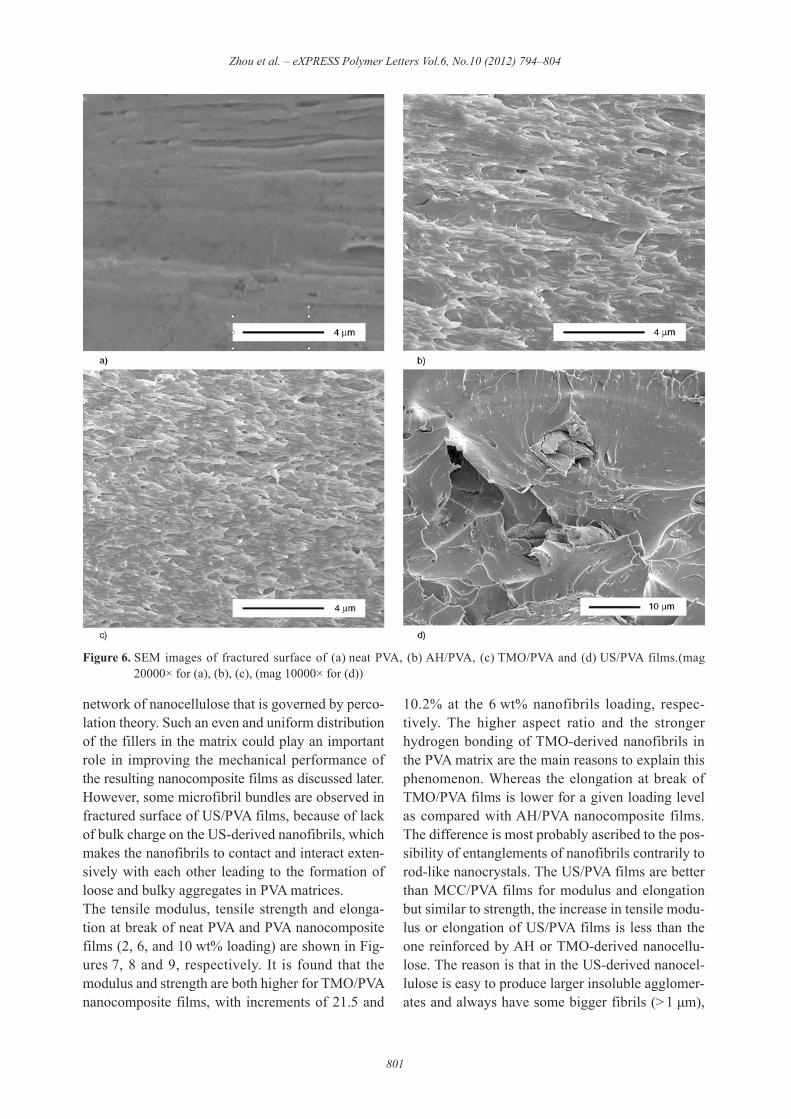
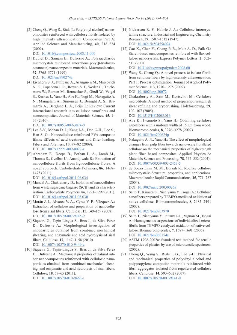
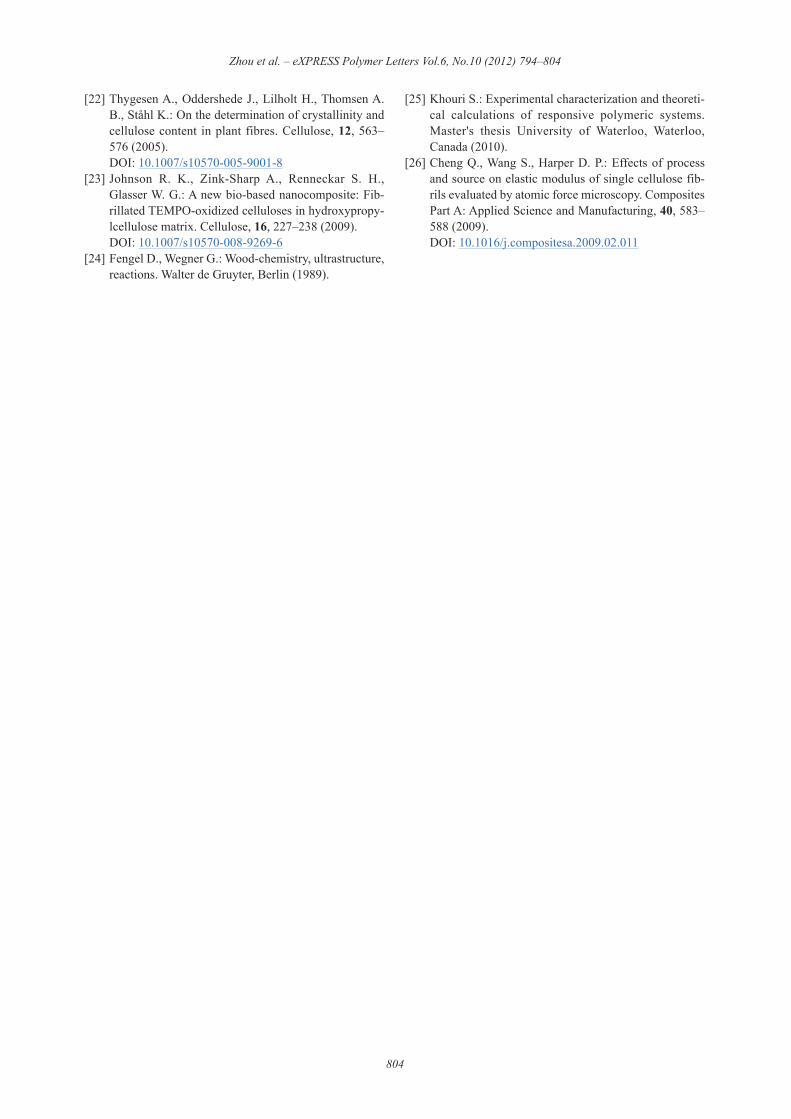
network of nanocellulose that is governed by perco-lation theory. Such an even and uniform distributionof the fillers in the matrix could play an importantrole in improving the mechanical performance ofthe resulting nanocomposite films as discussed later.However, some microfibril bundles are observed infractured surface of US/PVA films, because of lackof bulk charge on the US-derived nanofibrils, whichmakes the nanofibrils to contact and interact exten-sively with each other leading to the formation ofloose and bulky aggregates in PVA matrices.The tensile modulus, tensile strength and elonga-tion at break of neat PVA and PVA nanocompositefilms (2, 6, and 10 wt% loading) are shown in Fig-ures 7, 8 and 9, respectively. It is found that themodulus and strength are both higher for TMO/PVAnanocomposite films, with increments of 21.5 and
10.2% at the 6 wt% nanofibrils loading, respec-tively. The higher aspect ratio and the strongerhydrogen bonding of TMO-derived nanofibrils inthe PVA matrix are the main reasons to explain thisphenomenon. Whereas the elongation at break ofTMO/PVA films is lower for a given loading levelas compared with AH/PVA nanocomposite films.The difference is most probably ascribed to the pos-sibility of entanglements of nanofibrils contrarily torod-like nanocrystals. The US/PVA films are betterthan MCC/PVA films for modulus and elongationbut similar to strength, the increase in tensile modu-lus or elongation of US/PVA films is less than theone reinforced by AH or TMO-derived nanocellu-lose. The reason is that in the US-derived nanocel-lulose is easy to produce larger insoluble agglomer-ates and always have some bigger fibrils (> 1 µm),
Zhou et al. – eXPRESS Polymer Letters Vol.6, No.10 (2012) 794–804
801
Figure 6. SEM images of fractured surface of (a) neat PVA, (b) AH/PVA, (c) TMO/PVA and (d) US/PVA films.(mag20000# for (a), (b), (c), (mag 10000# for (d))
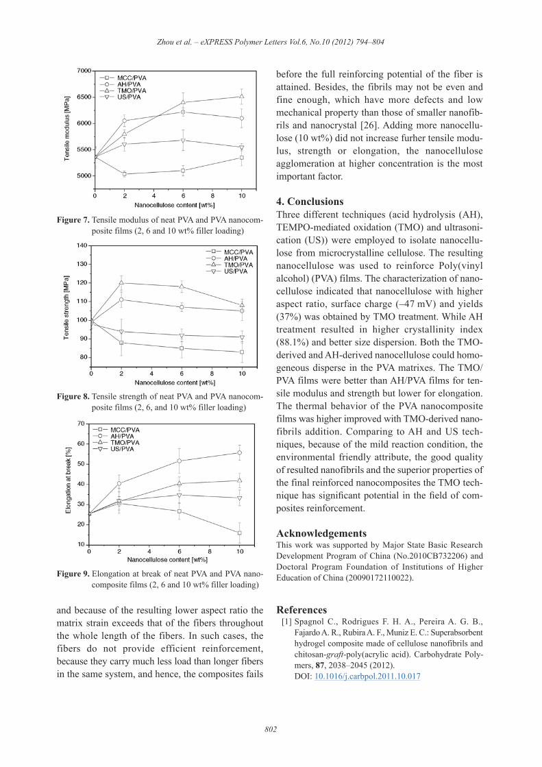
and because of the resulting lower aspect ratio thematrix strain exceeds that of the fibers throughoutthe whole length of the fibers. In such cases, thefibers do not provide efficient reinforcement,because they carry much less load than longer fibersin the same system, and hence, the composites fails
before the full reinforcing potential of the fiber isattained. Besides, the fibrils may not be even andfine enough, which have more defects and lowmechanical property than those of smaller nanofib-rils and nanocrystal [26]. Adding more nanocellu-lose (10 wt%) did not increase furher tensile modu-lus, strength or elongation, the nanocelluloseagglomeration at higher concentration is the mostimportant factor.
4. ConclusionsThree different techniques (acid hydrolysis (AH),TEMPO-mediated oxidation (TMO) and ultrasoni-cation (US)) were employed to isolate nanocellu-lose from microcrystalline cellulose. The resultingnanocellulose was used to reinforce Poly(vinylalcohol) (PVA) films. The characterization of nano -cellulose indicated that nanocellulose with higheraspect ratio, surface charge (–47 mV) and yields(37%) was obtained by TMO treatment. While AHtreatment resulted in higher crystallinity index(88.1%) and better size dispersion. Both the TMO-derived and AH-derived nanocellulose could homo-geneous disperse in the PVA matrixes. The TMO/PVA films were better than AH/PVA films for ten-sile modulus and strength but lower for elongation.The thermal behavior of the PVA nanocompositefilms was higher improved with TMO-derived nano -fibrils addition. Comparing to AH and US tech-niques, because of the mild reaction condition, theenvironmental friendly attribute, the good qualityof resulted nanofibrils and the superior properties ofthe final reinforced nanocomposites the TMO tech-nique has significant potential in the field of com-posites reinforcement.
AcknowledgementsThis work was supported by Major State Basic ResearchDevelopment Program of China (No.2010CB732206) andDoctoral Program Foundation of Institutions of HigherEducation of China (20090172110022).
References [1] Spagnol C., Rodrigues F. H. A., Pereira A. G. B.,
Fajardo A. R., Rubira A. F., Muniz E. C.: Superabsorbenthydrogel composite made of cellulose nanofibrils andchitosan-graft-poly(acrylic acid). Carbohydrate Poly-mers, 87, 2038–2045 (2012).DOI: 10.1016/j.carbpol.2011.10.017
Zhou et al. – eXPRESS Polymer Letters Vol.6, No.10 (2012) 794–804
802
Figure 8. Tensile strength of neat PVA and PVA nanocom-posite films (2, 6, and 10 wt% filler loading)
Figure 9. Elongation at break of neat PVA and PVA nano -composite films (2, 6 and 10 wt% filler loading)
Figure 7. Tensile modulus of neat PVA and PVA nanocom-posite films (2, 6 and 10 wt% filler loading)
[2] Cheng Q., Wang S., Rials T.: Poly(vinyl alcohol) nanoc -omposites reinforced with cellulose fibrils isolated byhigh intensity ultrasonication. Composites Part A:Applied Science and Manufacturing, 40, 218–224(2009).DOI: 10.1016/j.compositesa.2008.11.009
[3] Dubief D., Samain E., Dufresne A.: Polysaccharidemicrocrystals reinforced amorphous poly(&-hydroxy-octanoate) nanocomposite materials. Macromolecules,32, 5765–5771 (1999).DOI: 10.1021/ma990274a
[4] Eichhorn S. J., Dufresne A., Aranguren M., MarcovichN. E., Capadona J. R., Rowan S. J., Weder C., Thiele-mans W., Roman M., Renneckar S., Gindl W., VeigelS., Keckes J., Yano H., Abe K., Nogi M., Nakagaito A.N., Mangalam A., Simonsen J., Benight A. S., Bis-marck A., Berglund L. A., Peijs T.: Review: Currentinternational research into cellulose nanofibres andnanocomposites. Journal of Materials Science, 45, 1–33 (2010).DOI: 10.1007/s10853-009-3874-0
[5] Lee S-Y., Mohan D. J., Kang I-A., Doh G-H., Lee S.,Han S. O.: Nanocellulose reinforced PVA compositefilms: Effects of acid treatment and filler loading.Fibers and Polymers, 10, 77–82 (2009).DOI: 10.1007/s12221-009-0077-x
[6] Abraham E., Deepa B., Pothan L. A., Jacob M.,Thomas S., Cvelbar U., Anandjiwala R.: Extraction ofnanocellulose fibrils from lignocellulosic fibres: Anovel approach. Carbohydrate Polymers, 86, 1468–1475 (2011).DOI: 10.1016/j.carbpol.2011.06.034
[7] Mandal A., Chakrabarty D.: Isolation of nanocellulosefrom waste sugarcane bagasse (SCB) and its character-ization. Carbohydrate Polymers, 86, 1291–1299 (2011).DOI: 10.1016/j.carbpol.2011.06.030
[8] Morán J. I., Alvarez V. A., Cyras V. P., Vázquez A.:Extraction of cellulose and preparation of nanocellu-lose from sisal fibers. Cellulose, 15, 149–159 (2008).DOI: 10.1007/s10570-007-9145-9
[9] Siqueira G., Tapin-Lingua S., Bras J., da Silva PerezD., Dufresne A.: Morphological investigation ofnanoparticles obtained from combined mechanicalshearing, and enzymatic and acid hydrolysis of sisalfibers. Cellulose, 17, 1147–1158 (2010).DOI: 10.1007/s10570-010-9449-z
[10] Siqueira G., Tapin-Lingua S., Bras J., da Silva PerezD., Dufresne A.: Mechanical properties of natural rub-ber nanocomposites reinforced with cellulosic nano -particles obtained from combined mechanical shear-ing, and enzymatic and acid hydrolysis of sisal fibers.Cellulose, 18, 57–65 (2011).DOI: 10.1007/s10570-010-9463-1
[11] Nickerson R. F., Habrle J. A.: Cellulose intercrys-talline structure. Industrial and Engineering ChemistryResearch, 39, 1507–1512 (1947).DOI: 10.1021/ie50455a024
[12] Cao X., Chen Y., Chang P. R., Muir A. D., Falk G.:Starch-based nanocomposites reinforced with flax cel-lulose nanocrystals. Express Polymer Letters, 2, 502–510 (2008).DOI: 10.3144/expresspolymlett.2008.60
[13] Wang S., Cheng Q.: A novel process to isolate fibrilsfrom cellulose fibers by high-intensity ultrasonication,Part 1: Process optimization. Journal of Applied Poly-mer Science, 113, 1270–1275 (2009).DOI: 10.1002/app.30072
[14] Chakraborty A., Sain M., Kortschot M.: Cellulosemicrofibrils: A novel method of preparation using highshear refining and cryocrushing. Holzforschung, 59,102–107 (2005).DOI: 10.1515/HF.2005.016
[15] Abe K., Iwamoto S., Yano H.: Obtaining cellulosenanofibers with a uniform width of 15 nm from wood.Biomacromolecules, 8, 3276–3278 (2007).DOI: 10.1021/bm700624p
[16] Nakagaito A. N., Yano H.: The effect of morphologicalchanges from pulp fiber towards nano-scale fibrillatedcellulose on the mechanical properties of high-strengthplant fiber based composites. Applied Physics A:Materials Science and Processing, 78, 547–552 (2004).DOI: 10.1007/s00339-003-2453-5
[17] de Souza Lima M. M., Borsali R.: Rodlike cellulosemicrocrystals: Structure, properties, and applications.Macromolecular Rapid Communications, 25, 771–787(2004).DOI: 10.1002/marc.200300268
[18] Saito T., Kimura S., Nishiyama Y., Isogai A.: Cellulosenanofibers prepared by TEMPO-mediated oxidation ofnative cellulose. Biomacromolecules, 8, 2485–2491(2007).DOI: 10.1021/bm0703970
[19] Saito T., Nishiyama Y., Putaux J-L., Vignon M., IsogaiA.: Homogeneous suspensions of individualized micro -fibrils from TEMPO-catalyzed oxidation of native cel-lulose. Biomacromolecules, 7, 1687–1691 (2006).DOI: 10.1021/bm060154s
[20] ASTM 1708-2002a: Standard test method for tensileproperties of plastics by use of microtensile specimens(2002).
[21] Cheng Q., Wang S., Rials T. G., Lee S-H.: Physicaland mechanical properties of polyvinyl alcohol andpolypropylene composite materials reinforced withfibril aggregates isolated from regenerated cellulosefibers. Cellulose, 14, 593–602 (2007).DOI: 10.1007/s10570-007-9141-0
Zhou et al. – eXPRESS Polymer Letters Vol.6, No.10 (2012) 794–804
803
[22] Thygesen A., Oddershede J., Lilholt H., Thomsen A.B., Ståhl K.: On the determination of crystallinity andcellulose content in plant fibres. Cellulose, 12, 563–576 (2005).DOI: 10.1007/s10570-005-9001-8
[23] Johnson R. K., Zink-Sharp A., Renneckar S. H.,Glasser W. G.: A new bio-based nanocomposite: Fib-rillated TEMPO-oxidized celluloses in hydroxypropy-lcellulose matrix. Cellulose, 16, 227–238 (2009).DOI: 10.1007/s10570-008-9269-6
[24] Fengel D., Wegner G.: Wood-chemistry, ultrastructure,reactions. Walter de Gruyter, Berlin (1989).
[25] Khouri S.: Experimental characterization and theoreti-cal calculations of responsive polymeric systems.Master's thesis University of Waterloo, Waterloo,Canada (2010).
[26] Cheng Q., Wang S., Harper D. P.: Effects of processand source on elastic modulus of single cellulose fib-rils evaluated by atomic force microscopy. CompositesPart A: Applied Science and Manufacturing, 40, 583–588 (2009).DOI: 10.1016/j.compositesa.2009.02.011
Zhou et al. – eXPRESS Polymer Letters Vol.6, No.10 (2012) 794–804
804