Effect of MMP-2 gene silencing on radiation-induced DNA ...
7
SHORT REPORT Open Access Effect of MMP-2 gene silencing on radiation-induced DNA damage in human normal dermal fibroblasts and breast cancer cells Gugalavath Shailender 1* , Seema Kumari 1 , Patnala Kiranmayi 2 and Rama Rao Malla 1* Abstract Introduction: Diagnostic and therapeutic ionizing radiation (IR) is one of the well known long term risk factors of breast cancer. Extremely lethal consequences of IR causes double-strand breaks, which are mainly responsible for genomic instability, altered gene expression, and cell death. Findings: This study evaluated the effect of matrix metalloproteinases-2 (MMP-2) gene silencing using MMP-2 shRNA expression plasmids (pMMP-2) on IR induced cytotoxicity and DNA damage by MTT, dead green, γH2AX and comet assays in human normal dermal fibroblasts (HDFs) and MCF-7 human breast cancer cells. IR has decreased the viability of HDFs and MCF-7 cells with increasing IR (2-10Gy). IR induced DNA damage in both HDFs and MCF-7 cells. However, pMMP-2 transfection has increased the viability of irradiated HDFs (10Gy) and significantly decreased the viability of irradiated MCF-7 cells (10Gy). Further, DNA damage in terms of γH2AX foci decreased with pMMP-2 transfection in irradiated HDFs (10Gy) and increased in irradiated MCF-7 cells (10Gy). In addition, MMP-2 gene silencing using pMMP-2 decreased comet tail length in irradiated HDFs but increased in irradiated MCF-7 cells. Conclusions: The results conclude that pMMP-2 has protected HDFs and sensitized the MCF-7 cells from IR induced DNA damage. This differential response might be due to IR induced MMP-2 distinctive ROS generation in HDFs and MCF-7 cells. Keywords: Ionizing radiation, Matrix metalloproteinase, DNA damage, Breast cancer, Fibroblasts Introduction The exposure of humans to ionizing radiation (IR) in- duces several types of genetic and somatic mutations leading to several types of cancers including breast can- cer [1]. However, a single dose or fractionated dose of radiation has severe effects on the adjacent normal tissue during radiotherapy [2]. The high dose of radiation in- duces early and late skin effects and secondary neoplasm during radiotherapy of breast cancer [3]. Normal or cancer tissue is composed of extracellular matrix (ECM) and cellular constituents mainly fibroblasts. Fibroblasts have a major role in structural integrity, tissue repair, and deposition of the ECM and regulation of epithelial cell differentiation, inflammation and wound healing [4]. However, cancer-associated fibroblasts have a major role in proliferation, invasion, tumor progression, metastasis, and angiogenesis. Various studies have highlighted that dermal fibroblasts are pertinent and condemnatory target cells for IR [5]. Therefore, the deve- lopment of novel and competent non-toxic radiopro- tectors is of great interest against the radiation-induced damages. Radioprotectors are important to safeguard the normal tissues during radiotherapy of breast cancer [6]. The naturally occurring thiol- and sulphur- containing compounds, pharmacological agents and phytoche- micals present radioprotection, but non-specific, and showed side effects like cephalalgia, nausea, sickness and vomiting [7, 8]. © The Author(s). 2019 Open Access This article is distributed under the terms of the Creative Commons Attribution 4.0 International License (http://creativecommons.org/licenses/by/4.0/), which permits unrestricted use, distribution, and reproduction in any medium, provided you give appropriate credit to the original author(s) and the source, provide a link to the Creative Commons license, and indicate if changes were made. The Creative Commons Public Domain Dedication waiver (http://creativecommons.org/publicdomain/zero/1.0/) applies to the data made available in this article, unless otherwise stated. * Correspondence: [email protected]; [email protected] 1 Cancer Biology Lab, Department of Biochemistry and Bioinformatics, Institute of Science, GITAM (Deemed to be University), Visakhapatnam, Andhra Pradesh, India Full list of author information is available at the end of the article Shailender et al. Genes and Environment (2019) 41:16 https://doi.org/10.1186/s41021-019-0131-x
Transcript of Effect of MMP-2 gene silencing on radiation-induced DNA ...

SHORT REPORT Open Access
Effect of MMP-2 gene silencing onradiation-induced DNA damage in humannormal dermal fibroblasts and breastcancer cellsGugalavath Shailender1*, Seema Kumari1, Patnala Kiranmayi2 and Rama Rao Malla1*
Abstract
Introduction: Diagnostic and therapeutic ionizing radiation (IR) is one of the well known long term risk factors ofbreast cancer. Extremely lethal consequences of IR causes double-strand breaks, which are mainly responsible forgenomic instability, altered gene expression, and cell death.
Findings: This study evaluated the effect of matrix metalloproteinases-2 (MMP-2) gene silencing using MMP-2shRNA expression plasmids (pMMP-2) on IR induced cytotoxicity and DNA damage by MTT, dead green, γH2AX andcomet assays in human normal dermal fibroblasts (HDFs) and MCF-7 human breast cancer cells. IR has decreasedthe viability of HDFs and MCF-7 cells with increasing IR (2-10Gy). IR induced DNA damage in both HDFs and MCF-7cells. However, pMMP-2 transfection has increased the viability of irradiated HDFs (10Gy) and significantly decreasedthe viability of irradiated MCF-7 cells (10Gy). Further, DNA damage in terms of γH2AX foci decreased with pMMP-2transfection in irradiated HDFs (10Gy) and increased in irradiated MCF-7 cells (10Gy). In addition, MMP-2 genesilencing using pMMP-2 decreased comet tail length in irradiated HDFs but increased in irradiated MCF-7 cells.
Conclusions: The results conclude that pMMP-2 has protected HDFs and sensitized the MCF-7 cells from IRinduced DNA damage. This differential response might be due to IR induced MMP-2 distinctive ROS generation inHDFs and MCF-7 cells.
Keywords: Ionizing radiation, Matrix metalloproteinase, DNA damage, Breast cancer, Fibroblasts
IntroductionThe exposure of humans to ionizing radiation (IR) in-duces several types of genetic and somatic mutationsleading to several types of cancers including breast can-cer [1]. However, a single dose or fractionated dose ofradiation has severe effects on the adjacent normal tissueduring radiotherapy [2]. The high dose of radiation in-duces early and late skin effects and secondary neoplasmduring radiotherapy of breast cancer [3].Normal or cancer tissue is composed of extracellular
matrix (ECM) and cellular constituents mainly fibroblasts.Fibroblasts have a major role in structural integrity, tissue
repair, and deposition of the ECM and regulation ofepithelial cell differentiation, inflammation and woundhealing [4]. However, cancer-associated fibroblasts have amajor role in proliferation, invasion, tumor progression,metastasis, and angiogenesis. Various studies havehighlighted that dermal fibroblasts are pertinent andcondemnatory target cells for IR [5]. Therefore, the deve-lopment of novel and competent non-toxic radiopro-tectors is of great interest against the radiation-induceddamages. Radioprotectors are important to safeguard thenormal tissues during radiotherapy of breast cancer [6].The naturally occurring thiol- and sulphur- containingcompounds, pharmacological agents and phytoche-micals present radioprotection, but non-specific, andshowed side effects like cephalalgia, nausea, sicknessand vomiting [7, 8].
© The Author(s). 2019 Open Access This article is distributed under the terms of the Creative Commons Attribution 4.0International License (http://creativecommons.org/licenses/by/4.0/), which permits unrestricted use, distribution, andreproduction in any medium, provided you give appropriate credit to the original author(s) and the source, provide a link tothe Creative Commons license, and indicate if changes were made. The Creative Commons Public Domain Dedication waiver(http://creativecommons.org/publicdomain/zero/1.0/) applies to the data made available in this article, unless otherwise stated.
* Correspondence: [email protected]; [email protected] Biology Lab, Department of Biochemistry and Bioinformatics,Institute of Science, GITAM (Deemed to be University), Visakhapatnam,Andhra Pradesh, IndiaFull list of author information is available at the end of the article
Shailender et al. Genes and Environment (2019) 41:16 https://doi.org/10.1186/s41021-019-0131-x
Matrix metalloproteinases are a broad family of Zn con-taining proteases including collagenases, gelatinases, stro-melysins, elastases and membrane-type [9]. Matrixmetalloproteinases-2 (MMP-2) is a member of gelatinasesub-family plays a crucial role in ECM turnover. Itdegenerates the component of the basement membrane,alters the interstitial collagens I and III, native collagen IV.Previous reports have described that IR increased theMMP-2 activity in human bronchial epithelial cells [10],rat astrocytes [11], rat mesangial cells, rat kidney tubuleepithelial cells [12], and human cultured fibroblasts [13].Further, increased synthesis and activity of MMP-2 aug-ment the biological aggressiveness of breast cancer [14].MMP-2 activity was targeted using various phytochemi-cals [15]. But these phytochemicals are neither specific tocancer cells nor normal cells. However, RNAi mediatedgene silencing protected normal cells [16–19]. The aim ofthe present investigation is to study the effect of MMP-2gene silencing by transfection of MMP-2 shRNA expres-sion plasmids (pMMP-2on radiation-induced DNAdamage in human normal dermal fibroblasts (HDFs) andMCF-7 cells.
MethodsChemicals and reagentsDulbecco’s modified Eagle’s medium (DMEM), Fetal bo-vine serum (FBS), MTT reagent, Dead green viabilitystain, γH2AX primary antibody, Alexa Fluor-555 conju-gated secondary antibody, Lipofectamine® 3000 Reagentwere procured from Invitrogen, USA. Vista green DNAdye and MMP-2 primary antibody was procured fromCell Biolabs, USA and Santa Cruz Biotechnologyrespectively.
Cell culture and maintenanceHuman normal dermal fibroblast cells (HDFs) were ob-tained from the cell bank, Hi-Media, Mumbai; humanbreast cancer cells (MCF-7) were obtained from NCCSPune. HDFs and MCF-7 cells were cultured in DMEMmedium supplemented with 10% FBS and 100 U/mLgentamicin and maintained at 37O C in 5% CO2 humidi-fied environment.
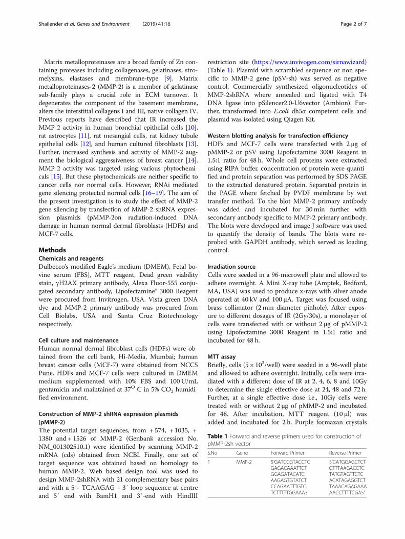
Construction of MMP-2 shRNA expression plasmids(pMMP-2)The potential target sequences, from + 574, + 1035, +1380 and + 1526 of MMP-2 (Genbank accession No.NM_001302510.1) were identified by scanning MMP-2mRNA (cds) obtained from NCBI. Finally, one set oftarget sequence was obtained based on homology tohuman MMP-2. Web based design tool was used todesign MMP-2shRNA with 21 complementary base pairsand with a 5′- TCAAGAG − 3′ loop sequence at centreand 5′ end with BamH1 and 3′-end with HindIII
restriction site (https://www.invivogen.com/sirnawizard)(Table 1). Plasmid with scrambled sequence or non spe-cific to MMP-2 gene (pSV-sh) was served as negativecontrol. Commercially synthesized oligonucleotides ofMMP-2shRNA where annealed and ligated with T4DNA ligase into pSilencer2.0-U6vector (Ambion). Fur-ther, transformed into E.coli dh5α competent cells andplasmid was isolated using Qiagen Kit.
Western blotting analysis for transfection efficiencyHDFs and MCF-7 cells were transfected with 2 μg ofpMMP-2 or pSV using Lipofectamine 3000 Reagent in1.5:1 ratio for 48 h. Whole cell proteins were extractedusing RIPA buffer, concentration of protein were quanti-fied and protein separation was performed by SDS PAGEto the extracted denatured protein. Separated protein inthe PAGE where fetched by PVDF membrane by wettransfer method. To the blot MMP-2 primary antibodywas added and incubated for 30 min further withsecondary antibody specific to MMP-2 primary antibody.The blots were developed and image J software was usedto quantify the density of bands. The blots were re-probed with GAPDH antibody, which served as loadingcontrol.
Irradiation sourceCells were seeded in a 96-microwell plate and allowed toadhere overnight. A Mini X-ray tube (Amptek, Bedford,MA, USA) was used to produce x-rays with silver anodeoperated at 40 kV and 100 μA. Target was focused usingbrass collimator (2 mm diameter pinhole). After expos-ure to different dosages of IR (2Gy/30s), a monolayer ofcells were transfected with or without 2 μg of pMMP-2using Lipofectamine 3000 Reagent in 1.5:1 ratio andincubated for 48 h.
MTT assayBriefly, cells (5 × 10³/well) were seeded in a 96-well plateand allowed to adhere overnight. Initially, cells were irra-diated with a different dose of IR at 2, 4, 6, 8 and 10Gyto determine the single effective dose at 24, 48 and 72 h.Further, at a single effective dose i.e., 10Gy cells weretreated with or without 2 μg of pMMP-2 and incubatedfor 48. After incubation, MTT reagent (10 μl) wasadded and incubated for 2 h. Purple formazan crystals
Table 1 Forward and reverse primers used for construction ofpMMP-2sh vector
S.No Gene Forward Primer Reverse Primer
1 MMP-2 5’GATCCGTACCTCGAGACAAATTCTGGAGATACATCAAGAGTGTATCTCCAGAATTTGTCTCTTTTTGGAAA3’
3’CATGGAGCTCTGTTTAAGACCTCTATGTAGTTCTCACATAGAGGTCTTAAACAGAGAAAAACCTTTTCGA5’
Shailender et al. Genes and Environment (2019) 41:16 Page 2 of 7

were dissolved in DMSO (100 μl) and absorbance wastaken at 590 nm using ELISA plate reader (Bio-Rad,USA) [19].
Dead green assayCells (1x10⁴cells/well) were irradiated with IR at 10Gyand further transfected with or without 2 μg of pMMP-2for 48 h, fixed with paraformaldehyde (4%) and per-meabilized with Triton-X 100(0.1%). Then 50 μL of deadgreen viability stain was added to each well andincubated for 30 min as per the manufacturer’s instruc-tions. Stained cells were visualized under a fluorescentmicroscope (Lynx Microscope, USA), at an excitation(Ex) wavelength of 488 nm and an emission (Em)wavelength of 515 nm and images were captured at 20xresolution.
Immunofluorescence assay of γH2AXγH2AX expression was determined using the immuno-fluorescence assay. The cells were irradiated with IR(10Gy) further transfected with or without 2 μg ofpMMP-2 and incubated for 48 h. Cells were fixed with4% paraformaldehyde and permeabilized. Then cellswere incubated with γH2AX mouse monoclonal anti-body followed by anti-mouse Alexa Fluor 555 goatantibody. Fluorescent microscopy was used to visualizeand capture image [19].
Alkaline comet assayThe cells were irradiated with IR at 10Gy and furthertransfected with or without 2 μg of pMMP-2 and incu-bated for 48 h. Collected cells were suspended in PBS.Subsequently, cell sample and 1% of low melting agarosewere diluted in 1:10 ratio, coated to comet slide in a vol-ume about 100-120ul allowed to incubate at 37 °C. Thenthe slide was plated in a lysis buffer at 4 °C for 60 minfollowed by incubation in pre-chilled alkaline solutionfor 30 min. Electrophoresis was carried out by using thealkaline electrophoresis solution at 25 V and 300 mA for20min. The slide was washed with distilled water andincubated with 100ul of Vista green DNA dye at roomtemperature for 15 min. The images were captured byfluorescent microscopy. Length of the comet was deter-mined using CASP Lab software [20].
ResultsTransfection efficacy of pMMP-2 in HDFs and MCF-7cellsTo evaluate the efficacy of MMP-2 shRNA construct,the monolayer of HDFs and MCF-7 cells were tran-siently transfected with MMP-2 shRNA for 48 h andexpression was analyzed by western blotting analysis.Western blotting analysis reveal that 3.5 fold decreasedexpression of the MMP-2 protein compared to pSV-sh
in HDFs (Additional file 1 Figure S1a). Whereas inMCF-7 cells transfected with pMMP-2 decreased 3 foldcompared to pSV-sh (Additional file 1 Figure S1b).
Effect of MMP-2 gene silencing on radiation-induced DNAdamage in HDFsThe cytotoxic effect of IR on the viability of HDFswas determined by MTT assay. The results showthat the viability of HDFs was decreased with an in-creased dose of IR (2-10Gy) at 24, 48 and 72 h. Theviability of HDFs was found to be 65 ± 3.4, 51 ± 3.9,45 ± 4.8% at 24, 48 and 72 h with 10Gy radiationdose (Fig. 1a). After determining the dose-responsefrom 2 to 10Gy, the effect of single dose radiationwith 10Gy at 48 h was used to evaluate the effect ofpMMP-2 on IR induced cytotoxicity in HDFs.Results indicate that the viability of irradiated HDFswith pMMP-2 transfection was increased 91 ± 4.0%compared to irradiated HDFs (Fig. 1b). The cyto-logical changes in pMMP-2 transfected irradiatedHDF cells at 10Gy were observed under phasecontrast microscopy. As compared to non-irradiatedcontrol, irradiated HDFs showed morphological changesincluding the disrupted cell membrane with characteristicsof apoptosis. However, pMMP-2 transfection apparentlydecreased the characteristics of apoptosis in irradiatedHDFs (Fig. 1c). Further, the effect of pMMP-2 on IRinduced cell death was evaluated using fluorescence-baseddead green viability staining method in HDFs. The resultsdisplay that fluorescence intensity of dead green viabilitystain was increased in irradiated HDFs compared to non-irradiated HDFs. However, pMMP-2 has decreased thefluorescence intensity of dead green viability stain in irra-diated HDFs indicating the increased viability of irradiatedHDFs. Mean fluorescence intensity (MFI) analysis byusing Image J indicate that percent of HDF cells deathwith IR was 62.5 ± 4.5% compared to non-irradiatedcontrol (8.9 + 2.5%).The viability of pMMP-2 treatedirradiated HDFs was 12.45 ± 3.9% after 48 h of post-irra-diation (Fig. 1d). This study further extended to evaluatethe effect of pMMP-2 transfection on radiation-inducedDNA damage in HDF cells using γH2AX antibody. Theresults show that γH2AX foci were significantly increasedwith 10Gy radiation dosage at 48 h compared to non-irra-diated control. Further, pMMP-2 has decreased γH2AXfoci in irradiated HDFs. The analysis of foci by Image Jdisplayed 8 ± 4.2, 75 ± 6.6 and 14 ± 4.5% of γH2AX foci innon-irradiated, irradiated and pMMP-2 transfected irra-diated HDFs (10Gy) at 48 h, respectively (Fig. 1e). Theeffect of IR on DNA fragmentation was determined by thealkaline comet assay and the tail length was measured byusing CASP Lab software. The results demonstrate thatthe tail length was increased about 20-folds with 10Gyradiation compared to non-irradiated control, whereas
Shailender et al. Genes and Environment (2019) 41:16 Page 3 of 7

silencing of the MMP-2 gene has decreased the cometlength to 4-folds in irradiated HDFs (Fig. 1f).
Effect of MMP-2 gene silencing on IR induced DNAdamage in MCF-7 cellsThe viability of MCF-7 cells was decreased in a dose-dependent manner at 24, 48 and 72 h. The viability of
MCF-7 cells was 71 ± 2.5, 55 ± 3.2 and 44 ± 4.8% at 24,48 and 72 h with 10Gy radiation at 48 h (Fig. 2a).However, pMMP-2 transfection reduced the viability ofirradiated MCF-7 cells by 23 ± 2.09% at 48 h (Fig. 2b).Morphological characteristics of apoptosis were featuredwith round shape, cell blebbing and shrinkage were in-creased with pMMP-2 transfection in irradiated MCF-7
Fig. 1 Effect of MMP-2 gene silencing in pMMP-2 transfected irradiated HDF cells. a Graphical representation of viability of HDF cells onirradiation (2-10Gy) analyzed by MTT assay. b Graphical representation of viability in pMMP-2 transfected HDF cells at single large dose i.e. 10Gyanalyzed by MTT assay. c Morphological characterization under phase contrast microscopy of HDF cells. d Fluorescence micrographs of deadgreen stain in HDF cells in control, irradiated and pMMP-2sh transfected irradiated HDF cells. e Representative images γH2AX foci obtained byimmunofluorescence microscopy in HDF cells in control, irradiated HDF cells and pMMP-2 transfected irradiated HDF cells. f Representativeimages of alkaline comet assay performed in HDF cells in control, irradiated and pMMP-2 transfected irradiated HDF cells
Shailender et al. Genes and Environment (2019) 41:16 Page 4 of 7

cells (Fig. 2c). Further, the effect of pMMP-2 on IRinduced cell death of MCF-7 by dead green assayshowed that MFI of irradiated MCF-7 was 58 ± 8.9% andpMMP-2 transfected irradiated MCF-7 was 89 ± 4.6%(Fig. 2d). pMMP-2 also showed DNA damage effectmore effectively in pMMP-2 transfected irradiated
MCF-7 cells, γH2AX foci analysis by Image J indicatedthat the percentof foci was 69 ± 9.4% in irradiated MCF-7cells and 9 ± 7.8% in non-irradiated MCF-7 cells, whereas91 ± 5.6% in pMMP-2 transfected irradiated MCF-7 cells(Fig. 2e). DNA fragmentation analyses by comet assayindicate that the comet tail length was increased 7-folds in
Fig. 2 Effect of MMP-2 gene silencing in pMMP-2 transfected irradiated MCF-7 cells. a Graphical representation of viability of MCF-7 cellsradiation (2-10Gy) analyzed by MTT assay. b Graphical representation of viability of pMMP-2 transfected MCF-7 cells at single large dose i.e. 10Gyanalyzed by MTT assay. c Morphological characterization under phase contrast microscopy of MCF-7 cells. d Fluorescence micrographs of deadgreen stain in MCF-7 cells in control, irradiated and pMMP-2 transfected irradiated MCF-7 cells. e Representative images γH2AX foci obtained byimmunofluorescence microscopy in MCF-7 cells in control, irradiated MCF-7 cells and pMMP-2 transfected irradiated MCF-7 cells. f Representativeimages of alkaline comet assay performed in MCF-7 cells in control, irradiated and pMMP-2 transfected irradiated MCF-7 cells
Shailender et al. Genes and Environment (2019) 41:16 Page 5 of 7

irradiated MCF-7 cells compared to non-irradiatedcontrol. However, pMMP-2 transfection further increasedthe tail length by 1.4 folds in irradiated MCF-7 (Fig. 2f).
DiscussionRadiotherapy is always on a sharp edge in killing thetumors and limiting the survival of critically importantadjacent normal tissues. Currently available radioprotec-tive agents are non-specific and not fulfilling the criteriathat have been approved for clinical use. Therefore, thedevelopment of target-specific radioprotective agents thatexclusively protect normal cells but damage cancer cells isa choice of interest. The present study observed that dose-response versus viability was decreased from 2-10Gy IR at24, 48 and 72 h in both HDFs and MCF-7 cells indicatingthe cytotoxic effect of IR. Previously, similar results werereported in murine skin fibroblasts [21], HEM normal hu-man cells [22] and primary lung fibroblasts [23]. Further,50% viability was observed with a single dose of IR (10Gy)at 48 h of post-radiation. Hence, the effect of MMP-2 genesilencing on the viability of HDFs and MCF-7 cells withIR (10Gy) at 48 h was evaluated similarly to an earlierstudy [24].Loss of cell viability of irradiated HDFs and MCF-7 cells
is due to DNA damage as detected by immunofluores-cence method using γH2AX antibody. The H2AX isquickly phosphorylated at serine 139 when DSBs arise inthe cells and form distinct γH2AX foci, an indicator ofDNA damage. It plays a critical role in the recruitment ofrepair or damage-signaling factors at the site of DNAdamage. Earlier, report on HF19 human fibroblastsobserved an increased γH2AX foci with radiation [25].This study demonstrated that the expression of γH2AXfoci was decreased with pMMP-2 transfection in irra-diated HDFs whereas increased in irradiated MCF-7 cells.Similarly, olive tail length due to DNA fragmentationdecreased in irradiated HDFs and increased in MCF-7cells. These results indicate DNA damage protection inHDFs and destruction in MCF-7 cells. Similarly, MnSODsiRNA showed a protective effect against oxidative stressand radiation exposure in mouse embryonic cells [26].Glyburide, a small molecule inhibitor of protective geneseffectively decreased radiation-induced cell death inT98G, U-87 MG, normal lung epithelial BEAS-2B and inprimary astrocytes [27]. Whereas silencing of HIF-1 αgene protected the HepG2 cells from low doses of CoCl2and radiation [28]. siRNA of pro-apoptotic genes, pkcδand BAX reduced radiation-induced DNA damage inprimary salivary gland cultures [18]. Silencing of Egr1attenuated radiation-induced apoptosis in normal tissue,while sensitized the two cancer cell lines due to differen-tial response based on the cellular context [29].Ideally, clinically useful radioprotectors should protect
normal cells and eliminate cancers. The differential
response to DNA damage on the silencing of the MMP-2gene in HDFs and MCF-7 cells might be due to the pro-death and pro-life activity of the pMMP-2. pMMP-2 haspro-death activity by causing DNA damage in HDFs andpro-life activity by inhibiting the DNA damage in MCF-7cells. IR induced MMP-2 modulates ROS generation [30],which in turn causes DNA damage and cell death [31].Silencing of MMP-2 gene reduced the DNA damage inHDFs, and protected cardiomyocyte from ischemia-reper-fusion injury [32] and reduced the osteogenic trans-formation of fibroblasts by inhibiting the SMP/Smadpathway in ankylosing spondylitis [33]. The pro-death acti-vity of the pMMP-2 enhanced DNA damage with IR inMCF-7 cells. Similarly, silencing of the MMP-2 gene, usingMMP-2 siRNA transfection inhibited radiation-enhancedviability of glioma cells [34] and also suppressed the growthof esophageal carcinoma cells [35].However, pMMP-2 pro-death and pro-life activity may be due to ROS generationthat promotes anti- or pro-tumor effects with differentmechanisms and genomic instability [31]. Additionalstudies will be required to determine the differentialmechanism in HDFs and MCF-7 cells.
ConclusionThis study observed dose-dependent IR induced cyto-toxicity in HDFs and MCF-7 cells with 50% reductionof viability by 10Gy at 48 h. Silencing of MMP-2using pMMP-2 reduced the IR induced cytotoxicityand DNA damage and increased the viability of HDFs.However, MMP-2 gene silencing increased the cytoto-xicity and DNA damage and decreased the viability ofMCF-7 cells. This opposite effect of MMP-2 silencing inHDFs and MCF-7 cells might be due to the differentialregulation of ROS generation.
Additional file
Additional file 1: Figure S1. Expression of MMP-2 gene on silencingwith MMP-2sh vector. a Western blot analysis of MMP-2 protein in HDFstransfected with pMMP-2. b Western blot analysis of MMP-2 protein inMCF-7 cells transfected with pMMP-2. (JPG 245 kb)
AbbreviationsECM: Extracellular matrix; HDF: Human normal Dermal Fibroblasts; IR: Ionizingradiation,; MFI: Mean Fluorescence Intensity; MMP: Matrix metalloproteinases;pMMP-2: Plasmid MMP-2 shRNA; siRNA: small interfering RNA
AcknowledgmentsThanks to DST-FIST Laboratory, GITAM (Deemed to be University) for support.We thank the authorities of GITAM (Deemed to be University) for continuoussupport and encouragement. Authors thank Mr.Manas Malla for grammarcorrections.
Authors’ contributionsMR and GS participated in study design, analysis and interpretation of thedata. GS has executed all the experiments. MR and GS drafted and editedthe manuscript. SK and PK helped in reviewing and editing of themanuscript. All the authors read and approved the final manuscript.
Shailender et al. Genes and Environment (2019) 41:16 Page 6 of 7

FundingThis project was financially supported by DRDO-LSRB, India (Project F.No. CCR&D (TM) /81/48222/LSRB 282/SH&DD2014 dated 08.12.2014.
Availability of data and materialsNot applicable.
Ethics approval and consent to participateNot applicable.
Consent for publicationNot applicable.
Competing interestsThe authors declare that they have no competing interests.
Author details1Cancer Biology Lab, Department of Biochemistry and Bioinformatics,Institute of Science, GITAM (Deemed to be University), Visakhapatnam,Andhra Pradesh, India. 2Department of Biotechnology, Institute of Science,GITAM Deemed to be University, Visakhapatnam, Andhra Pradesh, India.
Received: 29 April 2019 Accepted: 12 July 2019
References1. Baskar R, Lee KA, Yeo R, Yeoh K-W. Cancer and radiation therapy: current
advances and future directions. Int J Med Sci. 2012;9:193.2. Kim JH, Jenrow KA, Brown SL. Mechanisms of radiation-induced normal tissue
toxicity and implications for future clinical trials. Radiat Oncol J. 2014;32:103–15.3. Barnett GC, West CML, Dunning AM, Elliott RM, Coles CE, Pharoah PDP, et al.
Normal tissue reactions to radiotherapy: towards tailoring treatment doseby genotype. Nat Rev Cancer. 2009;9:134–42.
4. Bonnans C, Chou J, Werb Z. Remodeling the extracellular matrix indevelopment and disease. Nat Rev Mol Cell Biol. 2014;15:786–801.
5. Karagiannis GS, Poutahidis T, Erdman SE, Kirsch R, Riddell RH, Diamandis EP.Cancer-associated fibroblasts drive the progression of metastasis throughboth paracrine and mechanical pressure on cancer tissue. Mol Cancer Res.2012;10:1403–18.
6. Citrin D, Cotrim AP, Hyodo F, Baum BJ, Krishna MC, Mitchell JB.Radioprotectors and mitigators of radiation-induced normal tissue injury.Oncologist. 2010;15:360–71.
7. Arora R, Gupta D, Chawla R, Sagar R, Sharma A, Kumar R, et al.Radioprotection by Plant Products: Present Status and Future Prospects.Phytother Res. 2005;22:1–22.
8. Kamran MZ, Ranjan A, Kaur N, Sur S, Tandon V. Radioprotective Agents:Strategies and Translational Advances. Med Res Rev. 2016;36:461–93.
9. Shapiro SD, Senior RM. Matrix metalloproteinases. Matrix degradation andmore. Am J Respir Cell Mol Biol. 1999;20:1100–2.
10. Araya JUN, Maruyama M, Sassa K, Fujita T, Maruyama M, Sassa K, et al. Ionizingradiation enhances matrix metalloproteinase-2 production in human lungepithelial cells. Am J Physiol Lung Cell Mol Physiol. 2001;280:30–8.
11. Sawaya R, Tofilon PJ, Ali-osman F, Liotta LA, William G, Rao J. Induction OfTissue-Type Plasminogen Activator And 72-Kda Type-Iv Collagenase ByIonizing Radiation In Rat Astrocytes. Int I Cancer. 1994;218:214–8.
12. Malley YO, Robbins MEC. Irradiation of rat mesangial cells alters theexperssion of gene products associated with the development of renalfibrosis. Radiat Res. 2014;152:160–9.
13. Hierrtnann G, Wlaschek M, Ts L, Prenzel K, Goerz G. UVA irradiationstimulates the synthesis of various matrix-metalloproteinases ( MMPs ) incultured human fibroblasts. Exp Dermatol. 1993;2:92–8.
14. Pellikainen JM, Ropponen KM, Kataja VV, Kellokoski JK, Eskelinen MJ, KosmaV-M. Expression of matrix metalloproteinase (MMP)-2 and MMP-9 in breastcancer with a special reference to activator protein-2, HER2, and prognosis.Clin Cancer Res. 2004;10:7621–8.
15. Hassan ZK, Daghestani MH. Curcumin effect on MMPs and TIMPs genes in abreast cancer cell line. Asian Pac J Cancer Prev. 2012;13:3259–64.
16. Arany S, Benoit DSW, Dewhurst S, Ovitt CE. Nanoparticle-mediated gene silencingconfers radioprotection to salivary glands in vivo. Mol Ther. 2013;21:1182–94.
17. Liu X-D, Ma S-M, Liu Y, Liu S-Z, Sehon A. Short hairpin RNA and retroviralvector-mediated silencing of p53 in mammalian cells. Biochem Biophys ResCommun. 2004;324:1173–8.
18. Arany S, Xu Q, Hernady E, Benoit DSW, Dewhurst S, Ovitt CE. Pro-apoptoticgene knockdown mediated by nanocomplexed siRNA reduces radiationdamage in primary salivary gland cultures. J Cell Biochem. 2012;113:1955–65.
19. Malla RR, Gopinath S, Alapati K, Gorantla B, Gondi CS, Rao JS. uPAR andcathepsin B inhibition enhanced radiation-induced apoptosis ingliomainitiating cells. Neuro Oncol. 2012;14:745–60.
20. Nagarajan R, Muthusamy G, Balupillai A, Govindasamy K, Ramasamy K,Ponniresan V, et al. Modified comet assays for the detection of cyclobutanepyrimidine dimers and oxidative base damages. J Radiat Cancer Res. 2017:82.
21. Davis GD, Masilamoni JG, Arul V, Kumar MS, Baraneedharan U, Paul SF, et al.Radioprotective effect of dl-α-lipoic acid on mice skin fibroblasts. 2009. 331–340.
22. Long M. World Congress on Medical Physics and Biomedical EngineeringMay 26-31, 2012. Beijing, China: Springer Science & Business Media; 2013.
23. Judge JL, Owens KM, Pollock SJ, Woeller CF, Thatcher TH, Williams JP, et al. IonizingRadiation Induces Myofibroblast Differentiation via Lactate Dehydrogenase[Internet]. Am J Physiol Lung Cell Mol Physiol. 2015; ajplung.00153.2015.
24. Veldwijk MR, Zhang B, Wenz F, Herskind C. The biological effect of largesingle doses: a possible role for non-targeted effects in cell inactivation.PLoS One. 2014;9:e84991.
25. Leatherbarrow EL, Harper JV, Cucinotta FA, O’Neill P. Induction andquantification of γ-H2AX foci following low and high LET-irradiation[Internet]. Int J Radiat Biol. 2006. 111–118.
26. Belikova NA, Glumac A, Rafikov R, Jiang J, Greenberger JS, Kagan VE, et al.Radioprotection by short-term oxidative preconditioning: role of manganesesuperoxide dismutase. FEBS Lett. 2009;583:3437–42.
27. Jiang J, McDonald PR, Dixon TM, Franicola D, Zhang X, Nie S, et al. Syntheticprotection short interfering RNA screen reveals glyburide as a novelradioprotector. Radiat Res. 2009;172:414–22.
28. Jin W, Wang J, Xu S, Xiao L, Chen G, Zhang W, et al. Radioprotective effecton HepG2 cells of low concentrations of cobalt chloride: induction ofhypoxia-inducible factor-1 alpha and clearance of reactive oxygen species. JRadiat Res. 2013;54:203–9.
29. Zhao DY, Jacobs KM, Hallahan DE, Thotala D. Silencing Egr1 AttenuatesRadiation-Induced Apoptosis in Normal Tissues while Killing CancerCells and Delaying Tumor Growth. Mol Cancer Ther. 2015;14:2343–52.
30. Zhang HJ, Zhao W, Venkataraman S, Robbins MEC, Buettner GR, Kregel KC,et al. Activation of matrix metalloproteinase-2 by overexpression ofmanganese superoxide dismutase in human breast cancer MCF-7 cellsinvolves reactive oxygen species. J Biol Chem. 2002;277:20919–26.
31. Reczek CR, Chandel NS. The Two Faces of Reactive Oxygen Species inCancer. Annu Rev Cancer Biol. Ann Rev. 2017;1:79–98.
32. Lin H-B, Cadete VJJ, Sra B, Sawicka J, Chen Z, Bekar LK, et al. Inhibition of MMP-2expression with siRNA increases baseline cardiomyocyte contractility and protectsagainst simulated ischemic reperfusion injury. Biomed Res Int. 2014;2014:810371.
33. Yuan B, Wu Z. MMP-2 silencing reduces the osteogenic transformation offibroblasts by inhibiting the activation of the BMP/Smad pathway inankylosing spondylitis. Oncol Lett. 2018;15:3281–6.
34. Badiga AV, Chetty C, Kesanakurti D, Are D, Gujrati M, Klopfenstein JD, et al.MMP-2 siRNA inhibits radiation-enhanced invasiveness in glioma cells. PLoSOne. 2011;6:e20614.
35. Shen Y-G, Feng W, Xu Y-J, Jiao N-N, Sun D-Q, Qu W-D, et al. Effects of RNAsilencing of matrix metalloproteinase-2 on the growth of esophagealcarcinoma cells in vivo. Oncol Lett. 2017;13:1119–24.
Publisher’s NoteSpringer Nature remains neutral with regard to jurisdictional claims inpublished maps and institutional affiliations.
Shailender et al. Genes and Environment (2019) 41:16 Page 7 of 7