Effect of Mercuric Chloride on Some Hematological ... · the formation of cyanmethemoglobin were...
11
IBIMA Publishing International Journal of Veterinary Medicine: Research & Reports http://www.ibimapublishing.com/journals/IJVMRR/ijvmrr.html Vol. 2013 (2013), Article ID 183410, 11 pages DOI: 10.5171/2013.183410 _____________ Cite this Article as: Aliakbar Hedayati and Zahra Ghaffari (2013), “Effect of Mercuric Chloride on Some Hematological, Biochemical Parameters in Silver Carp (Hypophthalmichthys Molitrix),” International Journal of Veterinary Medicine: Research & Reports, Vol. 2013 (2013), Article ID 183410, DOI: 10.5171/2013.183410 Research Article Effect of Mercuric Chloride on Some Hematological, Biochemical Parameters in Silver Carp (Hypophthalmichthys Molitrix) Aliakbar Hedayati 1 and Zahra Ghaffari 2 1 Department of Fishery, Faculty of Fisheries and Environment, Gorgan University of Agricultural Science and Natural Resources, Gorgan, Iran 2 Gorgan University of Agricultural Science and Natural Resources, Gorgan, Iran Correspondence should be addressed to: Zahra Ghaffari; [email protected] Received 17 June 2013; Accepted 25 July 2013; Published 18 October 2013 Academic Editor: Mahmut Selvi Copyright © 2013 Aliakbar Hedayati and Zahra Ghaffari. Distributed under Creative Commons CC-BY 3.0 Abstract The mutual action between a toxicant and a biological system can be calculated using hematological, biochemical biomarkers. The immunological, hematological and biochemical indices of silver carp (Hypophthalmichthys molitrix) were investigated under low (10%LC50) and high (50%LC50) concentrations of mercury chloride for 96h. In both concentrations Ht, Hb, RBC, lymphocyte were significantly (p<0/05, p<0/01) lower than controls group whereas MCH, MCHC, WBC, Glucose, neutrophil, eosinophil were significantly (p<0/05, p<0/01) larger contrasted to the respective controls group. In low and high concentrations MCV, cortisols respectively were lower than control group. A significant decrease in high concentrations in cortisol level may be as cortisol suppression due to high stresses. The consequential discoveries of this study are that mercury chloride concentrations (low and high) may cause some substitutions in the hematological, biochemical and immunological parameters of the studied fish, so estimation of these indices, could supply a useful indicator of mercury chloride of water bodies. It appears that MCH, eosinophil in low concentrations and lymphocyte in high concentrations is appropriate biomarkers of mercury chloride in silver carp. Keywords: Aquatic ecosystems; Heavy metals; Toxicology; Pollutions. Introduction Heavy metals have been announced to exert a vast range of metabolic, physiological, ecological and behavioral influences on fish (Soengas et al., 1996). Mercury is non- biodegradable and non- advantageous heavy metals and their role in the cell is not understood (Bailey et al., 1999). Mercury cycles in the environment as a result of normal and anthropogenic bustles. Anyway, the amount of mercury liberated via human
Transcript of Effect of Mercuric Chloride on Some Hematological ... · the formation of cyanmethemoglobin were...

IBIMA Publishing
International Journal of Veterinary Medicine: Research & Reports
http://www.ibimapublishing.com/journals/IJVMRR/ijvmrr.html
Vol. 2013 (2013), Article ID 183410, 11 pages
DOI: 10.5171/2013.183410
_____________
Cite this Article as: Aliakbar Hedayati and Zahra Ghaffari (2013), “Effect of Mercuric Chloride on Some
Hematological, Biochemical Parameters in Silver Carp (Hypophthalmichthys Molitrix),” International Journal of
Veterinary Medicine: Research & Reports, Vol. 2013 (2013), Article ID 183410, DOI: 10.5171/2013.183410
Research Article
Effect of Mercuric Chloride on Some
Hematological, Biochemical Parameters in
Silver Carp (Hypophthalmichthys Molitrix)
Aliakbar Hedayati1 and Zahra Ghaffari
2
1Department of Fishery, Faculty of Fisheries and Environment, Gorgan University of Agricultural Science
and Natural Resources, Gorgan, Iran
2Gorgan University of Agricultural Science and Natural Resources, Gorgan, Iran
Correspondence should be addressed to: Zahra Ghaffari; [email protected]
Received 17 June 2013; Accepted 25 July 2013; Published 18 October 2013
Academic Editor: Mahmut Selvi
Copyright © 2013 Aliakbar Hedayati and Zahra Ghaffari. Distributed under Creative Commons CC-BY 3.0
Abstract
The mutual action between a toxicant and a biological system can be calculated using
hematological, biochemical biomarkers. The immunological, hematological and biochemical
indices of silver carp (Hypophthalmichthys molitrix) were investigated under low (10%LC50) and
high (50%LC50) concentrations of mercury chloride for 96h. In both concentrations Ht, Hb, RBC,
lymphocyte were significantly (p<0/05, p<0/01) lower than controls group whereas MCH, MCHC,
WBC, Glucose, neutrophil, eosinophil were significantly (p<0/05, p<0/01) larger contrasted to the
respective controls group. In low and high concentrations MCV, cortisols respectively were lower
than control group. A significant decrease in high concentrations in cortisol level may be as cortisol
suppression due to high stresses. The consequential discoveries of this study are that mercury
chloride concentrations (low and high) may cause some substitutions in the hematological,
biochemical and immunological parameters of the studied fish, so estimation of these indices, could
supply a useful indicator of mercury chloride of water bodies. It appears that MCH, eosinophil in
low concentrations and lymphocyte in high concentrations is appropriate biomarkers of mercury
chloride in silver carp.
Keywords: Aquatic ecosystems; Heavy metals; Toxicology; Pollutions.
Introduction
Heavy metals have been announced to exert a
vast range of metabolic, physiological,
ecological and behavioral influences on fish
(Soengas et al., 1996). Mercury is non-
biodegradable and non- advantageous heavy
metals and their role in the cell is not
understood (Bailey et al., 1999). Mercury
cycles in the environment as a result of
normal and anthropogenic bustles. Anyway,
the amount of mercury liberated via human

International Journal of Veterinary Medicine: Research & Reports 2
_______________
Aliakbar Hedayati and Zahra Ghaffari (2013), International Journal of Veterinary Medicine: Research & Reports,
DOI: 10.5171/2013.183410
activities severely increased since the
commencement of the industrial age (Wang
et al., 2004). The unfastened of
manufacturing, domestic and urban wastes
produced by human activities to aquatic
ecosystems potentially induces different
combinations of stresses to wildlife. The
evaluation of environmental perturbations
requires the explanation of stress effects
throughout the pecking order of biological
organization, from molecular and cellular
levels up to organism and people levels (Moore
2002). To do so, the expansion of
distinctive biomarkers to examine the in vivo
effects of contaminants is a preference
necessity to show the action mechanisms of
toxicants. As aquatic ecosystems are the
most prominent final containers to
industrial and urban waste ejections
(Hoffman et al., 1995), an essential target in
ecotoxicology is to assess the risks for fishes
and human populations. Hematology
furnishes an index of physiological position
of fish and the use of blood picture of fish is
an impressive tool for detection of
alterations in practical state of organism
(Rambhaskar and Rao 1987). Behavioral
changes in animal are symptomatic of
internal interferences in the body functions,
such as disturbances in metabolic routes and
ionic imbalance in blood serum (Lewis and
Lewis 1971; Shah 2002). Blood indices are
more frequently utilized when clinical
diagnosis of fish physiology are used to
ascertain sublethal and chronic exposure of
contaminants (Kim et al., 2008). Fish
hematological indices are very susceptible to
water contaminants, and its commutation due
to the hematological and immunological
parameters can be utilized as toxicity indices
of xenobiotics (Sancho et al., 2000). however
fish blood indices have been increasingly
tested in marine environmental monitoring
as important parameters of physiological
changes in the presence of contaminant,
the absence of basic knowledge about the
hematological reaction to the main stressors
of at risk species is the most important
leakage for using these indices in
environmental monitoring plans (Affonso et
al., 2002). Examinations on the toxic effect of
metals on fish are joined by the analysis of
exchanges in some haematological and
biochemical blood indices (Hoyle et al., 2007).
The mutual action between a toxicant and a
biological system can be calculated using
hematological, biochemical biomarkers
(Li et al., 2010). The change in
hematological (RBC and WBC count, Hb,
Hct, MCV, MCH and MCHC), biochemical
(glucose and cortisol) biomarkers and
immunological (lymphocyte, neutrophil
and eosinophil) parameters are greatly
used to evaluate the toxic stress, integrity
of the immune system and tissue damage
(Nemcsok and Benedeczky 1990; Talas and
Gulhan 2009; Kavitha et al., 2010). The
changes in these biomarkers are
frequently susceptible to environmental
or physiological changes and are simply
quantifiable and supply an integrated
measure of the physiological changes in
organisms (Remyla et al., 2008).
Materials and Methods
Juvenile samples of silver carp with mean
weight 200gr provided by fisheries research
laboratory. Fish were adapted to laboratory
conditions for 7 days in a 400L tank with
dechlorinated tap water. During adaption, all
fish were fed with trading pellet twice a day.
fish were exposed to a low concentration of
10% LC50 (0/09 mg/l) mercury chloride
and high concentration of 50% LC50 (0/27
mg/l) for a period of 24h, 48h and 96h static
toxicity examination, presented in tank of
400L, each including 21 fish. One group
control was conserved in a fiberglass tank
without accessing toxicant by storing 21 fish.
For each experimental time were carried out
three repeats. PH, dissolved oxygen,
temperature and conductivity were
monitored during the experiment (Hedayati
et al., 2010).
Fish were without delay anesthetized with
200ppm clove power. Blood samples rapidly

3 International Journal of Veterinary Medicine: Research & Reports
_______________
Aliakbar Hedayati and Zahra Ghaffari (2013), International Journal of Veterinary Medicine: Research & Reports,
DOI: 10.5171/2013.183410
were taken from tail blood vessel by
heparinized syringes. Appointments of the
blood indices were carried out on fresh
blood. Amounts of blood leukocytes and
erythrocytes were carried out at 1:30
dilution by diluting heparinized blood with
Giemsa stain. Cells were enumerated using a
hemocytometer Neubauer below the light
microscope (Stevens 1997)
According to Beutler et al., (2001) the
leukocyte differential tab was made in
peripheral blood smears stained with Merck
Giemsa, giving the neutrophils extent of
differential neutrophils and the mononuclear
value of differential lymphocytes too
eosinophil and monocyte.
After sampling Hematocrite values were
shortly ascertained by putting new blood in
glass capillary tubes and centrifuged in a
microhematocrit centrifuge at 10000 rpm for
5 min (Hettich, Germany). With assist of a
microhematocrit were accomplished
Hematocrite readerships.
According to Lee et al., (1998) by measuring
the formation of cyanmethemoglobin were
found hemoglobin levels (Hb mg/l).
Erythrocytes Indices (MCH or Mean
Corpuscular Hemoglobin, MCHC or Mean Cell
Hemoglobin Concentration and MCV or Mean
Corpuscular Volume) were computed from
RBC, Ht, and Hb (Lee et al., 1998).
To determine meaningful disagreements to
evaluate the effect of methyl mercury on
blood indices was used an analysis of
variance (ANOVA) with Duncan Post Hoc.
Pearson coefficients of correlation(r) were
evaluated between mercury concentrations
and blood indices to investigate associations
between bioaccumulation and its effects.
Multiple regressions were used to ascertain
the kinship between mercury concentration
and blood parameters. The data were
analyzed by means of statistics at p<0/05.
Data are told as means ± standard deviation (
SDX ± ).
Results
In the present examination the 96 h LC 50
value of mercury chloride to (H. molitrix)
was determined to be 0/55 mg/l.
Results of Survey Blood Factors in Low
Concentrations (10%Lc50)
Fish exposed to mercury for 96h presented a
significant change in hematological (Hb, Ht,
MCH, MCHC, MCV, RBC), biochemical
(glucose, cortisol) and immunological
(neutrophil, lymphocyte, eosinophil)
parameters concentrations toward the
control groups (p<0/05), whereas among
significant indices MCV, RBC, Ht, Hb,
Lymphocyte in fish exposed to mercury for
96h were significantly lower than control
groups (P< 0.05) and WBC, MCH, MCHC,
glucose, cortisol, neutrophil, eosinophil were
significantly (P< 0.05) greater compared to
the control groups (Fig 3-10) . In low
concentrations, the correlation between
mercury with all parameters was statistically
tested by analyzing the data procured during
the mercury exposed. MCH, MCHC, cortisol,
glucose, neutrophil and eosinophils levels
showed significant (p<0/01) positive
correlations but RBC, lymphocyte (p<0/01)
and Ht (p<0/05) exerted significant
correlation with mercury exposure that
altogether were negative in 10%Lc50
concentrations. To appointment the
relationship between mercury
concentrations with biochemical,
hematological and immunological activity
accustomed to curve estimation regressions.
Only the MCH and eosinophil levels showed
significant linear regression (p<0/05) Y = a ±
bX with mercury (Fig. 1).

International Journal of Veterinary Medicine: Research & Reports 4
_______________
Aliakbar Hedayati and Zahra Ghaffari (2013), International Journal of Veterinary Medicine: Research & Reports,
DOI: 10.5171/2013.183410
Fig .1: Regression Curve M.C.H, Eosinophil of Silver Carp during Exposure to Mercury
Chloride in Low Concentrations
Results of Survey Blood Factors in High
Concentrations (50%Lc50)
Change in all the blood factors was similar to
low concentrations except MCV, cortisol (Fig.
3, 9).
MCV levels were significantly greater (P<
0.05) than controls groups and cortisol levels
were significantly lowers (P< 0.05) toward
the control groups.
MCH, glucose, eosinophil levels showed
significant (p<0/01) positive correlations but
RBC (P<0/01), Ht and lymphocyte (p<0/05)
levels displayed significant negative
correlations with mercury exposure.
Only eosinophils and lymphocyte introduced
significant linear regression (p<0/05) Y = a ±
bX with mercury (Fig. 2).
Fig .2: Regression Curve Lymphocyte, Eosinophil of Silver Carp during Exposure to Mercury
Chloride in High Concentrations

5 International Journal of Veterinary Medicine: Research & Reports
_______________
Aliakbar Hedayati and Zahra Ghaffari (2013), International Journal of Veterinary Medicine: Research & Reports,
DOI: 10.5171/2013.183410
Fig .3: MCV Change of Silver Carp during
Exposure to Mercury Chloride in Low and
High Concentrations
Fig .4: WBC Change of Silver Carp during
Exposure to Mercury Chloride in Low and
High Concentrations
Fig .5: Ht, Hb Change of Silver Carp during
Exposure to Mercury Chloride in Low and
High Concentrations
Fig .6: MCH, MCHC Change of Silver Carp
during Exposure to Mercury Chloride in
Low and High Concentrations
Fig .7: Lymphocyte, Neutrophil Changes of
Silver Carp during Exposure to Mercury
Chloride in Low and High Concentrations
Fig .8: RBC Changes of Silver Carp during
Exposure to Mercury Chloride in Low and
High Concentrations
International Journal of Veterinary Medicine: Research & Reports 6
_______________
Aliakbar Hedayati and Zahra Ghaffari (2013), International Journal of Veterinary Medicine: Research & Reports,
DOI: 10.5171/2013.183410
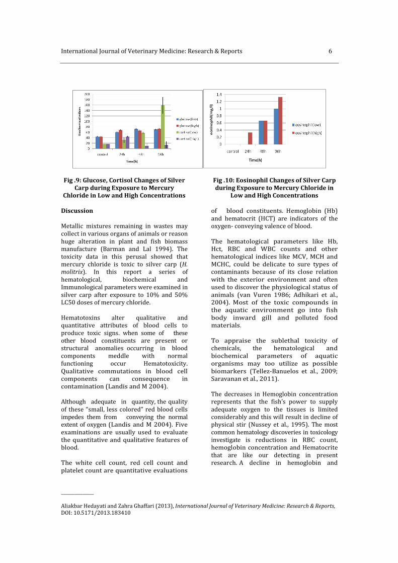
Fig .9: Glucose, Cortisol Changes of Silver
Carp during Exposure to Mercury
Chloride in Low and High Concentrations
Fig .10: Eosinophil Changes of Silver Carp
during Exposure to Mercury Chloride in
Low and High Concentrations
Discussion
Metallic mixtures remaining in wastes may
collect in various organs of animals or reason
huge alteration in plant and fish biomass
manufacture (Barman and Lal 1994). The
toxicity data in this perusal showed that
mercury chloride is toxic to silver carp (H.
molitrix). In this report a series of
hematological, biochemical and
Immunological parameters were examined in
silver carp after exposure to 10% and 50%
LC50 doses of mercury chloride.
Hematotoxins alter qualitative and
quantitative attributes of blood cells to
produce toxic signs. when some of these
other blood constituents are present or
structural anomalies occurring in blood
components meddle with normal
functioning occur Hematotoxicity.
Qualitative commutations in blood cell
components can consequence in
contamination (Landis and M 2004).
Although adequate in quantity, the quality
of these “small, less colored” red blood cells
impedes them from conveying the normal
extent of oxygen (Landis and M 2004). Five
examinations are usually used to evaluate
the quantitative and qualitative features of
blood.
The white cell count, red cell count and
platelet count are quantitative evaluations
of blood constituents. Hemoglobin (Hb)
and hematocrit (HCT) are indicators of the
oxygen- conveying valence of blood.
The hematological parameters like Hb,
Hct, RBC and WBC counts and other
hematological indices like MCV, MCH and
MCHC, could be delicate to sure types of
contaminants because of its close relation
with the exterior environment and often
used to discover the physiological status of
animals (van Vuren 1986; Adhikari et al.,
2004). Most of the toxic compounds in
the aquatic environment go into fish
body inward gill and polluted food
materials.
To appraise the sublethal toxicity of
chemicals, the hematological and
biochemical parameters of aquatic
organisms may too utilize as possible
biomarkers (Tellez-Banuelos et al., 2009;
Saravanan et al., 2011).
The decreases in Hemoglobin concentration
represents that the fish’s power to supply
adequate oxygen to the tissues is limited
considerably and this will result in decline of
physical stir (Nussey et al., 1995). The most
common hematology discoveries in toxicology
investigate is reductions in RBC count,
hemoglobin concentration and Hematocrite
that are like our detecting in present
research. A decline in hemoglobin and

7 International Journal of Veterinary Medicine: Research & Reports
_______________
Aliakbar Hedayati and Zahra Ghaffari (2013), International Journal of Veterinary Medicine: Research & Reports,
DOI: 10.5171/2013.183410
Hematocrite levels can reveal an anemic
situation (Kumar et al., 1999). Panigrahi and Misra (1987) saw reductions
in hemoglobin outturn and red blood cell
count of the fish Anabas scandens treated
with mercury. Reduction in Hct, Red blood
cell (RBC) count and Hb was studied in fish
Tinca, tinca exposed to lead and mercuric
chloride (Shah and Altindag 2004).
The decline in RBC counts in the current
study might have resulted from inhibition of
RBC manufacture by the mercury chloride.
Likewise, Li et al., (2011) announced a
reduction of total content of RBC in the blood
of rainbow trout (Oncorhynchus mykiss)
when exposed to verapamil (VPR), a
cardiovascular medicine. Mostly, the
reduction in RBC counts in fish may imply the
anemic condition of the fish under stress
situations (Li et al., 2011). Furthermore,
amplitude of toxicants in the gill area may
injury the structure of gill resulting
hemolysis and toxicant induced damaged
osmoregulation may lead to a diminution in
RBC counts (Kavitha et al., 2010; Saravanan
et al., 2011). When erythrocytes have low
hemoglobin extent reflects Microcytic
hypochromic anemia.
The declined number of RBC in fish caused
by toxicant exposure has been announced
by Allin and Wilson (2000) and
Chowdhury et al., (2004). The increase in MCV in high
concentrations can too result from an
expansion of unripe RBC (Carvalho and
Fernandes 2006). In the present study the
important increase of MCV (in high
concentrations) and MCH during low
and high concentrations might be caused
by the above said reason. The expansion of
MCV studied in individuals of
H.malabaricus exposed to MeHg may be
illustrated by the existence of a larger
amount of older or larger red blood cells as
delineated by Hardig and Hoglund (1983).
In the later phases the high percentage of
unripe red blood cells in the circulation
might be the cause for MCV decrease in low
concentrations. The significant expansion
of MCHC value might be resulted from
sphaerocytosis as mentioned by Sobecka
(2001).
The expansion in MCV (in high
concentrations) and MCH value show the
anemia was of a macrocytic type as
submitted by Talas and Gulhan (2009).
WBCs are involved in the adjustment of
immunological work in many organisms
and the saw increase in WBC count in
mercury chloride treated fish shows a
generalized immune reply and a defensive
response to mercury chloride (Witeska
2004; Saravanan et al., 2011). Rousing
effect of the toxicant on immune system
and liberate of lymphocytes from
lymphomyeloid tissue as a protection
mechanism may also redound to expansion
in WBC count in fish (Ates et al., 2008).
Toxicants in the aquatic environment
may cause influence at cellular or
molecular level which results in
significant changes in the biochemical
parameters of the organisms (Kavitha et
al., 2010). Between the several
biochemical parameters, blood glucose
level in toxicant treated animals has been
greatly utilized as an indicator of
environmental stress (Nemcsok and Boross
1982).
The blood glucose level has been utilized as
an indicator of environmental stress and
returned the changes in carbohydrate
metabolism below hypoxia and stress
conditions. Tseng (2004) announced that
chronic exposure of arsenic or its methylated
metabolites caused Diabetes mellitus in rats
and this condition may be accountable for
hyperglycemia. Therefore an elevation of
blood glucose level in the current study
might be caused by gluconeogenesis to
supply energy for the increased metabolic
claims instituted by mercury chloride stress.

International Journal of Veterinary Medicine: Research & Reports 8
_______________
Aliakbar Hedayati and Zahra Ghaffari (2013), International Journal of Veterinary Medicine: Research & Reports,
DOI: 10.5171/2013.183410
In the present study, a significant increase in
cortisol level was observed in fish exposed to
low concentrations.
Other stressors activate the hypothalamus-
pituitary-interrenal (HPI) axis, resulting in a
cortisol liberate that causes secondary stress
answers.
A significant decrease in high concentrations
in cortisol level may be as Cortisol
suppression due to high stresses. However,
the need for further research is felt in this
area.
Our consequences show expansion in
differential neutrophil and decrease in
lymphocyte. It has been approved that the
monocytes and neutrophils raised while
lymphocytes decrease during different
stressors in cultured fish Oreochromis
aureus Silveira-Coffignya et al., (2004). It is
thought that neutrophils and monocytes
have phagocytic stir, which might elucidate
their increased proportion during infectious
conditions. Darwish et al., (2001) too
discovered an increase in neutrophil
amounts in channel catfish exposed to high
doses of potassium permanganate.
Regarding eosinophilia, Abdel-Ghafar and El-
Khayat showed that the most common
cause of peripheral eosinophilia in patients
of the third world was parasites.
The consequential discoveries of this study
are that mercury chloride concentrations
(low and high) may cause some substitutions
in the hematological, biochemical and
immunological parameters of the studied
fish, so estimation of these indices, could
supply a useful indicator of mercury chloride
of water bodies. It appears that MCH,
eosinophil in low concentrations and
Lymphocyte in high concentrations is
appropriate biomarkers of mercury chloride
in silver carp (H. molitrix).
References
Abdel-Ghafar, F. & El-Khayat, H. (1988). Ain
Shams Med. J. 39 (1988)221
Adhikari, S., Sarkar, B., Chatterjee, A.,
Mahapatra, C. T. & Ayyappan, S. (2004).
“Effects of Cypermethrin and Carbofuran on
Haematological Parameters and Prediction of
their Recovery in a Freshwater Teleost,
Labeo rohita (Hamilton),” Ecotoxicology and
Environmental Safety 58:220-226
Affonso, E. G., Polez, V. L. P., Corre, C. F.,
Mazon, A. F., Araujo, M. R. R., Moraes, G. &
Ratin, F. T. (2002). 'Blood Parameters and
Metabolites in the Teleosts fish Colossoma
Macropomum Exposed to Sulfide or Hypoxia,'
Comparative Biochemistry and Physiology
C133:375-382
Allin, C. J. & Wilson, R. W. (2000). “Effects of
Pre-Acclimation to Aluminium on the
Physiology and Swimming Behaviour of
Juvenile Rainbow Trout (Oncorhynchus
Mykiss) during a Pulsed Exposure,” Aquatic
Toxicology 51 (2):213-224
Ates, B., Orun, I., Talas, Z. S., Durmaz, G. &
Yilmaz, I. (2008). “Effects of Sodium Selenite
on Some Bio-chemical and Hematological
Parameters of Rainbow Trout (Oncorhynchus
Mykiss Walbaum, 1792) Exposed to Pb2+ and
Cu2+,” Fish Physiology and Biochemistry
34:53-59
Bailey, S. E., Olin, T. J., Bricka, R. M. & Adrian,
D. D. (1999). “A Review of Potentially Low-
cost Sorbents for Heavy Metals,” Water
Research 33:2469-2479
Barman, S. C. & Lal, M. M. (1994).
'Accumulation of Heavy Metal (Zn, Cu,
Cd and Pb) in Soil and Cultivated Vegetables
and Weeds Grown in Industrially Polluted
Fields,' Journal of Environmental Biology
15:107-115
9 International Journal of Veterinary Medicine: Research & Reports
_______________
Aliakbar Hedayati and Zahra Ghaffari (2013), International Journal of Veterinary Medicine: Research & Reports,
DOI: 10.5171/2013.183410
Beutler, E., Lichtman, M. A., Coller, B. S. &
Seligsohn, U. (2001). 'Hematology,' sixth ed.
McGraw-Hill, USA
Carvalho, C. S. & Fernandes, M. N. (2006).
“Effect of Temperature on Copper Toxicity
and Hematological Responses in the
Neotrophical Fish, Prochilodus Scrofa at Low
and High pH,” Aquaculture 251:109-117
Chowdhury, M. J., Pane, E. F. & Wood, C. M.
(2004). “Physiological Effects of Dietary
Cadmium Acclimation and Waterborne
Cadmium Challenge in Rainbow Trout:
Respiratory, Ionoregulatory, and Stress
Parameters,” Comparative Biochemistry and
Physiology Part C: Toxicology & Pharmacology
139:163-173
Darwish, A. M., Griffin, B. R., Straus, D. L. &
Mitchell, A. J. (2001). “Histological and
Haematological Evaluation of Potassium
Permanganate Exposure in Channel Catfish,”
Journal of Aquatic Animal Health 14:134-144
Hardig, J. & Hoglund, L. B. (1983). “Seasonal
and Ontogenetic Effects on Methaemoglobin
and Reduced Glutathione Content in the
Blood of Reared Baltic Salmon,” Comparative
Biochemistry and Physiology 75:27-34
Hedayati, A., Safahieh, A., Savari, A. & Ghofleh
Marammazi, J. (2010). “Detection of Mercury
Chloride Acute Toxicity in Yellowfin Sea
Bream (Acanthopagrus Latus),” World
Journal of Fish and Marine Sciences 2: 86-92
Hoffman, J. D., Rattner, B. A., Burton, G. A. &
Cairns, J. (1995). Handbook of Ecotoxicology,
Lewis Publishers, London
Hoyle, I., Shaw, B. J. & Handy, R. D. (2007).
“Dietary Copper Exposure in the African
Walking Catfish, Clarias Gariepinus: Transient
Osmoregulatory Disturbances and Oxidative
Stress,” Aquatic Toxicology 83(1):62-72
Kavitha, C., Malarvizhi, A., Kumaran, S. S. &
Ramesh, M. (2010). “Toxicological Effects of
Arsenate Exposure on Hematological,
Biochemical and Liver Transaminases
Activity in an Indian Major Carp, Catla Catla,”
Food and Chemical Toxicology 48:2848-2854
Kim, S. G., Park, D. K., Jang, S. W., Lee, J. S.,
Kim, S. S. & Chung, M. H. (2008). “Effects of
Dietary Benzo Pyrene on Growth and
Hematological Parameters in Juvenile
Rockfish, Sebastes Schlegeli (Hilgendorf),”
Bulletin of Environmental Contamination and
Toxicology 81:470-474
Kumar, S., Lata, S. & Gopal, K. (1999).
“Deltamethrin Induced Physiological Changes
in Freshwater Catfish Heteropneustes
Fossilis,” Bulletin of Environmental
Contamination and Toxicology 62: 254-258
Landis, W. G. & Yu, M. H. (2004).
'Introduction to Environmental Toxicology,'
Crc Press. pp: 509
Lee, R. G., Foerster, J., Jukens, J., Paraskevas,
F., Greer, J. P. & Rodgers, G. M. (1998).
'Wintrobe’s—Clinical Hematology,' 10th ed.
Lippincott Williams & Wilkins, New York, USA
Lewis, S. D. & Lewis, W. M. (1971). 'The Effect
of Zinc and Copper on the Osmolality of
Blood Serum of the Channel Catfish, Ictalurus
Punctatus and Golden Shiner, Notemigonus
Crysoleucas,' Transactions of American
Fisheries Society 4:639-643
Li, Z. H., Velisek, J., Zlabek, V., Grabic, R.,
Machova, J., Kolarova, J., Li, P. & Randak, T.
(2011). “Chronic Toxicity of Verapamil on
Juvenile Rainbow Trout (Oncorhynchus
Mykiss): Effects on Morphological Indices,
Hematological Parameters and Antioxidant
Responses,” Journal of Hazardous Materials
185: 870-880
Li, Z. H., Velisek, J., Zlabek, V., Grabic, R.,
Machova, J., Kolarova, J. & Randak, T. (2010).
“Hepatic Antioxidant Status and
Hematological Parameters in Rainbow Trout,
Oncorhynchus Mykiss, after Chronic Exposure
to Carbamazepine,” Chemico-Biological
Interactions 183:98-104
Moore, M. N. (2002). “Biocomplexity: The
Post-genome Challenge in Ecotoxicology,”
Aquatic Toxicology 59:1-15

International Journal of Veterinary Medicine: Research & Reports 10
_______________
Aliakbar Hedayati and Zahra Ghaffari (2013), International Journal of Veterinary Medicine: Research & Reports,
DOI: 10.5171/2013.183410
Nemcsok, J. & Benedeczky, I. (1990). "Effect
of Sublethal Concentrations of Phenol on
Some Enzyme Activities and Blood Sugar
Level of Carp, (Cyprinus Carpio L.),”
Environmental Monitoring and Assessment
14: 377-383
Nemcsok, J. & Boross, L. (1982).
“Comparative Studies on the Sensitivity of
Different Fish Species to Metal Pollution,”
Acta Biologica Academiae Scientarum
Hungarica 33: 23-27
Nussey, G., Van Vuren, J. H. J. & Du Preez, H. H.
(1995). 'Effects of Copper on Haematology
and Osmoregulation of the Mozambique
Tilapia, Oreochromis Mossambicus
(Cichlidae),' Comparative Biochemistry and
Physiology 111:369-380
Panigrahi, A. K. & Misra, B. N. (1987).
'Toxicological Effects of Mercury on a Fresh
Water Fish Anabas Scandens, CUV and VAL
and their Ecological Implications,'
Environmental Pollution.16:31-39
Rambhaskar, B. & Rao, K. S. (1987).
“Comparative Haematology of Ten Species of
Marine Fish from Visakhapatnam Coast,”
Journal of Fish Biology. 30: 59-66
Remyla, S. R., Ramesh, M., Sajwan, K. S.
& Kumar, K. S. (2008). “Influence of Zinc on
Cadmium Induced Haematological and
Biochemical Responses in a Freshwater
Teleost Fish Catla Catla,” Fish Physiology and
Biochemistry 34:169-174
Sancho, E., Ceron, J. J. & Ferrando, M. D.
(2000). “Cholinesterase Activity and
Hematological Parameters as Biomarkers of
Sublethal Molinate Exposure in Anguilla
Anguilla,” Ecotoxicology and Environmental
Safety 46:81-86
Saravanan, M., Karthika, S., Malarvizhi, A. &
Ramesh, M. (2011). “Ecotoxicological Impacts
of Clofibric Acid and Diclofenac in Common
Carp (Cyprinus Carpio) Fingerlings:
Hematological, biochemical, Ionoregulatory
and Enzymological Responses,” Journal of
Hazardous Materials 195:188-194
Shah, S. L. (2002). “Behavioural
Abnormalities of Cyprinion Watsoni on
Exposure to Copper and Zinc,” Turkish
Journal of Zoology, 26:137-140
Shah, S. L. & Altindag, A. (2005). 'Alterations
in the Immunological Parameters of Tench
(Tinca Tinca L.) after Acute and Chronic
Exposure to Lethal and Sublethal Mercury
Treatments,' Bulletin of Environmental
Contamination and Toxicology 73:911-918
Silveira-Coffignya, R., Prieto-Trujilloa, A. &
Ascencio-Valle, F. (2004). “Effects of Different
Stressors in Haematological Variables in
Cultured Oreochromis Aureus S.,"
Comparative Biochemistry and Physiology
Part C: Toxicology & Pharmacology 39:245-
250
Sobecka, E. (2001). 'Changes in the Iron
Leveling the Organs and Tissues of Wells
Catfish, Silurus Glanis L. Caused by Nickel,'
Acta. Ichthyol. Piscat. 31 (2):127-143
Soengas, J. L., Agra-Lago, M. J., Carballo, B.,
Andres, M. D. & Veira, J. A. R. (1996). “Effects
of an Acute Exposure to Sublethal
Concentrations of Cadmium on Liver
Carbohydrates Metabolism of Atlantic
Salmon, (Salmo Salar),” Bulletin of
Environmental Contamination and Toxicology,
57(4):625-631
Stevens, M. L. (1997). Fundamentals of
Clinical Hematology, WB Saunders,
Philadelphia, PA
Talas, Z. S. & Gulhan, M. F. (2009). “Effects of
Various Propolis Concentrations on
Biochemical and Hematological Parameters
of Rainbow Trout (Oncorhynchus Mykiss),”
Ecotoxicology and Environmental Safety
72:1994-1998
Tellez-Banuelos, M. C., Santerre, A., Casas-
Solis, J., Bravo-Cuellar, A. & Zaitseva, G.

11 International Journal of Veterinary Medicine: Research & Reports
_______________
Aliakbar Hedayati and Zahra Ghaffari (2013), International Journal of Veterinary Medicine: Research & Reports,
DOI: 10.5171/2013.183410
(2009). “Oxidative Stress in Macrophages
from Spleen of Nile Tilapia (Oreochromis
Niloticus) Exposed to Sublethal
Concentration of Endosulfan,” Fish & Shellfish
Immunology 27:105-111
Tseng, C.- H. (2004). “The Potential Biological
Mechanisms of Arsenic-induced Diabetes
Mellitus," Toxicology and Applied
Pharmacology 197:67-83
Van Vuren, J. H. J. (1986). “The Effects of
Toxicants on the Hematology of Labeo
Umbratus (Teleostei Cyprinidae),”
Comparative Biochemistry and Physiology
Part C: Comparative Pharmacology 83:155-
159
Wang, Q., Kim, D., Dionysiou, D. D., Sorial, G.,
A. & Timberlake, D. (2004). “Sources and
Remediation for Mercury Contamination in
Aquatic Systems-A Literature Review,”
Environmental Pollution, 131(2):323-336
Witeska, M. (2004). 'The Effect of Toxic
Chemicals of Blood Cell Morphology in Fish,'
Fresenius Environ. Bull. 13 (12):1379-1384


![ToxicEffectsofMercuryontheCardiovascularandCentral ...(Hg(CNO) 2), which is used as an explosive detonator [8, 25]. Among the mercuric mercury compounds, mercuric chloride (HgCl 2)](https://static.fdocuments.us/doc/165x107/60d2d8c6db79240e210ea4a7/toxiceffectsofmercuryonthecardiovascularandcentral-hgcno-2-which-is-used.jpg)