EFFECT OF BORON DEFICIENCY ON THE HISTOLOGY OF GARDEN BEET
28
EFFECT OF BORON DEFICIENCY ON THE HISTOLOGY OF GARDEN BEET AND CABBAGE ' By JAMES P. JOLIVETTE, instructor, and J. C. WALKER, professor of plant pathology, Wisconsin Agricultural Experiment Station ^ INTRODUCTION When garden beet (Beta vulgaris L.) and cabbage {Brassica olerácea var. capitata L.) are grown in nutrient solution or in soil deficient in boron, marked external malformation, together with internal discolora- tion and necrosis, occurrs in several organs of the plants. The appear- ance of macroscopic symptoms is quite sudden if boron deficiency is acute. Signs of the disease are similar in seedlings of both species and in the mature plants of garden beet, but they differ markedly in the mature plants of cabbage. Since beet and cabbage differ widely in anatomy, they were particularly suitable subjects for a comparative study of the nature and extent of histological changesleading to and accompanying macroscopic symptoms of boron deficiency. Investigators undertook to study the histological changes induced in plants by boron deficiency soon after establishment of the fact that boron is essential to normal plant growth. Interest in the subject was further stimulated by the discoveries of Mes (^0) ^ and Brandenburg (4) that a number of unexplained plant diseases were due, primarily, to boron deficiency. The effects of boron deficiency have been inves- tigated by others in sugar beet (Beta vulgaris L.) (^^), broadbean (Vicia faba L.) (5, 32), potato (Solanum tuberosum L.) (24), tobacco (Nicotiana tabacum L.) (22,26), tomato (Lycopersiconesculentum Mül.) (15, 23), pea (Pimm sativum L.) (28), rutabaga (Brassica campestris var. napobrassica DC.) (6, 17), carrot (Daucus carota L.) (33), sugar- cane (Saccharum officinarum L.) (19), com (Zea mays L.) (7), apple (Malus pumila Mill.) (18), grapefruit (Citrus grandis Osbeck) (12), and orange (Citrus sinensis Osbeck) (12). These reports vary from mere mention of affected tissues to detailed histological studies. These studies showed that the most extensive pathological changes caused by boron deficiency occur in the meristematic regions and in the pith. Frequently the first evidence of degeneration appeared at or near the apical growing point. Later, abnormalities were common in vascular tissues throughout the plant and in the mesophyll of expanding leaf laminae. Often differentiation was retarded or sup- pressed. Younger differentiated elements were misshapen, aiid hy- pertrophy and hyperplasia, accompanied or followed by necrosis, were common in the cambial zone. Some workers found inter- and intra- cellular deposits in the necrotic areas. The histological changes found 1 Received for publication March 11, 1942. This study was supported in part by the Wisconsin Alumni Research Foundation and the American Potash Institute. Certain nontechnical assistance was provided by the Work Projects Administration, project No. 65-1-53-2349. The photographs were made by Eugene Herrling, Wisconsin Agricultural Experiment Station. 2 The writers wish to acknowledge the helpful advice of Prof. Emma L. Fisk, Department of Botany, University of Wisconsin, during the course of this investigation. 3 Italic numbers in parentheses refer to Literature Cited, p. 181. Journal of Agricultural Research, Vol. 66, No. 14 Washington, D. C. Feb. 15,1943 Key No. Wis.-136 (167)
Transcript of EFFECT OF BORON DEFICIENCY ON THE HISTOLOGY OF GARDEN BEET
EFFECT OF BORON DEFICIENCY ON THE HISTOLOGY OF GARDEN BEET AND CABBAGE '
By JAMES P. JOLIVETTE, instructor, and J. C. WALKER, professor of plant pathology, Wisconsin Agricultural Experiment Station ^
INTRODUCTION
When garden beet (Beta vulgaris L.) and cabbage {Brassica olerácea var. capitata L.) are grown in nutrient solution or in soil deficient in boron, marked external malformation, together with internal discolora- tion and necrosis, occurrs in several organs of the plants. The appear- ance of macroscopic symptoms is quite sudden if boron deficiency is acute. Signs of the disease are similar in seedlings of both species and in the mature plants of garden beet, but they differ markedly in the mature plants of cabbage. Since beet and cabbage differ widely in anatomy, they were particularly suitable subjects for a comparative study of the nature and extent of histological changesleading to and accompanying macroscopic symptoms of boron deficiency.
Investigators undertook to study the histological changes induced in plants by boron deficiency soon after establishment of the fact that boron is essential to normal plant growth. Interest in the subject was further stimulated by the discoveries of Mes (^0) ^ and Brandenburg (4) that a number of unexplained plant diseases were due, primarily, to boron deficiency. The effects of boron deficiency have been inves- tigated by others in sugar beet (Beta vulgaris L.) (^^), broadbean (Vicia faba L.) (5, 32), potato (Solanum tuberosum L.) (24), tobacco (Nicotiana tabacum L.) (22,26), tomato (Lycopersiconesculentum Mül.) (15, 23), pea (Pimm sativum L.) (28), rutabaga (Brassica campestris var. napobrassica DC.) (6, 17), carrot (Daucus carota L.) (33), sugar- cane (Saccharum officinarum L.) (19), com (Zea mays L.) (7), apple (Malus pumila Mill.) (18), grapefruit (Citrus grandis Osbeck) (12), and orange (Citrus sinensis Osbeck) (12). These reports vary from mere mention of affected tissues to detailed histological studies.
These studies showed that the most extensive pathological changes caused by boron deficiency occur in the meristematic regions and in the pith. Frequently the first evidence of degeneration appeared at or near the apical growing point. Later, abnormalities were common in vascular tissues throughout the plant and in the mesophyll of expanding leaf laminae. Often differentiation was retarded or sup- pressed. Younger differentiated elements were misshapen, aiid hy- pertrophy and hyperplasia, accompanied or followed by necrosis, were common in the cambial zone. Some workers found inter- and intra- cellular deposits in the necrotic areas. The histological changes found
1 Received for publication March 11, 1942. This study was supported in part by the Wisconsin Alumni Research Foundation and the American Potash Institute. Certain nontechnical assistance was provided by the Work Projects Administration, project No. 65-1-53-2349. The photographs were made by Eugene Herrling, Wisconsin Agricultural Experiment Station.
2 The writers wish to acknowledge the helpful advice of Prof. Emma L. Fisk, Department of Botany, University of Wisconsin, during the course of this investigation.
3 Italic numbers in parentheses refer to Literature Cited, p. 181.
Journal of Agricultural Research, Vol. 66, No. 14 Washington, D. C. Feb. 15,1943
Key No. Wis.-136
(167)
168 Journal oj Agricultural Research voi. ee, No. 4
in boron-deficient sugar-beet seedlings by Rowe {21) were of partic- ular interest to the writers because of the anatomical similarity be- tween the sugar beet and the garden beet. A comparison between these and the abnormalities found in garden beet will be brought out later in this paper.
MATERIALS AND METHODS
Most of the plants used in these studies were grown in sand-nutrient cultures. The remainder came from a field of Poygan silty clay loam near Winneconne, Wis., or from one of Miami silt loam at Madison, Wis.
The clay pots in which the plants were grown were given two coats of varnish to prevent them from absorbing salts. A one-half inch layer of small stones was placed in the bottom of each pot to insure good drainage. A thin layer of glass wool was placed over the stones, and the pots were then filled with washed white quartz sand.
In preliminary work in sand-nutrient culture, Shivers Best three- salt solution ^ {25), to which small amounts of ferrous sulfate (FeS04.7H20), zinc sulfate (ZnS04.5H20), manganous chloride (MnCl2.4H20), and copper sulfate (CUSO4.5H2O) were added, was diluted 10 times and used as the standard. The solution was modi- fied ^ in later experiments to obtain more vigorous growth, but no marked differences in. the histology or morphology of the plants grown in the original and the modified solution were observed. These two solutions will be referred to as minus-boron solutions. When it was desired to obtain normal growth, boric acid (H3BO3) was added to the solution at the rate of 0.75 p. p. m. of boron. A modification of the continuous-flow system described by Allison and Shive (Í), adjusted to deliver approximately 1 liter per day to each 10-inch clay pot, was used to supply the solution. The salts and water used in these studies were found by microchemical test ^ to be free from boron.
Plants started from seed in minus-boron sand-nutrient cultures seldom developed more than two or three true leaves. Therefore, when plants at later stages of development were desired, it was neces- sary to grow them for some time in the complete solution before transferring them to the minus-boron solution. Small beet plants grown in normal nutrient, including those with five or six leaves, were removed and their roots washed free of sand. Some were then trans- planted to minus-boron solution and others back to complete solution to give plants in both types of culture the same start. A better method for larger plants was the shifting of entire pots from the complete to the minus-boron solution. When this shift was made a certain amount of boron was carried over in the sand and the appearance of boron- deficiency symptoms was delayed 2 to 4 weeks. Cabbage plants were handled similarly, except that seedlings up to the eighth-leaf stage were transplanted. In all cases in which plants were transferred from one solution to the other, similar plants were left under the original conditions as controls.
Garden beets of the varieties Detroit Dark Ked (Ferry strain). Good For All, and Woodruff Improved Short Top were used in the
4 MgS04.7H20, 0.0150 M; KR2POÍ, 0.018 M; and CaCN03)2.4H20, 0.0052 M. « MgS04.7H20, 0.0004 M; KH2PO4,0.0016 M; and Ca('N03)?.4F20,0.0016 M: this solution was used with-
out dilution. The sar^e microelements as used in addition to Shive's Best solution were incorporated. 6 The writers are indebted to Dr. Kermit C. Berger, Department of Soils, University of Wisconsin, for
making these tests.
Feb. 15,1943 Bovou Deficiency in Garden Beet and Cabbage 169
field and greenhouse studies. The cabbage varieties used were Jersey Queen, Resistant Detroit, All Head Select, Marion Market, Wisconsin All Seasons, and Wisconsin Ballhead.
Care was exercised to collect material as nearly comparable as could be obtained from normal and boron-deficient plants, taking into account the relatively slower growth of those in the deficient nutrient solution.
After preliminary trials with several fixatives, a solution of formalin, ethyl alcohol, and acetic acid, mixed in the ratio of 5-90-5, was selected. All material was prepared for infiltration by the ethyl-butyl alcohol method and then embedded in tissue-mat paraffin with a melting point at 52° to 54° C. Relatively thick sections were required in many cases since the pathological changes induced by boron de- ficiency caused the tissue to crumble and to tear easily. Most of the material was stained with safranine and fast green. For study of certain structures of the phloem in beets, safranine and orange G, Delafield's haematoxylin and safranine, and a modification of Mayer's haemalum and orange G were used.
EXPERIMENTAL RESULTS
PATHOLOGICAL HISTOLOGY OF BORON-DEFICIENT GARDEN BEET
The macroscopic symptoms of the boron-deficiency disease, known as internal black spot, have already been described {29, 30), The normal anatomy of the sugar beet has been described fully by Art- schwager {2, 3), Esau (5, 9,10,11) and Hayward {1^). In general the facts apply also to the garden beet. The histology of boron-deficient jJants will be discussed in relation to the various organs affected.
ROOT AND HYPCíCOTYL
The effect of a deficiency of boron on root and hypocotyl is con- sidered first with regard to plants in which little or no tertiary thicken- ing had occurred. The most noticeable abnormality at this stage was found in certain cells of the phloem resembling companion cells. Many of these were entirely or partly filled with a dense substance which stained heavily with safranine, orange G, or haematoxylin (pi. 1, B, G), The lumen contents were densest near the cell wall. Near the center of the cell they were granular or made up of a mosaic of dark- and light-stained areas. Similar degenerated phloem cells were found in the roots of plants showing a marginal necrosis of the lower leaves which resulted from an excessive supply of boron (pi. 1, D). This suggests that the response is not specific for boron deficiency.
In content these cells were similar to those described by Rowe {21) in the phloem of sugar-beet seedlings receiving an inadequate boron supply. Some of them abutted the sieve tubes and appeared to be companion cells rather than the abnormal sieve tubes found by Rowe {21) in boron-deficient sugar beets. Others were not associated with the sieve tubes a|id their identity remains uncertain. They may be phloem-parenchyma or undifferentiated phloem cells, but in any case they do not appear to be sieve tubes. In normal plants the dense cytoplasm in the companion cells stained heavily, but except near the nucleus the lumen was not completely filled at any one level, and the contents were not as dense as the material in the cells of boron-

170 Journal of Agricultural Research voi. 66, No. 4
deficient plants filled with darkly stained material. As a result, relatively, few heavily stained cells were seea in cross sections of normal plants (pi. 1, ^) as compared with the number in abnormal plants (pi. IjByC). Usually the other tissues of the root and hypocotyl of boron-deficient plants were normal at this stage, but occasionally large thin-walled cells replaced the normal tabular cambial cells between the secondary xylem and phloem. In a few specimens a number of the cells which would normxally function in initiating the first tertiary ring were hypertrophied and had proliferated while adjacent endodermal cells were crushed.
In slightly older plants thick-walled xylem elements had differ- entiated in the first two tertiary rings and the phloem could just be distinguished in the third ring. Pathological changes were more extensive in these than in younger plants. In the secondary phloem the degenerated cells were quite numerous. In both primary and secondary xylem intercellular brown deposits appeared, particularly near the thick-walled xylem elements, some of which were filled with darkly stained material. Cells adjoining the deposits were frequently distorted in size and shape. In the first tertiary ring, discolored or degenerated phloem cells were fairly numerous. The disorganization in the xylem of this ring was more pronounced than in the secondary xylem, especially in proximity to differentiated vessels. Here groups of parenchyma cells had discolored walls and contained discolored protoplasts. Many such cells had collapsed. Near the periphery of such areas there was less discoloration of both walls and protoplasts, while enlargement and proliferation of bordering parenchyma cells tended to wall off the necrotic region from the normal tissue. Although most of the pathological areas included only two or three thick- walled xylem elements and adjacent parenchyma cells, a few involved all the xylem and cambium of a bundle or of two adjacent bundles. When the cambium was included, the normal cells were replaced to some extent by spherical to cubical thin-walled cells.
The second and third tertiary rings in some plants at this stage were normal; in others there was slight cell proliferation or enlargement and a few discolored and crushed cells in some of the bundle initials. These occurred near the few differentiated thick-walled elements in the second ring and in an occasional phloem group in the third ring. It is probable that the degree and location of the pathological changes in these rings were determined by the relative acuteness of boron deficiency in the plants examined. No abnormalities were found in the fourth ring, in which only a few phloem cells had differentiated.
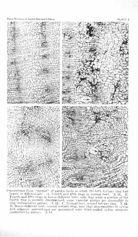
Plants similar to those just described were continued on boron- deficient nutrient and examined 1 month later to determine the effect of a longer period of boron starvation. By this time there was considerable interzonal parenchyma in the first four rings. A few thick-walled vessels were present in some bundles, of the third tertiary ring of the ''taproot^' ^, but only the phloem groups had differentiated in the fourth and fifth rings. In the well-differentiated bundles of the first and second rings the pathological changes and tissues in- volved were the same as those described for the first ring of younger plants except that a greater proportion of the xylem of affected bundles was usually involved (pi. 2, D). At this stage in the third
7 "Taproot" as used here includes root and hypocotyl.

Boron Deficiency in Garden Beet and Cabbage PLATE 1
1 '^■•f v-'î >i ■■ ■ ■ ''v >'*V,«JÎ-
.■K Transoctions of garden beet seedlings. A, Lower hypocotyl of normal plant.
X 155. B, Lower hypocotyl of boron-deficient plant; note deeply stained phloem cells. X 310. C, Lower hypotocyl of boron-deficient plant. "Many of the abnormal phloem cells are only partly plugged. Compare with A and B. X 207. I), Hypocotyl of plant receiving an excess of boron; note numerous cells filled with darkly stained materials in the secondary phloem and the early differentiation of xylem elements in the tertiary ring. X 105.
Boron Deíiciency ¡n Garden Beet and Cabbage
1 'v^^^-:/f ;-^ PLATE 2
vio
H-
TranscctioM.s from "taproot" of garden beets in which the fiftli tertiary rinj; had begun to differentiate. A, Fourth and fifth rings in normal beet. 'X 51. B, Fourth and fifth ring.s in boron-deficient beet; note tliat while a portion of the fourth ring is entirely disorganized, some vascular groups are discernible in ring (indicated by arrow). X 42. C, Xormal beet, second tertiary ring. X 42. D, Boron-deficient beet, second tertiary ring; note that abnormalities in xylem and adjacent parenchyma are a.ssociáted with thick-walled xvlem elements (indicated by arrow). X 44.

Feb. 15,1943 Borou Deficiency in Garden Beet and Cabbage 171
and fourth rings there were some abnormal areas as in the younger plants, but there were also larger areas of disorganized tissues involv- ing all or part of several bundles and adjacent interzonal parenchyma in one or both rings (pi. 2, B). Scattered throughout such areas were groups of crushed parenchymatous cells enclosing brown deposits among them as well as an occasional misshapen vessel segment, sieve tube, and companion cell. Near the margin of these areas the brown deposits were for the most part intercellular, some cells appear- ing normal except for a slight discoloration of one or two walls. Interspersed among the patches of necrotic cells were groups of small rapidly dividing cells and enlarged, thin-walled ones. Those adjacent to the necrotic tissues sometimes appeared to be so oriented as to wall off that region. However, the larger areas of proliferated cells which were arranged in radial rows appeared to be part of the degener- ate area rather than the result of a growth response to the necrosis. Many of the larger cells had their long axis in a horizontal plane. The few isolated normal phloem groups in disorganized areas probably had differentiated before boron deficiency became acute. The areas of disorganized tissues in the third and fourth rings were macroscop- ically visible as typical internal black spot lesions, while those in the two inner rings were not, indicating that a considerable amount of the brown deposit and many necrotic cells were necessary before there was any macroscopic evidence of the pathological changes in the boron-deficient plants. No abnormalities were found in the fifth tertiary ring of plants at this age.
In still older plants which had three or more tertiary rings in which xylem elements had differentiated, pathological changes were confined to two or three rings much as was found to be the situation in sugar beets by Rowe (21). The fact that the inner rings were usually normal suggested that they had differentiated before the available boron was used up. In the ring in which only a few thick-v/alled xylem elements had developed and in the next one or two outer rings there were disorganized and necrotic areas, similar to, but more ex- tensive than, those in comparable rings of younger plants. Frequently the ratio of necrosis to hypertrophy and hyperplasia was greater in older plants. The youngest outer rings in which a few bundle initials were present were normal.
Field-grown beets showing internal black spot symptoms contained abnormalities similar to those just described (pi. 3). The apparent confinement of symptoms to interzonal parenchyma in larger field beets can be attributed in part at least to suppression of vascular differentiation in the affected areaj to an increased amount of inter- zonal parenchyma, and to a higher ratio of necrosis to proliferation in the affected parenchyma. In some field specimens, disorganized and necrotic areas appeared in the more mature inner rings with or without degeneration in the yoimger rings. It is probable that there was a deficiency in boron during the period of rapid development in the affected areas and that the supply of boron became adequate again before much differentiation occurred in succeeding normal rings. In roots of some plants abnormalities were confined to a single small area in one layer of interzonal parenchyma. The abnormalities found were the same as those in larger spots except that the area was com- pletely separated from normal tissue by a zone of actively dividing, thin-walled cells.

172 Journal qf Agricultural Research voi. 66, No. 4
In larger beets degenerated areas in the interzonal parenchyma frequently assumed a definite pattern (pi. 3, (7). Near the center of the degenerate area was a mass of crushed, dead cells, brown inter- cellular and intracellular deposits, and a few distorted parenchymatous cells with discolored walls. The region adjacent to this area, and between it and the center of the root, was composed of numerous small, and a few enlarged, thin-walled cells all of which were oriented radially (pi. 3 (7, a), whüe the adjacent tissues in the direction of the periphery consisted of many hypertrophied and a few hyperplastic cells with their long axes parallel or nearly parallel to that of the main necrotic area (pi. 3 C, 6).
PETIOLE OF THE LEAF
True leaves in a vegetative crown differentiate successively from the apical meristem just above the cotyledonary node.
Since the newly initiated leaves of the beet develop as older ones become senescent, it was possible to obtain material at several stages of the disease from a single plant. Therefore, in histological studies, both the nature of the pathological changes induced by boron defici- ency and the variation in the degree of these changes causing the different macroscopic symptoms could be determined. While the size and shape of the leaf varies among varieties, the types of tissue and their arrangement are similar in all varieties examined.
Abnormalities in older leaves were found in the petioles of old malformed leaves and in. some which were macroscopically normal as well as in malformed ones (pi. 4, D). As in the root and hypocotyl many phloem c4Is contained excessive amounts of heavily stained, granular material. Frequently the cambial zone was composed of a wide band of thin-walled cells some of which were hypertrophied and others small and rapidly dividing. A few misshapen xylem elements were interspersed among the thin-walled cells. In bimdles in which cell enlargement and proliferation were extensive, a few discolored cell walls and some brown deposits were present (pi. 4, D). In such bundles there was a decrease in the amount of normal differentiated xylem and phloem tissue. The extent of vascular disorganization varied in different bundles at a given level (pi. 4, B) and at different levels in the same petiole. No abnormalities were found in any other tissues in petioles of older, apparently normal leaves.
In the petioles of younger leaves which showed macroscopic symp- toms degeneration appeared earlier and progressed farther. The magnitude of microscopic degeneration in these was in direct propor- tion to the severity of external symptoms. In the vascular bundles heavily stained phloem cells were present but were less numerous than in petioles of older leaves. A few of the younger sieve tubes and com- panion cells were short and irregularly shaped. The cambial zone contained many hypertrophied and a few small thin-walled cells. In some bundles a few of the giant cells extended from the xylem to the phloem. In others the cells in the cambial zone near the phloem were much larger than those adjacent to the xylem (pi, 5, (7). Scat- tered among the thin-walled cells near the xylem were strands of distorted scalariform vessel segments which were oriented at various angles. Often successive elements failed to join or were only partly connected. There was seldom any evidence that these strands were

Boron Deficiency in (.iarden B«t and Cabbage
f^'^-3^^\YW^-"^ :r,
^ ^ .-r ;
"Taproots" of boron-deficient garden beet'- .1 and li Adiaccnt lran-.octions near periphery of a young field-grown plant. Xoto that .^oino \aspiilar tis^-iie in the severely diseased region is not disorganized; slight degeneration in one bundle of the inner ring. X 26. C, Transection from a taproot near maturity. Note the radial rows of proliferated cells (a) inside the necrotic area and the hypertrophied cells (ft) outside the necrotic area. Note also the necrosis aVound a xylem element in upper left corner of photograpn. X 44. D, Longisection of young beet near periphery. Note the difference in the extent of degeneration at the different levels. X 18.

Boron Deficiency in Garden Beet and Cabbage
^1^««-, /•,
'^ K>f^^'^ ':'Jr.
PUATE 4
'J>'i . V
■é)
- 'yM r. >f> -
^
i> Tr
X*-' *#.
Tmtisectioii of petioles from older leaves of garden beet. A, Normal petiole. X 39. B, Boron-deficient petiole; note the wide zone of thin-walled cells in the cambial region of the two lower bnndles. X 30. C, Knlargemeiit from A. X 145. I), Enlargement from B; note the wall discoloration in seme of the hypcrtrophied cells. X 91.

Feb. 15,1943 Boron Deficiency in Garden Beet and Cabbage 173
connected with the older xylem tissues, in which only a few of the vessel segments were misshapen. In a few specimens some xylem parenchyma cells were hypertrophied.
In the more severely affected bundles there was considerable cell discoloration and disintegration in the cambial zone. Although in a few specimens the discoloration first appeared near the differentiated tissues (pi. 5, C), the first evidence of degeneration in most cases was a slight discoloration of adjacent walls of 2 or 3 cells, usually hyper- trophied ones, in the cambial zone similar to that shown in older leaves: (pi. 4, D). As such an area of discolored tissue enlarged, some cells near the center were crushed and discolored. Others retained their form but were darkened by heavily stained deposits. In acute cases the necrotic areas included portions of the xylem, phloem, bundle caps, starch sheath, and adjacent parenchyma as well as the cambial zone (pi. 5, C), In some bundles (pi. 5, 5, C) there was considerable necrosis in the phloem and starch sheath, but little or none in the bundle cap between them. When a portion of the xylem bundle cap or starch sheath was involved in a necrotic area, the adjacent cells were often hypertrophied (pi. 5, B, C). Paren- chyma cells between badly disorganized bundles were also much enlarged (pi. 5, C). In addition to degeneration in and near the vascular bundles, necrotic areas were frequently present in sub- epidermal tissue. These areas were most common at the petiole margin and at the ribs, regions where coUenchyma would normally be present. Cells abutting the necrotic tissue were frequently mis- shapen and hypertrophied. In a few specimens epidermal cells were involved in the necrotic area.
Changes in petioles of very small, stunted leaves were similar to those just described, except that there were less hypertrophy and proliferation and more cell deposits in the cambial zone (pi. 5, B).
Examination of petioles of unilaterally developed leaves described elsewhere {29, 30) revealed that the bundles on one side showed slight if any pathological change while those on the other side had undergone extensive degeneration (pi. 4, B). This could account for both the unilateral development of the lamina and the intensification of red color in the poorly developed portion. Degeneration and the accom- panying decrease in differentiation of vascular tissue would retard leaf development and impede the movement of materials to and from the affected region and the resultant accumulation of sugars would lead to increased pigmentation. The necrosis and proliferation found in the subepidermal tissue of the petiole might well account for the external cross-hatching symptoms (pi. 5, B, D) occasionally observed in petioles of boron-deficient garden beet (30).
LAMINA OF THE LEAP
In the lamina of the normal beet leaf, xylem, phloem, and cambium are found in the midrib and larger veins, but xylem and phloem or xylem only is found in the smaller veins. The single-cell-layered epidermis is composed of irregularly shaped cells. Stomata, which are present on both dorsal and ventral surfaces, are slightly more numerous on the former. The mesophyll consists of two or three layers of palisade cells on the ventral side, which grade into spongy parenchyma cells on the dorsal side. Cells of both contain numerous
509338—43 3

174 Journal of Agricultural Research voi. 66, No. 4
chloroplasts. It is possible that, as found by Artschwager (2) in sugar beet, the ratio of pahsade to spongy parenchyma cells is higher in thick leaves.
In the mesophyll of the lamina of young leaves from boron-deficient plants both cell enlargement and cell proliferation occurred. Some of the hypertroph i ed spongy parenchyma cells were elongated with their longitudinal axis parallel to the leaf surface. The chloroplasts did not appear to be quite so numerous nor quite so green as those in normal plants. In the reddened leaves examined, the pigmentation was confined to the mesophyll cells just beneath the epidermis, except at the midrib and the larger veins. In these regions the red pigment and a brown pigment were found in two or three subepidermal cell layers. Necrotic areas were sometimes found in younger leaves, particularly near the expanding margin of the lamina. In the larger veins and in the midrib there was vascular disorganization similar to that found in the petiole.
FLORAL AXIS
As in the other organs of the plant, the changes found in the slightly difiPerentiated tissues near the growing point of the floral axis were similar to those in the more mature tissues, except for the degree to which the various tissTies were affected. In the slightly differentiated regions darkly stained phloem cells similar to those found in the root and hypocotyl were present in most bundles, but were particularly numerous in those which were otherwise quite normal. All or a portion of the cambial tissues were composed of hypertrophied and hyperplastic cells (pi. 6, B), The few thick-walled vessels present in the xylem were scattered among numerous, radially elongated, thin- walled cells (pi. 6, 0). Frequently some of the parenchyma cells in the vicinity of the innermost xylem elements were crushed and heavily stained. Occasionally a xylem element had degenerated. Pith cells in large areas adjacent to such bundles were*also greatly enlarged and horizontally elongated (pi. 6, B). Infrequently a crushed cell or a few cells with discolored walls were found in these abnormal areas. Patches of necrotic cells which were found in the subepidermal col- lenchyma layers occasionally extended to include epidermal cells. At some levels of the stem these necrotic areas extended inward and connected with similar areas in the vascular tissues.
In regions of the floral axis in which tertiary thickening was well under way heavily stained phloem cells were present in both secondary and tertiary phloem. The numerous thin-walled, hypertrophied cells interspersed among the primary and secondary xylem elements were not so much enlarged as similar cells near the apex (compare pi. 6, B and C w^ith D). The number of already differentiated xylem elements in this region may have been a factor in checking cell enlargement. The areas of crushed cells among the xylem elements and in abutting pith tissue were larger and more numerous than in younger tissue (pi. 6, D). Areas of enlarged and proliferated cells found in the pith were similar to those in younger tissues except for the greater amount of necrosis. In a few cases small islands of normal parenchyma cells which extended but a few cells horizontally and vertically were found near the center of an area of abnormal pith cells. In the tertiary ring the cambial zone was often composed of a mosaic of large, radi-

Boron Deficiency in Garden Beet and Cabbage PLATE 5
Transactions from gardon beet petioles. A, Normal leaf. X 22. B, Biiron-defieient yonng leaf; note extensive necrosis and accumulation of deposits in the vascular bundles, the hvpertrophied cells adjacent to the two largest bundles, and the subepidermal necrosis. X 31. C, Boron-deficient leaf near the base of the lamma; note necrosis in phloem and starch sheath in one bundle (indicated by arrow) and necrosis in the lower wing. X 20. D, Port ion of petiole which showed cross liatching; note abnormal areas at the top and along the dorsal margin. X 33.

Boron Díficiency in Garden Bret and Cabbftgc PLATE 6
4 If ^^.l^ ■%, ■^ -^'/j
I* • , ^ ' Í , h -' , ■* si" \ 1
i ^' V ,, -•/ \
•7 '
'1 raii-octionv fioin lli)i,il ^U'lk-- of garcirn l)c( I ,1 Noriiul pLiiit iioar ¡'.pox X 2(). h, "Huruii-deficieiu, plaiii iioar apex; note necrosis and deposits at junc- ture of pith and vascular tissues and the assoi^^iation of pith and cortical degen- eration with vascular degeneration. X 23. C, Enlargement from B: note the radial elongation of xylem parenchyma cells. X 68. D, Four internode.s below B; note that hypertrophy and hyperplasia are more extensive in the region of the secondary cambiuiu than in the xylem parenchvma. X 21.

Feb. 15,1943 BoTou Deficiency in Garden Beet and Cabbage 175
ally elongated and small, rapidly dividing cells arranged in radial rows (pi. 6, D). The changes here were similar to those found in the bundles of petioles of boron-deficient beet plants. Parenchyma cells in the tertiary xylem elements were similar to those around secondary xylem elements. The areas of abnormal tissue in the subepidermal layers of coUenchyma and adjacent epidermal cells varied from small, discolored cell groups to large areas in which a considerable number of cells had disintegrated. As in similar degeneration nearer the stem apex, such areas sometimes extended into and involved adjacent vascular bundles. Along the margin of the subepidermal lesions the cells were hypertrophied and intercellular deposits accumulated. Farther away from the lesion small groups of proliferated cells were interspersed among the enlarged cortical cells.
PATHOLOGICAL HISTOLOGY OF BORON-DEFICIENT CABBAGE
The macroscopic symptoms of the boron-deficiency disease in cab- bage seedlings grown in sand-nufrient culture and in older plants growing in the field have been described by Walker, McLean, and Jolivette (81). Marked histological abnormalities were found in both seedlings and headed plants of cabbage. In seedlings these were con- fined for the most part to vascular tissue except near the apical meri- stem and in expanding leaves. In plants grown in complete solution for 2 or 3 months before being placed in minus-boron solution and in those grown on boron-deficient soils, degeneration in the stem was confined to the pith, although the changes in the younger leaves were similar to those in comparable leaves in seedlings. In view of the differences in tissues most affected in seedlings and older plants they will be considered separately. The normal histology of root, hypo- cotyl, and stem of cabbage has already been described bv Smith and Walker (l?7), Larson (16), and Havis (13).
ROOT, HYPOCOTYL, AND STEM OF YOUNG CABBAGE PLANTS
In young plants the disorganization in the vascular system of boron-deficient plants was similar in root, hypocotyl, and stem. In all or part of the vascular ring the normal tabular cells were absent from the cambium. In their place was a zone of hyperplastic and hypertrophied cells, most of which were thin-walled and radially elongated (pi. 7, B, D). In some plants the cambial zone became several times the width of the normal vascular ring in the stem, and the entire ring at a given level was involved (pi. 7, B). In others abnormalities were present in only a portion of the vascular ring and the affected tissue was less hypertrophied. Frequently groups of discolored and crushed undifferentiated cells were found in the abnormal cambial zone, particularly near the xylem or phloem (pi. 7, B, D). The first sign of such degeneration was a slight discolora- tion of adjacent walls of two or three cells and occasional brown intercellular deposits. At later stages the degenerated areas became quite large. In the center of the large areas the cells were discolored or crushed and more of the brown deposit had accumulated. Near the margin, cells were only slightly discolored. The proportion of necrotic and crushed cells to enlarged and proliferated cells was often high when only a part of the bundles was affected.

176 Journal of Agricultural Research voi. 66, No. 4
Small groups of misshapen xylem elements were scattered through- out the major part of the abnormal cambial zone (pi. 7, D^ and 8, A),
"This'^suggests that-much of the undiffer^ntiated tissure was potentially xylem in nature. In some specimens a thin layer of smal 1 cellswitifi dense contents was present near the differentiated phloem (pi. 7, B). The fact that a few distorted phloem groups but no abnormal vessel segments were found outside this layer suggested that it was a non- functioning cambium.
In boron-deficient plants there was less differentiated xylem and phloem tissue than in normal plants, and what there was tended to mature earlier. This was particularly noticeable in the early ligni- fication in the xylem and in pericycle fibers. A similar tendency for some cells to mature early in boron-deficient plants was noted in pea roots by Sommer and Sorokin (^8).
In the root and hypocotyl, a condition of hypertrophy, hyperplasia, and necrosis similar to that described for the vascular system was sometimes found in the periderm fpl. 7, D).
Near the stem apex, abnormalities were sometimes found in the cortex and epidermis in addition to those in the vascular tissues. The affected areas in the cortical parenchyma contained enlarged cells and proliferated cells. Some brown deposits were present among the cells. In late stages some of the affected cells disintegrated.
In the cortical, ray, and pith parenchyma cells of the remainder of the stem, the hypocotyl, and the root, starch accumulation, which may have been a direct or indirect effect of boron starvation, was noticed. This symptom was most noticeable in plants grown in the complete nutrient solution for 3 or 4 weeks before being placed in minus-boron solution, and was sometimes found when no other micro- scopic symptoms were noted. In all cases little or no starch was found in comparable normal plants fixed at the same time as the boron-deficient plants.
LEAVES OF YOUNG PLANTS»
When plants were grown from seed in minus-boron, sand nutrient cultures, the few leaves that developed were affected very early and therefore the effect of boron starvation could be studied on very young tissues only. However, when plants were supplied with complete solution for several weeks before being transferred to minus-boron cultures, successive leaves were at different stages of development when the supply of boron became inadequate. In these it was pos sible to compare the nature and extent of degeneration caused by boron deficiency in very young leaves with that in leaves at various stages of development.
The most extensive degeneration in the leaf petiole was found in the vascular tissue. While abnormalities varied in magnitude with the age of the leaf, they were in nature similar at various ages. Most bundles in the larger vascular strands were not entirely separated by parenchyma rays as in normal petioles. Instead their xylem elements were merged into one solid core or arc. In extreme cases there was considerable horizontal elongation of the thin-walled cells among the differentiated xylem elements. Usually instead of a normal cambium between the layer of differentiated xylem and the phloem of each of the several bundles there was a zone of misshapen cells similar to

Boron Deficiency in Garden Beet and Cabbage PLATE 7
Transect ions from young cabbage plants. /I, Normal stem. X 65. B, Boron- deficiont stem; noto layer of small cells suggestive of a degenerated cambium (indicated by arrow) and the small necrotic areas scattered through the zone of thin-walled cells in the cambial /.one. X 33. C, Normal root. X 47. O, Boron-deficient root; note the thick-walled xylem elements scattered in the abnormal tissue as indicated bv arrow. X 23.

Boron Deficienzy in Garden Beet and Cabbaçe PLATE 8
«S=*git??*"*'
. --^ife'^aSi.^ if^;.,
I^^MaáMi#lf Longisections from boron-starved cabbage. A, Hypocotyl of a young plant;
note ihe flistorted xyleiii elements (indicated by arrow) scattered in the undif- ferentiated tissue. X 53. B, Stem of plant approaching head stage; note that although there is evidence of cell division and enlargement near the vasoilar tissue at left, degeneration is confined to the central part of the pith. X 19.

Feb. 15,1943 Borou Deficiency in Garden Beet and Cabbage 177
but not so extensive as that found in the root and stem (pi. 9, D). In some specimens the hypertrophy of the cells near the xylem was greater than that in other cells in the undifferentiated zone. Fre- quently small areas of necrotic cells were found in the undifferentiated tissue (pi. 9, D). Occasionally a few misshapen xylem elements were found in the inner portion of the undiiferentiated zone. In such cases the necrotic areas usually centered about these elements. As in the root and stem, a layer of heavily stained cells, which appeared to be an abnormal cambial layer, was sometimes discernible in this area located a few cells inward from the differentiated phloem. A few misshapen degenerated cells resembling sieve tubes were found adjacent to the normal differentiated phloem. In the larger vascular strands the phloem of each bundle was usually separated from that of neighboring bundles by radial bands of parenchyma.
In the cortical parenchyma starch grains were numerous. In regions where cross hatching occurred, necrotic and proliferated cell groups were found involving subepidermal and, to a lesser extent, epidermal tissues.
As was found in the garden beet, degeneration occurred in some of the older, apparently normal leaves. In plants with macroscopic symptoms the histological changes occupied larger areas than in those in which symptoms were not visible.
In a study of a limited amount of leaf-lamina material, changes similar to those in the petiole were found in the vascular tissue. In thickened leaves of boron-deficient plants there was an increase in the size of some cells and in the number of cells present in the mesophyll. In it the cells appeared to be packed more tightly than in normal tissues. Chloroplasts were equal in size or slightly larger than those in normal plants.
OLDER PLANTS
The normal anatomy of plants grown for 3 or 4 months in complete nutrient solution and of plants transplanted into the field was similar to that of several-week-old seedlings except that more secondary tissue was present in the vascular ring and more pith in the stem of the older plants and the cortex was wider. In some specimens a 2- or 3-mm. core of tissue in the center of the pith was water-soaked or green. Cells in this area, however, showed no evidence of de- generation.
When the boron supply became inadequate for older plants which had not yet headed, symptoms similar to those in leaves of boron- starved younger plants were sometimes present in the outer leaves. However, the major degeneration, internal necrosis, appeared in the central part of the pith throughout the stem and upper hypocotyl, regions in which no symptoms except starch accumulation appeared in younger cabbage plants. In studies of a limited number of larger roots of older plants, no apparent abnormalities were found. For this reason histological studies on that portion of the plant have been left for a later investigation.
In the pith microscopic changes involved a slightly larger area than the macroscopically visible symptoms. In plants transferred to a boron-deficient solution after having grown in complete solution for 2 or 3 months, a greater proportion of the pith was involved than in normal plants transplanted into boron-deficient fields. Nevertheless,

178 Journal of Agricultural Research voi. 66, No. 4
the changes were the same except that there were fewer cells with thickened walls in the pith of the plants grown in sand culture (p. S, B, and 10, B), At and near the margin of the affected pith, isolated groups of abnormal cells were found, while near the center the majority of the cells were abnormal (pi. 10., B). The marginal cell groups usually occupied an area which was roughly spherical. These areas contained several types of abnormal tissue arranged in a fairly consistent pattern (pi. 10, (7, D). In the center of the degenerated tissue were thick- walled cells which sometimes contained deposits of a brown material. In larger areas some of these cells were crushed or disintegrated. Scattered among or surrounding these was a zone of large cells some of which had partly thickened walls. Others had thin cross walls. Such cells were similar to those first described except that degeneration had not progressed so far. The size of this zone varied but was roughly proportional to the size of the affected area. The walls of most of these cells and of the other thick-walled ones in the abnormal tissue took safranine when the safranine and fast-green stain were applied. All other pith cells took fast green.
Surrounding the zone of large cells in the more extensive degenerate areas was one of small, thin-walled, rapidly dividing cells which in some cases entirely walled off the inner tissue. Intermingled with and surrounding this layer was a one- or two-cell zone of sclerotic cells (pi. 10, D) which had scalariform, reticulate, or pitted walls. Prolifera- tion sometimes occurred just outside this zone, but the daughter cells were nearer normal size than in the hyperplastic tissue near the center of the disorganized area. Some of the isolated abnormal cell groups later coalesced. lù such cases it was difficult to make out distinct patterns. In the central portion of affected pith tissue the several types of cells mentioned were intermingled in random fashion (pi. 10, JB). It is .probable that such a case represented several ^coa- lesced groups of abnormal cells. Occasionally islands of normal parenchyma cells, similar to those in the pith of the floral stem of garden beet, were found.
DISCUSSION
The pathological changes in boron-deficient cabbage and garden beet have many points in common. The greatest degree of break-down in beets at all stages and in cabbage in early development was found in those regions in which cell division and tissue differentiation were most active. The reactions appeared to be intimately associated with cell metabolism rather than with a particular tissue. In beet seedlings in which secondary thickening had just begun the principal break-down was in phloem cells. In those in which tertiary thickening had begun degjeneration was in the xylem as well as in the phloem of differentiated rings. In somewhat older ^^taproots'' most necrosis was found in rings in which early differentiation was under way, less in the next older rings, and none in the oldest rings. In young cabbage plants the chief abnormalities in root and stem were also found in the differentiat- ing vascular tissues. The greatest difference in the reaction of cabbage as compared with that of beet was found in the older plants in which the head had developed. Even though there was still meristematic activity in the vascular tissue, disorganization was confined almost entirely to the central portion of the pith.

Boron Deficirncy in Garden Beet and Cabbage PLATE
Traiisection.s fiom cabbaso petioles. A, Normal peiiolo. X 30. Ji, Boron- deficient petiole. X 24. C, P^iilargement from A. X 140. D, Elnlargemeiit from /?; note the abnormal cells and nccrotic areas in the undifferentiated tissue and the position of the differentiated .xvlem as compared to that in C. X 144.

Boron OrfiriT.ry in Garden Rcrt and Cabhanf PLATE 10
^^ ■'■'V--li 't^ Central part of pith of headed cabbage. A, Transection from a normal plant.
X 35. /?, I^oiiKi.seotion from a boron-deficient plant; note the thick-wallod cells at lower margin of small degenerate area in upper right of photograph and liroliforation and deposit accumulation in tissue at left of photograph. X 21. C, Transection from boron-deficient plant at margin of degenerate pith area. X 28. D, Small degenerate area adjacent to C; note the cross wails in cells in the center of the area and the w-all thickenings (indicated by arrow) in the surroimding ring of cells. X 120.

Feb. 15,1943 Boron Deficiency in Garden Beet and Cabbage 179
It was evident in roots, stems, and leaves of both species that micro- scopic abnormal development occurred in advance of macroscopic symptoms and that the former was usually more extensive than could be recognized macroscopically. It was also to be noted that micro- scopic changes occurred which did not progress to the point where macroscopic symptoms became visible. Sudden severe manifesta- tions of disease thus may often have been preceded by slow, pro- tracted invisible degeneration. Factors such as increased moisture and temperature favorable for sudden increases in growth rates of these plants would then contribute to a sudden appearance of extensive visible disease symptoms.
The details of histological changes, though often varied in their grosser macroscopic manifestations, usually followed a fairly uniform pattern. Cell wall discoloration, hyperplasia, and hypertrophy were all common initial cellular reactions in thin-walled differentiating tissue. As death of a group of cells occurred, increased cell division or cell growth commonly followed in the vicinity of the dead cells. This often led to islands of pathologic tissue as noted in the pith of maturing cabbage. Where continued cell activity predominated, as in beet roots and young cabbage stems, normal differentiation of vascular elements was checked. In other cases abnormal differentia- tion of thick-walled cells appeared as in cabbage-pith lesions. It is thus evident that the fundam.ental effect of boron deficiency in both species is one of disturbance of growth-regulation processes, while the course of events following the initial response varies with the tissues concerned.
The picture of pathological histology indicates strongly that boron is particularly important in the metabolic processes concerned in cell division and tissue differentiation. It may also be of peculiar value to the development of storage tissue. One of tbe physiological characteristics of boron pointed out previously (ßO) is its ready mobility in the plant when first introduced and its tendency to become immobile rather rapidly in the tissue.
The first pathologic effects of boron deficiency, then,' may be con- sidered physiological, exhibited locally by disturbance of growth relations and manifested by both hypertrophie and hyperplastic cell reaction. Necrosis follows and byproducts released from dead or dying cells may then also become factors influencing cell growth. General physiological effects follow through disturbed tissue differ- entiation and function. Abnormal organization of the xylem and phloem with reduced amounts of elements adapted to translocation leads to an accumulation of sugars in developing beet leaves and con- sequent excessive formation of pigment. Further vascular disloca- tion leads to stunted growth in tissues normally supplied by the xylem and phloem concerned. Thus boron deficiency commonly leads to such general effects as stunting, malformation of organs, unilateral growth, and death. Furthermore, mild shortage of boron insufficient to produce macroscopic symptoms may lead only to microscopic injury to transporting tissue. It is in such instances that the appli- cation of borax to growing crops commonly results in increased growth, which is reflected in greater yields, even though deficiency symptoms do not appear in untreated controls {SO).

180 Journal of Agricultural Research voi. 66, NO. 4
SUMMARY
This investigation comprises a study of histological changes which occur in garden beet and cabbage grown with a deficiency of boron in the nutrient suppUed to greenhouse sand culture, and under natural conditions in the field.
In beet root and hypocotyl the first sign of disease in young seed- lings is in the contents of certain phloem cells and occasional hyper- trophy of cambial cells. In slightly older plants degeneration appears in primary and secondary xylem as intercellular brown deposits, accompanied by distortion of cells. Groups of disintegrated cells are frequently surrounded by a border of proliferating cells which may involve one or more bundles, thus giving rise eventually to macroscopic necrotic areas. In roots which are somewhat older when the defi- ciency of boron becomes acute, the most severe necrosis occurs in the tertiary ring or rings most active in differentiation at the time.
Abnormal development in beet leaves also centers in the bundles of the petiole. It is found commonly in old leaves which were normal macroscopically and in which it has occurred too late to influence gross morphology. In younger leaves the magnitude of histological change is in direct proportion to the severity of external symptoms. In petioles of unilaterally developed leaves the bundles on one side show slight, if any, pathological change while those on the other side have undergone extensive degeneration. Cell enlargement and pro- liferation extend to the mesophyll and spongy parenchyma of the leaf lamina.
In the floral axis, proliferation of cells occurs in the cambium and in the pith. Necrotic areas occur in the pith and in the vascular region of secondary and tertiary rings and are sometimes connected with similar regions in the cortex, where the lesion occasionally involved epidermal cells.
In young cabbage plants extensive proliferation of cells in the region of the cambium of root, hypocotyl, and stem results in a band of meristematic tissue several times the usual width of the undifferenti- ated zone between phloem and xylem. There is correspondingly less differentiation of vessels and phloem elements. Necrotic areas develop in this undifferentiated area. Abnormalities are to be seen occasion- ally in the cortex of the stem apex but necrosis in the pith is rare until the plant has approached the head stage. When boron deficiency symptoms appear in the field they are usually confined to this region. In cabbage leaves disturbances in differentiation of the vascular bundles in petiole and lamina are the chief evidences of the disease.
In both species the shortage of boron appears to affect chiefiy the metabolic processes concerned in cell división, cell differentiation, and possibly storage. The initial reaction of cells is seen in cell-wall dis- coloration, discoloration of cell contents, excessive cell division, and abnormal enlargement. Necrotic areas which follow are made up of disintegrated cells which are often sm*rounded by a region of abnor- mally active tissue consisting of hyperplastic cells, hypertrophied cells, and sometimes peculiarly differentiated ones.
The pathological histology of internal black spot of garden beet and internal breakdown in the pith of cabbage is the same in nutrient- grown and field-grown plants. In both species extensive histological changes occur before macroscopic symptoms appear.

Feb. 15,1943 BoroTi Deficiency in Garden Beet and Cabbage 181
LITERATURE CITED
1) ALLISON, R. V., and SHIVE, J. W. 1923. STUDIES ON THE RELATION OF AERATION AND CONTINUOUS RENEWAL
OF NUTRIENT SOLUTION TO THE GROWTH OF SOYBEANS IN ARTI- FICIAL CULTURE. Amer. Jour. Bot. 10: 554-566, illus.
2) ARTSCHWAGER, E. 1926. ANATOMY OF THE VEGETATIVE ORGANS OF THE SUGAR BEET. JOUF.
Agr. Res. 33: 143-176, illus. 3)
1927, DEVELOPMENT OF FLOWERS AND SEED IN THE SUGAR BEET. Jour. Agr. Res. 34: 1-25, illus.
4) BRANDENBURG, E.
1931. DIE HERZ- UND TROCKENFäULE DER RüBEN ALS BORMANGEL ER-
SCHEINUNG. Phytopath. Ztschr. 3: 499-517, illus. 5) BRENCHLEY, W. E., and THORNTON, H. G.
1925. THE RELATION BETWEEN THE DEVELOPMENT, STRIJCTURE AND FUNCTIONING OF THE NODULES ON VICIA FABA, AS INFLUENCED BY THE PRESENCE OR ABSENCE OF BORON IN THE NUTRIENT MEDIUM. Roy. Soc. [London] Proc, Ser. B., 98: 373-399, illus.
6) DENNIS, R. W. G., and O'BRIEN, D. G. 1937. BORON IN AGRICULTURE. West of Scot. Agr. Col. Res. Bui. 5,
98 pp., illus. 7) ELTINGE, E. T.
1936. EFFECT OF BORON DEFICIENCY UPON THE STRUCTURE OF ZEA MAYS. Plant Physiol. 11: 765-778, illus.
;8) ESAU, K. 1933. PATHOLOGICAL CHANGES IN THE ANATOMY OF LEAVES OF THE SUGAR
BEET, BETA VULGARIS L., AFFECTED BY CURLY TOP. Phyto- pathology 23: 679-712, illus.
1934. ONTOGENY OF PHLOEM IN THE SUGAR BEET (BETA VULGARIS L,)i Amer. Jour. Bot. 21: 632-644, illus.
1935. ONTOGENY OF THE PHLOEM IN SUGAR BEETS AFFECTED BY THE CURLY-TOP DISEASE. Amer. Jour. Bot. 22: 149-163, illus.
1935. INITIAL LOCALIZATION AND SUBSEQUENT SPREAD OF CURLY-TOP SYMPTOMS IN THE SUGAR BEET. Hilgardia 9: 395-436, illus..
(12) HAAS, A. R. C, and KLOTZ, L. J. 1931. SOME ANATOMICAL AND PHYSIOLOGICAL CHANGES IN CITRUS PRO-
DUCED BY BORON DEFICIENCY. Hilgardia 5: [175J-197, illus. (13) HAVIS, A. L.
1940. A DEVELOPMENTAL ANALYSIS OF KOHLRABI AND CABBAGE STEMS. Jour. Agr. Res. 61: 459-470, illus.
(14) HAYWARD, H. E. 1938. THE STRUCTURE OP ECONOMIC PLANTS. 674 pp., illus. New York.
(15) JOHNSTON, E. S., and DORE, W. H.
1929. THE INFLUENCE OF BORON ON THE CHEMICAL COMPOSITION AND GROWTH OF THE TOMATO PLANT. Plant Physiol. 4: 31-62, illus.
(16) LARSON, R. H. 1934. WOUND INFECTION AND TISSUE INVASION BY PLASMODIOPHORA
BRASSicAE. Jour. Agr. Res. 49: 607-624, illus. (17) LöHNis, M. P.
1937. BROWN HEART IN SWEDES (BRASSICA NAPUS RAPIFERA). Wageuiugen, The Netherlands, Col. Agr. (Abstract in Chron. Bot. 4: 9. 1938.)
(18) MACARTHUR, M. 1940. HISTOLOGY OF SOME PHYSIOLOGICAL DISORDERS OF THE >APPLE FRUIT.
Canad. Jour. Res., Sect. C, 18: 26-34, illus. (19) MARTIN, J. P.
1934. BORON DEFICIENCY SYMPTOMS IN SUGARCANE. Hawaü. Planters Rec. 38: 95-107, illus.
(20) MES, M. 1930. PHYSIOLOGICAL DISEASE SYMPTOMS OF TOBACCO. PhytOpath.
Ztschr. 2: t593]-614, illus.

182 Journal oj Agricultural Research voi. 66, NO.4
(21) RowE, E. A. 1936. A STUDY OF HEART-EOT OF YOUNG SUGAR-BEET PLANTS GROWN IN
CULTURE SOLUTIONS. Ann. Bot. [London] 50: [735]-746, illus. (22) SCHREVEN, D. A. VAN.
1934. EXTERNAL AND INTERNAL SYMPTOMS OF BORON DEFICIENCY IN TOBACCO. Tjjdschr. over Plantenziekten 40: 97-129, illus. (In Dutch. English summary, pp. 122-125.)
(23) — 1935. EXTERNAL AND INTERNAL SYMPTOMS OF BORON DEFICIENCY IN
TOMATO. Tijdschr. over Plantenziekten 41: 1-26, illus. (In Dutch. English summary, pp. 22-23.)
(24) 1939. THE INFLUENCE OF TWELVE CHEMICAL ELEMENTS ON THE STATE OF
HEALTH OF THE POTATO PLANT. [Wageningen] Landbouwhooge- seh. Meded., deel 43, verhandeling 1, 166 pp., illus. (In Dutch. English summarv, pp. [126]-142.)
(25) SHIVE, J. W. 1915. A THREE-SALT NUTRIENT SOLUTION FOR PLANTS. Amer. Jour. Bot.
2: 157-160. (26)
1936. THE ADEQUACY OF THE BORON AND MANGANESE CONTENT OF NATURAL NITRATE OF SODA TO SUPPORT PLANT GROWTH IN SAND CULTURE. N. J. Agr. Expt. Sta. Bui. 603, 36 pp., illus.
(27) SMITH, R., and WALKER, J. C. 1930. A CYTOLOGICAL STUDY OF CABBAGE PLANTS IN STRAINS SUSCEPTIBLE
OR RESISTANT TO YELLOWS. Jour. Agr. Res. 41: 17-35, illus. (28) SOMMER, A. L., and SOROKIN, H.
1928. EFFECT OF THE ABSENCE OF BORON AND OF SOME OTHER ESSENTIAL ELEMENTS ON THE CELL AND TISSUE STRUCTURE OF THE ROOT TIPS OF pisuM SATivuM. Plant Physiol. 3: 237-260, illus.
(29) WALKER, J. C. 1939. INTERNAL BLACK SPOT OF GARDEN BEET. Phytopathology 29:
120-128, illus. (30) JoLiVETTE, J. P., and MCLEAN, J. G.
1942. BORON DEFICIENCY IN GARDEN AND SUGAR BEET. Jour. Agr. Res. 65: 97-123, illus.
(31) WALKER, J. C, MCLEAN, J. G., and JOLIVETTE, J. P. 1941. THE BORON DEFICIENCY DISEASE IN CABBAGE. Jour. Agr. Res. 62:
573-587, illus. (32) WARINGTON, K.
1926. THE CHANGES INDUCED IN THE ANATOMICAL STRUCTURE OF VICIA FABA BY THE ABSENCE OF BORON FROM THE NUTRIENT SOLUTION. Ann. Bot. [London] 40: [27]-42, illus.
(33) 1940. THE GROWTH AND ANATOMICAL STRUCTURE OF THE CARROT (DAUCUS
CAROTA) AS AFFECTED BY BORON DEFICIENCY. Ann. Appl. Boil. 27: 176-183, illus.
U. S.G:VERNMENT PRINTING OFFICE: 1943
m

INFORMATION IN REGARD TO THE POMO Y OF THE JOURNAL OF AGRICITLTXJRAI RESEARCH AND SUGGESTIONS TO AUTHORS
1. The Journal accepts articles only from the United States Department of Agriculture and the State agricultural experiment stations-
2. Each article submitted must bear the formal approval of the chief of the department bureau or the director of the experiment station from which it emanates. The letter of transmittal must state that the manuscript has been read and approved by one or more persons (named) familiar with the subject, that the data as represented by the tables, graphs, summaries, and conclusions have been approved from the statistical viewpoint by someone (named) compe- tent to judge) and that the computations have been verified.
3. Manuscripts originating at the State agricultural experiment stations should be forwarded to the chairman of the committee acting for tho Association of Land-Grant Colleges and Univemiies, and those originating in the Department should be ¿ansmitted to the Division of Publications, which will forward them for approve to the committee acting for the Departnient. Each manuscript is numbered and edited in the order Teceived. ^ ^^ ^ ,
4. The Style Manual of the Government Printing Office and Webster s New International X)ictionáry a^ followed in matters of orthography* capitahzation, and hyphenation. , * , ^. v, / x ^ i
5. A recent cQpy of the Journal shötuld be consulted and followed as to style, especially in regard to tables, illustrations, and literature citations.
6. Paper 8 x 10^^>r8H x 11 inches, of good grade and medium weight, should be used. . ^. ./ xi T Y i^
7. Ail material except tables and quotations of more than three unes shoula be double-spaced; Thesemay be single-spaced, \, \ . ,. , .
8. A table of contents properly indented to show the intended relationship between the different headings should aceompany the manuscrip^^
9. Following the name of the author on the first page there should be &^^^ his official title aaid the name of the division, bureau, or station with which he^ is connected; :,^ . , ,, , ^ r - , i. i^ ^ u • -^u
10. Each jpagi^of the manuscript should he numbered and should begin with a new para^ph; that is, no paragraph should carry over froni T>ne page to the next unless it is lön#r than one page. - ^ - - ^ , \,i AA. \'
11. Each footnote should be inserted in the text nnmediately after the line bearing the footnote reference. , .j,
12. Each table should be typed on a separate^heet, or on several if necessary. The page (or pages) carrying the table should immediately follow that containing the first reference to it. Each table should be referred to in the text and be numbered in the oid^ictf reference. ,i , . i. ^ ú í.
13. The illustrations in the Journal are usually shown as text figures, ^ut to bring out fine detail plates may be used. Text figures ahd plates are each numbered in the order of refer^ce. Each text-figure legend should be inserted in the text underneath the line carrying the first reference to it. Legends for plates should accompany the manuscript^ but should not be inserted in the text. All legends i^otild be double-spaced and furnished in duplicate. ^ ^^ _
14. The major pajrts or units of illustrations are designated by capital itahc letters; the subparts or subumts by lower-case italic letters. No final lettering on illustrations should be ffcttempted, particularly on photograi>hs. All lettering and necessary drafting wM be doné in the Section of Illustrations of the Division of Publications. Eequired letterings or markings should be indicated in the margins or lightíy in pencil on the illustrations. - — , .
15. Graphs should be sent in final form* if possible, except for the lettermg. If pr^ared in tentative form the curves^nd bars should be earrfully indicated «6 that they may^ be aceiirately redrawn. ■ ^ i ^ .
16. ThepMe or figiire nuinber ajidthe tiUe of theaccompanymgmanuscrip^^ should be lightly vrritten (not typed) on tue back of each illustration. All photo- graphs shöiüd be submitted unmounted, enclosed in an envelope. _
17. Only references cited in the text should be Usted in the literature Citations. If there are seven or more they should be giveu at the end of the paper under the heading "titerature Cited." If fewer than seven they should be given as footnotes. AU numbers referring to literature citations should be enclosed m parentheses in the text. The footnote reference to the first citation in the manu- script should be worded as foUows: "ItaUc numbers in parentheses refer to Lrter-
18. For further information consult MisceUaneous PubUcation NO; 3 issued by the Joint Committee on PoHcy and Manuscripts. It may be obtamed from the Diyisioarof Publicatrons, Ümted State» Depaitment of Agriculture. ,

CONSERVATION OF SCHOLARLY JOURNALS
One of the most difficult tasks in library reconstruction after the first World War was that of completing foreign institutional sets of American scholarly, scientific, and technical periodicals. The attempt to avoid a duplication of that situation is now the concern of a special committee of the American Library Association, headed by John R. Russellj the Librarian of the University of Rochester.
Because of the imminent paper shortage^ attempts are being made to collect old periodicals for pulp. The Committee hopes to enlist the cooperation of subscribers to this Journal in pre- venting the sacrifice of this type of material to the pulp demand.
Questions concerning the project or concerning the value of particular periodicals should be directed to Wayne M. Hartwell, Executive Assistant to the Committee o^i Aid to Libraries in War Areas, Rush Rhees Library, University of Rochester, Rochester, New York.