Effect of biomimetic 3D environment of an injectable polymeric scaffold on MG-63 osteoblastic-cell...
11
Effect of biomimetic 3D environment of an injectable polymeric scaffold on MG-63 osteoblastic-cell response Shalini Verma, Neeraj Kumar ⁎ Department of Pharmaceutics, National Institute of Pharmaceutical Education and Research (NIPER), Sector 67, S. A. S. Nagar (Mohali)-160062, India abstract article info Article history: Received 29 December 2009 Received in revised form 7 May 2010 Accepted 8 June 2010 Available online 12 June 2010 Keywords: Biomimetic 3D scaffold MG-63 P-15 PLGA Surface modification 2D Vs 3D culture Solid PLGA microspheres were fabricated and characterized in terms of their in vitro degradation behaviour. Microsphere scaffolds were then modified covalently by P-15 (GTPGPQGIAGQRGVV) to obtain a 3D bioactive collagen surrogate matrix for bone filling applications. These scaffolds were characterized for surface topography, hydrophilicity and evaluated for their effect on osteoblastic activity of MG-63 cell line vis-a-vis 2D monolayer culture. AFM and contact angle experiments indicated enhanced nano-level roughness and hydrophilicity on P-15 modification. Modified scaffolds showed enhanced cell attachment, proliferation, extracellular matrix formation, mineralization and collagen type-I expression when compared to unmodified microspheres, prerequisite for bone filling applications. On long term in vitro cell culture, however, decreased cell viability was observed which may be attributed to the acidic microenvironment generated due to polymer degradation and reduction in nutrient diffusion through the copious ECM formed in 3D scaffolds. Though a higher cell count could be obtained in 2D monolayer cell culture, it was overshadowed by weak cell attachment, poor phenotypic characteristics, decreased cell viability and low mineralization levels, over 28 day cell culture studies. Results indicate that P-15 modified microsphere scaffolds may provide a natural, biomimetic 3D environment and may be successfully exploited for non-invasive bone filling applications. © 2010 Elsevier B.V. All rights reserved. 1. Introduction One of the major challenges of bone tissue engineering is to design a matrix that is capable of mimicking the natural properties of bone while providing a temporary scaffold for tissue regeneration. Initially, materials intended for implantation were designed to be “bio-inert”. However, the current focus has shifted towards the design of deliberately “bioactive” materials that integrate with biological molecules or cells and regenerate tissues [1,2]. In the case of bone filling applications, materials should preferably be both osteoconduc- tive (support bone growth and encourage the in-growth of surround- ing bone) and osteoinductive (capable of promoting the differentiation of progenitor cells down an osteoblastic lineage), as well as capable of osseointegration (integrate into surrounding bone). Osteoconductive matrices are fabricated from biodegradable materials of natural origin and most commonly from synthetic polymers such as poly (lactic-co-glycolic) acid (PLGA). The matrix used as a scaffold should satisfy certain requirements. They should be designed to allow diffusion of nutrients to the transplanted cells and guide cell organization, attachment, migration and differentiation. They should also be biodegradable and bioresorbable. Literature reports support the fact that changes in scaffold surface chemistry and topography alter cellular activity [3,4]. Therefore, surface may need to be characterized or even altered to facilitate bone tissue regeneration. Surface modification of biomaterials with bioactive molecules such as a native long-chain of extracellular matrix (ECM) proteins (e.g. fibronectin, vitronectin, and laminin) as well as short peptide sequences derived from intact ECM proteins (e.g. RGD found in fibronectin, collagen, and vitronectin; REDV found in fibronectin; GTPGPQGIAGQRGVV (P-15) found in collagen) is an attractive approach to develop biomimetic niches which interact bio-molecu- larly with the cells to control their function, guiding the spatially and temporally complex multicellular processes, and facilitating tissue regeneration [5–9]. The use of short cell-binding peptides is however advantageous over long-chain native ECM proteins as they are flexible, experience minimal steric effect, have usually lower immunogenicity, can be easily synthesized and purified at relatively low costs and are more stable than large ECM proteins during the surface modification and sterilization processes [10]. Though a number of scaffold types have been explored for tissue engineering and regeneration applications, many groups have recently demonstrated the potential of polymeric microsphere scaffolds/micro- carriers as transplantation matrices [11–15]. Our group has also previously reported solid PLCL microspheres for growth and Materials Science and Engineering C 30 (2010) 1118–1128 ⁎ Corresponding author. Tel.: + 91 172 2292057; fax: + 91 172 2214692. E-mail address: [email protected] (N. Kumar). 0928-4931/$ – see front matter © 2010 Elsevier B.V. All rights reserved. doi:10.1016/j.msec.2010.06.005 Contents lists available at ScienceDirect Materials Science and Engineering C journal homepage: www.elsevier.com/locate/msec
-
Upload
shalini-verma -
Category
Documents
-
view
213 -
download
1
Transcript of Effect of biomimetic 3D environment of an injectable polymeric scaffold on MG-63 osteoblastic-cell...

Materials Science and Engineering C 30 (2010) 1118–1128
Contents lists available at ScienceDirect
Materials Science and Engineering C
j ourna l homepage: www.e lsev ie r.com/ locate /msec
Effect of biomimetic 3D environment of an injectable polymeric scaffold on MG-63osteoblastic-cell response
Shalini Verma, Neeraj Kumar ⁎Department of Pharmaceutics, National Institute of Pharmaceutical Education and Research (NIPER), Sector 67, S. A. S. Nagar (Mohali)-160062, India
⁎ Corresponding author. Tel.: +91 172 2292057; fax:E-mail address: [email protected] (N. Kumar).
0928-4931/$ – see front matter © 2010 Elsevier B.V. Adoi:10.1016/j.msec.2010.06.005
a b s t r a c t
a r t i c l e i n f oArticle history:Received 29 December 2009Received in revised form 7 May 2010Accepted 8 June 2010Available online 12 June 2010
Keywords:Biomimetic 3D scaffoldMG-63P-15PLGASurface modification2D Vs 3D culture
Solid PLGA microspheres were fabricated and characterized in terms of their in vitro degradation behaviour.Microsphere scaffolds were then modified covalently by P-15 (GTPGPQGIAGQRGVV) to obtain a 3D bioactivecollagen surrogate matrix for bone filling applications. These scaffolds were characterized for surfacetopography, hydrophilicity and evaluated for their effect on osteoblastic activity of MG-63 cell line vis-a-vis2D monolayer culture.AFM and contact angle experiments indicated enhanced nano-level roughness and hydrophilicity on P-15modification. Modified scaffolds showed enhanced cell attachment, proliferation, extracellular matrixformation, mineralization and collagen type-I expression when compared to unmodified microspheres,prerequisite for bone filling applications. On long term in vitro cell culture, however, decreased cell viabilitywas observed which may be attributed to the acidic microenvironment generated due to polymerdegradation and reduction in nutrient diffusion through the copious ECM formed in 3D scaffolds. Though ahigher cell count could be obtained in 2D monolayer cell culture, it was overshadowed by weak cellattachment, poor phenotypic characteristics, decreased cell viability and low mineralization levels, over28 day cell culture studies.Results indicate that P-15 modified microsphere scaffolds may provide a natural, biomimetic 3Denvironment and may be successfully exploited for non-invasive bone filling applications.
+91 172 2214692.
ll rights reserved.
© 2010 Elsevier B.V. All rights reserved.
1. Introduction
One of the major challenges of bone tissue engineering is to designa matrix that is capable of mimicking the natural properties of bonewhile providing a temporary scaffold for tissue regeneration. Initially,materials intended for implantation were designed to be “bio-inert”.However, the current focus has shifted towards the design ofdeliberately “bioactive” materials that integrate with biologicalmolecules or cells and regenerate tissues [1,2]. In the case of bonefilling applications, materials should preferably be both osteoconduc-tive (support bone growth and encourage the in-growth of surround-ing bone) and osteoinductive (capable of promoting thedifferentiation of progenitor cells down an osteoblastic lineage), aswell as capable of osseointegration (integrate into surrounding bone).
Osteoconductive matrices are fabricated from biodegradablematerials of natural origin and most commonly from syntheticpolymers such as poly (lactic-co-glycolic) acid (PLGA). The matrixused as a scaffold should satisfy certain requirements. They should bedesigned to allow diffusion of nutrients to the transplanted cells andguide cell organization, attachment, migration and differentiation.
They should also be biodegradable and bioresorbable. Literaturereports support the fact that changes in scaffold surface chemistry andtopography alter cellular activity [3,4]. Therefore, surface may need tobe characterized or even altered to facilitate bone tissue regeneration.Surface modification of biomaterials with bioactive molecules such asa native long-chain of extracellular matrix (ECM) proteins (e.g.fibronectin, vitronectin, and laminin) as well as short peptidesequences derived from intact ECM proteins (e.g. RGD found infibronectin, collagen, and vitronectin; REDV found in fibronectin;GTPGPQGIAGQRGVV (P-15) found in collagen) is an attractiveapproach to develop biomimetic niches which interact bio-molecu-larly with the cells to control their function, guiding the spatially andtemporally complex multicellular processes, and facilitating tissueregeneration [5–9]. The use of short cell-binding peptides is howeveradvantageous over long-chain native ECM proteins as they areflexible, experience minimal steric effect, have usually lowerimmunogenicity, can be easily synthesized and purified at relativelylow costs and are more stable than large ECM proteins during thesurface modification and sterilization processes [10].
Though a number of scaffold types have been explored for tissueengineering and regeneration applications, many groups have recentlydemonstrated the potential of polymeric microsphere scaffolds/micro-carriers as transplantation matrices [11–15]. Our group has alsopreviously reported solid PLCL microspheres for growth and

1119S. Verma, N. Kumar / Materials Science and Engineering C 30 (2010) 1118–1128
proliferation of epithelial andmyoblast cells [16]. Microcarriers providea three-dimensional (3D) environment for cell growth and at the sametime, offer an attractive alternative to avoid the limitations of current exvivo cell expansion methods for clinical-scale production (2D cultureplates) [17]. Biodegradable microsphere scaffolds can potentially beused in vitro as a mouldable and injectable scaffold for cellular delivery[18,19]. Moreover, one can also load specific growth factors or ECMproteins to promote cell proliferation and differentiation on thesescaffolds. With the growing popularity of non-invasive arthroscopicprocedures, injectable scaffolds are particularly promising for boneregeneration, especially for treating irregularly shaped defects inapplications such as vertebroplasty, unicameral bone cysts, tumourresection surgeries, osteoporotic bone fractures and other clinicalconditions requiring bone defect filling.
In the light of above discussion, the present study aimed atmodifying the hydrophobic surface of PLGA microsphere scaffoldswith a biomimetic cell adhesion peptide; P-15 (GTPGPQGIAGQRGVV)(MediScient, USA), a synthetic analogue of cell-binding domain of α1(I) chain sequence of human type-I collagen, so as to impart cellrecognition signals for development of an osteoinductive, injectablebone filler. We evaluated P-15 modified PLGA microsphere scaffolds(P-15 MS) for attachment and proliferation of osteoblast-like cell line(MG-63). The response of cells to 2D monolayer and 3D microspherescaffold environment was investigated during 28 days by in vitro cellculture studies. The in vitro degradation of the microsphere scaffoldswas studied since it has a profound effect on cell viability and growthand also on in vivo fate of the scaffold after implantation. An attempthas been made to correlate the degradation pattern of 3D solidmicrospheres to their in vitro cell culture behaviour.
2. Materials and methods
2.1. Microsphere scaffold fabrication
Microspheres were prepared by solvent evaporation method.PLGA 50:50 (I.V. 0.55–0.75 dL/g, DURECT Corporation) solution indichloromethanewas added dropwise into poly vinyl alcohol solution(PVA, 30,000–70,000 Da, Sigma) while stirring to form an o/w emulsion. After evaporation of organic solvent (4 h), microsphereswere recovered, washed and dried at room temperature (RT) undervacuum. Microsphere size was calculated using IMT i-Solutionimaging software (Version 7.5).
2.2. Surface modification of microspheres for induction of biomimeticfeatures
The biomimetic surface modification was performed by covalentlycoupling P-15 peptide on to microsphere surface by carbodiimidecoupling procedure. Free COOH groups on the surface of microsphereswere determined by simple acid–base titration in aqueousmedium [20].Themolar ratio of free COOH: N-hydroxy succimide (NHS) (Sigma): (N-(3-Dimethylaminopropyl)-N′-ethyl carbodiimide hydrochloride (EDC)(Sigma)was kept as 1:1.5:2 for the reaction. TheCOOHgroupswere firstactivated in coupling solution (containingNHS andEDC) for 30 min afterwhich P-15 was added to reactionmixture (final concentration: 100 μg/ml). The reactionwas performed at pH5.5 for 24 hwith slight shaking atRT. After completion of the reaction, the microspheres were washedseveral times with distilled water, dried under vacuum at RT and storedat−20 °C. P-15, being an aliphatic peptide, lacks UV absorbance. Hence,spectrofluorimetric derivatization of hydrolyzed P-15, using o-phtha-laldehyde-N-acetyl cysteine (OPA-NAC), was developed and used forestimation of coupling efficiency. Reaction of OPA with SH-groupcontaining molecules such as NAC and primary amino groups (aminoacids) results in a 1-alkylthio-2-alkyl-substituted isoindole [21]. Theresulting isoindole derivatives reveal a fluorescence excitation (λex345 nm) and emission (λem 450 nm) spectra. Coupling efficiency was
calculated indirectlybydepletionmethod,where thedifferencebetweeninitial (Total amount of P-15 initially added into the reaction medium)and final (Total amount of P-15 remaining in the reaction medium andobtained in the washings, after 24 h) concentration of P-15 in thereaction mixture gave an indirect measure of coupling efficiency. Serialdilutions of known concentrations of P-15 spiked with EDC and NHSwere used as standards for preparation of calibration curve.
2.3. Characterization of P-15 modified scaffold
2.3.1. Surface topographyAtomic force microscopy (AFM) was performed to study the effect
of P-15 surface modification on surface roughness and topography ofmicrosphere scaffolds. Five scans (3×3 μm) were analyzed for bothunmodified (MS) and P-15 modified microsphere (P-15 MS) scaffoldsusing BioScope II microscope (Veeco Instruments Inc.). Images of thesurfaces were obtained in tapping mode using Rotating tipped EtchedSilicon Probe (RTESP) Phosphorous (n) doped cantilevers (VeecoInstruments). The topography of the surface and average roughness(Ra) were obtained using the built-in software (NanoScope 7.10image processing software). Height phase images were recorded withthe maximum available number of pixels (512×512).
2.3.2. Contact angle measurementThe change in hydrophobicity of polymeric surface after P-15
modification was studied by contact angle measurements usingsessile drop method, using a 5 μl water droplet in a telescopicgoniometer (FTA 1000 Drop Shape Instrument, U.S.A.) until thelargest contact angle was achieved without increasing the solid/liquidinterfacial area. A smooth and uniform surface is a prerequisite forreliable contact angle determination. This was, however, impossiblefor microsphere scaffolds due to their size and shape. Hence, films ofPLGA were fabricated on glass slides and modified with P-15, asmentioned in section 2.2. Films thus obtained were used for contactangle determination (n=9) [22].
2.4. Degradation studies
The in vitro degradation behaviour of microsphere scaffolds wasassessed by placing 20 mg MS in 1.0 ml of phosphate buffer (100 mM,pH 7.4) with sodium azide (0.02% w/v) as a bactericide. The sampleswere kept in a shaking water bath maintained at 37 °C, 100 cycles/minduring the study period. At various time points, over a span of 60 days,the pH of degradationmediumwasmeasured as an indicative of extentof degradation.MSwereweighed after removal of degradationmediumto get thewetweight and thenwere vacuumdried to a constantweightat a temperature below 40 °C. The percentage mass loss and degree ofhydration were calculated as per Eqs. (1) and (2) respectively.
%Mass loss = Wo−Wdð Þ=Wo × 100 ð1Þ
Degree of hydration = ðWw–WdÞ=Wd ð2Þ
where, Wo=original weight of MS; Ww=wet weight of MS;Wd=dry weight of MS after vacuum drying to constant weight.
At predetermined intervals, molecular weight changes were trackedby GPC using Styragel® HR3 & HR4 column (7.8×300 mm) withchloroform as solvent at 30 °C and mono-disperse polystyrene ascalibration standards. The copolymer composition was determined by1H NMR where the samples dissolved in a small quantity of deuteriatedchloroform (CDCl3) were analyzed by Bruker (Avane DPX 300) in thechemical shift range (δ) of 0–13 ppm using tetramethylsilane as areference standard. Changes in surface morphology, during scaffolddegradation, were studied by scanning electronmicroscopy (SEM). DriedMS were mounted on the stub, gold sputter coated (30 s) and observedusing S-3400 N scanning electron microscope (Hitachi, Japan).

Fig. 1. AFM topographies for (A) MS, and (B) P-15 MS. The Ra value and vertical axis scale was 5.33±0.45 nm and 300 nm for PLGA MS; and 200.20±13.37 nm and 3 μm, for P-15PLGA MS, respectively.
Fig. 2. Degradation study parameters and scanning electron micrographs of MS showing sequential events during degradation* A) Weight average molecular weight (Mw) loss@ andweight loss; B)Degreeof hydration andchange inmediumpH;Morphological changes at (C) 0 h, (D)15 days, (E) 27 days and (F) 42 days at 100×magnification. Insets correspond to500×magnification of respective images. *Study was performed in phosphate buffer (0.1 M, pH 7.4) at 37 °C and 100 rpm, without media replacement (n=4) @ As determined by GPC.
1120 S. Verma, N. Kumar / Materials Science and Engineering C 30 (2010) 1118–1128

Fig. 4. Cell viability and proliferation onMS, P-15 MS (A) and 2Dmonolayer culture (B),during in vitro cell culture study. Initial MG-63 cell seeding: 0.2 million/well in MS andP-15 MS; 40,000 per well in 2-D plate. P*b0.050 Vs MS at day 1 and day 7, P#b0.001 Vsday 1 and day 28 for both MS and P-15 MS, [email protected] Vs day 1 and day 21 for P-15 MS,P^b0.001 Vs day 28 for P-15MS, P%b0.050 Vs day 21 and day 28 for P-15MS, P□b0.001Vs day 1, day 7 and day 28 for 2D, P♦b0.001 Vs day 21 for 2D, P∇b0.001 Vs day 1 andday 28 for 2D. Error bars represent mean±SD, n=3. (One way ANOVA followed byStudent–Newman–Keuls Test).
Fig. 3. Cell attachment efficiency of MS, P-15 MS and 2D monolayer culture, asdetermined by cell counting after trypsinization followed by trypan blue staining, after24h of cell seeding. P*b0.050 Vs 2D, P#b0.050 Vs MS, n=3 (One way ANOVA followedby Student–Newman–Keuls test).
1121S. Verma, N. Kumar / Materials Science and Engineering C 30 (2010) 1118–1128
2.5. In vitro cell culture studies
Osteoblast-like cells (MG-63) (NCCS, Pune) were used for in vitrocell culture studies. The cells were cultured in Dulbecco's ModifiedEagle Medium (DMEM) (Life Technologies, India) supplemented with10% foetal bovine serum (FBS) (Life Technologies, India) and 1%antibiotic (Penicillin (100 IU/ml), streptomycin (100 μg/ml) andamphotericin B (2.5 mg/ml) (Calbiochem, Germany), in 5% CO2 at37 °C. Scaffold sterilization was done by placing the samples (MS andP-15 MS) in absolute ethanol for 2 h followed by UV exposure(30 min). The sterilized microspheres (22 mg/well) were thentransferred to Costar® Ultra low cluster 24 well plates (Corning,Catalogue #3473) and seeded with 0.2 million cells/well for 3D cellculture studies. For 2D monolayer cell culture studies, 40,000 cellswere directly seeded on each well of Costar® tissue culture treated 24well plate (Corning, Catalogue #3526). During the first 24 h, cellswere cultured in absence of FBS, to eliminate the effect of serumproteins on initial cell attachment. Later, the medium was replacedwith DMEM containing 1% antibiotic and 10% FBS.
2.5.1. Cell attachment and proliferation studiesCell adhesion was determined on 2Dmonolayer culture, MS and P-
15 MS after 24 h of cell seeding. Briefly, the medium was aspirated;samples were washed by PBS to remove the unattached cells and theattached cell count was determined by trypsinization followed bytrypan blue staining. MTT assay was used to study whether P-15modification on 3D microsphere surface had an effect on the longterm cell viability and proliferation of osteoblast-like MG-63 cells onmicrosphere scaffolds, over a period of 28 days. 2D monolayer culturewas also performed and compared with the 3D scaffolds (MS as wellas P-15 MS).
2.5.2. Cell morphologyThe morphology of cells growing on 2D monolayer culture and 3D
scaffolds was studied using optical microscopy. Such visualizationhelped in determining the pattern of cell growth and cellulararrangement of cell–scaffold constructs.
The morphology of cell–scaffold constructs was further exploredby SEM, where the constructs were gently washed with PBS followedby an overnight fixation using 2.5% glutaraldehyde solution bufferedwith 0.2 M cacodylate buffer (pH 7.4), at RT. After this, cells weredehydrated in ethanol/distilled water mixture from 50% to 100%ethanol in steps of 10% for 10 min each. Samples were then air-dried,gold sputter coated (30 s) and observed using S-3400 N scanningelectron microscope (Hitachi, Japan).
2.5.3. Cytoskeleton organizationThe cytoskeleton arrangement, of cells cultured on MS and P-15
MS, was observed by fluorescent labelling of actin filaments withRhodamine 110 phalloidin (Biotium Inc., USA), as per manufacturer's
instructions followed by imaging by confocal microscopy (OlympusFluoview FV 1000, Japan). The images were collected as z-sections andmultiple sections were projected onto one plane for presentation.
2.5.4. Protein estimationGeneration of matrix proteins with required quantity and quality is
valuable evidence towards the formation of functional tissues via tissueengineering. Hence, Bradford colorimetric assay was used to determinethe protein content in both 2D and 3D scaffolds as a quantitativemeasure of matrix protein production, using bovine serum albumin(BSA) as a standard.
2.5.5. Cell differentiation studiesAlkaline phosphatase (ALP) activity and osteopontin (OPN)
content were determined as a marker of cell differentiation on day21. For this, cell culture medium was aspirated and samples wererinsed with PBS. In case of 2Dmonolayer culture, cells were harvestedby trypsinization. Cells obtained in 2D monolayer culture andmicrosphere–cell constructs (MS and P-15 MS) were placed in500 μl lysis buffer containing 50 mM Tris (pH 6.8), 0.1% triton-X 100and 2 mMmagnesium chloride and sonicated for 3× 10 s at 60 Amp inice. ALP activity was determined in the cell lysate using Autozyme ALPkinetic kit (Accurex Biomedical Pvt. Ltd.) as per manufacturer'sinstructions. For OPN content determination, rat OPN ELISA kit (#CSB-E08393 r, Cusabio Biotech Co. Ltd.) was used. The effect of P-15

Fig. 5. Microscopic observation of 2D plate (A), MS (B) and P-15 MS (C) scaffolds after day 1 (1), day 7 (2), day 14 (3), day 21 (4) and day 28 (5) of in vitro cell culture studies.
1122S.V
erma,N
.Kum
ar/Materials
Scienceand
EngineeringC30
(2010)1118
–1128
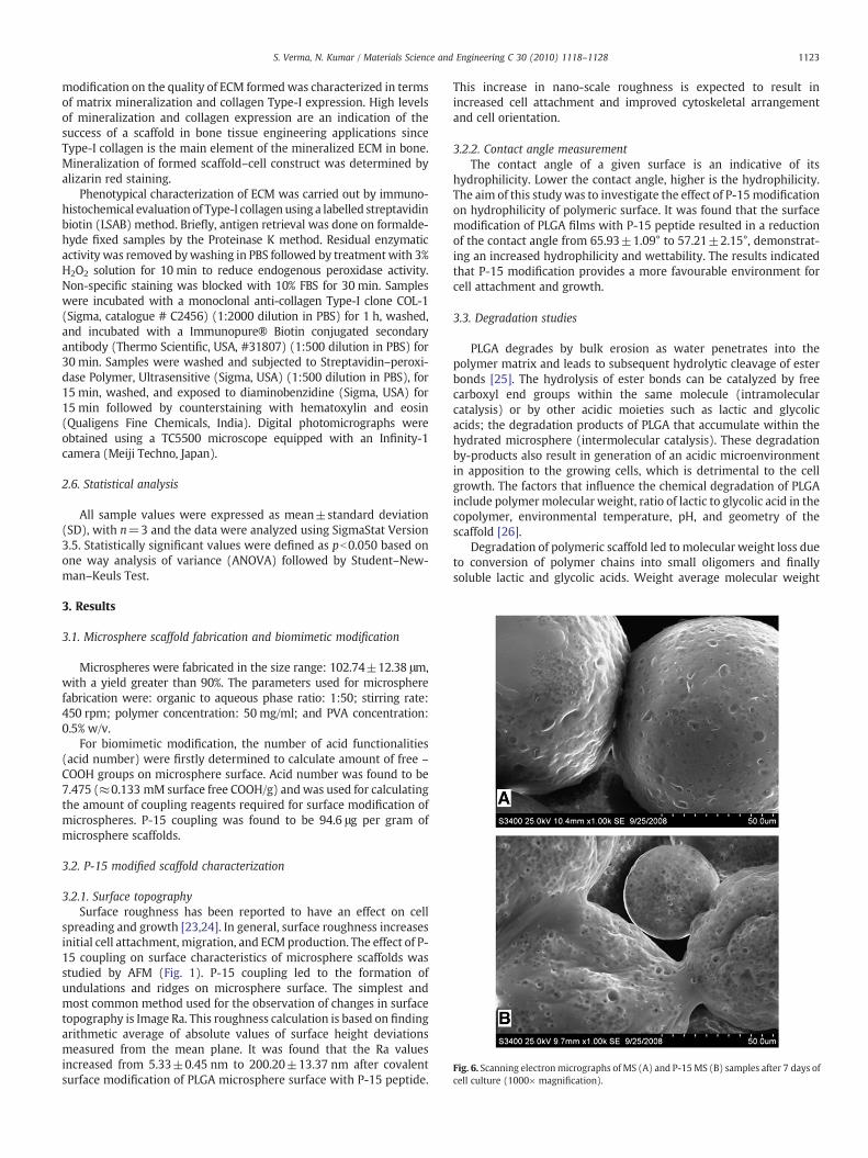
Fig. 6. Scanning electronmicrographs of MS (A) and P-15MS (B) samples after 7 days ofcell culture (1000× magnification).
1123S. Verma, N. Kumar / Materials Science and Engineering C 30 (2010) 1118–1128
modification on the quality of ECM formedwas characterized in termsof matrix mineralization and collagen Type-I expression. High levelsof mineralization and collagen expression are an indication of thesuccess of a scaffold in bone tissue engineering applications sinceType-I collagen is the main element of the mineralized ECM in bone.Mineralization of formed scaffold–cell construct was determined byalizarin red staining.
Phenotypical characterization of ECM was carried out by immuno-histochemical evaluation of Type-I collagen using a labelled streptavidinbiotin (LSAB) method. Briefly, antigen retrieval was done on formalde-hyde fixed samples by the Proteinase K method. Residual enzymaticactivity was removed bywashing in PBS followed by treatmentwith 3%H2O2 solution for 10 min to reduce endogenous peroxidase activity.Non-specific staining was blocked with 10% FBS for 30 min. Sampleswere incubated with a monoclonal anti-collagen Type-I clone COL-1(Sigma, catalogue # C2456) (1:2000 dilution in PBS) for 1 h, washed,and incubated with a Immunopure® Biotin conjugated secondaryantibody (Thermo Scientific, USA, #31807) (1:500 dilution in PBS) for30 min. Samples were washed and subjected to Streptavidin–peroxi-dase Polymer, Ultrasensitive (Sigma, USA) (1:500 dilution in PBS), for15 min, washed, and exposed to diaminobenzidine (Sigma, USA) for15 min followed by counterstaining with hematoxylin and eosin(Qualigens Fine Chemicals, India). Digital photomicrographs wereobtained using a TC5500 microscope equipped with an Infinity-1camera (Meiji Techno, Japan).
2.6. Statistical analysis
All sample values were expressed as mean±standard deviation(SD), with n=3 and the data were analyzed using SigmaStat Version3.5. Statistically significant values were defined as pb0.050 based onone way analysis of variance (ANOVA) followed by Student–New-man–Keuls Test.
3. Results
3.1. Microsphere scaffold fabrication and biomimetic modification
Microspheres were fabricated in the size range: 102.74±12.38 μm,with a yield greater than 90%. The parameters used for microspherefabrication were: organic to aqueous phase ratio: 1:50; stirring rate:450 rpm; polymer concentration: 50 mg/ml; and PVA concentration:0.5% w/v.
For biomimetic modification, the number of acid functionalities(acid number) were firstly determined to calculate amount of free –
COOH groups on microsphere surface. Acid number was found to be7.475 (≈0.133 mM surface free COOH/g) andwas used for calculatingthe amount of coupling reagents required for surface modification ofmicrospheres. P-15 coupling was found to be 94.6 μg per gram ofmicrosphere scaffolds.
3.2. P-15 modified scaffold characterization
3.2.1. Surface topographySurface roughness has been reported to have an effect on cell
spreading and growth [23,24]. In general, surface roughness increasesinitial cell attachment, migration, and ECMproduction. The effect of P-15 coupling on surface characteristics of microsphere scaffolds wasstudied by AFM (Fig. 1). P-15 coupling led to the formation ofundulations and ridges on microsphere surface. The simplest andmost common method used for the observation of changes in surfacetopography is Image Ra. This roughness calculation is based on findingarithmetic average of absolute values of surface height deviationsmeasured from the mean plane. It was found that the Ra valuesincreased from 5.33±0.45 nm to 200.20±13.37 nm after covalentsurface modification of PLGA microsphere surface with P-15 peptide.
This increase in nano-scale roughness is expected to result inincreased cell attachment and improved cytoskeletal arrangementand cell orientation.
3.2.2. Contact angle measurementThe contact angle of a given surface is an indicative of its
hydrophilicity. Lower the contact angle, higher is the hydrophilicity.The aim of this study was to investigate the effect of P-15modificationon hydrophilicity of polymeric surface. It was found that the surfacemodification of PLGA films with P-15 peptide resulted in a reductionof the contact angle from 65.93±1.09° to 57.21±2.15°, demonstrat-ing an increased hydrophilicity and wettability. The results indicatedthat P-15 modification provides a more favourable environment forcell attachment and growth.
3.3. Degradation studies
PLGA degrades by bulk erosion as water penetrates into thepolymer matrix and leads to subsequent hydrolytic cleavage of esterbonds [25]. The hydrolysis of ester bonds can be catalyzed by freecarboxyl end groups within the same molecule (intramolecularcatalysis) or by other acidic moieties such as lactic and glycolicacids; the degradation products of PLGA that accumulate within thehydrated microsphere (intermolecular catalysis). These degradationby-products also result in generation of an acidic microenvironmentin apposition to the growing cells, which is detrimental to the cellgrowth. The factors that influence the chemical degradation of PLGAinclude polymermolecular weight, ratio of lactic to glycolic acid in thecopolymer, environmental temperature, pH, and geometry of thescaffold [26].
Degradation of polymeric scaffold led tomolecular weight loss dueto conversion of polymer chains into small oligomers and finallysoluble lactic and glycolic acids. Weight average molecular weight

1124 S. Verma, N. Kumar / Materials Science and Engineering C 30 (2010) 1118–1128
(Mw) of MS decreased exponentially with degradation time through-out the degradation period (Fig. 2A). Apparent degradation rateconstant (KMw) and degradation half life (t1/2Mw) were calculated tobe 0.032 week−1 and 9.44 weeks, respectively. A sharp weight losswas observed after an initial lag time of about 20 days, with almost40% weight loss in 30 days (Fig. 2A). The degree of hydration was alsoinvestigated as a measure of polymer degradation. MS exhibited ahigh degree of hydration and swelling due to the presence of free –
COOH groups in the polymer (Fig. 2B). As hydration occurred,microspheres swelled and increased in size due to water ingress.After about 20 days of degradation, the extent of degradationexceeded the hydration resulting in a steep fall in the degree ofhydration (Fig. 2B). At the same time, pH of the degradation mediumfell (7.4 to 3.9 in about 27 days) and then reached a plateau indicatingthat PLGA microspheres were completely degraded. A shift wasobserved in the copolymer composition from 50 mol% at day 0 to70 mol% at 42 day as preferential cleavage occurred at the glycolic–glycolic and glycolic–lactic bonds thereby causing a faster release ofglycolic acid. The initially smooth and spherical microspheres alsoshowed visible signs of bulk degradation as indicated by SEM results(Fig. 2C–F).
3.4. In vitro cell culture studies
3.4.1. Cell adhesion, viability and proliferation studiesCell–matrix/cell–scaffold interactions are the basis of initial cell
attachment and also influence the cell phenotype and functions.
Fig. 7. Cytoskeletal actin distribution and organization of MG-63 cells grown on 2-D plate (A)Dotted circle represents microsphere perimeter.
During the first 24 h of the study, FBS was excluded from cell culturemedium so as to avoid the effect of serum proteins on initial cellattachment. P-15 MS showed significantly higher cell attachmentefficiency thanMS (Fig. 3). Thismay be attributed to the ability of P-15to enhance cell adhesion and to reduce apoptosis levels in serum-freeconditions [27]. A significantly higher cell count was observed in P-15MS than on MS due to enhanced cell attachment and biomimeticsurface environment provided by P-15 peptide until day 21 (Fig. 4A).For both 2D plate and 3D microsphere scaffolds, cell viability andcorresponding cell count increased till 14 day and later decreased. Thecell viability remained significantly higher on 2D plate than on bothMS and P-15 MS on 7th, 14th and 21st day, as multiple layers ofcellular clusters were formed in 2D plate. However, on 28th day, cellattachment on plate surface was completely lost and led todetachment of almost all cells in the form of loose clusters. Althoughcell viability declined during the later phase of study, cell attachmentto scaffolds was maintained in 3D scaffolds until the end of the study,unlike in 2D monolayer culture, indicating the beneficial effect of 3Denvironment in maintaining the cell growth.
3.5. Morphology
Cell morphology and cellular arrangement on 2D monolayerculture plates as well as on 3D scaffold–cell constructs, was studiedusing optical microscopy and SEM. In 2D cultures, a uniform layer offlattened cells with very few rounded cells could be seen on 7th day(Fig. 5A2). As the study progressed, clusters of rounded dead cells
, MS (B) and P-15MS (C), after day 7 (1), day 14 (2) and day 21 (3) of in vitro cell culture.
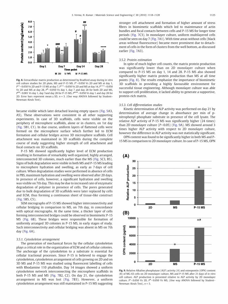
Fig. 8. Extracellular matrix production as determined by Bradford assay during in vitrocell culture studies for 2D plate, MS and P-15 MS. P*b0.050 Vs 2D and MS at day 1,P**b0.050 Vs 2D and P-15MS at day 7, P***b0.050 Vs 2D andMS at day 14, P****b0.050Vs 2D and MS at day 28, P#b0.050 Vs day 1, day 7 and day 28 for both 2D and MS,P##b0.001 Vs day 1, day 7and day 28 for P-15 MS, P###b0.050 Vs day 1 and day 28 for2D. Error bars represent mean±SD, n=3. (One way ANOVA followed by Student–Newman–Keuls Test).
Fig. 9. Relative Alkaline phosphatase (ALP) activity (A) and osteopontin (OPN) content(B) of MG-63 cells on 2D monolayer culture, MS and P-15 MS after 21 days of in vitrocell culture. ALP production is presented relative to ALP activity by 2D monolayerculture. P*b0.050 Vs 2D, P#b0.050 Vs MS, (One way ANOVA followed by Student–Newman–Keuls Test), n=3.
1125S. Verma, N. Kumar / Materials Science and Engineering C 30 (2010) 1118–1128
became visible which later detached leaving empty spaces (Fig. 5A3,A5). These observations were consistent in all other supportingexperiments. In case of 3D scaffolds, cells were visible on theperiphery of microsphere scaffolds, alone or in clusters, on 1st day(Fig. 5B1, C1). In due course, uniform layers of flattened cells wereformed on the microsphere surface which further led to ECMformation and cellular bridges across 3D microsphere scaffolds. Cellattachment was maintained in 3D scaffolds during the completecourse of study suggesting higher strength of cell attachment andfocal contacts on 3D scaffolds.
P-15 MS showed significantly higher level of ECM productionresulting in formation of remarkably well organized, highly arranged,interconnected 3D colonies, much earlier than the MS (Fig. 5C3, B5).Signs of bulk degradationwere visible in bothMS and P-15MS leadingto microsphere hydration and swelling, as early as 7 days of cellculture. When degradation studies were performed in absence of cellsin PBS,maximumhydration and swellingwere observed after 20 days.In presence of cells, however, a significant hydration and swellingwas visible on 7th day. Thismay be due to increased rate of enzymaticdegradation of polymer in presence of cells. The pores generateddue to bulk degradation of 3D scaffolds were later replaced by cellsand ECM, thus forming a continuous sheet of tissue-like construct(Fig. 5B5, C5).
SEMmicrographs of P-15 MS showed higher interconnectivity andcellular bridging in comparison to MS, on 7th day, in concordancewith optical micrographs. At the same time, a thicker layer of cellsforming interconnected bridges could be observed in biomimetic P-15MS (Fig. 6B). These bridges were responsible for formation ofuniformly arranged 3D colonies in P-15 MS, in early stages of study.Such interconnectivity and cellular bridging was absent in MS on 7thday (Fig. 6A).
3.5.1. Cytoskeleton arrangementThe generation of mechanical forces by the cellular cytoskeleton
plays a critical role in the organization of ECM and of cellular colonies.The anchorage of the cytoskeleton to a substrate is essential forcellular tractional processes. Since P-15 is believed to engage thecytoskeleton, cytoskeleton arrangement of cells growing on 2D and on3D MS and P-15 MS was studied using fluorescent labelling of cellswith Rhodamine 110 phalloidin. Day 14 images showed a uniformcytoskeleton network interconnecting the microsphere scaffolds inboth P-15 MS and MS (Fig. 7B2, C2). On day 21, the cytoskeletonarrangement in MS was lost (Fig. 7B3). However, a uniformcytoskeleton arrangement was still maintained in P-15 MS suggesting
stronger cell attachment and formation of higher amount of stressfibres in biomimetic scaffolds which led to maintenance of actinbundles and focal contacts between cells and P-15 MS for longer timeperiods (Fig. 7C3). In monolayer culture, uniform multilayered cellscould be seen on day 7 (Fig. 7A1).With time areas without cells (blackareas without fluorescence) became more prominent due to detach-ment of cells in the form of clusters from thewell bottom, as discussedearlier (Fig. 7A3).
3.5.2. Protein estimationIn spite of much higher cell counts, the matrix protein production
was significantly lower than on 2D monolayer culture whencompared to P-15 MS on day 1, 14 and 28. P-15 MS also showedsignificantly higher matrix protein production than MS at all timepoints (Fig. 8). The results emphasize the importance of biomimetic3D scaffolds in providing a highly favourable environment forsuccessful tissue engineering. Although monolayer culture was ableto support cell proliferation, it lacked ability to generate a supportive,protein-rich matrix.
3.5.3. Cell differentiation studiesKinetic determination of ALP activity was performed on day 21 by
determination of average change in absorbance per min of p-nitrophenyl phosphate substrate in presence of the cell lysate. Therelative ALP activity of P-15 MS was significantly higher (24 times)than 2D monolayer culture (Pb0.05) (Fig. 9A). MS showed around 3times higher ALP activity with respect to 2D monolayer culture,however the difference in ALP activity was not statistically significant.
OPN contentwas found to be significantly higher in bothMS and P-15MS in comparison to 2Dmonolayer culture. In case of P-15MS, OPN

Fig. 10. Mineralization of extracellular matrix in 2D (A), MS (B) and P-15 MS (C) scaffolds after day 7(1), day 14 (2) and day 21 (3) of in vitro cell culture indicated by alizarin redstaining.
1126 S. Verma, N. Kumar / Materials Science and Engineering C 30 (2010) 1118–1128
content was around 22-fold higher than 2D monolayer culture. Thiswas 2.6 fold in biomimetic P-15 MS than MS (Pb0.05) (Fig. 9B).
Alizarin red staining showed minimal mineralization in monolayerculture. Cellular clusterswhichwere on the verge of detaching fromwellbottom showed some signs of mineralization on day 21 in 2D culture(Fig. 10A3). On the contrary, 3D culture systems showed formation ofmineralized matrix at an early stage. P-15 MS showed a strong redcoloration indicating higher mineralization than MS (Fig. 10C3).
During Immunohistochemistry studies for collagen expression(28th day), cell clusters were washed off after the antigen retrievalstep in case of monolayer culture due to poor phenotypic character-istics and weak cellular attachment at the well bottom. Cellularattachment was however maintained, on 3D microsphere scaffolds inboth MS and P-15 MS. Collagen Type-I expression was much higher inP-15 MS in comparison to MS (Fig. 11B).
4. Discussion
This study aimed to evaluate the potential of P-15 modifiedbiomimetic PLGAmicrosphere scaffolds as injectable bone filler. A sizeof around 100 μm was selected while fabricating the microspherescaffolds as it was expected to provide an optimum balance betweenthe requirement to maximize the cell surface to volume ratio and theability of each micro-carrier to support sufficient cell growth beyondthe initial seeding while maintaining the injectability. To inducebiomimetism, the surface of hydrophobic PLGA microspheres was
modified with P-15 peptide, 766GTPGPQGIAGQRGVV780, a syntheticanalogue of the cell-binding domain of α(I) chain of collagen type-I,on account of previous reports indicating a key role of P-15 inosteogenesis and ultimately bone regeneration [28,29]. P-15 isresponsible for generating appropriate biomimetic environment forenhanced viable cell attachment, cell bonding and the initiation of acascade of events (migration, alignment, proliferation and differen-tiation) that are necessary for optimal bone formation [27,30–32]. Itincreases Type-I collagen, alkaline phosphatase and BMP-2 geneexpression thus stimulating osteoblastic activity and synergising theeffects of osteogenic factors to induce osteoblastic differentiation [28].Clinically, an anorganic bovine-derived hydroxyapatite matrix/cell-binding peptide (ABM/P-15) has been found to significantly improvethe outcomes of regeneration of infrabony periodontal defectsfollowing treatment when compared to open flap debridement [33].
It is reported that surfaces with textures such as nodes, pores, orrandom patterns are often associated with marked changes of cellmorphology, cell activities, and production of regulatory factors whencompared to smooth surfaces [3]. The effect of P-15 modification onthe surface topography and hydrophilicity of scaffold was assessedand it was found that P-15 peptide conjugation led to an increase insurface hydrophilicity as well as nano-level roughness. Y. Wan et al.reported enhanced cell adhesion strength due to the nano-scale ormicro-scale roughness, when compared with a controlled smoothsurface [4]. The width and the depth of a surface topographicalstructure can also influence the cell responses, since cells can orient

Fig. 11. Immunohistochemistry of Type-I Collagen on MS (A) and P-15 MS (B), after28 days of cell culture.
1127S. Verma, N. Kumar / Materials Science and Engineering C 30 (2010) 1118–1128
themselves along the groove and ridge, the phenomenon commonlyreferred as “contact guidance” [23,24]. Reports suggest that thealterations in adhesion structures due to the surface topography maybe responsible for differences in cell signalling, which lead to changesin the cellular function like mineralization [34]. It was expected thatthe changes in scaffold topography due to P-15 modification couldpossibly have marked effects on the cellular response of the scaffold.
The synchronization between polymer degradation and replace-ment by natural tissue produced from cells is considered to beessential for the success of any tissue engineered scaffold [35]. Thus, invitro degradation studies were performed to have an idea about thedegradation characteristics which may affect biological cellularprocesses including cell viability, growth, tissue regeneration, andhost response [36]. During degradation, scaffolds underwent hydra-tion, swelled and later showed visible signs of bulk degradation alongwith generation of soluble, acidic degradation by-products resultingin acidification of external medium. However, during cell cultureevaluation, hydration and swelling was visible much earlier (around7 days) due to an added effect of enzymatic degradation in cellularenvironment. Thus, it is imperative to consider the effect of biologicalenvironment on the scaffold degradation while choosing a polymerfor a specific tissue engineering application. Suitable measures mayalso be required to minimize the effect of microenvironment pHacidification due to polymer degradation. During cell cultureevaluation, the polymer showed faster degradation and the degradingpolymer was replaced by growing cells and ECM.
The ability of cells to adhere to the scaffold, leading to productionand organization of ECM is also a central and key step to successfultissue engineering. It was observed that P-15 MS showed enhancedcell-binding capacity due to its biomimetic nature which resulted insignificantly higher viable cell attachment in comparison to MS. Theimproved hydrophilicity and nano-level roughness generated due toP-15 surface modification probably also contributed to the improvedcell attachment. Cell proliferation was analyzed until 28 days and it
was found that viable cell count increased until 14 days, but wassignificantly reduced up to the 28th day. Biomimetic scaffolds showeda higher cell proliferation till 21 days in comparison to unmodifiedmicrospheres. After this time period, factors such as nutrient diffusionand hypoxia probably became limiting for cell growth and survival,thereby overshadowing the biomimetic effect of P-15. Acidification ofthe microenvironment pH due to by-products generated duringpolymer degradation also contributed to decreased cell viability. Onday 21, a uniform layer of microsphere scaffolds interconnected withintercellular bridges could be observed in P-15MS. Contraction due toforce transfer through biomimetic environment provided by P-15resulted in microsphere clustering, 3D colony formation and orderingof cells thus initiating physiological processes leading to cell sheetformation. This observation could be attributed to the fact that P-15MS behaved as a collagen surrogate matrix (CSM), and were able tomimic the physiological interactions of collagen with cells, therebyleading to haptotactic cell migration, activation of signalling path-ways, induction of growth factors, cell differentiation, tissue remodel-ling and morphogenesis [37].
Deposition of bone in physiological conditions involves timedsecretion, deposition and removal of a complex array of ECM proteinswhich appear in a defined temporal and spatial sequence [38]. Hence,matrix protein production is an essential component for healthy bonetissue formation. Abundant matrix protein formation, which isconsidered as a benchmark for successful tissue-construct formation,contributed in generation of uniform 3D colonies by holding theseparatemicrospheres together, provided a uniform sheet-like arrange-ment to the scaffold–cell constructs and helped in maintaining thecellular arrangement on P-15MS scaffolds during the long term studies.Optical microscopy and SEM studies corroborated the aforementionedobservations. Biomimetic modification could further contribute tomaintain the cytoskeleton arrangement for longer time periods invitro. In spite of higher cell count in 2D plate, protein contentwas foundto bemuch less, suggesting that cell phenotype, extracellularmatrix andcytoskeleton arrangement could not be maintained in monolayerculture for longer time periods. Instead, cells were washed off duringthe processing due to weak attachment to the plate surface.
The formation of a collagenous matrix with mineralization is aprerequisite for a good bone repair process as mineralization plays arole in dictating and spatially orienting the deposition of ECM. Thecomposition of the extracellular framework is dominated by a class ofmolecules known as collagens, each with unique features suited for itsfunction and location. Ninety percent of the organic matrix of bone ismade up by collagen Type-I, and 10% by a variety of non-collagenousproteins [38]. Biomimeticmicrospheres developed significantly higherlevels of mineralized ECM and showed enhanced collagen Type-Iexpression. High ALP activity and OPN production, even in the absenceof an osteogenic medium, indicated the osteoinductive nature of P-15and its ability tomodulate cell differentiation andmorphogenesis, thussupporting its possible role in skeletal reconstruction.
These results suggest the fact that P-15 modified osteoconductivePLGA microsphere scaffolds encompass the biocompatibility, me-chanical strength and biodegradability of PLGA, along with the celladhesive, proliferative and differentiative properties of osteoinductiveP-15, and also eliminate the limitations of monolayer culture due tothe 3D biomimetic environment provided for cell growth. In the caseof 2Dmonolayer culture, though a higher cell count is obtained, it wasovershadowed by the weak cell attachment, poor phenotypiccharacteristics, low mineralization levels and cell death in the latertime points.
5. Conclusions
The present study substantiates the importance of biomimetic 3Dscaffolds for providing and maintaining a natural environmentconducive for successful tissue regeneration and also correlates the
1128 S. Verma, N. Kumar / Materials Science and Engineering C 30 (2010) 1118–1128
degradation behaviour of polymeric scaffolds to cell survival. P-15modified microsphere scaffolds showed a significantly higher cellattachment, proliferation and ECM formation, with enhanced miner-alization and collagen Type-I expression, when compared to unmod-ified scaffolds. The results demonstrate the suitability of P-15modified PLGA microspheres as a potential injectable scaffold fornon-invasive bone tissue engineering applications.
Acknowledgements
Authors are thankful to Professor Rajendra S. Bhatnagar, Chiefscientist and CEO, ENTAM, LLC, USA for providing the financialsupport to carry-out the research work and to Professor P. Ramarao(Director, NIPER) for allowing the use of SEM, AFM and CLSM atCentre for Pharmaceutical Nanotechnology. Technical support formicroscopic techniques from Mr. Dinesh and Dr. Vijender Beniwal isduly acknowledged.
Appendix A. Supplementary data
Supplementary data associated with this article can be found, inthe online version, at doi:10.1016/j.msec.2010.06.005.
References
[1] C. Sanchez, H. Arribart, M.M. Guille, Nat. Mater. 4 (2005) 277.[2] A.B. Sanghvi, K.P. Miller, A.M. Belcher, C.E. Schmidt, Nat. Mater. 4 (2005) 496.[3] R.G. Flemming, C.J. Murphy, G.A. Abrams, S.L. Goodman, P.F. Nealey, Biomaterials
20 (1999) 573.[4] Y. Wan, Y. Wang, Z. Liu, X. Qu, B. Han, J. Bei, S. Wang, Biomaterials 26 (2005) 4453.[5] B.C. Tai, C. Du, S. Gao, A.C. Wan, J.Y. Ying, Biomaterials 31 (2009) 48.[6] M. Pegueroles, C. Aparicio, M. Bosio, E. Engel, F.J. Gil, J.A. Planell, G. Altankov, Acta
Biomater. 6 (2010) 291.[7] R.F. Neiva, Y.-P. Tsao, R. Eber, J. Shotwell, E. Billy, H.-L. Wang, J. Periodontol. 79
(2008) 291.[8] A. Andukuri, W. P. Minor, M. Kushwaha, J. M. Anderson, H. W. Jun, Nanomed. 6
(2010) 289.
[9] C. Milburn, J. Chen, Y. Cao, G.M. Oparinde, M.O. Adeoye, A. Beye, W.O. Soboyejo,Mater. Sci. Eng: C 29 (2009) 306.
[10] H. Shin, S. Jo, A.G. Mikos, Biomaterials 24 (2003) 4353.[11] N.R. Mercier, H.R. Costantino, M.A. Tracy, L.J. Bonassar, Biomaterials 26 (2005)
1945.[12] K.D. Newman, M.W. McBurney, Biomaterials 25 (2004) 5763.[13] B.P. Chan, T.Y. Hui, C.W. Yeung, J. Li, I. Mo, G.C.F. Chan, Biomaterials 28 (2007)
4652.[14] Y. Senuma, S. Franceschin, J.G. Hilborn, P. Tissières, I. Bisson, P. Frey, Biomaterials
21 (2000) 1135.[15] M. Borden, S.F. El-Amin, M. Attawia, C.T. Laurencin, Biomaterials 24 (2003) 597.[16] K. Garkhal, S. Verma, K. Tikoo, N. Kumar, J. Biomed, Mater. Res. A 82 (2007) 747.[17] S.W. Kang, S.W. Seo, C.Y. Choi, B.S. Kim, Tissue Eng. 14 (2008) 25.[18] J.M. Melero-Martin, M.A. Dowling, M. Smith, M. Al-Rubeai, Biomaterials 27 (2006)
2970.[19] F. Gabler, S. Frauenschuh, J. Ringe, C. Brochhausen, P. Gotz, C.J. Kirkpatrick, M.
Sittinger, H. Schubert, R. Zehbe, Biomol. Eng. 24 (2007) 515.[20] R.C. Mehta, B.C. Thanoo, P.P. Deluca, J. Control. Release 41 (1996) 249.[21] D. Lochmann, S. Stadlhofer, J. Weyermann, A. Zimmer, Int. J. Pharm. 283 (2004)
11.[22] Y. Zhu, K.S. Chian, M.B. Chan-Park, P.S. Mhaisalkar, B.D. Ratner, Biomaterials 27
(2006) 68.[23] Y. Wan, X. Qu, J. Lu, C. Zhu, L. Wan, J. Yang, J. Bei, S. Wang, Biomaterials 25 (2004)
4777.[24] P.T. Ohara, R.C. Buck, Exp. Cell Res. 121 (1979) 235.[25] M. Sandor, D. Enscore, P. Weston, E. Mathiowitz, J. Control. Release 76 (2001) 297.[26] J. Panyam,M.M. Dali, S.K. Sahoo,W.Ma, S.S. Chakravarthi, G.L. Amidon, R.J. Levy, V.
Labhasetwar, J. Control. Release 92 (2003) 173.[27] T. Hanks, B.L. Atkinson, Biomaterials 25 (2004) 4831.[28] X.B. Yang, R.S. Bhatnagar, S. Li, R.O. Oreffo, Tissue Eng. 10 (2004) 1148.[29] A.H. Valentin, J. Weber, Keio J. Med. 53 (2004) 166.[30] J.J. Qian, R.S. Bhatnagar, J. Biomed. Mater. Res. 31 (1996) 545.[31] H. Nguyen, J.J. Qian, R.S. Bhatnagar, S. Li, Biochem. Biophys. Res. Commun. 311
(2003) 179.[32] A. Palmieri, F. Pezzetti, G. Brunelli, I. Zollino, L. Scapoli, M. Martinelli, M. Arlotti, F.
Carinci, J. Biomed. Sci. 14 (2007) 777.[33] A. Kasaj, B. Rohrig, C. Reichert, B. Willershausen, Clin. Oral Investig. 5 (2008) 5.[34] B. Nebe, F. Lüthen, R. Lange, P. Becker, U. Beck, J. Rychly, Mater. Sci. Eng: C 24
(2004) 619.[35] H.J. Sung, C. Meredith, C. Johnson, Z.S. Galis, Biomaterials 25 (2004) 5735.[36] J.E. Babense, J.M. Anderson, L.V. McIntire, A.G. Mikos, Adv. Drug Deliv. Rev. 33
(1998) 111.[37] R. Bhatnagar, S. Li, Conf. Proc. IEEE Eng. Med. Biol. Soc. 7 (2004) 5021.[38] M. Riminucci, P. Bianco, Braz. J. Med. Biol. Res. 36 (2003) 1027.