Effect of antibiotics inhibiting cell wall synthesis on growth of
13
Instructions for use Title Effect of antibiotics inhibiting cell wall synthesis on growth of staphylococcal L-phase variant Author(s) MAKINO, Toshikazu Citation Journal of the Faculty of Science, Hokkaido University. Series 5, Botany, 14(2): 135-146 Issue Date 1988 Doc URL http://hdl.handle.net/2115/26438 Type bulletin File Information 14(2)_P135-146.pdf Hokkaido University Collection of Scholarly and Academic Papers : HUSCAP
Transcript of Effect of antibiotics inhibiting cell wall synthesis on growth of

Instructions for use
Title Effect of antibiotics inhibiting cell wall synthesis on growth of staphylococcal L-phase variant
Author(s) MAKINO, Toshikazu
Citation Journal of the Faculty of Science, Hokkaido University. Series 5, Botany, 14(2): 135-146
Issue Date 1988
Doc URL http://hdl.handle.net/2115/26438
Type bulletin
File Information 14(2)_P135-146.pdf
Hokkaido University Collection of Scholarly and Academic Papers : HUSCAP

Journ. Fac. Sci., Hokkaido Univ. Ser. V (Botany), 14(2): 135-146, 1988.
Effect of antibiotics inhibiting cell wall synthesis on growth of staphylococcal L-phase variant
Toshikazu MAKINO
Penicillin, a typical inhibitor of cell wall synthesis, inhibited the colony formation of L
phase variant of Staphylococcus au reus , which has no cell wall.
The inhibition was observed only when the L-colony was formed in a culture medium
with a low cncentration of N aCI (0.35 M). Lowering concentation of yeast extract and
incubation temperature suppressed inhibition. L-colony formation in a culture medium
without Mg++ was dependent on the presence of penicillin.
The same effects, inhibition and dependence, were observed in the treatment with 0-
cycloserine and bacitracin. However, bacitracin inhibited L-colony formation even in the
medium with 0.9 M NaCI. Vancomycin inhibited formation of L-colony but did not
support L-colony formation without Mg++. Phosphomycin neither inhibited nor supported
L-colony formation.
The cell wall structure was not detected by electoron microscopic observation in the L
phase variant cell grown under the condition in which L-colony formation was inhibited by
penicillin.
In a previous study (MAKINO, 1983), the induction of staphylococcal Lphase variant was investigated. Protoplast, prepared by the treatment of parental cells with lysostaphin, was readily transformed to L-phase variant on the surface of an agar medium which contained an osmotic stablizer (N aCl), Mg++, serum and penicillin in addition to nutrient components. Penicillin was added to prevent colony formation of the remaining parental type cells. Although L-phase variant was not susceptible to penicillin because it does not synthesize cell wall, inibition by and dependence on penicillin were observed under a certain restricted condition. Penicillin is a typical inhibitor of cell wall synthesis. It combines with penicillin binding protein (GEORGO-PAPADAKOU et at., 1976), which is found in cell membrane and inhibits the action of transpeptidase. Therefore, any effect of penicillin on cell wall-less bacteria is inconsistent with the current view.
The purpose of this communication was to describe a condition which affects the action of penicillin on L-phase variant and to present some possible explanations of its mechanism.

136 T. Makino
Materials and Methods
Bacterial strain Staphylococcus aureus 209P was used throughout this experiment.
Culture Tryptic soy broth (Difco, Detroit) supplemented with 0.5% yeast extract
(Difco, Detroit) was used for the growth of parental cells. The basal medium for L-phase variant cells consisted of 2% casamino acid (Difco, Detroit), 0.5% yeast extract, 1% Na-Iactate (Nakarai, Kyoto), 5.2% NaCl and 0.8% Nobel agar (Difco, Detroit), and pH was adjusted to 7.0 with Tris. Casein (0.005%) and Mg-acetate (20 mM) were autoclaved separately and added to the culture medium before preparation of agar plate (CL YS-agar medium). Chemicals
Lysostaphin and antibiotics except penicillin were purchased from Sigma (St. Louis). Penicillin was purchased from BanyO (Tokyo). Induction of L-phase variant
Cells harvested from the middle of a logarithmic culture (cell density was about 109/ml) were washed once with physiological saline and resuspended in the original volume of 1.4 M NaCl solution with 0.01 M Tris-HCI buffer, pH 7.4. After treatment with 20 j.tg/ml lysostaphin at 37°C for 3 hours, almost all the parental cells were transformed to protoplast. The protoplast was spread on the surface of CL YS-agar medium after appropriate dilution. A typical fried egg shaped L-form colony was formed after incubation at 3TC 3 days. Electron microscopi microscopic observation
The sample of electron microscopic observation was prepared by the rapid-freezing and substitution fixation method (AMAKO et al., 1986). Lphase variant cells grown on CL YS-agar medium at 3TC were mounted directly on a Teflon speciment support.
This part of the research was carried out by KAZUNOBU AMAKO and AKIKO UMEDA at School of Medicine in KyOshO University.
Rresults
Inhibition of L-colony formation by penicillin Under the ordinary condition in which the culture medium contained 0.9
M NaCl, penicillin did not inhibit L-colony formation. However, when the concentration of N aCI was decreased, L-colony formation was ihibited by penicillin (Fig. 1). Minimu~ inhibitory concentration for L-phase variant was the same as that for parental cells (0.2 unit/mI). Fig. 1 also shows that

Effect of penicillin on L-phase variant
Inhibition
CFU ("!oj
100
300 80
200 60
40
100 20
~, 0~~~~~ __ ~~_'_-~6~_~-4~_~_~ o 0.1 0.2 0.3 0.4 0.5 0.6 0.7 O.B 0.9
NaCI (M J
Fig.1. Effect of NaCl on inhibition of L-colony formation by penicillin. 0: control, .: with penicillin (1 unit/ml), L:.: inhibition rate.
CFU 400r-~~~~~'---~~~~~~
300
200
100
10 100
Mg++ (mM J
Fig. 2. Effect of Mg++ on inhibition of L-colony formation by penicillin. L-colony was formed on CL YS-agar medium of which N aCI concentration was reduced to 0.35 mM. 0: control, .: with penicillin (1 unit/mI).
137

138 T. Makino
Inhibition
CFU (%)
400 100
0 A 300 j~'
l5: t:/
d 200 50
!5.
100
Yeast extract (%)
Fig. 3. Effect of yeast extract on inhibition of L-colony formation by penicillin. L-colony was formed on CL YS-agar medium of which NaCl concentration was reduced to 0.35 M. Symbols are as defined in the legend to Figure 1.
CFU 400
300
200'
100
o
20· 3D· 37·
0.2 0.4 0.8 0.2 0.4 0.8 0.2 0.4 0.8
Yeast extract (%)
Fig. 4. Effect of temperature on inhibition of L-colony formation by penicillin. L-colony was formed on CLYS-agar medium of which concentration was reduced to 0.35 M. CJ: control, _: with penicillin (1 unit/m!)

Effect of penicillin on L-phase variant 139
0.3 M of N aCI was the minimum concentration required to protect the Lphase variant cell from low osmolarity.
Fig. 2 shows the effect of Mg++ on L-colony formation and its inhibition by penicillin. Mg++ was essential for the L-colony formation. Higher concentrations of Mg++ suppressed the penicillin inhibition.
Yeast extract was also effective factor of penicillin inhibition. As yeast extract was an essential nutrient for L-phase variant, less than 0.1% of yeast extract could not support the growth of L-phase variant but higher concent· rations of yeast extract gave a higher inhibition rate. The highest inhibition rate was demonstrated at a concentration of 0.8% of yeast extract (Fig. 3).
CFU
1.00
300
200
100
o 400
300
200
yeast extract (0.2"10)
100 ~
oll 6.25 6.50 675 700 725
pH
20·C
(08%)
37°C
625 650 6.75 7.00 725 pH
Fig. 5. Effect of pH on inhibition of L-colony formation by penicillin. Lcolony was formed on CLYS-agar medium of which NaCl concentration was reduced to 0.35 M. Symbols are as defined in the legened to Figure 4.

140 T. Makino
CFU
yeast extract (0.8'/,) 400 20·C
( 0.2'/,)
37·C
300
200
~~L 100
0 '" a a a ~ ~ 8 8 N '" ~ a
- N IS") 0 _ N
a a 0 ci d 0000..-
EDTA( mM )
Fig. 6. Effect of EDT A on inhibition of L-colony formation by penicillin. L-colony was formed on CL YS-agar medium of which concentration of NaCl was reduced to 0.35 M. Symbols are as defined in the legende to Figure 4.
CFU
400~~~~~~--~~~~~
300
200
100
10
Mg++( mM)
Fig. 7. Dependence of L-colony formation on penicillin. 0: control, .: with penicillin (1 unit/mt)
Effect of penicillin on L-phase variant 141
Incubation temperature was another factor which affected the penicillin inhibition of L-phase variant (Fig. 4). At 20°C, penicillin did not show any significant effect on L-colony formation. At 30"C, inhibition was demonstrated but its rate was less than that at 3TC. Fig. 4 also shows the effect of yeast extract on penicillin inhibition at various temperatures.
Fig. 5 shows tfe effect of pH, yeast extract and incubation temperature on penicillin inhibition. Yeast extract and temperature gave almost the same results as those described above. There was no significant effect of pH on the susceptibility of L-phase variant to penicillin in the range of pH tested (6.25-7.25).
Fig. 6 shows the effect of EDT A on L-colony formation and its inhibition by penicillin. The cells growing at 20°C and with 0.8% yeast extract had higher sensitivity to EDT A. At a concentration of 0.5 mM, the colony size became small and it was hard to count individual colonies. No colony was detected at 1.0 mM of EDT A. The cells growing at 3TC and with 0.2% of yeast extract were comparatively resistant to EDTA. Inhibition by penicillin was observed when concentration of EDT A was 1.0 mM. Dependence of L-colony formation on penicillin
CFU 400
300
200
100
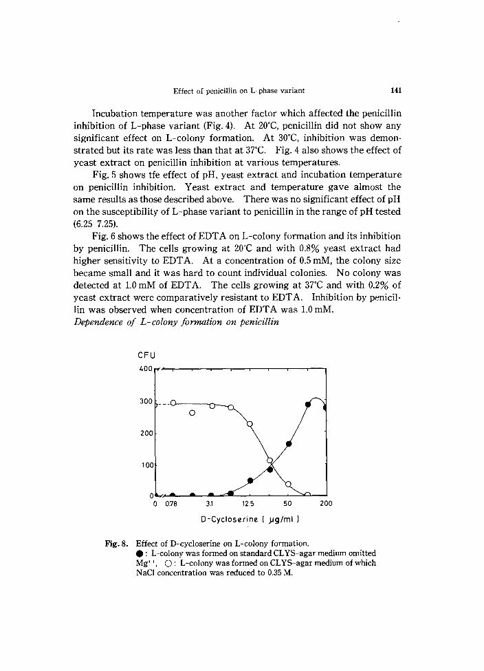
0 0 0.78 3.1 12.5 50 200
D-Cycloserine ( ).Jg/ml )
Fig_ 8. Effect of D-cycloserine on L-colony formation . • : L-colony was formed on standard CL YS-agar medium omitted Mg++, 0: L-colony was formed on CLYS-agar medium of which N aCl concentration was reduced to 0.35 M.

142 T. Makino
L-phase variant required Mg++ for its colony formation; however, as shown in Fig. 7, if penicilin was present, it could grow without Mg++. In this case the amnount of penicillin required was 0.02 unit/ml, which was much less than MIC. Effeet of other antibiotics which inhibit cell wall synthesis
Antibiotics other than penicillin which are known as inhibitors of cell wall synthesis were examined for their ability to affect L-colony formation. D-Cycloserine inhibited and supported L-colony formation by the same way as penicillin (Fig. 8). Bacitracin also inhibited L-colony formation in the medium with 0.35 M NaCl and supported it without Mg++. However, it inhibited L-colony formation even in the medium with 0.9 M N aCI (Fig. 9).
Vancomycin did not support L-colony formation without Mg++ but in· hibited it under both conditions of 0.35 M and 0.9 M N aCI (Fig. 10). Phospho· mycin neither inhibited nor supported L-colony formation. Electron microscopic observation of the L-phase variants grown with 0.35 M NaCl
Since there must be a possibility that the L-phase variant grown with 0.35 M NaCI can synthesize cell wall, the cell structures were examined by an electron microscope. Fig. 11 shows that no structures resembling cell wall were recognized, even in the cells grown in the medium with 0.35 M NaCI and 0.8% yeast extract.
CFU 400r-~--~--r-~--~--r-~--~~
300
Bactracin (}Jg/mll
Fig. 9. Effect of bacitracin on L-colony formation. Symbols are as defin· ed in the legened to Figure 8.

300
200
100
Effect of penicillin on L- phase variant
4
o
• 8 16 31 63
Vancomycin(ng) 125 250
Fig. 10. Effect of vancomycin on L-colony formation. Symbols are as defined in the legened to Figure 8.
Fig. 11. Thin sectioned L-phase variant cell grown On CL YS-agar medium of which NaCI concentration was reduced to 0.35 M. Bar = l,um
143

144 T. Makino
Discussion
One of the possible explanations of the inhibition of L-colony formation by penicilin was the synthesis of cell wall by L-phase variant grown in the medium with low concentration of NaCl. This possibility was ruled out by the electron microscopic observation. Although chemical analysis of cell wall components might be necessary, at the present stage the number of cells obtainable was insufficient because L-phase variant cells can grow only on the surface of agar medium and most part of the colony is formed in the agar layer. However, the L-phase cells grown under the restricted condition were also susceptible to low osmolarity (data not shown). This meant that they did not retain an enough amount of cell wall structure to prevent the cells from plasmolysis due to low osmolarity.
All the antibiotics which inhibit L-colony formation had a common characteristic: the cells inhibited by these antibiotics accumulated uri dine nucleotide peptide (PARK, 1952; PARK and STROMINGER, 1957; PARK, 1958; 1961; REYNOLDS, 1964). On the contrary, the antibiotic that had no effect, phosphomycin, is known to inhibit NAC-pyruvate transpeptidase (MATA et at., 1969). Some relationship may exists between the accumulation of uridine nucleotide peptide and the inhibition by and depedence on penicillin of L-colony formation.
There was an interesting similarity between the penicillin inhibition of L-phase variant and methicillin resistant Staphytococcus au reus (MRSA). As is well known MRSA isolates fequently have a characteristic referred to as heterogeneity (BARBAER, 1964; DYKE, 1969; SABATH and WALLACE, 1971; SABATH et at., 1972; SABATH,1977). This term refers to the fact that within a given strain of MRSA, only a small proportion of CFU are able to express resistance to B-Iactam antibiotics. Modification of growth conditions, incubation temperature, an increase in osmolarity, and the addition of EDT A, can increase the proportion of CFU that expresses resistance to various degrees. In the case of penicillin inhibition of L-phase variant, osmolarity, temperature and EDT A also gave a similar effect. In MRSA, penicillin binding protein 2 (PBP 2) was replaced with PBP2a which had low affinity to penicillin and its relation with the phenotypic expression of MRSA was suggested (HARTMAN and THOMASZ, 1984; 1986). While the presence of PBP in L-phase cell was reported (MARTIN et at., 1980), its detail remained to be elucidated. Effect of culture conditions on PBP formation in L-phase cells now under investigation.
One of the characteristic aspects of L-phase variant was pleuromor-
Effect of penicillin on L-phase variant 145
phism (DIENS, 1970; HIJIMANS et al., 1969). The cell division of L-phase variant was irregular, and sometimes even the budding of daughter cell was observed. This phenomenon seemed to be the result of the loss of cell wall. In other words, the loss of cell wall induced irregular cell division. It was conjectured that the cell wall synthesizing system may have the capability to control cell division. It is well known and established evidence that the bacterial chromosome is attached to the cell surface, providing a means of regulating both initiation of DNA replication and chromosome segregation. SANDER and KEYMAN (1988) described an association of DNA, including newly synthesized regions, with a specific region of the cytoplasmic membrane, and the attachment was prevented by inhibitor of cell wall synthesis, vancomycin and bacitracin.
A clinical isolate of Staphylococcus au reus which was tolerant to a number of B-lactam antibiotics showed initially stimulated and then severely inhibited RNA synthesis by the treatment with oxacillin, an inhibitor of cell wall synthesis. Protein synthesis of this strain was not inhibited initially but the rate of protein synthesis declined after 50 min of antibiotic treatment (JABLONSKI and MYCHA]LONKA, 1988).
The phenomena above mentioned might be suggestive to explain the mechanism of growth inhibition of L-phase variant with cell wall inhibitingantibiotics.
References
AMAKO, K., MURATA, K. and UMEDA, A. 1986. Structure of the envelope of Escherichia coli
observed by rapid-freezing and substitution fixation method. Microbiol.
Immunol. 27: 95-99. BARBAER, M. 1964. Naturally occuring methicillin-resistant Staphylococci. J. Gen.
Microbiol. 35: 183-190. DIENS, L. 1970. Biology and morphology of L-forms with a note on the relation of L-forms
to Mycoplasma. p.285-312. In SHARP, J. (ed.), The role of Mycoplasma and L
form of bacteria in disease. Charles C. Thomas Publ. Co, Springfield. DYKE, K. G. H. 1969. Penicillinase production and intrinsic resistance to penicillins in
methiciIIn resistant cultures of Staphylococcus au reus . J. Gen. Microbiol. 2: 261 -278.
GEORGOPAPADAKOU, N. H., BARBARA, A. and MAURIZ, R. 1976. Possible physiological functions of penicillin-binding proteins in Staphylococcus aureus. Antimicrob. Agents. Chemother. 29: 333-336.
HARTMAN, B. J. and TOMASZ, A. 1984. Low affinity penicillin-binding protein associated with B-lactam resistance in Staphyococcus aureus. J. Bacteriol. 158: 513-516.
--- and 1986. Expression of methicillin resistance in heterogenous strains of

146 T. Makino
Staphylococcus aureus. Antimicrob. Agents Chern other. 29: 85-92. HUIMAN, W., VAN BOVEN, C. P. A. and CLASENER, H. A. L. 1969. Fundamental biology of
the L-phase of bacteria. P.67-143. In HAYFLICK, L.(ed.). The Myco
lasmatales and the L-phase bacteria. North-Holland Publ. Co., Amsterdam.
JABLONSKI, P. E. and MCHA]LONKA, M. 1988. Oxacillin-induced inhibition of protein and
RNA synthesis in a tolerant Staphylococcus cureus isolate. J. Bact. 170 : 1831-1836.
MATA, J. M., HERNANDEZ, H. and MOCHALES, S.1969. Phosphomycin, a new antibiotic
produced by strains of Streptomyces. Science. 166: 122-123.
MAKINO, T. 1983. Induction of L-phase variant from protoplast of Staphylococcus aureus.
Microbiol. Immunol. 27: 749-755. MARTIN, H. Fl., SCHILE, W. and SCHIEFER, H. G. 1980. Differentiation of Mycoplasmatales
from bacterial protoplast L-forms by assay for penicillin binding proteins. Arch.
Microbiol. 127: 297-299.
PARK, J. T. 1952. Uridine-5'-pyrophosphate derivatives III. Amino acid-containing deri
vatives. J. BioI. Chern. 194: 897-904.
--- and STROMINGER, J. L. 1957. Mode of action of penicillin. Biochemical basis for
the mechanism of action of penicillin and for its selective toxicity. Science. 125 :
99-101.
--- 1958. Inhibition of cell wall synthesis in Staphylococcus au reus by chemicals which
cause accumulation of wall precursor. Biochem. J. 70: 2p. ~--- 1961. Inhibition of synthesis of bacterial mucopeptide or protein by certain
antibiotics and its possible significance for microbiology and medicine. p. 338
-343. In Antimicrob. Agents Ann. 1960. Plenum Press. New York.
REYNOLDS, P. E. 1964. Mucopeptide synthesis in Staphylococcus aureus after treatment
with vancomycin. J. Gen. Microbiol. 35: v.
SABATH, L. D., and WALLACE, S. J. E'71. Factors influencing methicilin resistance in
Staphylococci. Ann. N. Y. Acad. Sci. 182 : 258-266.
and GERSTEIN, D. A. 1972. Suppression of intrinsic resistance to
methicillin and other penicillin in Staphylococcus aureus. Antimicrob. Agents
Chemother. 2: 350-355.
1977. Chemical and Physical factors influencing methicillin resistance of
Staphylococcus aureus J. Antimicrob. Chern other. 3: 47-51.
SANDLER, N. and KEYMAN, A.1988. Membrane binding and release of Bacillus subtilis
DN A as a function of the cell cycle. J. Gen. Microbiol. 134: 1155-1163.