Effect of amphotericin B lipid formulation on immune response in aspergillosis
12
International Journal of Pharmaceutics 188 (1999) 19 – 30 Effect of amphotericin B lipid formulation on immune response in aspergillosis Sandeep Saxena a , P.K. Bhatnagar b , P.C. Ghosh a , P. Usha Sarma b, * a Department of Biochemistry, Uni6ersity of Delhi South Campus, Benito Juarez Road, New Delhi, 21, India b Centre for Biochemical Technology, Mall Road, Delhi, 7, India Received 25 September 1998; received in revised form 18 May 1999; accepted 8 June 1999 Abstract The immune response against Aspergillus fumigatus has been studied during infection and therapy in order to understand the mechanism of pathogenesis and the effect of treatment with amphotericin B. With this in view an animal model of aspergillosis was developed in Balb/c mice by intravenous injection of an optimized dose of 3.6 ×10 6 A. fumigatus spores. Infection due to Aspergillus was well established by histopathological examination and fungal load in the animal. Lesions and eosinophil infiltration was observed in the infected tissues which indicated the involvement of a Type I hypersensitivity response. Evaluation of serological parameters indicated high levels of interleukin-4 (IL-4) and A. fumigatus specific IgG antibodies. The reduction in fungal load and modulation of immune response in the infected mice was studied following treatment with amphotericin B/cholesterol hemisuccinate vesicles (ABCV). The results clearly indicated significant reduction in the fungal load, disappearance of eosinophils and lesions with the appearance of macrophages and neutrophils in the infected lung tissue, a decrease in IL-4 (fourfold) and a concomitant increase of interferon-g (IFN-g; twofold) with an improvement in general condition of mice. In the non-treated mice, the rise of IL-4 level indicated the association of T H 2 cell response with susceptibility to infection while the increase of IFN-g in the treated group suggested that T H 1 cell response may be involved in resistance to Aspergillus infection. © 1999 Published by Elsevier Science B.V. All rights reserved. Keywords: Aspergillus fumigatus ; Immuneregulation; Amphotericin B; Sterol vesicles; Cytokines; Lesions www.elsevier.com/locate/ijpharm 1. Introduction Aspergillosis, a fungal disease caused by Asper - gillus fumigatus comprises two major clinical forms, allergic and invasive (Rinaldi 1983; Bodey and Vartivarian 1989). Invasive aspergillosis re- mains a very important cause of fungal infections in immunosuppressed hosts especially organ transplant cases with mortality approaching 100% (Kusne et al., 1992; McWhinney et al., 1993; Walsh and Pizzo, 1994). The threat of allergic aspergillosis has also been realized in the last few years (Rosenberg et al., 1997). The factors in- volved in the pathogenesis of A. fumigatus infec- * Corresponding author. Tel.: +91-11-7256158; fax: +91- 11-7257471. E-mail address: u – [email protected] (P.U. Sarma) 0378-5173/99/$ - see front matter © 1999 Published by Elsevier Science B.V. All rights reserved. PII:S0378-5173(99)00200-8
-
Upload
sandeep-saxena -
Category
Documents
-
view
217 -
download
2
Transcript of Effect of amphotericin B lipid formulation on immune response in aspergillosis
International Journal of Pharmaceutics 188 (1999) 19–30
Effect of amphotericin B lipid formulation on immuneresponse in aspergillosis
Sandeep Saxena a, P.K. Bhatnagar b, P.C. Ghosh a, P. Usha Sarma b,*a Department of Biochemistry, Uni6ersity of Delhi South Campus, Benito Juarez Road, New Delhi, 21, India
b Centre for Biochemical Technology, Mall Road, Delhi, 7, India
Received 25 September 1998; received in revised form 18 May 1999; accepted 8 June 1999
Abstract
The immune response against Aspergillus fumigatus has been studied during infection and therapy in order tounderstand the mechanism of pathogenesis and the effect of treatment with amphotericin B. With this in view ananimal model of aspergillosis was developed in Balb/c mice by intravenous injection of an optimized dose of 3.6×10⁶
A. fumigatus spores. Infection due to Aspergillus was well established by histopathological examination and fungalload in the animal. Lesions and eosinophil infiltration was observed in the infected tissues which indicated theinvolvement of a Type I hypersensitivity response. Evaluation of serological parameters indicated high levels ofinterleukin-4 (IL-4) and A. fumigatus specific IgG antibodies. The reduction in fungal load and modulation ofimmune response in the infected mice was studied following treatment with amphotericin B/cholesterol hemisuccinatevesicles (ABCV). The results clearly indicated significant reduction in the fungal load, disappearance of eosinophilsand lesions with the appearance of macrophages and neutrophils in the infected lung tissue, a decrease in IL-4(fourfold) and a concomitant increase of interferon-g (IFN-g; twofold) with an improvement in general condition ofmice. In the non-treated mice, the rise of IL-4 level indicated the association of TH2 cell response with susceptibilityto infection while the increase of IFN-g in the treated group suggested that TH1 cell response may be involved inresistance to Aspergillus infection. © 1999 Published by Elsevier Science B.V. All rights reserved.
Keywords: Aspergillus fumigatus ; Immuneregulation; Amphotericin B; Sterol vesicles; Cytokines; Lesions
www.elsevier.com/locate/ijpharm
1. Introduction
Aspergillosis, a fungal disease caused by Asper-gillus fumigatus comprises two major clinicalforms, allergic and invasive (Rinaldi 1983; Bodey
and Vartivarian 1989). Invasive aspergillosis re-mains a very important cause of fungal infectionsin immunosuppressed hosts especially organtransplant cases with mortality approaching 100%(Kusne et al., 1992; McWhinney et al., 1993;Walsh and Pizzo, 1994). The threat of allergicaspergillosis has also been realized in the last fewyears (Rosenberg et al., 1997). The factors in-volved in the pathogenesis of A. fumigatus infec-
* Corresponding author. Tel.: +91-11-7256158; fax: +91-11-7257471.
E-mail address: u–[email protected] (P.U. Sarma)
0378-5173/99/$ - see front matter © 1999 Published by Elsevier Science B.V. All rights reserved.
PII: S0 378 -5173 (99 )00200 -8

S. Saxena et al. / International Journal of Pharmaceutics 188 (1999) 19–3020
tion are not well understood even though therehave been many studies carried out on differentanimal models of aspergillosis.
An allergic bronchopulmonary aspergillosismodel was developed by Kurup et al. (1992) byintraperitoneal and intranasal exposure of Balb/cmice to particulate A. fumigatus antigen. Theyobserved blood and lung eosinophilia and in-crease in the levels of IgG1 and IgE antibodies(Kurup et al., 1994, 1997). Invasive diseasewhich is characterized by hyphal invasion anddestruction of pulmonary tissue is often fatal.An invasive model for aspergillosis was devel-oped by Nagai et al. (1995) using immunosup-presants before and after intravenousadministration of A. fumigatus spores.
In the present study, an optimized dose of3.6×106 A. fumigatus spores without any im-munosuppresants was administered intravenouslyfor developing infection which was confirmed byhistopathological and immunological parameters.A lipid formulation, amphotericin B cholesterolhemisuccinate vesicles (ABCV) which was foundto be significantly less toxic than free ampho-tericin B (amphotericin B deoxycholate;AmBDOC) was developed and used for treatingthe infected mice (Saxena et al., 1998). There-fore the current work is aimed at elucidating therole played by various factors involved in sup-pressing or promoting this infection. Humoralresponse and cytokine profile in infected, ABCVtreated and control groups of mice were exam-ined.
Many workers have investigated cytokine pro-duction by CD4TH cells in order to determinethe pattern of susceptibility and resistance tofungal infections (Romani et al., 1992; Cenci etal., 1995, 1997). They reported that resistantmice produced interferon-g (IFN-g) whereas insusceptible mice, interleukin-4 (IL-4) productionwas observed. Other workers have also reportedan increase in survival of infected mice by exter-nal administration of IFN-g and IL-4 receptorwhich suggested that IL-4 and IFN-g are in-volved in susceptibility and resistance to A. fu-migatus respectively (Romani et al., 1991;Puccetti et al., 1994; Nagai et al., 1995; Cenci etal., 1997).
We detected A. fumigatus specific antibodiesand observed TH1–TH2 cross-regulation in im-mune response against A. fumigatus. It was ob-served that non-treated mice produced TH2cytokines whereas ABCV-treated mice whichshowed improved survival produced TH1 cytoki-nes. Therefore it seems that activation of TH1 orTH2 cytokines is an important factor in diseaseprogression (Clerici and Shearer, 1993).
2. Materials and methods
2.1. Animals
Male Balb/c mice (20 g body wt) were ob-tained from the laboratory animal facility ofNational Institute of Nutrition, India and main-
Table 1Plan of infection and treatment for immunological studies
Description of Schedule of saline/spore injection Schedule of treatmentgroup
Mice not injected No treatment doneNormal controlInjected with physiological saline by i.v. route Intravenously administered with 12 mg/kg wt ABCVDrug control
24 h after injection of salineIntravenously administered with 3.6×10⁶ sporesNon-treated Intravenously administered with Tris–HCl buffer 24
h after injection of sporesof A. fumigatusIntravenously administered with 12 mg/kg wt ABCVIntravenously administered with 3.6×10⁶ sporesTreated24 h after injection of sporesof A. fumigatus

S. Saxena et al. / International Journal of Pharmaceutics 188 (1999) 19–30 21
Fig. 1. Effect of intravenous administration of A. fumigatusspores on the % survival of Balb/c mice. Each infected groupconsisted of 20 mice. �, 1.8×10⁶ spore injected mice; × ,3.6×106 spore injected mice,�, 1.8×10⁷ spore injected mice.Values are expressed as mean of % survival in two experi-ments9S.E.M.
gen, USA. All other chemicals used in the studywere of analytical grade.
2.4. De6elopment of a murine model for aspergillo-sis
The fungus was cultured on Sabouraud Dex-trose agar medium for 48 h. The spores weresuspended in normal sterile saline and counted ina hemocytometer. Groups of animals were in-jected with 0.25 ml of 0.15 M saline containingspores varying from 1.8×10⁶ to 1.8×10⁷ fordevelopment of Aspergillus infection.
2.5. Preparation of ABCV
ABCV employed for the treatment of infectedmice was prepared as described earlier (Saxena etal., 1998). In brief, amphotericin B and choles-terol hemisuccinate were cosolubilised inmethanol and the resulting dry film was hydratedwith Tris–HCl buffer to obtain ABCV.
2.5.1. Comparison of toxicity of ABCV and freeamphotericin B
Toxicity due to administration of free ampho-tericin B (amphotericin B deoxycholate; AmBDOC)is well documented in the literature. In an attemptto reduce the toxicity of amphotericin B, ABCVwas developed and its acute toxicity, in vitrotoxicity and nephrotoxicity were compared withAmBDOC. Acute toxicity was evaluated by intra-venously administering the drug and observingany mortality within 2 h. To determine the maxi-mum tolerated dose, survival was checked for aperiod of 4 days. The different doses chosen forthe evaluation of nephrotoxicity and in vitro toxi-city of ABCV and AmBDOC were based on themaximum tolerated dose of the formulation. Invitro toxicity of ABCV to erythrocytes was deter-mined as described earlier (Saxena et al., 1998). Inbrief, RBCs were drawn from Balb/c mice, incu-bated with ABCV or AmBDOC at 37°C for 1 hand hemoglobin released was measured. Similarlynephrotoxicity was evaluated by measuring serumcreatinine levels of mice injected with ABCV orAmBDOC.
tained in the animal house of University of DelhiSouth Campus.
2.2. Microorganism used for infection
A. fumigatus, a pathogenic strain isolated froman invasive patient was a kind gift from Dr N.A.Kshirsagar of KEM Hospital, Mumbai.
2.3. Drugs, lipids and general chemicals
Amphotericin B and cholesterol hemisuccinatewere obtained from Sigma (St Louis, MO). Cy-tokine detection kits were obtained from Endo-

S. Saxena et al. / International Journal of Pharmaceutics 188 (1999) 19–3022
2.6. Schedule of infection and drug administrationto 6arious groups of mice
We used 3.6×10⁶ spores of A. fumigatus todevelop infection in Balb/c mice as 25% micesurvived for 15 days giving sufficient time forstudy of an immune response. Based on observa-tions of the condition of the mice, their survivaland fungal load, 12 mg/kg wt ABCV was used asa model treatment for elucidating the immuneprofile during cure of infection. Immune re-sponses were examined in the infected mice withand without the administration of drug with ap-propriate controls as described in Table 1.
2.7. Study of 6arious parameters to determine thetherapeutic efficacy of ABCV
The efficacy of the drug was evaluated on thebasis of1. the general condition of mice;2. survival;3. fungal load, and4. the histopathology of their lung tissue.
General condition and survival of mice waschecked everyday for 15 days. Fungal load invarious organs of infected mice was determined asdescribed earlier (Saxena et al., 1998).
2.8. Histopathology of the lung tissue at ultrastruc-tural le6el
Histopathology of the infected tissues was stud-ied by the use of electron microscopy.
2.8.1. Scanning electron microscopyMice were sacrificed, lung tissue excised and
washed thoroughly in normal saline. It was slicedinto small segments and fixed for 4–16 h in 2.5%glutaraldehyde buffered with 0.1 M sodium phos-phate buffer pH 7.2. After fixation, the tissuesamples were washed in sodium phosphate bufferand post fixed in 1% osmium tetroxide for 2 h at4°C. Following fixation, the specimens were dehy-drated in a series of acetone on ice, critical pointdried using liquid CO2, coated with gold andobserved in a Philips SEM 505 electron micro-scope at 20 kV.
2.8.2. Transmission electron microscopyThe lung tissue samples were fixed and post
fixed as described above. Specimens were bufferwashed, dehydrated in acetone and embedded inTAAB resin (TAAB Labs, Reading, UK). Ultra-thin sections (60–70 nm) were cut using a Re-ichert Ultracut E microtome, mounted on
Table 2Fungal load (CFU/organ) in A. fumigatus infected Balb/c mice with and without treatment with amphotericin Ba
Colony counts [log10 (mean CFU9S.E.)]Treatment Dosage (mg/kg)
Liver SpleenKidney Lung
3.7493.04 2.5491.673.1092.174.0193.02Non-treated –3.1592.11 2.0391.07**AmBDOC 1 3.7392.76* 3.4592.99
3.1592.58** 2.8591.92**ABCV 2 3.1492.23** 1.7991.33**2.8791.94**2.3991.76** 1.5590**3.1492.10**ABCV 4
2.9992.34** 2.3691.21** 2.0790.92**ABCV 1.5590**8U.D.U.D.ABCV 12 U.D. 2.1091.52**
a CFU, colony forming units; U.D., undetectable. The CFU was determined in each group of mice after 5 days of injection of3.6×106 spores. Values are expressed as log10 mean CFU of two readings of two animals in each group9S.E.M. Analysis ofvariance between the non-treated and treated values was heterogeneous.
* t-values were significant at 5% level within the respective organ.** t-values were significant at 1% level within the respective organ.
S. Saxena et al. / International Journal of Pharmaceutics 188 (1999) 19–30 23
Fig. 2. Erythrocyte lysis caused by amphotericin B whendelivered through AmBDOC and ABCV. Values are expressedas mean of % lysis of two experiments9S.E.M. �, AmBDOC;�, ABCV.
2.10. Cytokine analysis
Serum levels of IL-4, IFN-g, interleukin-2 (IL-2) and tumor necrosis factor-a (TNF-a) weredetermined by ELISA method as given in theprotocols provided with the kits. In brief, anti-mouse cytokine precoated plate was incubatedwith plate reagent, standards and samples for 2 hat 37°C. After washing, antimouse cytokine per-oxidase was added and incubated for 1 h at 37°C.After a second washing tetramethylbenzidine(TMB) substrate was added and kept in the darkfor 30 min after which reaction was stopped withH2SO4. The absorbance of the plate was read at450 nm. A standard graph was made for eachcytokine and the serum values for different groupsof mice were estimated from the standard graph.
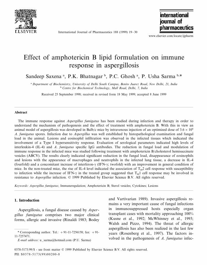
Fig. 3. Changes in serum creatinine in Balb/c mice treated withAmBDOC or ABCV. Each treatment group consisted of fourmice. Values are expressed as mean of serum creatinine (mg/dl) of two experiments9S.E.M.
uncoated 300 mesh copper grids and doublestained with uranyl acetate (10 min) and leadcitrate (10 min). Sections were examined using aPhilips CM10 transmission electron microscope.
2.9. Indirect ELISA
Indirect ELISA was carried out as described byBanerjee et al. (1995) with some modifications. Inbrief, antigen-coated plates were incubated with1:25 diluted serum for 3 h at 37°C. Protein Aperoxidase was then added for detection of IgGantibodies by use of substrate O-phenylenedi-amine. Colour developed was measured againstblank at 490 nm in a NUNC ELISA reader.

S. Saxena et al. / International Journal of Pharmaceutics 188 (1999) 19–3024
Fig. 4. Acute mortality of mice treated with AmBDOC orABCV. Numbers at the top of each bar indicate the number ofdeaths per number of mice injected within 2 h of administra-tion of drug. Each treatment group consisted of five mice.Values are expressed as mean of two experiments9S.E.M.
ity between non-treated and treated survival ra-tios on each day. Sera from four mice wereassayed in duplicates on each day for antibody orcytokine levels. Values were expressed as themean of two readings of two experiments9S.E.M.
3. Results
3.1. Animal model for aspergillosis
The survival of mice was dependent on the dose
Fig. 5. Effect of different doses of ABCV on survival of A.fumigatus infected Balb/c mice. Amphotericin B treatment wasgiven 24 h after injection of 3.6×10⁶ spores. × , infectedmice; �, infected mice treated with 2 mg/kg wt ABCV; �, 4mg/kg wt ABCV; �, 8 mg/kg wt ABCV; , 12 mg/kg wtABCV; �, 1 mg/kg wt AmBDOC. Each treatment groupconsisted of 20 mice. Values are expressed as mean of %survival of two experiments9S.E.M.
For each sample, duplicates were run and themean values were expressed for individualcytokines.
2.11. Statistical analysis of colony forming unitsand sur6i6al
The colony forming units data were statisticallyanalyzed by applying the Student’s t-test to checkfor heterogeneity between non-treated and treatedmeans. The survival data was analyzed using thex2-test on a 2×2 table to find out the heterogene-
S. Saxena et al. / International Journal of Pharmaceutics 188 (1999) 19–30 25
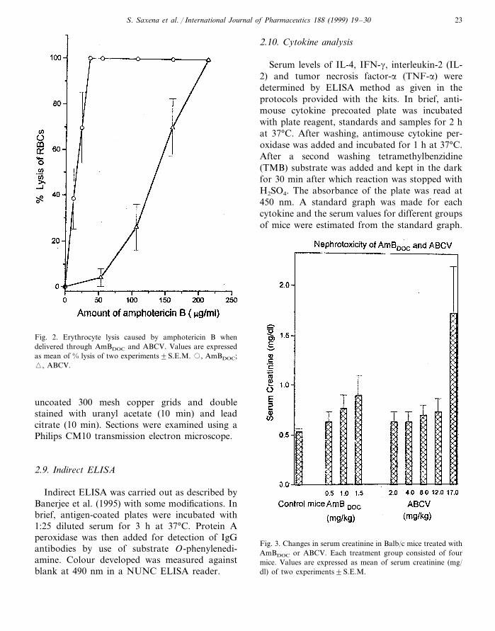
Fig. 6. Scanning electron micrographs of lung tissue of Balb/c mice on the 5th day. (a) In lung tissue of infected mice typical lesionswere observed in which fungal spores colonize and invade tissues, ×353. (b) Treated mice did not show lesions in the lung tissuewhich is suggestive of control of infection, ×353.
of spores injected (Fig. 1). A spore dose of 1.8×10⁷ resulted in acute infection with high mortality.On the 2nd and 3rd day, variation in the percentsurvival was observed between the two separatelycarried out experiments but all the mice died bythe 4th day. On other hand, when 3.6×10⁶ sporeswere administered, 25% of the infected animalssurvived even till the 15th day. The optimizeddose of 3.6×10⁶ spores was used for study ofimmune response to A. fumigatus. Aspergillus in-fection in the mice was well established by thepresence of eosinophils and lesions in infectedtissue with an increase in A. fumigatus specificantibodies. Table 2 shows the colony formingunits in different organs which reflect the dissemi-nation of this infection.
3.2. Toxicity of ABCV to Balb/c mice
Figs. 2–4 show the effect of free amphotericinB and ABCV on erythrocyte lysis, nephrotoxicityand acute toxicity in Balb/c mice respectively.ABCV was about six times less toxic thanAmBDOC to mouse erythrocytes. Nephrotoxicitywas also significantly reduced when amphotericinB was delivered using cholesterol hemisuccinatevesicles. Mice receiving 12 mg/kg wt ABCV hadlower serum creatinine levels in comparison to
mice administered with 1.5 mg/kg wt AmBDOC.The maximum tolerated dose was observed to be2 mg/kg wt via intravenous route for AmBDOC
and it increased to 17 mg/kg wt when cholesterolhemisuccinate vesicles were used for delivery ofamphotericin B. As we have reported earlier, thereduction in toxicity of amphotericin B whendelivered by way of cholesterol hemisuccinatevesicles is due to altered biodistribution (Wang etal., 1995; Saxena and Ghosh, 1998).
3.3. Sur6i6al, fungal load and general condition ofinfected and treated mice
Mice infected with A. fumigatus showed loss ofgait and suffered convulsions, from the 2nd to 3rdday and by the 5–6th day, 50% of the animalsdied. However, the remaining mice showed animprovement in their general condition and sur-vived until the 16th day. Mice treated with 12mg/kg wt ABCV showed a general loss of gaitand mild convulsions but 92.5% of these animalssurvived (Fig. 5). Moreover, mice in this grouprecovered earlier and by the 6th day, they wereobserved to be normal. Table 2 indicates colonyforming units isolated from various tissues oftreated and non-treated mice. Fungal load in in
S. Saxena et al. / International Journal of Pharmaceutics 188 (1999) 19–3026
fected mice was observed to be reduced by 99%with 12 mg/kg wt ABCV treatment. Whentreated with 2, 4 and 8 mg/kg wt ABCV, thedecrease in the fungal load was observed to be79, 86 and 92% respectively. Around 43% de-crease in fungal load was observed when treat-ment was done with 1 mg/kg wt AmBDOC. Sincemaximum therapeutic efficacy was achieved at
12 mg/kg wt, we chose this as the model treat-ment for all subsequent histopathological andimmunological experiments. The x2-test revealsthat there was heterogeneity in survival ratiosbetween non-treated and ABCV-treated micefrom the 4th day as indicated by the x2-value(5%) whereas the AmBDOC-treated group did notshow a significant difference at x2 (5%).
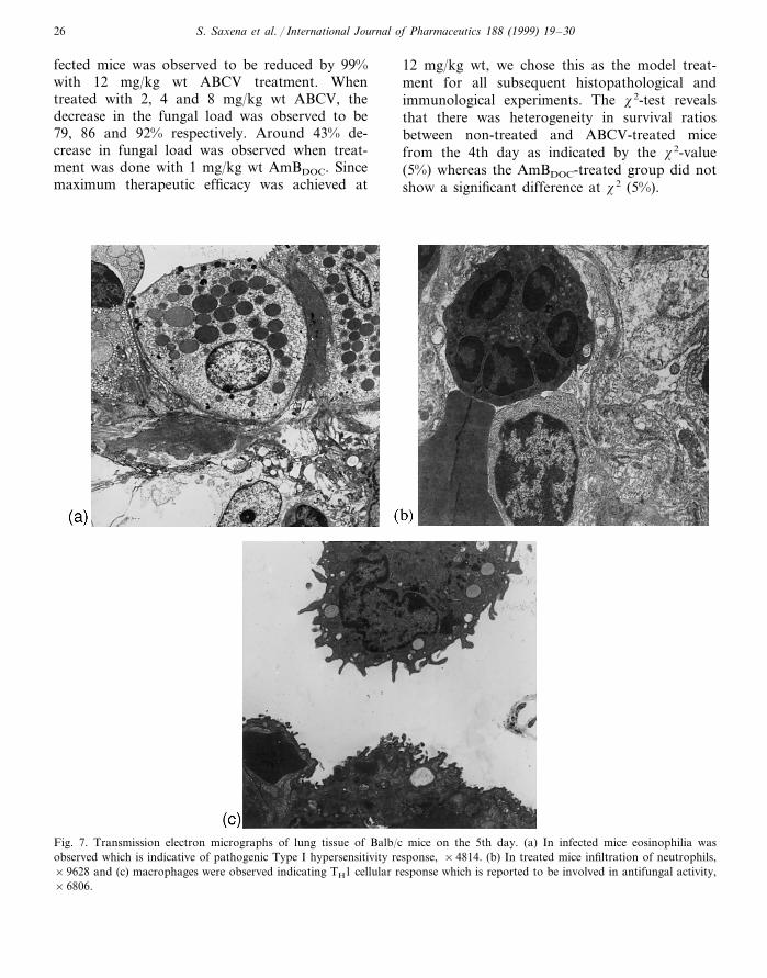
Fig. 7. Transmission electron micrographs of lung tissue of Balb/c mice on the 5th day. (a) In infected mice eosinophilia wasobserved which is indicative of pathogenic Type I hypersensitivity response, ×4814. (b) In treated mice infiltration of neutrophils,×9628 and (c) macrophages were observed indicating TH1 cellular response which is reported to be involved in antifungal activity,×6806.

S. Saxena et al. / International Journal of Pharmaceutics 188 (1999) 19–30 27
Fig. 8. A. fumigatus specific IgG antibodies levels in infectedand treated mice. ABCV treatment was 24 h after injection of3.6×106 spores. �, normal mice; × , infected mice; ,infected mice treated with 12 mg/kg wt ABCV. Four micewere sacrificed from each group on various days and serumobtained and evaluated for antibody levels in duplicate. Valuesare expressed as mean of optical density (O.D.) readings oftwo experiments9S.E.M.
the fungus in the form of neutrophils andmacrophages was observed.
3.5. Le6el of A. fumigatus specific antibodies ininfected and treated mice
The specific antibodies against diagnosticallyrelevant antigens of A. fumigatus were not detectedbefore the 9th day after spore challenge. Howeverspecific IgG antibodies were detected on the 9thand 12th days by ELISA. A sixfold increase inantibody level in non-treated mice (on the 9th day)as compared to around threefold increase in treatedmice (Fig. 8) was noticed. A heavy fungal load(Table 2) in non-treated mice corresponds well withincreased level of antibodies.
Fig. 9. IL-4 levels in infected and treated mice. ABCV treatmentwas 24 h after injection of 3.6×10⁶ spores. �, normal mice;× , infected mice; , infected mice treated with 12 mg/kg wtABCV. Four mice were sacrificed from each group on variousdays and serum obtained and evaluated for IL-4 levels induplicate. Values are expressed as mean of two experiments9S.E.M.
3.4. Histopathology of lung tissue of infected andtreated mice
Scanning electron micrographs confirm the util-ity of the infection model as lesions were demon-strated in lung tissue of mice infected with 3.6×10⁶
A. fumigatus spores. Such lesions were not detectedin normal and treated mice which confirms thattreatment with ABCV circumvented the spread offungus (Fig. 6). Transmission electron microscopyof infected lung tissue shows infiltration ofeosinophils suggesting a Type I hypersensitivityresponse due to severe infection of Aspergillus (Fig.7a). Fig. 7b,c shows a reduction in eosinophilia inthe treated group of mice and the host response to
S. Saxena et al. / International Journal of Pharmaceutics 188 (1999) 19–3028
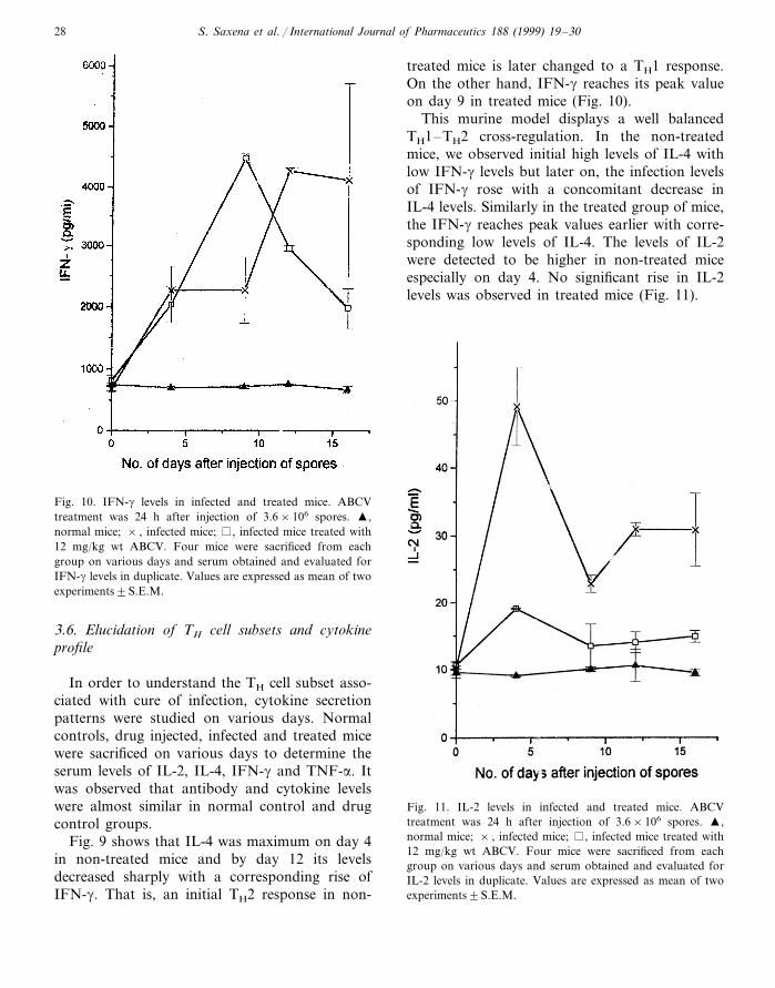
Fig. 10. IFN-g levels in infected and treated mice. ABCVtreatment was 24 h after injection of 3.6×10⁶ spores. �,normal mice; × , infected mice; , infected mice treated with12 mg/kg wt ABCV. Four mice were sacrificed from eachgroup on various days and serum obtained and evaluated forIFN-g levels in duplicate. Values are expressed as mean of twoexperiments9S.E.M.
treated mice is later changed to a TH1 response.On the other hand, IFN-g reaches its peak valueon day 9 in treated mice (Fig. 10).
This murine model displays a well balancedTH1–TH2 cross-regulation. In the non-treatedmice, we observed initial high levels of IL-4 withlow IFN-g levels but later on, the infection levelsof IFN-g rose with a concomitant decrease inIL-4 levels. Similarly in the treated group of mice,the IFN-g reaches peak values earlier with corre-sponding low levels of IL-4. The levels of IL-2were detected to be higher in non-treated miceespecially on day 4. No significant rise in IL-2levels was observed in treated mice (Fig. 11).
Fig. 11. IL-2 levels in infected and treated mice. ABCVtreatment was 24 h after injection of 3.6×10⁶ spores. �,normal mice; × , infected mice; , infected mice treated with12 mg/kg wt ABCV. Four mice were sacrificed from eachgroup on various days and serum obtained and evaluated forIL-2 levels in duplicate. Values are expressed as mean of twoexperiments9S.E.M.
3.6. Elucidation of TH cell subsets and cytokineprofile
In order to understand the TH cell subset asso-ciated with cure of infection, cytokine secretionpatterns were studied on various days. Normalcontrols, drug injected, infected and treated micewere sacrificed on various days to determine theserum levels of IL-2, IL-4, IFN-g and TNF-a. Itwas observed that antibody and cytokine levelswere almost similar in normal control and drugcontrol groups.
Fig. 9 shows that IL-4 was maximum on day 4in non-treated mice and by day 12 its levelsdecreased sharply with a corresponding rise ofIFN-g. That is, an initial TH2 response in non-
S. Saxena et al. / International Journal of Pharmaceutics 188 (1999) 19–30 29
4. Discussion
Many investigators have demonstrated in vivoand in vitro that TH1 cytokines are associatedwith resistance to fungal infections (Rex et al.,1991; Roilides et al., 1993; Nagai et al., 1995)while others observed that TH2 cytokines are pro-duced in susceptible mice (Romani et al., 1992;Cenci et al., 1997). However, not many studieshave been carried out on the immune responseduring treatment of infection which will lead to abetter understanding of the factors involved in thetreatment process. In the present communication,we have used ABCV for treating the infected miceand the immune response was evaluated duringinfection and therapy in order to understand therole played by various factors in suppressing orpromoting this fungal infection.
We have also observed that the TH2 cell re-sponse is associated with susceptibility during as-pergillosis. In the non-treated mice which showedheavy fungal load, low survival and lesions in theinfected tissue, a TH2 response with the presenceof eosinophils was observed. The presence ofeosinophilia in non-treated mice indicates Type Ihypersensitivity reaction to Aspergillus antigen. Ithas been reported in the literature that IL-4causes an increase in IgE levels leading to mastcell degranulation and release of mediators whichserves to attract a large number of eosinophils tothe site. Eosinophil activation leads to the releaseof a number of inflammatory mediators leading toexcessive tissue damage (Kuby, 1994). Thereforein the current study, increased levels of IL-4 andeosinophils observed in the non-treated mice cor-relate well with high mortality. Other investiga-tors have also reported lung eosinophilia duringA. fumigatus infection in mice (Kurup et al., 1992)and humans (Rosenberg et al., 1997).
Association of TH1 cellular response with resis-tance to A. fumigatus as reported by others alsoseems to be true in the present study (Cenci et al.,1997). In the treated mice, a rise in IFN-g levelscoincides with improvement in the condition ofmice (around the 6th day). Moreover, it wasobserved that eosinophilia and IL-4 levels werereduced significantly indicating the inhibition ofTH2 response and there was heavy infiltration of
macrophages and neutrophils. It is well docu-mented in literature that IFN-g brings about itsantifungal activity through activation of cells suchas macrophages and neutrophils (Kuby, 1994).
In the present study it was also observed thatthere was enhanced levels of TNF-a in treatedmice (unpublished data). It has been reported thatactivation of macrophages by IFN-g promotesTNF-a production (Kuby, 1994). Therefore it ap-pears that activation of macrophages and neu-trophils by IFN-g and TNF-a respectively incombination with amphotericin B bring abouteffective fungicidal action.
Cases of Aspergillus species causing invasive,life threatening infections in immunocompromisedhosts such as those suffering from AIDS andundergoing organ transplants continue to be re-ported (McWhinney et al., 1993; Morrison et al.,1994). Documented invasive aspergillosis has ahigh mortality rate surpassing 60% even withantifungal therapy (Rinaldi, 1983). Thereforethere is a need for safer and effective therapeuticmeasures. This understanding of the immune re-sponse during infection and treatment will pavethe way for combined therapy wherein the treat-ment is based not only on antifungal property butalso on modulation of immune response for bettermanagement of disease.
In the current report an infectious model foraspergillosis was developed and characterized byhistopathological and immunological parameters.The non-treated mice showed high mortality withheavy fungal load, poor general condition, lesionsin infected tissue, high levels of A. fumigatusantibodies and TH2 cellular responses. Treatedmice showing TH1 response were able to circum-vent the infection and showed an improved sur-vival rate.
Acknowledgements
Sandeep Saxena is grateful to the Governmentof India for financial assistance. Financial supportfrom the Department of Biotechnology andCouncil of Scientific and Industrial Research,Government of India is acknowledged.

S. Saxena et al. / International Journal of Pharmaceutics 188 (1999) 19–3030
References
Banerjee, B., Madan, T., Sharma, G.L., Prasad, H.K., Nath,I., Sarma, P.U., 1995. Characterization of glycoproteinantigen (45KD) of Aspergillus fumigates. Serodiagn. Im-munother. Infect. Dis. 7, 147–152.
Bodey, G.P., Vartivarian, S., 1989. Aspergillosis. Eur. J. Clin.Microbiol. Infect. Dis. 8, 413–437.
Cenci, E., Mencacci, A., Spaccapelo, R., Tonnetti, L., Mosci,P., Enssle, K.H., Puccetti, P., Romani, L., Bistoni, F.,1995. T helper cell type 1 (Th1)- and (Th2)- like responsesare present in mice with gastric candidiasis but protectiveimmunity is associated with Th1 development. J. Infect.Dis. 171, 1279–1288.
Cenci, E., Perito, S., Enssle, K.H., Mosci, P., Latge, J.P.,Romani, L., Bistoni, F., 1997. TH1 and TH2 cytokines inmice with invasive aspergillosis. Infect. Immun. 65, 564–570.
Clerici, M., Shearer, G.M., 1993. A TH1�TH2 switch is acritical step in the etiology of HIV infection. Immunol.Today 14, 107–111.
Kuby, J., 1994. Immunology, 2nd edn. W.H. Freeman, SanFrancisco, CA.
Kurup, V.P., Mauze, S., Choi, H., Seymour, B.W.P., Coff-man, R.L., 1992. A murine model of allergic bronchopul-monary aspergillosis with elevated eosinophils and IgE. J.Immunol. 148, 3783–3788.
Kurup, V.P., Seymour, B.W.P., Choi, H., Coffman, R.L.,1994. Particulate Aspergillus fumigatus antigens elicit a TH2
response in BALB/c mice. J. Allergy Clin. Immunol. 93,1013–1020.
Kurup, V.P., Choi, H., Murali, P.S., Resnick, A., Fink, J.N.,Coffman, R.L., 1997. Role of particulate antigens of As-pergillus in murine eosinophilia. Int. Arch. Allergy Im-munol. 112, 270–278.
Kusne, S., Torre-Cisneros, J., Manez, R., Irish, W., Martin,M., Fung, J., Simmons, R.L., Starzl, T.E., 1992. Factorsassociated with invasive lung aspergillosis and the signifi-cance of positive Aspergillus culture after liver transplanta-tion. J. Infect. Dis. 166, 1379–1383.
McWhinney, P.H.M., Kibbler, C.C., Hamon, M.D., Smith,O.P., Gandhi, L., Berger, L.A., Walesby, R.K., Hoffbrand,A.V., Prentice, H.G., 1993. Progress in the diagnosis andmanagement of aspergillosis in bone marrow transplanta-tion: 13 years experience. Clin. Infect. Dis. 17, 397–404.
Morrison, V.A., Haake, R.J., Weisdorf, D.J., 1994. Non-can-dida fungal infections after bone marrow transplantation:risk factors and outcome. Am. J. Med. 96, 497–503.
Nagai, H., Guo, J., Choi, H., Kurup, V., 1995. Interferon-gand tumor necrosis factor-a protect mice from invasiveaspergillosis. J. Infect. Dis. 172, 1554–1560.
Puccetti, P., Mencacci, A., Cenci, E., Spaccapelo, R., Mosci,P., Enssle, K.H., Romani, L., Bistoni, F., 1994. Cure ofmurine candidiasis by recombinant soluble interleukin-4receptor. J. Infect. Dis. 169, 1325–1331.
Rex, J.H., Benett, J.E., Gallin, J.I., Malech, H.L., DeCarlo,E.S., Melnick, D.A., 1991. In 6i6o interferon-g therapyaugments the in 6itro ability of chronic granulomatousdisease PMNs to damage Aspergillus hyphae. J. Infect. Dis.163, 849–852.
Rinaldi, M.G., 1983. Invasive aspergillosis. Rev. Infect. Dis. 5,1061–1077.
Roilides, E., Uhlig, K., Venzon, D., Pizzo, P.A., Walsh, T.J.,1993. Enhancement of oxidative response and damagecaused by human PMNs to Aspergillus fumigatus hypae bygranulocyte colony-stimulating factor and gamma inter-feron. Infect. Immun. 61 (4), 1185–1193.
Romani, L., Mocci, S., Bietta, C., Lanfaloni, L., Puccetti, P.,Bistoni, F., 1991. TH1 and TH2 cytokine secretion patternsin murine candidiasis: association of TH1 responses withacquired resistance. Infect. Immun. 59, 4647–4654.
Romani, L., Mencacci, A., Grohmann, U., Mocci, S., Mosci,P., Pucetti, P., Bistoni, F., 1992. Neutralizing antibody tointerleukin-4 induces systemic protection and T helper type1-associated immunity in murine candidiasis. J. Exp. Med.176, 19–25.
Rosenberg, M., Patterson, R., Mintzer, R., Cooper, B.J.,Roberts, M., Harris, K.E., 1997. Clinical and immunologiccriteria for the diagnosis of allergic bronchopulmonaryaspergillosis. Ann. Intern. Med. 86, 405–414.
Saxena, S., Ghosh, P.C., 1998. Pharmacokinetics of ampho-tericin B delivered through cholesterol hemisuccinate vesi-cles in normal and A. fumigatus infected mice. J.Antimicrob. Chemother., in press.
Saxena, S., Khan, J.A., Ghosh, P.C., 1998. Toxicity andtherapeutic efficacy of amphotericin B delivered throughcholesterol hemisuccinate vesicles in treatment of experi-mental murine aspergillosis. J. Antimicrob. Chemother. 42,635–642.
Walsh, T.J., Pizzo, P.A., 1994. Aspergillosis. In: Hoeprich,P.D., Jordan, C., Ronald, A.R. (Eds.), Infectious Diseases,5th edn. Lippincott, Philadelphia, PA, pp. 541–547.
Wang, L.H., Fielding, R.M., Smith, P.C., Guo, L.S.S., 1995.Comparative tissue distribution and elimination of ampho-tericin B colloidal dispersion (Amphocil) and fungizoneafter repeated dosing in rats. Pharm. Res. 12, 275–283.
.


![Aspergillosis - Youngstown State Universitypeople.ysu.edu/~crcooper01/Aspergillosis[1]- Katie Jacquie Qazi.pdf•People with Aspergillosis are in three distinct groups •Healthy immune](https://static.fdocuments.us/doc/165x107/5e3883b0e2f2970b7b1c24ad/aspergillosis-youngstown-state-crcooper01aspergillosis1-katie-jacquie-qazipdf.jpg)