Edita: Universidad de Extremadura - core.ac.uk · al Consejo de Departamento de Fisiología que la...
248
-
Upload
nguyenkhue -
Category
Documents
-
view
213 -
download
0
Transcript of Edita: Universidad de Extremadura - core.ac.uk · al Consejo de Departamento de Fisiología que la...


Edita: Universidad de Extremadura Servicio de Publicaciones Caldereros 2. Planta 3ª Cáceres 10071 Correo e.: [email protected] http://www.unex.es/publicaciones

Universidad de Extremadura
Escuela Universitaria de Enfermería y Terapia Ocupacional.
Departamento de Fisiología.
INFLAMACIÓN Y ENVEJECIMIENTO EN EL MÚSCULO LISO.
PAPEL PROTECTOR DE LA MELATONINA
Memoria presentada por el licenciado Pedro Julián Gómez Pinilla para optar al grado de Doctor por la Universidad de Extremadura y dirigida por los doctores María José Pozo Andrada y Pedro Javier Camello Almaraz
Cáceres, Marzo 2007


Modelo1 Asunto: Rtdo. Impreso de Conformidad Defensa Tesis para su Conocimiento y Difusión Destinatario: Sr. Director de Departamento
Como Director/es de la Tesis doctoral titulada: �Inflamación y envejecimiento del músculo liso. Papel protector de la melatonina� realizada por D/Dª Pedro Julián Gómez Pinilla, en el Departamento de Fisiología, de la cual se adjuntan dos ejemplares para el cumplimiento de lo establecido en el artículo 141.1 de los Estatutos de la Universidad de Extremadura.
INFORMO/INFORMAMOS al Consejo de Departamento de Fisiología que la elaboración de la Tesis ha concluido y que la misma cumple con los criterios de calidad necesarios para que el doctorando pueda optar al Título de Doctor, por lo que:
SOLICITO/SOLICITAMOS del Consejo de Departamento que otorgue su conformidad para la presentación de la Tesis a la Comisión de Doctorado.
Cáceres, a 4 de Septiembre de 2006
Vº Bº Vº Bº Fdo. Pedro Javier Camello Almaraz Fdo. María José Pozo Andrada
Comisiónde
DoctoradoU
nive
rsid
ad d
e Ex
trem
adur
a


Durante la realización de la presente tesis he disfrutado una beca predoctoral financiada por la Junta de Extremadura (FEDER). Además, este trabajo ha sido sufragado por los proyectos SAF2001-0295 y BFU 2004�0637 del Ministerio de Ciencia y Tecnología, 2PR03A020 de la consejería de Infraestructura y desarrollo tecnológico de la Junta de Extremadura y 2003/62 de la Consejería de Sanidad y Consumo de la Junta de Extremadura.

Agradecimientos
Me gustaría aprovechar esta oportunidad para manifestar mi agradecimiento y cariño a
Che. Por dirigir mi tesis y por su cariño hacia todos los que la rodeamos, más bien los que
convivimos con ella. Muchas gracias por tus observaciones y reflexiones juiciosas y sopesadas,
por tu dirección, eficacia y por tus muestras de amistad, compresión y generosidad. Sin
embargo su actitud más admirable es la perseverancia que posee. Gracias maestra.
Mi agradecimiento se dirige también al Dr. Pedro Javier Camello Almaraz, quien tan
sabiamente me ayudó a dar los primeros pasos en el mundo de la investigación. Tu ayuda
técnica fue muy importante para el desarrollo de la presente tesis. Su paciencia y cordura es
muy importante en este laboratorio. Gracias maestro.
Quisiera subrayar mi gratitud hacia la Dra Cristina Camello Almaraz porque a su
manera también ha colaborado en la realización de esta Tesis. A Rosario porque me ha ayudado
muchísimo en el trabajo experimental, porque siempre estás dispuesta a ayudar y también por la
paciencia que en ocasiones tiene. Una gran parte de esta tesis también es tuya. A Sara porque
me ayudó mucho más de lo que ella se imagina, ya que me explicó muchos trucos y porque me
abrió muchas puestas. Ella siempre está en nuestra mente. A Bea que últimamente comparte
mucho tiempo con nosotros y me hace recordar los tiempos en los que yo estaba empezando.
Aunque se nos va a Barcelona. También a Fran por los ratos pasados en el �imaging�. Espero
que seas feliz entre nosotros. Gracias amigos.
A los doctores del Departamento de Fisiología de la Facultad de Veterinaria; Ginés
Salido, José Antonio Pariente, Antonio González, Juan Antonio Rosado, Ana Lajas y José
Antonio Tapia, por los momentos que hemos vivido dentro y fuera del laboratorio. A los
becarios; María Granados, Pedro Cosme, JJ, Alba, Inés, Lauro y Miguel Ángel, que aunque
hemos trabajado poco tiempo juntos de vez en cuando nos solidarizamos con el gremio. Quiero
aprovechar esta oportunidad para agradecer a Sergio Damián Paredes su desinteresada ayuda
con la determinación de melatonina. A Puri y Mercedes les agradezco mucho su apoyo técnico.
También quiero dirigir mi agradecimiento a los compañeros del laboratorio de Lund por ser tan
suecos. Especialmente a María Gómez porque allí se convirtió en la otra Che. Gracias amigos.
id10590906 pdfMachine by Broadgun Software - a great PDF writer! - a great PDF creator! - http://www.pdfmachine.com http://www.broadgun.com

PJ Gómez-Pinilla
A mis amigos Juan Nogales, el kimiko, mi primo Joaquín, Andrea, Mireia y David,
Juan Marín, Juanma, María del Mar, porque hemos pasados realmente muy buenos momentos
juntos, creo que nunca olvidaremos el día de la caja del gato y el día del karaoke. A mis
compañeros de piso Nestor, David, y Miguel por preocuparse por mi paradero y por
aguantarme cuando llego del laboratorio. También a mis compañeros de promoción Thales,
Isaac, Eusebio, Ernesto y muchos más que seguro que me olvido por compartir aquellos
maravillosos años de estudiante. Gracias amigos.
Gracias a mis padres, porque me dieron los medios y la confianza necesarios para
poder elegir y desarrollar libremente todo lo que he conseguido tanto a nivel personal como
profesional. Ya sabéis que ésta es también vuestra tesis. Gracias también al resto de mi familia,
porque vuestro apoyo y amistad han sido muy importantes para mí. Especialmente a mi
sobrino, ya que si no fuera por él, últimamente sólo me dedicaría a trabajar en esta tesis.
Gracias familia.
Y finalmente a todas aquellas persona que quedan en el tintero y que de una forma u
otra han colaborado en la realización de esta tesis.
También quiero agradecer a la Escuela Universitaria de Enfermería y Terapia
ocupacional de la Universidad de Extremadura por permitirme el uso de las instalaciones.

�La verdad es dificil de encontrar, por eso la buscamos tanto�
Santiago Ramón y Cajal, 1892
id10707750 pdfMachine by Broadgun Software - a great PDF writer! - a great PDF creator! - http://www.pdfmachine.com http://www.broadgun.com


A mis Padres, ellos saben el por qué.


ÍNDICE
1. Resumen. ..��������������������������������. 21
2. Introducción ������������������������������...� 24
2.1 Fisiología de la vesícula biliar ���������������������..�.�. 27
2.2 Fisiología de la vejiga urinaria ...��..�������������������.... 28
2.3. Contracción del músculo liso �.����������������������.... 32
2.3.1 El aparato contráctil en el músculo liso ��������...������.�����.. 32
2.3.2 Mecanismo de contracción del músculo liso ������������..������.. 33
2.4. Homeostasis del ión Ca2+ en el músculo liso �.�����������������. 34
2.4.1 Entrada de iones Ca2+ desde el medio extracelular ���������������.�. 34
2.4.2 Liberación de calcio desde los depósitos intracelulares �������������..� 35
2.4.3 Mecanismos para la extrusión de Ca2+ �.�����������������.�..� 37
2.4.4 Recaptación de Ca2+ hacia los depósitos ��..�����������������.. 37
2.5. Sensibilización al calcio �������������������..������.� 38
2.5.1 Fosforilación de MLC20 independiente de calcio ���... ������������... 39
2.5.2 Inhibición de la MLCP ��������������������������� 39
2.5.3 Vías de señalización reguladoras de la sensibilidad al calcio ������������.. 41
2.5.3.1 Activación de la GTPasa monomérica de bajo peso molecular Rho A ��.����..� 42
2.5.3.2 Cinasa asociada a RhoA (ROCK) .........................................................��.����..� 42
2.5.3.3 Activación de la proteína cinasa C (PKC) ....................................................����..� 43
2.5.4 Desensibilización al calcio ������������������������..� 44
2.5.5 Expresión y participación de las vías de sensibilización
al calcio en el músculo liso. ���������������������.................. 44
2.5.6 Alteración de las vías de sensibilización al calcio
en el músculo liso. �������������������������................ 44
id22705859 pdfMachine by Broadgun Software - a great PDF writer! - a great PDF creator! - http://www.pdfmachine.com http://www.broadgun.com

2.6. Envejecimiento ��...���������������������������.. 45
2.6.1 Teorías del envejecimiento ������������������������..� 46
2.6.1.1 La teoría de los radicales libres ����������������������� 46
2.6.1.2 Teoría mitocondrial del envejecimiento ������.�������������.. 47
2.6.1.3 Teoría basada en la longitud de los telómeros ..����������������... 48
2.6.1.4 Teoría genética ����������������������������.� 48
2.6.2 Envejecimiento y señalización celular. ��������������������.... 48
2.6.3 Enfermedades relacionadas con el envejecimiento ����������������. 49
2.6.4. Efecto del envejecimiento sobre la vejiga urinaria ���������������� 50
2.6.4.1 Efectos sobre el ciclo de la micción �..�����.��������������. 51
2.6.4.2 Efectos sobre la inervación de la vejiga urinaria ����������������. 51
2.6.4.3 Efectos del envejecimiento sobre el detrusor �������........................................... 52
2.6.4.4 Tatamiento de las alteraciones de la
vejiga urinaria asociadas al envejecimiento �...����������������.. 53
2.6.5. El envejecimiento del tracto gastrointestinal ������������������. 54
2.6.5.1 Efectos sobre el sistema nervioso entérico ������������������.. 54
2.6.5.2 Efectos del envejecimiento sobre el músculo liso
del tracto gastrointestinal �������������������������. 55
2.6.5.3 Efectos del envejecimiento sobre la vesícula biliar ............................................................. 55
2.7. La colecistitis acalculosa ...������������������������...� 56
2.7.1 Modelos experimentales ��������������������������.. 56
2.7.2 Agentes pro-inflamatorios �������������������������... 56
2.7.3 Efectos de la colecistitis sobre la inervación de la vesícula biliar ������.����.. 57
2.7.4 Efectos de la colecistitis sobre el músculo liso de la vesícula biliar ........................................ 58
2.7.5 Posibles estrategias terapeúticas para la colecistitis acalculosa. �..���������.. 58

2.8 La melatonina ������������������...�����������.� 59
2.8.1 Melatonina: Síntesis y secreción .����������������������.� 59
2.8.2 Receptores para la melatonina ������������������������. 59
2.8.3 Acciones biológicas de la melatonina ��������������������.� 60
2.8.4 Capacidad antioxidante de la melatonina �....�����������������... 60
2.8.5 Posibles indicaciones terapeúticas de la melatonina ..��������������� 60
2.8.5.1 Indicaciones terapeúticas de la melatonina
en el tracto gastrointestinal ................................................................................................... 61
2.8.5.2 Indicaciones terapeúticas de la melatonina frente
a alteraciones del tracto urinario inferior �������������������. 62
2.8.6 Melatonina, envejecimiento y enfermedades
relacionadas con la edad ��������������������������.. 63
2.8.7 Melatonina y colecistitis ��������������������������. 64
Bibliografría ...................................................................................................................................... 64
3. Justificación y objetivos ��������������������������� 87
4. Meterial y métodos ����������������������������... 91
5. Resultados ������������������������������...� 95
5.1 Trabajo experimental I:
Effect of melatonin on age-associated changes in guinea pig urinary bladder function ��.. 97
5.2 Trabajo experimental II:
Ageing impairs neurogenic contraction in guinea pig urinary bladder.
The role of oxidative stress and melatonin ������������������...... 107
5.3 Trabajo experimental III:
Melatonin recovers impaired contractility in aged guinea pig urinary bladder �����... 125
5.4 Trabajo experimental IV:

Calcium extrusion in aged smooth muscle cells ����������������...... 143
5.5 Trabajo experimental V:
Melatonin treatment reverts aged-related changes in guinea pig gallbladder
Neuromuscular transmission and contractility �����������������.... 163
5.6 Trabajo experimental VI:
Effect of melatonin on gallbladder neuromuscular function in acute cholecystitis ���..... 183
5.7 Trabajo experimental VII:
Changes in guinea pig gallbladder smooth muscle calcium
homeostasis by acute acalculous cholecystitis �����������������.... 199
5.8 Trabajo experimental VIII:
Protective effect of melatonin on calcium homeostasis
and contractility in acute cholecystitis ����������..����������... 215
6. Discusión general .................................................................................................................... 233
7. Conclusiones �...����������������������������� 239

Abreviaturas
ABREVIATURAS
[Ca2+]i: Concentración intracelular de calcio. 2-APB: 2-aminoetoxidifenilborano.
ACh: Acetilcolina.
ADP: Adenosina-5�- difosfato.
AMPc: Adenosina 3´,5�-monofosfato cíclico. ANOVA: Análisis de varianza. ATP: Adenosina-5�- trifosfato.
ADP: Adenosina difosfato.
BK: Canales de K+ de gran conductancia sensibles a Ca2+.
cADPR: ADP ribosa cíclica. CaM: Calmodulina.
CaMKII: Proteína kinasa II dependiente de Ca2+-calmodulina. CBDL: Ligadura del conducto biliar común. CCE: Entrada capacitativa de calcio.
CCK: Colecistokinina.
CGRP: Péptido relacionado con el gen de calcitonina. CICR: Liberación de calcio inducida por calcio
CO: Monóxido de carbono. DAG: Diacilglicerol.
DHP: Dihidropiridina.
DMSO: Dimetilsulfóxido.
DTT: Ditiotreitol.
EC50: Dosis efectiva 50. EGTA: Ácido etilénglicol-bis (â-aminoetiléter)N,N,N�,N�-tetraacético. ES: Solución enzimática.. GDP: Guanosina-5-difosfato.
GMPc: Guanosina- 3�,5�-monofosfato cíclico. GTP: Guanosina-5-trifosfato.
IP3: Inositol 1,4,5-trifosfato. IP3R: Receptor de inositol 1,4,5-trifosfato.
KATP: Canales de K+ sensibles a metabolitos como el ATP.
K-HS: Solución Krebs-Henseleit.
MLC: Cadena ligera de miosina
MLC20: Cadena ligera reguladora de miosina de 20 kDa. MLC17: Cadena ligera esencial de miosina de 17 kDa.
MLCK: Kinasa de la cadena ligera de miosina.
MLCP: Fosfatasa de la cadena ligera de miosina.
NKA: Neuroquinina A
id10846484 pdfMachine by Broadgun Software - a great PDF writer! - a great PDF creator! - http://www.pdfmachine.com http://www.broadgun.com
PJ Gómez-Pinilla
NO: Óxido nítrico. NPY: Neuropéptido Y PACAP: Polipéptido activador de la adenilato ciclasa pituitaria. PBS: Tampón fosfato salino. Pi: Fósforo inorgánico. PKA: Proteína kinasa dependiente de AMPc. PKC: Proteína kinasa C. PKG: Proteína kinasa dependiente de GMPc. PLC: Fosfolipasa C.
ROCC: Canales de calcio operados por receptor.
RyR: Receptor de rianodina.
SERCA: Bomba de Ca2+ dependientes de ATP y Mg2+.
SMOCC: Canales de calcio operados por segundos mensajeros. SOCC: Canales de calcio operados por depósito. SP: Substancia P. STOCs: Corrientes espontáneas salientes transitorias. TPS: Tapsigagina.
TRPC: Familia de proteínas homólogas a las proteínas TRP. VIP: Péptido intestinal vasoactivo. VOCC: Canales de calcio operados por voltaje.
1. Resumen
id10955484 pdfMachine by Broadgun Software - a great PDF writer! - a great PDF creator! - http://www.pdfmachine.com http://www.broadgun.com


Resumen 23
Son muy escasos los estudios encaminados a analizar los efectos del envejecimiento sobre los órganos y tejidos. En la presente tesis se analizan los efectos del envejecimiento sobre la vejiga urinaria y la vesícula biliar, para ello se han utilizado cobayas envejecidos como modelo animal de envejecimiento. Así, se ha encontrado que el envejecimiento provoca inestabilidad de la vejiga urinaria junto con una disminución en la respuesta contráctil. Una vez realizados los estudios funcionales, se procedió a analizar los efectos del envejecimiento sobre la inervación de la vejiga y sobre el detrusor. Encontrándose que los animales envejecidos presentan una denervación funcional de las fibras excitatorias y una hiperactivación de los componentes relajantes, cambios que podrían explicar la menor capacidad contráctil de la vejiga urinaria envejecida. A nivel del detrusor, el envejecimiento provoca una sobre carga de calcio y una mayor capacidad de movilización de este ión, que se ve favorecida por las alteraciones en los mecanismos de extrusión. Además, el envejecimiento disminuye la contribución de los mecanismos excitatorios independientes de los incrementos en la concentración de calcio citosólico junto con una alteración de la maquinaria contráctil que provoca una menor contractilidad de la vejiga urinaria envejecida y consecuentemente la aparición de volumen residual.
El envejecimiento del tracto gastrointestinal es un hecho contrastado pero muy poco estudiado y mucho menos en la vesícula biliar. Nosotros, en la presente tesis, hemos analizado mecanísticamente los efectos del envejecimiento sobre la inervación y el músculo liso de la vesícula biliar. Aquí, el envejecimiento produce una denervación funcional y un cambio en el componente nervioso relajante de la vesícula biliar. A nivel del músculo liso la entrada de calcio y el contenido de F-actina es menor en animales envejecidos, hechos que explican la presencia de una menor capacidad contráctil.
Debido a sus propiedades antioxidantes, los animales envejecidos se trataron con melatonina. Dicho tratamiento resultó en la reducción del estrés oxidativo y en la normalización tanto de la función, inervación y contractilidad de la vejiga urinaria y de la vesícula biliar de cobaya. Indicando que el daño tisular asociado al envejecimiento probablemente es debido a un incremento en el estrés oxidativo y/o reducción en los mecanismos antioxidantes.
La colecistitis acalculosa es una situación fisiopatológica con una incidencia y grado de mortalidad creciente. Para analizar el impacto de la inflamación sobre la vesícula biliar hemos utilizado un modelo experimental muy aceptado que es la ligadura del conducto biliar común. El mantenimiento de dicha ligadura durante dos días incrementó la acumulación de radicales libres y redujo los mecanismos celulares antioxidantes. Este incremento en el estrés oxidativo podría explicar la reducción de la respuesta contráctil de las vesículas colecistíticas. Sin embargo, en la presente tesis se han analizado más aspectos del músculo liso vesicular, encontrando que la inflamación afecta a la señalización mediada por calcio y la maquinaria contráctil. Además, la colecistitis produce una denervación funcional de las fibras eferentes junto con una mayor excitabilidad de la inervación aferente contráctil.
El tratamiento con melatonina en éstos animales reduce el estrés oxidativo y a pesar de normalizar algunos parámetros como la señal de calcio, no recupera la contractilidad del
id10998484 pdfMachine by Broadgun Software - a great PDF writer! - a great PDF creator! - http://www.pdfmachine.com http://www.broadgun.com

24 PJ Gómez-Pinilla
órgano. Un hecho importante del tratamiento con melatonina es que aumentó la velocidad de recuperación de la contractilidad de la vesícula biliar tras la retirada de la obstrucción.
Nuestros resultados demuestran que la melatonina es una herramienta farmacológica muy interesante para el tratamiento de alteraciones asociadas a un incremento en el estrés oxidativo como son el envejecimiento y la inflamación.

2. Introducción
id11101359 pdfMachine by Broadgun Software - a great PDF writer! - a great PDF creator! - http://www.pdfmachine.com http://www.broadgun.com


Introducción 27
2.1. Fisiología de la vesícula biliar
La vesícula biliar es un órgano del tracto gastrointestinal que durante los períodos interdigestivos produce bilis concentrada a partir de bilis hepática diluida. Para ello, absorbe la mayoría del agua y electrolitos de la secreción hepática, produciendo un líquido viscoso y de color dorado. Durante esta fase de almacenamiento la vesícula se relaja, mientras que posteriormente y en respuesta a la ingesta de alimentos, la vesícula se contrae y vierte su contenido al duodeno donde participa en el proceso digestivo.
En el cobaya, la vesícula biliar tiene unas dimensiones de 1 centímetro de largo y 0.5 de ancho aproximadamente y se encuentra localizada en la superficie ventral del hígado. Este órgano se puede dividir en tres áreas: fundus, que es la porción más distal, cuerpo, localizado a nivel central y cuello, estrechamiento que desemboca en el conducto cístico, que al unirse con el conducto hepático forma el conducto biliar común que desemboca en el duodeno a través del esfínter de Oddi..
La pared de la vesícula biliar está formada por tres capas: la mucosa interna, la muscular y la serosa externa . El mayor aporte arterial a este órgano procede de la arteria cística y aunque parte de la sangre drena a la rama cística de la vena porta, la mayoría de las venas vesiculares vierten su contenido al interior de los capilares hepáticos. El nervio simpático esplácnico y el parasimpático vago inervan la vesícula biliar y los conductos biliares.
El vaciado y posiblemente el llenado de la vesícula está sujeto a un control neural que incluye fibras nerviosas eferentes, aferentes y el plexo intrínseco, localizado en las capas submucosa y subserosa. Los nervios eferentes son aparentemente todos colinérgicos (Talmage et al.,1992) pero también expresan péptido vasoactivo intestinal (VIP), neuropéptido Y (NPY), somatostatina, polipéptido pituitario activador de la adenilato ciclasa (PACAP), takininas y óxido nítrico sintasa, si bien esta enzima nunca se expresa en neuronas que también sintetizan VIP (Mawe et al.,1997). Además, en la vesícula existen fibras nerviosas aferentes que contienen sustancia P (SP) y péptido relacionado con el gen de la calcitonina (CGRP) (Goehler et al.,1988).
La hormona colecistocinina (CCK), liberada en respuesta a la ingesta de alimentos, y el neurotransmisor acetilcolina (ACh) son los principales agentes que promueven contracción de la vesícula biliar.
La CCK induce una contracción miógena mediada por su unión a receptores de tipo A del sarcolema (Deweerth et al.,1993). Estos receptores se encuentran acoplados a la proteína Gi3, que a su vez activa a la fosfolipasa C específica de fosfatidilinositoles (PI-PLC). Dicha activación
Figura 1. Anatomía del árbol biliar.
id11171937 pdfMachine by Broadgun Software - a great PDF writer! - a great PDF creator! - http://www.pdfmachine.com http://www.broadgun.com

28 PJ Gómez-Pinilla
genera los mensajeros intracelulares inositol 1,4,5-trifosfato (IP3), que libera calcio desde depósitos intracelulares, y diacilglicerol (DAG), que activa la PKC. Otra fuente de DAG en respuesta a la CCK es el generado a partir del ácido fosfatídico resultante de la degradación de la fosfatidilcolina por la fosfolipasa D (Alcon et al.,2002). La contracción inducida por CCK también está parcialmente mediada por la activación de la entrada de calcio a través de canales de tipo L (Alcon et al.,2000). Además, la CCK actúa presinápticamente en los ganglios intramurales incrementando la liberación de acetilcolina desde terminales vagales (Mawe,1991).
En el caso de la ACh, la contracción tiene lugar tras su unión a receptores presentes en la membrana de las células musculares. Dicha contracción depende de la entrada de Ca2+ a través de canales de tipo L (Alcon et al.,2000) y es mediada por receptores muscarínicos, principalmente el subtipo M3. Las señales intracelulares involucradas en la contracción inducida por ACh son la generación de IP3 y DAG por acción de la PLC, la activación de la PKC, la fosforilación en tirosina y la inhibición de la síntesis de AMPcíclico (Alcon et al.,2000).
Durante la fase de llenado que ocurre en los períodos de ayuno, la vesícula sufre un proceso de dilatación. Esta expansión se puede producir de forma pasiva, debido a la existencia de componentes fibroelásticos en la pared de la vesícula, o de forma activa, mediante la relajación de la estructura. Esta relajación activa puede estar mediada por incrementos en los niveles de AMPc inducidos por los neurotransmisores VIP y PACAP (Dahlstrand et al.,1989; Ryan y Ryave,1978), por elevaciones en el GMPc en respuesta al óxido nítrico, y por CO (Alcon et al.,2001b). Recientemente, en nuestro laboratorio se han descrito los efectos inhibitorios del AMPc sobre la señal de Ca2+ y la contracción asociada a ésta (Morales et al.,2004).
2.2. Fisiología de la vejiga urinaria
La uretra y la vejiga urinaria forman el denominado tracto urinario inferior, que junto con el tracto urinario superior (riñones y uréteres) conforman el aparato urinario. En los riñones la producción de orina es constante como resultado del filtrado glomerular en las nefronas. Esta orina pasa a la vejiga a través de los uréteres y en ella se almacena hasta alcanzar un umbral de llenado, y posteriormente se elimina a través de la uretra. La vejiga urinaria es un órgano globoso, de unos 2 cm de largo por 1 cm de ancho en el caso del cobaya, con el eje mayor orientado caudoventralmente. Desde un punto de vista funcional, puede dividirse en cuerpo y base (Elbadawi,1996). La base de la vejiga comprende las estructuras anatómicas situadas por debajo del nivel de los orificios uretrales, mientras que el cuerpo se sitúa por encima de éstos. La zona de la base localizada en la cara dorsal comprendida entre la desembocadura de los uréteres y el orificio inferior que se comunica con la uretra (meato uretral), se denomina trígono vesical debido a su forma de triángulo invertido.
Pre
sión
de
la
vejig
a
Fase de llenado Fase de vaciado
Detrusor
Uretra
Relajado Relajado Relajado
Contraída Contraída ContraídaSe relaja
Se contrae
Control Simpático Simpático SimpáticoParasimpático
Pre
sión
de
la
vejig
a
Fase de llenado Fase de vaciado
Detrusor
Uretra
Relajado Relajado Relajado
Contraída Contraída ContraídaSe relaja
Se contrae
Control Simpático Simpático SimpáticoParasimpático
Figura 2. Esquema del ciclo de la micción.

Introducción 29
La vejiga urinaria desempeña por tanto dos funciones fisiológicamente relacionadas. En primer lugar, se encarga de almacenar la orina durante la fase de llenado, para una vez alcanzada la capacidad fisiológica, expulsar la orina durante la fase de vaciado. La alternancia de ambas fases se denomina ciclo de la micción.La pared de la vejiga urinaria está formada principalmente por tres capas o túnicas: la mucosa interna o urotelio, la capa muscular o detrusor y una capa externa compuesta por tejido conectivo denominada adventicia (Elbadawi,1996). El urotelio tiene, además de la función de barrera protectora frente a sustancias tóxicas de la orina, un papel activo en el almacenamiento y en el vaciado de la orina (revisado por Ferguson) (Ferguson,1999). Bajo el urotelio se dispone un plexo nervioso, de función principalmente aferente, que llega a alcanzar la base del urotelio y que es relativamente escaso en el cuerpo, progresivamente más denso en la base y particularmente rico en el trígono (Gabella y Davis,1998).
La capa muscular del cuerpo de la vejiga urinaria o detrusor está compuesta por fibras musculares lisas que se disponen circular y longitudinalmente al azar. Dicha localización permite a la vejiga acomodarse al contenido sin que se produzcan incrementos de presión intravesical (revisado por Turner) (Turner y Brading,1997). Sin embargo, en la base de la vejiga las fibras musculares lisas se disponen en 3 capas: longitudinal interna, circular media y longitudinal externa. En el trígono el músculo detrusor es más grueso y menos distensible, y consta de dos capas distintas. La profunda o músculo trigonal profundo que es indiferenciable del detrusor del cuerpo, mientras que el músculo trigonal superficial es morfológicamente distinto, más fino y con un engrosamiento a lo largo de su borde superior. En el cuello y el nacimiento de la uretra, las fibras musculares se disponen circularmente y se condensan para constituir el esfínter interno de la vejiga.
El músculo detrusor presenta actividad contráctil espontánea como resultado de la existencia de potenciales de acción miógenos, pero ésta no está asociada a episodios de micción y es inhibida por el urotelio. Aunque la actividad fásica está presente en todas las especies, existen diferencias interespecíficas e incluso depende de la parte de la vejiga urinaria considerada (Buckner et al.,2002). Aunque se considera que los potenciales de acción espontáneos del músculo detrusor son los responsables de la actividad contráctil fásica, no todos los potenciales de acción desencadenan contracción, lo que indica la existencia de un acoplamiento eléctrico pobre entre las células musculares del detrusor (revisado por Fry,) (Fry y Wu,1997).
Durante la fase de almacenamiento o llenado la orina llega a la vejiga de forma continua a través de los uréteres, el detrusor se acomoda paulatinamente al contenido sin que haya un aumento significativo en la presión intravesical a pesar del llenado. A lo largo de esta fase se mantienen cerrados el cuello vesical o esfínter interno, el músculo liso de la uretra y el músculo estriado que recubre la uretra, también denominado esfínter externo, único componente que se encuentra bajo
Ligamento medio
Uréter
Ligamentolateral
Cuerpo de la vejiga
Entrada delos uréteres
Esfinter uretralinterno
Esfinter uretralexterno
Trígono
Cuello
Próstata
Uretraprostática
Uretramembranosa
Ligamento medio
Uréter
Ligamentolateral
Cuerpo de la vejiga
Entrada delos uréteres
Esfinter uretralinterno
Esfinter uretralexterno
Trígono
Cuello
Próstata
Uretraprostática
Uretramembranosa
Figura 3. Anatomía del tracto urinario inferior.

30 PJ Gómez-Pinilla
control voluntario. Todo ello hace que la presión en la uretra sea mayor que la existente en el cuerpo de la vejiga urinaria alcanzándose así la continencia. El mantenimiento de la contracción durante la fase de almacenamiento se debe al sistema nervioso simpático. Éste a su vez inhibe los ganglios y, de este modo, disminuye la participación de las vías excitatorias parasimpáticas durante la fase de llenado (Degroat,1993).
La fase de vaciado se produce cuando la vejiga alcanza su máxima capacidad y el esfínter externo se relaja voluntariamente, aumenta la luz de la uretra y se relaja el cuello vesical, permitiendo así el vaciado. En esta fase es fundamental la contracción del detrusor, aumentando así la presión intravesical hasta hacerla superior a la existente en la uretra, condición necesaria para la expulsión de la orina. La contracción del detrusor es regulada por el sistema nervioso parasimpático.
La función del tracto urinario inferior de almacenar y liberar periódicamente la orina se controla por circuitos neurales complejos asentados en cerebro, médula espinal y ganglios periféricos. El cuerpo de la vejiga urinaria está inervado principalmente por el parasimpático pélvico, procedente del centro sacro de la micción, mientras que el cuello vesical y la uretra proximal son inervados por el nervio simpático hipogástrico y el esfínter externo se encuentra inervado por el nervio pudendo. En total el detrusor recibe señal de entre 2000-2500 neuronas, muchas de las cuales forman ganglios que pueden llegar a contener hasta más de cuarenta somas (Gabella,1990). La vejiga urinaria es un órgano que se encuentra muy inervado también por fibras sensoriales que contienen principalmente taquiquininas (Lecci et al.,2000).
En condiciones normales la ACh es el neurotransmisor predominante en la transmisión neuromuscular y, junto con la noradrenalina y el ATP, es el principal neurotransmisor que controla la actividad contráctil del músculo detrusor. La ACh tiene un papel excitador sobre el detrusor a través de la activación de receptores muscarínicos, donde los más comunes son M2 y M3, siendo los primeros más abundantes pero los segundos están más involucrados en la contracción (Wang et al.,1995). Los M3 inducen contracción por liberación de calcio desde depósitos a través de la vía intracelular PLC/IP3 y entrada de calcio desde el medio extracelular, mientras que los receptores de tipo M2 provocan contracción inhibiendo la adenilato ciclasa (Caulfield y Birdsall,1998). En el
Figura 4. Inervación del tracto urinario inferior.

Introducción 31
detrusor, también existen receptores presinápticos M1 y M4 cuyo papel predominante es la neuromodulación de la transmisión eferente (D'Agostino et al.,2000).
La distribución de los receptores adrenérgicos es heterogénea a lo largo de la vejiga (Gosling et al.,1999). Dicha distribución es muy importante desde el punto de vista funcional. El cuerpo contiene una alta densidad de -receptores cuya estimulación provoca una relajación mediada por incrementos en los niveles de AMPc durante la fase de llenado, mientras que contiene sólo algunos receptores adrenérgicos del tipo . Sin embargo, la base de la vejiga es rica en receptores adrenérgicos contráctiles y tiene un número pequeño de -receptores relajantes, lo que ayuda a mantener la continencia durante la fase de llenado (Andersson et al.,1999). El ATP es un neurotransmisor excitatorio que actúa a través de receptores que además son canales iónicos (P2X) y receptores acoplados a proteínas G (P2Y). En este caso hay muchas diferencias interespecíficas en la respuesta contráctil del detrusor y en el tipo de receptor involucrado (Longhurst y Levendusky,2001). El óxido nítrico (NO) se considera el candidato más probable para mediar parcial o totalmente las relajaciones del tracto urinario inferior, sin embargo, se desconoce el papel del NO en el detrusor (Andersson y Persson,1993; James et al.,1993). En las neuronas intramurales de la vejiga urinaria de cobaya se ha encontrado la coexistencia de la enzima óxido nítrico sintasa (NOS) con acetilcolinesterasa, VIP, CGRP y sustancia P (Zhou y Ling,1998).
Los neurotransmisores inhibitorios (VIP) y excitadores (neuropéptido Y (NPY), endotelina, angiotensina y prostanoides) también están presentes en neuronas que inervan el detrusor, pero existen muchas diferencias interespecíficas respecto a su presencia (Andersson y Wein,2004).
La inervación sensorial o vías aferentes discurren por los nervios pélvico, hipogástrico y pudendo y regulan tanto la continencia como la micción, pero también están implicadas en sensaciones térmicas y de dolor. Las fibras sensoriales, identificadas tanto bajo el urotelio como en el músculo detrusor, se dividen en mielinizadas (fibras-A), capaces de detectar distensiones pasivas, y fibras no mielinizadas, que responden a la irritación química de la mucosa y temperaturas frías y que participan en la nocicepción (Janig,1986).
Los principales neurotransmisores sensoriales de la vejiga son sustancia P (SP), neurokinina A (NKA), péptido relacionado con el gen de la calcitonina (CGRP), péptido activador de la adenilato ciclasa pituitaria (PACAP) y las encefalinas (Lecci et al.,2000). Los tres primeros (SP, NKA y CGRP) pertenecen al grupo de las taquiquininas, que tienen tanto función aferente o sensorial como función eferente periférica, actuando directamente sobre las células musculares de la vejiga mediante receptores de membrana específicos (Lecci et al.,2000): NK1 para la sustancia P, NK2 para NKA y CGRPr para el CGRP (Maggi et al.,1987). La localización de los receptores está muy relacionada con la función eferente. Los receptores NK1 y CGPRr se localizan en los vasos sanguíneos sub-uroteliales y del detrusor, donde las taquiquininas producen vasodilatación (Burcher et al.,2000). Además, los receptores NK2 se encuentran principalmente en las fibras musculares del detrusor, donde la NKA produce contracción (Burcher et al.,2000) y la subsiguiente micción . Las funciones de las taquiquininas están controladas por prostanoides y NO (Andersson y Hedlund,2002; Maggi et al.,1987).
Las taquiquininas también son liberadas por neuronas aferentes primarias sensibles a capsaicina (una molécula contenida en el pimiento rojo ampliamente utilizada como herramienta farmacológica específica para revelar la participación de vías aferentes) (Szallasi y Blumberg,1999). También el ATP tiene función aferente, al ser liberado desde la cara serosa del urotelio en respuesta a la distensión mecánica de la vejiga donde es capaz de activar los nervios sensoriales mediante
32 PJ Gómez-Pinilla
receptores del tipo P2X3 (Ferguson,1999). Andersson en 2004 realizó una amplia revisión sobre la inervación de la vejiga urinaria (Andersson y Arner,2004).
2.3. Contracción del músculo liso
Al igual que ocurre en otros tipos musculares, el aumento en la concentración de Ca2+ citoplasmático es el principal evento que inicia la contracción muscular lisa. En este apartado se describirá tanto el aparato contráctil presente en el músculo liso como los mecanismos que conducen a la contracción.
2.3.1 El aparato contráctil en el músculo liso
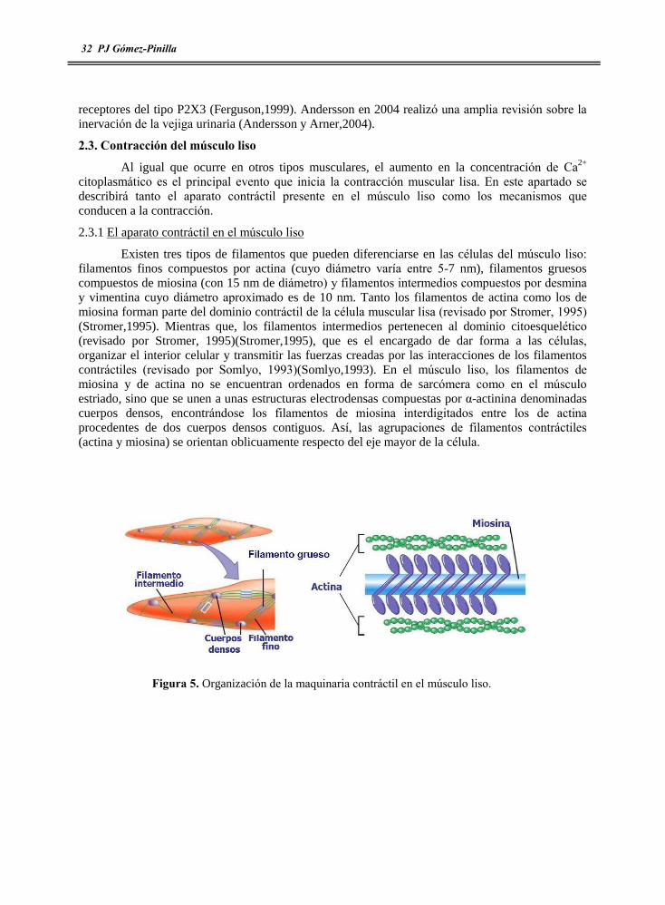
Existen tres tipos de filamentos que pueden diferenciarse en las células del músculo liso: filamentos finos compuestos por actina (cuyo diámetro varía entre 5-7 nm), filamentos gruesos compuestos de miosina (con 15 nm de diámetro) y filamentos intermedios compuestos por desmina y vimentina cuyo diámetro aproximado es de 10 nm. Tanto los filamentos de actina como los de miosina forman parte del dominio contráctil de la célula muscular lisa (revisado por Stromer, 1995) (Stromer,1995). Mientras que, los filamentos intermedios pertenecen al dominio citoesquelético (revisado por Stromer, 1995)(Stromer,1995), que es el encargado de dar forma a las células, organizar el interior celular y transmitir las fuerzas creadas por las interacciones de los filamentos contráctiles (revisado por Somlyo, 1993)(Somlyo,1993). En el músculo liso, los filamentos de miosina y de actina no se encuentran ordenados en forma de sarcómera como en el músculo estriado, sino que se unen a unas estructuras electrodensas compuestas por á-actinina denominadas cuerpos densos, encontrándose los filamentos de miosina interdigitados entre los de actina procedentes de dos cuerpos densos contiguos. Así, las agrupaciones de filamentos contráctiles (actina y miosina) se orientan oblicuamente respecto del eje mayor de la célula.
Figura 5. Organización de la maquinaria contráctil en el músculo liso.

Introducción 33
Los filamentos finos están compuestos por actina, una proteína globular (actina G) muy ubicua de 42 KDa que polimeriza para formar un filamento helicoidal de dos hebras (actina F) (Strzeleckagolaszewska et al.,1984). En las estrías de la hélice se insertan otro tipo de proteínas de naturaleza inhibitoria (caldesmonina y calponina) que regulan la actividad de la actina (Sobieszek y Bremel,1975). Los filamentos finos son polares, un extremo se inserta en un cuerpo denso mientras que el otro extremo del filamento queda libre y rodeado de filamentos gruesos compuestos por miosina. (Bond y Somlyo,1982).
Los filamentos gruesos están formados por moléculas de miosina, proteína de unos 480 KDa compuesta por la asociación no covalente de 6 cadenas proteicas, un par de cadenas pesadas y dos pares de cadenas ligeras (MLCs) (Sellers y Adelstein,1982). Las dos cadenas pesadas, de 200 KDa cada una, forman una cola helicoidal rígida cuya agregación forma el filamento de miosina, que aparece interdigitado entre filamentos finos de actina. En el extremo contrario a la cola existe una cabeza globular rodeada por dos cadenas ligeras distintas: una cadena ligera de 20 KDa denominada cadena ligera reguladora (MLC20) y una cadena de 17 KDa o cadena ligera esencial (MLC17). Cada cabeza globular posee un sitio de unión a la actina y capacidad para hidrolizar Mg2+-ATP activada por la unión a la actina. En la parte de las cadenas pesadas que sobresale del filamento de miosina se localizan dos zonas bisagra que permiten la rotación de la cabeza globular con respecto al filamento de miosina (Sellers y Adelstein,1982).
2.3.2 Mecanismo de contracción del músculo liso
El mecanismo básico para la generación de fuerza consiste en la interacción cíclica entre la actina y la miosina, proceso denominado �ciclo de los puentes cruzados� que permite el deslizamiento de filamentos gruesos y finos y el consiguiente desarrollo de fuerza o acortamiento celular.
En el músculo liso, al aumentar la concentración de calcio intracelular, éste se une a la calmodulina, proteína que une de forma reversible 4 iones Ca2+. Una vez formado el complejo Ca2+-calmodulina, ésta sufre un cambio conformacional y se une a la cinasa de la cadena ligera reguladora de miosina (MLCK). La formación del complejo terciario (Ca2+-Calmodulina-MLCK) conduce a la fosforilación de la cadena MLC20 y provoca un cambio conformacional en la cabeza globular de las moléculas de miosina que aumenta su capacidad de hidrolizar el Mg2+-ATP activada por la presencia de actina (Horowitz et al.,1996). Esto desencadena el ciclo de los puentes cruzados y la consiguiente contracción celular. El primero se basa en las diferentes afinidades de las moléculas de miosina por las de actina dependiendo de si tienen unido ATP o ADP (Ikebe et al.,1987).
Figura 6. Inicio de la contráctil en el músculo liso.

34 PJ Gómez-Pinilla
Cuando disminuye la concentración de Ca2+ citoplasmático la actividad de la MLCK disminuye, lo que supone el cese del ciclo de los puentes cruzados. Además esto se suele ver reforzado por la activación de la fosfatasa de la cadena ligera de la miosina (MLCP) que provoca la desfosforilación de la MLC20, favoreciendo así la relajación del músculo. Por tanto, en el músculo liso el factor clave en el control de la contracción es el nivel de fosforilación de la MLC20, directamente regulado por la fosfatasa MLCP y la cinasa dependiente de Ca2+/calmodulina MLCK.
2.4. Homeostasis del ión Ca2+ en el músculo liso
Como hemos visto, la concentración de Ca2+ citoplasmático ([Ca2+]) es uno de los factores que controla el estado contráctil de las células musculares lisas. El aumento de Ca2+]c esta mediado por la entrada de Ca2+ extracelular y/o la liberación desde depósitos intracelulares. La posterior disminución de Ca2+]c se debe a la extrusión de iones Ca2+ al medio extracelular y/o al secuestro en depósitos intracelulares como retículo sarcoplásmico o mitocondrias.
2.4.1. Entrada de iones Ca2+ desde el medio extracelular
El calcio entra al citosol de las células musculares lisas desde el medio extracelular durante los periodos de despolarizacion de la membrana, distorsiones mecánicas o estimulación por agonistas a través de distintos tipos de canales tanto específicos como inespecíficos.
Canales dependientes de voltaje (VDCC). La mayor parte del Ca2+ que activa el aparato contráctil en las células musculares lisas penetra desde el medio extracelular a través de canales VDCC durante la fase de despolarización del potencial de acción. Estos canales sufren inactivación por despolarizaciones mantenidas o de gran magnitud y por elevaciones de la [Ca2+]c que a su vez es dependiente de voltaje (Giannattasio et al.,1991). Los VDCC más importantes en el detrusor y vesícula biliar son los de tipo L, muy selectivos para el Ca2+ (especificidad que se pierde en ausencia de iones divalentes) (revisado por Tsien) (Tsien et al.,1987). Estos canales poseen un alto umbral de activación y una alta conductancia y las corrientes a través de ellos son prolongadas ya que su inactivación es lenta. Otra característica de los canales de tipo L es su sensibilidad a las dihidropiridinas, fenilalkilaminas y benzodiazepinas. En el detrusor también se han descrito canales VDCC de tipo T cuya apertura e inactivación se produce a potenciales más electronegativos.
Canales no selectivos. Además de los canales VDCC, el Ca2+ puede entrar en la célula por otros tipos de canales, como los canales catiónicos no selectivos. Dentro de este grupo hay varios tipos: los canales operados por segundos mensajeros (SMOC), regulados desde el interior celular mediante la producción de
Figura 7. Mecanismos homeostáticos del calcio en el músculo liso.
PLCGp
PMCA
Ca2+
2 H+
Ca2+
3 Na+
Inter.Na+/Ca2+
NAADP
NaadpRIP3R RyR
IP3
cADPR
SERCA
VDCC SOCC
SM
ROC SMOC
PLCGp
PMCA
Ca2+
2 H+
Ca2+
3 Na+
Inter.Na+/Ca2+
NAADP
NaadpRIP3R RyR
IP3
cADPR
SERCA
VDCC SOCC
SMSM
ROC SMOC

Introducción 35
segundos mensajeros, los canales asociados a receptores (ROC), cuya apertura se produce directamente mediante la unión de un ligando al receptor que es un canal iónico y canales catiónicos sensibles a estiramiento, (Wellner y Isenberg,1993).
En algunos tipos celulares, incluido el músculo liso el vaciamiento de los depósitos de Ca2+ intracelulares activa una vía de entrada de Ca2+, proceso denominado entrada de Ca2+ operada por depósito o entrada capacitativa de Ca2+ (CCE) y postulado por primera vez por Putney en 1986 en células no excitables (Putney,1986). La comunicación entre los depósitos y los canales responsables de dicha entrada no está clara, habiéndose postulado desde comunicación directa entre el receptor del depósito con el canal de la membrana citoplasmática (revisado por Irvine) (Irvine,1990) hasta la existencia de mensajeros difusibles (Pandol y Schoeffieldpayne,1990). En este sentido se han postulado varios modelos para explicar la comunicación depósito-membrana plasmática (Rosado et al.,2005; Rosado y Sage,2000). Aunque la CCE es un mecanismo que tradicionalmente ha sido descrito en células no excitables, recientemente miembros de nuestro laboratorio han descrito la presencia de CCE en la vesícula biliar de cobaya y que ésta entrada se asocia a la contracción del músculo. La participación del citoesqueleto de actina en este proceso, la regulación de dicha entrada por AMPc y la dependencia de la expresión de los canales TRPCs (posibles candidatos a ser los canales capacitativos) de los niveles citosólicos de Ca2+, se han descrito en el músculo liso de la vesícula biliar por miembros de nuestro grupo de investigación (Morales et al.,2004; Morales et al.,2005b; Morales et al.,2007).
2.4.2 Liberación de calcio desde los depósitos intracelulares
Otra fuente de iones Ca2+ para la contracción muscular son los depósitos de Ca2+ intracelulares. Los principales reservorios de Ca2+ dentro de la célula muscular son el retículo sarcoplásmico y la mitocondria en ciertas circunstancias, aunque también se han propuesto como depósitos de calcio el aparato de Golgi y la envoltura nuclear (Sanders,2001). Los depósitos liberan Ca2+ al citosol mediante canales específicos activados en respuesta a segundos mensajeros. Los principales canales intracelulares que liberan Ca2+ desde los depósitos son el receptor de inositol 1,4,5-trifosfato (IP3) y el receptor de rianodina (RyR) (Berridge y Irvine,1989).
El receptor de IP3. En el músculo liso, al igual que en otros tipos celulares, una variedad de agonistas se unen a receptores acoplados a proteínas G que activan la fosfolipasa C, que a partir de fosfatidil inositol 4,5 bifosfato genera IP3. Este IP3 se une a su receptor situado en el retículo sarcoplásmico, lo que provoca la liberación de Ca2+ desde ese depósito (Berridge y Irvine,1989). Dicha liberación está modulada por la concentración de Ca2+ tanto del lumen del retículo como del citoplasma celular. Así, se ha descrito que el Ca2+ luminal controla el receptor de IP3 en hepatocitos de rata y en células musculares lisas (Missiaen et al.,1992), ya que es necesaria una mayor cantidad de IP3 para vaciar los depósitos que tienen menor contenido en Ca2+ que aquellos que están más llenos. El Ca2+ citoplasmático tiene dos efectos distintos sobre el receptor de IP3 dependiendo de la concentración del ión y del subtipo de receptor de IP3. Un incremento en la concentración intracelular de Ca2+ (desde niveles basales hasta 300 nM) facilita la apertura del receptor, que por tanto puede funcionar como un canal de liberación de Ca2+ inducida por Ca2+ (CICR), lo que acelera la liberación desde los depósitos. En el caso de los receptores de IP3 tipo 1 este efecto activador deja de ser eficaz para [Ca2+]i superior a 300 nM, pasando a tener efectos negativos sobre la liberación de Ca2+, aunque en otros subtipos esto no es así (Hagar et al.,1998).
El receptor de rianodina (RyR). La rianodina, un alcaloide de origen vegetal, capaz de liberar Ca2+ desde los depósitos intracelulares cuando se une a un receptor ampliamente distribuido en el retículo sarcoplásmico. En condiciones fisiológicas, donde la rianodina no está presente,

36 PJ Gómez-Pinilla
existen dos ligandos endógenos para este receptor: el propio Ca2+ y la ADPribosa cíclica. El efecto estimulante del Ca2+ citosólico sobre su propia liberación explicó inicialmente el proceso de CICR, aunque como acabamos de ver este proceso también puede incluir receptores de IP3. La [Ca2+]i necesaria para activar la apertura es relativamente elevada (cercana al nivel micromolar), aunque en miocitos de detrusor se ha comprobado que la entrada de Ca2+ a través de canales sensibles a DHP, puede activar el fenómeno de CICR (Collier et al.,2000) por la aparición transitoria de microdominios de alto [Ca2+]i en la cercanía de los RyR (Moore et al.,2004) .
La potenciación en la apertura de ambos tipos de receptor por la presencia de Ca2+ permite la interacción entre distintas vías de liberación de Ca2+, ya que si los canales están lo suficientemente próximos el Ca2+ liberado a través de uno de ellos estimula la liberación de Ca2+ por el otro tipo. Este tipo de interacción podría conducir a la generación de ondas de Ca2+ regenerativas. En vesícula biliar de cobaya recientemente se ha descrito la presencia de un depósito de calcio que expresa tanto receptores para el IP3 como RYR, posee pérdidas espontáneas y su recarga es a través de la bomba SERCA, aunque la característica más importante de dicho depósito es que la liberación a través de receptores para el IP3 tiene capacidad pro-contráctil mientras que el calcio liberado a través de RYRs tiene un papel pro-relajante (Morales et al.,2005a).
Otro agonista endógeno de los RyR, la ADP-ribosa cíclica (ADPRc), es un derivado del NAD presente en el músculo liso. La acción de la ADP-ribosa-cíclica sobre el receptor de rianodina es compleja y requiere proteínas accesorias y la calmodulina como cofactor (revisado en Lee)(Lee,2001). Mediante estudios de reconstitución, farmacológicos y funcionales se ha comprobado que las características del canal sensible a ADPRc son similares a las del canal de rianodina (Perez et al.,1998).
Los canales de rianodina median en las células musculares lisas, un tipo de liberación de Ca2+ denominado spark a través del cual el Ca2+ puede inhibir la contracción muscular. Un spark de Ca2+ consiste en una elevación de [Ca2+]i transitoria, intensa y localizada en una pequeña zona del citosol adyacente a los depósitos. A diferencia de los sparks del músculo cardíaco, que colaboran en la contracción (Lederer et al.,2004), en el músculo liso la disposición de los depósitos que los originan permite que los sparks activen canales de membrana hiperpolarizantes que contribuyen a la inhibición de la contracción (Nelson et al.,1995). Así, se ha comprobado en diferentes tipos de músculos lisos, incluyendo el músculo detrusor y el de la vesícula biliar, que los sparks están asociados a corrientes de potasio transitorias espontáneas (denominadas STOCs) producidas por la activación de canales de tipo BK, que generan hiperpolarización celular (Herrera et al.,2001; Nelson et al.,1995; Pozo et al.,2002). Además en el detrusor se ha descrito la presencia de �unidades liberadoras de calcio� compuestas por receptores de rianodina localizados cercanos al plasmalema y allí contactan con los receptores de tipo BK (Moore et al.,2004).
Mientras que los sparks son bloqueados por la rianodina (que en función de la concentración utilizada inhibe el RyR o lo activa, vaciando el depósito,), existen incrementos transitorios rápidos en la concentración citoplasmática de Ca2+ que no son bloqueados por rianodina pero sí por xestospongina C, un bloqueante de los receptores de IP3, lo que indica que se deben al receptor de IP3. En el músculo liso de colon se han denominado �puffs� para diferenciarlos de los sparks asociados al RyR. Una diferencia existente entre los puffs y los sparks es que los primeros activan también los canales de K+ de tipo SK (Bayguinov et al.,2000). No se ha descrito la presencia de este tipo de eventos (puffs) en células musculares del detrusor. Los puffs de Ca2+ pueden ser importantes en el acoplamiento entre los receptores acoplados a proteínas G y la activación de canales iónicos dependientes de Ca2+ presentes en la membrana plasmática.

Introducción 37
Dado que las concentraciones citoplasmáticas elevadas del ión calcio pueden resultar muy tóxicas para la célula y también para provocar la relajación muscular, la célula dispone de mecanismos que reducen la [Ca2+]i como son el transporte de Ca2+ al medio extracelular y la recaptación hacia los depósitos.
2.4.3 Mecanismos para la extrusión de Ca2+
Este transporte se puede producir a través de una bomba de calcio denominada PMCA o mediante un intercambiador Na+/Ca2+. La velocidad de descenso de la concentración citoplasmática de Ca2+ hasta niveles basales depende de la concentración alcanzada, es mayor después de un aumento prolongado de la concentración citoplásmática de Ca2+ y es un proceso aparentemente saturable (Becker et al.,1989). De estos mecanismos, el cuantitativamente más importante varía dependiendo del tejido estudiado, aunque en el caso del músculo detrusor parece ser más importante el intercambiador.
La bomba PMCA (plasma membrana Ca2+ ATPase) es eléctricamente neutra porque el Ca2+ bombeado al espacio extracelular es intercambiado por dos H+, que son expulsados al exterior por intercambiadores H+/Na+ de la membrana plasmática. En el músculo liso la PMCA se activa por calmodulina y por fosforilaciones, que eliminan su autoinhibición e incrementan la afinidad de la PMCA por el Ca2+ y la velocidad de extrusión de éste (Zhang y Muallem,1992). El potencial de membrana también modula la actividad de la bomba PMCA, de tal forma que la despolarización de membrana del músculo liso estimula a la bomba PMCA y acelera la salida Ca2+, mientras que la hiperpolarización de membrana la inhibe (Furukawa et al.,1989).
La extrusión de Ca2+ por el intercambiador Na+/Ca2+ utiliza la energía del gradiente electroquímico de Na+ y parece seguir una estequiometría 3 Na+ /1 Ca2+. Pero el sistema de intercambio Na+/Ca2+, no es un sistema unidireccional, sino que puede mover Ca2+ en ambas direcciones dependiendo del gradiente transmembrana para el Na+. Así, el aumento del Na+ intracelular o la disminución de Na+ en el medio extracelular aumenta la entrada de Ca2+ en la célula, todo lo contrario al proceso de extrusión detallado anteriormente. De hecho, en el músculo liso detrusor, en el que este sistema parece ser predominante (Liu et al.,2006), se ha postulado que el intercambiador puede ser tanto un sistema de extrusión como una vía de entrada en función de los cambios de la concentración intracelular de Na+ al excitarse la célula muscular (Wu y Fry,2001). Existiendo mucha controversia sobre la contribución relativa del intercambiador Na+/Ca2+ en el músculo liso .
2.4.4 Recaptación de Ca2+ hacia los depósitos
La entrada de Ca2+ dentro de las organelas tiene lugar en contra de gradiente de concentración por lo que se requiere energía para producirse. En el caso del retículo sarcoplásmico, la energía es proporcionada por una ATPasa especializada denominada SERCA (smooth endoplasmic reticulum Ca2+ ATPase), mientras que en la mitocondria, la fuerza conductora es aportada por el potencial de membrana fruto de la cadena respiratoria, negativo en el interior.
En el retículo sarcoplásmico, principal almacén de Ca2+ en el músculo liso y responsable de mantener bajas concentraciones citoplasmática de Ca2+ (Devine et al.,1972), la captación de calcio a través de la SERCA genera un gradiente de concentración entre el citoplasma y la luz del retículo de entre tres y cuatro órdenes de magnitud. Se estima que la concentración de calcio total en el lumen del retículo sarcoplásmico puede ser superior a 15 mM, si bien la mayoría de dicho calcio se encuentra unido a las proteínas calsecuestrina y calreticulina, así la concentración de Ca2+ libre presente en la luz del retículo es inferior. En el retículo sarcoplásmico se producen pérdidas o salida

38 PJ Gómez-Pinilla
de Ca2+ de forma espontánea y a favor de gradiente que podrían conducir a contracción, pero las bombas SERCA se encargan de reintroducir dicho calcio al depósito evitando así la contracción e induciendo un recambio del Ca2+ del depósito. La velocidad de este proceso depende del tipo celular, así en el músculo liso de la vesícula biliar la velocidad de intercambio es relativamente rápida, con una vida media de pocos minutos (Morales et al.,2005a), por lo que al disponer las células en un medio libre de Ca2+ el depósito se vacía en apenas 2-3 minutos.
La actividad de las bombas SERCA está inhibida por una pequeña proteína transmenbranal denominada fosfolambano, sin embargo las bombas SERCA pueden ser inhibidas específicamente por tapsigargina (Tps) o ácido ciclopiazónico (CPA), lo que experimentalmente ha supuesto una buena herramienta farmacológica para poner de manifiesto el papel tan importante que desempeñan estas bombas y los depósito que las albergan (Ganitkevich,1999).
En algunos tipos de músculo liso se ha descrito la coexistencia de dos vías diferentes para el rellenado del retículo sarcoplásmico. Así, en arterias mesentéricas existe un depósito que es rellenado por una bomba de Ca2+ con actividad ATPasa y otra vía que no necesita de la bomba de Ca2+. Esta última vía de llenado se bloquea por nifedipina, un bloqueante del canal de Ca2+ dependiente de voltaje, y aumenta con Bay K 8644, un activador de ese canal, por lo que se ha postulado que dicho depósito estaría unido a la membrana plasmática por una conexión sensible a voltaje que contribuiría al llenado del depósito de forma independiente a las bombas de Ca2+ del tipo SERCA.
Desde los años 50 se sabe que las mitocondrias acumulan grandes cantidades de Ca2+ gracias a la presencia en la membrana interna mitocondrial de un transportador de Ca2+ unidireccional. Este transporte utiliza como energía el gradiente eléctrico negativo de la membrana interna mitocondrial producido debido al bombeo de protones hacia el citosol por la cadena de transporte de electrones, de modo que por ejemplo la utilización de CCCP, un protonóforo que colapsa este gradiente, aumenta la concentración citoplasmática de Ca2+ en miocitos vasculares (Greenwood et al.,1997). Recientemente hemos revisado la participación de la mitocondria en la homeostasis del calcio (Camello-Almaraz et al.,2006).
Se ha descrito que la entrada de Ca2+ dentro de la mitocondría podría ser más importante cuando la concentración citoplasmática de Ca2+ son mayores, mientras que la entrada dentro del retículo sarcoplámico, vía SERCA, es más importante cuando los niveles de Ca2+ citoplasmático son mas bajos. (Nicholls,2005).
2.5 Sensibilización al calcio
La fosforilación del residuo Ser19 de la MLC20 mediante la MLCK es esencial para estimular la actividad ATPasa de la cabeza de miosina y, de este modo, iniciar la interacción entre la actina y la miosina que desencadenan la contracción. Tal y como se ha comentado anteriormente, en este fenómeno está directamente implicado el ion calcio, ya que, de no unirse a la calmodulina, la MLCK no se activaría y no se produciría la contracción. Sin embargo, un hecho constatable es que el calcio desencadenante del proceso inicial de la contracción se retira del citoplasma de manera rápida y efectiva por la recaptación del ion al RS o los mecanismos de extrusión anteriormente descritos. A pesar de esta disminución de la [Ca2+]c y de la actividad de la MLCK, la realidad fisiológica del músculo liso refleja que la fosforilación de la MLC20 y, por lo tanto la contracción, se mantienen. Himpens y cols. propusieron en 1990 que el acoplamiento farmacomecánico puede modular la contracción alterando la sensibilidad del aparato contráctil a la [Ca2+]i mediante un mecanismo acoplado a proteínas G, actuando conjuntamente con el mecanismo clásico de la

Introducción 39
contracción que lleva implícito el incremento en la [Ca2+]i.(Somlyo y Somlyo,1994) Este mecanismo de contracción �no clásico� se ha denominado �sensibilización al calcio�.
Así, el nivel de fosforilación de la MLC20 y por tanto el grado de contracción, es determinado por el balance entre las actividades de fosforilación y desfosforilación de la cadena ligera y por tanto por la relación de actividad MLCK/MLCP (Somlyo y Somlyo,1994). El mecanismo de sensibilización al calcio en el músculo liso cobra gran importancia en el mantenimiento de la respuesta contráctil y alteraciones de este mecanismo pueden explicar condiciones patológicas de este tejido (Murthy,2006; Somlyo y Somlyo,1994).
Tanto la activación de la fosforilación de la MLC20 por mecanismos independientes de la [Ca2+]i, como la inhibición de la desfosforilación de esta cadena, darán lugar a un aumento global en los niveles de P-MLC20 para un determinado incremento de [Ca2+]i y por tanto a una mayor sensibilidad al Ca2+ por parte de los filamentos contráctiles.
2.5.1 Fosforilación de MLC20 independiente de [Ca2+]i
La actividad ATPásica de la miosina, relacionada con la contracción del músculo liso, depende de la fosforilación de los residuos Ser19 o Thr18 de la MLC20. En 1996, Amano y cols. pusieron de manifiesto que la Rho-cinasa (ROCK) inducía la fosforilación del residuo Ser19 sin estar ésta asociada a cambios en el calcio citosólico (Amano et al.,1996). Posteriormente, se han descrito otras cinasas como ILK y ZIP cinasa que son capaces de fosforilar la MLC independientemente del calcio , si bien estas cinasas promueven fosforilación tanto de Ser19 como de Thr18, mientras que la MLCK dependiente de Ca2+ sólo fosforila el residuo Ser19. A pesar de que ROCK es capaz de fosforilar ambos residuos en células no musculares, en células de músculo liso sólo fosforila Ser19 (Amano et al.,1996).
2.5.2 Inhibición de la MLCP.
Estructuralmente, la MLCP de músculo liso está compuesta por tres subunidades, una subunidad catalítica fosfatasa de tipo 1 (isoforma ) de 38 kDa (PP1c), y dos subunidades reguladoras: la subunidad reguladora/diana llamada subunidad diana de la cadena de miosina (MYPT) de 110-130 kDa; y la subunidad de función desconocida y de tamaño pequeño (20 kDa), llamada M20. La familia de los MYPT tiene muchas isoformas que proceden del splicing de un único gen. Todas ellas incluyen en el extremo N-terminal una región con repeticiones de ankirinas (7ankirinas) por la que se une a la MLC20 y entre este dominio y el extremo amino, se localiza el dominio por el que se une a la PP1c. La unión con la subunidad M20 se establece en el extremo C-terminal del MYPT (Ito et al.,2004). La unión de MYPT generalmente MYPT1 a la MLC20, además de servir de plataforma para la posterior interacción de la subunidad catalítica- MLC20, reduce la Km de la PP1c por su sustrato, aumentando su actividad. En el extremo carboxilo de la MYPT también existe otro sitio para la unión al filamento de miosina, cuya fosforilación (Thr850) puede inhibir esta unión, impidiendo la posterior interacción con la MLC20. Por tanto la subunidad MYPT1 y sus interacciones con el resto de las subunidades, el sustrato y los filamentos de miosina son esenciales para la acción fosfatasa del trímero (Hirano et al.,2004).
Las perturbaciones en las interacciones proteína-proteína descritas arriba conducen a una disminución de la actividad fosfatasa. Así, el ácido araquidónico (AA) que disocia la subunidad MYPT1 de la PP1c, inhibe la MLCP y aumenta la sensibilización de la maquinaria contráctil al Ca2+ (Hirano et al.,2004). La PKC, que fosforila le región de ankirinas de MYPT1, reduce su interacción con PP1c y MLC20 causando sensibilización y contracción (Hirano et al.,2004). Del mismo modo, la contracción sostenida producida en vena porta por PGF2á, está asociada a una disociación de
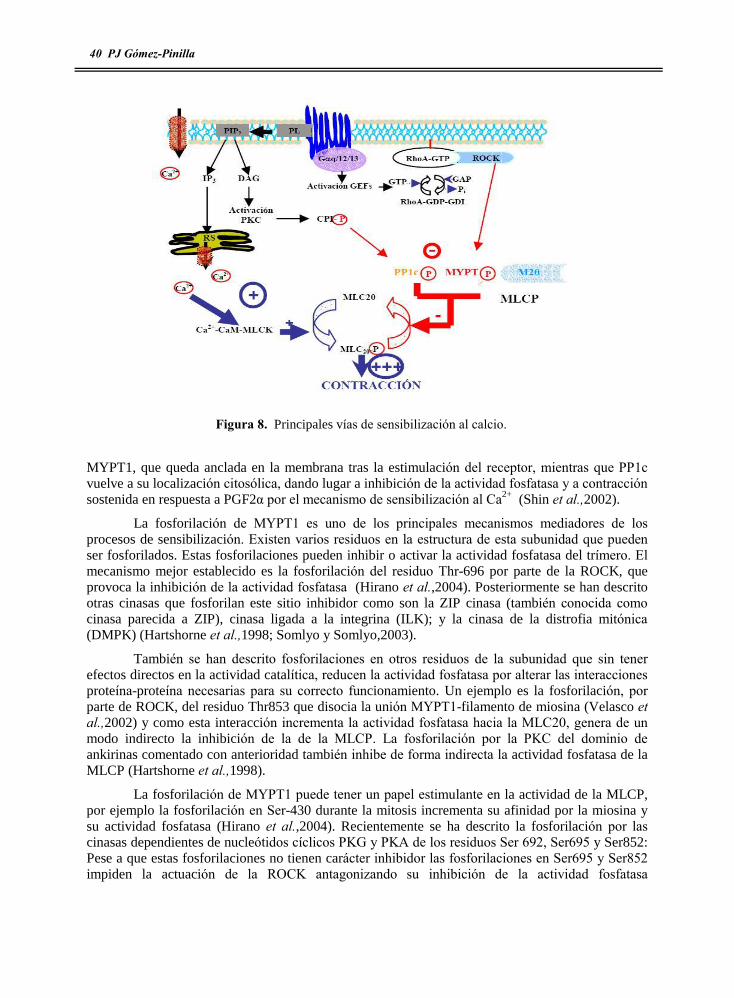
40 PJ Gómez-Pinilla
MYPT1, que queda anclada en la membrana tras la estimulación del receptor, mientras que PP1c vuelve a su localización citosólica, dando lugar a inhibición de la actividad fosfatasa y a contracción sostenida en respuesta a PGF2á por el mecanismo de sensibilización al Ca2+ (Shin et al.,2002).
La fosforilación de MYPT1 es uno de los principales mecanismos mediadores de los procesos de sensibilización. Existen varios residuos en la estructura de esta subunidad que pueden ser fosforilados. Estas fosforilaciones pueden inhibir o activar la actividad fosfatasa del trímero. El mecanismo mejor establecido es la fosforilación del residuo Thr-696 por parte de la ROCK, que provoca la inhibición de la actividad fosfatasa (Hirano et al.,2004). Posteriormente se han descrito otras cinasas que fosforilan este sitio inhibidor como son la ZIP cinasa (también conocida como cinasa parecida a ZIP), cinasa ligada a la integrina (ILK); y la cinasa de la distrofia mitónica (DMPK) (Hartshorne et al.,1998; Somlyo y Somlyo,2003).
También se han descrito fosforilaciones en otros residuos de la subunidad que sin tener efectos directos en la actividad catalítica, reducen la actividad fosfatasa por alterar las interacciones proteína-proteína necesarias para su correcto funcionamiento. Un ejemplo es la fosforilación, por parte de ROCK, del residuo Thr853 que disocia la unión MYPT1-filamento de miosina (Velasco et al.,2002) y como esta interacción incrementa la actividad fosfatasa hacia la MLC20, genera de un modo indirecto la inhibición de la de la MLCP. La fosforilación por la PKC del dominio de ankirinas comentado con anterioridad también inhibe de forma indirecta la actividad fosfatasa de la MLCP (Hartshorne et al.,1998).
La fosforilación de MYPT1 puede tener un papel estimulante en la actividad de la MLCP, por ejemplo la fosforilación en Ser-430 durante la mitosis incrementa su afinidad por la miosina y su actividad fosfatasa (Hirano et al.,2004). Recientemente se ha descrito la fosforilación por las cinasas dependientes de nucleótidos cíclicos PKG y PKA de los residuos Ser 692, Ser695 y Ser852: Pese a que estas fosforilaciones no tienen carácter inhibidor las fosforilaciones en Ser695 y Ser852 impiden la actuación de la ROCK antagonizando su inhibición de la actividad fosfatasa
Figura 8. Principales vías de sensibilización al calcio.

Introducción 41
(Wooldridge et al.,2004).Un estudio realizado por Takizawa y cols en 2002 demostró que la fosforilación de MYPT1 es muy resistente a la defosforilación mediada por fosfatasas, especialmente cuando se trata del residuo inhibitorio Thr-696. Esto quiere decir que, una vez la subunidad MYPT1 se ha fosforilado, permanece en este estado durante mucho tiempo. Por lo tanto, el control de la MLCP a través de MYPT1 es una regulación a largo plazo (Takizawa et al.,2002).
La inhibición directa de la subunidad PP1c, conduce obviamente a una pérdida en la capacidad fosfatasa de la MLCP. Dicha inhibición la produce la proteína inhibitoria dependiente de fosforilación por PKC o CPI-17, siendo por tanto otro mediador de la sensibilización al calcio, pero en este caso independiente de la fosforilación de MYPT-1. El CPI-17 y su posible regulación a través de la PKC se describió por primera vez en aorta porcina, siendo su función principal regular la extensión de la fosforilación de MLC20 que ocurre en la contracción del músculo liso (Eto et al.,1995). El CPI-17 es un péptido con un peso molecular de 17 kDa cuya fosforilación en el residuo Thr-38 inhibie la PP1c (Eto et al.,1995). El sitio Thr-38 se fosforila generalmente por la PKC, aunque también pueden hacerlo otras cinasas como ZIPL, PKN, ILK, PAK y ROCK (Hirano et al.,2004). Se ha demostrado que la fosforilación de CPI tiene lugar tras la estimulación con el agonista y su defosforilación ocurre antes de que termine el estímulo que activó a la cinasa. Por eso, se cree que la fosforilación de CPI-17 tiene una función importante en la fosforilación de MLC20 durante el ciclo de relajación-contracción inducido por agonistas . De todos modos, la relevancia del CPI-17 en la vía de la sensibilización al calcio depende del nivel de expresión de la proteína, mayor en músculo liso tónico que en fásico (Kitazawa et al.,2003).
2.5.3 Vías de señalización reguladoras de la sensibilización al Ca2+
Según se describe en la sección previa, es evidente la implicación de una compleja red de proteínas cinasas e interacciones proteína-proteína en los mecanismos de sensibilización al Ca2+. Además de los mecanismos descritos, la estimulación de diferentes vías intracelulares que conducen a la activación de las diferentes cinasas que intervienen en este mecanismo, complica mucho más este proceso de sensibilización al calcio.
Los mecanismos sensibilizadores al Ca2+ en músculo liso se describieron asociados a la estimulación de receptores de membrana acoplados a proteínas G. Los receptores acoplados a proteínas G constituyen una gran familia de proteínas cuya función principal es traducir estímulos externos en señales intracelulares. Basándose en su homología con la rodopsina, contienen 7 hélices transmembranales cuyo extremo amino se encuentra en el exterior y el carboxilo en el interior de la membrana. Estos receptores interaccionan con proteínas G mediante sus dominios intracelulares participando éstas en la señalización como segundos mensajeros (Kroeze et al.,2003).
En 1994, Alfred Gilman y Martin Rodbel recibieron el premio nobel por el descubrimiento de las proteínas G (proteínas fijadoras de nucleótidos de guanosina) que regulan los procesos intracelulares mediante un intercambio de GDP por GTP tras ser activadas por sus receptores específicos. Las proteínas G pertenecen al grupo de las GTPasas entre las que encontramos las proteínas G triméricas (formadas por las subunidades , y ã) y las proteínas G monoméricas o de bajo peso molecular (Gilman,1987). La superfamilia de las GTPasas de bajo peso molecular comprende más de 100 proteínas estructuralmente relacionadas que experimentan cambios en su conformación espacial y localización subcelular dependientes del nucleótido guanina. La superfamilia se ha dividido en 5 subfamilias: Ras, Rho, Rab, Arf y Ran. Hasta la fecha se han descrito diez Rho GTPasas diferentes en mamíferos (algunas de ellas con múltiples isoformas): Rho (isoformas A, B, C), Rac (isoformas 1, 2, 3), Cdc42 (isoformas Cdc42Hs, G25K), Rnd1/Rho6, Rnd2/Rho7, Rnd3/RhoE, RhoD, RhoG, TC10 y TTF. La función principal de esta subfamilia es

42 PJ Gómez-Pinilla
regular el estado del citoesqueleto de actina y procesos dependientes del mismo como fagocitosis, pinocitosis, migración celular, morfogénesis y crecimiento axonal, aunque también se ha descrito su intervención en transcripción genética, metabolismo lipídico y contracción del músculo liso .
2.5.3.1 Activación de la GTPasa monomérica de bajo peso molecular Rho A
La GTPasa monomérica RhoA parece ser la principal reguladora de la vía de la sensibilización al calcio participando en la inhibición de la fosfatasa de la cadena ligera de miosina a través de su efector ROCK. Los receptores acoplados a proteínas G triméricas Gáq y Gá12,13 son los iniciadores de la vía de la sensibilización al calcio mediada por RhoA/ROCK a través de mecanismos complejos. En estado de reposo de la célula muscular lisa, la proteína RhoA se encuentra localizada en el citosol asociada a GDP, encontrándose su cola hidrofóbica insertada en una región hidrofóbica de la proteína RhoGDI (inhibidor de la disociación del GDP) que mantiene a esta proteína hidrófoba en solución en el citosol. Este complejo impide el intercambio de GDP por GTP incluso cuando la concentración de GTP supera a la de GDP. El complejo GDP-RhoA-RhoGDI mantiene por tanto a la RhoA en estado inactivo, debiendo disociarse tanto del GDP como de RhoGDI para unirse al GTP y pasar a estado activo (Gosser et al.,1997; Somlyo y Somlyo,2003). El intercambio de nucleótidos (GTP por GDP) precede la translocación de RhoA a la membrana, su disociaciación de GDI y su activación (Somlyo y Somlyo,2003) . Para que se produzca el intercambio de nucleótidos se necesita la participación de los factores de intercambio de nucleótidos (GEFs), de los que se han descrito diferentes tipos: PDZ-RhoGEF, LARG, Vav y p115RhoGEF, entre otros. Estas proteínas contienen un dominio homólogo a DBL (DH) responsable de la actividad intercambiadora de nucleótidos, un dominio homólogo a pleckstrina (PH) relacionado con las interacciones proteína-proteína y proteína �fosfatidilinositol y un dominio parecido a RGS (regulador de la señalización de proteínas G) denominado RGSL, por el que se asocian con las subunidades á de las proteínas triméricas (Gá q y G á 12,13), lo que permite su activación en respuesta a la activación de receptores acoplados a estas proteínas. Los factores encargados de inhibir a RhoA se denominan GAP (proteína activadora de la actividad GTPasa), que, actuando a través de las Gá, activa la hidrólisis de GTP-RhoA a GDP-RhoA, que se liga a GDI pasando al citosol y a estado inactivo (Gong et al.,2001).
Algunos subtipos de RhoGEFs pueden activarse por fosforilación a través de proteínas cinasas como c-Src, FAK, o paxilina. La inhibición variable de la activación de Rho A en presencia de inhibidores de diferentes tirosinas cinasas, deja entrever la participación de más de una tirosina cinasa y fosfatasa en la regulación de RhoGEF dependiendo del agonista (Barker et al.,2004).
Las tirosinas cinasas también participan en los mecanismos de sensibilización al Ca2+ como consecuencia de intervenir en la translocación de RhoGEFs y ROCK a la membrana por fosforilación de las proteínas paxilina, p125FAK y p130CAS. Las interacciones lípido-proteína o proteína- proteína que se producen en la membrana plasmática favorecen tanto el intercambio de nucleótidos asociados a RhoA como la activación de ROCK, efectora de RhoA en el mecanismo de sensibilización al Ca2+ (Somlyo y Somlyo,2003).
2.5.3.2 Cinasa asociada a Rho A (ROCK)
La ROCK es una serina-treonina cinasa muy ubicua. Actualmente se conocen dos isoformas de esta enzima ROCKII/ROK, y ROCKI/ROK ambas de 160 kDa de peso molecular y presentes en músculo liso (Noma et al.,2006). Las ROCKs son la diana de la GTPasa monomérica RhoA, que cuando se encuentra translocada en la membrana y en estado activo, interacciona con ROCK produciendo un cambio conformacional, la autofosforilación de la cinasa y su activación. Para que esto ocurra, ROCK también debe traslocarse a la membrana, desconociéndose los

Introducción 43
mecanismos íntimos de dicha translocación. La ROCK puede activarse directamente por el ácido araquidónico producido en respuesta a la estimulación con agonistas, conduciendo a una contracción mediada por mecanismos de sensibilización al Ca2+ (Somlyo y Somlyo,2003), pero en este caso independientes de la RhoA.
Tal y como se ha comentado con anterioridad, el principal sustrato de ROCK es el MYPT1 lo que conlleva a la inhibición de MLCP, aunque esta inhibición también puede estar relacionada con la fosforilación de CPI-17. Tanto ROCKI como ROCKII son inhibidas específicamente por Y-27632 (Somlyo y Somlyo,2003), habiéndose usado ampliamente este inhibidor para el estudio de esta vía.
2.5.3.3 Activación de la proteína cinasa C (PKC)
La estimulación de receptores acoplados a proteínas G no sólo activa la vía de la RhoA sino que también activa la familia de las proteínas cinasas C, implicadas en la traducción de un gran número de señales. La PKC es una enzima que fue descrita originalmente como una proteína cinasa activada por calcio y dependiente de fosfolípidos. En la actualidad su análisis bioquímico y molecular ha revelado que la familia la constituyen diferentes subespecies con estructuras estrechamente relacionadas. Las isoformas de la PKC se clasifican en tres grupos en base a su estructura y regulación por cofactores. Las que primero se descubrieron fueron las PKC convencionales (cPKC): PKC, dos variantes PKC1 y PKC 2 , y PKC. Estas cPKC se distinguen de las demás porque su función está regulada por calcio ya que su dominio C2 contiene un lugar de unión para el ion. El siguiente grupo, las PKC nóveles (nPKC) lo constituyen la PKC, PKC, PKC, PKC y PKCque son similares al grupo anterior, diferenciándose fundamentalmente en que carecen de la región C2 y por tanto no requieren Ca2+ para su activación. El último grupo lo integran las PKC atípicas y cuyas características estructurales radican en la presencia de un solo dominio mano de zinc rico en cisteína, que son dependientes de la fosfatidilserina pero no se estimulan por DAG, ésteres de forbol ni Ca2+ (Nishizuka,1995).
En músculo liso, se ha descrito la presencia de las diferentes isoformas de PKC , estando relacionado el patrón de expresión con el tipo de músculo liso o estado de diferenciación de los miocitos. Así, en la aorta de rata las isoformas presentes son PKC, y mientras que en la arteria mesentérica están estas presentes además de la PKC y y la PKC está regulada negativamente cuando las células musculares de fenotipo �contráctil� pasan a fenotipo �secretor� . En el músculo liso la PKC promueve la fosforilación de la Gi que se traduce en una activación de la adenilato ciclasa, inhibe canales de K+ de tipo BK, y voltaje-dependientes, activa las ATPasas de Ca2+ PMCA y SERCA, facilitando la extrusión de ion que explica la naturaleza transitoria del incremento en Ca2+ que sus agonistas promueven, y también puede fosforilar y activar el intercambiador Na+/H+ y por tanto incrementar el pH intracelular (Barman et al.,2004).
La PKC también fosforila proteínas reguladoras del citoesqueleto y filamentos contráctiles del músculo liso. Así se ha descrito que la PKC fosforila a vinculina, proteínas del citoesqueleto localizadas en las placas de adhesión focal, por lo que controla la forma y adhesión celular. Tal y como se ha descrito con anterioridad el CPI-17 es sustrato de la PKC, inhibiendo la MLCP e induciendo sensibilización al Ca2+, reforzada por sus efectos sobre la subunidad MYPT1 pero contrarrestados por la fosforilación con carácter relajante que produce en la MLC20 y MLCK (Inagaki et al.,1987). La estimulación de esta vía se ha estudiado ampliamente mediante el uso de ésteres de forbol como PDBu y el uso de inhibidores específicos de esta cinasa como GF109203X (Toullec et al.,1991), herramienta farmacológica muy utilizada para poner de manifiesto la participación de la vía PKC/CPI-17 en la sensibilización al calcio en diferentes tejidos.

44 PJ Gómez-Pinilla
2.5.4 Desensibilización al calcio
También se producen en el músculo liso relajación muscular sin que se produzca una reducción en la [Ca2+], lo que se conoce como desensibilización al calcio Los mecanismos implicados en la relajación de la contracción sostenida de la célula muscular lisa estimulan la actividad de la MLCP, mediante la inhibición de RhoA o sus dianas (como MYPT1) y defosforilación del CPI-17. En algunos casos esta relajación se produce por la activación de la PKA o de la PKG en respuesta a incremento en sus nucleótidos específicos (AMPc y GMPc).
La PKG desensibiliza la célula mediante diferentes mecanismos: desinhibición de la MLCP acelerando la defosforilación de CPI-17 (Bonnevier et al.,2004), e inhibición de la vía RhoA/ROCK a través de la fosforilación del residuo Ser-188 de RhoA. Se cree que PKG inhibe la vía de RhoA/ROCK fosforilando la GTP-RhoA activa antes de disociarse del GDI, previniendo así la unión de RhoA a ROCK y otros efectores (Somlyo y Somlyo,2003). Tal y como se ha descrito previamente, la fosforilación de residuos cercanos a los fosforilados por ROCK en la subunidad MYPT, puede inhibir la activación de la vía RhoA/ROCK dando lugar a la relajación de la contracción mantenida por ROCK. También es posible que la PKG incremente la actividad de la MLCP por interacción de los dominios cremallera de leucina de PKG1 y MYPT1 (Murthy,2006).
En cuanto a la PKA, su importancia radica en su capacidad de fosforilar las proteínas G13 inhibiendo la vía de la RhoA , además de fosforilar el MYPT1. Tanto PKA como PKG aceleran la inactivación de Gq unida a GTP finalizando la activación de la vía RhoA (Murthy,2006).
2.5.5 Expresión y participación de las vías de sensibilización al calcio en el músculo liso
El mecanismo de la sensibilización al calcio se ha descrito en multitud de músculos lisos, habiéndose demostrado, que la vía PKC/CPI-17 es más importante en músculos tónicos mientras que la vía RhoA/ROK tiene una mayor participación en músculos fásicos(Woodsome et al.,2001). A pesar de que tanto la expresión como la participación de estas vías está ampliamente generalizada, existen multitud de diferencias entre especies y órganos estudiados lo que quizás esté relacionado con la aproximación experimental utilizada para su estudio (tejido intacto, permeabilizado, wester blot, etc. Esta vía se ha descrito en la traquea (Schaafsma et al.,2005), bronquios (Sakai et al.,2005), estómago (Ratz et al.,2002), ileón (Sward et al.,2000), pared vascular (Seko et al.,2003), miometrio (Friel et al.,2004), uréteres (Hong et al.,2005), uretra (Malmqvist et al.,2004) y vasos del cuerpo cavernoso (Chang et al.,2005). Recientemente se ha descrito que en la vesícula biliar de oveja, cobaya y humanos se expresa ROCK y que su activación está implicada en la respuesta contráctil inducida por estimulaciones eléctricas de campo, CCK, acetilcolina , entrada capacitativa e incluso por despolarización con KCl (Buyukafsar et al.,2006; Quinn et al.,2006; Sahan-Firat et al.,2005).
Respecto a la vejiga urinaria se han descritos mecanismos de sensibilización al calcio en humanos, conejo, rata y cobaya. En este tejido se ha demostrado que los agentes colinérgicos activan sólo la vía de la RhoA/ROCK o de ambas vías de sensibilización, en función de que se estudiaran en tejido intacto o permeabilizado, no existiendo un explicación satisfactoria para este fenómeno (Peters et al.,2006).
2.5.6 Alteración de las vías de sensibilización al calcio en el músculo liso
Quizás una de las posibles razones que convierten al mecanismo de sensibilización al calcio en objeto de estudio es su relación con las enfermedades. Existen multitud de situaciones patológicas en las que la función alterada es el resultado de una desregulación de la vía de la sensibilización al calcio. En la mayoría de los casos coincide con una sobreexpresión o
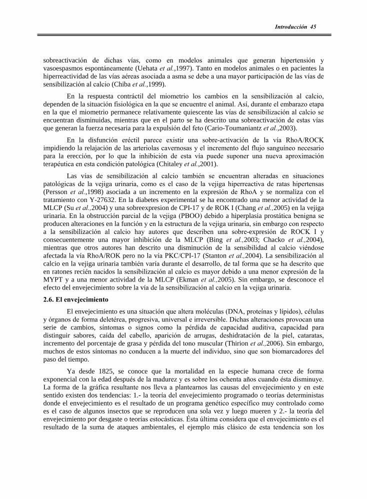
Introducción 45
sobreactivación de dichas vías, como en modelos animales que generan hipertensión y vasoespasmos espontáneamente (Uehata et al.,1997). Tanto en modelos animales o en pacientes la hiperreactividad de las vías aéreas asociada a asma se debe a una mayor participación de las vías de sensibilización al calcio (Chiba et al.,1999).
En la respuesta contráctil del miometrio los cambios en la sensibilización al calcio, dependen de la situación fisiológica en la que se encuentre el animal. Así, durante el embarazo etapa en la que el miometrio permanece relativamente quiescente las vías de sensibilización al calcio se encuentran disminuidas, mientras que en el parto se ha descrito una sobreactivación de estas vías que generan la fuerza necesaria para la expulsión del feto (Cario-Toumaniantz et al.,2003).
En la disfunción eréctil parece existir una sobre-activación de la vía RhoA/ROCK impidiendo la relajación de las arteriolas cavernosas y el incremento del flujo sanguíneo necesario para la erección, por lo que la inhibición de esta vía puede suponer una nueva aproximación terapéutica en esta condición patológica (Chitaley et al.,2001).
Las vías de sensibilización al calcio también se encuentran alteradas en situaciones patológicas de la vejiga urinaria, como es el caso de la vejiga hiperreactiva de ratas hipertensas (Persson et al.,1998) asociada a un incremento en la expresión de RhoA y se normaliza con el tratamiento con Y-27632. En la diabetes experimental se ha encontrado una menor actividad de la MLCP (Su et al.,2004) y una sobreexpresion de CPI-17 y de ROK I (Chang et al.,2005) en la vejiga urinaria. En la obstrucción parcial de la vejiga (PBOO) debido a hiperplasia prostática benigna se producen alteraciones en la función y en la estructura de la vejiga urinaria, sin embargo con respecto a la sensibilización al calcio hay autores que describen una sobre-expresión de ROCK I y consecuentemente una mayor inhibición de la MLCP (Bing et al.,2003; Chacko et al.,2004), mientras que otros autores han descrito una disminución de la sensibilidad al calcio viéndose afectada la vía RhoA/ROK pero no la vía PKC/CPI-17 (Stanton et al.,2004). La sensibilización al calcio en la vejiga urinaria también varía durante el desarrollo, de tal forma que se ha descrito que en ratones recién nacidos la sensibilización al calcio es mayor debido a una menor expresión de la MYPT y a una menor actividad de la MLCP (Ekman et al.,2005). Sin embargo, se desconoce el efecto del envejecimiento sobre la vía de la sensibilización al calcio en la vejiga urinaria.
2.6. El envejecimiento
El envejecimiento es una situación que altera moléculas (DNA, proteínas y lípidos), células y órganos de forma deletérea, progresiva, universal e irreversible. Dichas alteraciones provocan una serie de cambios, síntomas o signos como la pérdida de capacidad auditiva, capacidad para distinguir sabores, caída del cabello, aparición de arrugas, deshidratación de la piel, cataratas, incremento del porcentaje de grasa y pérdida del tono muscular (Thirion et al.,2006). Sin embargo, muchos de estos síntomas no conducen a la muerte del individuo, sino que son biomarcadores del paso del tiempo.
Ya desde 1825, se conoce que la mortalidad en la especie humana crece de forma exponencial con la edad después de la madurez y es sobre los ochenta años cuando ésta disminuye. La forma de la gráfica resultante nos lleva a plantearnos las causas del envejecimiento y en este sentido existen dos tendencias: 1.- la teoría del envejecimiento programado o teorías deterministas donde el envejecimiento es el resultado de un programa genético específico muy controlado como es el caso de algunos insectos que se reproducen una sola vez y luego mueren y 2.- la teoría del envejecimiento por desgaste o teorías estocásticas. Ésta última considera que el envejecimiento es el resultado de la suma de ataques ambientales, el ejemplo más clásico de esta tendencia son los

46 PJ Gómez-Pinilla
roedores donde la desaparición de sus dientes por uso, es el punto crítico al no poder comer más y por tanto mueren.
En mamíferos la esperanza de vida está estrechamente relacionada con el tamaño corporal y con el peso del cerebro en su caso, sin embargo los humanos poseemos una esperanza de vida superior a la que nos corresponde teóricamente debido a una falta de depredadores, un nivel bajo de producción de radicales libres, una baja insaturación de los ácidos grasos y un alto nivel de enzimas reparadoras del DNA. Sin embargo, evolutivamente una longevidad más allá de la edad de procreación podría ser una desventaja para la especie provocándose una competencia por los recursos. Así mismo, el incremento de la longevidad va en paralelo con una disminución de la calidad de vida desde el punto de vista de la salud, sobretodo en edades avanzadas. La esperanza de vida también depende del sexo, así en humanos la esperanza de vida es de tres a cuatro años superior en mujeres, sin embargo la calidad de vida de los hombres es mayor, de tal forma que el 44 % de los hombres con 80 años se mantienen robustos e independientes, mientras que para el caso de las mujeres es sólo el 28 %. En las mujeres alrededor de los cincuenta aparece la menopausia y asociada a ella es muy común la aparición de ciertas enfermedades que disminuyen la calidad de vida. De hecho, la terapia de sustitución hormonal reduce los efectos adversos de la menopausia y protege de la aparición de enfermedades post-menopausia como la enfermedad de Alzheimer (Birge,1996). Los efectos del envejecimiento sobre el sistema reproductor femenino, es el mejor ejemplo de envejecimiento programado. Sin embargo, existen multitud de síntomas indicando que el envejecimiento es el resultado del deterioro general debido al ambiente. Tal es el caso de la disminución del filtrado en los riñones, la aparición de enfermedades cardiovasculares, la reducción de masa ósea y muscular y la pérdida de elasticidad de la piel. Por ello, actualmente está muy aceptado que el envejecimiento es una consecuencia colectiva de factores genéticos y ambientales.
2.6.1 Teorías del envejecimiento
Puesto que en los países desarrollados la esperanza de vida es elevada, las posibles causas que conducen al envejecimiento han sido ampliamente investigadas y agrupadas en varias teorías. Dichas teorías se pueden agrupar en cuatro: teorías a nivel de individuo, de órgano, celular y molecular. Las teorías sistémicas o a nivel de animal adscriben el envejecimiento de todo el organismo a una disminución de la función de determinados sistemas fisiológicos. Las teorías a nivel de órgano apoyan que el envejecimiento es debido a una alteración hormonal y/o inmune. Las teorías celulares relacionan los cambios que se producen en los elementos estructurales y funcionales de las células con el paso del tiempo. Las teorías moleculares proponen que la duración de la vida de cualquier especie está gobernada por la acumulación de lípidos, proteínas o ácidos nucleicos cuyo funcionamiento se encuentra alterado.
2.6.1.1 Teoría de los radicales libres
Una teoría molecular y mixta, es la teoría del envejecimiento por radicales libres. Fue propuesta por Harman en 1956 del siguiente modo:�el envejecimiento resulta de los efectos nocivos generados por los radicales libres que se producen en el curso del metabolismo celular normal y que son acumulados a lo largo de toda la vida de un organismo�. De acuerdo con la teoría de Harman, el envejecimiento es consecuencia del acúmulo de radicales libres en las células, debido a una sobreproducción o a una disminución en los mecanismos antioxidantes, que desencadena estrés oxidativo y consecuentemente daño celular (Harman,1956). Los radicales libres son moléculas inestables caracterizadas por poseer electrones impares o no apareados en su orbital más externo. Dicha inestabilidad hace que reaccionen con otras moléculas convirtiéndolas a su vez en especies reactivas, dando lugar a reacciones en cadena. Las principales especies reactivas de oxígeno son: el

Introducción 47
oxígeno singlete, el anión superóxido, el peróxido de hidrógeno y el radical hidroxilo, además las especies reactivas de nitrógeno como el óxido nítrico y el anión peroxinitrito que también tienen efectos perjudiciales sobre la célula. Los radicales libres pueden provenir tanto de fuentes metabólicas endógenas como de agresiones externas. Puesto que los radicales libres son compuestos muy dañinos para la célula, ésta posee mecanismos de protección frente a los agentes oxidantes generados como la enzima superóxido dismutasa, que reduce los niveles del anión superóxido, la catalasa que cataboliza el agua oxigenada generada por la superóxido dismutasa y el sistema glutatión que detoxifica la célula de agua oxigenada. Las principales dianas para las especies reactivas son: lípidos los cuales sufren peroxidación lipídica, proteínas donde la oxidación proteica provoca una inactivación funcional y una mayor subceptibilidad a proteasas, hidratos de carbono cuya oxidación produce especies reactivas que desestabilizan a otras proteínas y ácidos nucleicos los cuales pueden sufrir mutaciones y alteraciones estructurales, inutilizando así a estas macromoléculas. Las mutaciones provocadas por los radicales libres son una de las causas más importantes del envejecimiento y de la transformación maligna de diferentes células (Burcham y Kuhan,1996). En nuestros días la teoría de los radicales libres como inductores del envejecimiento está bastante aceptada puesto que una gran cantidad de evidencias experimentales la apoyan .
2.6.1.2 Teoría mitocondrial del envejecimiento
Es una teoría que trata de ligar las tendencias estocásticas con las deterministas y postula que la mitocondria produce radicales libres que conducen a daño en el DNA mitocondrial y sobre la propia organela, induciendo la pérdida de la función mitocondrial y consecuentemente, una alteración de la fuente de energía celular (Miquel et al.,1980).
Las mitocondrias son orgánulos subcelulares que generan aeróbicamente energía en forma de ATP utilizando la cadena de transporte de electrones y las enzimas encargadas de la síntesis de ATP. La cadena de transporte de electrones o cadena respiratoria está formada por cuatro complejos intercomunicados por la coenzima Q y el citocromo C. El paso de los electrones desde el O2 hasta el H2O provoca el bombeo de H+ que genera un gradiente electroquímico utilizado para la síntesis de ATP proceso que se denomina fosforilación oxidativa. La ruptura o desacoplamiento de la cadena respiratoria conlleva una producción de especies reactivas. De hecho, el envejecimiento está asociado con un incremento en la producción de superóxido por parte de la cadena respiratoria y una disminución de la fosforilación oxidativa (Corda et al.,2001). La producción de radicales libre por la cadena de transporte de electrones se puede convertir en un círculo vicioso ya que las mismas especies reactivas pueden alterar a la propia cadena y hacerla más inefectiva, provocando así la pérdida de funcionalidad de la mitocondria. Así mismo, la mitocondria posee mecanismos para defenderse frente a excesos de radicales libres como son la coenzima Q y su propio DNA que codifica para los diferentes miembros de la cadena de transporte de electrones (Berneburg et al.,1999). Sin embargo, ante un estrés oxidativo excesivo estos mecanismos de defensa resultan inefectivos y consecuentemente el aporte energético se frena. Está ampliamente aceptado que dichos mecanismos de defensa no son muy efectivos, ya que el DNA mitocondrial no está muy protegido al no poseer histonas y ser las enzimas reparadoras de dicho DNA menos efectivas que las nucleares (Brunk y Terman,2002). Además, se ha descrito que con la edad disminuye la cantidad de coenzima Q (Kalen et al.,1989).
Otro parámetro que controla la acumulación de mitocondrias dañadas es la velocidad de degradación de las mismas. En este sentido, se ha postulado que más que la gravedad del daño, es una menor degradación de mitocondrias dañadas el factor que conduce a su acumulación (Degrey,1997). Así mismo, la velocidad de división celular está relacionada inversamente con el acumulo de mitocondrias dañadas ya que tras una división celular el daño inflingido a la membrana

48 PJ Gómez-Pinilla
mitocondrial disminuye porque los lípidos sintetizados de nuevo no se encuentran dañados. De cualquier modo, este planteamiento sólo es aplicable al daño asociado a la membrana y no al DNA mitocondrial (Degrey,1997).
A su vez, la mitocondria participa en los fenómenos de apoptosis o muerte celular programada, que se inducen por una sobrecarga de calcio en la mitocondria. Ésta provoca la apertura del poro de transición mitocondrial y la salida de citocromo C desde la mitocondria al citoplasma, evento que inicia la destrucción celular por las enzimas caspasas. Actualmente existe mucha controversia sobre si esta teoría es o no una teoría distinta de la de los radicales libres.
2.6.1.3 Teoría basada en la longitud de los telómeros
Dicha teoría molecular se basa en que los telómeros son el reloj biológico que conduce al envejecimiento provocado por el acortamiento de los mismos. Los telómeros son fragmentos de DNA de alrededor 10000 pares de bases compuestos por determinadas repeticiones localizadas en los extremos de los cromosomas. La longitud de los telómeros va disminuyendo a lo largo de las divisiones celulares debido a que los cromosomas se separan antes de su replicación total, provocándose así un acortamiento de los mismos. En humanos, durante los primeros nueve meses de vida, la longitud de los telómeros se reduce hasta la mitad debido al gran número de divisiones celulares que se producen en ese periodo de la vida. Es entonces cuando disminuye el número de divisiones y consecuentemente la velocidad de acortamiento de los telómeros (Rohme,1981). De hecho, cuando experimentalmente se activa la enzima telomerasa (encargada de elongar los telómeros) esa relación entre envejecimiento y acortamiento de los telómeros desaparece (Mathon et al.,2001). La desaparición de la protección de los cromosomas por los telómeros provoca la pérdida de genes. Además la presencia de cromosomas desnudos provoca la conexión entre cromosomas y aumenta la probabilidad de que se produzcan mutaciones y consecuentemente incrementa el número de anormalidades.
Existen algunos síndromes que conducen a envejecimiento acelerado debido a una alta velocidad de acortamiento de los telómeros como son síndrome de Werner , Huntchinson-Gilford , Bloom y síndrome de Down. Igualmente, las alteraciones que se producen en las enzimas reparadoras de modificaciones resultantes de replicaciones alteradas también llevan a síndromes caracterizados por un envejecimiento acelerado como es el caso del síndrome de Cockayne (Tuo et al.,2003) , xeroderma pigmentosum y ataxia telangiectasia (Weizman et al.,2003). Todos estos síndromes en los que la aparición de envejecimiento está relacionada con la longitud de los telómeros, ponen de manifiesto la relación existente entre longitud de los telómeros y longevidad.
2.6.1.4 Teoría genética
Según esta teoría, perteneciente al nivel celular, la longevidad está regulada y/o controlada por cientos de genes que se expresan juntos para formar una red que mantiene el metabolismo y homeostasis de la célula, y son alteraciones en dichos genes las que conducen al envejecimiento (Holliday,2000). Algunos de estos genes son age-1, Chico, clk-1, daf-2 (Hamet y Tremblay,2003) y pueden ser clasificados en cuatro categorías: genes anti-estrés, genes relacionados con el metabolismo energético, genes involucrados en la prevención de mutaciones y genes que protegen la homeostasis y señalización celular mediada por hormonas (Bohr et al.,2002).
2.6.2 Envejecimiento y señalización celular.
Independientemente de las causas que lleven al envejecimiento, que parece ser un fenómeno multifactorial más que debido a una única causa, éste provoca daño en los lípidos, proteínas y ácidos nucleicos que conducen a alteraciones en el funcionamiento celular. El acúmulo

Introducción 49
de radicales libres asociado al envejecimiento puede generar oxidaciones y glucosilaciones de las proteínas no mediadas enzimaticamente que conducen a su agregación y a la pérdida de elasticidad de los tejidos, característica muy común en los tejidos envejecidos. Pero la agregación y pérdida de función de las proteínas no es la única alteración que provoca el envejecimiento, sino que también puede inducir isomerizaciones, racemizaciones, y deaminaciones (Baynes y Thorpe,2000). El envejecimiento también afecta a la maquinaria encargada de sintetizar las proteínas y puede provocar la síntesis de proteínas no funcionales. Además, el envejecimiento puede alterar los mecanismos celulares que conducen a la degradación de las proteínas.
En células envejecidas los cromosomas se encuentran más compactos, lo que indica que hay una menor expresión génica. El daño sobre grandes zonas de los cromosomas es más perjudicial que mutaciones puntuales y a su vez en el envejecimiento las enzimas reparadoras se encuentran alteradas, lo que conduce a una mayor acumulación del daño.
Así, modificaciones en las proteínas y en los ácidos nucleicos provocan alteraciones de las vías de señalización celular, aunque existen multitud de diferencias entre tejidos y especies. Respecto a los receptores, los que poseen actividad tirosina cinasa son más susceptibles al envejecimiento que los acoplados a proteínas G, que o no cambian o incluso incrementan su participación para compensar el daño sobre los primeros (Yeo et al.,2002). Asociados al envejecimiento en varios tipos celulares, incluido el músculo liso, se han descrito cambios cuantitativos y cualitativos de los receptores para los factores de crecimiento incluyendo una menor expresión y una menor activación (Yeo et al.,2002). Estas alteraciones en los receptores con actividad tirosina cinasa se han atribuido a cambios en el reciclaje de receptores por endocitosis, debido a un retraso en el mecanismo mediado por clatrinas o a una inhibición a través de la caveolina (Park et al.,2000). Además de alterar los receptores con actividad tirosina cinasa, el envejecimiento también puede afectar a los miembros de las vías intracelulares localizados tras los receptores como Ras, PLC, PLD y PKC cuya activación disminuye con la edad, estando también alterada la translocación de algunos factores de transcripción (Park et al.,2000; Yeo et al.,2002).
Respecto a los receptores acoplados a proteínas G, los efectos no están tan claros. En el corazón se ha descrito una disminución de la densidad de receptores â-adrenergicos y una menor cantidad de la enzima adenilato ciclasa (Shu y Scarpace,1994), mientras que en el cerebro dichos cambios parecen ser dependientes de la región considerada y no se descartan posibles alteraciones en la PKA y CREB . La activación de receptores acoplados a proteínas G puede conducir a la formación de IP3 a través de la PLC, pero el efecto del envejecimiento sobre dicha formación varía mucho según los tejidos (Huang et al.,1991).
2.6.3 Enfermedades relacionadas con el envejecimiento.
Las alteraciones o daños sobre los órganos, tejidos, células o moléculas inducidas por el envejecimiento conducen a enfermedades como artritis, osteoporosis, cardiopatías, cáncer, enfermedad de Alzeheimer, enfermedad de Parkinson, incontinencia urinaria, etc� denominadas enfermedades asociadas al envejecimiento ya que su incidencia incrementa con la edad (Reiter et al.,1998).
La enfermedad de Alzheimer suele aparecer a partir de los 50 años, afecta a unos 15 millones de personas en el mundo y es la mayor causa de pérdida de capacidad cognitiva en la población envejecida. Está ampliamente aceptado que el acúmulo de especies reactivas asociado al envejecimiento provoca las principales alteraciones neuro-morfo-fisiológicas presentes en esta enfermedad como son: formación de placas â-amiloideas, acúmlo de neurofilamentos y pérdida neuronal sobre todo a nivel del hipocampo y cortex cerebral (Chen y Fernandez,2001). La
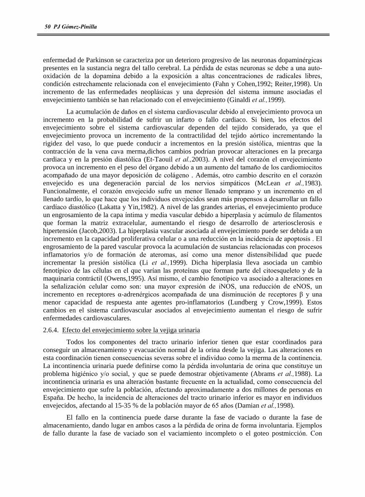
50 PJ Gómez-Pinilla
enfermedad de Parkinson se caracteriza por un deterioro progresivo de las neuronas dopaminérgicas presentes en la sustancia negra del tallo cerebral. La pérdida de estas neuronas se debe a una auto-oxidación de la dopamina debido a la exposición a altas concentraciones de radicales libres, condición estrechamente relacionada con el envejecimiento (Fahn y Cohen,1992; Reiter,1998). Un incremento de las enfermedades neoplásicas y una depresión del sistema inmune asociadas el envejecimiento también se han relacionado con el envejecimiento (Ginaldi et al.,1999).
La acumulación de daños en el sistema cardiovascular debido al envejecimiento provoca un incremento en la probabilidad de sufrir un infarto o fallo cardiaco. Si bien, los efectos del envejecimiento sobre el sistema cardiovascular dependen del tejido considerado, ya que el envejecimiento provoca un incremento de la contractilidad del tejido aórtico incrementando la rigidez del vaso, lo que puede conducir a incrementos en la presión sistólica, mientras que la contracción de la vena cava merma,dichos cambios podrían provocar alteraciones en la precarga cardiaca y en la presión diastólica (Et-Taouil et al.,2003). A nivel del corazón el envejecimiento provoca un incremento en el peso del órgano debido a un aumento del tamaño de los cardiomiocitos acompañado de una mayor deposición de colágeno . Además, otro cambio descrito en el corazón envejecido es una degeneración parcial de los nervios simpáticos (McLean et al.,1983). Funcionalmente, el corazón envejecido sufre un menor llenado temprano y un incremento en el llenado tardío, lo que hace que los individuos envejecidos sean más propensos a desarrollar un fallo cardíaco diastólico (Lakatta y Yin,1982). A nivel de las grandes arterias, el envejecimiento produce un engrosamiento de la capa íntima y media vascular debido a hiperplasia y acúmulo de filamentos que forman la matriz extracelular, aumentando el riesgo de desarrollo de arteriosclerosis e hipertensión (Jacob,2003). La hiperplasia vascular asociada al envejecimiento puede ser debida a un incremento en la capacidad proliferativa celular o a una reducción en la incidencia de apoptosis . El engrosamiento de la pared vascular provoca la acumulación de sustancias relacionadas con procesos inflamatorios y/o de formación de ateromas, así como una menor distensibilidad que puede incrementar la presión sistólica (Li et al.,1999). Dicha hiperplasia lleva asociada un cambio fenotípico de las células en el que varían las proteínas que forman parte del citoesqueleto y de la maquinaria contráctil (Owens,1995). Así mismo, el cambio fenotípico va asociado a alteraciones en la señalización celular como son: una mayor expresión de iNOS, una reducción de eNOS, un incremento en receptores á-adrenérgicos acompañada de una disminución de receptores â y una menor capacidad de respuesta ante agentes pro-inflamatorios (Lundberg y Crow,1999). Estos cambios en el sistema cardiovascular asociados al envejecimiento aumentan el riesgo de sufrir enfermedades cardiovasculares.
2.6.4. Efecto del envejecimiento sobre la vejiga urinaria
Todos los componentes del tracto urinario inferior tienen que estar coordinados para conseguir un almacenamiento y evacuación normal de la orina desde la vejiga. Las alteraciones en esta coordinación tienen consecuencias severas sobre el individuo como la merma de la continencia. La incontinencia urinaria puede definirse como la pérdida involuntaria de orina que constituye un problema higiénico y/o social, y que se puede demostrar objetivamente (Abrams et al.,1988). La incontinencia urinaria es una alteración bastante frecuente en la actualidad, como consecuencia del envejecimiento que sufre la población, afectando aproximadamente a dos millones de personas en España. De hecho, la incidencia de alteraciones del tracto urinario inferior es mayor en individuos envejecidos, afectando al 15-35 % de la población mayor de 65 años (Damian et al.,1998).
El fallo en la continencia puede darse durante la fase de vaciado o durante la fase de almacenamiento, dando lugar en ambos casos a la pérdida de orina de forma involuntaria. Ejemplos de fallo durante la fase de vaciado son el vaciamiento incompleto o el goteo postmicción. Con
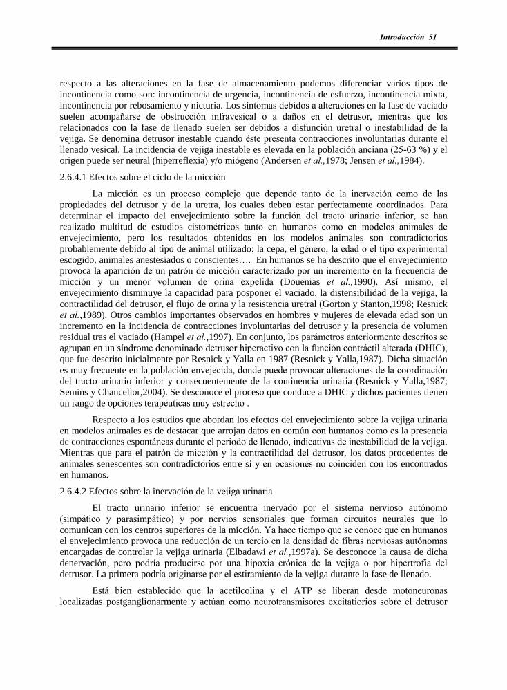
Introducción 51
respecto a las alteraciones en la fase de almacenamiento podemos diferenciar varios tipos de incontinencia como son: incontinencia de urgencia, incontinencia de esfuerzo, incontinencia mixta, incontinencia por rebosamiento y nicturia. Los síntomas debidos a alteraciones en la fase de vaciado suelen acompañarse de obstrucción infravesical o a daños en el detrusor, mientras que los relacionados con la fase de llenado suelen ser debidos a disfunción uretral o inestabilidad de la vejiga. Se denomina detrusor inestable cuando éste presenta contracciones involuntarias durante el llenado vesical. La incidencia de vejiga inestable es elevada en la población anciana (25-63 %) y el origen puede ser neural (hiperreflexia) y/o miógeno (Andersen et al.,1978; Jensen et al.,1984).
2.6.4.1 Efectos sobre el ciclo de la micción
La micción es un proceso complejo que depende tanto de la inervación como de las propiedades del detrusor y de la uretra, los cuales deben estar perfectamente coordinados. Para determinar el impacto del envejecimiento sobre la función del tracto urinario inferior, se han realizado multitud de estudios cistométricos tanto en humanos como en modelos animales de envejecimiento, pero los resultados obtenidos en los modelos animales son contradictorios probablemente debido al tipo de animal utilizado: la cepa, el género, la edad o el tipo experimental escogido, animales anestesiados o conscientes�. En humanos se ha descrito que el envejecimiento provoca la aparición de un patrón de micción caracterizado por un incremento en la frecuencia de micción y un menor volumen de orina expelida (Douenias et al.,1990). Así mismo, el envejecimiento disminuye la capacidad para posponer el vaciado, la distensibilidad de la vejiga, la contractilidad del detrusor, el flujo de orina y la resistencia uretral (Gorton y Stanton,1998; Resnick et al.,1989). Otros cambios importantes observados en hombres y mujeres de elevada edad son un incremento en la incidencia de contracciones involuntarias del detrusor y la presencia de volumen residual tras el vaciado (Hampel et al.,1997). En conjunto, los parámetros anteriormente descritos se agrupan en un síndrome denominado detrusor hiperactivo con la función contráctil alterada (DHIC), que fue descrito inicialmente por Resnick y Yalla en 1987 (Resnick y Yalla,1987). Dicha situación es muy frecuente en la población envejecida, donde puede provocar alteraciones de la coordinación del tracto urinario inferior y consecuentemente de la continencia urinaria (Resnick y Yalla,1987; Semins y Chancellor,2004). Se desconoce el proceso que conduce a DHIC y dichos pacientes tienen un rango de opciones terapéuticas muy estrecho .
Respecto a los estudios que abordan los efectos del envejecimiento sobre la vejiga urinaria en modelos animales es de destacar que arrojan datos en común con humanos como es la presencia de contracciones espontáneas durante el periodo de llenado, indicativas de inestabilidad de la vejiga. Mientras que para el patrón de micción y la contractilidad del detrusor, los datos procedentes de animales senescentes son contradictorios entre sí y en ocasiones no coinciden con los encontrados en humanos.
2.6.4.2 Efectos sobre la inervación de la vejiga urinaria
El tracto urinario inferior se encuentra inervado por el sistema nervioso autónomo (simpático y parasimpático) y por nervios sensoriales que forman circuitos neurales que lo comunican con los centros superiores de la micción. Ya hace tiempo que se conoce que en humanos el envejecimiento provoca una reducción de un tercio en la densidad de fibras nerviosas autónomas encargadas de controlar la vejiga urinaria (Elbadawi et al.,1997a). Se desconoce la causa de dicha denervación, pero podría producirse por una hipoxia crónica de la vejiga o por hipertrofia del detrusor. La primera podría originarse por el estiramiento de la vejiga durante la fase de llenado.
Está bien establecido que la acetilcolina y el ATP se liberan desde motoneuronas localizadas postganglionarmente y actúan como neurotransmisores excitatiorios sobre el detrusor

52 PJ Gómez-Pinilla
(Hoyle,1994). Sin embargo, la importancia relativa de estos dos neurotransmisores depende de la especie estudiada. Por ejemplo, en ratas y cobayas ambos neurotransmisores inducen contracción del detrusor, mientras que en humanos, en condiciones normales las contracciones de la vejiga son exclusivamente colinérgicas y sólo en determinadas situaciones patológicas el componente purinérgico tiene relevancia (Wu y Fry,1998). Así, en el 2001, Yoshida describió que el envejecimiento en humanos provoca una disminución de la participación colinérgica y un mayor papel de la inervación purinérgica, aunque la respuesta contráctil inducida por ATP y acetilcolina no se afectaba, indicando la existencia de una alteración de la liberación de los neurotransmisores (Yoshida et al.,2001). De hecho, cuando se midieron las cantidades de acetilcolina y ATP liberadas en respuesta a estimulaciones eléctricas de campo, se encontró una correlación negativa entre la edad y la cantidad de acetilcolina liberada, mientras que para el caso de la liberación de ATP, la correlación era positiva (Yoshida et al.,2004). Al igual que ocurría con los parámetros procedentes de estudios funcionales, el efecto del envejecimiento en modelos animales no es similar a lo descrito en humanos, ya que en los modelos animales la inervación colinérgica o no cambia (Lluel et al.,2000) o se incrementa (Latifpour et al.,1990)mientras que la inervación purinérgica presenta un incremento similar al encontrado en humanos (Kageyama et al.,2000) o no se altera (Lluel et al.,2000).
A pesar de la importancia de la inervación aferente como sensor del estado de llenado de la vejiga urinaria, no se ha descrito el efecto del envejecimiento sobre dicha inervación en humanos. En modelos animales se ha encontrado una denervación sensorial asociada al envejecimiento (Chai et al.,2000) con disminución de los nervios sensoriales que contienen PACAP, sin afectar a los que contienen CGRP, SP y VIP (Mohammed et al.,2002).
2.6.4.3 Efectos del envejecimiento sobre el detrusor
Actualmente se acepta que el detrusor de los ancianos es diferente al de los jóvenes. Sin embargo, se desconoce si dichos cambios causan las alteraciones típicas del detrusor envejecido o son meramente efectos secundarios del envejecimiento.
El envejecimiento provoca un incremento del espacio entre los fascículos de fibras musculares que forman el detrusor (Elbadawi et al.,1997b). Dicha separación es debida a un mayor acúmulo de colágeno, fibroblastos e infiltración de tejido conectivo que puede ser responsable, en parte al menos, de los típicos cambios funcionales asociados al envejecimiento como disminución de la distensibilidad, de la capacidad y de la contractilidad vesical, así como la aparición de volumen residual (Hald y Horn,1998). Este distanciamiento entre fibras también produce un menor acoplamiento eléctrico entre células, puesto de manifiesto por la presencia de una mayor resistencia eléctrica y una menor expresión de conexina 45, 43 y 40, principales componentes de las uniones íntimas entre células (Sui et al.,2003). Estos hallazgos no explican la inestabilidad del detrusor, pero sí podrían ser la causa de la menor contractilidad encontrada en el detrusor envejecido. Si a este menor acoplamiento eléctrico, le sumamos que el detrusor envejecido produce menos sustratos energéticos como ATP (Yu et al.,1997), las posibilidades de encontrarnos un detrusor con la capacidad contráctil disminuida son altas. Se ha demostrado también que esta disminución en la fosforilación oxidativa y consecuentemente en la síntesis de ATP asociada al envejecimiento, es debida a un proceso de anoxia (Lu et al.,2000). Este menor metabolismo energético conduce a que el mantenimiento de las contracciones a lo largo del tiempo disminuya con la edad , puesto que en el detrusor envejecido existe una menor cantidad de energía disponible (van Mastrigt y Griffiths,1986). Aunque una menor disponibilidad de energía podría ser la causa de la aparición de hipocontractilidad del detrusor, otras causas como menor aporte sanguíneo, cambio en los

Introducción 53
neurotransmisores liberados, alteraciones en la señalización celular y en las proteínas contráctiles podría conducir también a una menor contractilidad del detrusor asociada al envejecimiento.
En humanos, el envejecimiento reduce el número de receptores colinérgicos de tipo M2 y M3 presentes en las fibras musculares del detrusor, sin embargo la respuesta contráctil inducida por agonistas colinérgicos no está afectada por el envejecimiento (Yoshida et al.,2001). Además, en ratas en las que se ha descrito una menor expresión de receptores muscarínicos, la expresión de los subtipos de proteínas G asociadas a estos receptores no está afectada por el envejecimiento, lo que podría explicar la falta de efecto sobre la respuesta contráctil (Schneider et al.,2005). Sin embargo, estudios funcionales �in vivo� han puesto de manifiesto que la menor contractilidad de la vejiga podría deberse a la menor expresión de los receptores muscarínicos (Siroky,2004).
En modelos animales de envejecimiento y ancianos se ha encontrado una reducción en la expresión y función de receptores â-adrenérgicos, que se traduce en una menor respuesta relajante inducida por los agonistas de dichos receptores (Fraeyman et al.,2000). A nivel intracelular, se ha descrito un incremento en las proteínas Gi asociado al envejecimiento, que inhiben a la adenilato ciclasa, y conducen a una menor producción de AMPc (Derweesh et al.,2000), a pesar de no existir cambios en los efectores del AMPc (Nishimoto et al.,1995). Esta menor capacidad relajante del detrusor podría determinar la menor complianza y el menor intervalo de micción asociado al envejecimiento.
El ión calcio es el principal mensajero intracelular encargado de regular el estado contráctil del detrusor. Los estudios realizados para determinar el efecto del envejecimiento sobre la homeostasis de dicho ión describen un incremento en la entrada de calcio a través de los canales L asociada a un aumento de la cinética de activación y disminución de la velocidad de inactivación (Gallegos y Fry,1994). La presencia de otros canales iónicos como canales de K+ y Cl- activados por Ca2+ (Fry y Wu,1998) podrían regular la activación-inactivación de los canales de tipo L.
2.6.4.4 Tatamiento de las alteraciones de la vejiga urinaria asociadas al envejecimiento
Puesto que en la población mayor de 65 años la incidencia de vejiga inestable es de alrededor del 30 % (Geirsson et al.,1993), la mayoría de las estrategias terapéuticas están dirigidas a paliar la presencia de esa hiperactividad vesical (Andersson et al.,1999). Los agentes terapéuticos más utilizados en este sentido son los antimuscarínicos, ya que la mayoría de las contracciones de la vejiga son colinérgicas y su eficacia bloqueando las contracciones depende de la vía de administración, siendo más efectivos cuando se aplican intravesicalmente (Cardozo y Stanton,1979). Sin embargo, el uso de antimuscarínicos está muy limitado debido a la existencia de un gran número de efectos secundarios como son: parálisis del tracto gastrointestinal, estreñimiento, taquicardia y sequedad de las mucosas. Otros agentes utilizados para paliar la inestabilidad del detrusor son los antagonistas de los canales de calcio como nifedipina o verapamilo y activadores de los canales de potasio como pinacidil y cromakalina, a pesar de que el uso de estos agentes ha resultado efectivo en el tejido vascular, en la vejiga urinaria los resultados son contradictorios (Andersson,1992). Otra alternativa para tratar la hiperactividad de la vejiga es el uso de antagonistas de los receptores á-adrenergicos y agonistas de los receptores â-adrenérgicos, pero sus efectos son moderados y los efectos secundarios inevitables (Castleden y Morgan,1980; Jensen, Jr.,1981). En base a la importancia que está tomando la sensibilización al calcio en el músculo detrusor, el tratamiento con inhibidores de esta vía podrían ser una herramienta farmacológica interesante para evitar la hiperactividad del detrusor (Peters et al.,2006). En la actualidad existen muchas más alternativas terapéuticas para paliar la hiperactividad vesical, incluyendo tratamientos hormonales y

54 PJ Gómez-Pinilla
agentes con efectos mixtos, sin embargo la presencia de graves efectos secundarios es el principal factor limitante para su uso (Andersson et al.,1999; Andersson,2002; Andersson y Hedlund,2002).
2.6.5. El envejecimiento del tracto gastrointestinal
Al igual que ocurre con otros sistemas orgánicos, la incidencia de alteraciones del tracto gastrointestinal se incrementa en personas mayores de 65 años (Firth y Prather,2002). Dichas alteraciones conducen a disfagia, reflujo gastro-esofágico, dispepsia, desordenes en la función colónica, sobrecrecimiento bacteriano, estreñimiento e incontinencia fecal (Firth y Prather,2002) y son debidas a que el envejecimiento afecta a la absorción, secreción y motilidad del tracto gastrointestinal. Así, los efectos del envejecimiento varían a lo largo del tracto gastrointestinal, puesto que existen áreas con mayor riesgo de desarrollar alteraciones como son el tracto gastrointestinal superior (particularmente la orofarínge y el esófago) y las regiones distales como el colon (Phillips y Powley,2001).
En el caso del esófago se ha descrito que en ancianos las contracciones esofágicas presentan una menor velocidad y duración, provocando una disminución en el tránsito esofágico y disfagia (Castell,1990). Aunque parece que el envejecimiento no provoca cambios en la función del estómago, en ocasiones se ha descrito un retraso en el vaciamiento gástrico en ancianos (Clarkston et al.,1997). Las alteraciones provocadas por el envejecimiento sobre al intestino delgado incluyen la disminución del peristaltismo postpandrial, la reducción en la frecuencia de migración del complejo motor y de la propagación de contracciones agrupadas (Anuras y Sutherland,1984). Dichas alteraciones llevan asociadas un menor tránsito a nivel del colon y consecuentemente estreñimiento, el cual puede estar también asociado a una mayor pasividad del anciano y mejorará mediante una dieta rica en fibra y agua (Cheskin et al.,1995). El envejecimiento de la región ano-rectal se caracteriza por una menor distensibilidad, pérdida de sensibilidad rectal y menor presión basal del esfínter anal interno, que conducen a incontinencia fecal (Akervall et al.,1990). De forma similar a lo descrito en la vejiga urinaria, en el tracto gastriontestinal los trastornos asociados al envejecimiento son debidos a alteraciones en el sistema nervioso entérico y al propio músculo liso.
2.6.5.1 Efectos sobre el sistema nervioso entérico
En multitud de estudios realizados en humanos y en animales de laboratorio se ha descrito una reducción en el número de neuronas del sistema nervioso entérico (Saffrey,2004). La velocidad de deterioro suele ser lineal y el resultado final oscila entre un 20 y 60 % de reducción, dependiendo de la especie y de la región analizada. Así en humanos el esófago sufre un 30 % (Meciano et al.,1995), el colón un 40 % (Gomes et al.,1997) y el intestino delgado un 40 % de reducción (de Souza et al.,1993), mientras que la inervación del estómago permanece prácticamente inalterada (Madsen,1992). En cuanto a las subpoblaciones neuronales más vulnerables, se ha descrito que el plexo submucoso sufre una menor denervación (15-20 % de reducción) que el plexo mientérico (40 % de reducción) (Wade,2002). Además, se conoce que la inervación colinérgica es más susceptible que la inervación nitrérgica, que permanece inalterada o poco afectada por el envejecimiento (Wade y Cowen,2004). En los nervios colinérgicos, se ha descrito que la síntesis de acetilcolina permanece invariable, sin embargo la liberación del neurotransmisor es menor, debido a una reducción en la actividad del canal de calcio de tipo N presente en las terminaciones nerviosas. De hecho con el uso de ionóforos para el calcio, la liberación de acetilcolina se restaura hasta niveles similares a jóvenes (Roberts et al.,1994). Además de una menor liberación de acetilcolina, se ha descrito un incremento en la actividad acetilcolinesterasa asociada al envejecimiento que se traduce en una menor participación de las fibras colinérgicas en la respuesta contráctil del tracto gastrointestinal (Kobashi et al.,1985).

Introducción 55
Parece que el incremento en las especies reactivas de oxígeno es la principal causa de pérdida neuronal asociada al envejecimiento (Hall y Wiley,1998). De hecho se ha demostrado que las dietas hipocalóricas reducen la producción de especies reactivas de oxígeno en las neuronas entéricas, y consecuentemente, pueden provocar una recuperación de dicho plexo. Además, asociado al envejecimiento se ha descrito que, tanto la generación de ciertas sustancias en el sistema inmune gastrointestinal (Degiorgio et al.,1995) como la reducción de los factores neurotróficos secretados por las células de Schwann y de los sistemas neuroprotectores, están implicadas en la aparición de algunas patologías neurales del tracto gastrointestinal (Degiorgio et al.,1995) .
2.6.5.2 Efectos del envejecimiento sobre el músculo liso del tracto gastrointestinal
Aunque en la mayoría del músculo liso que forma parte del tracto gastrointestinal el envejecimiento provoca una pérdida de la contractilidad que se traduce en una menor peristalsis, el músculo más estudiado a nivel celular es el colon. En el esófago se ha descrito un engrosameinto de la capa muscular y fibrosis que conducen a una menor cinética esofágica (Lock,2001). En el año 2003 Bitar describió que las células procedentes del colon de ratas envejecidas presentaban una menor longitud, lo que podría ser un impedimento estérico para el deslizamiento de los filamentos contráctiles, presentando así una menor velocidad de acortamiento y una menor respuesta contráctil (Bitar,2003). Otro efecto del envejecimiento que provoca pérdida de la contractilidad del músculo liso es la reducción en la señal de calcio, tanto a nivel de entrada de calcio desde el medio extracelular como de la liberación desde depósito intracelulares de calcio (Lopes et al.,2006). Además, la reducción en la respuesta contráctil del colon de ratas asociado al envejecimiento, parece deberse a una menor expresión y activación de algunas isoformas de la PKC, de la vía Src-MAPK, de la maquinaría contráctil y de las proteínas reguladoras de la contracción como es la HSP-27 (Bitar et al.,2002). Al igual que en el colon, el envejecimiento provoca una reducción de las vías intracelulares que conducen a contracción del recto apareciendo así incontinencia fecal (Bitar,2005).
2.6.5.3 Efectos del envejecimiento sobre la vesícula biliar
La incidencia de formación de cálculos biliares incrementa con la edad (Diehl,1991) como consecuencia de un incremento en la secreción biliar y absorción intestinal de colesterol. Además, la disminución en la síntesis y secreción hepática de las sales biliares y la menor contractilidad de la vesícula biliar conducen a la formación de dichos cálculos biliares a medida que avanza la edad (Wang,2002). También se ha descrito que la formación de cálculos biliares es una enfermedad multifactorial en la que están implicados los genes litogénicos denominados Lith y condiciones ambientales como una dieta rica en sustancias litogénicas (Wang,2002).
En humanos se ha descrito un incremento en el volumen residual de bilis tras la contracción (Sacchetti et al.,1973) y una disminución en la sensibilidad a CCK por parte del músculo liso vesical asociado al envejecimiento. Dicha pérdida de sensibilidad se pone de manifiesto por la necesidad de una mayor liberación de CCK desde el duodeno en individuos envejecidos para alcanzar la misma contracción que en jóvenes (Khalil et al.,1985). La pérdida de sensibilidad a colecistokinina por parte del músculo liso aparece también en conejo y en cobaya (Poston et al.,1988). En este último caso el envejecimiento provoca una disminución de la respuesta contráctil a CCK �in vivo� e �in vitro�, debida a una disminución en la densidad de receptores para la CCK (Poston et al.,1988). No se conocen efectos del envejecimiento sobre la función neuromuscular de la vesícula biliar.

56 PJ Gómez-Pinilla
2.7 La colecistitis acalculosa
Las enfermedades del tracto biliar son bastante comunes y suponen un coste económico superior al de cualquier otra enfermedad gastrointestinal. Una alteración en la motilidad de la vesícula puede ocurrir en presencia o en ausencia de cálculos biliares. La colecistitis acalculosa es una enfermedad que se caracteriza por la aparición de inflamación aguda, pérdida de contractibilidad, una menor eyección y un aumento del volumen de la vesícula biliar a pesar de la ausencia de cálculos biliares. Sin embargo, se desconoce la relación causal existente entre el éstasis biliar y la colecistitis acalculosa.
Esta enfermedad es bastante frecuente en ancianos y en pacientes ingresados en unidades de cuidados intensivo y la tasa de mortalidad en pacientes que la presentan es elevada (Kalliafas et al.,1998). Sin embargo, la etiología exacta de la colecistitis acalculosa aguda no se ha podido determinar debido, sobre todo, a la complejidad clínica de los pacientes que la padecen.
2.7.1 Modelos experimentales.
Puesto que la disponibilidad de muestras humanas es muy limitada, gran parte de los estudios sobre la colecistitis acalculosa se han realizado en modelos experimentales en animales, tratando de imitar las condiciones que se producen en la colecistitis acalculosa humana.
Los modelos animales desarrollados se pueden dividir en dos grandes grupos: los que utilizan sustancias químicas para inducir la inflamación y los que se basan en procesos físicos como la inducción de obstrucción. Los primeros se fundamentan en la infusión de lisolecitina (Kaminski et al.,1995) o la instilación del lipopolisacárido (LPS) proveniente de Escherichia coli (Prystowsky y Rege,1997). El uso de sustancias pro-inflamatorias en combinación con la retirada de la bilis almacenada es una maniobra utilizada para imitar los primeros paso del proceso inflamatorio (Al Jiffry et al.,2004). Entre los modelos basados en fenómenos fisicos se encuentran: la introducción de cálculos dentro de la vesícula (Kaminski et al.,1985), la ligadura del conducto cístico en combinación con la sobresaturación de colesterol en la bilis (Roslyn et al.,1980) o la ligadura del conducto biliar común (Myers et al.,1988).
El modelo de ligadura del conducto biliar común provoca una inflamación aguda comparable a la que ocurre en la colecistitis acalculosa aguda humana, sin producir cambios en el índice litogénico biliar. Por ese motivo y porque no requiere demasiadas maniobras que provoquen artefactos, es un modelo muy utilizado para el estudio del proceso inflamatorio y de sus efectos sobre la vesícula biliar, siendo éste el modelo de elección en la presente tesis.
2.7.2 Agentes pro-inflamatorios.
Como ocurre en otros procesos inflamatorios, la colecistitis está asociada con la liberación de componentes paracrinos desde leucocitos. Existen estudios describiendo la producción de quininas como un evento temprano en la cascada inflamatoria seguido de la producción de otros mediadores inflamatorios (Al Jiffry et al.,2004) . Estos mediadores pro-inflamatorios asociados a la colecistitis aguda son: las prostaglandinas (PG), las especies reactivas de oxígeno (ROS) y la histamina.
Al igual que en la pancreatitis aguda, las infecciones hepática y la colitis , en la colecistitis aguda las endotelinas (ET) son uno de los mediadores inflamatorios primarios, actuando en las primeras fases del proceso inflamatorio (Al Jiffry et al.,2004). De hecho, se ha descrito que las células epiteliales de la mucosa producen altos niveles de ET-1 y ET-2 en la vesícula biliar humana con colecistitis aguda (Al Jiffry et al.,2001)).
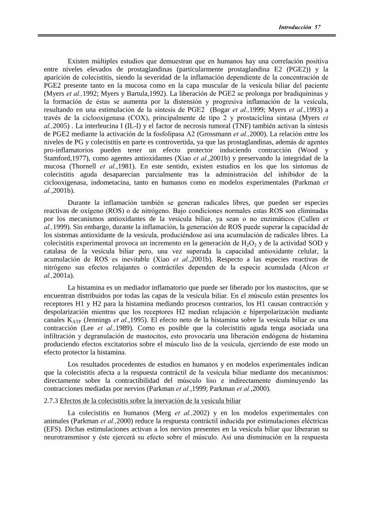
Introducción 57
Existen múltiples estudios que demuestran que en humanos hay una correlación positiva entre niveles elevados de prostaglandinas (particularmente prostaglandina E2 (PGE2)) y la aparición de colecistitis, siendo la severidad de la inflamación dependiente de la concentración de PGE2 presente tanto en la mucosa como en la capa muscular de la vesícula biliar del paciente (Myers et al.,1992; Myers y Bartula,1992). La liberación de PGE2 se prolonga por bradiquininas y la formación de éstas se aumenta por la distensión y progresiva inflamación de la vesícula, resultando en una estimulación de la síntesis de PGE2 (Bogar et al.,1999; Myers et al.,1993) a través de la ciclooxigenasa (COX), principalmente de tipo 2 y prostaciclina sintasa (Myers et al.,2005) . La interleucina I (IL-I) y el factor de necrosis tumoral (TNF) también activan la síntesis de PGE2 mediante la activación de la fosfolipasa A2 (Grossmann et al.,2000). La relación entre los niveles de PG y colecistitis en parte es controvertida, ya que las prostaglandinas, además de agentes pro-inflamatorios pueden tener un efecto protector induciendo contracción (Wood y Stamford,1977), como agentes antioxidantes (Xiao et al.,2001b) y preservando la integridad de la mucosa (Thornell et al.,1981). En este sentido, existen estudios en los que los síntomas de colecistitis aguda desaparecían parcialmente tras la administración del inhibidor de la ciclooxigenasa, indometacina, tanto en humanos como en modelos experimentales (Parkman et al.,2001b).
Durante la inflamación también se generan radicales libres, que pueden ser especies reactivas de oxígeno (ROS) o de nitrógeno. Bajo condiciones normales estas ROS son eliminadas por los mecanismos antioxidantes de la vesícula biliar, ya sean o no enzimáticos (Cullen et al.,1999). Sin embargo, durante la inflamación, la generación de ROS puede superar la capacidad de los sistemas antioxidante de la vesícula, produciéndose así una acumulación de radicales libres. La colecistitis experimental provoca un incremento en la generación de H2O2 y de la actividad SOD y catalasa de la vesícula biliar pero, una vez superada la capacidad antioxidante celular, la acumulación de ROS es inevitable (Xiao et al.,2001b). Respecto a las especies reactivas de nitrógeno sus efectos relajantes o contráctiles dependen de la especie acumulada (Alcon et al.,2001a).
La histamina es un mediador inflamatorio que puede ser liberado por los mastocitos, que se encuentran distribuidos por todas las capas de la vesícula biliar. En el músculo están presentes los receptores H1 y H2 para la histamina mediando procesos contrarios, los H1 causan contracción y despolarización mientras que los receptores H2 median relajación e hiperpolarización mediante canales KATP (Jennings et al.,1995). El efecto neto de la histamina sobre la vesícula biliar es una contracción (Lee et al.,1989). Como es posible que la colecistitis aguda tenga asociada una infiltración y degranulación de mastocitos, esto provocaría una liberación endógena de histamina produciendo efectos excitatorios sobre el músculo liso de la vesícula, ejerciendo de este modo un efecto protector la histamina.
Los resultados procedentes de estudios en humanos y en modelos experimentales indican que la colecistitis afecta a la respuesta contráctil de la vesícula biliar mediante dos mecanismos: directamente sobre la contractibilidad del músculo liso e indirectamente disminuyendo las contracciones mediadas por nervios (Parkman et al.,1999; Parkman et al.,2000).
2.7.3 Efectos de la colecistitis sobre la inervación de la vesícula biliar
La colecistitis en humanos (Merg et al.,2002) y en los modelos experimentales con animales (Parkman et al.,2000) reduce la respuesta contráctil inducida por estimulaciones eléctricas (EFS). Dichas estimulaciones activan a los nervios presentes en la vesícula biliar que liberaran su neurotransmisor y éste ejercerá su efecto sobre el músculo. Así una disminución en la respuesta
58 PJ Gómez-Pinilla
contráctil inducida por EFS podría indicar que la inflamación aguda provoca una denervación de la vesícula biliar. Al menos en cobaya, esta disminución de la respuesta inducida por estimulación eléctrica se debe a una menor participación del componente colinérgico contráctil junto a una mayor participación del componente nitrérgico inhibitorio que estaba ausente en los animales control (Parkman et al.,2000). Estos resultados están en conflicto con los estudios estructurales y funcionales que demuestran la existencia de fibras nitrérgicas en la vesícula biliar (Mawe et al.,1997; Mourelle et al.,1993).
2.7.4 Efectos de la colecistitis sobre el músculo liso de la vesícula biliar
Desde el punto de vista histológico la colecistitis provoca una invasión de células inflamatorias (principalmente neutrófilos y eosinófilos), edema e infiltración de fibroblastos tanto a nivel submucoso como en la capa muscular (Parkman et al.,2000).
Está ampliamente demostrado que la colecistitis disminuye la respuesta contráctil de la vesícula biliar �in vivo� e �in vitro�. Esta disminución se debe en parte a una disfunción miogéna, ya que agentes que actúan directamente sobre el músculo como el betanecol y/o despolarizaciones, provocan una alteración de las contracciones vesiculares .
En humanos parece que la alteración de la respuesta contráctil asociada a la colecistitis aguda se localiza a nivel de la maquinaria contráctil, ya que los receptores para la CCK y los canales L no parecen estar afectados(Xiao et al.,2001a). Además, en modelos experimentales se ha demostrado que el daño provocado por la inflamación parece localizarse en la membrana celular de la fibra muscular, ya que la contracción es normal cuando las células se estimulan con agonistas que atraviesan la membrana celular sin necesidad de unirse a sus receptores de membrana, como pueden ser los activadores de proteínas G, IP3 y DAG. Estas alteraciones en los receptores acoplados a proteínas G y en canales iónicos asociados a membrana son debidas a una disminución del contenido de fosfolípidos de membrana y a un incremento en la peroxidación lipídica como consecuencia del aumento de ROS durante la inflamación (Xiao et al.,2002; Xiao et al.,2003).Puesto que no existen estudios sobre la homeostasis del Ca2+ en este estado patológico de la vesícula biliar, una parte de esta tesis se ha centrado en los efectos que provoca la colecistitis sobre los cambios en la [Ca2+]c en respuesta a estímulos y la posible relación entre dichas alteraciones a nivel celular y los cambios en la motilidad de la vesícula.
2.7.5 Posibles estrategias terapeúticas para la colecistitis acalculosa
En contraste con los ácidos hidrofóbicos cuya acumulación y precipitación conduce a colecistitis, el enriquecimiento de la bilis con ácidos hidrofílicos como el ácido ursodeoxycólico reduce la incidencia de cólicos biliares, de colecistitis aguda y las alteraciones que la colecistitis provoca sobre el músculo liso de la vesícula biliar (Akriviadis et al.,1997). Ya en los años ochenta se conocía que el tratamiento con indometacina (un inhibidor de la síntesis de prostaglandinas) podía aliviar el dolor asociado a los cólicos biliares y sus efectos se atribuían a sus propiedades para revertir los defectos en la absorción/secreción que ocurren con la inflamación (Parkman et al.,2001a). Veinte años después, con el uso del modelo experimental de la ligadura del conducto biliar común, se ha demostrado que el tratamiento con indometacina reduce la inflamación y protege parcialmente la respuesta contráctil del músculo liso de la vesícula biliar, durante las primeras 24 horas tras la ligadura (Myers et al.,2005). Estudios más recientes han puesto de manifiesto que el incremento de la liberación de PGE asociado a la colecistitis es debido a un incremento en la expresión de COX-2. Estos resultados sugieren que el tratamiento con inhibidores de COX-2 (como la indometacina) durante las primeras etapas, podría prevenir el incremento en la liberación de PGE y disminuir o prevenir la aparición de este síndrome clínico.

Introducción 59
2.8 La melatonina
La melatonina (N-acetil-metoxitriptamina) es una neuro-hormona que se ha estudiado en profundidad para determinar su función exacta y su posible uso como agente farmacológico. El interés puesto en esta hormona radica en que se ha demostrado que en determinadas situaciones, la melatonina es capaz de paliar los daños inducidos por las especies reactivas de oxígeno sobre los lípidos, proteínas y ácidos nucleicos. A partir de estas observaciones se ha sugerido que la melatonina sería eficaz para combatir los síndromes asociados la acumulación de radicales libres y por ello se ha probado su posible papel terapéutico en multitud de situaciones fisiopatológicas.
2.8.1 Melatonina: Síntesis y secreción
La melatonina se produce a partir del aminoácido triptófano, el cual sufre una hidroxilación y posteriormente una descarboxilación, formándose respectivamente 5-hidroxitriptófano y serotonina. Las enzimas encargadas de estos primeros pasos tienen una distribución muy ubicua. Sin embargo, la transformación de serotonina en melatonina está restringida a ciertos órganos en los que se localizan las enzimas N-acetiltransferasa e hidroxiindol-O-metiltransferasa. Dichas enzimas, actuando de forma secuencial, conducen a la conversión de serotonina en melatonina (Huether,1993). La principal fuente de melatonina es la glándula pineal, pero se ha descrito que otros tejido pueden sintetizar y secretar melatonina. Entre ellos se incluyen la retina, las glándulas lacrimales, el intestino, el epitelio de los bronquios, el páncreas, las glándulas suprarrenales, el tiroides, el timo, el tracto urogenital y la placenta (Huether,1993). Es de especial relevancia la producción de melatonina en las células enterocromafines del intestino, habiéndose estimado que la cantidad de melatonina normalizada por gramo de tejido es 400 veces superior a la existente en la glándula pineal (Cavallo,1993; Haimov et al.,1995).
En la mayoría de individuos y animales jóvenes, la producción de melatonina por la glándula pineal sigue un ritmo circadiano con máximos de producción y secreción por la noche, que van disminuyendo durante el día. La presencia de este potente ritmo circadiano regulado por la luz, está controlada por un tracto óptico-pineal que va desde la retina hasta la glándula pineal pasando por el núcleo supraquiamático. Así, durante la fase oscura el núcleo supraquiasmático estimula al ganglio cervical superior y a la glándula pineal provocando la secreción de melatonina (Reiter,1992). Cuando la luz llega a la retina, el núcleo supraquiasmático deja de excitar la glándula pineal y frena la producción y secreción de melatonina. Los pinealocitos, células endocrinas de la glándula pineal, tras la síntesis de la melatonina vierten su contenido al torrente sanguíneo, de tal forma que el incremento nocturno de la síntesis y secreción de melatonina, se refleja rápidamente en los niveles sanguíneos de dicha hormona. Un hecho importante es que la presencia de este ritmo circadiano de producción y secreción de melatonina de la glándula pineal está limitada a individuos jóvenes y adultos, ya que el envejecimiento produce una disminución notable de la síntesis de melatonina (Henden et al.,1992). Esta reducción se debe a una menor expresión de los receptores á y â-adrenérgicos de la membrana del pinealocito (Poeggeler et al.,1993) y a una pérdida de las neuronas glutamatrérgicas, las cuales regulan la síntesis de melatonina y son muy sensibles al envejecimiento (Hazlerigg et al.,2001).
2.8.2 Receptores para la melatonina
En humanos y en animales superiores se han descrito tres tipos de receptores para la melatonina, que pueden localizarse en la membrana plasmática (MT1 y MT2) o a nivel citoplasmático (MT3 también denominado QR2). Los dos primeros se caracterizan por ser receptores acoplados a proteínas G que conducen a la formación de inositoles fosfato e inhiben la adenilato y guanilato ciclasa, mientras que los receptores citoplasmáticos parecen estar involucrados

60 PJ Gómez-Pinilla
en procesos antioxidantes. Los receptores para la melatonina pueden localizarse a nivel neural (núcleo supraquiasmático o en la capa flexiforme interna de la retina) y a nivel no neuronal como en la pars tuberalis de la hipófisis y en arterias caudales y cerebrales (Deacon y Arendt,1994).
A pesar de existir cientos de trabajos describiendo las acciones beneficiosas de la melatonina sobre la mayoría de los procesos fisiopatológicos, es difícil determinar si las acciones de la melatonina son mediadas a través de sus receptores o a través de su actividad antioxidante. En este sentido la substancia luzindol (un bloqueante de los receptores para la melatonina) es una herramienta muy útil para determinar si los efectos de la melatonina están mediados o no por receptores.
2.8.3 Acciones biológicas de la melatonina
La melatonina, gracias a su secreción en forma circadiana, informa al sistema endocrino sobre las condiciones ambientales, especialmente sobre el fotoperiodo. En este aspecto las principales funciones de la melatonina son: la regulación de los ritmos de otras hormonas, de los ciclos sueño-vigilia, de la época reproductiva y de la temperatura corporal (Tan et al.,1993).
Además, entre las acciones de la melatonina se encuentra una potente capacidad antioxidante, desarrollando un papel defensivo contra el estrés oxidativo. Esto la convierte en una herramienta farmacológica cuyos efectos beneficiosos han sido ampliamente recogidos en la bibliografía.
2.8.4 Capacidad antioxidante de la melatonina
La capacidad antioxidante de la melatonina se basa en sus propiedades como neutralizador de radicales libres y en su habilidad para activar otros mecanismos antioxidantes. La melatonina es altamente difusible y por tanto es capaz de ejercer sus efectos directos como antioxidante en la membrana citoplasmática, en el citoplasma y en el núcleo. Sus características estructurales que le permiten actuar como un potente secuestrador de radicales libres son: el grupo metilo localizado en posición 5 del indol y el grupo acetilo, de tal forma que sin estos grupos su capacidad para neutralizar radicales libres merma e incluso se puede convertir en un compuesto pro-oxidante (Reiter et al.,2002). Pero además, la melatonina puede promover la activación de las defensas antioxidantes endógenas por su efecto estimulador sobre la superóxido dismutasa, la catalasa, la gutatión reductasa y la glutation peroxidasa (Arendt et al.,1988). Adicionalmente, algunos metabolitos de la melatonina como la N-acetil-5-metoxikynureamina, son también muy eficiente reduciendo el daño ocasionado por los radicales libres, así se ha definido la cascada antioxidante de la melatonina.
2.8.5 Posibles indicaciones terapeúticas de la melatonina
Es indudable que las propiedades antioxidantes de la melatonina la convierten en una herramienta farmacológica muy efectiva para tratar los efectos devastadores provocados por el envejecimiento, en el que la acumulación de radicales libre juega un papel fundamental. Este aspecto será revisado más ampliamente en otro apartado.
El uso de la melatonina para tratar los desórdenes del sueño resultó muy satisfactorio en multitud de casos (Arendt y Deacon,1997). Además, su uso está muy indicado frente a desórdenes circadianos provocados por la ceguera, cambios de turnos de trabajo y vuelos transcontinentales en los que se cruzan varias zonas horarias (Sainz et al.,2003). Adicionalmente, a la melatonina se le han atribuido propiedades oncostáticas , cardioprotectoras e inmunomoduladoras (Huether,1993).

Introducción 61
2.8.5.1 Indicaciones terapéuticas de la melatonina en el tracto gastrointestinal
El tracto gastrointestinal es la principal fuente extrapineal de melatonina, encontrándose en una concentración de hasta 400 veces mayor que en la glándula pineal (Bubenik et al.,1999). En la bilis se detectan niveles de melatonina 10-40 veces superior al resto del tracto gastrointestinal (Lane y Moss,1985), como consecuencia de ser el hígado la vía metabólica de la melatonina (Huether,1993) y la bilis su principal vía de excreción aunque también se excreta melatonina activa en la bilis que se concentra en la vesícula biliar (Huether,1994) . sin embargo aún no se ha descrito la función de dicha neuro-hormona en el tracto gastrointestinal. La producción y secreción de la melatonina por parte del tracto gastrointestinal esta totalmente regulada por la ingesta (Bubenik y Pang,1994). La gran abundancia de melatonina indica que podría ser un regulador local de la función gastrointestinal , de hecho en ratones la melatonina regula el tránsito intestinal (BarajasLopez et al.,1996). En cobayas se ha descrito que los efectos de la melatonina sobre la motilidad digestiva se producen a través del bloqueo de los canales activados por acetilcolina localizados a nivel postsináptico . Pero los efectos pro o anti-cinéticos de la melatonina sobre el tracto gastrointestinal dependen de la dosis y de la duración del tratamiento, así tratamientos crónico y a bajas dosis tienen efectos estimulatorios (Cho et al.,1989).
Interesantemente, la melatonina puede considerarse como un posible agente protector en varias patologías del tracto gastrointestinal. Existen un gran número de evidencias experimentales que apoyan el uso terapéutico de la melatonina. Estudios bastantes antiguos indicaban que la melatonina tenía efectos protectores y sanadores frente a la formación de úlceras gástricas (Melchiorri et al.,1997). Estudios más recientes han propuesto que la prevención de las lesiones gástricas por parte de la melatonina se debe probablemente a su efecto antiserotoninérgico, que bloquea la reducción del flujo sanguíneo inducida por la serotonina, restaurándose así la microcirculación . En su acción gastroprotectora están implicados los receptores para la melatonina de tipo MT2, la prostaglandina E2 debido a una sobreexpresión y sobreactivación de COX-2 y la liberación de CGRP desde los nervios sensoriales (Kato et al.,2001). Además de los efectos beneficiosos sobre la circulación, las propiedades antioxidantes de la melatonina desempeñan un papel importante en la recuperación del estómago (Cho et al.,1989). Así, la administración intragástrica de melatonina a ratas con úlceras gástricas disminuye significativamente la incidencia de ulceración y el tamaño de las lesiones, ya que la melatonina provoca un descenso del contenido de radicales libres en la pared del estómago y en plasma (Sener-Muratoglu et al.,2001). Los efectos gastroprotectores de la melatonina son comparables a los del omeprazol y la famotidina (Bandyopadhyay et al.,2002) y en este sentido la efectividad de la melatonina supera la de otros antioxidantes clásicos como las vitaminas C, E, etc.. y la de fármacos como la ranitidina (Sjoblom y Flemstrom,2003). Además, la melatonina también protege la mucosa del tracto gastrointestinal estimulando la secreción de bicarbonato desde los enterocitos. Dicha secreción se produce mediante la estimulación de los receptores de la melatonina de tipo MT2 que provocan un incremento del calcio citoplasmático en el enterocito y la consecuente secreción de bicarbonato (Lu et al.,2005).
En el síndrome de intestino irritable la melatonina podría tener efectos terapéuticos, ya que es efectiva aliviando el dolor abdominal, la distensión abdominal y la tenesmo, a pesar de no mejorar las alteraciones del sueño que presentan este tipo de pacientes (Head y Jurenka,2003). De forma similar a lo que ocurre con las úlceras gástricas, en el síndrome de intestino irritable, el tratamiento con melatonina redujo el número de daños en la mucosa intestinal inducidos por un modelo de colitis experimental (Dong et al.,2003). Además de reducir el daño, la melatonina disminuye la cantidad de NO en el colon y la liberación de prostaglandina E, debido a una menor expresión de la enzima óxido nítrico sintasa inducible y de la COX-2 en la mucosa del colon

62 PJ Gómez-Pinilla
(Fuss,2003; Sans et al.,1999). Así mismo, se ha descrito que la inflamación inducida por los modelos de colitis experimental se debe a un incremento en la expresión de moléculas proinflamatorias, una excesiva activación del factor de transcripción NF-êB y una menor participación del factor de transcripción IêB (Li et al.,2005). El tratamiento con melatonina redujo la síntesis de moléculas proinflamatorias y normalizó la participación de los factores de transcripción NF-êB y IêB (Sartor,1997). Además, algunos estudios demuestran que la inflamación del intestino se debe a una alteración de la inmunoregulación (Mahida,2000) estando los macrófagos involucrados en este proceso. Esta hipótesis defiende que los macrófagos inician el ataque a las células epiteliales presentes en la mucosa del colon y que este es el primer paso que provoca daño en el colon (Li et al.,2002). Los macrófagos secretan citoquinas proinflamatorias (tales como IL-1, IL6 y TNF-á) junto a radicales libres como NO, iniciando la cascada inflamatoria e incrementando el daño al colon. Se ha demostrado que la administración de la melatonina intranal disminuye los síntomas de colitis y disminuye el daño a la mucosa. Dichos efectos beneficiosos de la melatonina están mediados por una disminución en la liberación de IL-1, TNF-á y NO, ya que la melatonina puede reprimir la expresión de la enzima óxido nítrico sintasa inducible (iNOS) y suprimir la actividad del factor de transcripción NF-êB, que es el principal factor de transcripción involucrado en la regulación de la expresión de citoquinas proinflamatorias y genes relacionados con la iNOS (Ozacmak et al.,2005).
Mediante el uso de modelos de isquemia-reperfusión para imitar las condiciones patológicas que en ocasiones ocurren en el organismo, se ha puesto de manifiesto otras posibles aplicaciones terapeúticas de la melatonina. Tal es el caso del modelo de isquemia-reperfusión en el íleon, el cual provocó una reducción de la respuesta contráctil de dicho tejido, probablemente debida a una acumulación de especies reactivas de oxígeno y al daño tisular. En este caso el tratamiento con melatonina redujo el daño tisular, la acumulación de radicales libres debido a un incremento en la actividad glutatión reductasa y consecuentemente mejoró la respuesta contráctil del íleon (Fraschini et al.,1998).
Pese a que la melatonina tiene efectos inmunoestimulantes, no hay evidencias directas de dicho efecto en el tracto gastrointestinal. Si bien, existen evidencias indirectas que indican que la administración de melatonina a ratas incrementa el tamaño de las placas de Peyer, el principal componente del sistema inmune localizado en el tracto gastrointestinal (Marquez et al.,2006). Sin embargo, se ha demostrado que el tratamiento con melatonina puede tener efectos perjudiciales debido a la sobre-estimulación del sistema inmune en patologías como la enfermedad de Crohn y la colitis experimental (Drake et al.,2004).
2.8.5.2 Indicaciones terapeúticas de la melatonina frente a alteraciones del tracto urinario inferior
Los estudios sobre las posibles aplicaciones terapeúticas de la melatonina frente a las alteraciones del tracto urinario inferior son escasos. Así, se ha descrito que el tratamiento con melatonina en pacientes que sufrían nocturia redujo la frecuencia de micción durante la noche, aunque se desconoce el mecanismo por el que la melatonina ejerce dicho efecto (Paskaloglu et al.,2004). En base a las propiedades antioxidantes de la melatonina, su uso resultó útil en un modelo experimental de isquemia-reperfusión de la vejiga urinaria, maniobra que redujo la respuesta contráctil de la vejiga y el contenido de glutatión reducido e incrementó la peroxidación lipídica. Los efectos perjudiciales de la isquemia-reperfusión fueron eliminados tras el tratamiento con melatonina, que restauró la respuesta contráctil, probablemente debido a una disminución del estrés oxidativo (Maestroni et al.,1988).

Introducción 63
2.8.6 Melatonina, envejecimiento y enfermedades relacionadas con la edad
Como se ha descrito anteriormente, el envejecimiento es un fenómeno multifactorial y por ello aún no se ha conseguido llegar a una teoría consensual que explique este proceso. Sin embargo hay algunas teorías que asignan a la glándula pineal y a la melatonina un papel fundamental en el proceso del envejecimiento. Se sabe que los niveles de melatonina disminuyen gradualmente a lo largo de la vida, lo que disminuye la calidad del sueño y altera otros ritmos circadianos. Además, la melatonina posee propiedades inmunomoduladoras, que al perderse conducirían a envejecimiento. Y por último, la melatonina es un potente protector contra el estrés oxidativo ya sea directa o indirectamente. De hecho, la acumulación de radicales libres asociada al envejecimiento puede contribuir a incrementar la incidencia y severidad de algunas enfermedades relacionadas con la edad.
El envejecimiento de la sociedad actual es un hecho, que tiene asociado problemas relacionados con una menor calidad de vida de este segmento poblacional teniendo además repercusiones económicas y sociales. Desde el punto de vista de la salud, el principal problema es el incremento en la incidencia de determinadas enfermedades como son la enfermedad de Alzheimer y enfermedad de Parkinson, arteriosclerosis, enfermedades neoplásicas, incontinencia urinaria y/o fecal�. las cuales merman de forma importante la calidad de vida de las personas e incrementan su grado de dependencia.
Existen teorías que relacionan el envejecimiento con la melatonina. Así, se ha descrito que el envejecimiento es debido a que el sistema inmune es menos efectivo, a una desregulación de los ritmos circadianos y a un acumulo de radicales libres. Parece ser que la menor secreción de melatonina es la principal causa que conduce a las alteraciones anteriores y por tanto al envejecimiento (Reiter et al.,1998). Sin embargo, la evidencia experimental más importante que relaciona a la melatonina con el envejecimiento es que la producción y secreción de la melatonina disminuye con la edad. Dicha pérdida tiene una serie de consecuencias entre las que destacan las alteraciones de los ritmos circadianos que conducen a cronopatologías como alteraciones del sueño y consecuentemente, al deterioro de la salud (Skwarlo-Sonta,2002). El papel regulador del sistema inmune por parte de la melatonina disminuye con la edad, provocando una reducción en el número de las células inmunes, una menor proliferación de linfocitos, diferentes niveles de expresión de citocinas y la alteración del índice de fagocitosis, de modo que la administración exógena de melatonina provoca un restablecimiento del sistema inmune . Pero quizás, las propiedades antioxidantes de la melatonina o más bien su ausencia, sean la principal causa que conduzca al acúmulo de radicales libres asociado al envejecimiento (Miles y Dement,1980).
El daño oxidativo desempeña un papel importante en la patogénesis de algunas enfermedades neurodegenerativas características de la población envejecida , habiéndose descrito que la melatonina tiene efectos terapéuticos en pacientes con enfermedad de Alzheimer y enfermedad de Parkinson . En ciertas neoplasias la melatonina redujo la velocidad de división celula, aunque parece ser que dichos efectos son mediados a través del sistema inmune (Parola et al.,1996).
En conclusión, la menor producción de melatonina en ancianos juega un papel muy importante en las enfermedades relacionadas con la edad. Esto sugiere que la pérdida de este agente antioxidante, regulador de ritmos circadianos e inmunomodulador, puede tener relación con el incremento de la incidencia y la severidad de ciertas patologías. Así, la administración de melatonina puede tener un uso terapéutico y en esta tesis se ha puesto de manifiesto dicho uso sobre las alteraciones que provoca el envejecimiento en la vesícula biliar y en la vejiga urinaria.
64 PJ Gómez-Pinilla
2.8.7 Melatonina y ligadura del conducto biliar común.
Los modelos de colecistitis acalculosa experimental inducidos por la ligadura del conducto biliar común, además de producir inflamación de la vesícula biliar, provocan daño hepático (Ohta et al.,2003; Polat y Emre,2006) probablemente asociados a un incremento del estrés oxidativo. Por este motivo, el tratamiento con melatonina mostró efectos beneficiosos gracias a su capacidad antioxidante directa o indirecta (Montilla et al.,2001) siendo mucho más efectiva que la vitamina E y que S-adenosil-L-metionina (Barlas et al.,2004). Además de estos efectos relacionados con su capacidad antioxidante, en el hígado la melatonina inhibió la infiltración de neutrófilos en el tejido (Padillo et al.,2004), redujo la apoptosis y necrosis (Ohta et al.,2003; Reiter et al.,2000) y su efecto anti inflamatorio previno la translocación del factor de transcripción NF-kâ además de reducir la síntesis de citocinas proinflamatorias como interleucinas y el factor de necrosis tumoral .
Sin embargo, se desconoce si la melatonina ejerce efectos beneficiosos sobre la inflamación y los cambios que ella produce en la inervación y el propio músculo liso de la vesícula biliar, habiéndose abordado estos temas en varios trabajos que integran la presente tesis.
BIBLIOGRAFÍA
1. Abrams P, Blaivas JG, Stanton SL, Andersen JT. The standardisation of terminology of lower urinary tract function. The International Continence Society Committee on Standardisation of Terminology. Scand J Urol Nephrol Suppl 1988; 114: 5-19.
2. Akervall S, Nordgren S, Fasth S, Oresland T, Pettersson K, Hulten L. The effects of age, gender, and parity on rectoanal functions in adults. Scand J Gastroenterol 1990; 25: 1247-1256.
3. Akriviadis EA, Hatzigavriel M, Kapnias D, Kirimlidis J, Markantas A, Garyfallos A. Treatment of biliary colic with diclofenac: a randomized, double-blind, placebo-controlled study. Gastroenterology 1997; 113: 225-231.
4. Al Jiffry BO, Meedeniya AC, Chen JW, Toouli J, Saccone GT. Endothelin-1 induces contraction of human and Australian possum gallbladder in vitro. Regul Pept 2001; 102: 31-39.
5. Al Jiffry BO, Shaffer EA, Woods CM, Menadue M, Young F, Oliver J, Thomas AC, Toouli J, Saccone GT. Endogenous endothelin increases gallbladder tone and leads to acute cholecystitis in the Australian possum. Neurogastroenterol Motil 2004; 16: 125-133.
6. Alcon S, Camello PJ, Garcia LJ, Pozo MJ. Activation of tyrosine kinase pathway by vanadate in gallbladder smooth muscle. Biochem Pharmacol 2000; 59: 1077-1089.
7. Alcon S, Morales S, Camello PJ, Hemming JM, Jennings L, Mawe GM, Pozo MJ. A redox-based mechanism for the contractile and relaxing effects of NO in the guinea-pig gall bladder. J Physiol 2001a; 532: 793-810.

Introducción 65
8. Alcon S, Morales S, Camello PJ, Pozo MJ. Contribution of different phospholipases and arachidonic acid metabolites in the response of gallbladder smooth muscle to cholecystokinin. Biochem ical Pharmacology 2002; 64: 1157-1167.
9. Alcon S, Morales S, Camello PJ, Salido GM, Miller SM, Pozo MJ. Relaxation of canine gallbladder to nerve stimulation involves adrenergic and non-adrenergic non-cholinergic mechanisms. Neurogastroenterol Motil 2001b; 13: 555-566.
10. Amano M, Ito M, Kimura K, Fukata Y, Chihara K, Nakano T, Matsuura Y, Kaibuchi K. Phosphorylation and activation of myosin by Rho-associated kinase (Rho-kinase). J Biol Chem 1996; 271: 20246-20249.
11. Andersen JT, Jacobsen O, Worm-Petersen J, Hald T. Bladder function in healthy elderly males. Scand J Urol Nephrol 1978; 12: 123-127.
12. Andersson KE. Clinical pharmacology of potassium channel openers. Pharmacol Toxicol 1992; 70: 244-254.
13. Andersson KE. Overactive bladder - Pharmacological aspects. Scand J Urol Nephrol Suppl 2002; 36: 72-81.
14. Andersson KE, Appell R, Cardozo LD, Chapple C, Drutz HP, Finkbeiner AE, Haab F, Vela NR. The pharmacological treatment of urinary incontinence. BJU Int 1999; 84: 923-947.
15. Andersson KE y Arner A. Urinary bladder contraction and relaxation: Physiology and pathophysiology. Physiol Rev 2004; 84: 935-986.
16. Andersson KE y Hedlund P. Pharmacologic perspective on the physiology of the lower urinary tract. Urology 2002; 60: 13-20.
17. Andersson KE y Persson K. The L-Arginine Nitric-Oxide Pathway and Nonadrenergic, Noncholinergic Relaxation of the Lower Urinary-Tract. Gen Pharmacol 1993; 24: 833-839.
18. Andersson KE y Wein AJ. Pharmacology of the lower urinary tract: basis for current and future treatments of urinary incontinence. Pharmacol Rev 2004; 56: 581-631.
19. Anuras S y Sutherland J. Small intestinal manometry in healthy elderly subjects. J Am Geriatr Soc 1984; 32: 581-583.
20. Arendt J, Aldhous M, Wright J. Synchronization of A Disturbed Sleep-Wake Cycle in A Blind Man by Melatonin Treatment. Lancet 1988; 1: 772-773.
21. Arendt J y Deacon S. Treatment of circadian rhythm disorders - Melatonin. Chronobiol Inter 1997; 14: 185-204.
22. Bandyopadhyay U, Biswas K, Chatterjee R, Bandyopadhyay D, Chattopadhyay I, Ganguly CK, Chakraborty T, Bhattacharya K, Banerjee RK. Gastroprotective effect of Neem (Azadirachta indica)
66 PJ Gómez-Pinilla
bark extract: possible involvement of H(+)-K(+)-ATPase inhibition and scavenging of hydroxyl radical. Life Sci 2002; 71: 2845-2865.
23. BarajasLopez C, Peres AL, EspinosaLuna R, ReyesVazquez C, PrietoGomez B. Melatonin modulates cholinergic transmission by blocking nicotinic channels in the guinea-pig submucous plexus. Eur J Pharmacol 1996; 312: 319-325.
24. Barker TH, Grenett HE, MacEwen MW, Tilden SG, Fuller GM, Settleman J, Woods A, Murphy-Ullrich J, Hagood JS. Thy-1 regulates fibroblast focal adhesions, cytoskeletal organization and migration through modulation of p190 RhoGAP and Rho GTPase activity. Exp Cell Res 2004; 295: 488-496.
25. Barlas A, Cevik H, Arbak S, Bangir D, Sener G, Yegen C, Yegen BC. Melatonin protects against pancreaticobiliary inflammation and associated remote organ injury in rats: role of neutrophils. J Pineal Res 2004; 37: 267-275.
26. Barman SA, Zhu S, White RE. Protein kinase C inhibits BKCa channel activity in pulmonary arterial smooth muscle. Am J Physiol Lung Cell Mol Physiol 2004; 286: L149-L155.
27. Bayguinov O, Hagen B, Bonev AD, Nelson MT, Sanders KM. Intracellular calcium events activated by ATP in murine colonic myocytes. Am J Physiol Cell Physiol 2000; 279: C126-C135.
28. Baynes JW y Thorpe SR. Glycoxidation and lipoxidation in atherogenesis. Free Radic Biol Med 2000; 28: 1708-1716.
29. Becker PL, Singer JJ, Walsh JV, Fay FS. Regulation of Calcium-Concentration in Voltage-Clamped Smooth-Muscle Cells. Science 1989; 244: 211-214.
30. Berneburg M, Grether-Beck S, Kurten V, Ruzicka T, Briviba K, Sies H, Krutmann J. Singlet oxygen mediates the UVA-induced generation of the photoaging-associated mitochondrial common deletion. J Biol Chem 1999; 274: 15345-15349.
31. Berridge MJ y Irvine RF. Inositol Phosphates and Cell Signaling. Nature 1989; 341: 197-205.
32. Bing W, Chang SH, Hypolite JA, DiSanto ME, Zderic SA, Rolf L, Wein AJ, Chacko S. Obstruction-induced changes in urinary bladder smooth muscle contractility: a role for Rho kinase. Am J Physiol Ren Physiol 2003; 285: F990-F997.
33. Birge SJ. Is there a role for estrogen replacement therapy in the prevention and treatment of dementia? J Am Geriatr Soc 1996; 44: 865-870.
34. Bitar KN. Aging and Gi smooth muscle fecal incontinence: Is bioengineering an option 3. Exp Gerontol 2005; 40: 643-649.
35. Bitar KN. Aging and neural control of the GI tract: V. Aging and gastrointestinal smooth muscle: from signal transduction to contractile proteins. Am J Physiol Gastrointest Liver Physiol 2003; 284: G1-G7.

Introducción 67
36. Bitar KN, Ibitayo A, Patil SB. HSP27 modulates agonist-induced association of translocated RhoA and PKC-alpha in muscle cells of the colon. J Appl Physiol 2002; 92: 41-49.
37. Bogar LJ, Bartula LL, Parkman HP, Myers SI. Enhanced bradykinin-stimulated prostaglandin release in the acutely inflamed guinea pig gallbladder is due to new synthesis of cyclooxygenase 1 and prostacyclin synthase. J Surg Res 1999; 84: 71-76.
38. Bohr VA, Brosh RM, von Kobbe C, Opresko P, Karmakar P. Pathways defective in the human premature aging disease Werner syndrome. Biogerontology 2002; 3: 89-94.
39. Bond M y Somlyo AV. Dense Bodies and Actin Polarity in Vertebrate Smooth-Muscle. J Cell Biol 1982; 95: 403-413.
40. Bonnevier J, Fassler R, Somlyo AP, Somlyo AV, Arner A. Modulation of Ca2+ sensitivity by cyclic mucleotides in smooth muscle from protein kinase G-deficient mice. J Biol Chem 2004; 279: 5146-5151.
41. Brunk UT y Terman A. The mitochondrial-lysosomal axis theory of aging - Accumulation of damaged mitochondria as a result of imperfect autophagocytosis. Eur J Biochem 2002; 269: 1996-2002.
42. Bubenik GA, Hacker RR, Brown GM, Bartos L. Melatonin concentrations in the luminal fluid, mucosa, and muscularis of the bovine and porcine gastrointestinal tract. J Pineal Res 1999; 26: 56-63.
43. Bubenik GA y Pang SF. The Role of Serotonin and Melatonin in Gastrointestinal Physiology - Ontogeny, Regulation of Food-Intake, and Mutual Serotonin-Melatonin Feedback. J Pineal Res 1994; 16: 91-99.
44. Buckner SA, Milicic I, Daza AV, Coghlan MJ, Gopalakrishnan M. Spontaneous phasic activity of the pig urinary bladder smooth muscle: characteristics and sensitivity to potassium channel modulators. Br J Pharmacol 2002; 135: 639-648.
45. Burcham PC y Kuhan YT. Introduction of carbonyl groups into proteins by the lipid peroxidation product, malondialdehyde. Biochem Biophys Res Commun 1996; 220: 996-1001.
46. Burcher E, Zeng XP, Strigas J, Shang F, Millard RJ, Moore KH. Autoradiographic localization of tachykinin and calcitonin gene-related peptide receptors in adult urinary bladder. J Urol 2000; 163: 331-337.
47. Buyukafsar K, Akca T, Tiffik RN, Sahan-Firat S, Aydin S. Contribution of Rho-kinase in human gallbladder contractions. Eur J Pharmacol 2006; 540: 162-167.
48. Camello-Almaraz C, Gomez-Pinilla PJ, Pozo MJ, Camello PJ. Mitochondrial reactive oxygen species and Ca2+ signaling. Am J Physiol Cell Physiol 2006; 291: C1082-C1088.
49. Cardozo LD y Stanton SL. An objective comparison of the effects of parenterally administered drugs in patients suffering from detrusor instability. J Urol 1979; 122: 58-59.

68 PJ Gómez-Pinilla
50. Cario-Toumaniantz C, Reillaudoux G, Sauzeau V, Heutte F, Vaillant N, Finet M, Chardin P, Loirand G, Pacaud P. Modulation of RhoA-Rho kinase-mediated Ca2+ sensitization of rabbit myometrium during pregnancy - role of Rnd3. J Physiol 2003; 552: 403-413.
51. Castell DO. Esophageal disorders in the elderly. Gastroenterol Clin North Am 1990; 19: 235-254.
52. Castleden CM y Morgan B. The effect of beta-adrenoceptor agonists on urinary incontinence in the elderly. Br J Clin Pharmacol 1980; 10: 619-620.
53. Caulfield MP y Birdsall NJM. International Union of Pharmacology. XVII. Classification of muscarinic acetylcholine receptors. Pharmacol Rev 1998; 50: 279-290.
54. Cavallo A. The Pineal-Gland in Human-Beings - Relevance to Pediatrics. J Pediatr 1993; 123: 843-851.
55. Chacko S, Chang SH, Hypolite JA, DiSanto ME, Wein AJ. Alteration of contractile and regulatory proteins following partial bladder outlet obstruction. Scand J Urol Nephrol 2004; 38: 26-36.
56. Chai TC, Andersson KE, Tuttle JB, Steers WD. Altered neural control of micturition in the aged F344 rat. Urol Res 2000; 28: 348-354.
57. Chang SH, Hypolite JA, Zderic SA, Wein AJ, Chacko S, DiSanto ME. Increased corpus cavernosum smooth muscle tone associated with partial bladder outlet obstruction is mediated via Rho-kinase. Am J Physiol Regul Integr Comp Physiol 2005; 289: R1124-R1130.
58. Chen M y Fernandez HL. Alzheimer movement re-examined 25 years later: is it a "disease" or a senile condition in medical nature? Front Biosci 2001; 6: E30-E40.
59. Cheskin LJ, Kamal N, Crowell MD, Schuster MM, Whitehead WE. Mechanisms of constipation in older persons and effects of fiber compared with placebo. J Am Geriatr Soc 1995; 43: 666-669.
60. Chiba Y, Sakai H, Suenaga H, Kamata K, Misawa M. Enhanced Ca2+ sensitization of the bronchial smooth muscle contraction in antigen-induced airway hyperresponsive rats. Res Commun Mol Pathol Pharmacol 1999; 106: 77-85.
61. Chitaley K, Webb RC, Mills TM. RhoA/Rho-kinase: a novel player in the regulation of penile erection. Int J Impot Res 2001; 13: 67-72.
62. Cho CH, Pang SF, Chen BW, Pfeiffer CJ. Modulating Action of Melatonin on Serotonin-Induced Aggravation of Ethanol Ulceration and Changes of Mucosal Blood-Flow in Rat Stomachs. J Pineal Res 1989; 6: 89-97.
63. Clarkston WK, Pantano MM, Morley JE, Horowitz M, Littlefield JM, Burton FR. Evidence for the anorexia of aging: gastrointestinal transit and hunger in healthy elderly vs. young adults. Am J Physiol 1997; 272: R243-R248.
64. Collier ML, Ji G, Wang YX, Kotlikoff MI. Calcium-induced calcium release in smooth muscle - Loose coupling between the action potential and calcium release. J Gen Physiol 2000; 115: 653-662.

Introducción 69
65. Corda S, Laplace C, Vicaut E, Duranteau J. Rapid reactive oxygen species production by mitochondria in endothelial cells exposed to tumor necrosis factor-alpha is mediated by ceramide. Am J Physiol Resp Cell Mol Biol 2001; 24: 762-768.
66. Cullen JJ, Conklin JL, Ephgrave KS, Oberley LW. The role of antioxidant enzymes in the control of opossum gallbladder motility. J Surg Res 1999; 86: 155-161.
67. D'Agostino G, Bolognesi ML, Lucchelli A, Vicini D, Balestra B, Spelta V, Melchiorre C, Tonini M. Prejunctional muscarinic inhibitory control of acetylcholine release in the human isolated detrusor: involvement of the M-4 receptor subtype. Br J Pharmacol 2000; 129: 493-500.
68. Dahlstrand C, Dahlstrom A, Ahlman H. Adrenergic and Vip-Ergic Relaxatory Mechanisms of the Feline Extrahepatic Biliary Tree. J Auton Nerv Syst 1989; 26: 97-106.
69. Damian J, Martin-Moreno JM, Lobo F, Bonache J, Cervino J, Redondo-Marquez L, Martinez-Agullo E. Prevalence of urinary incontinence among Spanish older people living at home. Eur Urol 1998; 34: 333-338.
70. de Souza RR, Moratelli HB, Borges N, Liberti EA. Age-induced nerve cell loss in the myenteric plexus of the small intestine in man. Gerontology 1993; 39: 183-188.
71. Deacon SJ y Arendt J. Phase-Shifts in Melatonin, 6-Sulphatoxymelatonin and Alertness Rhythms After Treatment with Moderately Bright Light at Night. Clin Endocrinol 1994; 40: 413-420.
72. Degiorgio R, Zittel TT, Parodi JE, Becker JM, Brunicardi FC, Go VLW, Brecha NC, Sternini C. Peptide Immunoreactivities in the Ganglionated Plexuses and Nerve-Fibers Innervating the Human Gallbladder. J Auton Nerv Syst 1995; 51: 37-47.
73. Degrey ADNJ. A proposed refinement of the mitochondrial free radical theory of aging. Bioessays 1997; 19: 161-166.
74. Degroat WC. Anatomy and Physiology of the Lower Urinary-Tract. Urol Clin North Am 1993; 20: 383-401.
75. Derweesh IH, Wheeler MA, Weiss RM. Alterations in G-proteins and beta-adrenergic responsive adenylyl cyclase in rat urinary bladder during aging. J Pharmacol Exp Ther 2000; 294: 969-974.
76. Devine CE, Somlyo AV, Somlyo AP. Sarcoplasmic Reticulum and Excitation-Contraction Coupling in Mammalian Smooth Muscles. J Cell Biol 1972; 52: 690-&.
77. Deweerth A, Pisegna JR, Wank SA. Cloning and Functional Expression of A Cckb Receptor from Guinea-Pig Gallbladder. Gastroenterology 1993; 104: A497-A497.
78. Diehl AK. Epidemiology and natural history of gallstone disease. Gastroenterol Clin North Am 1991; 20: 1-19.
79. Dong WG, Mei Q, Yu JP, Xu JM, Xiang L, Xu Y. Effects of melatonin on the expression of iNOS and COX-2 in rat models of colitis. World J Gastroenterol 2003; 9: 1307-1311.

70 PJ Gómez-Pinilla
80. Douenias R, Yalla SV, Badlani GH. Assessment of urinary dysfunction in the elderly. Clin Geriatr Med 1990; 6: 31-41.
81. Drake MJ, Mills IW, Noble JG. Melatonin pharmacotherapy for nocturia in men with benign prostatic enlargement. J Urol 2004; 171: 1199-1202.
82. Ekman M, Fagher K, Mia W, Stakeberg K, Arner A. Decreased phosphatase activity, increased Ca2+ sensitivity, and myosin light chain phosphorylation in urinary bladder smooth muscle of newborn mice. J Gen Physiol 2005; 125: 187-196.
83. Elbadawi A. Functional anatomy of the organs of micturition. Urol Clin North Am 1996; 23: 177-&.
84. Elbadawi A, Hailemariam S, Yalla SV, Resnick NM. Structural basis of geriatric voiding dysfunction. VI. Validation and update of diagnostic criteria in 71 detrusor biopsies. J Urol 1997a; 157: 1802-1813.
85. Elbadawi A, Hailemariam S, Yalla SV, Resnick NM. Structural basis of geriatric voiding dysfunction. VII. Prospective ultrastructural/urodynamic evaluation of its natural evolution. J Urol 1997b; 157: 1814-1822.
86. Et-Taouil K, Safar M, Plante GE. Mechanisms and consequences of large artery rigidity. Can J Physiol Pharmacol 2003; 81: 205-211.
87. Eto M, Ohmori T, Suzuki M, Furuya K, Morita F. A novel protein phosphatase-1 inhibitory protein potentiated by protein kinase C. Isolation from porcine aorta media and characterization. J Biochem (Tokyo) 1995; 118: 1104-1107.
88. Fahn S y Cohen G. The oxidant stress hypothesis in Parkinson's disease: evidence supporting it. Ann Neurol 1992; 32: 804-812.
89. Ferguson DR. Urothelial function. Bju International 1999; 84: 235-242.
90. Firth M y Prather CM. Gastrointestinal motility problems in the elderly patient. Gastroenterology 2002; 122: 1688-1700.
91. Fraeyman N, Van d, V, Van Ermen A, Bazan A, Vanderheyden P, Van Emmelo J, Vandekerckhove J. Effect of maturation and aging on beta-adrenergic signal transduction in rat kidney and liver. Biochem Pharmacol 2000; 60: 1787-1795.
92. Fraschini F, Demartini G, Esposti D, Scaglione F. Melatonin involvement in immunity and cancer. Biol Signals Recept 1998; 7: 61-72.
93. Friel AM, Ravikumar N, Smith TJ, Morrison JJ. Rho A and ROCK I protein levels in human myometrium during pregnancy. J Soc Gynecol Investig 2004; 11: 227A-227A.
94. Fry CH y Wu C. Initiation of contraction in detrusor smooth muscle. Scand J Urol Nephrol 1997; 31: 7-14.

Introducción 71
95. Fry CH y Wu C. The cellular basis of bladder instability. Br J Urol 1998; 81: 1-8.
96. Furukawa K, Tawadaiwata Y, Shigekawa M. Modulation of Plasma-Membrane Ca-2+ Pump by Membrane-Potential in Cultured Vascular Smooth-Muscle Cells. J Biol Chem 1989; 106: 1068-1073.
97. Fuss IJ. Cytokine network in inflammatory bowel disease. Curr Drug Targets Inflamm Allergy 2003; 2: 101-112.
98. Gabella G. Intramural Neurons in the Urinary-Bladder of the Guinea-Pig. Cell Tissue Res 1990; 261: 231-237.
99. Gabella G y Davis C. Distribution of afferent axons in the bladder of rats. J Neurocytol 1998; 27: 141-155.
100. Gallegos CR y Fry CH. Alterations to the electrophysiology of isolated human detrusor smooth muscle cells in bladder disease. J Urol 1994; 151: 754-758.
101. Ganitkevich VY. Clearance of large Ca2+ loads in a single smooth muscle cell: examination of the role of mitochondrial Ca2+ uptake and intracellular pH. Cell Calcium 1999; 25: 29-42.
102. Geirsson G, Fall M, Lindstrom S. Subtypes of overactive bladder in old age. Age Ageing 1993; 22: 125-131.
103. Giannattasio B, Jones SW, Scarpa A. Calcium Currents in the A7R5 Smooth-Muscle Derived Cell-Line - Calcium-Dependent and Voltage-Dependent Inactivation. J Gen Physiol 1991; 98: 987-1003.
104. Gilman AG. G proteins: transducers of receptor-generated signals. Annu Rev Biochem 1987; 56: 615-649.
105. Ginaldi L, De Martinis M, D'Ostilio A, Marini L, Loreto MF, Quaglino D. Immunological changes in the elderly. Aging (Milano ) 1999; 11: 281-286.
106. Goehler LE, Sternini C, Brecha NC. Calcitonin Gene-Related Peptide Immunoreactivity in the Biliary Pathway and Liver of the Guinea-Pig - Distribution and Colocalization with Substance-P. Cell Tissue Res 1988; 253: 145-150.
107. Gomes OA, de Souza RR, Liberti EA. A preliminary investigation of the effects of aging on the nerve cell number in the myenteric ganglia of the human colon. Gerontology 1997; 43: 210-217.
108. Gong MC, Gorenne I, Read P, Jia T, Nakamoto RK, Somlyo AV, Somlyo AP. Regulation by GDI of RhoA/Rho-kinase-induced Ca2+ sensitization of smooth muscle myosin II. Am J Physiol Cell Physiol 2001; 281: C257-C269.
109. Gorton E y Stanton S. Urinary incontinence in elderly women. Eur Urol 1998; 33: 241-247.
110. Gosling JA, Dixon JS, Jen PYP. The distribution of noradrenergic nerves in the human lower urinary tract - A review. Eur Urol 1999; 36: 23-30.

72 PJ Gómez-Pinilla
111. Gosser YQ, Nomanbhoy TK, Aghazadeh B, Manor D, Combs C, Cerione RA, Rosen MK. C-terminal binding domain of Rho GDP-dissociation inhibitor directs N-terminal inhibitory peptide to GTPases. Nature 1997; 387: 814-819.
112. Greenwood IA, Helliwell RM, Large WA. Effect of inhibitors of mitochondrial and sarcoplasmic reticulum Ca2+ uptake on the time course of Ca2+-activated Cl- currents in smooth muscle cells. Biophys J 1997; 72: WP119-WP119.
113. Grossmann EM, Longo WE, Mazuski JE, Panesar N, Kaminski DL. Role of cytosolic phospholipase A2 in cytokine-stimulated prostaglandin release by human gallbladder cells. J Gastrointest Surg 2000; 4: 193-200.
114. Hagar RE, Burgstahler AD, Nathanson MH, Ehrlich BE. Type III InsP3 receptor channel stays open in the presence of increased calcium. Nature 1998; 396: 81-84.
115. Haimov I, Lavie P, Laudon M, Herer P, Vigder C, Zisapel I. Melatonin Replacement Therapy of Elderly Insomniacs. Sleep 1995; 18: 598-603.
116. Hald T y Horn T. The human urinary bladder in ageing. Br J Urol 1998; 82 Suppl 1: 59-64.
117. Hall KE y Wiley JW. Neural injury, repair and adaptation in the GI tract. I. New insights into neuronal injury: a cautionary tale. Am J Physiol 1998; 274: G978-G983.
118. Hamet P y Tremblay J. Genes of aging. Metabolism-Clinical and Experimental 2003; 52: 5-9.
119. Hampel C, Wienhold D, Benken N, Eggersmann C, ThUroff JW. Definition of overactive bladder and epidemiology of urinary incontinence. Urology 1997; 50: 4-14.
120. Harman D. Aging - A Theory Based on Free-Radical and Radiation-Chemistry. J Gerontol 1956; 11: 298-300.
121. Hartshorne DJ, Ito M, Erdodi F. Myosin light chain phosphatase: subunit composition, interactions and regulation. J Muscle Res Cell Motil 1998; 19: 325-341.
122. Hazlerigg DG, Morgan PJ, Messager S. Decoding photoperiodic time and melatonin in mammals: What can we learn from the pars tuberalis? Journal of Biological Rhythms 2001; 16: 326-335.
123. Head KA y Jurenka JS. Inflammatory bowel disease Part 1: ulcerative colitis--pathophysiology and conventional and alternative treatment options. Altern Med Rev 2003; 8: 247-283.
124. Henden T, Larsen TS, Lathrop DA. Effect of Veratrine on Rat Ventricular Myocyte Cellular Calcium. J Mol Cell Cardiol 1992; 24: S41-S41.
125. Herrera GM, Heppner TJ, Nelson MT. Voltage dependence of the coupling of Ca2+ sparks to BKCa channels in urinary bladder smooth muscle. Am J Physiol Cell Physiol 2001; 280: C481-C490.
126. Hirano K, Hirano M, Kanaide H. Regulation of myosin phosphorylation and myofilament Ca2+ sensitivity in vascular smooth muscle. J Smooth Muscle Res 2004; 40: 219-236.

Introducción 73
127. Holliday R. Somatic mutations and ageing. Mutat Res 2000; 463: 173-178.
128. Hong SK, Kwak C, Jeong BC, Kim BS, Kim HH. Involvement of Rho-kinase in the contractile mechanism of human ureteral smooth muscle. J Urol 2005; 173: 139-140.
129. Horowitz A, Menice CB, Laporte R, Morgan KG. Mechanisms of smooth muscle contraction. Physiol Rev 1996; 76: 967-1003.
130. Hoyle CH. Non-adrenergic, non-cholinergic control of the urinary bladder. World J Urol 1994; 12: 233-244.
131. Huang HM, Toralbarza L, Thaler H, Tofelgrehl B, Gibson GE. Inositol Phosphates and Intracellular Calcium After Bradykinin Stimulation in Fibroblasts from Young, Normal Aged and Alzheimer Donors. Neurobiology of Aging 1991; 12: 469-473.
132. Huether G. The Contribution of Extrapineal Sites of Melatonin Synthesis to Circulating Melatonin Levels in Higher Vertebrates. Experientia 1993; 49: 665-670.
133. Huether G. Melatonin Synthesis in the Gastrointestinal-Tract and the Impact of Nutritional Factors on Circulating Melatonin. Aging Clock 1994; 719: 146-158.
134. Ikebe M, Hartshorne DJ, Elzinga M. Phosphorylation of the 20,000-Dalton Light Chain of Smooth-Muscle Myosin by the Calcium-Activated, Phospholipid-Dependent Protein-Kinase - Phosphorylation Sites and Effects of Phosphorylation. J Biol Chem 1987; 262: 9569-9573.
135. Inagaki M, Nishi Y, Nishizawa K, Matsuyama M, Sato C. Site-specific phosphorylation induces disassembly of vimentin filaments in vitro. Nature 1987; 328: 649-652.
136. Irvine RF. Quantal Ca-2+ Release and the Control of Ca-2+ Entry by Inositol Phosphates - A Possible Mechanism. Febs Letters 1990; 263: 5-9.
137. Ito M, Nakano T, Erdodi F, Hartshorne DJ. Myosin phosphatase: Structure, regulation and function. Mol Cel Biochem 2004; 259: 197-209.
138. Jacob MP. Extracellular matrix remodeling and matrix metalloproteinases in the vascular wall during aging and in pathological conditions. Biomed Pharmacother 2003; 57: 195-202.
139. James MJ, Birmingham AT, Hill SJ. Partial Mediation by Nitric-Oxide of the Relaxation of Human Isolated Detrusor Strips in Response to Electrical-Field Stimulation. Br J Clin Pharmacol 1993; 35: 366-372.
140. Janig W. Spinal-Cord Integration of Visceral Sensory Systems and Sympathetic Nervous-System Reflexes. Prog Brain Res 1986; 67: 255-277.
141. Jennings LJ, Salido GM, Pozo MJ, Davison JS, Sharkey KA, Lea RW, Singh J. The source and action of histamine in the isolated guinea-pig gallbladder. Inflamm Res 1995; 44: 447-453.

74 PJ Gómez-Pinilla
142. Jensen D, Jr. Uninhibited neurogenic bladder treated with Prazosin. Scand J Urol Nephrol 1981; 15: 229-233.
143. Jensen KM, Bruskewitz RC, Madsen PO. Urodynamic findings in elderly males without prostatic complaints. Urology 1984; 24: 211-213.
144. Kageyama S, Fujita K, Suzuki K, Shinbo H, Masuda N, Uchida W. Effect of age on the responses of rat bladder detrusor strips to adenosine triphosphate. BJU Int 2000; 85: 899-904.
145. Kalen A, Appelkvist EL, Dallner G. Age-Related-Changes in the Lipid Compositions of Rat and Human-Tissues. Lipids 1989; 24: 579-584.
146. Kalliafas S, Ziegler DW, Flancbaum L, Choban PS. Acute acalculous cholecystitis: incidence, risk factors, diagnosis, and outcome. Am Surg 1998; 64: 471-475.
147. Kaminski DL, Deshpande YG, Li A, Dysart F, Nag M. Gallbladder mucosal protein secretion during development of experimental cholecystitis. Dig Dis Sci 1995; 40: 1157-1164.
148. Kaminski DL, Deshpande YG, Qualy J, Thomas LA. The role of prostaglandins in feline experimental cholecystitis. Surgery 1985; 98: 760-768.
149. Kato K, Asai S, Murai I, Nagata T, Takahashi Y, Komuro S, Iwasaki A, Ishikawa K, Arakawa Y. Melatonin's gastroprotective and antistress roles involve both central and peripheral effects. J Gastroenterol 2001; 36: 91-95.
150. Khalil T, Walker JP, Wiener I, Fagan CJ, Townsend CM, Jr., Greeley GH, Jr., Thompson JC. Effect of aging on gallbladder contraction and release of cholecystokinin-33 in humans. Surgery 1985; 98: 423-429.
151. Kitazawa T, Eto M, Woodsome TP, Khalequzzaman M. Phosphorylation of the myosin phosphatase targeting subunit and CPI-17 during Ca2+ sensitization in rabbit smooth muscle. J Physiol 2003; 546: 879-889.
152. Kobashi YL, Breuing EP, Markus RP. Age-related changes in the reactivity of the rat jejunum to cholinoceptor agonists. Eur J Pharmacol 1985; 115: 133-138.
153. Kroeze WK, Sheffler DJ, Roth BL. G-protein-coupled receptors at a glance. J Cell Sci 2003; 116: 4867-4869.
154. Lakatta EG y Yin FC. Myocardial aging: functional alterations and related cellular mechanisms. Am J Physiol 1982; 242: H927-H941.
155. Lane EA y Moss HB. Pharmacokinetics of melatonin in man: first pass hepatic metabolism 1. J Clin Endocrinol Metab 1985; 61: 1214-1216.
156. Latifpour J, Kondo S, O'Hollaren B, Morita T, Weiss RM. Autonomic receptors in urinary tract: sex and age differences. J Pharmacol Exp Ther 1990; 253: 661-667.

Introducción 75
157. Lecci A, Giuliani S, Tramontana M, Meini S, Santicioli P, Maggi CA. Tachykinin-mediated effect of nociceptin in the rat urinary bladder in vivo. Eur J Biochem 2000; 389: 99-102.
158. Lederer JW, Gmosim S, Song LS, Sobie EA, Hartmann H, Lukyanenko V. Ca2+ sparks in heart muscle. J Muscle Res Cell Motil 2004; 25: 602-603.
159. Lee HC. Physiological functions of cyclic ADP-ribose and NAADP as calcium messengers. Annu Rev Pharmacol Toxicol 2001; 41: 317-345.
160. Lee KY, Biancani P, Behar J. Calcium sources utilized by cholecystokinin and acetylcholine in the cat gallbladder muscle. Am J Physiol 1989; 256: G785-G788.
161. Li JH, Yu JP, Yu HG, Xu XM, Yu LL, Liu J, Luo HS. Melatonin reduces inflammatory injury through inhibiting NF-kappaB activation in rats with colitis. Mediators Inflamm 2005; 2005: 185-193.
162. Li YH, Yan ZQ, Brauner A, Tullus K. Activation of macrophage nuclear factor-kappa B and induction of inducible nitric oxide synthase by LPS. Respir Res 2002; 3: 23-
163. Li Z, Froehlich J, Galis ZS, Lakatta EG. Increased expression of matrix metalloproteinase-2 in the thickened intima of aged rats. Hypertension 1999; 33: 116-123.
164. Liu L, Ishida Y, Okunade G, Shull GE, Paul RJ. Role of plasma membrane Ca2+-ATPase in contraction-relaxation processes of the bladder: evidence from PMCA gene-ablated mice. Am J Physiol Cell Physiol 2006; 290: C1239-C1247.
165. Lluel P, Palea S, Barras M, Grandadam F, Heudes D, Bruneval P, Corman B, Martin DJ. Functional and morphological modifications of the urinary bladder in aging female rats. Am J Physiol Regul Integr Comp Physiol 2000; 278: R964-R972.
166. Lock G. Physiology and pathology of the oesophagus in the elderly patient. Best Pract Res Clin Gastroenterol 2001; 15: 919-941.
167. Longhurst PA y Levendusky M. Response of rat urinary bladder to purinergic stimulation. FASEB J 2001; 15: A582-A582.
168. Lopes GS, Ferreira AT, Oshiro ME, Vladimirova I, Jurkiewicz NH, Jurkiewicz A, Smaili SS. Aging-related changes of intracellular Ca2+ stores and contractile response of intestinal smooth muscle. Exp Gerontol 2006; 41: 55-62.
169. Lu SH, Wei YH, Chang LS, Lin AT, Chen KK, Yang AH. Morphological and morphometric analysis of human detrusor mitochondria with urodynamic correlation after partial bladder outlet obstruction. J Urol 2000; 163: 225-229.
170. Lu WZ, Gwee KA, Moochhalla S, Ho KY. Melatonin improves bowel symptoms in female patients with irritable bowel syndrome: a double-blind placebo-controlled study. Aliment Pharmacol Ther 2005; 22: 927-934.

76 PJ Gómez-Pinilla
171. Lundberg MS y Crow MT. Age-related changes in the signaling and function of vascular smooth muscle cells. Exp Gerontol 1999; 34: 549-557.
172. Madsen JL. Effects of gender, age, and body mass index on gastrointestinal transit times. Dig Dis Sci 1992; 37: 1548-1553.
173. Maestroni GJM, Conti A, Pierpaoli W. Role of the Pineal-Gland in Immunity .3. Melatonin Antagonizes the Immunosuppressive Effect of Acute Stress Via An Opiatergic Mechanism. Immunology 1988; 63: 465-469.
174. Maggi CA, Santicioli P, Conte B, Furio M, Meli A. The Capsaicin-Sensitive Innervation of the Lower Urinary-Tract. Fed Proc 1987; 46: 342-342.
175. Mahida YR. The key role of macrophages in the immunopathogenesis of inflammatory bowel disease. Inflamm Bowel Dis 2000; 6: 21-33.
176. Malmqvist U, Hedlund P, Sward K, Andersson KE. Female pig urethral tone is dependent on Rho guanosine triphosphatases and Rho-associated kinase. J Urol 2004; 171: 1955-1958.
177. Marquez E, Sanchez-Fidalgo S, Calvo JR, la de Lastra CA, Motilva V. Acutely administered melatonin is beneficial while chronic melatonin treatment aggravates the evolution of TNBS-induced colitis. J Pineal Res 2006; 40: 48-55.
178. Mathon NF, Malcolm DS, Harrisingh MC, Cheng LL, Lloyd AC. Lack of replicative senescence in normal rodent glia. Science 2001; 291: 872-875.
179. Mawe GM. The Role of Cholecystokinin in Ganglionic Transmission in the Guinea-Pig Gallbladder. J Physiol 1991; 439: 89-102.
180. Mawe GM, Talmage EK, Cornbrooks EB, Gokin AP, Zhang L, Jennings LJ. Innervation of the gallbladder: Structure, neurochemical coding, and physiological properties of guinea pig gallbladder ganglia. Microsc Res Tech 1997; 39: 1-13.
181. McLean MR, Goldberg PB, Roberts J. An ultrastructural study of the effects of age on sympathetic innervation and atrial tissue in the rat. J Mol Cell Cardiol 1983; 15: 75-92.
182. Meciano FJ, Carvalho VC, de Souza RR. Nerve cell loss in the myenteric plexus of the human esophagus in relation to age: a preliminary investigation. Gerontology 1995; 41: 18-21.
183. Melchiorri D, Sewerynek E, Reiter RJ, Ortiz GG, Poeggeler B, Nistico G. Suppressive effect of melatonin administration on ethanol-induced gastroduodenal injury in rats in vivo. Br J Pharmacol 1997; 121: 264-270.
184. Merg AR, Kalinowski SE, Hinkhouse MM, Mitros FA, Ephgrave KS, Cullen JJ. Mechanisms of impaired gallbladder contractile response in chronic acalculous cholecystitis. J Gastrointest Surg 2002; 6: 432-437.
185. Miles LE y Dement WC. Sleep and aging. Sleep 1980; 3: 1-220.

Introducción 77
186. Miquel J, Economos AC, Fleming J, Johnson JE. Mitochondrial Role in Cell Aging. Exp Gerontol 1980; 15: 575-591.
187. Missiaen L, Taylor CW, Berridge MJ. Luminal Ca2+ Promoting Spontaneous Ca2+ Release from Inositol Trisphosphate-Sensitive Stores in Rat Hepatocytes. J Physiol 1992; 455: 623-640.
188. Mohammed H, Hannibal J, Fahrenkrug J, Santer R. Distribution and regional variation of pituitary adenylate cyclase activating polypeptide and other neuropeptides in the rat urinary bladder and ureter: effects of age. Urol Res 2002; 30: 248-255.
189. Montilla P, Cruz A, Padillo FJ, Tunez I, Gascon F, Munoz MC, Gomez M, Pera C. Melatonin versus vitamin E as protective treatment against oxidative stress after extra-hepatic bile duct ligation in rats. J Pineal Res 2001; 31: 138-144.
190. Moore ED, Voigt T, Kobayashi YM, Isenberg G, Fay FS, Gallitelli MF, Franzini-Armstrong C. Organization of Ca2+ release units in excitable smooth muscle of the guinea-pig urinary bladder. Biophys J 2004; 87: 1836-1847.
191. Morales S, Camello PJ, Mawe GM, Pozo MJ. Characterization of intracellular Ca2+ stores in gallbladder smooth muscle. Am J Physiol Gastrointest Liver Physiol 2005a; 288: G507-G513.
192. Morales S, Camello PJ, Mawe GM, Pozo MJ. Cyclic AMP-mediated inhibition of gallbladder contractility: role of K+ channel activation and Ca2+ signaling. Br J Pharmacol 2004; 143: 994-1005.
193. Morales S, Camello PJ, Rosado JA, Mawe GM, Pozo MJ. Disruption of the filamentous actin cytoskeleton is necessary for the activation of capacitative calcium entry in naive smooth muscle cells. Cell Signal 2005b; 17: 635-645.
194. Morales S, Diez A, Puyet A, Camello PJ, Camello-Almaraz C, Bautista JM, Pozo MJ. Calcium controls smooth muscle TRPC gene transcription via the CaMK/calcineurin-dependent pathways. Am J Physiol Cell Physiol 2007; 292: C553-C563.
195. Mourelle M, Guarner F, Molero X, Moncada S, Malagelada JR. Regulation of gall bladder motility by the arginine-nitric oxide pathway in guinea pigs 3. Gut 1993; 34: 911-915.
196. Murthy KS. Signaling for contraction and relaxation in smooth muscle of the gut. Annu Rev Physiol 2006; 68: 345-374.
197. Myers S, Evans CT, Bartula L, Kalley-Taylor B, Habeeb AR, Goka T. Increased gall-bladder prostanoid synthesis after bile-duct ligation in the rabbit is secondary to new enzyme formation. Biochem J 1992; 288 ( Pt 2): 585-590.
198. Myers SI y Bartula L. Human cholecystitis is associated with increased gallbladder prostaglandin I2 and prostaglandin E2 synthesis. Hepatology 1992; 16: 1176-1179.

78 PJ Gómez-Pinilla
199. Myers SI, Bartula LL, Colvin MP, Parkman HP. Cholecystokinin (CCK) down regulates PGE2 and PGI2 release in inflamed Guinea pig gallbladder smooth muscle cell cultures. Prostaglandins Leukot Essent Fatty Acids 2005; 73: 121-126.
200. Myers SI, Evans CT, Inman L, Demian S, Bartula L, Kalley-Taylor B, Riva A. Acute cholecystitis potentiates bradykinin stimulated fibroblast prostanoid release in the rabbit. Mol Cell Endocrinol 1993; 95: 129-138.
201. Myers SI, Haley-Russell D, Parks L, Husband K. Common bile duct ligation in rabbit: a new model of acute cholecystitis description of histology and bile analysis. J Surg Res 1988; 45: 556-564.
202. Nelson MT, Cheng H, Rubart M, Santana LF, Bonev AD, Knot HJ, Lederer WJ. Relaxation of Arterial Smooth-Muscle by Calcium Sparks. Science 1995; 270: 633-637.
203. Nicholls DG. Mitochondria and calcium signaling. Cell Calcium 2005; 38: 311-317.
204. Nishimoto T, Latifpour J, Wheeler MA, Yoshida M, Weiss RM. Age-dependent alterations in beta-adrenergic responsiveness of rat detrusor smooth muscle. J Urol 1995; 153: 1701-1705.
205. Nishizuka Y. Protein kinase C and lipid signaling for sustained cellular responses. FASEB J 1995; 9: 484-496.
206. Noma K, Oyama N, Liao JK. Physiological role of ROCKs in the cardiovascular system. Am J Physiol Cell Physiol 2006; 290: C661-C668.
207. Ohta Y, Kongo M, Kishikawa T. Therapeutic effect of melatonin on cholestatic liver injury in rats with bile duct ligation. Adv Exp Med Biol 2003; 527: 559-565.
208. Owens GK. Regulation of differentiation of vascular smooth muscle cells. Physiol Rev 1995; 75: 487-517.
209. Ozacmak VH, Sayan H, Arslan SO, Altaner S, Aktas RG. Protective effect of melatonin on contractile activity and oxidative injury induced by ischemia and reperfusion of rat ileum. Life Sci 2005; 76: 1575-1588.
210. Padillo FJ, Cruz A, Navarrete C, Bujalance I, Briceno J, Gallardo JI, Marchal T, Caballero R, Tunez I, Muntane J, Montilla P, Pera-Madrazo C. Melatonin prevents oxidative stress and hepatocyte cell death induced by experimental cholestasis. Free Radic Res 2004; 38: 697-704.
211. Pandol SJ y Schoeffieldpayne MS. Cyclic-Gmp Mediates the Agonist-Stimulated Increase in Plasma-Membrane Calcium Entry in the Pancreatic Acinar Cell. J Biol Chem 1990; 265: 12846-12853.
212. Park WY, Park JS, Cho KA, Kim DI, Ko YG, Seo JS, Park SC. Up-regulation of caveolin attenuates epidermal growth factor signaling in senescent cells. J Biol Chem 2000; 275: 20847-20852.
213. Parkman HP, Bogar LJ, Bartula LL, Pagano AP, Thomas RM, Myers SI. Effect of experimental acalculous cholecystitis on gallbladder smooth muscle contractility. Dig Dis Sci 1999; 44: 2235-2243.

Introducción 79
214. Parkman HP, James AN, Bogar LJ, Bartula LL, Thomas RM, Ryan JP, Myers SI. Effect of acalculous cholecystitis on gallbladder neuromuscular transmission and contractility. J Surg Res 2000; 88: 186-192.
215. Parkman HP, James AN, Thomas RM, Bartula LL, Ryan JP, Myers SI. Effect of indomethacin on gallbladder inflammation and contractility during acute cholecystitis. J Surg Res 2001a; 96: 135-142.
216. Parkman HP, James AN, Thomas RM, Bartula LL, Ryan JP, Myers SI. Effect of indomethacin on gallbladder inflammation and contractility during acute cholecystitis. J Surg Res 2001b; 96: 135-142.
217. Parola M, Leonarduzzi G, Robino G, Albano E, Poli G, Dianzani MU. On the role of lipid peroxidation in the pathogenesis of liver damage induced by long-standing cholestasis. Free Radic Biol Med 1996; 20: 351-359.
218. Paskaloglu K, Sener G, Kapucu C, Ayanoglu-Dulger G. Melatonin treatment protects against sepsis-induced functional and biochemical changes in rat ileum and urinary bladder. Life Sci 2004; 74: 1093-1104.
219. Perez CF, Marengo JJ, Bull R, Hidalgo C. Cyclic ADP-ribose activates caffeine-sensitive calcium channels from sea urchin egg microsomes. Am J Physiol Cell Physiol 1998; 43: C430-C439.
220. Persson K, Pandita RK, Spitsbergen JM, Steers WD, Tuttle JB, Andersson KE. Spinal and peripheral mechanisms contributing to hyperactive voiding in spontaneously hypertensive rats. Am J Physiol Regul Integr Comp Physiol 1998; 44: R1366-R1373.
221. Peters SL, Schmidt M, Michel MC. Rho kinase: a target for treating urinary bladder dysfunction? Trends Pharmacol Sci 2006; 27: 492-497.
222. Phillips RJ y Powley TL. As the gut ages: timetables for aging of innervation vary by organ in the Fischer 344 rat. J Comp Neurol 2001; 434: 358-377.
223. Poeggeler B, Reiter RJ, Tan DX, Chen LD, Manchester LC. Melatonin, Hydroxyl Radical-Mediated Oxidative Damage, and Aging - A Hypothesis. J Pineal Res 1993; 14: 151-168.
224. Polat A y Emre MH. Effects of melatonin or acetylsalicylic acid on gastric oxidative stress after bile duct ligation in rats. J Gastroenterol 2006; 41: 433-439.
225. Poston GJ, Singh P, Maclellan DG, Yao CZ, Uchida T, Townsend CM, Jr., Thompson JC. Age-related changes in gallbladder contractility and gallbladder cholecystokinin receptor population in the guinea pig. Mech Ageing Dev 1988; 46: 225-236.
226. Pozo MJ, Perez GJ, Nelson MT, Mawe GM. Ca2+ sparks and BK currents in gallbladder myocytes: role in CCK-induced response. Am J Physiol Gastrointest Liver Physiol 2002; 282: G165-G174.
227. Prystowsky JB y Rege RV. Interleukin-1 mediates guinea pig gallbladder inflammation in vivo. J Surg Res 1997; 71: 123-126.
228. Putney JW. A Model for Receptor-Regulated Calcium Entry. Cell Calcium 1986; 7: 1-12.
80 PJ Gómez-Pinilla
229. Quinn T, Feighery R, Baird AW. Role of Rho-kinase in guinea-pig gallbladder smooth muscle contraction. Eur J Pharmacol 2006; 534: 210-217.
230. Ratz PH, Meehl JT, Eddinger TJ. RhoA kinase and protein kinase C participate in regulation of rabbit stomach fundus smooth muscle contraction. Br J Pharmacol 2002; 137: 983-992.
231. Reiter RJ. The Aging Pineal-Gland and Its Physiological Consequences. Bioessays 1992; 14: 169-175.
232. Reiter RJ. Oxidative damage in the central nervous system: protection by melatonin. Prog Neurobiol 1998; 56: 359-384.
233. Reiter RJ, Calvo JR, Karbownik M, Qi W, Tan DX. Melatonin and its relation to the immune system and inflammation. Ann N Y Acad Sci 2000; 917: 376-386.
234. Reiter RJ, Guerrero JM, Garcia JJ, Acuna-Castroviejo D. Reactive oxygen intermediates, molecular damage, and aging. Relation to melatonin. Ann N Y Acad Sci 1998; 854: 410-424.
235. Reiter RJ, Tan DX, Manchester LC, El Sawi M. Melatonin reduces oxidant damage and promotes mitochondrial respiration - Implications for aging. Increasing Healthy Life Span: Conventional Measures and Slowing the Innate Aging Process 2002; 959: 238-250.
236. Resnick NM y Yalla SV. Aging and its effect on the bladder. Semin Urol 1987; 5: 82-86.
237. Resnick NM, Yalla SV, Laurino E. The pathophysiology of urinary incontinence among institutionalized elderly persons. N Engl J Med 1989; 320: 1-7.
238. Roberts D, Gelperin D, Wiley JW. Evidence for age-associated reduction in acetylcholine release and smooth muscle response in the rat colon. Am J Physiol 1994; 267: G515-G522.
239. Rohme D. Aging and the Fusion Sensitivity Potential of Human-Cells in Culture - Relation to Tissue Origin, Donor Age, and Invitro Culture Level and Condition. Mech Ageing Dev 1981; 16: 241-253.
240. Rosado JA, Redondo PC, Sage SO, Pariente JA, Salido GM. Store-operated Ca2+ entry: vesicle fusion or reversible trafficking and de novo conformational coupling? J Cell Physiol 2005; 205: 262-269.
241. Rosado JA y Sage SO. The actin cytoskeleton in store-mediated calcium entry. J Physiol 2000; 526 Pt 2: 221-229.
242. Roslyn JJ, DenBesten L, Thompson JE, Jr., Silverman BF. Roles of lithogenic bile and cystic duct occlusion in the pathogenesis of acute cholecystitis. Am J Surg 1980; 140: 126-130.
243. Ryan JP y Ryave S. Effect of Vasoactive Intestinal Polypeptide on Gallbladder Smooth-Muscle Invitro. Am J Physiol 1978; 234: E44-E46.

Introducción 81
244. Sacchetti G, Mandelli V, Roncoroni L, Montanari C. Influence of age and sex on gallbladder emptying induced by a fatty meal in normal subjects. Am J Roentgenol Radium Ther Nucl Med 1973; 119: 40-45.
245. Saffrey MJ. Ageing of the enteric nervous system. Mech Ageing Dev 2004; 125: 899-906.
246. Sahan-Firat S, Tiftik RN, Nacak M, Buyukafsar K. Rho kinase expression and its central role in ovine gallbladder contractions elicited by a variety of excitatory stimuli. Eur J Pharmacol 2005; 528: 169-175.
247. Sainz RM, Mayo JC, Rodriguez C, Tan DX, Lopez-Burillo S, Reiter RJ. Melatonin and cell death: differential actions on apoptosis in normal and cancer cells. Cell Mol Life Sci 2003; 60: 1407-1426.
248. Sakai H, Hirano T, Chiba Y, Misawa M. Acetylcholine-induced phosphorylation and membrane translocation of CPI-17 in bronchial smooth muscle of rats. Am J Physiol Lung Cell Mol Physiol 2005; 289: L925-L930.
249. Sanders KM. Signal transduction in smooth muscle - Invited review: Mechanisms of calcium handling in smooth muscles. J Appl Physiol 2001; 91: 1438-1449.
250. Sans M, Panes J, Ardite E, Elizalde JI, Arce Y, Elena M, Palacin A, Fernandez-Checa JC, Anderson DC, Lobb R, Pique JM. VCAM-1 and ICAM-1 mediate leukocyte-endothelial cell adhesion in rat experimental colitis. Gastroenterology 1999; 116: 874-883.
251. Sartor RB. Pathogenesis and immune mechanisms of chronic inflammatory bowel diseases. Am J Gastroenterol 1997; 92: 5S-11S.
252. Schaafsma D, Gosens R, Bos IST, Meurs H, Zaagsma J, Nelemans SA. Role of contractile prostaglandins and Rho-kinase in growth factor-induced airway smooth muscle contraction. Respir Res 2005; 6:
253. Schneider T, Hein P, Michel-Reher MB, Michel MC. Effects of ageing on muscarinic receptor subtypes and function in rat urinary bladder. Naunyn Schmiedebergs Arch Pharmacol 2005; 372: 71-78.
254. Seko T, Ito M, Kureishi Y, Okamoto R, Moriki N, Onishi K, Isaka N, Hartshorne DJ, Nakano T. Activation of RhoA and inhibition of myosin phosphatase as important components in hypertension in vascular smooth muscle. Circ Res 2003; 92: 411-418.
255. Sellers JR y Adelstein RS. Cooperativity and the Reversible Phosphorylation of Smooth-Muscle Heavy-Meromyosin. Biophys J 1982; 37: A262-A262.
256. Semins MJ y Chancellor MB. Diagnosis and management of patients with overactive bladder syndrome and abnormal detrusor activity. Nat Clin Pract Urol 2004; 1: 78-84.
257. Sener-Muratoglu G, Paskaloglu K, Arbak S, Hurdag C, Ayanoglu-Dulger G. Protective effect of famotidine, omeprazole, and melatonin against acetylsalicylic acid-induced gastric damage in rats. Dig Dis Sci 2001; 46: 318-330.

82 PJ Gómez-Pinilla
258. Shin HM, Je HD, Gallant C, Tao TC, Hartshorne DJ, Ito M, Morgan KG. Differential association and localization of myosin phosphatase subunits during agonist-induced signal transduction in smooth muscle. Circ Res 2002; 90: 546-553.
259. Shu Y y Scarpace PJ. Forskolin Binding-Sites and G-Protein Immunoreactivity in Rat Hearts During Aging. Journal of Cardiovascular Pharmacology 1994; 23: 188-193.
260. Siroky MB. The aging bladder. Rev Urol 2004; 6 Suppl 1: S3-S7.
261. Sjoblom M y Flemstrom G. Melatonin in the duodenal lumen is a potent stimulant of mucosal bicarbonate secretion. J Pineal Res 2003; 34: 288-293.
262. Skwarlo-Sonta K. Melatonin in immunity: comparative aspects. Neuro Endocrinol Lett 2002; 23 Suppl 1: 61-66.
263. Sobieszek A y Bremel RD. Preparation and Properties of Vertebrate Smooth-Muscle Myofibrils and Actomyosin. Eur J Biochem 1975; 55: 49-60.
264. Somlyo AP. Myosin Isoforms in Smooth-Muscle - How May They Affect Function and Structure. J Smooth Muscle Res 1993; 14: 557-563.
265. Somlyo AP y Somlyo AV. Signal-Transduction and Regulation in Smooth-Muscle. Nature 1994; 372: 231-236.
266. Somlyo AP y Somlyo AV. Ca2+ sensitivity of smooth muscle and nonmuscle myosin II: Modulated by G proteins, kinases, and myosin phosphatase. Physiol Rev 2003; 83: 1325-1358.
267. Stanton MC, Delaney D, Zderic SA, Moreland RS. Partial bladder outlet obstruction abolishes the receptor- and G protein-dependent increase in calcium sensitivity in rabbit bladder smooth muscle. Am J Physiol Ren Physiol 2004; 287: F682-F689.
268. Stromer MH. Immunocytochemistry of the Muscle-Cell Cytoskeleton. Microsc Res Tech 1995; 31: 95-105.
269. Strzeleckagolaszewska H, Hinssen H, Sobieszek A. Influence of An Actin-Modulating Protein from Smooth-Muscle on Actin Myosin Interaction. FEBS Lett 1984; 177: 209-216.
270. Su XL, Changolkar A, Chacko S, Moreland RS. Diabetes decreases rabbit bladder smooth muscle contraction while increasing levels of myosin light chain phosphorylation. Am J Physiol Ren Physiol 2004; 287: F690-F699.
271. Sui GP, Coppen SR, Dupont E, Rothery S, Gillespie J, Newgreen D, Severs NJ, Fry CH. Impedance measurements and connexin expression in human detrusor muscle from stable and unstable bladders. BJU Int 2003; 92: 297-305.
272. Sward K, Dreja K, Susnjar M, Hellstrand P, Hartshorne DJ, Walsh MP. Inhibition of Rho-associated kinase blocks agonist-induced Ca2+ sensitization of myosin phosphorylation and force in guinea-pig ileum. J Physiol 2000; 522: 33-49.

Introducción 83
273. Szallasi A y Blumberg PM. Vanilloid (capsaicin) receptors and mechanisms. Pharmacol Rev 1999; 51: 159-211.
274. Takizawa N, Niiro N, Ikebe M. Dephosphorylation of the two regulatory components of myosin phosphatase, MBS and CPI17. FEBS Lett 2002; 515: 127-132.
275. Talmage EK, Pouliot WA, Cornbrooks EB, Mawe GM. Transmitter Diversity in Ganglion-Cells of the Guinea-Pig Gallbladder - An Immunohistochemical Study. J Comp Neurol 1992; 317: 45-56.
276. Tan DX, Poeggeler B, Reiter RJ, Chen LD, Chen S, Manchester LC, Barlowwalden LR. The Pineal Hormone Melatonin Inhibits Dna-Adduct Formation Induced by the Chemical Carcinogen Safrole In-Vivo. Cancer Letters 1993; 70: 65-71.
277. Thirion L, Pierard-Franchimont C, Arrese JE, Quatresooz P, Gaspard U, Pierard GE. [The skin and menopause]. Rev Med Liege 2006; 61: 159-162.
278. Thornell E, Jansson R, Svanvik J. Indomethacin intravenously--a new way for effective relief of biliary pain: a double-blind study in man. Surgery 1981; 90: 468-472.
279. Toullec D, Pianetti P, Coste H, Bellevergue P, Grandperret T, Ajakane M, Baudet V, Boissin P, Boursier E, Loriolle F, Duhamel L, Charon D, Kirilovsky J. The Bisindolylmaleimide Gf-109203X Is A Potent and Selective Inhibitor of Protein-Kinase-C. J Biol Chem 1991; 266: 15771-15781.
280. Tsien RW, Hess P, Mccleskey EW, Rosenberg RL. Calcium Channels - Mechanisms of Selectivity, Permeation, and Block. Annu Rev Biophys Chem 1987; 16: 265-290.
281. Tuo JS, Jaruga P, Rodriguez H, Bohr VA, Dizdaroglu M. Primary fibroblasts of Cockayne syndrome patients are defective in cellular repair of 8-hydroxyguanine and 8-hydroxyadenine resulting from oxidative stress. FASEB J 2003; 17: 668-674.
282. Turner WH y Brading AF. Smooth muscle of the bladder in the normal and the diseased state: Pathophysiology, diagnosis and treatment. Pharmacol Ther 1997; 75: 77-110.
283. Uehata M, Ishizaki T, Satoh H, Ono T, Kawahara T, Morishita T, Tamakawa H, Yamagami K, Inui J, Maekawa M, Narumiya S. Calcium sensitization of smooth muscle mediated by a Rho-associated protein kinase in hypertension. Nature 1997; 389: 990-994.
284. van Mastrigt R y Griffiths DJ. An evaluation of contractility parameters determined from isometric contractions and micturition studies. Urol Res 1986; 14: 45-52.
285. Velasco G, Armstrong C, Morrice N, Frame S, Cohen P. Phosphorylation of the regulatory subunit of smooth muscle protein phosphatase 1M at Thr850 induces its dissociation from myosin. FEBS Lett 2002; 527: 101-104.
286. Wade PR. Aging and neural control of the GI tract. I. Age-related changes in the enteric nervous system. Am J Physiol Gastrointest Liver Physiol 2002; 283: G489-G495.
84 PJ Gómez-Pinilla
287. Wade PR y Cowen T. Neurodegeneration: a key factor in the ageing gut. Neurogastroenterol Motil 2004; 16 Suppl 1: 19-23.
288. Wang DQ. Aging per se is an independent risk factor for cholesterol gallstone formation in gallstone susceptible mice. J Lipid Res 2002; 43: 1950-1959.
289. Wang P, Luthin GR, Ruggieri MR. Muscarinic Acetylcholine-Receptor Subtypes Mediating Urinary-Bladder Contractility and Coupling to Gtp-Binding Proteins. J Pharmacol Exp Ther 1995; 273: 959-966.
290. Weizman N, Shiloh Y, Barzilai A. Contribution of the Atm protein to maintaining cellular homeostasis evidenced by continuous activation of the AP-1 pathway in Atm-deficient brains. J Biol Chem 2003; 278: 6741-6747.
291. Wellner MC y Isenberg G. Properties of Stretch-Activated Channels in Myocytes from the Guinea-Pig Urinary-Bladder. J Physiol 1993; 466: 213-227.
292. Wood JR y Stamford IF. Prostaglandins in chronic cholecystitis. Prostaglandins 1977; 13: 97-106.
293. Woodsome TP, Eto M, Everett A, Brautigan DL, Kitazawa T. Expression of CPI-17 and myosin phosphatase correlates with Ca2+ sensitivity of protein kinase C-induced contraction in rabbit smooth muscle. J Physiol 2001; 535: 553-564.
294. Wooldridge AA, MacDonald JA, Erdodi F, Ma CY, Borman MA, Hartshorne DJ, Haystead TAJ. Smooth muscle phosphatase is regulated in vivo by exclusion of phosphorylation of threonine 696 of MYPT1 by phosphorylation of serine 695 in response to cyclic nucleotides. J Biol Chem 2004; 279: 34496-34504.
295. Wu C y Fry CH. Na(+)/Ca(2+) exchange and its role in intracellular Ca(2+) regulation in guinea pig detrusor smooth muscle. Am J Physiol Cell Physiol 2001; 280: C1090-C1096.
296. Wu C y Fry CH. The effects of extracellular and intracellular pH on intracellular Ca2+ regulation in guinea-pig detrusor smooth muscle. J Physiol 1998; 508 ( Pt 1): 131-143.
297. Xiao ZL, Andrada MJ, Biancani P, Behar J. Reactive oxygen species (H(2)O(2)): effects on the gallbladder muscle of guinea pigs. Am J Physiol Gastrointest Liver Physiol 2002; 282: G300-G306.
298. Xiao ZL, Biancani P, Carey MC, Behar J. Hydrophilic but not hydrophobic bile acids prevent gallbladder muscle dysfunction in acute cholecystitis. Hepatology 2003; 37: 1442-1450.
299. Xiao ZL, Chen Q, Biancani P, Behar J. Abnormalities of gallbladder muscle associated with acute inflammation in guinea pigs. Am J Physiol Gastrointest Liver Physiol 2001a; 281: G490-G497.
300. Xiao ZL, Chen Q, Biancani P, Behar J. Abnormalities of gallbladder muscle associated with acute inflammation in guinea pigs. Am J Physiol Gastrointest Liver Physiol 2001b; 281: G490-G497.
301. Yeo EJ, Jang IS, Lim HK, Ha KS, Park SC. Agonist-specific differential changes of cellular signal transduction pathways in senescent human diploid fibroblasts. Exp Gerontol 2002; 37: 871-883.

Introducción 85
302. Yoshida M, Homma Y, Inadome A, Yono M, Seshita H, Miyamoto Y, Murakami S, Kawabe K, Ueda S. Age-related changes in cholinergic and purinergic neurotransmission in human isolated bladder smooth muscles. Exp Gerontol 2001; 36: 99-109.
303. Yoshida M, Miyamae K, Iwashita H, Otani M, Inadome A. Management of detrusor dysfunction in the elderly: changes in acetylcholine and adenosine triphosphate release during aging. Urology 2004; 63: 17-23.
304. Yu HJ, Levin RM, Longhurst PA, Damaser MS. Effect of age and outlet resistance on rabbit urinary bladder emptying. J Urol 1997; 158: 924-930.
305. Zhang BX y Muallem S. Feedback Inhibition of Ca2+ Release by Ca2+ Is the Underlying Mechanism of Agonist-Evoked Intracellular Ca2+ Oscillations in Pancreatic Acinar-Cells. J Biol Chem 1992; 267: 24387-24393.
306. Zhou Y y Ling EA. Colocalization of nitric oxide synthase and some neurotransmitters in the intramural ganglia of the guinea pig urinary bladder. J Comp Neurol 1998; 394: 496-505.

86 PJ Gómez-Pinilla

3. Justificación y Objetivos
id11324593 pdfMachine by Broadgun Software - a great PDF writer! - a great PDF creator! - http://www.pdfmachine.com http://www.broadgun.com


Justificación y Objetivos 89
El envejecimiento de la población ha sido uno de los eventos demográficos más distintivos del siglo XX y de acuerdo con el incremento previsto en la expectativa de vida, la población anciana constituirá el 20% de la población total mundial en 50 años. El envejecimiento se entiende como un proceso lento, gradual y pasivo influenciado por la interrelación de múltiples factores relacionados con la genética del individuo y el medio ambiente. Está aceptado que el envejecimiento no es una enfermedad, pero la división entre envejecimiento normal y patológico es realmente estrecha y gran cantidad de ancianos sufren las denominadas �enfermedades relacionadas con el envejecimiento� entre las que se encuentran trastornos neurodegenerativos, enfermedad cardiovascular, diabetes, incontinencia y trastornos gastrointestinales. Estas enfermedades contribuyen a la incapacidad, disminución en calidad de vida y elevados costes en los sistemas públicos de salud, por lo que es necesario investigar en las bases celulares y moleculares del envejecimiento para identificar así dianas terapéuticas y desarrollar tratamientos que mitiguen sus efectos en los diferentes tejidos.
Entre los diferentes órganos compuestos mayoritariamente por músculo liso, la versícula biliar y vejiga urinaria comparten la característica de servir de reservorios en las rutas de excreción metabólica y por tanto están sometidos a una mayor influencia de moléculas potencialmente tóxicas, lo que puede hacerlos especialmente sensibles al envejecimiento. En el tracto urinario y más concretamente la vejiga urinaria, los estudios encaminados a conocer las alteraciones que produce el envejecimiento en los diferentes componentes del órgano no son numerosos y además existen discrepancias entre sus hallazgos. Más escasos aún son los esfuerzos dedicados a comprender las alteraciones que aparecen en el tracto digestivo en general y la vesícula biliar en particular.
La función primordial del músculo liso, contracción en respuesta a señales nerviosas y/o hormonales, se sustenta fundamentalmente en mecanismos de señalización celular que conducen a cambios en la concentración citosólica del ión Ca2+. Alteraciones en los diferentes componentes de la señal de calcio se traducen generalmente en modificaciones de la contractilidad del músculo y disfunción orgánica. Sin embargo, y a diferencia de los músculos estriados, la capacidad contráctil del músculo liso también está regulada por mecanismos independientes de la [Ca2+]i, como consecuencia del control de la fosforilación de la cadena ligera reguladora de la miosina por cinasas y fosfatasas específicas, proceso denominado mecanismos de sensibilización al Ca2+. Ello abre un nuevo punto sobre el que pueden incidir situaciones fisiológicas y/o patológicas como envejecimiento e inflamación repercutiendo en la capacidad contráctil global del músculo liso.
La melatonina, hormona producida principalmente por la glándula pineal, es actualmente considerada una excelente herramienta terapéutica en procesos que cursan con agresión por especies reactivas. Entre otras, ha mostrado su potencial en alteraciones neurodegenerativas asociadas a la enfermedad de Alzheimer, colitis y úlceras gástricas. Sin embargo, no existen estudios mostrando su efecto sobre envejecimiento de la vejiga urinaria y de la vesícula biliar. Puesto que tanto el envejecimiento como la inflamación comparten agresiones debidas a estrés oxidativo (gradual y crónico en un caso e intenso y agudo en el otro), sería interesante evaluar la eficacia de este tratamiento sobre el mismo órgano en las dos situaciones, envejecimiento a inflamación.
id11487312 pdfMachine by Broadgun Software - a great PDF writer! - a great PDF creator! - http://www.pdfmachine.com http://www.broadgun.com

90 PJ Gómez-Pinilla
Por todo ello, los objetivos de esta memoria han sido:
1.- Caracterizar los cambios que el envejecimiento produce en la función de ambos órganos, estudiando las alteraciones de la función neuromuscular y modificaciones en los mecanismos homeostáticos del ión calcio y sensibilización al mismo responsables de los cambios de la capacidad contráctil.
2.- Analizar el efecto de la inflamación aguda sobre la motilidad de la vesícula biliar, su regulación neural y mecanismos homeostáticos del calcio.
3.- Determinar el efecto de la administración �in vivo� de melatonina sobre los cambios que el envejecimiento produce en ambos órganos y los ocasionados por la colecistitis.

4. Material y Métodos
id11571796 pdfMachine by Broadgun Software - a great PDF writer! - a great PDF creator! - http://www.pdfmachine.com http://www.broadgun.com


Material y Metodos 93
Cada uno de los suiguientes trabajos experimentales que forman parte de la presente
memoria incluye un apartado en el que se detallan debidamente el material y métodos utilizados para su ejecución, por lo que no se ha considerado conveniente reincidir en los mismos.
id11678578 pdfMachine by Broadgun Software - a great PDF writer! - a great PDF creator! - http://www.pdfmachine.com http://www.broadgun.com

94 PJ Gómez-Pinilla

5. Resultados.
id10283250 pdfMachine by Broadgun Software - a great PDF writer! - a great PDF creator! - http://www.pdfmachine.com http://www.broadgun.com


Trabajo Experimental I 97
Journal of Urology (Accepted for publication, September 2006)
EFFECT OF MELATONIN ON AGE-ASSOCIATED CHANGES IN GUINEA PIG
URINARY BLADDER FUNCTION
Pedro J. Gomez-Pinilla1, Maria F. Gomez2, Petter Hedlund3, Karl Swärd2, Per Hellstrand2, Pedro J. Camello1, María J. Pozo1, and Karl-Erik Andersson*3,4
1 Dept of Physiology, Nursing School, University of Extremadura, 10071Cáceres, Spain
2Depts of Experimental Medical Science and 3 Laboratory Medicine, Lund University, Sweden, and 4Wake Forest Institute for Regenerative Medicine
Wake Forest University School of Medicine, Winston Salem, USA
Running head: melatonin & aged bladder function
*Correspondence to
Karl-Erik Andersson, MD, PhD
Wake Forest Institute for Regenerative Medicine
Wake Forest University School of Medicine
Medical Center Boulevard
Winston Salem, NC 27157
USA
Tel 336.713.1195
Fax 336.713.7290
Email: [email protected]
Key Words: urinary bladder, micturition, aging, cystometry, melatonin, guinea pig
98 PJ Gómez-Pinilla
CONTENTS
1- Abstract
2- Introduction
3- Material and methods
Animals
Catheter implantation
Urodinamic measurements in conscious, unrestrained guinea pigs
Statistical analysis
4- Results
Body and bladder weights
Cystometry
5- Discussion
6- Conclusion
7- Acknowledgements
8- References list
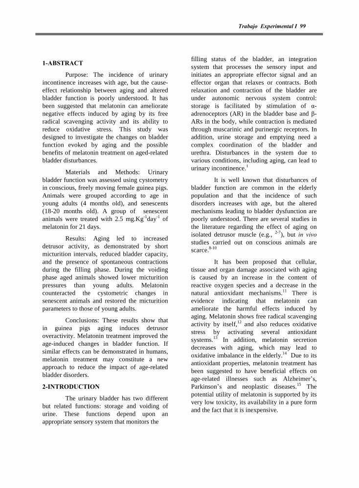
Trabajo Experimental I 99
1-ABSTRACT
Purpose: The incidence of urinary incontinence increases with age, but the cause-effect relationship between aging and altered bladder function is poorly understood. It has been suggested that melatonin can ameliorate negative effects induced by aging by its free radical scavenging activity and its ability to reduce oxidative stress. This study was designed to investigate the changes on bladder function evoked by aging and the possible benefits of melatonin treatment on aged-related bladder disturbances.
Materials and Methods: Urinary bladder function was assessed using cystometry in conscious, freely moving female guinea pigs. Animals were grouped according to age in young adults (4 months old), and senescents (18-20 months old). A group of senescent animals were treated with 2.5 mg.Kg-1day-1 of melatonin for 21 days.
Results: Aging led to increased detrusor activity, as demonstrated by short micturition intervals, reduced bladder capacity, and the presence of spontaneous contractions during the filling phase. During the voiding phase aged animals showed lower micturition pressures than young adults. Melatonin counteracted the cystometric changes in senescent animals and restored the micturition parameters to those of young adults.
Conclusions: These results show that in guinea pigs aging induces detrusor overactivity. Melatonin treatment improved the age-induced changes in bladder function. If similar effects can be demonstrated in humans, melatonin treatment may constitute a new approach to reduce the impact of age-related bladder disorders.
2-INTRODUCTION
The urinary bladder has two different but related functions: storage and voiding of urine. These functions depend upon an appropriate sensory system that monitors the
filling status of the bladder, an integration system that processes the sensory input and initiates an appropriate effector signal and an effector organ that relaxes or contracts. Both relaxation and contraction of the bladder are under autonomic nervous system control: storage is facilitated by stimulation of á-adrenoceptors (AR) in the bladder base and â-ARs in the body, while contraction is mediated through muscarinic and purinergic receptors. In addition, urine storage and emptying need a complex coordination of the bladder and urethra. Disturbances in the system due to various conditions, including aging, can lead to urinary incontinence.1
It is well known that disturbances of bladder function are common in the elderly population and that the incidence of such disorders increases with age, but the altered mechanisms leading to bladder dysfunction are poorly understood. There are several studies in the literature regarding the effect of aging on isolated detrusor muscle (e.g., 2-7), but in vivo studies carried out on conscious animals are scarce.8-10
It has been proposed that cellular, tissue and organ damage associated with aging is caused by an increase in the content of reactive oxygen species and a decrease in the natural antioxidant mechanisms.11 There is evidence indicating that melatonin can ameliorate the harmful effects induced by aging. Melatonin shows free radical scavenging activity by itself,12 and also reduces oxidative stress by activating several antioxidant systems.13 In addition, melatonin secretion decreases with aging, which may lead to oxidative imbalance in the elderly.14 Due to its antioxidant properties, melatonin treatment has been suggested to have beneficial effects on age-related illnesses such as Alzheimer�s, Parkinson�s and neoplastic diseases.15 The potential utility of melatonin is supported by its very low toxicity, its availability in a pure form and the fact that it is inexpensive.

100 PJ Gómez-Pinilla
To date, there seems to be no study aimed at investigating the benefits of melatonin treatment in bladder function during aging. Therefore, using continuous cystometry, we have studied and compared micturition in conscious young adult, and senescent guinea pigs. In addition, we have treated senescent guinea pigs with melatonin to investigate its effects on aging-induced changes in bladder function.
3-MATERIALS AND METHODS
Animals: Female guinea pigs were divided in two groups according to age: young adults (4 months old), senescents (18-20 months old). A group of senescent animals was treated orally with melatonin (2.5 mg.Kg-1day-1). This treatment was applied daily, 1 hour before the dark phase for 21 days. Animals were housed in light (12 h light-dark cycle) and temperature (20 ºC) controlled rooms and had free access to water and food. The experiments were performed according to European guidelines for animal research and approved by the Animal Ethics Committee, Court of the City of Lund, Sweden.
Catheter implantation: During anesthesia (ketamine 20 mg.Kg-1 and xylazine 5 mg.Kg-1, intraperitoneally) a lower midline abdominal incision was made to open the abdominal cavity and expose the urinary bladder, and a small incision was made at the back of the neck, creating a subcutaneous pouch. A polyethylene catheter (PE-10) was inserted into the dome of the urinary bladder and secured in place using a purse string suture. The bladder was emptied and the catheter was tunnelled subcutaneously and placed on the subcutaneous pouch at the back of the neck. The distal end of the catheter was tied off to avoid leakage.
Urodynamic measurements in conscious, unrestrained guinea pigs: Cystometry was performed without anesthesia
three days after the implantation of the catheters. The conscious and unrestrained guinea-pigs were placed in metabolic cages and the bladder catheters were connected via a T-tube to a pressure transducer (P23 DC; Statham Instruments Inc., Oxnard, CA, USA) and a microinjection pump (CMA 100; Carnegie Medicine AB, Solna, Sweden). Micturition volumes were recorded with a fluid collector connected to a force displacement transducer (FT 03 D, Grass Instrument Co., Quincy, MA, USA). Room-temperature saline was infused into the bladder continuously at a rate of 10 mL. h�1. Pressures and micturition volumes were recorded continuously with Acq Knowledge 3.5.3 software and a MP100 data acquisition system (Biopac Syst. Inc. Santa Barbara, CA) connected to a Grass polygraph (Model 7E, Grass Instrument Co). At the beginning of cystometry, the bladder was emptied via the bladder catheter.
The following cystometric parameters were analyzed: basal pressure (cm H20), threshold pressure (pressures just before micturition, cm H20), threshold pressure corrected (threshold pressure minus basal pressure, cm H20), compliance (ml of infused volume per cm H20 of increase in bladder pressure during filling phase, ml.cm H20
-1), maximal micturition pressure (cm H20), micturition duration (s), micturition interval (time between 2 subsequent micturitions, min) and micturition volume (ml). Three reproducible micturition cycles were analyzed, and the results averaged.
Statistical analysis: Data are presented as mean values ± SEM. Statistical analysis was based on one-way analysis of variance (ANOVA) followed by Bonferroni´s multiple comparison test. P values <0.05 were considered to be significant. To compare frequency distribution ÷2 analysis was used.
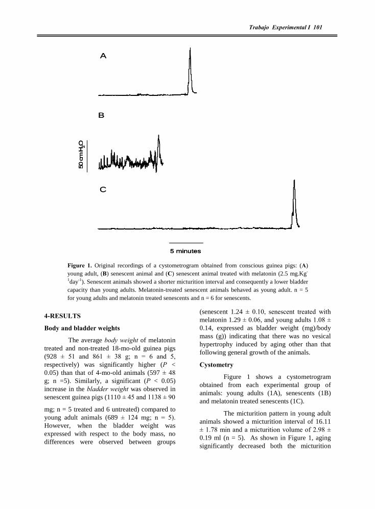
Trabajo Experimental I 101
4-RESULTS
Body and bladder weights
The average body weight of melatonin treated and non-treated 18-mo-old guinea pigs (928 ± 51 and 861 ± 38 g; n = 6 and 5, respectively) was significantly higher (P < 0.05) than that of 4-mo-old animals (597 ± 48 g; n =5). Similarly, a significant (P < 0.05) increase in the bladder weight was observed in senescent guinea pigs (1110 ± 45 and 1138 ± 90
mg; n = 5 treated and 6 untreated) compared to young adult animals (689 ± 124 mg; n = 5). However, when the bladder weight was expressed with respect to the body mass, no differences were observed between groups
(senescent 1.24 ± 0.10, senescent treated with melatonin 1.29 ± 0.06, and young adults 1.08 ± 0.14, expressed as bladder weight (mg)/body mass (g)) indicating that there was no vesical hypertrophy induced by aging other than that following general growth of the animals.
Cystometry
Figure 1 shows a cystometrogram obtained from each experimental group of animals: young adults (1A), senescents (1B) and melatonin treated senescents (1C).
The micturition pattern in young adult animals showed a micturition interval of 16.11 ± 1.78 min and a micturition volume of 2.98 ± 0.19 ml (n = 5). As shown in Figure 1, aging significantly decreased both the micturition
Figure 1. Original recordings of a cystometrogram obtained from conscious guinea pigs: (A) young adult, (B) senescent animal and (C) senescent animal treated with melatonin (2.5 mg.Kg-
1day-1). Senescent animals showed a shorter micturition interval and consequently a lower bladder capacity than young adults. Melatonin-treated senescent animals behaved as young adult. n = 5 for young adults and melatonin treated senescents and n = 6 for senescents.
A
B
C
5 minutes
50 c
mH
2O
A
B
C
5 minutes5 minutes
50 c
mH
2O
50
cm
H2O
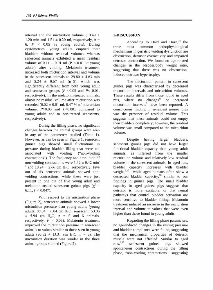
102 PJ Gómez-Pinilla
interval and the micturition volume (10.49 ± 1.20 min and 1.51 ± 0.20 ml, respectively, n = 6, P < 0.05 vs young adults). During cystometries, young adults emptied their bladders without residual volumes whereas senescent animals exhibited a mean residual volume of 0.11 ± 0.01 ml (P < 0.01 vs young adults) after voiding. Melatonin treatment increased both micturition interval and volume in the senescent animals to 29.80 ± 4.63 min and 5.24 ± 0.67 ml (n=5), which was significantly different from both young adult and senescent groups (P <0.05 and P< 0.01, respectively). In the melatonin-treated animals, almost no residual volume after micturition was recorded (0.02 ± 0.01 ml, 0.47 % of micturition volume, P>0.05 and P<0.001 compared to young adults and to non-treated senescents, respectively).
During the filling phase, no significant changes between the animal groups were seen in any of the parameters studied (Table 1). However, as can be seen in Figure 1, senescent guinea pigs showed small fluctuations in pressure during bladder filling that were not associated with voiding (�non-voiding contractions�). The frequency and amplitude of non-voiding contractions were 1.32 ± 0.42 min-
1 and 10.24 ± 2.66 cm H2O, respectively. Five out of six senescent animals showed non-voiding contractions, while these were just present in one out of five young adult and melatonin-treated senescent guinea pigs (÷2 = 6.11, P < 0.047).
With respect to the micturition phase (Figure 2), senescent animals showed a lower micturition pressure than young adults (young adults: 88.84 ± 6.04 cm H2O, senescent: 53.06 ± 5.94 cm H2O, n = 5 and 6 animals, respectively, P < 0.05). Melatonin treatment improved the micturition pressure in senescent animals to values similar to those seen in young adults (90.52 ± 13.31 cm H2O, n = 5). The micturition duration was similar in the three animal groups studied (Figure 2).
5-DISCUSSION
According to Hald and Horn,16 the three most common pathophysiological mechanisms in geriatric voiding dysfunction are obstruction, detrusor overactivity and impaired detrusor contraction. We found no age-related changes in the bladder/body weight ratio, suggesting that there was no obstruction-induced detrusor hypertrophy.
The micturition pattern in senescent guinea pigs was characterized by decreased micturition intervals and micturition volumes. These results differ from those found in aged rats, where no changes10 or increased micturition intervals9 have been reported. A conspicuous finding in senescent guinea pigs was the presence of residual volume. This suggests that these animals could not empty their bladders completely; however, the residual volume was small compared to the micturition volume.
Despite having larger bladders, senescent guinea pigs did not have larger functional bladder capacity than young adult animals, as inferred from the smaller micturition volume and relatively low residual volume in the senescent animals. In aged rats, bladder capacity increases with bladder weight,9,17 while aged humans often show a decreased bladder capacity,18 similar to our findings in guinea pigs. The small bladder capacity in aged guinea pigs suggests that detrusor is more excitable, or that neural pathways that control bladder activation are more sensitive to bladder filling. Melatonin treatment induced an increase in the micturition interval and volume to values that were even higher than those found in young adults.
Regarding the filling phase parameters, no age-induced changes in the resting pressure and bladder compliance were found, suggesting that the mechanical properties of detrusor muscle were not affected. Similar to aged rats,9,17 senescent guinea pigs showed spontaneous contractions during the filling phase, �non-voiding contractions�, suggesting

Trabajo Experimental I 103
an increased contractile activity in the detrusor muscle. However, it has been proposed that such activity in aging could also result from altered central nervous system control, which does not rule out aging-induced changes in the contractility of the detrusor smooth muscle or the intrinsic nerves in the bladder. Interestingly, melatonin treatment reduced the non-voiding contractions to the same level as found in young adults.
Although micturition duration was not altered by aging, micturition pressure was impaired in aged guinea pigs, in contrast to findings in senescent rats.9,17 Such a decrease could contribute to the residual volume described above. It cannot be excluded that the reduction of pressure was secondary to structural changes in aged detrusor muscle. However, there were no signs of detrusor hypertrophy, and the bladder compliance was similar in adult and senescent animals. A plausible explanation is that aging affects smooth muscle contractility and that this process is affected by melatonin. This would explain the low micturition pressure induced by aging and the elevated pressure reached in melatonin treated animals. Studies to test these possibilities have therefore been initiated. An age-related decrease in muscarinic receptor mediated activation of the voiding contraction6
or changes in sensory input9 could also contribute to the decreased micturition pressure in aged animals.
Melatonin is secreted by the pineal gland predominantly during the night, and in addition to its well-known role in setting circadian rhythms, it is a powerful antioxidant, acting either as a scavenger or through activation of antioxidant enzymes. Nocturnal production of melatonin is impaired in elderly individuals, and several clinical trials have demonstrated that exogenous administration of melatonin improves circadian disturbances.15 As proposed by Harman,11 aging is due in part to accumulation of reactive oxygen species. If true, age-induced impairment of melatonin secretion may participate in the increase of oxidative damage associated with aging.
In this study, melatonin treatment reversed the residual volume, detrusor overactivity, and the decrease of micturition pressure induced by aging. The effect of melatonin could be due to recovery of circadian rhythm (melatonin was administered at the onset of dark phase), the general improvement of physiological functions, or to local effects in the bladder. In agreement with the beneficial effects of melatonin in this study, it has been reported that aged human with bladder outflow obstruction and nocturia treated with melatonin
Figure 2. Age-related changes in the voiding phase parameters. Micturion pressure (A) in senescent guinea pigs was lower (P < 0.05) than in young adult and melatonin-treated senescent animals. Micturition duration (B) was similar in the three experimental groups. n = 5 for young adults and melatonin treated senescents and n = 6 for senescents.
Adults Senescent Melatonin0
50
100
150
***
Pressure(cmH 2O)
Micturition duration
Adults Senescents Melatonin0
10
20
30
40
Duration(s)
Micturition pressure
Adults Senescent Melatonin0
50
100
150
***
Pressure(cmH 2O)
Micturition duration
Adults Senescents Melatonin0
10
20
30
40
Duration(s)
Micturition pressure

104 PJ Gómez-Pinilla
showed a decrease in the frequency of micturition during night.19
The present study shows that melatonin can normalize age-induced urinary bladder dysfunction. Melatonin treatment in vivo has been found to protect rat urinary bladder against oxidative damage.20 Although the exact mechanism of action has not yet been elucidated, the effects of melatonin on altered bladder function in guinea pig suggest that it may be considered a potentially useful therapeutic agent for treating aged-related detrusor overactivity. Indeed, melatonin may be a powerful tool against age-related diseases in general.15
6-CONCLUSIONS
In the guinea pig, aging induces changes in micturition pattern suggesting an increased bladder activity during the filling phase, but also causes a decrease in the micturition pressure, possibly related to a decrease in the contractility of the detrusor. Melatonin treatment counteracted all these age-induced changes, suggesting that melatonin could be a candidate to palliate urinary bladder dysfunction evoked by aging.
7-ACKNOWLEDGEMENTS
Supported by Junta de Extremadura (2PR03A020) and Ministerio de Educacion y Ciencia (BFU 2004-0637). Pedro J Gómez-Pinilla is recipient of Doctoral Fellowship from Junta de Extremadura.
Table 1. Parameters related to the filling phase in the three groups of guinea pigs.
Adult Senescent Melatonin treated
Basal pressure (cm H2O) 19.5 ± 4.97 17.85 ± 2.21 18.52 ± 2.94
Threshold pressure (cm H2O) 23.27 ± 4.94 20.46 ± 2.53 24.04 ± 4.79
Threshold pressure corrected (cm H2O) 3.83 ± 1.05 3.04 ± 0.37 3.91 ± 1.14
Compliance (ml/cm H2O) 0.88 ± 0.21 0.67 ± 0.16 0.95 ± 0.25
Data from 5 adult, 6 senescent, and 5 senescent, melatonin-treated animals expressed as mean ± SEM
8-REFERENCE LIST
1. Andersson, K. E. and Wein, A. J.: Pharmacology of the lower urinary tract: basis for current and
future treatments of urinary incontinence. Pharmacol Rev, 56: 581, 2004.
2. Kolta, M. G., Wallace, L. J., and Gerald, M. C.: Age-Related-Changes in Sensitivity of Rat Urinary-Bladder to Autonomic Agents. Mechanisms of Ageing and Development, 27: 183, 1984.
3. Johnson, J. M., Skau, K. A., Gerald, M. C., and Wallace, L. J.: Regional Noradrenergic and Cholinergic Neurochemistry in the Rat Urinary-Bladder - Effects of Age. Journal of Urology, 139: 611, 1988.
4. Longhurst, P. A., Eika, B., Leggett, R. E., and Levin, R. M.: Comparison of Urinary-Bladder Function in 6 and 24 Month Male and Female Rats. Journal of Urology, 148: 1615, 1992.

Trabajo Experimental I 105
5. Munro, D. D. and Wendt, I. R.: Contractile and Metabolic Properties of Longitudinal Smooth-Muscle from Rat Urinary-Bladder and the Effects of Aging. Journal of Urology, 150: 529, 1993.
6. Yoshida, M., Homma, Y., Inadome, A., Yono, M., Seshita, H., Miyamoto, Y. et al.: Age-related changes in cholinergic and purinergic neurotransmission in human isolated bladder smooth muscles. Experimental Gerontology, 36: 99, 2001.
7. Schneider, T., Hein, P., Michel-Reher, M. B., and Michel, M. C.: Effects of ageing on muscarinic receptor subtypes and function in rat urinary bladder. Naunyn Schmiedebergs Arch Pharmacol, 372: 71, 2005.
8. Chun, A. L., Wallace, L. J., Gerald, M. C., Levin, R. M., and Wein, A. J.: Effect of Age on Invivo Urinary-Bladder Function in the Rat. Journal of Urology, 139: 625, 1988.
9. Chai, T. C., Andersson, K. E., Tuttle, J. B., and Steers, W. D.: Altered neural control of micturition in the aged F344 rat. Urological Research, 28: 348, 2000.
10. Lluel, P., Palea, S., Barras, M., Grandadam, F., Heudes, D., Bruneval, P. et al.: Functional and morphological modifications of the urinary bladder in aging female rats. American Journal of Physiology-Regulatory Integrative and Comparative Physiology, 278: R964, 2000.
11. Harman, D.: Free-Radical Theory of Aging. Mutation Research, 275: 257, 1992.
12. Tan, D. X., Manchester, L. C., Reiter, R. J., Qi, W. B., Karbownik, M., and Calvo, J. R.: Significance of melatonin in antioxidative defense system: Reactions and products. Biological Signals and Receptors, 9: 137, 2000.
13. Reiter, R. J., Tan, D. X., Osuna, C., and Gitto, E.: Actions of melatonin in the reduction of oxidative stress - A review. Journal of Biomedical Science, 7: 444, 2000.
14. Reiter, R. J.: The Aging Pineal-Gland and Its Physiological Consequences. Bioessays, 14: 169, 1992.
15. Poeggeler, B.: Melatonin, aging, and age-related diseases: perspectives for prevention, intervention, and therapy. Endocrine, 27: 201, 2005.
16. Hald, T. and Horn, T.: The human urinary bladder in ageing. Br J Urol, 82 Suppl 1: 59, 1998.
17. Lluel, P., Deplanne, V., Heudes, D., Bruneval, P., and Palea, S.: Age-related changes in urethrovesical coordination in male rats: relationship with bladder instability? American Journal of Physiology-Regulatory Integrative and Comparative Physiology, 284: R1287, 2003.
18. Castleden, C. M., Duffin, H. M., and Asher, M. J.: Clinical and Urodynamic Studies in 100 Elderly Incontinent Patients. British Medical Journal, 282: 1103, 1981.
19. Drake, M. J., Mills, I. W., and Noble, J. G.: Melatonin pharmacotherapy for nocturia in men with benign prostatic enlargement. Journal of Urology, 171: 1199, 2004.
20. Paskaloglu, K., Sener, G., Kapucu, C., and Ayanoglu-Dulger, G.: Melatonin treatment protects against sepsis-induced functional and biochemical changes in rat ileum and urinary bladder. Life Sciences, 74: 1093, 2004.

106 PJ Gómez-Pinilla

Trabajo Experimental II 107
American Juornal of Physiology Regulatory- Integrative and Comparative Physiology (Under Review)
AGING IMPAIRS NEUROGENIC CONTRACTION IN GUINEA PIG URINARY BLADDER. THE ROLE OF OXIDATIVE STRESS AND MELATONIN
Pedro J. Gomez-Pinilla, Maria J. Pozo and Pedro J. Camello*
Dept of Physiology, Nursing School,
University of Extremadura, 10071Cáceres, Spain
Running head: melatonin & aged bladder innervation
*Correspondence to:
Dr Pedro J Camello
Faculty of Veterinary Sciences, Dept of Physiology
Campus Universitario s/n
10071 Cáceres, Spain
Ph: +927-257100 Ext 1321/1290
Fax: +927-257110
Key Words: urinary bladder, neuromuscular function, electrical field stimulation, aging, melatonin, guinea pig
id3591656 pdfMachine by Broadgun Software - a great PDF writer! - a great PDF creator! - http://www.pdfmachine.com http://www.broadgun.com

108 PJ Gómez-Pinilla
CONTENTS
1- Abstract
2- Introduction
3- Material and methods
Animals and tissue preparation
Contraction recording of guinea pig urinary bladder smooth muscle strips
Malondialdehyde (MDA) and reduced glutathione (GSH) assays
Solutions and drugs
Quantification and statistic
4- Results
Excitatory innervation
Inhibitory innervation
Oxidative stress in aged guinea pig urinary bladder
5- Discussion
6- Acknowledgements
7- Reference list

Trabajo Experimental II 109
1-ABSTRACT
The incidence of urinary bladder disturbances increases with age, and free radical accumulation has been proposed as causal factors. We have recently described that age-induced alterations of micturition pattern are ameliorated by melatonin treatment. In the current study we investigate the association between changes on bladder innervation and oxidative stress in aging and the possible benefits of melatonin treatment as antioxidant and neuro-protector agent.
Urinary bladder neuromuscular function was assessed by electrical field stimulation (EFS) of isolated guinea pig detrusor strips. Neurotransmitter blockers were used to dissect pharmacologically the EFS-elicited contractile response. Female guinea pigs were grouped according to age in young adults (4 months old), and aged (18-20 months old). A group of aged animals were treated with 2.5 mg.Kg-1day-1 melatonin for 28 days.
EFS induced a neurogenic and frequency-dependent contraction which was impaired by aging. Age also decreased the sensitivity of the contraction to pharmacological blockade of purinergic (suramin application) and sensitive (capsaicin treatment) fibers, but increased the effect of blockade of nitrergic (L-NAME) and adrenergic (guanethidine) nerves. These changes were associated to an increased level of markers for oxidative stress. Melatonin treatment normalized oxidative levels and counteracted the ageing-associated changes in bladder innervation. In conclusion, these results show that aging modifies the functional profile of the urinary bladder plexus and simultaneously increases the oxidative damage to the organ.
Melatonin reduces oxidative stress and improves the age-induced changes in bladder neuromuscular function. This treatment may constitute a new pharmacological tool to reduce the impact of age-related bladder disorders.
2-INTRODUCTION
Continence and voiding of urine are two important processes controlled by a complex neural network with intrinsic and extrinsic components. In most animal species, bladder contraction is mediated by both cholinergic and nonadrenergic, noncholinergic (NANC) mechanisms (37). Under normal conditions, the micturition contraction in vivo and the contraction evoked by electrical stimulation of nerves in vitro is considered to be mainly mediated by muscarinic receptor stimulation (21). Though M3 receptors are not the more expressed cholinergic subtype, they account for the physiological voiding contraction (7). Adrenergic inputs to urinary bladder have been identified, but their functional role is not well established. The á-adrenergic receptors are mainly expressed in the bladder neck, where they keep the continence, while -adrenergic receptors are generally distributed in the bladder body, its activation leading to relaxation during the filling phase (13). In the bladder, the second more important excitatory neurotransmitter is ATP, which acts on two families of purinergic receptors: P2X, an ion channel receptor, and P2Y, a family of G protein-coupled receptors (5). ATP is released to bladder smooth muscle by both efferent and afferent synapses, the later serving as a mechanosensor pathway reporting about urinary bladder distension (11). Although nitrergic fibres are present in the urinary bladder direct relaxation of the detrusor smooth muscle in response to NO has not been reported, and therefore the role of NO may be to modulate other transmitters and/or to participate in afferent neurotransmission (1). Other neurotransmitters such as vasoactive intestinal polypeptide, endothelins, tachikinins, angiotensin and prostanoids have been found in the nervous plexus of the urinary bladder, and their functional role has been reviewed in (3).
It is well known that disturbances of bladder function are common in the elderly population and that the incidence of such disorders increases with age, but the altered

110 PJ Gómez-Pinilla
mechanisms leading to bladder dysfunction are poorly understood. Age-related changes in the innervation of the bladder are of particular interest in understanding how aging impairs contractility given the important role of nerves in the control of bladder function. There are several studies in the literature regarding the effect of aging on the neurotransmitter-induced responses in bladder smooth muscle. However, there is a great variability in the reported results, and some studies indicate age-dependent increases (23; 35) or decreases in ACh-induced responses (38), whereas others report no changes in carbachol-evoked contraction (28; 40) or increased norepinephine-elicited contractions (29; 35). Information about age-related changes in the neuromuscular function in the bladder is scarce. Yoshida et al reported in humans no changes in the global response to EFS (40), although aging caused a decrease and an increase in the cholinergic and purinergic components of EFS, respectively. In rats, aging decreases EFS-induced neurogenic contractions, but changes in the different components of the neurotransmission were not studied (28). There are no reports on the amelioration of age-related changes of neuromuscular function.
The age-induced damage has been associated to an increased reactive species production and a decrease in the cellular antioxidant mechanisms (16). In this regard, melatonin, a potent endogenous free radical scavenger and antioxidant which declines with age, has been proposed as a good candidate to palliate the ageing associated alterations (20). In fact, we have recently reported that melatonin exerts beneficial effects on the micturition pattern (15) and in the gallbladder neurotransmission (14) of aged guinea pigs. However, there is no information about the effects of melatonin on neural alterations induced by aging in the urinary bladder.
The current study was designed to investigate the effect of aging on neurally evoked urinary bladder contraction and the participation of the different neurotransmitters.
In addition, we wanted to assess whether the antioxidant properties of melatonin can ameliorate the alterations of bladder neuromuscular function associated to age.
3-MATERIALS AND METHODS
Animals and tissue preparation., Urinary bladders, isolated from 4-and 20-month-old female guinea pigs after deep halothane anaesthesia and cervical dislocation, were immediately placed in cold Krebs-Henseleit solution (K-HS; for composition see Solutions and drugs) at pH 7.35. The urinary bladder was cleaned of fatty tissue, opened longitudinally, washed with K-HS solution to remove urine remains and the urothelium was carefully dissected away.
A group of senescent animal was treated with melatonin (2.5 mg/Kg/day, per os) during 28 days just before the start of the dark phase (7 p.m.). All the experiments were carried out according to the guidelines of Animal Care and Use Committees of the University of Extremadura.
Contraction recording of guinea pig urinary bladder smooth muscle strips. Strips of detrusor muscle (~4 x 15 mm) were placed vertically in a 10 ml organ bath filled with K-HS maintained at 37 °C and gassed with 95% O2 - 5% CO2. Isometric contractions were measured using force displacement transducers interfaced with a Macintosh computer using MacLab hardware and dedicated software (ADInstruments; Colorado Spring, CO, USA). The muscle strips were placed under an initial resting tension of 1.5 g, and allowed to equilibrate for 60 min, with solution changes every 20 min. All the strips obtained from the same animal were used in a different experimental protocol.
Intrinsic nerves were activated by electrical field stimulation (EFS) with a pair of external platinum ring electrodes (0.7 mm in diameter) connected to a square wave stimulator (Cibertec CS9/3BO) programmed through Scope software application from Mac
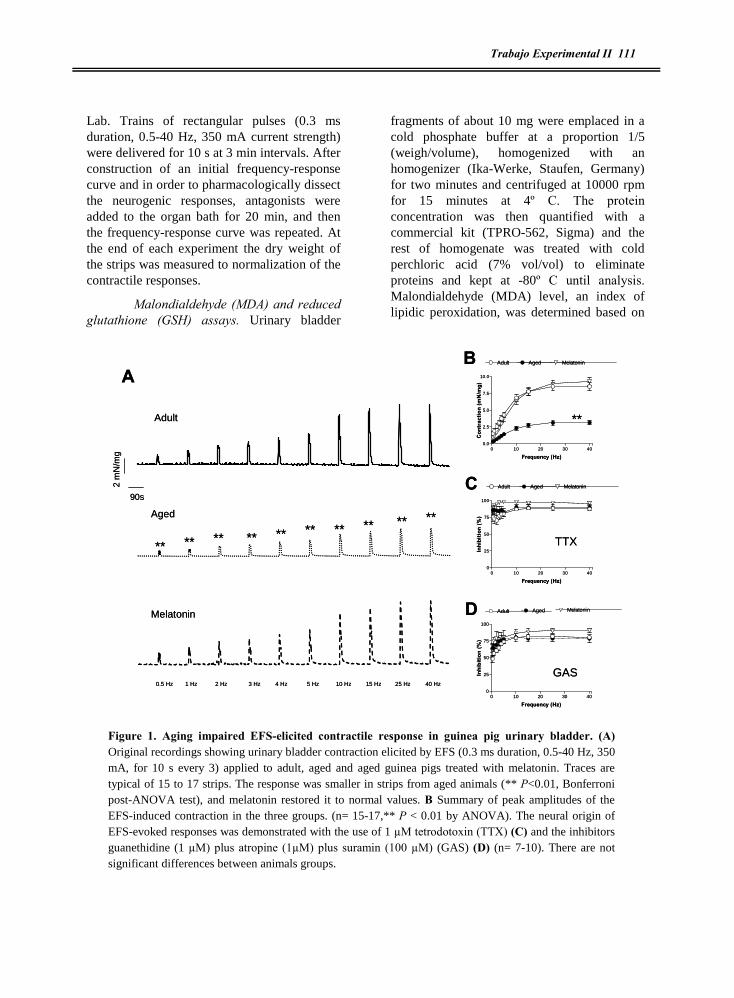
Trabajo Experimental II 111
Lab. Trains of rectangular pulses (0.3 ms duration, 0.5-40 Hz, 350 mA current strength) were delivered for 10 s at 3 min intervals. After construction of an initial frequency-response curve and in order to pharmacologically dissect the neurogenic responses, antagonists were added to the organ bath for 20 min, and then the frequency-response curve was repeated. At the end of each experiment the dry weight of the strips was measured to normalization of the contractile responses.
Malondialdehyde (MDA) and reduced glutathione (GSH) assays. Urinary bladder
fragments of about 10 mg were emplaced in a cold phosphate buffer at a proportion 1/5 (weigh/volume), homogenized with an homogenizer (Ika-Werke, Staufen, Germany) for two minutes and centrifuged at 10000 rpm for 15 minutes at 4º C. The protein concentration was then quantified with a commercial kit (TPRO-562, Sigma) and the rest of homogenate was treated with cold perchloric acid (7% vol/vol) to eliminate proteins and kept at -80º C until analysis. Malondialdehyde (MDA) level, an index of lipidic peroxidation, was determined based on
0 10 20 30 400
25
50
75
100
Adult Aged Melatonin
Frequency (Hz)
Inhi
bitio
n (%
)
0 10 20 30 400
25
50
75
100
Adult Aged Melatonin
Frequency (Hz)
Inhi
bitio
n (%
)
0 10 20 30 400.0
2.5
5.0
7.5
10.0
Adult Aged Melatonin
Frequency (Hz)
Con
trac
tion
(mN
/mg)
2 m
N/m
g
90s
Adult
Aged
Melatonin
0.5 Hz 1 Hz 2 Hz 3 Hz 4 Hz 5 Hz 10 Hz 15 Hz 25 Hz 40 Hz
GAS
TTX
AB
C
D
********************
**
0 10 20 30 400
25
50
75
100
Adult Aged Melatonin
Frequency (Hz)
Inhi
bitio
n (%
)
0 10 20 30 400
25
50
75
100
Adult Aged Melatonin
Frequency (Hz)
Inhi
bitio
n (%
)
0 10 20 30 400.0
2.5
5.0
7.5
10.0
Adult Aged Melatonin
Frequency (Hz)
Con
trac
tion
(mN
/mg)
2 m
N/m
g
90s
Adult
Aged
Melatonin
0.5 Hz 1 Hz 2 Hz 3 Hz 4 Hz 5 Hz 10 Hz 15 Hz 25 Hz 40 Hz
GAS
TTX
AB
C
D
********************
**
Figure 1. Aging impaired EFS-elicited contractile response in guinea pig urinary bladder. (A) Original recordings showing urinary bladder contraction elicited by EFS (0.3 ms duration, 0.5-40 Hz, 350 mA, for 10 s every 3) applied to adult, aged and aged guinea pigs treated with melatonin. Traces are typical of 15 to 17 strips. The response was smaller in strips from aged animals (** P<0.01, Bonferroni post-ANOVA test), and melatonin restored it to normal values. B Summary of peak amplitudes of the EFS-induced contraction in the three groups. (n= 15-17,** P < 0.01 by ANOVA). The neural origin of EFS-evoked responses was demonstrated with the use of 1 µM tetrodotoxin (TTX) (C) and the inhibitors guanethidine (1 µM) plus atropine (1µM) plus suramin (100 µM) (GAS) (D) (n= 7-10). There are not significant differences between animals groups.

112 PJ Gómez-Pinilla
colorimetric Recknagel´s method (39). Briefly, the samples were incubated with 0.4 % of tiobarbituric acid at 80º C for 20 minutes and later the sample absorbance at 550 nm was measured. Reduced glutathione determination was carried out following the Hissin and Hilf method (17): samples were incubated with 0.005 % of ortophtaldehide in the darkness at room temperature for 45 minutes and the fluorescent complex formed, indicative of reduced glutathione (GSH) level, was
measured with a fluorimeter (excitation 350 nm, emission 425 nm).
Solutions and drugs, The K-HS contained (in mM): 113 NaCl, 4.7 KCl, 2.5 CaCl2, 1.2 KH2PO4, 1.2 MgSO4, 25 NaHCO3, and 11.5 D-glucose. This solution had a final pH of 7.35 after equilibration with 95% O2-5% CO2. The phosphate buffer used to homogenize the tissue contained (in mM): NaCl 20, KCl 2.7, Na2HPO4 16, NaH2PO4 4, pH 7.4. Drug
0 10 20 30 40 500
1
2
3
4
5
6
7
8
9
Control Atropine (1 M)
Frequency (Hz)
Con
trac
tion
(mN
/mg)
0 10 20 30 40 500.0
2.5
5.0
7.5
10.0
Control Atropine (1M)
Frequency (Hz)
Con
trac
tion
(mN
/mg)
0 10 20 30 40 500.0
2.5
5.0
7.5
10.0
Control Atropine
Frequency (Hz)
Con
trac
tion
(mN
/mg)
0 10 20 30 400
10
20
30
40
50
60
70
Adult Aged Melatonin
Frequency (Hz)
Inhi
bitio
n (%
)
A B
C D
**
**
**
Figure 2. Aging did not alter the cholinergic component of the contraction in the guinea pig urinary bladder. Effect of 1 µM of atropine on EFS-elicited contractile response in guinea pig urinary bladder from adult (A), aged (B) and aged treated with melatonin guinea pigs (C). After EFS was performed in control conditions, strips were incubated for 20 min with atropine and EFS was repeated. Atropine reduced the contraction in a frequency-dependent manner with maximal inhibition (50 %) at 40 Hz. Inset shows corresponding original recording of 25 Hz evoked responses from each animal group in the absence and presence of atropine. (D) Summary of atropine effect on EFS response in the three guinea pig groups. No significant differences between groups were found at any of the frequencies tested. Data are from 25-13 urinary bladder stips. (** P < 0.01 by ANOVA).
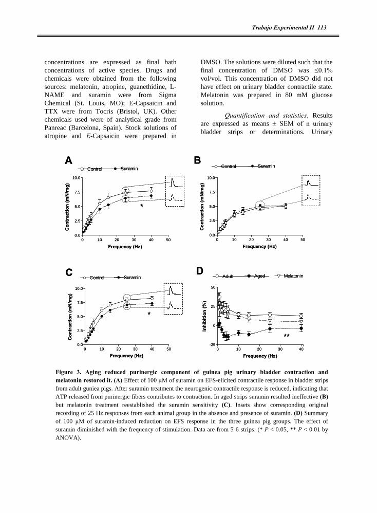
Trabajo Experimental II 113
concentrations are expressed as final bath concentrations of active species. Drugs and chemicals were obtained from the following sources: melatonin, atropine, guanethidine, L-NAME and suramin were from Sigma Chemical (St. Louis, MO); E-Capsaicin and TTX were from Tocris (Bristol, UK). Other chemicals used were of analytical grade from Panreac (Barcelona, Spain). Stock solutions of atropine and E-Capsaicin were prepared in
DMSO. The solutions were diluted such that the final concentration of DMSO was ≤0.1% vol/vol. This concentration of DMSO did not have effect on urinary bladder contractile state. Melatonin was prepared in 80 mM glucose solution.
Quantification and statistics. Results are expressed as means ± SEM of n urinary bladder strips or determinations. Urinary
0 10 20 30 40 500.0
2.5
5.0
7.5
10.0
Control Suramin
Frequency (Hz)
Con
trac
tion
(mN
/mg)
0 10 20 30 40 500.0
2.5
5.0
7.5
10.0
Control Suramin
Frequency (Hz)
Con
trac
tion
(mN
/mg)
0 10 20 30 40 500.0
2.5
5.0
7.5
10.0
Control Suramin
Frequency (Hz)
Con
trac
tion
(mN
/mg)
0 10 20 30 40-25
0
25
50
Adult Aged Melatonin
Frequency (Hz)
Inhi
bitio
n (%
)
A B
C D
*
*
**
0 10 20 30 40 500.0
2.5
5.0
7.5
10.0
Control Suramin
Frequency (Hz)
Con
trac
tion
(mN
/mg)
0 10 20 30 40 500.0
2.5
5.0
7.5
10.0
Control Suramin
Frequency (Hz)
Con
trac
tion
(mN
/mg)
0 10 20 30 40 500.0
2.5
5.0
7.5
10.0
Control Suramin
Frequency (Hz)
Con
trac
tion
(mN
/mg)
0 10 20 30 40-25
0
25
50
Adult Aged Melatonin
Frequency (Hz)
Inhi
bitio
n (%
)
A B
C D
*
*
**
Figure 3. Aging reduced purinergic component of guinea pig urinary bladder contraction and melatonin restored it. (A) Effect of 100 µM of suramin on EFS-elicited contractile response in bladder strips from adult guniea pigs. After suramin treatment the neurogenic contractile response is reduced, indicating that ATP released from purinergic fibers contributes to contraction. In aged strips suramin resulted ineffective (B) but melatonin treatment reestablished the suramin sensitivity (C). Insets show corresponding original recording of 25 Hz responses from each animal group in the absence and presence of suramin. (D) Summary of 100 µM of suramin-induced reduction on EFS response in the three guinea pig groups. The effect of suramin diminished with the frequency of stimulation. Data are from 5-6 strips. (* P < 0.05, ** P < 0.01 by ANOVA).

114 PJ Gómez-Pinilla
bladder tension is given in millinewtons (mN)/mg of tissue. Inhibition of contraction is calculated as the percentage decrease in the tension evoked by a treatment respect a previous control EFS performed in the same strip. All results from MDA and GSH determinations are given as nmol/mg of protein.
Statistical differences between animal groups and drug effects were determined using adequate analysis of variance (two ways ANOVA) followed by a Bonferroni�s post hoc test. Differences were considered significant at P < 0.05.
Figure 4. Aging reduced the role of excitatory sensory afferent fibers in the guinea pig urinary bladder. Effect of capsaicin on EFS-elicited contractile response in urinary bladder stips from adult (A), aged (B) and melatonin treated aged guniea pigs (C). After EFS was performed in control conditions, strips were incubated for 20 min with 10 µM capsaicin to promote afferent sensory denervation. Note that capsaicin reduced the EFS-induced contraction indicating that neural activation releases an excitatory component from sensory fibers. Insets show corresponding original recording of 25 Hz evoked contraction from each animal group in the absence and presence of 10 µM of capsaicin. (D) Summary of the reduction evoked by capsaicin treatment on EFS response in the three guinea pig groups. The sensory participation is smaller in bladder from aged animals. Data are from 5-9 urinary bladder strips. (* P < 0.05 and ** P < 0.01 by ANOVA).
0 10 20 30 40 500.0
2.5
5.0
7.5
10.0
Control Capsaicin
Frequency (Hz)
Con
trac
tion
(mN
/mg)
0 10 20 30 40 500.0
2.5
5.0
7.5
10.0
Control Capsaicin
Frequency (Hz)
Con
trac
tion
(mN
/mg)
0 10 20 30 400
10
20
30
40
50
60
Adult Aged Melatonin
Frequency (Hz)
Inhi
bitio
n (%
)
0 10 20 30 40 500.0
2.5
5.0
7.5
10.0
Control Capsaicin
Frequency (Hz)
Con
trac
tion
(mN
/mg)
A B
C D
*
**
**
*0 10 20 30 40 50
0.0
2.5
5.0
7.5
10.0
Control Capsaicin
Frequency (Hz)
Con
trac
tion
(mN
/mg)
0 10 20 30 40 500.0
2.5
5.0
7.5
10.0
Control Capsaicin
Frequency (Hz)
Con
trac
tion
(mN
/mg)
0 10 20 30 400
10
20
30
40
50
60
Adult Aged Melatonin
Frequency (Hz)
Inhi
bitio
n (%
)
0 10 20 30 40 500.0
2.5
5.0
7.5
10.0
Control Capsaicin
Frequency (Hz)
Con
trac
tion
(mN
/mg)
A B
C D
*
**
**
*

Trabajo Experimental II 115
4-RESULTS
Irrespective of the age of animals, electrical field stimulation of guinea pig urinary bladder produced a frequency-dependent contraction of about 10 seconds of duration reaching the maximal amplitude when the strips were stimulated at 25 or 40 Hz. As shown in figure 1, aging decreased the contractile response evoked by EFS, an effect reversed by melatonin treatment (Fig. 1A and 1B). To determine whether aging altered the neural origin of the EFS-evoked contraction, we used the nerve Na+ channel inhibitor tetrodotoxin (TTX, 1 µM). TTX almost abolished the response to EFS for all the frequencies tested in all the experimental groups (Fig. 1C). In keeping with this, we obtained similar results with a combined treatment with atropine (1 µM), suramin (100 µM) and guanethidine (1 µM), which blocks cholinergic, purinergic and adrenergic neurotransmission, respectively (Fig. 1D).
Excitatory innervation. Similar to several species, the main excitatory neurotransmitter in guinea pig urinary bladder is ACh. The cholinergic contribution to EFS-induced contractile response was determined using atropine (1 µM), which reduced the contraction in a frequency-dependent fashion in all experimental groups (Fig. 2A-C). Atropine effects ranged from 19 % inhibition at 0.5 Hz to 58 % inhibition at 40 Hz (Figure 2D), but it was unable to abolish the contraction, indicating the release of additional excitatory neurotransmitters in response to EFS. In order to characterize this non-cholinergic excitatory component we tested suramin, a purinergic antagonist, and capsaicin, which induces a sensory denervation when used at high concentration. In young adult guinea pigs 100 µM suramin reduced the EFS response in a negative frequency-dependent fashion (36 % inhibition at 0.5 Hz and 16 % inhibition at 40 Hz, Fig. 3A and 3D). By the contrary, suramin was ineffective in aged strips (Fig. 3B and 3D), suggesting that aging evokes a functional purinergic denervation in guinea pig urinary
bladder. Melatonin treatment of aged animals restored suramin effects on EFS-evoked response (35 % of inhibition at 0.5 Hz and 11 % of inhibition at 40 Hz, Fig. 3C and 3D).
As shown in figure 4, desensitization of afferent nerves by a high concentration of capsaicin (10 µM) reduced the response, revealing the contribution of afferent neurones in the contraction induced by EFS. Similar to the purinergic component, the afferent contribution was impaired by aging (Fig. 4B), although capsaicin conserved a slight effect on the contraction. Melatonin treatment also restored the excitability of the afferent innervation, increasing the sensitivity to capsaicin in aged strips to levels similar to those in young adult strips (Fig. 4).
To test the presence of interactions between purinergic and afferent innervation, the strips were treated with 100 µM suramin before or after application of 10 µM capsaicin. Both protocols resulted in additive effects of suramin and capsaicin (data not shown) indicating that these neural components do not interact. Following the same approach, we pre-treated the strips with 1 µM atropine before suramin or capsaicin to test respectively cholinergic modulation of purinergic and afferent excitatory neurotransmission. In young adult animals, suramin in the presence of atropine induced an inhibition of the EFS response which was similar to that obtained by suramin alone at low frequencies (suramin alone: 35.83 ± 6.53 % inhibition at 0.5 Hz, suramin after atropine: 29.59 ± 4.46 % inhibition at 0.5 Hz, compare effects of suramin in Fig. 3A & 5A) but it was higher at medium-high frequencies, suggesting that cholinergic nerves negatively modulate purinergic fibers. Surprisingly, in aged animals where application of 100 µM suramin alone did not have effects (see Fig. 3), suramin inhibited the EFS-elicited contractile response in presence of atropine (suramin alone: 0.98 ± 3.51 % inhibition at 0.5 Hz, suramin after atropine: 34.18 ± 4.96 % inhibition at 0.5 Hz, Fig. 5B), indicating that aging increases the cholinergic negative modulation of purinergic

116 PJ Gómez-Pinilla
innervation. In melatonin treated animals, the effect of suramin after atropine was similar to that in young animals, showing that at frequencies resembling physiological stimulation (0.5 Hz) there was no apparent cholinergic modulation of the purinergic neurotransmission (suramin alone: 31.71 ± 5.86 % inhibition at 0.5 Hz, suramin after atropine:
27.04 ± 2.33 % inhibition at 0.5 Hz, Figure 5C). Regarding the afferent sensitive fibers, capsaicin was unable to reduce the neurogenic response in presence of atropine (Fig. 5D, 5E, 5F), suggesting that in urinary bladder afferent innervation collaborates in the total response to EFS through activation of cholinergic fibers.
AAdult
0 10 20 30 400
2
4
6
8
10
12
Frequency (Hz)
Con
trac
tion
(mN
/mg)
Aged
B
0 10 20 30 400
2
4
6
8
10
12
Frequency (Hz)
Con
trac
tion
(mN
/mg)
CMelatonin
0 10 20 30 400
2
4
6
8
10
12
Frequency (Hz)
Con
trac
tion
(mN
/mg)
Control
Atropine
A + Sur
Control
AtropineA + Sur
Control
Atropine
A + Sur
DAdult
0 10 20 30 400
2
4
6
8
10
12
Frequency (Hz)
Con
trac
tion
(mN
/mg) Control
AtropineA + Cap
Aged
E
0 10 20 30 400
2
4
6
8
10
12
Frequency (Hz)
Con
trac
tion
(mN
/mg)
Control
AtropineA + Cap
MelatoninF
0 10 20 30 400
2
4
6
8
10
12
Frequency (Hz)
Con
trac
tion
(mN
/mg)
Control
AtropineA + Cap
Figure 5. Effects of cholinergic nerves on purinergic and sensory fibers in the urinary bladder. Effect of 100 µM of suramin after atropine (1 µM) on EFS-elicited contractile response in urinary bladder from adult (A), aged (B) and melatonin treated aged animals (C). Atropine does not block the inhibitory effect of suramin. Data are from 5-6 urinary bladder strips. (D), (E) and (F) In strips pretreated with atropine (1 µM) capsaicin (10 µM) resulted ineffective, indicating that cholinergic activation is necessary to release excitatory neurotransmitters from sensory nerves. Data are from 5-6 urinary bladder strips. (** P < 0.01 by ANOVA).
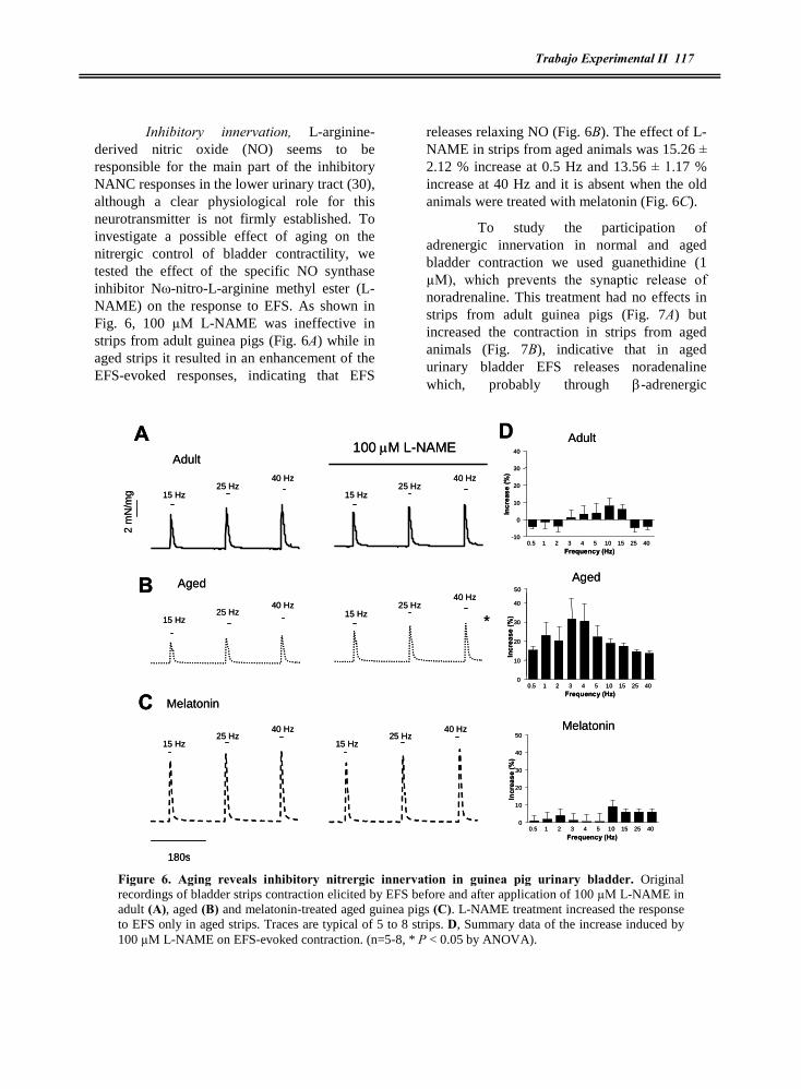
Trabajo Experimental II 117
Inhibitory innervation, L-arginine-derived nitric oxide (NO) seems to be responsible for the main part of the inhibitory NANC responses in the lower urinary tract (30), although a clear physiological role for this neurotransmitter is not firmly established. To investigate a possible effect of aging on the nitrergic control of bladder contractility, we tested the effect of the specific NO synthase inhibitor Nù-nitro-L-arginine methyl ester (L-NAME) on the response to EFS. As shown in Fig. 6, 100 µM L-NAME was ineffective in strips from adult guinea pigs (Fig. 6A) while in aged strips it resulted in an enhancement of the EFS-evoked responses, indicating that EFS
releases relaxing NO (Fig. 6B). The effect of L-NAME in strips from aged animals was 15.26 ± 2.12 % increase at 0.5 Hz and 13.56 ± 1.17 % increase at 40 Hz and it is absent when the old animals were treated with melatonin (Fig. 6C).
To study the participation of adrenergic innervation in normal and aged bladder contraction we used guanethidine (1 µM), which prevents the synaptic release of noradrenaline. This treatment had no effects in strips from adult guinea pigs (Fig. 7A) but increased the contraction in strips from aged animals (Fig. 7B), indicative that in aged urinary bladder EFS releases noradenaline which, probably through -adrenergic
2 m
N/m
g
180s
Adult
Aged
Melatonin
A
B
C
15 Hz25 Hz
40 Hz
15 Hz25 Hz
40 Hz
15 Hz25 Hz
40 Hz15 Hz
25 Hz40 Hz
15 Hz25 Hz
40 Hz
15 Hz25 Hz
40 Hz
100 M L-NAME
*
D
0.5 1 2 3 4 5 10 15 25 400
10
20
30
40
50
Frequency (Hz)
Incr
ease
(%)
50
0.5 1 2 3 4 5 10 15 25 40-10
0
10
20
30
40
Frequency (Hz)
Incr
ease
(%)
0.5 1 2 3 4 5 10 15 25 400
10
20
30
40
Frequency (Hz)
Incr
ease
(%)
Adult
Aged
Melatonin
Figure 6. Aging reveals inhibitory nitrergic innervation in guinea pig urinary bladder. Original recordings of bladder strips contraction elicited by EFS before and after application of 100 µM L-NAME in adult (A), aged (B) and melatonin-treated aged guinea pigs (C). L-NAME treatment increased the response to EFS only in aged strips. Traces are typical of 5 to 8 strips. D, Summary data of the increase induced by 100 µM L-NAME on EFS-evoked contraction. (n=5-8, * P < 0.05 by ANOVA).

118 PJ Gómez-Pinilla
receptors, leads to relaxation. This effect was 16.91 ± 2.15 % increase at 0.5 Hz and 17.09 ± 3.39 % increase at 40 Hz and it was reversed by treatment with melatonin (Fig. 7C).
The data presented above show that cholinergic neurons control purinergic and afferent fibres. To assess whether this control also occurred for the nitrergic and adrenergic innervation, we treated the strips with 100 µM L-NAME or 1 µM guanethidine in the presence of 1 µM atropine. Though L-NAME or guanethidine alone resulted ineffective in adult strips, in atropine-treated strips these inhibitors increased the EFS-induced response (Fig. 8A and 8B), indicating that in young adult guinea
pig urinary bladder the neural realease of NO and noradrenaline is inhibited by acetylcholine. The contraction in L-NAME and atropine treated strips was 160.27 ± 13.68 % respect to atropine treated strips at 0.5 Hz and 144.52 ± 12.16 % at 40 Hz. For guanethidine and atropine treated strips the response was 179.98 ± 10.30 % respect to atropine at 0.5 Hz and 152.81 ± 9.74 % at 40 Hz (Fig. 8). This cholinergic modulation of the inhibitory innervation is exclusive of adult animals, since it was absent in both aged and melatonin-treated animals (Fig.8).
Oxidative stress in aged guinea pig urinary bladder, MDA level, an index of lipidic
0
2
4
6
2 m
N/m
g
180s
Adult
Aged
Melatonin
A
B
C
15 Hz25 Hz
40 Hz
15 Hz25 Hz
40 Hz
15 Hz25 Hz
40 Hz15 Hz
25 Hz40 Hz
15 Hz25 Hz
40 Hz
15 Hz25 Hz
40 Hz
1 M Guanethidine
*
0.5 1 2 3 4 5 10 15 25 40-10
0
10
20
30
Frequency (Hz)
Incr
ease
(%)
0.5 1 2 3 4 5 10 15 25 400
10
20
30
40
Frequency (Hz)
Incr
ease
(%)
0.5 1 2 3 4 5 10 15 25 400
10
20
30
40
Frequency (Hz)
Incr
ease
(%)
D Adult
Aged
Melatonin
0
2
4
6
2 m
N/m
g
180s
Adult
Aged
Melatonin
A
B
C
15 Hz25 Hz
40 Hz
15 Hz25 Hz
40 Hz
15 Hz25 Hz
40 Hz15 Hz
25 Hz40 Hz
15 Hz25 Hz
40 Hz
15 Hz25 Hz
40 Hz
1 M Guanethidine
*
0.5 1 2 3 4 5 10 15 25 40-10
0
10
20
30
Frequency (Hz)
Incr
ease
(%)
0.5 1 2 3 4 5 10 15 25 400
10
20
30
40
Frequency (Hz)
Incr
ease
(%)
0.5 1 2 3 4 5 10 15 25 400
10
20
30
40
Frequency (Hz)
Incr
ease
(%)
D Adult
Aged
Melatonin
Figure 7. Aging evoked hipereactivity of inhibitory adrenergic innervation in guinea pig urinary bladder. Original recordings of urinary bladder contraction elicited by EFS in the presence and absence of 1 µM guanethidine in adult (A), aged (B) and melatonin-treated aged guinea pigs (C). Adrenergic depletion evoked by guanethidine increased the response to EFS indicative of noradrenaline-induced relaxation. Traces are typical of 6 to 11 strips. D, Summary data of guanethidine-induced increase on EFS-induced contractile. (n=6-11, * P < 0.05 by ANOVA).

Trabajo Experimental II 119
peroxidation, was significantly higher (P < 0.05) in bladders from aged animals (0.833 ± 0.097 nmol/mg of protein, n=7) than in those from young adult animals (0.552 ± 0.043 nmol/mg of protein, n=8). In parallel, aging caused a significant (P< 0.01) reduction in GSH levels from 5.04 ± 0.20 to 3.76 ± 0.29 nmol/mg of protein (adult and aged groups, respectively). Melatonin treatment reversed the elevations in MDA levels and the changes in GSH levels
evoked by ageing (MDA: 0.518 ± 0.084 nmol/mg of protein; GSH: 5.02 ± 0.40 nmol/mg of protein, n=6).
5-DISCUSSION
The results of the present study demonstrate that aging impairs EFS-evoked contractile response in guinea pig urinary bladder through functional impairment of excitatory nerves and sensitization of inhibitory
A Adult
**
0 10 20 30 400.0
2.5
5.0
7.5
10.0
12.5
Frequency (Hz)
Con
trac
tion
(mN
/mg)
A + L-NAME
Atropine
Control
Aged
0 10 20 30 400.0
2.5
5.0
7.5
10.0
12.5
Frequency (Hz)
Con
trac
tion
(mN
/mg)
Control
A + L-NAME
Atropine
Melatonin
0 10 20 30 400.0
2.5
5.0
7.5
10.0
12.5
Frequency (Hz)
Con
trac
tion
(mN
/mg)
Atropine
Control
A + L-NAME
B Adult
Aged
Melatonin
**
0 10 20 30 400.0
2.5
5.0
7.5
10.0
Frequency (Hz)
Con
trac
tion
(mN
/mg)
A + Guanet
Control
Atropine
0 10 20 30 400.0
2.5
5.0
7.5
10.0
Frequency (Hz)
Con
trac
tion
(mN
/mg)
A + GuanetControl
Atropine
0 10 20 30 400.0
2.5
5.0
7.5
10.0
Frequency (Hz)
Con
trac
tion
(mN
/mg)
A + Guanet
Control
Atropine
Figure 8. Aging suppresses cholinergic inhibition of relaxing innervation in the guinea pig urinary bladder. Effect of 100 µM L-NAME (A) or 1 µM guanethidine (B) after atropine (1 µM) pretreatment on EFS-elicited contraction in young, aged and melatonin-tretaed aged guinea pig urinary bladder. Note that L-NAME or guanethidine enhanced the effects of neural response only in young adult animals, indicating that in aged individuals cholinergic inhibition of relaxing nitrergic and adrenergic nerves is lost. Data are from 5-10 urinary bladder strips. (** P < 0.01 by ANOVA).

120 PJ Gómez-Pinilla
fibres, a process correlated with an increase in markers of oxidative stress. Melatonin treatment recovered the neurogenic contraction, reversed most of the age-induced neurotransmission alterations and restored the levels of oxidative stress markers to those of young adult individuals.
In our experimental conditions EFS of isolated detrusor induces a neurogenic contraction, as shown by the strong inhibition by TTX or by a combination of neurotransmission antagonists. This neurogenic contraction was lower in aged guinea pig detrusor, a finding in keeping with previous reports of age-associated loss of urinary bladder innervation (4; 12) and neuromuscular function (28), but disagrees with other findings (31; 40; 41). Similar to human and other species, the main excitatory neurotransmitter in guinea pig bladder was acetylcholine (21; 40). Our data indicate that the rest of the contraction is due to purinergic and afferent (capsaicin sensitive) fibres, in keeping with the reported presence of tachykinins (including substance P and neurokinins) in sensory afferent nerves of the urinary bladder in several species (25). Although tachykinins have an obvious afferent functions, they may contract detrusor after peripherical release, as shown in other organs such as the gastrointestinal tract (24). In our model aging induced a loss of purinergic neurotransmission and a functional neurodegeneration of sensory fibers, similar to previous reports in rat bladder (6), but did not alter the cholinergic component of the contraction, while in human bladder it evokes a decrease in the cholinergic neurotransmission and an increase in the purinergic component (40).
In addition to the changes reported above, we describe here for the first time the presence of neuromodulation between different neural components in urinary bladder contraction. The combination of atropine and suramin shows that in aged animals acetylcholine binding to muscarinic receptors inhibits the release of excitatory ATP from
purinergic fibres. Although the type of cholinergic receptor involved is out of the scope of our study, this mechanism can explain the loss of purinergic contraction in aged detrusor.
The present study shows that, in addition to the loss of excitatory neurotransmission, alterations in the inhibitory innervation also contribute to the impairment in contractility. Although NO is a well accepted non-adrenergic, non-cholinergic inhibitory neurotransmitter in other smooth muscles, such as vascular and gastrointestinal, its functional role on detrusor is controversial. NO synthase is present in detrusor muscle (10) but the release of NO in response to EFS and its relaxing effects are deceiving (19; 22). Similar to NO, the role of catecholamines in the control of urinary bladder is controversial (for review see (2). Inhibitory (â) and excitatory (á) (33) adrenergic receptors have been described, but the net effect of noradrenergic release on urinary bladder depends on multiple factors (species, age, sex, region of bladder,...). We found that only in aged strips the blockade of nitrergic (L-NAME) or adrenergic nerves (guanethidine) enhanced the EFS-evoked contraction, indicating that in aged bladders the relevance of inhibitory innervation is increased. In the case of young adult animals the release of these relaxing neurotransmitters is inhibited by cholinergic modulation of nitrergic and adrenergic terminals, and the loss of this neuromodulation during aging contributes to the impaired contractile response present in aged individuals. Therefore, as in the case of the inhibition of purinergic transmission by ACh in aged strips (see above), aging induces changes in the neuromodulation of the intrinsic plexus leading to impairment of contraction. To some extent, this is reminiscent of the alterations of cortical synapses observed in aged brain (18).
Although our data show that the loss of contraction is due to a specific pattern of alterations in the release of neurotransmitters and in the reciprocal neuromodulation, they do not exclude the possible participation of

Trabajo Experimental II 121
myogenic mechanisms, as we have found that aging also decreases myogenic contractions through changes in the process of calcium sensitization in detrusor cells (Experimental cap III).
Regarding the subcellular mechanisms leading to the observed effects of aging in bladder contraction, our finding that aging is associated with an increase in levels of oxidative stress markers (high lipidic peroxidation and low GSH levels) suggests that this factor contributes to the process. In urinary bladder several conditions associated to high levels of free radicals lead to altered detrusor responses (26; 27; 32; 36), and oxidative stress impairs the contraction in response to muscarinic activation (8). Moreover, this link is further supported by the normalization of the oxidative parameters in melatonin-treated animals. Melatonin is a potent scavenger and antioxidant agent (34) and the clear beneficial effects presented here is likely to be related to mitigation of oxidative stress. Beneficial effects of melatonin have been described in conditions of oxidative stress associated with cystitis (32) and in patients with nocturia (9) where melatonin normalizes the altered state of bladder.
Regarding the targets of melatonin, our results indicate that they can be the nerves, since melatonin-induced improvement of neuromuscular function is associated to the recovery of the sensitiveness to capsaicin,
suramin, L-Name and guanethidine. However, there is not a total recovery of neuromuscular function after melatonin treatment since it did not reverse the age-induced changes on neuromodulation of inhibitory nerves. Results from our group show that the effects of melatonin also take place in the muscle cells, since we have found that melatonin improves the contraction of guinea pig detrusor muscle (Experimental cap III). All together, it is clear that melatonin can target several mechanisms compromised by the aging process in urinary bladder, explaining the improvement of in vivo urinary bladder function recently reported by our group in this model (15). This conclusion is also supported by related results in aged guinea pig gallbladder, another smooth muscle rich reservoir organ where melatonin treatment normalizes the age-induced altered neural function in a similar way to that described in this report (14). In view of the clearly advantageous effects of melatonin in age-associated alterations, the use of this hormone as a protector of lower urinary tract is a promising alternative to less efficient actual treatments.
6-ACKNOWLEDGEMENTS
Supported by Junta de Extremadura (2PR03A020) and Ministerio de Educacion y Ciencia (BFU 2004-0637). Pedro J Gómez-Pinilla is recipient of Doctoral Fellowship from Junta de Extremadura.
7-REFERENCE LIST
1. Andersson KE. Overactive bladder - Pharmacological aspects. Scand Jl of Urol Nephrol 36: 72-81, 2002.
2. Andersson KE and Arner A. Urinary bladder contraction and relaxation: Physiology and pathophysiology. Physiol Rev 84: 935-986, 2004.
3. Andersson KE and Wein AJ. Pharmacology of the lower urinary tract: basis for current and future treatments of urinary incontinence. Pharmacol Rev 56: 581-631, 2004.
4. Brading AF and Turner WH. The Unstable Bladder - Towards A Common Mechanism. Br J Urol 73: 3-8, 1994.

122 PJ Gómez-Pinilla
5. Burnstock G and Williams M. P2 purinergic receptors: Modulation of cell function and therapeutic potential. J Pharmacol Exp Ther 295: 862-869, 2000.
6. Chai TC, Andersson KE, Tuttle JB and Steers WD. Altered neural control of micturition in the aged F344 rat. Urol Res 28: 348-354, 2000.
7. Chess-Williams R, Chapple CR, Yamanishi T, Yasuda K and Sellers DJ. The minor population of M-3-receptors mediate contraction of human detrusor muscle in vitro. J. Auton Pharmacol 21: 243-248, 2001.
8. de JR, Haenen GR, van Koeveringe GA, Dambros M, De Mey JG and van Kerrebroeck PE. Oxidative stress reduces the muscarinic receptor function in the urinary bladder. Neurourol Urodyn 2006.
9. Drake MJ, Mills IW and Noble JG. Melatonin pharmacotherapy for nocturia in men with benign prostatic enlargement. J. Urol 171: 1199-1202, 2004.
10. Ehren I, Adolfsson J and Wiklund NP. Nitric-Oxide Synthase Activity in the Human Urogenital Tract. Urol Res 22: 287-290, 1994.
11. Ferguson DR, Kennedy I and Burton TJ. ATP is released from rabbit urinary bladder epithelial cells by hydrostatic pressure changes - a possible sensory mechanism? J. Physiol 505: 503-511, 1997.
12. Gilpin SA, Gilpin CJ, Dixon JS, Gosling JA and Kirby RS. The Effect of Age on the Autonomic Innervation of the Urinary-Bladder. BJU 58: 378-381, 1986.
13. Goepel M, Wittmann A, Rubben H and Michel MC. Comparison of adrenoceptor subtype expression in porcine and human bladder and prostate. Urol Res 25: 199-206, 1997.
14. Gomez-Pinilla PJ, Camello-Almaraz C, Moreno R, Camello PJ and Pozo MJ. Melatonin treatment reverts age-related changes in Guinea pig gallbladder neuromuscular transmission and contractility. J Pharmacol Exp Ther 319: 847-856, 2006.
15. Gomez-Pinilla, P. J., Gomez. MF, Hedlund, P., Swärd, K., Hellstrand, P., Camello, P. J., Pozo, M. J., and Andersson, K. E. Effect of melatonin on age associated changes in guinea pig bladder function. J. Urol . Accepted 2006 September, 10.1016/j.juro.2006.11.071
16. Harman D. Aging - A Theory Based on Free-Radical and Radiation-Chemistry. J. Gerontol 11: 298-300, 1956.
17. Hissin PJ and Hilf R. A fluorometric method for determination of oxidized and reduced glutathione in tissues. Anal Biochem 74: 214-226, 1976.
18. Hof PR and Morrison JH. The aging brain: morphomolecular senescence of cortical circuits. Trends Neurosci 27: 607-613, 2004.
19. James MJ, Birmingham AT and Hill SJ. Partial Mediation by Nitric-Oxide of the Relaxation of Human Isolated Detrusor Strips in Response to Electrical-Field Stimulation. Br J Clin Pharmacol 35: 366-372, 1993.
20. Karasek M. Melatonin, human aging, and age-related diseases. Exp Gerontol 39: 1723-1729, 2004.
21. Kinder RB and Mundy AR. Atropine Blockade of Nerve-Mediated Stimulation of the Human Detrusor. BJU 57: 418-421, 1985.
22. Klarskov P. Noncholinergic, Nonadrenergic Inhibitory Nerve Responses of Bladder Outlet Smooth-Muscle Invitro. BJU 60: 337-342, 1987.
Trabajo Experimental II 123
23. Kolta MG, Wallace LJ and Gerald MC. Age-related changes in sensitivity of rat urinary bladder to autonomic agents. Mech Ageing Dev 27: 183-188, 1984.
24. Lecci A, Capriati A, Altamura M and Maggi CA. Tachykinins and tachykinin receptors in the gut, with special reference to NK2 receptors in human. Auton Neurosci 126-127: 232-249, 2006.
25. Lecci A and Maggi CA. Tachykinins as modulators of the micturition reflex in the central and peripheral nervous system. Regul Pept 101: 1-18, 2001.
26. Lin AT, Chen MT, Yang CH and Chang LS. Blood flow of the urinary bladder: effects of outlet obstruction and correlation with bioenergetic metabolism. Neurourol Urodyn 14: 285-292, 1995.
27. Liu GM, Saffore L, Imrey PB and Daneshgari F. Time course related changes in Contractility of detrusor muscle may explain variation in bladder dysfunction in diabetes. Diabetes 53: A369, 2004.
28. Lluel P, Deplanne V, Heudes D, Bruneval P and Palea S. Age-related changes in urethrovesical coordination in male rats: relationship with bladder instability? Am J Physiol Regul Integr Comp Physiol 284: R1287-R1295, 2003.
29. Lluel P, Palea S, Barras M, Grandadam F, Heudes D, Bruneval P, Corman B and Martin DJ. Functional and morphological modifications of the urinary bladder in aging female rats. Am J Physiol Regul Integr Comp Physiol 278: R964-R972, 2000.
30. Mumtaz FH, Khan MA, Thompson CS, Morgan RJ and Mikhailidis DP. Nitric oxide in the lower urinary tract: physiological and pathological implications. Bju International 85: 567-578, 2000.
31. Munro DD and Wendt IR. Contractile and Metabolic Properties of Longitudinal Smooth-Muscle from Rat Urinary-Bladder and the Effects of Aging. J. Urol150: 529-536, 1993.
32. Paskaloglu K, Sener G and Ayanoglu-Dulger G. Melatonin treatment protects against diabetes-induced functional and biochemical changes in rat aorta and corpus cavernosum. Eur.J. Pharmacol 499: 345-354, 2004.
33. Perlberg S and Caine M. Adrenergic Response of Bladder Muscle in Prostatic Obstruction - Its Relation to Detrusor Instability. Urology 20: 524-527, 1982.
34. Reiter RJ. Cytoprotective properties of melatonin: Presumed association with oxidative damage and aging. Nutrition 14: 691-696, 1998.
35. Saito M, Kondo A, Gotoh M, Kato K and Levin RM. Age-related changes in the response of the rat urinary bladder to neurotransmitters. Neurourol Urodyn 12: 191-200, 1993.
36. Saito M, Wada K, Kamisaki Y and Miyagawa I. Effect of ischemia-reperfusion on contractile function of rat urinary bladder: Possible role of nitric oxide. Life Sciences 62: L149-L156, 1998.
37. Taira N. Autonomic Pharmacology of Bladder. Ann Rev Pharmacol 12: 197-&, 1972.
38. Toyoshima A, Onodera S, Yoshinaga M, Takenaga K and Uchiyama T. [Effects of aging and the autonomic nervous system-related agents on the intravesical pressure of the bladder in situ in female rats]. Nippon Yakurigaku Zasshi 96: 103-115, 1990.
39. Waller RL and Recknagel RO. Determination of lipid conjugated dienes with tetracyanoethylene-14C: significance for study of the pathology of lipid peroxidation. Lipids 12: 914-921, 1977.
40. Yoshida M, Homma Y, Inadome A, Yono M, Seshita H, Miyamoto Y, Murakami S, Kawabe K and Ueda S. Age-related changes in cholinergic and purinergic neurotransmission in human isolated bladder smooth muscles. Exp Gerontol 36: 99-109, 2001.

124 PJ Gómez-Pinilla
41. Yu HJ, Wein AJ and Levin RM. Age-related differential susceptibility to calcium channel blocker and low calcium medium in rat detrusor muscle: Response to field stimulation. Neurourol Urodyn 15: 563-576, 1996.

Trabajo Experimental III 125
Juornal of Physiology (Under Review)
MELATONIN RECOVERS IMPAIRED CONTRACTILITY
IN AGED GUINEA PIG URINARY BLADDER
Pedro J. Gomez-Pinilla1, Maria F. Gomez2, Karl Swärd2, Petter Hedlund3, ,Per Hellstrand2, Pedro J. Camello1, Karl-Erik Andersson3,4, and María J. Pozo*1
1 Dept of Physiology, Nursing School, University of Extremadura, 10071Cáceres, Spain
Depts of 2Experimental Medical Science and 3 Laboratory Medicine, Lund University, Sweden, and 4Wake Forest Institute for Regenerative Medicine
Wake Forest University School of Medicine, Winston Salem, USA
Running head: melatonin & aged bladder contractility
*Correspondence to:
María J. Pozo
Department of Physiology
Nursing School
Avda Universidad s/n
10071 Cáceres
Spain
Phone: ++ 34-927-257450
Fax: ++ 34-927-257451
E-mail: [email protected]
Key Words: urinary bladder, neuromuscular function, electrical field stimulation, aging, melatonin, guinea pig
id4567937 pdfMachine by Broadgun Software - a great PDF writer! - a great PDF creator! - http://www.pdfmachine.com http://www.broadgun.com

126 PJ Gómez-Pinilla
CONTENTS.
1- Abstract
2- Introduction
3- Material and methods
Animals
Contraction recording of guinea pig urinary bladder smooth muscle strips
Cell isolation
Cell loading and [Ca2+]i determination
Analysis of protein expression and phosphorylation by western blot
Solutions and drugs
Quantification and statistic
4- Results
Contractile response to agonist challenge
Calcium signalling in urinary bladder smooth muscle cells
Calcium sensitization in guinea pig urinary bladder
5- Discussion
6- Acknowledgements
7- Reference list

Trabajo Experimental III 127
1-ABSTRACT
Urinary bladder disturbances are very frequent in the elderly population but the responsible mechanisms are poorly understood. This study evaluates the effects of ageing on detrusor myogenic contractile responses and the impact of melatonin treatment. The contractility of the strips from adult, aged and melatonin treated guinea pigs was evaluated by isometric tension recordings. Cytoplasmatic calcium concentration [Ca2+]i was estimated by epifluorescence microscopy in fura-2-loaded isolated detrusor smooth muscle cells, and the levels of protein expression and phosphorylation were quantified by western blotting. Ageing impairs the contractile response of detrusor strips to cholinergic and purinergic agonists and to membrane depolarization. The impaired contractility correlates with increased [Ca2+]i in response to the stimuli, suggesting a reduced Ca2+ sensitization. Indeed, the agonist-induced contractions in adult strips were sensitive to the blockade with Y27632, an inhibitor of Rho kinase (ROCK) and GF109203X, an inhibitor of PKC, but these inhibitors had negligible effects in aged strips. The reduced Ca2+ sensitization in aged tissues was confirmed by lower levels of expression of RhoA, ROCK, PKC and the two effectors CPI-17 and MYPT1 and no phosphorylation of CPI-17 and MYPT1 in response to agonists. Interestingly, melatonin treatment recovered impaired contractility via normalization of Ca2+ handling and Ca2+ sensitizations pathways. These results suggest that melatonin might be a novel therapeutic tool to palliate ageing-related urinary bladder contractile impairment.
INTRODUCTION
Urethra relaxation and bladder contraction are two critical steps to expel the urine during the voiding phase. Detrusor contraction is mainly cholinergic and purinergic in origin (Ambache & Zar, 1970). It is traditionally accepted that acetylcholine and ATP increase cytoplasmatic calcium
concentration ([Ca2+]i), which initiates smooth muscle contraction upon binding to calmodulin. The Ca2+-calmodulin complex induces calmodulin-dependent activation of myosin light chain kinase (MLCK), which catalyzes the phosphorylation of myosin regulatory light chain, crossbridge cycling and force development (Karaki et al., 1997). Dephosphorylation of the myosin regulatory light chain is performed by myosin light chain phosphatase (MLCP), leading to relaxation (Horowitz et al., 1996).
However, it is now well established that smooth muscle can contract independently of changes in [Ca2+]i. This process, referred as Ca2+ sensitization, is the result of changes in the sensitivity of the contractile machinery to Ca2+. Inhibition of MLCP, decreased MLC desphosphorylation and subsequent force development mediate Ca2+ sensitization (Somlyo & Somlyo, 2003). One main pathway for inhibition of the MLCP involves Rho-kinase (ROCK), the downstream intermediary of the small G protein RhoA, that inhibits MLCP through the 130 KDa subunit myosin phosphatase targeting 1 (MYPT1). Ca2+ sensitization can also be activated by the PKC/CPI-17 pathway, where activated protein kinase C (PKC) phosphorylates CPI-17 (C-Kinase-dependent phosphatase inhibitor of 17 KDa), which inhibits MLCP by mean of its catalytic subunit PP1â (Eto et al., 1995;Kitazawa et al., 1999;Kitazawa et al., 2000).
Ca2+ sensitization has been described in several smooth muscles, including that from the urinary bladder (Schneider et al., 2004;Durlu-Kandilci & Brading, 2006;Wibberley et al., 2003) and it seems to be altered in pathological conditions such as hypertension (Mukai et al., 2000), asthma (Chiba et al., 2004), and erectile dysfunction (Dai et al., 2004). Bladder dysfunction related to experimental diabetes, hypertension or partial bladder outlet obstruction (Su et al., 2004;Rajasekaran et al., 2005;Stanton et al.,

128 PJ Gómez-Pinilla
2003;Stanton et al., 2004) correlates with changes in Ca2+ sensitization.
It is well known that disturbances of bladder function are common in the elderly population and that the incidence of such disorders increases with age (Andersen et al., 1978), but the mechanisms leading to altered micturition cycle are poorly understood. There are several in vitro studies in the literature regarding the effect of ageing on detrusor smooth muscle strips (Longhurst et al., 1992;Lluel et al., 2000;Yoshida et al., 2001). According to these studies ageing affects the intrinsic neural network that controls the micturition cycle (Yoshida et al., 2001), but it also alters the myogenic responses to neurotransmitters (Longhurst et al., 1992;Lluel et al., 2000). However, little is known about the intracellular mechanisms underlying age-related detrusor dysfunction. Ageing-induced changes in Ca2+ sensitization mechanisms in smooth muscles have not been described, although up- or down-regulation of Ca2+ independent contraction during development have been shown for different smooth muscle preparations (Ekman et al., 2005;Bruce & Nixon, 1997).
In agreement with other reports, we have recently shown that ageing impairs urinary bladder function by shortening the micturition intervals, reducing bladder capacity, and by the presence of spontaneous contractions during the filling phase and lower micturition pressures during the voiding phase (Gomez-Pinilla et al., 2007;Bruce & Nixon, 1997). Furthermore, we show a beneficial effect of melatonin treatment, since it decreased bladder overactive and restored all the micturition parameters to those values found in adult animals. In the aged gallbladder, we have shown that melatonin improves contractility and that this is not associated with restoration of Ca2+ handling (Gomez-Pinilla et al., 2006a). These results suggest that melatonin can affect Ca2+ independent mechanisms resulting in the amelioration of age-related smooth muscle dysfunction.
In the current study we characterized aged-related changes in the contractile response of detrusor smooth muscle to agonist stimulation and investigated the possible alterations in agonist-induced Ca2+ mobilization and Ca2+ sensitization mechanisms. In addition, the effects of melatonin in both, Ca2+ dependent and independent contractions were evaluated.
3-MATERIALS AND METHODS
Animals: Female guinea pigs were divided in two groups according to age: young adults (4 months old) and aged (18-20 months old). Animals were housed in light (12 h light-dark cycle) and temperature (20 ºC) controlled rooms and had free access to water and food. A group of aged animals was treated orally with melatonin (2.5 mg.Kg-1day-1). Melatonin was dissolved in glucose solution (1.5%) and administered orally, every day at 7:00 pm (1 hour before the dark phase) for 28 days. The experiments were performed according to European guidelines for animal research and approved by the Animal Ethics Committees of the University of Extremadura and Lund University.
Contraction recording of guinea pig urinary bladder smooth muscle strips: Deep halothane anesthetized guinea pigs were killed by cervical dislocation, the urinary bladder was removed just above urethra and placed in a sylgard plate filled of K-HS. The urinary bladder was cleaned of fatty tissue, opened longitudinally and the urothelium was carefully removed. Longitudinal strips (~5 x 15 mm) of detrusor muscle were placed vertically in a 10 ml organ bath filled with K-HS maintained at 37 °C and gassed with 95% O2 - 5% CO2. Isometric contractions were measured using force displacement transducers, digitized using a MacLab hardware unit and dedicated software (ADInstruments; Colorado Spring, CO, USA). The muscle strips were mounted under an initial resting tension equivalent to 1.5 g load and allowed to equilibrate for 60 min, with solution changes every 20 min. Strips were challenged with different concentrations of bethanechol

Trabajo Experimental III 129
(BE), ATP and KCl and concentration-response curves were constructed for strips from adult, aged and aged melatonin-treated animals (MEL). At the end of each experiment the strips were dried and weighed to normalize detrusor contractile responses.
Cell isolation: Urinary bladder smooth muscle cells were dissociated enzymatically using the following method. Briefly, approximately 20 mg of detrusor was cut into small pieces and incubated for 35 min at 37°C in enzyme solution (ES, for composition see Solutions and drugs) supplemented with 1 mg/ml BSA, 1 mg/ml papain, and 1 mg/ml dithioerythritol. Next, the tissue was transferred to fresh ES containing 1 mg/ml BSA, 1 mg/ml collagenase, and 100 µM CaCl2 and incubated for 10 min at 37°C. The tissue was then washed three times using cold ES, and the single smooth muscle cells were isolated by several passages of the tissue pieces through the tip of a fire-polished glass Pasteur pipette. The obtained cell suspension was kept in ES at 4°C until use, generally within 6 h. Cell viability was routinely checked by trypan blue staining of cells founding a similar viability (~90 %) in all group of animals. Urinary bladder smooth muscle cell length was also similar in the different groups (adult: 52.02 ± 1.48 m; aged: 47.35 ± 2.52 m and melatonin: 48.20 ± 1.44 m, n = 74, 41 and 58 cells, respectively). All experiments involving isolated cells were performed at room temperature (22°C).
Cell loading and [Ca2+]i determination: [Ca2+]i was determined by epifluorescence microscopy using the fluorescent ratiometric Ca2+ indicator fura 2. Isolated cells were loaded with 4 µM fura 2-AM at room temperature for 25 min. An aliquot of cell suspension was placed in an experimental chamber made with a glass poly-D-lysine treated coverslip (0.17 mm thick) filled with Na+-HEPES solution (for composition see Solutions and drugs) and mounted on the stage of an inverted microscope (Eclipse TE2000-S; Nikon). After cell sedimentation, a gravity-fed system was used to
perfuse the chamber with Na+-HEPES solution in the absence or presence of the experimental agents. Cells were illuminated at 340 and 380 nm at 0.3 cycles/s (Optoscan, Cairn Research), and the emitted fluorescence was captured with a cooled digital charge-coupled device camera (ORCAII-ERG; Hamamatsu Photonics) and recorded using dedicated software (Metafluor, Universal Imaging). Fluorescence ratio (F340/F380) was calculated pixel by pixel and used to indicate the changes in [Ca2+]i. A calibration of the ratio for [Ca2+]i was not performed in view of the many uncertainties related to the binding properties of fura 2 with Ca2+ inside of smooth muscle cells.
Analysis of protein expression and phosphorylation by western blot: Detrusor pieces (~2 mg of dry weight) were quickly frozen, pulverized in liquid nitrogen, extracted in lysis buffer (for composition see Solutions and drugs) and then sonicated for 5 s. Lysates were centrifuged at 10,000 g for 15 min at 4ºC to remove nuclei and unlysed cells and the protein concentration was measured. Protein extracts (40 µg) were heat-denaturalized at 95ºC for 5 min with DTT, electrophoresed on 7.5 or 15 % polyacrylamide-SDS gels and then transferred to a nitrocellulose membrane. Membranes were blocked for 1 h at room temperature using 10% bovine serum albumin (BSA) and incubated overnight at 4ºC with affinity-purified polyclonal antibodies for ROCK I and II (1:500, Becton-Dickson Bioscience), RhoA, p-CPI-17, CPI-17, p-MYPT-1 and MYPT-1(1:500, Santa Cruz Biotechnology) and p-PKC (1:5000 Cell Signalling Technology). A mouse anti-á tubulin monoclonal antibody (1:1000, Santa Cruz Biotechnology) was used as loading control. After washing, the membranes were incubated for 1 h at room temperature with anti-mouse (1:10000, Amersham Biosciences) or anti-rabbit (1:7000, Santa Cruz Biotechnology) IgG-horseradish peroxidase conjugated secondary antibody. Bands were detected with the supersignal west pico chemiluminescent substrate (Pierce, IL, USA). The intensity of the
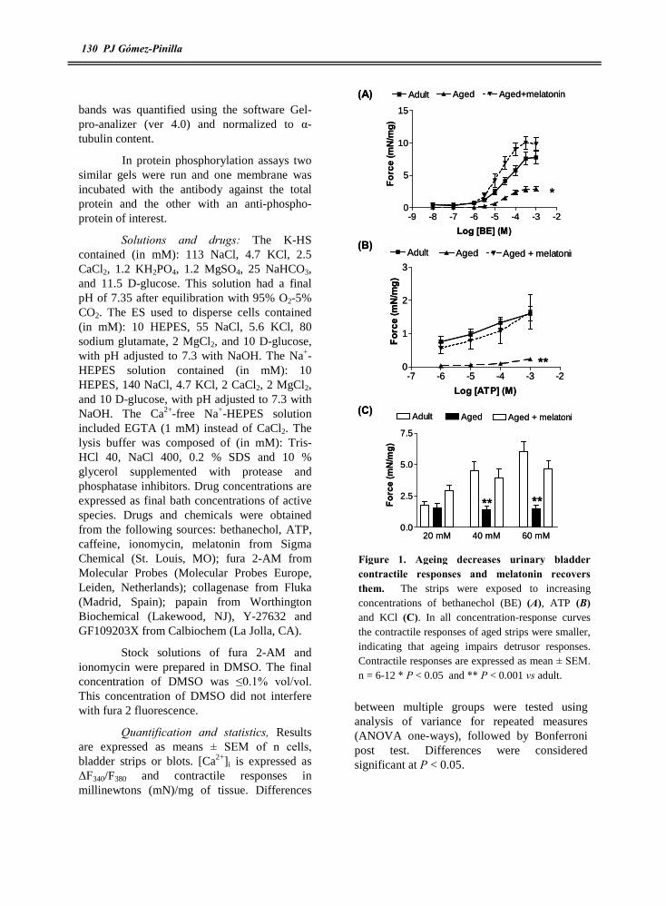
130 PJ Gómez-Pinilla
bands was quantified using the software Gel- pro-analizer (ver 4.0) and normalized to á-tubulin content.
In protein phosphorylation assays two similar gels were run and one membrane was incubated with the antibody against the total protein and the other with an anti-phospho-protein of interest.
Solutions and drugs: The K-HS contained (in mM): 113 NaCl, 4.7 KCl, 2.5 CaCl2, 1.2 KH2PO4, 1.2 MgSO4, 25 NaHCO3, and 11.5 D-glucose. This solution had a final pH of 7.35 after equilibration with 95% O2-5% CO2. The ES used to disperse cells contained (in mM): 10 HEPES, 55 NaCl, 5.6 KCl, 80 sodium glutamate, 2 MgCl2, and 10 D-glucose, with pH adjusted to 7.3 with NaOH. The Na+-HEPES solution contained (in mM): 10 HEPES, 140 NaCl, 4.7 KCl, 2 CaCl2, 2 MgCl2, and 10 D-glucose, with pH adjusted to 7.3 with NaOH. The Ca2+-free Na+-HEPES solution included EGTA (1 mM) instead of CaCl2. The lysis buffer was composed of (in mM): Tris-HCl 40, NaCl 400, 0.2 % SDS and 10 % glycerol supplemented with protease and phosphatase inhibitors. Drug concentrations are expressed as final bath concentrations of active species. Drugs and chemicals were obtained from the following sources: bethanechol, ATP, caffeine, ionomycin, melatonin from Sigma Chemical (St. Louis, MO); fura 2-AM from Molecular Probes (Molecular Probes Europe, Leiden, Netherlands); collagenase from Fluka (Madrid, Spain); papain from Worthington Biochemical (Lakewood, NJ), Y-27632 and GF109203X from Calbiochem (La Jolla, CA).
Stock solutions of fura 2-AM and ionomycin were prepared in DMSO. The final concentration of DMSO was ≤0.1% vol/vol. This concentration of DMSO did not interfere with fura 2 fluorescence.
Quantification and statistics, Results are expressed as means ± SEM of n cells, bladder strips or blots. [Ca2+]i is expressed as ÄF340/F380 and contractile responses in millinewtons (mN)/mg of tissue. Differences
between multiple groups were tested using analysis of variance for repeated measures (ANOVA one-ways), followed by Bonferroni post test. Differences were considered significant at P < 0.05.
-9 -8 -7 -6 -5 -4 -3 -20
5
10
15
Adult Aged Aged+melatonin
Log [BE] (M)
Forc
e (m
N/m
g)-7 -6 -5 -4 -3 -2
0
1
2
3
Adult Aged Aged + melatonin
Log [ATP] (M)Fo
rce
(mN
/mg)
20 mM 40 mM 60 mM0.0
2.5
5.0
7.5
Adult Aged Aged + melatonin
Forc
e (m
N/m
g)
(A)
(B)
(C)
*
**
** **
-9 -8 -7 -6 -5 -4 -3 -20
5
10
15
Adult Aged Aged+melatonin
Log [BE] (M)
Forc
e (m
N/m
g)-7 -6 -5 -4 -3 -2
0
1
2
3
Adult Aged Aged + melatonin
Log [ATP] (M)Fo
rce
(mN
/mg)
20 mM 40 mM 60 mM0.0
2.5
5.0
7.5
Adult Aged Aged + melatonin
Forc
e (m
N/m
g)
(A)
(B)
(C)
*
**
** **
Figure 1. Ageing decreases urinary bladder contractile responses and melatonin recovers them. The strips were exposed to increasing concentrations of bethanechol (BE) (A), ATP (B) and KCl (C). In all concentration-response curves the contractile responses of aged strips were smaller, indicating that ageing impairs detrusor responses. Contractile responses are expressed as mean ± SEM. n = 6-12 * P < 0.05 and ** P < 0.001 vs adult.

Trabajo Experimental III 131
4- RESULTS
Contractile response to agonist challenge
Cumulative additions of the muscarinic agonist BE (10 nM-1 mM) evoked concentration-dependent contractions in guinea pig urinary bladder strips (Figure 1A). Ageing impaired BE-induced contractile responses as indicated by the decrease in the maximal response (Emax) and the increase in the EC50
values (Emax: 7.91 ± 0.94 vs 2.90 ± 0.37 mN/mg; EC50: 24.05 ±4.05 vs 50.85 ±10.18 M; n = 12 and 6 for adult and aged, respectively; P < 0.01 and P < 0.05, adult vs aged). Similar to BE, when urinary bladder strips were challenged with increasing doses of
ATP, a concentration-dependent contractile response was obtained (Figure 1B), which was similarly changed in aged animals (Emax, 1.60 ± 0.21 vs 0.23 ± 0.05 mN/mg; n = 12 and 6, P < 0.001; adult vs aged). To explore if ageing affects depolarization-induced contractions we exposed the strips to medium containing 20, 40 and 60 mM KCl. In adult animals, KCl induced a dose-dependent contraction (Figure 1C) but in aged strips contractions were significantly reduced at 40 and 60 mM. Interestingly, melatonin treatment of aged animals reverted the ageing-related alterations in contractile responses described above (Figure 1).
Figure 2. Ageing increases cytosolic Ca2+ concentration ([Ca2+]i) responses to store depletion and membrane depolarization. Representative [Ca2+]i responses to 100 ìM of bethanechol (BE) (A), 100 ìM of ATP (C), 10 mM of caffeine (Caff) (C) and 60 mM of KCl (D) in fura-loaded urinary bladder smooth muscle cells from adult (solid line), aged (discontinuous line) and aged treated (dotted line) animals. In aged cells the calcium signalling is up-regulated and melatonin treatment restores Ca2+ signalling. Traces are typical of 6-34 cells. The ratio of fluorescence at 340 nm to fluorescence at 380 nm (F340/F380) is used as indicative of [Ca2+]i..
(A)
0.2 Ä
F34
0/F
38
0
10 s
BE BE BE
(C)
ATP ATP ATP
(D)Caff Caff Caff
60 mM KCl
50 s
0.2 Ä
F34
0/F
380
(B)0.
2 ÄF
340/
F3
80
10 s
0.2 Ä
F3
40/F
380
10 s
(A)
0.2 Ä
F34
0/F
38
0
10 s
BE BE BE
(C)
ATP ATP ATPATP ATP ATP
(D)Caff Caff Caff
60 mM KCl
50 s
0.2 Ä
F34
0/F
380
60 mM KCl
50 s
0.2 Ä
F34
0/F
380
(B)0.
2 ÄF
340/
F3
80
10 s
0.2 Ä
F3
40/F
380
10 s

132 PJ Gómez-Pinilla
Calcium signalling in urinary bladder smooth muscle cells.
The diminished contractile responses in aged bladders could be the result of a reduction in Ca2+ mobilization. In order to test this possibility, we quantified [Ca2+]i in response to the same stimuli as in Figure 1. Surprisingly, BE and ATP, which mainly release Ca2+ from intracellular stores through IP3 receptors (Hashitani et al., 2000), evoked higher [Ca2+]i increases in cells from aged than adult animals (adult: 0.291 ± 0.032 and 0.099 ± 0.010 ÄF340/F380; aged: 0.443 ± 0.041 and 0.204 ± 0.020 ÄF340/F380 for 100 M BE and ATP, respectively, n = 8-21 cells, P < 0.05 adult vs aged for BE and P < 0.001 adult vs aged for ATP). These responses had the typical profile of intracellular Ca2+ release, a transient peak followed by the rapid return to basal levels (Figure 2A & B). Similarly, Ca2+ release from ryanodine receptors was higher in aged cells as indicated by the response to 10 mM caffeine (adult: 0.426 ± 0.034 ÄF340/F380; aged: 0.664 ± 0.040 ÄF340/F380, n = 16 and 18 cells, P < 0.01, Figure 2C). These results indicate that Ca2+ stores content may be higher in aged cells. To test this possibility we applied a low level of ionomycin in Ca2+ free solution, a treatment which releases Ca2+ from intracellular stores while bypassing channels and receptors. [Ca2+]i transients induced by 50 nM of ionomycin in adult were smaller than in aged cells (adult: 0.069 ± 0.008 ÄF340/F380, aged: 0.174 ± 0.020 ÄF340/F380, n = 13 and 18 cells, respectively, P < 0.01). Similar differences were obtained in the presence of blockers of Ca2+ extrusion mechanisms (data not shown). In addition, ageing increased KCl-induced Ca2+ influx (adult: 0.170 ± 0.025 ÄF340/F380; aged: 0.247 ± 0.014 ÄF340/F380 , n = 6 and 10 cells, P < 0.05, Figure 2D).
As shown in figure 2, melatonin treatment was able to counteract ageing-induced changes in Ca2+ homeostasis. Thus, Ca2+ release from intracellular stores stimulated by BE, ATP and caffeine and extracellular Ca2+ influx activated by KCl, were similar to the
values obtained in the adult group and significantly different to the aged group (BE: 0.357 ± 0.036 ÄF340/F380; ATP: 0.118 ± 0.016 ÄF340/F380; caffeine: 0.471 ± 0.024 ÄF340/F380;
Contractile response to ATP
Y-27632 (5 M) GFX (5M) Both-25
0
25
50
75
100
Adult Aged Aged + melatonin
Inhi
bitio
n (%
)
Contractile response to KCl
Y-27632 (5 M) GFX (5M) Both-25
0
25
50
75
100
Adult Aged Aged + melatonin
Inhi
bitio
n (%
)
Contractile response to BE
Y-27632 (5 M) GFX (5M) Both-25
0
25
50
75
Adult Aged Aged + melatonin
Inhi
bitio
n (%
)
(A)
(B)
(C)
*
* * *
***
Figure 3. Ageing depresses contractile response sensitivitiness to Y-27632 and GF109203X, whereas melatonin treatment recovers it. Effects of 5 ìM Y-27632, 5 ìM GF109203X or the combination of both inhibitors on 100 ìM o BE- (A), 100 ìM ATP- (B) and 60 mM KCl- (C) evoked contractions in urinary bladder strips. Data are expressed as mean ± SEM. n = 6-15 strips. * P < 0.01. vs adults.
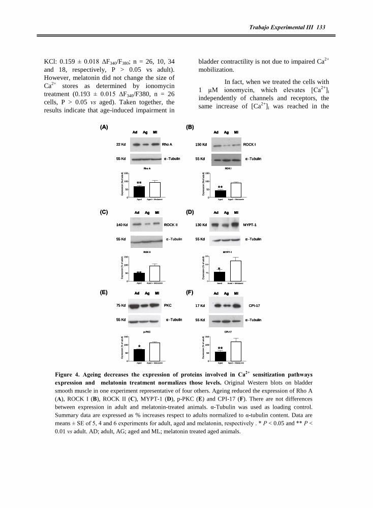
Trabajo Experimental III 133
KCl: 0.159 ± 0.018 ÄF340/F380; n = 26, 10, 34 and 18, respectively, P > 0.05 vs adult). However, melatonin did not change the size of Ca2+ stores as determined by ionomycin treatment (0.193 ± 0.015 ÄF340/F380, n = 26 cells, P > 0.05 vs aged). Taken together, the results indicate that age-induced impairment in
bladder contractility is not due to impaired Ca2+ mobilization.
In fact, when we treated the cells with 1 µM ionomycin, which elevates [Ca2+]i independently of channels and receptors, the same increase of [Ca2+]i was reached in the
Ad Ag Ml
22 Kd
55 Kd
Rho A
á -Tubulin
Ad Ag Ml
140 Kd
55 Kd
ROCK II
á -Tubulin
Ad Ag Ml
130 Kd
55 Kd
MYPT-1
á -Tubulin
Ad Ag Ml
75 Kd
55 Kd
PKC
á -Tubulin
Ad Ag Ml
17 Kd
55 Kd
CPI-17
á -Tubulin
(A) (B)
(C) (D)
(E) (F)
****
***
* **
Ad Ag Ml
150 Kd
55 Kd
ROCK I
á -Tubulin
Rho A
Aged Aged + Melatonin0
50
100
150
Exp
ress
ion
(%of
adu
lt)
ROK I
Aged Aged + Melatonin0
50
100
150
Exp
ress
ion
(%of
adu
lt)
ROK II
Aged Aged + Melatonin0
50
100
150
Exp
ress
ion
(% o
f adu
lt)
MYPT-1
Aged Aged + Melatonin0
50
100
150
Exp
ress
ion
(%
of a
dult)
p-PKC
Aged Aged + Melatonin0
50
100
150
Exp
ress
ion
(% o
f adu
lt)
CPI-17
Aged Aged + Melatonin0
50
100
150
Exp
ress
ion
(% o
f adu
lt)
Ad Ag Ml
22 Kd
55 Kd
Rho A
á -Tubulin
Ad Ag Ml
22 Kd
55 Kd
Rho A
á -Tubulin
Ad Ag Ml
140 Kd
55 Kd
ROCK II
Ad Ag Ml
140 Kd
55 Kd
ROCK II
á -Tubulin
Ad Ag Ml
130 Kd
55 Kd
MYPT-1
á -Tubulin
Ad Ag Ml
130 Kd
55 Kd
MYPT-1
á -Tubulin
Ad Ag Ml
75 Kd
55 Kd
PKC
á -Tubulin
Ad Ag Ml
75 Kd
55 Kd
PKC
á -Tubulin
Ad Ag Ml
17 Kd
55 Kd
CPI-17
á -Tubulin
Ad Ag Ml
17 Kd
55 Kd
CPI-17
á -Tubulin
(A) (B)
(C) (D)
(E) (F)
****
***
* **
Ad Ag Ml
150 Kd
55 Kd
ROCK I
á -Tubulin
Ad Ag Ml
150 Kd
55 Kd
ROCK I
á -Tubulin
Rho A
Aged Aged + Melatonin0
50
100
150
Exp
ress
ion
(%of
adu
lt)
ROK I
Aged Aged + Melatonin0
50
100
150
Exp
ress
ion
(%of
adu
lt)
ROK II
Aged Aged + Melatonin0
50
100
150
Exp
ress
ion
(% o
f adu
lt)
MYPT-1
Aged Aged + Melatonin0
50
100
150
Exp
ress
ion
(%
of a
dult)
p-PKC
Aged Aged + Melatonin0
50
100
150
Exp
ress
ion
(% o
f adu
lt)
CPI-17
Aged Aged + Melatonin0
50
100
150
Exp
ress
ion
(% o
f adu
lt)
Figure 4. Ageing decreases the expression of proteins involved in Ca2+ sensitization pathways expression and melatonin treatment normalizes those levels. Original Western blots on bladder smooth muscle in one experiment representative of four others. Ageing reduced the expression of Rho A (A), ROCK I (B), ROCK II (C), MYPT-1 (D), p-PKC (E) and CPI-17 (F). There are not differences between expression in adult and melatonin-treated animals. á-Tubulin was used as loading control. Summary data are expressed as % increases respect to adults normalized to á-tubulin content. Data are means ± SE of 5, 4 and 6 experiments for adult, aged and melatonin, respectively . * P < 0.05 and ** P < 0.01 vs adult. AD; adult, AG; aged and ML; melatonin treated aged animals.
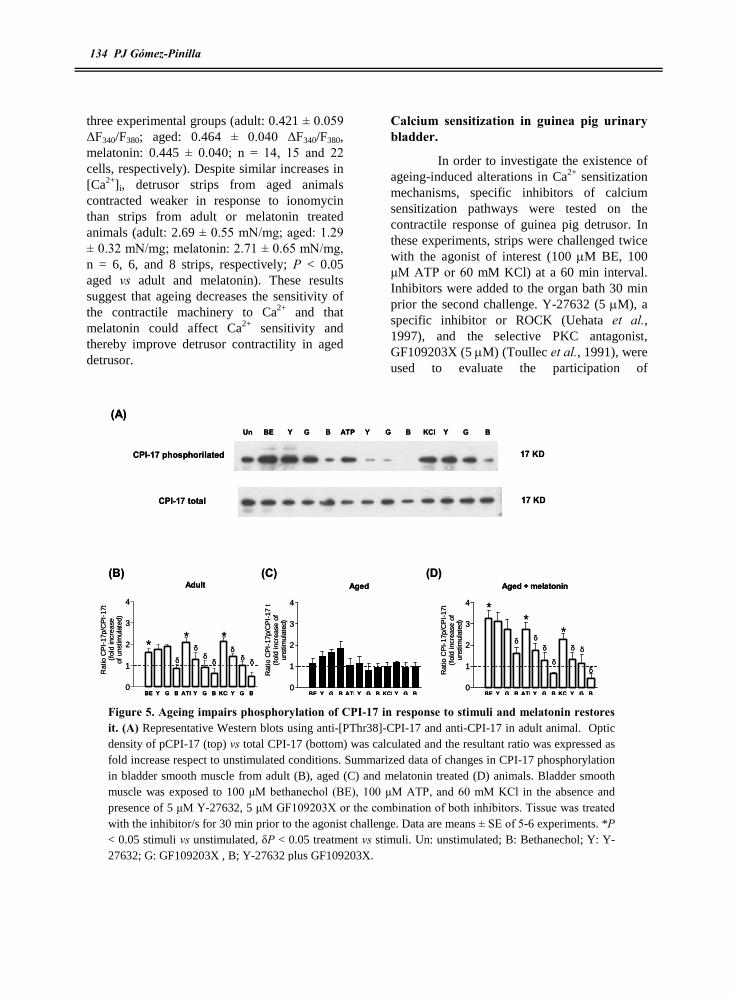
134 PJ Gómez-Pinilla
three experimental groups (adult: 0.421 ± 0.059 ÄF340/F380; aged: 0.464 ± 0.040 ÄF340/F380, melatonin: 0.445 ± 0.040; n = 14, 15 and 22 cells, respectively). Despite similar increases in [Ca2+]i, detrusor strips from aged animals contracted weaker in response to ionomycin than strips from adult or melatonin treated animals (adult: 2.69 ± 0.55 mN/mg; aged: 1.29 ± 0.32 mN/mg; melatonin: 2.71 ± 0.65 mN/mg, n = 6, 6, and 8 strips, respectively; P < 0.05 aged vs adult and melatonin). These results suggest that ageing decreases the sensitivity of the contractile machinery to Ca2+ and that melatonin could affect Ca2+ sensitivity and thereby improve detrusor contractility in aged detrusor.
Calcium sensitization in guinea pig urinary bladder.
In order to investigate the existence of ageing-induced alterations in Ca2+ sensitization mechanisms, specific inhibitors of calcium sensitization pathways were tested on the contractile response of guinea pig detrusor. In these experiments, strips were challenged twice with the agonist of interest (100 M BE, 100 ìM ATP or 60 mM KCl) at a 60 min interval. Inhibitors were added to the organ bath 30 min prior the second challenge. Y-27632 (5 M), a specific inhibitor or ROCK (Uehata et al., 1997), and the selective PKC antagonist, GF109203X (5 M) (Toullec et al., 1991), were used to evaluate the participation of
Un BE Y G B Y G B Y G BATP KCl
CPI-17 phosphorilated
CPI-17 total
17 KD
17 KD
(A)
(B) (C) (D)Aged
BE Y G B ATPY G B KCl Y G B0
1
2
3
4
Rat
io C
PI-
17p/
CP
I-17
t(f
old
incr
ease
of
unst
imul
ated
)
Adult
BE Y G B ATPY G B KCl Y G B0
1
2
3
4
Rat
io C
PI-
17p/
CP
I-17
t(f
old
incr
ease
of u
nstim
ulat
ed)
Aged + melatonin
BE Y G B ATPY G B KCl Y G B0
1
2
3
4
Rat
io C
PI-
17p/
CP
I-17
t(f
old
incr
ease
of
unst
imul
ated
)
** *
**
*
ä
ää
ä
äää
ä
äää
ää ä
Figure 5. Ageing impairs phosphorylation of CPI-17 in response to stimuli and melatonin restores it. (A) Representative Western blots using anti-[PThr38]-CPI-17 and anti-CPI-17 in adult animal. Optic density of pCPI-17 (top) vs total CPI-17 (bottom) was calculated and the resultant ratio was expressed as fold increase respect to unstimulated conditions. Summarized data of changes in CPI-17 phosphorylation in bladder smooth muscle from adult (B), aged (C) and melatonin treated (D) animals. Bladder smooth muscle was exposed to 100 ìM bethanechol (BE), 100 ìM ATP, and 60 mM KCl in the absence and presence of 5 ìM Y-27632, 5 ìM GF109203X or the combination of both inhibitors. Tissue was treated with the inhibitor/s for 30 min prior to the agonist challenge. Data are means ± SE of 5-6 experiments. *P < 0.05 stimuli vs unstimulated, äP < 0.05 treatment vs stimuli. Un: unstimulated; B: Bethanechol; Y: Y-27632; G: GF109203X , B; Y-27632 plus GF109203X.

Trabajo Experimental III 135
RhoA/ROCK and PKC/CPI-17 pathways, respectively. In adult animals the contractile response evoked by 100 M of BE was not affected neither by Y-27632 nor by GF109203X, but when the strips were co-incubated with both antagonists the response to BE was inhibited by 57% (Figure 3A). In aged strips, the inhibitors did not have any significant effects when administered either separately or in combination (Figure 3A). In adults, the contractile response to ATP was sensitive to both Y-27632 (44 % of inhibition), and GF109203X (34% of inhibition) and when both inhibitors were tested together the effect was additive (74 % of inhibition). Both compounds had only small inhibitory effects on the purinergic contractile response in aged animals (Figure 3B). Y-27632 even caused an increase in ATP-evoked contraction (10 % of increase). A similar pattern of response was obtained when strips were exposed to depolarizing medium (60 mM KCl). Strips from adult guinea pigs were sensitive to Y-27632, GF109203X and a combination of both inhibitors (51, 43. and 76 % of inhibition, respectively), whereas the inhibitory effects were smaller in aged strips (P <0.01). Interestingly, after melatonin treatment the contractile responses of aged strip recovered sensitivity to Y-27632, GF109203X and the combination of both drugs (Figure 3). These results suggest that ageing impairs Ca2+ sensitization mechanisms and melatonin treatment restores them.
To further test whether Ca2+ sensitization mechanisms are affected by ageing we determined the expression of proteins involved in Ca2+ sensitization pathways. As shown in Figure 4 ageing induced a significant reduction of the main components of RhoA/ROCK pathway that ranged from 33 % of reduction in Rho A expression to 58 % of reduction in ROCK I expression (Figure 4 A-D). In addition, the expression of phospho-PKC and its effector CPI-17 were reduced by 30% and 43 % in aged detrusor (Figure 4E & F). Melatonin treatment normalized the expression of all tested proteins (Figure 4). The lowered
level of expression of these proteins in aged detrusor, may explain the impairment in contractile responses found in aged bladder. Accordingly, the melatonin-induced normalization of expression levels could account for the improvement of contractile responses found in treated guinea pigs.
In both Ca2+ sensitization pathways, phosphorylation of the last component (MYPT for RhoA/ROCK pathway and CPI-17 for PKC/CPI-17 pathway) is essential to inhibit the activity of MLCP and cause contraction. Thus, apart from measuring the total level of expression of these proteins, we quantified the ratio phospho-protein/total protein. In addition, we tested the effect of the inhibitors used in organ bath experiments (Y27632 and GF109203X) to verify the specificity of both compounds in Ca2+ sensitization pathways.
Phosphorylation of CPI-17 (at Thr-38) and MYPT1 (at Thr-853) in response to BE, ATP and depolarization, in the absence and presence of the blockers, was examined using anti-phosphoprotein-specific antibodies. As predicted, in adult and melatonin treated animals a low level of p-CPI-17 was detected at resting conditions, while p-CPI-17 increased after challenge with 100 ìM BE, 100 ìM ATP and 60 mM of KCl (Figure 5). This effect was not present in aged tissues. These results are consistent with the activation of PKC by the contractile agonists in adult and melatonin treated tissues, leading to CPI-17 phosphorylation and Ca2+ sensitization. This was confirmed by assaying the phosphorylation levels of PKC in response to the stimuli, since its activation leads to autophosphorylation (Blobe et al., 1996) (adult, BE: 1.63 ± 0.10, ATP: 2.27 ± 0.2, KCl: 1.89 ± 0.15 fold increase respect to unstimulated; aged, BE: 0.99 ± 0.14, ATP: 1.03 ± 0.16, KCl: 1.40 ± 0.27 fold increase respect to unstimulated; melatonin treatment 1.70 ± 0.17, ATP: 2.27 ± 0.29, KCl: 1.92 ± 0.20 fold increase respect to unstimulated; n = 4 experiments; P < 0.05). According to the effects in contraction (see Figure 3), CPI-17 phosphorylation in adult and

136 PJ Gómez-Pinilla
melatonin-treated detrusor in response to BE was only reduced by co-incubation withY27632 and GF109203X, whereas phosphorylation in response to ATP or KCl was reduced by both antagonists in an additive mode (Figure 5B and D). In aged smooth muscle, there was not increased phosphorylation in response to stimuli, (Figure 5C).
Regarding the phosphorylation status of MYPT1 at Thr-853, in adult and melatonin-treated animals, bladder exhibited a low basal level of p-MYPT1 and stimulation with 100 ìM BE, 100 ìM ATP and 60 mM KCl evoked a significant increase in phosphorylation. Under BE challenge, MYPT1 phosphorylation was only inhibited by Y-27632 plus GF109203X treatment, but when the tissue was stimulated with ATP and KCl, p-MYPT1 levels were reduced by both Y-27632 and GF109203X. Y27632 was more effective than GF109203X
inhibiting MYPT1 phosphorylation in response to these stimuli (Figure 6A, B & D). In detrusor from aged animals, phosphorylation levels of MYPT were not affected by agonist challenge or blocker treatments (Figure 6C).
5- DISCUSSION
The results of the present study indicate that ageing decreases urinary bladder contractile response through a decrease in Ca2+ sensitization mechanisms. Paradoxically, the diminished contractility found in aged detrusor correlates with increased Ca2+ mobilization. Further, we show that treatment of aged animals with melatonin recovered the contractile response of bladder smooth muscle as the result of normalization of Ca2+ dependent and independent mechanisms.
It is well known that urinary bladder disturbances, as a consequence of alterations in
Un BE Y G B Y G B Y G BATP KCl
MYPT phosphorylated
MYPT total
130 KD
130 KD
(A)
(B) (C) (D)
Adult
BE y g b ATP y g b KCl y g b0
1
2
3
4
5
Rat
io M
YP
Tp/
MY
PTt
(fo
ldin
crea
se o
f un
stim
ulat
ed)
Senescent
BE y g b ATP y g b KCl y g b0
1
2
3
4
5
Rat
io M
YP
Tp/
MY
PTt
(fo
ldin
crea
se o
f un
stim
ulat
ed)
Melatonin
BE y g b ATP y g b KCl y g b0
1
2
3
4
5
Rat
io M
YP
Tp/
MY
PTt
(fo
ldin
crea
se o
f un
stim
ulat
ed)
*ä
**
ä ä
ää
ää
ää
**
ä
**
**
ää
ä
ä
ä ää
Figure 6. Ageing impairs phosphorylation of MYPT1 in response to stimuli and melatonin restores it. (A) Representative Western blots using anti-[PThr853]-MYPT1 and anti-MYPT1 in adult animal. Optic density of pMYPT1 (top) vs total MYPT1 (bottom) was calculated and the resultant ratio was expressed as fold increase respect to unstimulated conditions. Summarized data of changes in MYPT1 phosphorylation in bladder smooth muscle from adult (B), aged (C) and melatonin treated (D) animals. Bladder smooth muscle was exposed to 100 ìM bethanechol (BE), 100 ìM ATP, and 60 mM KCl in the absence and presence of 5 ìM Y-27632, 5 ìM GF109203X or the combination of both inhibitors. Tissue was treated

Trabajo Experimental III 137
the micturition cycle, increase with age (Andersen et al., 1978), but little is known about the underlying changes in bladder smooth muscle that lead to those alterations. Conflicting findings have been reported depending on the species and agonists used (Longhurst et al., 1992;Lluel et al., 2000;Yoshida et al., 2001). In these studies none of the two mechanisms leading to contraction were analysed (Ca2+ mobilization and Ca2+ sensitization). Here, we found that ageing impairs the contractile response to cholinergic and purinergic stimuli, which can explain the functional data recently published by us showing that ageing decreases micturition pressure (Gomez-Pinilla et al., 2007).
In urinary bladder, the ageing-evoked impairment in contractile response has been previously associated to structural changes in the detrusor, such as increased collagen content, alterations in cell junctions and/or increased space between muscle cells (Elbadawi, 1995). In colonic and gallbladder smooth muscle, a reduced contractile response was attributed to inhibition of the Src Kinase pathway, reduced activation of PKC, decreased association between HSP-27 with the contractile machinery and alterations in Ca2+ homeostasis (Bitar, 2003;Gomez-Pinilla et al., 2006a). In this paper, we explore whether changes in Ca2+ signalling and Ca2+ sensitization can explain the ageing-related impairment in detrusor contractility.
Intracellular Ca2+ levels play an important role in the modulation of contraction-relaxation cycle of smooth muscle cells. The origin of Ca2+ can be extracellular or from internal stores, but independently of the source, increases in [Ca2+]i lead to contraction in a calmodulin- and MLCK-dependent way (Horowitz et al., 1996;Karaki et al., 1997). In this study, we describe that in aged smooth muscle cells, Ca2+ release from intracellular stores and extracellular Ca2+ influx are up-regulated. This could be due to changes in the size of the stores, in extrusion pathways, or in the expression and or conductance of different
Ca2+ channels. Here, we demonstrate that intracellular Ca2+ stores in aged cells have a larger Ca2+ content, which supports the increase in Ca2+ release through both IP3 and ryanodine receptor channels. Reduction of Ca2+ extrusion mechanisms operating in plasma membrane could also be responsible for the increased Ca2+ signalling in aged bladder smooth muscle cells. Unpublished studies from our group indicate that the Ca2+ elimination rate is slower in aged cells than in control cells, and that this is related to a lower PMCA contribution (Experimental Cap IV). Since this pump is easily altered by endogenous oxidants (Zaidi & Michaelis, 1999), extrusion impairment could be due to ageing-induced oxidative stress, which could also change the expression and properties of Ca2+ channels. In this regard, higher L-type Ca2+ influx in detrusor myocytes isolated from unstable or obstructed bladder have been related to changes on the time- and voltage- dependent kinetics of the channels (Gallegos & Fry, 1994). In gastrointestinal inflamed cells an increase in the number of L-type Ca2+ channels and changes in the pharmacological profile of these channels have been described (Liu et al., 2001;Gomez-Pinilla et al., 2006b). The higher [Ca2+]i in response to KCl-induced depolarization could be also explained by more effective Ca2+ induce Ca2+ release (CICR) mechanisms described in bladder smooth muscle cells (Collier et al., 2000). A detailed characterization of the age- and melatonin-induced changes in number and properties of Ca2+ channels is beyond the scope of this study and will be addressed in future work.
The ageing process has been associated to changes in Ca2+ homeostasis and cytotoxicity, leading to oxidative stress, organ dysfunction and/or degenerative diseases (Verkhratsky & Toescu, 1998). However, what is clear in our study is that changes in Ca2+ homeostasis can not explain the impairment in bladder contractility. Instead, we present evidence for a reduced Ca2+ sensitization as a possible mechanism underlying aged-related impairment of bladder contractility. First,

138 PJ Gómez-Pinilla
stimulation with ionomycin induced the same increase in [Ca2+]i in both adult and aged tissues, whereas it yielded a reduced contractile response in aged muscle. Second, specific inhibitors of the Ca2+ sensitization pathways inhibited the contractile response induced by G-protein coupled receptor stimulation and Ca2+ influx in adult bladder smooth muscle, but had less effect in aged strips. Third, the expression of protein components of RhoA/ROCK and PKC/CPI-17 pathways was diminished in aged bladder. Fourth, the lack of activation of the final effectors MYPT1 and CPI-17 in response to stimuli in aged detrusor.
Ca2+ sensitization was first demonstrated in urinary bladder contraction in 2001 (Jezior et al., 2001) and since then, several papers have been published describing the participation of calcium sensitization in different species including humans (Takahashi et al., 2004;Schneider et al., 2004;Jezior et al., 2001;Woodsome et al., 2001;Wibberley et al., 2003;Fleichman et al., 2004;Durlu-Kandilci & Brading, 2006). Variable contribution from different Ca2+ sensitization pathways and sometimes conflicting evidence has been described depending on the nature of the stimuli and species. Thus, a predominant role for the PKC pathway in cholinergic responses has been shown in rat but not in guinea pig bladder (Durlu-Kandilci & Brading, 2006), whereas other studies claim for a role of RhoA/ROCK pathway (Wibberley et al., 2003;Fleichman et al., 2004). In humans, participation of both pathways (Takahashi et al., 2004) and a major participation of RhoA/ROCK pathway has been reported (Schneider et al., 2004). In adult guinea pig urinary bladder we have found that bethanechol-evoked contractions were not sensitive to either Y-27632 or GF109203X alone, but co-incubation with both antagonists evoked a 50 % inhibition. In addition, BE causes an increase in MYPT1 and CPI-17 phosphorylation that were not inhibited neither by Y-27632 nor GF109203X but were sensitive to both inhibitors when added together. These results suggest a potential cross-talk mechanism
between pathways, upstream phosphorylation of CPI-17 and MYPT1. Therefore, only when both pathways were down-regulated simultaneously, BE-mediated contraction and MYPT1 and CPI-17 phosphorylation could be inhibited. This cross-talk seems to be unique to BE-stimulation, as ATP-and KCl-induced contractions and MYPT1 and CPI-17 phosphorylation were also sensitive to both inhibitors when applied independently. Agonist-induced CPI-17 phosphorylation was inhibited by Y-27632, consistent with Thr-38 of CPI-17 also being a substrate of ROCK (Koyama et al., 2000) and agonist-induced MYPT1 phosphorylation was also decreased by GF109203X, which could reflect the phosphorylation of MYPT1 by PKC (Toth et al., 2000). Our results with KCl are in line with accumulating evidence showing that membrane depolarization by high K+ induces Ca2+-dependent Rho activation and MLCP inhibition through phosphorylation of MYPT1 and CPI-17 (Mita et al., 2002;Sakurada et al., 2003). It has recently been demonstrated in vascular smooth muscle that PI3Kinase is essential for these Ca2+-mediated responses, and that PKC does not mediate CPI-17 phosphorylation (Azam et al., 2007). In bladder smooth muscle, both depolarization-induced contraction and CPI-17 and MYPT1 phosphorylation in adult animals was inhibited by both GF109203X and Y27362, which highlights potential differences in the upstream CPI-17 and MYPT1 regulators between these tissues.
Independently of the source of activation, our data indicate that ageing evokes a loss of calcium sensitization mechanisms which probably underlies the diminished contractile responses in aged bladder smooth muscle. The lack of effect of the inhibitors on bladder contractility, suggests that Ca2+ sensitization does not contribute to contraction in strips from aged animals. This was supported by the reduced expression in RhoA, ROCK, PKC, MYPT1 and CPI-17 and reduced MYPT1 and CPI-17 phosphorylation in response to

Trabajo Experimental III 139
cholinergic and purinergic agonists or membrane depolarization.
Aged-induced oxidative damage of proteins related to Ca2+ sensitization could account for the down regulation of PKC and RhoA pathways found in our study. In agreement with this hypothesis, diabetes-induced decrease in detrusor smooth muscle force, which is associated to an increase in oxidative stress (Changolkar et al., 2005), has been related to diminished Ca2+ sensitivity (Su et al., 2004). Similarly, in partial outlet bladder obstruction, where it has also been shown oxidative insult (Levin et al., 2002) the Ca2+ sensitivity of force is lower than in control bladder (Stanton et al., 2003;Stanton et al., 2004). On the other hand, acute exposure of smooth muscle to reactive oxygen species (ROS) has been shown to induce RhoA translocation and MYPT1 phosphorylation (Jin et al., 2004). This discrepancy may be possibly related to differences in the effects of acute and chronic exposure to ROS.
Melatonin was able to increase not only the expression levels of several proteins that mediate Ca2+ sensitization, but also the phosphorylation of two final effectors, MYPT1 and CPI-17. This increased Ca2+ sensitization resulted in improved contractility in strips from aged animals, as shown by the larger effect of the inhibitors of ROCK and PKC. The improved contractile response of aged detrusor may explain our previous results showing that melatonin treatment could revert the aged-induced reduction in micturition pressure in
aged animals (Gomez-Pinilla et al., 2007). In inflamed bladder, it has been shown that melatonin recovers the contractile response to cholinergic stimulation (Paskaloglu et al., 2004). Melatonin treatment also resulted in normalization of Ca2+ homeostasis, which agrees with the effects reported for this hormone in aged gallbladder smooth muscle (Gomez-Pinilla et al., 2006a). The mechanism of action of melatonin is still not clear, but this hormone is a potent free radical scavenger and antioxidant (Tan et al., 2002) and under the experimental conditions of this study causes a decrease in lipid peroxidation and an increase in the natural antioxidant defence, GSH (Experimental Cap II).
In summary, we document significant age-related changes in the contractility of detrusor smooth muscle, which can be explained by altered Ca2+ homeostasis and decreased Ca2+ sensitization. Melatonin treatment recovered smooth muscle contractility by restoring Ca2+ signalling and increasing Ca2+ sensitization. The beneficial effects of melatonin may represent an unexplored area for the development of new therapeutic approaches for treating aged-related detrusor disturbances.
6-ACKNOWLEDGEMENTS
Supported by Junta de Extremadura (2PR03A020) and Ministerio de Educacion y Ciencia (BFU 2004-0637). Pedro J Gómez-Pinilla is recipient of Doctoral Fellowship from Junta de Extremadura.
7- REFERENCE LIST
Ambache N & Zar MA (1970). Non-Cholinergic Transmission by Post-Ganglionic Motor Neurones in Mammalian Bladder. J Physiol 210, 761-&.
Andersen JT, Jacobsen O, Wormpetersen J, & Hald T (1978). Bladder Function in Healthy Elderly Males. Scand J Urol Nephrol 12, 123-127.
Azam MA, Yoshioka K, Ohkura S, Takuwa N, Sugimoto N, Sato K, & Takuwa Y (2007). Ca2+-Independent, Inhibitory Effects of Cyclic Adenosine 5'-Monophosphate on Ca2+ Regulation of

140 PJ Gómez-Pinilla
Phosphoinositide 3-Kinase C2{alpha}, Rho, and Myosin Phosphatase in Vascular Smooth Muscle. J Pharmacol Exp Ther 320, 907-916.
Bitar KN (2003). Aging and neural control of the GI tract V. Aging and gastrointestinal smooth muscle: from signal transduction to contractile proteins. Am J Physiol Gastrointest Liver Physiol 284, G1-G7.
Blobe GC, Stribling S, Obeid LM, & Hannun YA (1996). Protein kinase C isoenzymes: regulation and function. Cancer Surv 27, 213-248.
Bruce L & Nixon GF (1997). Increased sensitization of the myofilaments in rat neonatal portal vein: A potential mechanism. Exp Physiol 82, 985-993.
Changolkar AK, Hypolite JA, Disanto M, Oates PJ, Wein AJ, & Chacko S (2005). Diabetes induced decrease in detrusor smooth muscle force is associated with oxidative stress and overactivity of aldose reductase. J Urol 173, 309-313.
Chiba Y, Ueno A, Sakai H, & Misawa M (2004). Hyperresponsiveness of bronchial but not tracheal smooth muscle in a murine model of allergic bronchial asthma. Inflamm Res 53, 636-642.
Collier ML, Ji G, Wang Y, & Kotlikoff MI (2000). Calcium-induced calcium release in smooth muscle: loose coupling between the action potential and calcium release. J Gen Physiol 115, 653-662.
Dai Y, Chitaley K, Webb RC, Lewis RW, & Mills TM (2004). Topical application of a Rho-kinase inhibitor in rats causes penile erection. Int J Impot Res 16, 294-298.
Durlu-Kandilci NT & Brading AF (2006). Involvement of Rho kinase and protein kinase C in carbachol-induced calcium sensitization in beta-escin skinned rat and guinea-pig bladders. Br J Pharmacol 148, 376-384.
Ekman M, Fagher K, Mia W, Stakeberg K, & Arner A (2005). Decreased phosphatase activity, increased Ca2+ sensitivity, and myosin light chain phosphorylation in urinary bladder smooth muscle of newborn mice. J Gen Physiol 125, 187-196.
Elbadawi A (1995). Pathology and Pathophysiology of Detrusor in Incontinence. Urol Clin North Am 22, 499-512.
Eto M, Ohmori T, Suzuki M, Furuya K, & Morita F (1995). A Novel Protein Phosphatase-1 Inhibitory Protein Potentiated by Protein-Kinase-C - Isolation from Porcine Aorta Media and Characterization. J Biochem (Tokyo) 118, 1104-1107.
Fleichman M, Schneider T, Fetscher C, & Michel MC (2004). Signal transduction underlying carbachol-induced contraction of rat urinary bladder. II. Protein kinases. J Pharmacol Exp Ther 308, 54-58.
Gallegos CR & Fry CH (1994). Alterations to the electrophysiology of isolated human detrusor smooth muscle cells in bladder disease. J Urol 151, 754-758.
Gomez-Pinilla PJ, Camello-Almaraz C, Moreno R, Camello PJ, & Pozo MJ (2006a). Melatonin treatment reverts age-related changes in Guinea pig gallbladder neuromuscular transmission and contractility. J Pharmacol Exp Ther 319, 847-856.
Gomez-Pinilla PJ, Gomez MF, Hedlund P, Sward K, Hellstrand P, Camello PJ, Pozo MJ, & Andersson KE (2007). Effect of melatonin on age-associated changes in guinea pig urinary bladder function. J Urol.
Gomez-Pinilla PJ, Morales S, Camello-Almaraz C, Moreno R, Pozo MJ, & Camello PJ (2006b). Changes in guinea pig gallbladder smooth muscle Ca2+ homeostasis by acute acalculous cholecystitis. Am J Physiol Gastrointest Liver Physiol 290, G14-G22.

Trabajo Experimental III 141
Hashitani H, Bramich NJ, & Hirst GDS (2000). Mechanisms of excitatory neuromuscular transmission in the guinea-pig urinary bladder. J Physiol 524, 565-579.
Horowitz A, Menice CB, Laporte R, & Morgan KG (1996). Mechanisms of smooth muscle contraction. Physiol Rev 76, 967-1003.
Jezior JR, Brady JD, Rosenstein DI, McCammon KA, Miner AS, & Ratz PH (2001). Dependency of detrusor contractions on calcium sensitization and calcium entry through LOE-908-sensitive channels. Br J Pharmacol 134, 78-87.
Jin L, Ying Z, & Webb RC (2004). Activation of Rho/Rho kinase signaling pathway by reactive oxygen species in rat aorta. Am J Physiol Heart Circ Physiol 287, H1495-H1500.
Karaki H, Ozaki H, Hori M, MitsuiSaito M, Amano KI, Harada KI, Miyamoto S, Nakazawa H, Won KJ, & Sato K (1997). Calcium movements, distribution, and functions in smooth muscle. Pharmacol Rev 49, 157-230.
Kitazawa T, Eto M, Woodsome TP, & Brautigan DL (2000). Agonists trigger G protein-mediated activation of the CPI-17 inhibitor phosphoprotein of myosin light chain phosphatase to enhance vascular smooth muscle contractility. J Biol Chem 275, 9897-9900.
Kitazawa T, Takizawa N, Ikebe M, & Eto M (1999). Reconstitution of protein kinase C-induced contractile Ca2+ sensitization in Triton X-100-demembranated rabbit arterial smooth muscle. J Physiol 520, 139-152.
Koyama M, Ito M, Feng J, Seko T, Shiraki K, Takase K, Hartshorne DJ, & Nakano T (2000). Phosphorylation of CPI-17, an inhibitory phosphoprotein of smooth muscle myosin phosphatase, by Rho-kinase. FEBS Lett 475, 197-200.
Levin RM, Kawashima Y, Leggett RE, Whitbeck C, Horan P, & Mizutani K (2002). Effect of oral Kohki tea on bladder dysfunction induced by severe partial outlet obstruction. J Urol 167, 2260-2266.
Liu X, Rusch NJ, Striessnig J, & Sarna SK (2001). Down-regulation of L-type calcium channels in inflamed circular smooth muscle cells of the canine colon. Gastroenterology 120, 480-489.
Lluel P, Palea S, Barras M, Grandadam F, Heudes D, Bruneval P, Corman B, & Martin DJ (2000). Functional and morphological modifications of the urinary bladder in aging female rats. Am J Physiol Reg Integ Comp Physiol 278, R964-R972.
Longhurst PA, Eika B, Leggett RE, & Levin RM (1992). Comparison of Urinary-Bladder Function in 6 and 24 Month Male and Female Rats. J Urol 148, 1615-1620.
Mita M, Yanagihara H, Hishinuma S, Saito M, & Walsh MP (2002). Membrane depolarization-induced contraction of rat caudal arterial smooth muscle involves Rho-associated kinase. Biochem J 364, 431-440.
Mukai Y, Shimokawa HA, Matoba TS, Kandabashi T, & Takeshita A (2000). Role of Rho-kinase in the pathogenesis of hypertensive vascular disease. Circulation 102, 96.
Paskaloglu K, Sener G, Kapucu C, & Ayanoglu-Dulger G (2004). Melatonin treatment protects against sepsis-induced functional and biochemical changes in rat ileum and urinary bladder. Life Sciences 74, 1093-1104.
Rajasekaran M, Wilkes N, Kuntz S, & Albo ME (2005). Rho-kinase inhibition suppresses bladder hyperactivity in spontaneously hypertensive rats. Neurourol Urodyn 24, 295-300.

142 PJ Gómez-Pinilla
Sakurada S, Takuwa N, Sugimoto N, Wang Y, Seto M, Sasaki Y, & Takuwa Y (2003). Ca2+-dependent activation of Rho and Rho kinase in membrane depolarization-induced and receptor stimulation-induced vascular smooth muscle contraction. Circ Res 93, 548-556.
Schneider T, Fetscher C, Krege S, & Michel MC (2004). Signal transduction underlying carbachol-induced contraction of human urinary bladder. J Pharmacol Exp Ther 309, 1148-1153.
Somlyo AP & Somlyo AV (2003). Ca2+ sensitivity of smooth muscle and nonmuscle myosin II: modulated by G proteins, kinases, and myosin phosphatase. Physiol Rev 83, 1325-1358.
Stanton MC, Clement M, Macarak EJ, Zderic SA, & Moreland RS (2003). Partial bladder outlet obstruction alters Ca2+ sensitivity of force, but not of MLC phosphorylation, in bladder smooth muscle. Am J Physiol Renal Physiol 285, F703-F710.
Stanton MC, Delaney D, Zderic SA, & Moreland RS (2004). Partial bladder outlet obstruction abolishes the receptor- and G protein-dependent increase in calcium sensitivity in rabbit bladder smooth muscle. Am J Physiol Renal Physiol 287, F682-F689.
Su X, Changolkar A, Chacko S, & Moreland RS (2004). Diabetes decreases rabbit bladder smooth muscle contraction while increasing levels of myosin light chain phosphorylation. Am J Physiol Renal Physiol 287, F690-F699.
Takahashi R, Nishimura J, Hirano KT, Seki N, Naito S, & Kanaide H (2004). Ca2+ sensitization in contraction of human bladder smooth muscle. J Urol 172, 748-752.
Tan DX, Reiter RJ, Manchester LC, Yan MT, El Sawi M, Sainz RM, Mayo JC, Kohen R, Allegra M, & Hardeland R (2002). Chemical and physical properties and potential mechanisms: melatonin as a broad spectrum antioxidant and free radical scavenger. Curr Top Med Chem 2, 181-197.
Toth A, Kiss E, Gergely P, Walsh MP, Hartshorne DJ, & Erdodi F (2000). Phosphorylation of MYPT1 by protein kinase C attenuates interaction with PP1 catalytic subunit and the 20 kDa light chain of myosin. FEBS Lett 484, 113-117.
Toullec D, Pianetti P, Coste H, Bellevergue P, Grandperret T, Ajakane M, Baudet V, Boissin P, Boursier E, Loriolle F, Duhamel L, Charon D, & Kirilovsky J (1991). The Bisindolylmaleimide Gf-109203X Is A Potent and Selective Inhibitor of Protein-Kinase-C. J Biol Chem 266, 15771-15781.
Uehata M, Ishizaki T, Satoh H, Ono T, Kawahara T, Morishita T, Tamakawa H, Yamagami K, Inui J, Maekawa M, & Narumiya S (1997). Calcium sensitization of smooth muscle mediated by a Rho-associated protein kinase in hypertension. Nature 389, 990-994.
Verkhratsky A & Toescu EC (1998). Calcium and neuronal ageing. Trend Neurosc 21, 2-7.
Wibberley A, Chen ZX, Hu ED, Hieble JP, & Westfall TD (2003). Expression and functional role of Rho-kinase in rat urinary bladder smooth muscle. Br J Pharmacol 138, 757-766.
Woodsome TP, Eto M, Everett A, Brautigan DL, & Kitazawa T (2001). Expression of CPI-17 and myosin phosphatase correlates with Ca2+ sensitivity of protein kinase C-induced contraction in rabbit smooth muscle. J Physiol 535, 553-564.
Yoshida M, Homma Y, Inadome A, Yono M, Seshita H, Miyamoto Y, Murakami S, Kawabe K, & Ueda S (2001). Age-related changes in cholinergic and purinergic neurotransmission in human isolated bladder smooth muscles. Exp Gerontol 36, 99-109.
Zaidi A & Michaelis ML (1999). Effects of reactive oxygen species on brain synaptic plasma membrane Ca(2+)-ATPase. Free Radic Biol Med 27, 810-821.
Trabajo Experimental IV 143
Cell Calcium (Under review)
CALCIUM EXTRUSION IN AGED SMOOTH MUSCLE CELLS
Pedro J. Gomez-Pinilla1, María J. Pozo1, Akemishi Baba2, Toshio Matsuda2,
and Pedro J. Camello1 *
1 Departhment of Physiology, Nursing School,
University of Extremadura, (Cáceres), Spain.
2 Graduate School of Pharmaceutical Sciences,
Osaka University, Japan.
Running head: Calcium extrusion and aging
*Correspondence to:
Dr Pedro J Camello
Dept of Physiology
Fac Veterinary
University of Extremadura
Campus universitario, 10071 Cáceres
Spain
Phone: + 927-257100
Fax: + 927-257110
Key Words: calcium signals, PMCA, NCX, SERCA, smooth muscle, urinary bladder, aging, guinea pig.
id5516937 pdfMachine by Broadgun Software - a great PDF writer! - a great PDF creator! - http://www.pdfmachine.com http://www.broadgun.com

144 PJ Gómez-Pinilla
CONTENTS
1- Summary
2- Introduction
3- Experimental procedures
Cell isolation
Cell loading and [Ca2+]i determination
Solutions and drugs
Data processing and statistics
4- Results
5- Discussion
6- Acknowledgements
7- References list

Trabajo Experimental IV 145
1-SUMMARY
We have investigated the effects of aging in Ca2+ extrusion mechanisms in smooth muscle bladder cells from 3 and 20 month old guinea pigs using fluorescence microscopy and fura-2. Cells were challenged with a pulse of KCl immediately before perfusion with a Ca2+ free solution containing no inhibitors (control, untreated cells) or inhibitors of plasma membrane Ca2+ pump (PMCA, 1 mM La3+), Na+/Ca2+ exchanger (NCX, 1 µM SEA0400, 15 min pretreatment) or the sarcoendoplasmic calcium pump (SERCA, 1 µM thapsigargin, 15 min pretreatment). The [Ca2+]i decay followed a two phase exponential function, and showed a smaller recovery and a slower rate for aged cells. Treatment of adult cells with the inhibitors slowed the decay and allowed to estimate a relative contribution of 55% for NCX, 27% for PMCA and 31% for SERCA. Combination of two inhibitors at the same time showed the presence of interaction between extrusion mechanisms. In aged cells, the contribution of PMCA was decreased, and both NCX and SERCA showed complex interactions between each other, inducing globally a decrease of the rate of extrusion as revealed by the accelerated decay in response to their respective inhibitors. In conclusion, in smooth muscle cells aging induces a decrease in the overall Ca2+ extrusion activity and strong modifications in the interactions between the activities of the main Ca2+ removing mechanisms.
2-INTRODUCTION
Numerous stimulus and conditions operate cellular responses through changes in cytosolic Ca2+ concentration ([Ca2+]i), which in turn regulate multiple cellular functions, from contraction or secretion to gene regulation. The presence of age-related changes in these calcium signals leads to the proposal of a �calcium theory of aging� (Khachaturian, 1987). [Ca2+]i signals are shaped by cytosolic Ca2+ binding capacity and Ca2+ transport mechanisms. [Ca2+]i increases are due to Ca2+
influx from extracellular medium and release from internal stores (mainly sarcoendoplasmic reticulum). Subsequent recovery of [Ca2+]i to basal levels is achieved by active transport either to external medium, via plasma membrane Ca2+ pumps (PMCA) and Na+/Ca2+ exchange (NCX), or into subcellular stores through sarcoendoplasmic reticulum Ca2+ (SERCA) pumps. The relative importance of these systems depends on both the type and status of the cell (Camello et al., 1996).
The effects of aging in Ca2+ homeostatic parameters have been studied mainly in excitable cells (for reviews see Toescu & Verkhratsky, 2000; Toescu & Verkhratsky, 2004). Thus, aged neurones display impairment of mechanisms such as Ca2+ buffering and Ca2+ extrusion (Smith, 1988; Martinez-Serrano et al., 1992; Kirischuk et al., 1996; Brewer et al., 2006), expression (Vanterpool et al., 2006) and operation (Gant et al., 2006) of sarcoplasmic Ca2+ release channels, and refilling of intracellular neural pools (Toescu & Verkhratsky, 2003; Vanterpool et al., 2005). In cardiac cells, aging increases the frequency of spontaneous localized Ca2+ release events (sparks) (Howlett et al., 2006) and reduces depolarization-evoked [Ca2+]i responses (Isenberg et al., 2003; Howlett et al., 2006), while skeletal muscle SERCA and brain PMCA activities are impaired by age-related redox modifications (Squier & Bigelow, 2000).
Reports on smooth muscle Ca2+ signals during aging are scanty. Arterial myocites show age-induced alterations in Ca2+ release through ryanodine and IP3 receptors (Rubio et al., 2002; Del et al., 2006) and impaired SERCA function (Maloney & Wheeler-Clark, 1996), while intestinal smooth muscle cells from aged rats show increases in both Ca2+ release from intracellular stores (Lopes et al., 2006) and calcium entry (Xiong et al., 1993). It has also been reported that aged arterial muscle shows increased [Ca2+]i (Del et al., 2006) and proliferation rate (Szabo et al., 1993). To our knowledge, there is no additional information in

146 PJ Gómez-Pinilla
other smooth muscle types with the exception of changes in sensitivity to Ca2+ blockers in bladder muscle strips (Yu et al., 1996) and our recent description of impairment of Ca2+ influx in aged gallbladder smooth muscle (Gomez-Pinilla et al., 2006). These changes are likely to be related to age-related functional changes of smooth muscle, such as loss of gallbladder contraction (Ishizuka et al., 1993; Gomez-Pinilla et al., 2006) or impaired arterial tone and shift to hypertension (Maloney & Wheeler-Clark, 1996; Geary & Buchholz, 2003).
Bladder smooth muscle shows contractility alterations during aging ((Pagala et al., 2001); Experimental cap III), and some of them have been postulated to be related to changes in Ca2+ homeostasis (Yu et al., 1996). Ca2+ extrusion systems are main determinants in bladder contraction (Liu et al., 2006). Therefore, in the present study we have studied the possible modification of smooth muscle Ca2+ clearance mechanisms evoked by aging.
3-EXPERIMENTAL PROCEDURE
Cell isolation. Single cells from detrusor smooth muscle of young adults (4 month old) or aged guinea pigs (20 month old) were obtained by enzymatic digestion. After anaesthesia and cervical dislocation, urinary bladder was removed, placed in a sylgard plate filled of Krebs-Henseleit solution (K-HS, see Solutions
Figure 1. [Ca2+]i decay after KCl-evoked depolarization in urinary bladder smooth muscle cells. A Original recording of fura-2 ratio of fluorescence of a single bladder muscle cell showing the protocol used to study Ca2+ clearance. Cells were stimulated with a pulse of 60 mM KCl solution and then they were perfused with a Ca2+ free solution (1 mM EGTA) in absence (control) or presence (not shown) of Ca2+ transport inhibitors. For thapsigargin or SEA0400 cells were pretreated for 10 min in Ca2+ free solution. B Averaged record of Ca2+ decay from untreated young bladder cells., showing actual data (symbols) and a fitted two phase exponential decay function (solid line). The two dashed lines correspond to the two exponential components of the function. C Clearance plot obtained from data in panel B by time derivation of the fitted equation (d[Ca2+]i /dt) and subsequent plot against [Ca2+]i (ratio). The line corresponds to a fourth order polynomial function.
A
B
C
0.7 0.8 0.9
0.00
0.01
0.02
Ratio
dCa/
dt(d
Rat
io/d
t)
0 100 200 300
0.70
0.75
0.80
0.85
0.90
Time (s)
Rat
io (F
340/
F 380
)
0.6
0.7
0.8
0.9
1.0
KCl
200 s
+ Ca 2+
- Ca 2+
Rat
io (F
340/
F 380
)
A
B
C
0.7 0.8 0.9
0.00
0.01
0.02
Ratio
dCa/
dt(d
Rat
io/d
t)
0 100 200 300
0.70
0.75
0.80
0.85
0.90
Time (s)
Rat
io (F
340/
F 380
)
0.6
0.7
0.8
0.9
1.0
KCl
200 s
+ Ca 2+
- Ca 2+
Rat
io (F
340/
F 380
)

Trabajo Experimental IV 147
and Drugs), trimmed free of fatty tissue and opened along its longitudinal axis. Later the bladder was pinned to the sylgard plate and the urothelium was removed carefully. Subsequently, about 20 mg of urinary bladder muscle was cut into small pieces and incubated during 35 min at 37°C in enzyme solution (ES, for composition see Solutions and drugs) supplemented with 1 mg/ml BSA, 1 mg/ml papain, and 1 mg/ml dithioerythritol. The tissue was then transferred to fresh ES containing 1 mg/ml BSA, 1 mg/ml collagenase, and 100 µM CaCl2 and incubated for 10 min at 37°C. The tissue was then washed three times using ES, and the single smooth muscle cells were isolated by several passages of the tissue pieces through the tip of a fire-polished pipette. The resultant cell suspension was kept in ES at 4°C until use, generally within 6 h. Both cell viability (assayed by fast trypan blue staining) and cell length was similar in young and adult cells (adult: 52.02 ± 1.48 µm; aged: 47.35 ± 2.52 µm). Experiments were performed at room temperature.
Cell loading and [Ca2+]i determination. [Ca2+]i was determined by epifluorescence microscopy using the fluorescent ratiometric Ca2+ indicator fura 2. Isolated cells were loaded with 4 µM fura 2-AM at room temperature for 25 min. An aliquot of cell suspension was placed in an experimental chamber made with a glass poly-
D-lysine treated coverslip (0.17 mm thick) filled with Na+-HEPES solution (for composition see Solutions and drugs) and mounted on the stage of an inverted microscope (Diaphot T2000; Nikon). After cell sedimentation, a gravity-fed system was used to perfuse the chamber with Na+-HEPES solution in the absence or presence of experimental agents. For deesterification of the dye, >20 min were allowed to elapse before Ca2+ measurements were started. Cells were illuminated at 340 and 380 nm by a computer-controlled monochromator (Optoscan, Cairn Research) at 0.3 Hz, and the emitted fluorescence was selected by a 500-nm long-
pass filter. The emitted images were captured with a cooled digital charge-coupled device camera (ORCAII-ER; Hamamatsu Photonics) and recorded using dedicated software (Metafluor, Universal Imaging). The ratio of fluorescence at 340 nm to fluorescence at 380 nm (F340/F380) was calculated pixel by pixel and used to indicate the changes in [Ca2+]i. A calibration of the ratio for [Ca2+]i was not performed in view of the many uncertainties related to the binding properties of fura 2 with Ca2+ inside of smooth muscle cells.
Solutions and drugs. The K-HS contained (in mM): 113 NaCl, 4.7 KCl, 2.5 CaCl2, 1.2 KH2PO4, 1.2 MgSO4, 25 NaHCO3, and 11.5 D-glucose. This solution had a final pH of 7.35 after equilibration with 95% O2-5% CO2. The ES used to disperse cells was made up of (in mM): 10 HEPES, 55 NaCl, 5.6 KCl, 80 sodium glutamate, 2 MgCl2, and 10 D-glucose, with pH adjusted to 7.3 with NaOH. The Na+-HEPES solution contained (in mM): 10 HEPES, 140 NaCl, 4.7 KCl, 2 CaCl2, 2 MgCl2, and 10 D-glucose, with pH adjusted to 7.3 with NaOH. The Ca2+-free Na+-HEPES solution was prepared by substituting EGTA (1 mM) for CaCl2. Drug concentrations are expressed as final bath concentrations of active species. Drugs and chemicals were obtained from the following sources: 1,4-dithio-DLthreitol, lanthanum and thapsigargin were from Sigma Chemical (St. Louis, MO); fura 2-AM was from Molecular Probes (Molecular Probes Europe, Leiden, Netherlands); collagenase was from Fluka (Madrid, Spain); and papain was from Worthington Biochemical (Lakewood, NJ). SEA0400 (2-[4-[(2,5-difluorophenyl)methoxy]phenoxy]-5-ethoxyaniline) was synthesized in Taisho Pharmaceutical Co., Ltd. (Saitama, Japan). Other chemicals used were of analytical grade from Panreac (Barcelona, Spain). Stock solutions of fura 2-AM and thapsigargin were prepared in DMSO. The solutions were diluted such that the final concentrations of DMSO were ≤1% vol/vol, which do not interfere with fura 2 fluorescence.

148 PJ Gómez-Pinilla
Data processing and statistics. To analyze [Ca2+]i clearance the ratio traces were clipped from the start of the decay and averaged for presentation purposes and to fit exponential decay equations (see below). When comparing between different experimental conditions, the raw data were previously normalized following the formula
Normalizedi=(rawi - rawprevious)/(rawstart � rawprevious),
where rawstart and rawprevious are respectively the original ratio values at the beginning of the decay and before stimulation with KCl, allowing comparison of the recovery period between traces with different absolute ratios.
The average decay (either raw or normalized) was fitted with a two phase exponential decay
y=plateau+span1 *exp(-k1*x)+ span2 *exp(-k2*x)
where y and x are ratio and time (s), Plateau, Span1 and Span2 are the ratio at the end of the decay and the initial and second amplitude associated to each exponential component, and K1 and K2 (s-1)are the rate constants of the first and second exponential components. The fitting procedure was constrained to Plateau levels ≥ 90% of the value measured at the end of the recorded decay. Correlation was always higher than 90%. Fitting with a single phase exponential decay gave always a worst goodness of fit.
To evaluate and visualize the contribution of different mechanisms in the removal of Ca2+ clearance graphs (d[Ca2+]i/dt vs [Ca2+]i) were constructed as follows: 1) after fitted with the two phase exponential described
above, the fitted line was derived (dRatio/dt) and plotted versus ratio values obtained from the exponential fit; 2) the clearance plots of comparable experimental groups (control vs treated, young vs aged) were graphed together and fitted with a fourth order polynomial equation (Wanaverbecq et al., 2003). For clarity, only the fitted polynomial fit was graphed in most of the figures. To visualize the component associated to an extrusion mechanism, the plot obtained in presence of an inhibitor was subtracted from the plot from untretated cells.
To perform statistical comparison of the rate of decay of different conditions, the time to reach 50% of the initial ratio (t1/2) was calculated for each individual cell and subsequently averaged. The same procedure was followed for the plateau attained at the end of the decay (final recovery). Estimation of the contribution of each homeostatic mechanism was performed calculating the percentage decrease in average t1/2 induced by the inhibitor of the desired mechanism (see Results section) using average of the untreated group (either adult or aged) as control (100%).
Statistical differences between means were determined by Student�s t-test. Differences were considered significant at P < 0.05.
4-RESULTS
To study Ca2+ extrusion we followed the protocol shown in figure 1. Bladder smooth muscle cells were stimulated with a K+-rich depolarizing solution (60 mM KCl) until a steady-state was achieved, and then were perfused with a

Trabajo Experimental IV 149
Ca2+ -free solution to decrease [Ca2+]i level. Previous to KCl application, cells were perfused for 15 min with a Ca2+ -free medium in absence (control, untreated cells) or presence of Ca2+ transport inhibitors (SEA0400- or thapsigargin-treated cells), which were also present during KCl application. As observed, removal of KCl induced a decay in [Ca2+]i, which approached to resting levels within 300 seconds. The kinetic of this decay was used as an index of Ca2+ extrusion mechanisms. The [Ca2+]i profile was fitted with a two phase exponential decay (fig 1B), which was used to build clearance plots (dRatio/dt vs Ratio, see Experimental Procedures) (fig 1C), in order to visualize the speed of the calcium decay along
the recovery process (from stimulated to resting [Ca2+]i). This observed pattern, with an early fast decay followed by a slower phase, is suggestive of at least two components or mechanisms in the Ca2+ clearance process (Wanaverbecq et al., 2003).
Figure 2A shows the average raw [Ca2+]i decays for young adult and aged bladder smooth muscle cells. The resting ratio value immediately before KCl application, corresponding to basal [Ca2+]i for non stimulated cells in Ca2+ -free, was slightly higher in old cells (0.6947 ± 0.014 F340/F380, n=63) than in young adult cells (0.6919 ± 0.011 F340/F380, n= 92), as it was the ratio value for the KCl-induced [Ca2+]i increase at the initiation of
Figure 2. Effect of aging in Ca2+ clearance activity. A and B Average [Ca2+]i decays after a KCl depolarizing pulse in young and aged smooth muscle cells. For the sake of clarity error bars are not shown. The solid lines correspond to two phase exponential decay functions fitted to observed raw (A) or normalized (B) fura-2 ratio. C First and second rate constants (s-1) and final plateau ratio for the fitted exponential decays of panels A and B. D Clearance plots for normalized extrusion decays in young and aged cells.
A
C
B
D
0 100 200 300
0.65
0.70
0.75
0.80
0.85
0.90
0.95
Adult youngAged
Time (s)
Rat
io (F
340/F
380)
0.00 0.25 0.50 0.75 1.00
0.00
0.02
0.04
0.06
0.08
Adult youngAged
Ratio
dCa/
dt
0 100 200 300
0.00
0.25
0.50
0.75
1.00
Adult youngAged
Time (s)
Nor
mal
ized
ratio
0.00
0.25
0.50
0.75
Plat
eau
(Rat
io)
0.0000
0.0025
0.0050
0.0075
0.0100
Seco
ndra
teco
nsta
nt
YoungOld
YoungNorm
OldNorm
0.0
0.1
0.2
Firs
trat
eco
nsta
nt
YoungOld
YoungNorm
OldNorm
YoungOld
YoungNorm
OldNorm
A
C
B
D
0 100 200 300
0.65
0.70
0.75
0.80
0.85
0.90
0.95
Adult youngAged
Time (s)
Rat
io (F
340/F
380)
0.00 0.25 0.50 0.75 1.00
0.00
0.02
0.04
0.06
0.08
Adult youngAged
Ratio
dCa/
dt
0 100 200 300
0.00
0.25
0.50
0.75
1.00
Adult youngAged
Time (s)
Nor
mal
ized
ratio
0.00
0.25
0.50
0.75
Plat
eau
(Rat
io)
0.0000
0.0025
0.0050
0.0075
0.0100
Seco
ndra
teco
nsta
nt
YoungOld
YoungNorm
OldNorm
YoungOld
YoungNorm
OldNorm
0.0
0.1
0.2
Firs
trat
eco
nsta
nt
YoungOld
YoungNorm
OldNorm
YoungOld
YoungNorm
OldNorm
YoungOld
YoungNorm
OldNorm
YoungOld
YoungNorm
OldNorm
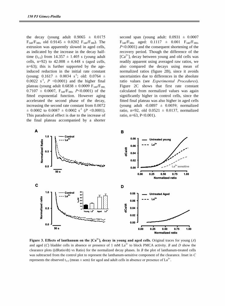
150 PJ Gómez-Pinilla
the decay (young adult 0.9065 ± 0.0175 F340/F380, old 0.9145 ± 0.0202 F340/F380). The extrusion was apparently slowed in aged cells, as indicated by the increase in the decay half-time (t1/2) from 14.357 ± 1.405 s (young adult cells, n=92) to 42.008 ± 6.448 s (aged cells, n=63); this is further supported by the age-induced reduction in the initial rate constant (young: 0.1617 ± 0.0034 s-1; old: 0.0764 ± 0.0022 s-1, P <0.0001) and the higher final plateau (young adult 0.6838 ± 0.0009 F340/F380, 0.7107 ± 0.0007, F340/F380, P<0.0001) of the fitted exponential function. However aging accelerated the second phase of the decay, increasing the second rate constant from 0.0072 ± 0.0002 to 0.0087 ± 0.0002 s-1 (P <0.0001). This paradoxical effect is due to the increase of the final plateau accompanied by a shorter
second span (young adult: 0.0931 ± 0.0007 F340/F380, aged: 0.1117 ± 0.001 F340/F380, P<0.0001) and the consequent shortening of the recovery period. Though the difference of the [Ca2+]i decay between young and old cells was readily apparent using averaged raw ratios, we also compared the decays using mean of normalized ratios (figure 2B), since it avoids uncertainties due to differences in the absolute ratio values (see Experimental Procedures). Figure 2C shows that first rate constant calculated from normalized values was again significantly higher in control cells, since the fitted final plateau was also higher in aged cells (young adult -0.0897 ± 0.0059; normalized ratio, n=92, old 0.0521 ± 0.0137, normalized ratio, n=63, P<0.001).
Figure 3. Effects of lanthanum on the [Ca2+]i decay in young and aged cells. Original traces for young (A) and aged (C) bladder cells in absence or presence of 1 mM La3+ to block PMCA activity. B and D show the clearance plots ((dRatio/dt) vs Ratio) for the normalized decay phases. In B the plot of lanthanum-treated cells was subtracted from the control plot to represent the lanthanum-sensitive component of the clearance. Inset in C represents the observed t1/2 (mean ± sem) for aged and adult cells in absence or presence of La3+.
A B
C D
0.0
0.5
1.0
50 s
La3+Control
Nor
mal
ized
Rat
io
0.00 0.25 0.50 0.75 1.00
0.00
0.02
0.04
0.06
0.08
La3+ sensitive
Normalized ratio
dCa/
dt
0.0
0.5
1.0
50 s
Nor
mal
ized
ratio
0.00 0.25 0.50 0.75 1.00
0.00
0.02
0.04
0.06
0.08
Normalized ratio
dCa/
dt
La3+Control
0
10
20
30
40
50
60
Young La3+ La3+Aged
La
Untreated young3+
La
Untreated Aged3+
t 1/2
(s)
t 1/2
(s)

Trabajo Experimental IV 151
Irrespective of small differences between absolute values for the initial phase of the [Ca2+]i decay, clearance operates at a significantly impaired rate in senescent cells for most of the [Ca2+]i range, as inferred by comparing the clearance plot of aged and young adult bladder cells, which shows a substantial loss of clearance activity in aged cells (more than 50%).
Ca2+ extrusion from cytosol is operated by three main transport mechanisms: PMCA pumps of the plasma membrane, Na+/Ca2+ exchange with extracellular medium and SERCA pumps of the sarcoplasmic reticulum. To test the contribution of these mechanisms in young and aged cells, we used specific inhibitors: 1 µM SEA-0400 for NCX, 1 mM
La3+ for PMCA and 1 µM thapsigargin for SERCA pumps. The relative importance of these systems in young and aged cells was estimated through the effects of the inhibitors on the clearance plots and on the t1/2 and final recovery values measured in each condition.
Figure 3 shows that the treatment of young adult cells with La3+ did not change the observed final plateau (control: -0.0276 ± 0.0189, n=92, La3+ -0.0257 ± 0.0214, n=59, not significant) but slowed up the initial part of the decay (first rate constant: 0.1815 ± 0.0008 vs 0.0955 ± 0.0005 s-1, control and La3+
respectively, P<0.0001). However, the t1/2 was slightly but not significantly increased (figure 3C). Clearance plots show that lanthanum retards the initial phase of the decay, when the
Figure 4. Effects of inhibition of Na+/Ca2+ exchange on the [Ca2+]i decay in young and aged cells. Original traces for young (A) and aged (C) bladder cells in absence or presence of 1 µM SEA0400 to inhibit NCX function. B and D show the clearance plots for the normalized decay phases. In B the [Ca2+]i �dependent rate of decay of SEA0400-treated cells was subtracted from the rate of untreated cells to represent the NCX-dependent component of the clearance. Inset in C represents the observed t1/2 (mean ±sem) for aged and adult cells in absence or presence of SEA0400.
A B
C D
0.0
0.5
1.0
50 s
SEA0400Control
Nor
mal
ized
Rat
io
0.0
0.5
1.0
50 s
Nor
mal
ized
Rat
io
0.00 0.25 0.50 0.75 1.00
0.00
0.02
0.04
0.06
0.08 Untreated youngSEA0400
SEA0400 sensitive
Normalized ratio
dCa/
dt
SEA0400Control
0.00 0.25 0.50 0.75 1.00
0.00
0.02
0.04
0.06
0.08
Normalized ratio
dCa/
dt
Untreated agedSEA0400
0
10
20
30
40
50
60
Young SEA SEAAged
t 1/2
(s)
P<0.01

152 PJ Gómez-Pinilla
fractional decay is above 50% of the initial ratio value (fig 3B), indicating the contribution of PMCA to this phase of the extrusion. By the contrary, when aged cells were perfused with La3+ there was no slow up on the rate of the decay, as it can be observed in figure 3C-D. Moreover, the total recovery was impaired in lanthanum-treated cells, as indicated by the plateau values (0.0873 ± 0.0288, n=63, vs 0.1769 ± 0.0339, n=61, control and treated cells respectively, P<0.05), although the t1/2 was almost intact. These result indicate that, in aged cells PMCA activity has a low contribution on Ca2+ extrusion at high [Ca2+]i levels but increases its role for near-resting values.
Treatment of young cells with SEA0400 to inhibit NCX activity resulted in a clear inhibition of the decay, as shown in figure 4A. The drug also increased the final normalized plateau from -0.0276 ± 0.0189 (n=92) to 0.1159 ± 0.0447 (n=36, P<0.005). Although SEA0400 did no change the fitted rate constants of the decay, it enhanced t1/2 significantly (fig 4C) and decreased the instantaneous decay rate even for moderate [Ca2+]i levels, when the fractional recovery was even lower than 50% (Fig 4B). In aged cells, application of SEA0400 also reduced the final recovery (untreated 0.0873 ± 0.0288, n=63, treated cells 0.2340 ± 0.0439, n=12, normalized plateau, P<0.05), but induced a slight acceleration of the first half of the decay, with a small and not significant decrease in observed half-life (figure 4C). The clearance plots showed that in aged cells the instantaneous rate lost the sensitivity to SEA0400 at high [Ca2+]i levels, at which even a slight increase in speed was observed, but it retained sensitivity for basal levels (fig 4D).
Thapsigargin, a specific inhibitor of the sarcoplasmic reticulum SERCA pumps, did not change the measured final recovery (untreated young cells -0.0276 ± 0.0189, n=92, thapsigargin-treated -0.0680 ± 0.0358, n=35, normalized ratio, not significant). However, TPS retarded the initial part of the extrusion, as shown by the initial phase of the clearance plot
(normalized ratios > 0.5), although the increase of half-life of decay was not statistically significant (figure 5). TPS induced no retard or even a slight acceleration in the rate of decay when [Ca2+]i approaches to resting levels (see figure 5B). By the contrary, in aged cells TPS increased the final plateau from 0.0873 ± 0.0288 (n=63) to 0.2022 ± 0.0562 (n=14, P<0.05, one tailed test), but decreased the clearance half-life (Fig 5C), suggesting an acceleration of the initial phase of the decay. This is further supported by the examination of the clearance plot, which reveals that in thapsigargin-treated aged cells the initial speed of the recovery is higher than in untreated cells, dropping slightly below the later when [Ca2+]i approaches to resting values (fig 5D). This effect is opposite to that observed in young adult cells (fig 5B), indicating that aging changes the role of SERCA activity in Ca2+ clearance.
The previous results indicate that the most important mechanism to decrease [Ca2+]i in young adult bladder smooth muscle is the NCX exchanger (SEA0400 sensitive), with PMCA (La3+ sensitive) and SERCA-operated pools (TPS sensitive) operating when intracellular calcium is high or moderately high. In senescent cells the apparent conclusion is that none of these mechanisms are responsible for Ca2+ clearance, given that the inhibitors did not impair the extrusion (or even enhanced it) (for an estimation of the relative contributions see Experimental Procedures and Table I). To confirm the effects of inhibiting extrusion mechanisms, we applied simultaneously inhibitors for two of the transport routes. Under this condition, the remaining activity is due to the third route (or to alternative Ca2+ transport systems). The corresponding clearance plots are shown in figure 6. The combination of TPS plus La3+ in young cells induced a retard higher than that attained by either TPS or La3+ alone, as indicated by the increased t1/2 and by comparison of the clearance plots (see figures 6, 3 and 5). In addition, the average normalized plateau at the end of the recorded period
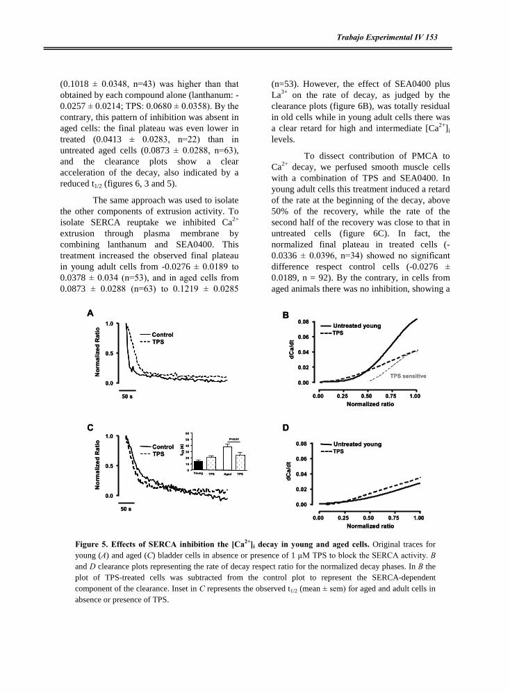
Trabajo Experimental IV 153
(0.1018 ± 0.0348, n=43) was higher than that obtained by each compound alone (lanthanum: -0.0257 ± 0.0214; TPS: 0.0680 ± 0.0358). By the contrary, this pattern of inhibition was absent in aged cells: the final plateau was even lower in treated (0.0413 ± 0.0283, n=22) than in untreated aged cells (0.0873 ± 0.0288, n=63), and the clearance plots show a clear acceleration of the decay, also indicated by a reduced t1/2 (figures 6, 3 and 5).
The same approach was used to isolate the other components of extrusion activity. To isolate SERCA reuptake we inhibited Ca2+ extrusion through plasma membrane by combining lanthanum and SEA0400. This treatment increased the observed final plateau in young adult cells from -0.0276 ± 0.0189 to 0.0378 ± 0.034 (n=53), and in aged cells from 0.0873 ± 0.0288 (n=63) to 0.1219 ± 0.0285
(n=53). However, the effect of SEA0400 plus La3+ on the rate of decay, as judged by the clearance plots (figure 6B), was totally residual in old cells while in young adult cells there was a clear retard for high and intermediate [Ca2+]i levels.
To dissect contribution of PMCA to Ca2+ decay, we perfused smooth muscle cells with a combination of TPS and SEA0400. In young adult cells this treatment induced a retard of the rate at the beginning of the decay, above 50% of the recovery, while the rate of the second half of the recovery was close to that in untreated cells (figure 6C). In fact, the normalized final plateau in treated cells (-0.0336 ± 0.0396, n=34) showed no significant difference respect control cells (-0.0276 ± 0.0189, n = 92). By the contrary, in cells from aged animals there was no inhibition, showing a
A B
C D
0.0
0.5
1.0
50 s
TPSControl
Nor
mal
ized
Rat
io
0.0
0.5
1.0
50 s
Nor
mal
ized
Rat
io
0.00 0.25 0.50 0.75 1.00
0.00
0.02
0.04
0.06
0.08
TPS sensitive
TPS
Normalized ratio
dCa/
dt
0.00 0.25 0.50 0.75 1.00
0.00
0.02
0.04
0.06
0.08
Normalized ratio
dCa/
dt
TPSControl
Untreated young
TPSUntreated young
0
10
20
30
40
50
60
Young TPS TPSAged
P<0.01
t 1/2
(s)
t 1/2
(s)
Figure 5. Effects of SERCA inhibition the [Ca2+]i decay in young and aged cells. Original traces for young (A) and aged (C) bladder cells in absence or presence of 1 µM TPS to block the SERCA activity. B and D clearance plots representing the rate of decay respect ratio for the normalized decay phases. In B the plot of TPS-treated cells was subtracted from the control plot to represent the SERCA-dependent component of the clearance. Inset in C represents the observed t1/2 (mean ± sem) for aged and adult cells in absence or presence of TPS.

154 PJ Gómez-Pinilla
shortened half-life (figure 6D) and an accelerated rate of decay from high to resting [Ca2+]i levels (figure 6C), when treated cells decline slightly slower than untreated cells.
To estimate the age-related changes of each Ca2+ removal system in our experimental conditions we followed the method described by (Liu et al., 2006). The overall rate constant of the decay, estimated as the inverse of observed half-life values (t1/2
-1 or Total), can be considered to be the sum of three main components: T = PMCA + SEA + SERCA. The contribution of each component was calculated
as i = (1- X/CONTROL)*100, i.e. the percentage decrease in the rate constant in response to a treatment. Table I shows relative contributions of each system in young adult and in aged cells. In adult cells the main contributor is NCX (more than 50%), with contribution for PMCA and SERCA around 30%. The sum of the three components (114%) is higher than 100%, suggesting some kind of interaction between one another. We also calculated the contribution for two systems at a time, corresponding to the effect of the combination of two inhibitors. The sum of this value plus the contribution of the third system should
Figure 6. Effects of combined inhibition of PMCA, NCX and SERCA on the rate of clearance in young and aged bladder smooth muscle cells. Cells were treated with a combination of inhibitors of two of the three main Ca2+ removing mechanisms to isolate the clearance activity of NCX (A, TPS plus La3+), SERCA (B, SEA0400 plus La3+ ) or PMCA (C, TPS plus SEA0400). Plots for untreated young and aged cells are also combined for comparison. Panel D shows the mean ± sem of the measured t1/2 for each experimental condition.
A B
C D
0.00 0.25 0.50 0.75 1.00
0.00
0.02
0.04
0.06
0.08
YoungYoung TPS+LaAgedAged TPS+La
Normalized ratio
dCa/
dt
0.00 0.25 0.50 0.75 1.00
0.00
0.02
0.04
0.06
0.08YoungYoung TPS + SEA
Aged TPS + SEAAged
Normalized ratio
dCa/
dt
0.00 0.25 0.50 0.75 1.00
0.00
0.02
0.04
0.06
0.08 YoungYoung SEA+La
3+
AgedAged SEA+La3+
Normalized ratio
dCa/
dt
0
10
20
30
40
50
60
La3+
TPSSEA0400
+ + + ++ + + +
+ + + +
Young Aged
**
**
t ½(S
)

Trabajo Experimental IV 155
approach 100; for example, contribution of SERCA (31.04, TPS) plus contribution of NCX and PMCA (57.25, SEA0400+La3+) amounts 88.03%. The most striking feature is the low apparent contribution for combined SERCA and NCX (only 11.58%, TPS+SEA0400, far from the expected 73%). Therefore, PMCA activity is somehow inhibited or disguised by NCX and SERCA, so that when activities of NCX and SERCA are intact lanthanum treatment induces a 27% decrease, while the application of SEA0400 plus TPS decreases the rate only by 11% because PMCA is no longer inhibited and its contribution is increased (~88%). Although to a much lesser extent, a similar situation was found for SERCA: its contribution is slightly increased from 31% (TPS treatment, with NCX and PMCA operative) to 43% for SEA0400 and La3+ treatment, indicating some inhibition by NCX and PMCA activity. By the contrary, in the case of NCX the estimated contribution was decreased by inhibition of PMCA and SERCA from 55% (SEA0400 alone) to 38% (100-62, La3++TPS), indicating that its role in Ca2+ extrusion is enhanced in presence of SERCA and PMCA pumps.
When this approach was used in aged cells, we obtained negative contribution in all conditions, except for lanthanum-treated cells. These figures result from a trade-off between extrusion from the cytosol and inhibition of other Ca2+ transport systems, and are in keeping with the observed acceleration of the decay in response to inhibitors. Similar to adult young cells, the contribution of PMCA was also inhibited by NCX and SERCA (TPS plus SEA0400, which leave PMCA as the main route for extrusion, induced a clear acceleration of the decay), but was weaker than in young adult cells in lanthanum-treated cells (less than 10%). SERCA apparently inhibits the other transport routes, given the strong increase on decay rate in response to TPS (72%). In the case of NCX, it seems also to inhibit the other transporters, but contrary to young cells, NCX was apparently inhibited by the other routes,
since inhibition of SERCA and PMCA (TPS+La3+) strongly enhanced the rate of decay. Therefore, the effect of aging is not a simple loss of sensitivity to the inhibitors (which should induce no effect instead of acceleration), but a complex set of interactions between the main Ca2+ extrusion systems.
5-DISCUSSION
The present study shows that both the speed and the pharmacology of the Ca2+ extrusion mechanisms in smooth muscle are modified by aging. To our knowledge this is the first detailed account of how aging alters each of the main Ca2+ clearing systems.
Our results indicate that bladder smooth muscle cells remove Ca2+ from cytosol using the three �classic� transport systems, i.e. SERCA reuptake into stores, and NCX exchange and PMCA pumps located at the sarcolemma. The most active system is the NCX, as revealed by the effect of the specific inhibitor SEA0400 (Matsuda et al., 2001) on both the amplitude and the speed of the recovery. The dominance of this system seems to spread to both elevated and near-resting [Ca2+]i levels, as judged by examination of the clearance plots, while inhibition of PMCA or SERCA seems less effective at low calcium levels.
Our estimates of the contribution of each Ca2+ transport system (NCX 56%, PMCA 27% and SERCA 31%) are in line with recent reports about contractility of bladder muscle strips from normal and PMCA gene-ablated mice (Liu et al., 2006). These authors also found that cooperation between the systems is not always additive, as indicated by our finding that combination of SEA0400 with either La3+ or TPS did not increase inhibition. Only when La3+ and TPS were combined there was addition between the relative contributions of PMCA and SERCA. Moreover, addition of the three contributions exceeded 100%, which also suggests the presence of interaction between the transport systems. Therefore, our data reinforce the concept that in physiological conditions

156 PJ Gómez-Pinilla
Ca2+ clearing mechanisms do not operate independently. This cooperation has been previously postulated to explain experimental data regarding contractility (Liu et al., 2006), electrophysiology (Orio et al., 2002; Gibson & Muzyamba, 2004) and [Ca2+]i signals (Imaizumi et al., 1999; Liang et al., 2004; Poburko et al., 2004). So, it has been proposed that spatial arrangements of transport systems can induce a vectorial movement of Ca2+ ions from SERCA-driven internal stores to plasma membrane transporters (Liang et al., 2004; Poburko et al., 2004). Following this, inhibition of two �consecutive� transporters would be redundant, therefore yielding less than additive effect. The physical basis for this mechanism is based not only in the �superficial barrier� theory (van et al., 1986; Poburko et al., 2004) but in the presence of both NCX and PMCA in smooth muscle caveolae (Daniel et al., 2006). In fact, bladder smooth muscle cells show similar examples of tight apposition of different Ca2+ transport systems, such as clusters of channels in plasmalemmal caveolae which operate as a functional unit in [Ca2+]i signals (Moore et al., 2004) and buffering of plasmalemmal Ca2+ current by SERCA pumps (Yoshikawa et al., 1996).
This study indicates that the mechanisms responsible for Ca2+ extrusion in urinary bladder smooth muscle are impaired in senescent cells. Thus, overall Ca2+ extrusion was inhibited in aging cells compared to young cells irrespective of the actual [Ca2+]i level. In addition, inhibition of PMCA, NCX or SERCA, which impaired the rate of clearance in control cells, did not retard extrusion in senescent cells, with the exception of a minor inhibition of the final recovery. Therefore, it seems that in aged cells the initial phase of the recovery, when [Ca2+]i is at least moderately high, is not driven by the three classic transport systems examined in our study. When calcium approaches basal levels these extrusion routes would regain importance. Mitochondrial uptake could serve as a compensatory extrusion mechanism because they accumulate calcium upon [Ca2+]i
signals and release it later to the cytosol (Duchen, 2000). In fact, in some excitable cells mitochondrial Ca2+ uptake has been reported to compensate for age-related SERCA impairment (Murchison & Griffith, 1998). However, other reports failed to find this effect (Pottorf et al., 2000), and senescent neurones show a progressive depolarization of the mitochondrial membrane (Xiong et al., 2004), incompatible with a rapid increase in Ca2+ uptake necessary to participate in the [Ca2+]i decay pattern studied in our experiments. Also, the mitochondria from aged bladder muscle shows impaired activity for key metabolic enzymes of the organelle (Lin et al., 2000), suggesting a loss of mitochondrial activity in this organ. Ongoing work in our laboratory is assessing this possibility.
A possibility to explain the apparent no participation of the classical Ca2+ extrusion mechanisms in aged smooth muscle cells could be the existence of inhibitory relationship among them, as will be discussed below, or changes in the sensitivity of these transporters to the normal inhibitors. In line with this, a striking observation is the enhanced extrusion rate in presence of thapsigargin or SEA0400 in aged cells, which also reveals that the transporters do participate in the extrusion (otherwise the inhibitors would be without effect). Several explanations could account for this result. First, SERCA inhibition could stimulate PMCA pumps situated in close apposition, a mechanism previously reported for thapsigargin-induced acceleration of Ca2+ decays (Morgan & Jacob, 1998). Second, during the KCl pulse SERCA pumps could �prime� intracellular stores with a fast turnover, so that continuous leak from this pool would �retard� the posterior decay. A similar picture has been previously reported in pancreatic acinar cells (Camello et al., 2000). Under this condition, if the Ca2+ release from this pool to the cytosol is faster than the reuptake activity, the net effect of SERCA inhibition would be an apparent acceleration of Ca2+ extrusion.
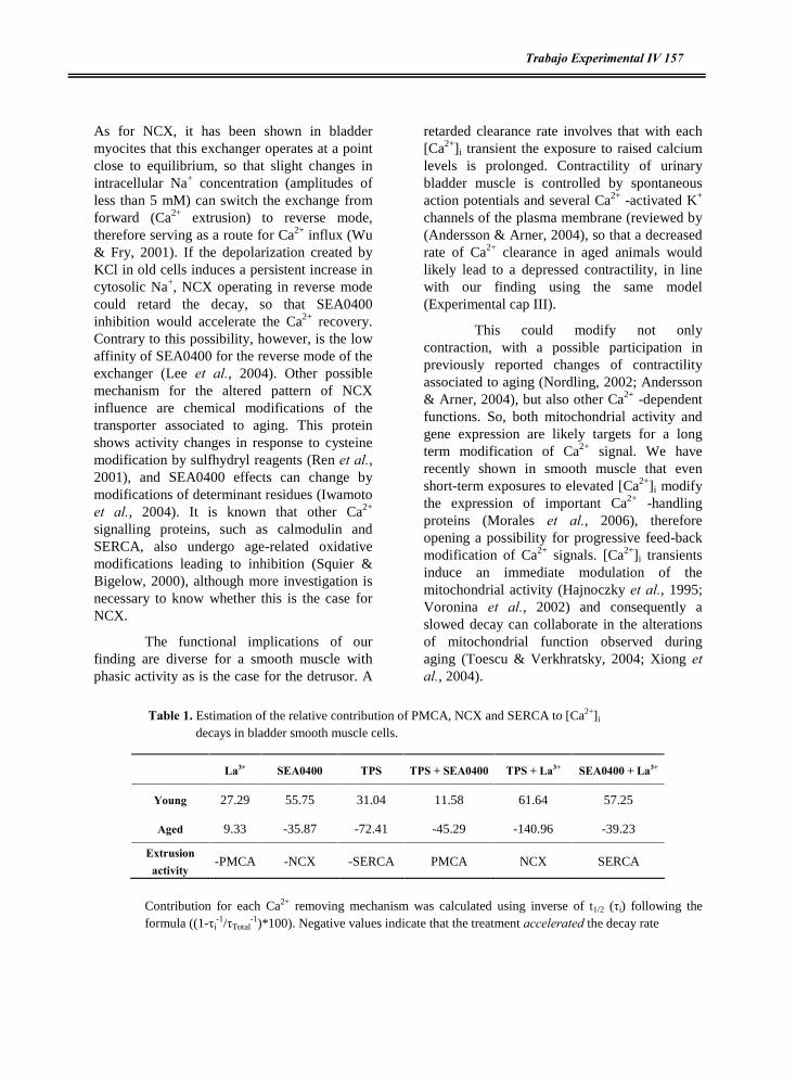
Trabajo Experimental IV 157
As for NCX, it has been shown in bladder myocites that this exchanger operates at a point close to equilibrium, so that slight changes in intracellular Na+ concentration (amplitudes of less than 5 mM) can switch the exchange from forward (Ca2+ extrusion) to reverse mode, therefore serving as a route for Ca2+ influx (Wu & Fry, 2001). If the depolarization created by KCl in old cells induces a persistent increase in cytosolic Na+, NCX operating in reverse mode could retard the decay, so that SEA0400 inhibition would accelerate the Ca2+ recovery. Contrary to this possibility, however, is the low affinity of SEA0400 for the reverse mode of the exchanger (Lee et al., 2004). Other possible mechanism for the altered pattern of NCX influence are chemical modifications of the transporter associated to aging. This protein shows activity changes in response to cysteine modification by sulfhydryl reagents (Ren et al., 2001), and SEA0400 effects can change by modifications of determinant residues (Iwamoto et al., 2004). It is known that other Ca2+ signalling proteins, such as calmodulin and SERCA, also undergo age-related oxidative modifications leading to inhibition (Squier & Bigelow, 2000), although more investigation is necessary to know whether this is the case for NCX.
The functional implications of our finding are diverse for a smooth muscle with phasic activity as is the case for the detrusor. A
retarded clearance rate involves that with each [Ca2+]i transient the exposure to raised calcium levels is prolonged. Contractility of urinary bladder muscle is controlled by spontaneous action potentials and several Ca2+ -activated K+ channels of the plasma membrane (reviewed by (Andersson & Arner, 2004), so that a decreased rate of Ca2+ clearance in aged animals would likely lead to a depressed contractility, in line with our finding using the same model (Experimental cap III).
This could modify not only contraction, with a possible participation in previously reported changes of contractility associated to aging (Nordling, 2002; Andersson & Arner, 2004), but also other Ca2+ -dependent functions. So, both mitochondrial activity and gene expression are likely targets for a long term modification of Ca2+ signal. We have recently shown in smooth muscle that even short-term exposures to elevated [Ca2+]i modify the expression of important Ca2+ -handling proteins (Morales et al., 2006), therefore opening a possibility for progressive feed-back modification of Ca2+ signals. [Ca2+]i transients induce an immediate modulation of the mitochondrial activity (Hajnoczky et al., 1995; Voronina et al., 2002) and consequently a slowed decay can collaborate in the alterations of mitochondrial function observed during aging (Toescu & Verkhratsky, 2004; Xiong et al., 2004).
Table 1. Estimation of the relative contribution of PMCA, NCX and SERCA to [Ca2+]i
decays in bladder smooth muscle cells.
Contribution for each Ca2+ removing mechanism was calculated using inverse of t1/2 (ôi) following the formula ((1-ôi
-1/ôTotal-1)*100). Negative values indicate that the treatment accelerated the decay rate
La3+ SEA0400 TPS TPS + SEA0400 TPS + La3+ SEA0400 + La3+
Young 27.29 55.75 31.04 11.58 61.64 57.25
Aged 9.33 -35.87 -72.41 -45.29 -140.96 -39.23
Extrusion activity
-PMCA -NCX -SERCA PMCA NCX SERCA

158 PJ Gómez-Pinilla
A practical consequence of our study is related to the effect of inhibitors in aged cells. The finding that inhibitors of Ca2+ extrusion mechanisms can accelerate the [Ca2+]i decay in aged individuals poses potential side-effects in the therapeutical use of this type of compounds, as is the case for NCX inhibitors in some cardiovascular and renal diseases (Iwamoto & Kita, 2004) because these drugs could worsen the impairment of aged smooth muscle cells.
6-ACKNOWLEDGEMENTS
Pedro J Gómez-Pinilla is recipient of a PhD Fellowship of Junta de Extremadura (Spain). The study was supported by Spanish Ministery of Education (BFU 2004-0637) and Junta de Extremadura (2PR03A020).
7- REFERENCE LIST
Andersson KE & Arner A (2004). Urinary bladder contraction and relaxation: physiology and pathophysiology. Physiol Rev 84, 935-986.
Brewer GJ, Reichensperger JD, & Brinton RD (2006). Prevention of age-related dysregulation of calcium dynamics by estrogen in neurons. Neurobiol Aging 27, 306-317.
Camello C, Pariente JA, Salido GM, & Camello PJ (2000). Role of proton gradients and vacuola H+-ATPases in the refilling of intracellular calcium stores in exocrine cells. Curr Biol 10, 161-164.
Camello P, Gardner J, Petersen OH, & Tepikin AV (1996). Calcium dependence of calcium extrusion and calcium uptake in mouse pancreatic acinar cells. J Physiol 490 ( Pt 3), 585-593.
Daniel EE, El-Yazbi A, & Cho WJ (2006). Caveolae and calcium handling, a review and a hypothesis. J Cell Mol Med 10, 529-544.
Del CC, Ostrovskaya O, McAllister CE, Murray K, Hatton WJ, Gurney AM, Spencer NJ, & Wilson SM (2006). Effects of aging on Ca2+ signaling in murine mesenteric arterial smooth muscle cells. Mech Ageing Dev 127, 315-323.
Duchen MR (2000). Mitochondria and calcium: from cell signalling to cell death. J Physiol 529 Pt 1, 57-68.
Gant JC, Sama MM, Landfield PW, & Thibault O (2006). Early and simultaneous emergence of multiple hippocampal biomarkers of aging is mediated by Ca2+-induced Ca2+ release. J Neurosci 26, 3482-3490.
Geary GG & Buchholz JN (2003). Selected contribution: Effects of aging on cerebrovascular tone and [Ca2+]i. J Appl Physiol 95, 1746-1754.
Gibson JS & Muzyamba MC (2004). Modulation of Gardos channel activity by oxidants and oxygen tension: effects of 1-chloro-2,4-dinitrobenzene and phenazine methosulphate. Bioelectrochemistry 62, 147-152.
Gomez-Pinilla PJ, Camello-Almaraz C, Moreno R, Camello PJ, & Pozo MJ (2006). Melatonin treatment reverts age-related changes in Guinea pig gallbladder neuromuscular transmission and contractility. J Pharmacol Exp Ther 319, 847-856.
Hajnoczky G, Robb-Gaspers LD, Seitz MB, & Thomas AP (1995). Decoding of cytosolic calcium oscillations in the mitochondria. Cell 82, 415-424.

Trabajo Experimental IV 159
Howlett SE, Grandy SA, & Ferrier GR (2006). Calcium spark properties in ventricular myocytes are altered in aged mice. Am J Physiol Heart Circ Physiol 290, H1566-H1574.
Imaizumi Y, Ohi Y, Yamamura H, Ohya S, Muraki K, & Watanabe M (1999). Ca2+ spark as a regulator of ion channel activity. Jpn J Pharmacol 80, 1-8.
Isenberg G, Borschke B, & Rueckschloss U (2003). Ca2+ transients of cardiomyocytes from senescent mice peak late and decay slowly. Cell Calcium 34, 271-280.
Ishizuka J, Murakami M, Nichols GA, Cooper CW, Greeley GH, Jr., & Thompson JC (1993). Age-related changes in gallbladder contractility and cytoplasmic Ca2+ concentration in the guinea pig. Am J Physiol 264, G624-G629.
Iwamoto T & Kita S (2004). Development and application of Na+/Ca2+ exchange inhibitors. Mol Cell Biochem 259, 157-161.
Iwamoto T, Kita S, Uehara A, Imanaga I, Matsuda T, Baba A, & Katsuragi T (2004). Molecular determinants of Na+/Ca2+ exchange (NCX1) inhibition by SEA0400. J Biol Chem 279, 7544-7553.
Khachaturian ZS (1987). Hypothesis on the regulation of cytosol calcium concentration and the aging brain. Neurobiol Aging 8, 345-346.
Kirischuk S, Voitenko N, Kostyuk P, & Verkhratsky A (1996). Age-associated changes of cytoplasmic calcium homeostasis in cerebellar granule neurons in situ: investigation on thin cerebellar slices. Exp Gerontol 31, 475-487.
Lee C, Visen NS, Dhalla NS, Le HD, Isaac M, Choptiany P, Gross G, Omelchenko A, Matsuda T, Baba A, Takahashi K, Hnatowich M, & Hryshko LV (2004). Inhibitory profile of SEA0400 [2-[4-[(2,5-difluorophenyl)methoxy]phenoxy]-5-ethoxyaniline] assessed on the cardiac Na+-Ca2+ exchanger, NCX1.1. J Pharmacol Exp Ther 311, 748-757.
Liang W, Buluc M, van BC, & Wang X (2004). Vectorial Ca2+ release via ryanodine receptors contributes to Ca2+ extrusion from freshly isolated rabbit aortic endothelial cells. Cell Calcium 36, 431-443.
Lin AT, Hsu TH, Yang C, & Chang LS (2000). Effects of aging on mitochondrial enzyme activity of rat urinary bladder. Urol Int 65, 144-147.
Liu L, Ishida Y, Okunade G, Shull GE, & Paul RJ (2006). Role of plasma membrane Ca2+-ATPase in contraction-relaxation processes of the bladder: evidence from PMCA gene-ablated mice. Am J Physiol Cell Physiol 290, C1239-C1247.
Lopes GS, Ferreira AT, Oshiro ME, Vladimirova I, Jurkiewicz NH, Jurkiewicz A, & Smaili SS (2006). Aging-related changes of intracellular Ca2+ stores and contractile response of intestinal smooth muscle. Exp Gerontol 41, 55-62.
Maloney JA & Wheeler-Clark ES (1996). Reduction in sarcoplasmic reticulum Ca2+-ATPase activity contributes to age-related changes in the calcium content and relaxation rate of rabbit aortic smooth muscle. J Hypertens 14, 65-74.
Martinez-Serrano A, Blanco P, & Satrustegui J (1992). Calcium binding to the cytosol and calcium extrusion mechanisms in intact synaptosomes and their alterations with aging. J Biol Chem 267, 4672-4679.
160 PJ Gómez-Pinilla
Matsuda T, Arakawa N, Takuma K, Kishida Y, Kawasaki Y, Sakaue M, Takahashi K, Takahashi T, Suzuki T, Ota T, Hamano-Takahashi A, Onishi M, Tanaka Y, Kameo K, & Baba A (2001). SEA0400, a novel and selective inhibitor of the Na+-Ca2+ exchanger, attenuates reperfusion injury in the in vitro and in vivo cerebral ischemic models. J Pharmacol Exp Ther 298, 249-256.
Moore ED, Voigt T, Kobayashi YM, Isenberg G, Fay FS, Gallitelli MF, & Franzini-Armstrong C (2004). Organization of Ca2+ release units in excitable smooth muscle of the guinea-pig urinary bladder. Biophys J 87, 1836-1847.
Morales S, Diez A, Puyet A, Camello P, Camello-Almaraz C, Bautista J, & Pozo MJ (2006). Calcium controls smooth muscle TRPC gene transcription via the CAMK/calcineurin-dependent pathways. Am J Physiol Cell Physiol.
Morgan AJ & Jacob R (1998). Differential modulation of the phases of a Ca2+ spike by the store Ca2+-ATPase in human umbilical vein endothelial cells. J Physiol 513 ( Pt 1), 83-101.
Murchison D & Griffith WH (1998). Increased calcium buffering in basal forebrain neurons during aging. J Neurophysiol 80, 350-364.
Nordling J (2002). The aging bladder--a significant but underestimated role in the development of lower urinary tract symptoms. Exp Gerontol 37, 991-999.
Orio P, Rojas P, Ferreira G, & Latorre R (2002). New disguises for an old channel: MaxiK channel beta-subunits. News Physiol Sci 17, 156-161.
Pagala MK, Tetsoti L, Nagpal D, & Wise GJ (2001). Aging effects on contractility of longitudinal and circular detrusor and trigone of rat bladder. J Urol 166, 721-727.
Poburko D, Kuo KH, Dai J, Lee CH, & van BC (2004). Organellar junctions promote targeted Ca2+ signaling in smooth muscle: why two membranes are better than one. Trends Pharmacol Sci 25, 8-15.
Pottorf WJ, Duckles SP, & Buchholz JN (2000). Adrenergic nerves compensate for a decline in calcium buffering during ageing. J Auton Pharmacol 20, 1-13.
Ren X, Kasir J, & Rahamimoff H (2001). The transport activity of the Na+-Ca2+ exchanger NCX1 expressed in HEK 293 cells is sensitive to covalent modification of intracellular cysteine residues by sulfhydryl reagents. J Biol Chem 276, 9572-9579.
Rubio C, Moreno A, Briones A, Ivorra MD, D'Ocon P, & Vila E (2002). Alterations by age of calcium handling in rat resistance arteries. J Cardiovasc Pharmacol 40, 832-840.
Smith DO (1988). Cellular and molecular correlates of aging in the nervous system. Exp Gerontol 23, 399-412.
Squier TC & Bigelow DJ (2000). Protein oxidation and age-dependent alterations in calcium homeostasis. Front Biosci 5, D504-D526.
Szabo P, Weksler D, Whittington E, & Weksler BB (1993). The age-dependent proliferation of rat aortic smooth muscle cells is independent of differential splicing of PDGF A-chain mRNA. Mech Ageing Dev 67, 79-89.
Toescu EC & Verkhratsky A (2000). Parameters of calcium homeostasis in normal neuronal ageing. J Anat 197 Pt 4, 563-569.
Toescu EC & Verkhratsky A (2003). Neuronal ageing from an intraneuronal perspective: roles of endoplasmic reticulum and mitochondria. Cell Calcium 34, 311-323.

Trabajo Experimental IV 161
Toescu EC & Verkhratsky A (2004). Ca2+ and mitochondria as substrates for deficits in synaptic plasticity in normal brain ageing. J Cell Mol Med 8, 181-190.
Van BC, Lukeman S, Leijten P, Yamamoto H, & Loutzenhiser R (1986). The role of superficial SR in modulating force development induced by Ca entry into arterial smooth muscle. J Cardiovasc Pharmacol 8 Suppl 8, S111-S116.
Vanterpool CK, Pearce WJ, & Buchholz JN (2005). Advancing age alters rapid and spontaneous refilling of caffeine-sensitive calcium stores in sympathetic superior cervical ganglion cells. J Appl Physiol 99, 963-971.
Vanterpool CK, Vanterpool EA, Pearce WJ, & Buchholz JN (2006). Advancing age alters the expression of the ryanodine receptor 3 isoform in adult rat superior cervical ganglia. J Appl Physiol 101, 392-400.
Voronina S, Sukhomlin T, Johnson PR, Erdemli G, Petersen OH, & Tepikin A (2002). Correlation of NADH and Ca2+ signals in mouse pancreatic acinar cells. J Physiol 539, 41-52.
Wanaverbecq N, Marsh SJ, Al-Qatari M, & Brown DA (2003). The plasma membrane calcium-ATPase as a major mechanism for intracellular calcium regulation in neurones from the rat superior cervical ganglion. J Physiol 550, 83-101.
Wu C & Fry CH (2001). Na+/Ca2+ exchange and its role in intracellular Ca2+ regulation in guinea pig detrusor smooth muscle. Am J Physiol Cell Physiol 280, C1090-C1096.
Xiong J, Camello PJ, Verkhratsky A, & Toescu EC (2004). Mitochondrial polarisation status and [Ca2+]i signalling in rat cerebellar granule neurones aged in vitro. Neurobiol Aging 25, 349-359.
Xiong Z, Sperelakis N, Noffsinger A, & Fenoglio-Preiser C (1993). Changes in calcium channel current densities in rat colonic smooth muscle cells during development and aging. Am J Physiol 265, C617-C625.
Yoshikawa A, van BC, & Isenberg G (1996). Buffering of plasmalemmal Ca2+ current by sarcoplasmic reticulum of guinea pig urinary bladder myocytes. Am J Physiol 271, C833-C841.
Yu HJ, Wein AJ, & Levin RM (1996). Age-related differential susceptibility to calcium channel blocker and low calcium medium in rat detrusor muscle: response to field stimulation. Neurourol Urodyn 15, 563-576.

162 PJ Gómez-Pinilla

Trabajo Experimental V 163
The Journal of Pharmacology and Experimental Therapeutics (Accepted for publication, August 9, 2006)
MELATONIN TREATMENT REVERTS AGED-RELATED CHANGES IN GUINEA PIG GALLBLADDER NEUROMUSCULAR TRANSMISSION AND CONTRACTILITY
Pedro J. Gomez-Pinilla, Cristina Camello-Almaraz, Rosario Moreno,
Pedro J. Camello and María J. Pozo*
Department of Physiology, Nursing School,
University of Extremadura, (10071) Cáceres, Spain
Running head: Melatonin and aged gallbladder
*Correspondence to:
Maria J. Pozo
Department of Physiology
Nursing School
Avda Universidad s/n
10071 Cáceres
Spain
Telephone: + 34-927-257450
Fax: + 34-927-257451
E-mail: [email protected]
Key Words: gallbladder, aging, guinea pig, neruogenic, miogenic and melatonin
id5996953 pdfMachine by Broadgun Software - a great PDF writer! - a great PDF creator! - http://www.pdfmachine.com http://www.broadgun.com

164 PJ Gómez-Pinilla
CONTENTS
1- Abstract
2- Introduction
3- Material and methods
Tissue preparation
Measurement of melatonin in serum
Contraction recording of guinea pig gallbladder smooth muscle strips
Cell isolation
Cell loading and [Ca2+]i determination
F-actin Content Measurement
Solutions and drugs
Quantification and statistics
4- Results.
Aged related changes in efferent innervation in EFS-induced gallbladder responses
Aged related changes in sensory innervation
Aged related changes in gallbladder contractility
Aged related changes in calcium handling
Aged-related changes in contractile machinery.
5- Discussion
6- Acknowledgements
7- References list
Abbreviations:
EFS, electrical field stimulation; CGRP, calcitonin gene-related peptide; CCK, cholecystokinin; GI, gastrointestinal tract; ACh, acetylcholine; BSA, bovine serum albumin; DTT, 1,4-dithio-DLthreitol; Phosphate-buffered saline solution , PBS; Nonidet P-40, octylphenyl-polyethylene glycol; FITC-phalloidin, fluorescein isothiocyanate-labeled phalloidin; L-NAME, Nù-nitro-L-arginine methyl ester; Bt3-Ins(1,4,5)P3-PM, 2,3,6-Tri-O-Butyryl-myo-Inositol-1,4,5-trisphosphate-Hexakis(propionoxymethyl) Ester; mN, milliNewtons; ANOVA, Analysis of variance; CCE, capacitative calcium entry; IP3, D-myo-inositol 1,4,5-triphosphate.

Trabajo Experimental V 165
1-ABSTRACT
The incidence of gallbladder illness increases with age but the altered mechanisms leading to gallbladder dysfunction are poorly understood. Here we determine the age-related alterations in gallbladder contractility and the impact of melatonin treatment. Isometric tension changes in response to electrical field stimulation (EFS) and to agonists were recorded from guinea pig gallbladder muscle strips. [Ca2+]i was determined by epifluorescence microscopy in fura-2 loaded isolated gallbladder smooth muscle cells, and F-actin content was quantified by confocal microscopy. Aging reduced neurogenic contractions, which was associated to the impairment of nitrergic innervation and to increased responsiveness of capsaicin-sensitive relaxant nerves, possibly involving calcitonin gene-related peptide (CGRP). Melatonin treatment for four weeks restored neurogenic responses to normal values, with an associated recovery of nitrergic function and the disappearance of the capsaicin-sensitive component. Aging also reduced the contractile responses to cholecystokinin (CCK) and Ca2+ influx. The impaired contractility only correlated with diminished Ca2+ mobilization in response to activation of Ca2+ influx. Melatonin improved contractility and increased smooth muscle F-actin content without changing Ca2+ homeostasis. In conclusion, aging impairs gallbladder function as the result of changes in the inhibitory neuromodulation of smooth muscle contractility and the reduction in the myogenic response to contractile agonists. Impaired contractility seems to be related to decreased Ca2+ influx and damage of contractile proteins. Melatonin significantly ameliorated these age-related changes.
2-INTRODUCTION
Population aging was one of the most distinctive demographic events of the 20th century and according to the increase in the mean life expectancy, the elderly population will constitute the 20% of the world population in the next 50 years (Centers for Disease Control And Prevention (CDC), 2003). Aging is understood
mainly as a slow, gradual, and passive process influenced by interplay of multiple genetic and environmental factors. It is widely accepted that aging is not a disease but the borderline between pathological and normal aging is quite narrow. Although there are many kinds of �non-disease� aging manifestations including, blood vessel stiffening, skin slacking, joint stiffening, etc, the molecular structures of aged organisms are abnormal (Bailey, 2001). These molecular abnormalities could be responsible of the increase in the incidence and prevalence in the elderly of the so called age-related diseases such as neurodegenerative disorders (Troulinaki and Tavernarakis, 2005), cardiovascular disease (Ferrari, et al., 2003), diabetes (Winer and Sowers, 2004), incontinence (Nasr and Ouslander, 1998), etc.
Regarding the gastrointestinal tract (GI), the prevalence of dysfunctions related to motility is higher in older than in younger adults (Camilleri, et al., 2000). However, there are very few studies concerning the cellular and molecular mechanisms whose alteration could be responsible for such us dysfunctions in aging. Thus, it has been reported that neuronal loss occurs with age in the myenteric plexus of the GI in several species including man (Gomes, et al., 1997). However, it is unclear whether cell death affects all classes of myenteric neurons nonselectively or is confined to specific phenotypes (Wade and Cowen, 2004). Another important component that could be affected by aging, leading to contractility impairment, is smooth muscle itself. In this regard, information regarding the putative changes in cellular mechanisms of smooth muscle contraction is scanty (Bitar and Patil, 2004) . Acetylcholine-induced contractile responses are impaired in colon (Roberts, et al., 1994) which could be due to the limited cell length distribution found in aged-animals but also to alteration of the intracellular cell signalling pathways related to activation of protein kinase C and cytoskeleton reorganization (Bitar, 2003). There is little information regarding the possible changes in Ca2+ homeostasis during aging in smooth muscle

166 PJ Gómez-Pinilla
in general and gastrointestinal smooth muscle in particular, where it has been described an aged-related decline in the L-type Ca2+ currents both in rat and human colon smooth muscle cells (Xiong, et al., 1993;Xiong, et al., 1995).
Knowledge of the molecular mechanisms involved in aging is required to develop strategies to preserve the quality of life of the increasingly aging population. Degenerative changes associated with aging have been related to progressive damage by reactive oxygen and nitrogen species in those situations where the anti-oxidative defence systems fails to eliminate them (Sohal and Allen, 1990). Recently, melatonin, the main hormone of the pineal gland was proposed as a protective agent against macromolecular destruction associated with longevity (Reiter, et al., 1996). The protective effects of melatonin could be related to the ability of the hormone to synchronize circadian rhythms and thereby to reduce biological stress, as well as to its direct free radical scavenging activity and its indirect antioxidants properties (Reiter, et al., 2002).
In the current study we explored the aged-related alterations of neuromuscular transmission, gallbladder contractility and smooth muscle Ca2+ homeostasis. Our results show that aging-induced gallbladder impairment is primarily related to changes in the inhibitory innervation of the organ and to a decrease in the myogenic response to excitatory hormones and neurotransmitters. Melatonin treatment reversed the functional impairment caused by aging at the level of both the neuromuscular transmission and the myogenic contractility despite being ineffective in the restoration of Ca2+ homeostasis.
3-MATERIALS AND METHODS
Tissue preparation: Gallbladders were removed from 4-and 20-mo-old female guinea pigs after deep halothane anaesthesia and cervical dislocation, and were immediately placed in cold Krebs-Henseleit solution (for composition see Solutions and drugs) at pH 7.35. The gallbladder was opened from the end of the cystic duct to the base, and trimmed of any adherent liver tissue.
After the preparation was washed with the nutrient solution to remove residual bile, the mucosa was carefully dissected away.
A group of aged animals was treated orally with melatonin (2.5 mg/Kg/day). Melatonin was dissolved in glucose solution (1.5%) and placed in the oropharynx by a syringe. This treatment was applied daily at the same time, just before the light in the animal house was switched off (7 p.m.). All the experiments were conducted according to the guidelines of Animal Care and Use Committees of the University of Extremadura.
Measurement of melatonin in serum: Melatonin levels were determined in the different age groups by using a commercial radioimmunoassay kit (IBL, Hamburg), according to the manufacturers instruction. The kit consisted of 125I-melatonin (0.68 µCi/ml), rabbit anti-melatonin serum, melatonin standards, delipidizing agent, assay buffer, precipitating antiserum, and controls (lyophilized plasma samples). Results were expressed in pg/ml.
Contraction recording of guinea pig gallbladder smooth muscle strips: Gallbladder strips (measuring ~3 x 10 mm) were placed vertically in a 10 ml organ bath filled with Krebs-Henseleit solution maintained at 37 °C and gassed with 95% O2 - 5% CO2. Isometric contractions were measured using force displacement transducers that were interfaced with a Macintosh computer using a MacLab hardware unit and software (ADInstruments; Colorado Spring, CO, USA). The muscle strips were placed under an initial resting tension equivalent to 1.5 g load and allowed to equilibrate for 60 min, with solution changes every 20 min. Every strip coming from a given animal was used in a different experimental protocol.
Intrinsic nerves were activated by electrical field stimulation (EFS) with a pair of external platinum ring electrodes (0.7 mm in diameter) connected to a square wave stimulator (Cibertec CS9/3BO) programmed through Scope software application from MacLab (AD
Trabajo Experimental V 167
Instruments). Trains of stimuli (0.3 ms duration, 5-40 Hz, 350 mA current strength) were delivered for 10 s at 3 min intervals. After construction of a frequency response curve and in order to pharmacologically dissect the neurogenic responses, antagonists were added to the organ bath for 20 min, and then the EFS protocols were repeated.
In some experiments the contractile effects of acetylcholine (ACh), cholecystokinin (CCK), KCl and Ca2+ restoration after intracellular Ca2+ store depletion were tested.
Cell isolation: Gallbladder smooth muscle cells were dissociated enzymatically using a previously described method (Pozo, et al., 2002). Briefly, the gallbladder was cut into small pieces and incubated for 34 min at 37°C in enzyme solution (for composition see Solutions and drugs) supplemented with 1 mg/ml BSA, 1 mg/ml papain, and 1 mg/ml DTT. Next, the tissue was transferred to fresh enzyme solution containing 1 mg/ml BSA, 1 mg/ml collagenase, and 100 µM CaCl2 and incubated for 9 min at 37°C. The tissue was then washed three times using cold enzyme solution, and the single smooth muscle cells were isolated by several passages of the tissue pieces through the tip of a fire-polished glass Pasteur pipette. The resultant cell suspension was kept in enzyme solution at 4°C until use, generally within 6 h. All experiments involving isolated cells were performed at room temperature (22°C). The cell length was similar in the three age groups (young adult: 44.68 ± 1.62; aged: 45.49 ± 1.83; melatonin: 43.07 ± 1.36 µm, n = 98, 61 and 96 cells, respectively)
Cell loading and [Ca2+]i determination: [Ca2+]i was determined by epifluorescence microscopy using the fluorescent ratiometric Ca2+ indicator fura 2 as previously described (Morales, et al., 2004). Isolated cells were loaded with 4 µM fura 2-AM at room temperature for 25 min. An aliquot of cell suspension was placed in an experimental chamber made with a glass poly-D-lysine treated coverslip (0.17 mm thick) filled with Na+-HEPES solution (for composition see
Solutions and drugs) and mounted on the stage of an inverted microscope (Eclipse TE2000-S; Nikon). After cell sedimentation, a gravity-fed system was used to perfuse the chamber with Na+-HEPES solution in the absence or presence of the experimental agents. Cells were illuminated at 340 and 380 nm by a computer-controlled monochromator (Optoscan, Cairn Research) at 0.3 cycles/s, and the emitted fluorescence was selected by a 510/40-nm band-pass filter. The emitted fluorescence images were captured with a cooled digital charge-coupled device camera (ORCAII-ER; Hamamatsu Photonics) and recorded using dedicated software (Metafluor, Universal Imaging). The ratio of fluorescence at 340 nm to fluorescence at 380 nm (F340/F380) was calculated pixel by pixel and used to indicate the changes in [Ca2+]i. A calibration of the ratio for [Ca2+]i was not performed in view of the many uncertainties related to the binding properties of fura 2 with Ca2+ within smooth muscle cells.
F-actin Content Measurement: The F-actin content of gallbladder smooth muscle cells was determined according to a previously published procedure (Morales, et al., 2005b). Briefly, samples of gallbladder smooth muscle cell suspensions (200 µl) in Na+-HEPES solution were transferred to 200 µl ice-cold 3% (w/v) formaldehyde in PBS (for composition see Solution and drugs) for 10 min. Fixed cells were permeabilised by incubation for 10 min with 0.025 % (v/v) Nonidet P-40 detergent dissolved in PBS. Cells were then incubated for 30 min with fluorescein isothiocyanate-labeled phalloidin (FITC-phalloidin; 1 µM) in PBS supplemented with 0.5 % (w/v) bovine serum albumin (BSA). After incubation, the cells were collected by centrifugation for 2 min at 10000 x g and resuspended in PBS. Staining of actin filaments was measured using a confocal laser-scanning system (model MRC-1024, Bio-Rad) with excitation wavelength of 488 nm and emission at 515 nm. The cell F-actin content was quantified as arbitrary units of fluorescence using the ImageJ software (NIH, Bethesda).
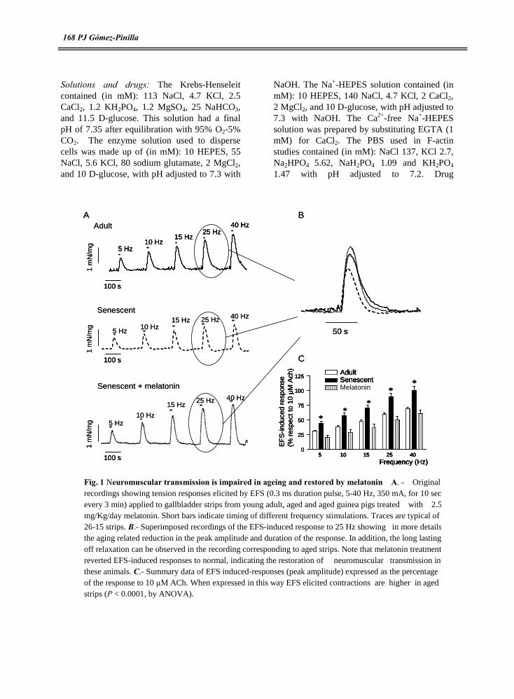
168 PJ Gómez-Pinilla
Solutions and drugs: The Krebs-Henseleit contained (in mM): 113 NaCl, 4.7 KCl, 2.5 CaCl2, 1.2 KH2PO4, 1.2 MgSO4, 25 NaHCO3, and 11.5 D-glucose. This solution had a final pH of 7.35 after equilibration with 95% O2-5% CO2. The enzyme solution used to disperse cells was made up of (in mM): 10 HEPES, 55 NaCl, 5.6 KCl, 80 sodium glutamate, 2 MgCl2, and 10 D-glucose, with pH adjusted to 7.3 with
NaOH. The Na+-HEPES solution contained (in mM): 10 HEPES, 140 NaCl, 4.7 KCl, 2 CaCl2, 2 MgCl2, and 10 D-glucose, with pH adjusted to 7.3 with NaOH. The Ca2+-free Na+-HEPES solution was prepared by substituting EGTA (1 mM) for CaCl2. The PBS used in F-actin studies contained (in mM): NaCl 137, KCl 2.7, Na2HPO4 5.62, NaH2PO4 1.09 and KH2PO4 1.47 with pH adjusted to 7.2. Drug
100 s
1 m
N/m
g
5 Hz10 Hz
15 Hz25 Hz
40 HzAdult
1 m
N/m
g
5 Hz10 Hz
15 Hz 25 Hz 40 HzSenescent
Senescent + melatonin
1 m
N/m
g 5 Hz10 Hz
15 Hz25 Hz 40 Hz
50 s
EF
S-in
duce
dre
spon
se
(% r
espe
ctto
10 µ
M A
ch)
5 10 15 25 400
25
50
75
100
125AdultSenescentMelatonin
Frequency (Hz)
**
*
**
100 s
100 s
A
C
B
100 s100 s
1 m
N/m
g
5 Hz10 Hz
15 Hz25 Hz
40 Hz
5 Hz10 Hz
15 Hz25 Hz
40 HzAdult
1 m
N/m
g
5 Hz10 Hz
15 Hz 25 Hz 40 HzSenescent
Senescent + melatonin
1 m
N/m
g 5 Hz10 Hz
15 Hz25 Hz 40 Hz
50 s50 s
EF
S-in
duce
dre
spon
se
(% r
espe
ctto
10 µ
M A
ch)
5 10 15 25 400
25
50
75
100
125AdultSenescentMelatonin
Frequency (Hz)
**
*
**
EF
S-in
duce
dre
spon
se
(% r
espe
ctto
10 µ
M A
ch)
5 10 15 25 400
25
50
75
100
125AdultSenescentMelatonin
Frequency (Hz)
**
*
**
5 10 15 25 400
25
50
75
100
125AdultSenescentMelatonin
Frequency (Hz)
**
*
**
100 s100 s
100 s100 s
A
C
B
Fig. 1 Neuromuscular transmission is impaired in ageing and restored by melatonin A. - Original recordings showing tension responses elicited by EFS (0.3 ms duration pulse, 5-40 Hz, 350 mA, for 10 sec every 3 min) applied to gallbladder strips from young adult, aged and aged guinea pigs treated with 2.5 mg/Kg/day melatonin. Short bars indicate timing of different frequency stimulations. Traces are typical of 26-15 strips. B.- Superimposed recordings of the EFS-induced response to 25 Hz showing in more details the aging related reduction in the peak amplitude and duration of the response. In addition, the long lasting off relaxation can be observed in the recording corresponding to aged strips. Note that melatonin treatment reverted EFS-induced responses to normal, indicating the restoration of neuromuscular transmission in these animals. C.- Summary data of EFS induced-responses (peak amplitude) expressed as the percentage of the response to 10 µM ACh. When expressed in this way EFS elicited contractions are higher in aged strips (P < 0.0001, by ANOVA).
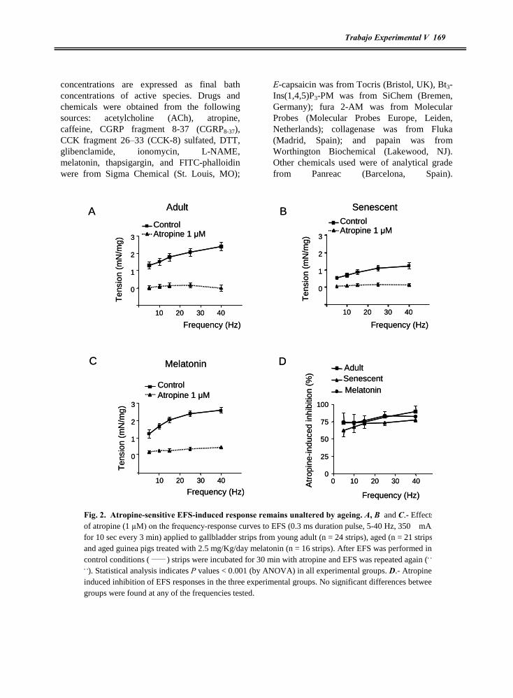
Trabajo Experimental V 169
concentrations are expressed as final bath concentrations of active species. Drugs and chemicals were obtained from the following sources: acetylcholine (ACh), atropine, caffeine, CGRP fragment 8-37 (CGRP8-37), CCK fragment 26�33 (CCK-8) sulfated, DTT, glibenclamide, ionomycin, L-NAME, melatonin, thapsigargin, and FITC-phalloidin were from Sigma Chemical (St. Louis, MO);
E-capsaicin was from Tocris (Bristol, UK), Bt3-Ins(1,4,5)P3-PM was from SiChem (Bremen, Germany); fura 2-AM was from Molecular Probes (Molecular Probes Europe, Leiden, Netherlands); collagenase was from Fluka (Madrid, Spain); and papain was from Worthington Biochemical (Lakewood, NJ). Other chemicals used were of analytical grade from Panreac (Barcelona, Spain).
10 20 30 40
0
1
2
3
ControlAtropine 1 ìM
Adult
Frequency (Hz)
Ten
sion
(mN
/mg)
10 20 30 40
0
1
2
3
Senescent
Frequency (Hz)
Ten
sion
(mN
/mg)
Melatonin
ControlAtropine 1 ìM
Frequency (Hz)
Ten
sion
(mN
/mg)
Atr
opin
e-in
duce
din
hibi
tion
(%)
10 20 30 40
0
1
2
3
AdultSenescentMelatonin
Frequency (Hz)
0 10 20 30 400
25
50
75
100
A
C D
BControlAtropine 1 ìM
10 20 30 40
0
1
2
3
ControlAtropine 1 ìM
Adult
Frequency (Hz)
Ten
sion
(mN
/mg)
10 20 30 40
0
1
2
3
Senescent
Frequency (Hz)
Ten
sion
(mN
/mg)
Melatonin
ControlAtropine 1 ìM
Frequency (Hz)
Ten
sion
(mN
/mg)
Atr
opin
e-in
duce
din
hibi
tion
(%)
10 20 30 40
0
1
2
3
AdultSenescentMelatonin
Frequency (Hz)
0 10 20 30 400
25
50
75
100
A
C D
BControlAtropine 1 ìM
Fig. 2. Atropine-sensitive EFS-induced response remains unaltered by ageing. A, B and C.- Effects of atropine (1 µM) on the frequency-response curves to EFS (0.3 ms duration pulse, 5-40 Hz, 350 mA, for 10 sec every 3 min) applied to gallbladder strips from young adult (n = 24 strips), aged (n = 21 strips) and aged guinea pigs treated with 2.5 mg/Kg/day melatonin (n = 16 strips). After EFS was performed in control conditions ( _____ ) strips were incubated for 30 min with atropine and EFS was repeated again (. . .
. .). Statistical analysis indicates P values < 0.001 (by ANOVA) in all experimental groups. D.- Atropine-induced inhibition of EFS responses in the three experimental groups. No significant differences between groups were found at any of the frequencies tested.

170 PJ Gómez-Pinilla
Stock solutions of atropine, capsaicin, fura 2-AM, thapsigargin, ionomycin, were prepared in DMSO, and FITC-phalloidin was prepared in ethanol. The solutions were diluted such that the final concentration of DMSO was ≤0.1% vol/vol. This concentration of DMSO did not interfere with fura 2 fluorescence. Melatonin was prepared in 80 mM glucose solution.
Quantification and statistics: Results are expressed as means ± SEM of n cells or
gallbladder strips. Gallbladder tension is given in milliNewtons (mN)/mg of tissue. All results from [Ca2+]i determinations are given as ÄF340/F380. Statistical differences between means were determined by Student�s t-test. Differences between multiple groups were tested using two-way analysis of variance (ANOVA) followed by Bonferroni´s post hoc test. Differences were considered significant at P < 0.05.
1 m
N/m
g
100 s
Senescent
100 s
L-NAME (100 µM)
1 m
N/ m
g
Adult
1 m
N/m
g
100 s
Senescent + melatonin
Adult
0
1
2
3
4 Control
L-NAME
Senescent
0
1
2
3
4ControlL-NAME
Melatonin
0 10 20 30 400
1
2
3
4 ControlL-NAME
Ten
sion
(mN
/mg)
Ten
sion
(mN
/mg)
Ten
sion
(mN
/mg)
Frequency (Hz)
A B
1 m
N/m
g
100 s
Senescent
100 s
L-NAME (100 µM)
1 m
N/ m
g
Adult
1 m
N/m
g
100 s
Senescent + melatonin
Adult
0
1
2
3
4 Control
L-NAME
Senescent
0
1
2
3
4ControlL-NAME
Melatonin
0 10 20 30 400
1
2
3
4 ControlL-NAME
Ten
sion
(mN
/mg)
Ten
sion
(mN
/mg)
Ten
sion
(mN
/mg)
Frequency (Hz)
A B
Fig. 3.- Nitrergic function is impaired in ageing and restored by melatonin. A.- Original recordings of the effects of L-NAME (100 µM) on the EFS (0.3 ms duration pulse, 5-40 Hz, 350 mA, for 10 sec every 3 min) applied to gallbladder strips from young adult, aged and aged guinea pigs treated with 2.5 mg/Kg/day melatonin . After EFS was performed in control condition s strips were incubated for 30 min with L-NAME and EFS was repeated again. Vertical dotted lines indicate the timing of EFS. B.- Sumary data of the frequency-response curves to EFS. Data are mean ± SEM. n = 10, 6 and 5 strips for adult, aged and aged treated with melatonin, respectively. Note that L-NAME does not have effects on aged gallbladder strips, indicating the loss of nitrergic function and that melatonin treatment induces the recovery of the nitrergic responses. P < 0.01 & P < 0.001 (by ANOVA) control vs L-NAME for young adult and aged plus melatonin experimental groups.

Trabajo Experimental V 171
4-RESULTS
Aged related changes in efferent innervation in EFS-induced gallbladder responses
EFS induced a frequency-dependent gallbladder contraction (a representative trace is shown in Fig. 1A) with a maximum amplitude of 2.23 ± 0.18 mN/mg tissue at the frequency of 40 Hz (n = 26, Table 1). When gallbladder strips
from aged animals were electrically stimulated, a significant decrease in the EFS-induced contraction at all the frequencies tested was recorded (Fig. 1A, Table 1). The diminished response was reflected by reductions in both the amplitude of the peak and the duration of the contraction (P < 0.001, two-way ANOVA for both parameters, Fig. 1B, Table 1). In addition, there was a long lasting off-relaxation (amplitude
0
0 10 20 30 400
1
2
ControlCapsaicin
Frequency (Hz)
0
1
2
ControlCapsaicin
1
2
3ControlCapsaicin
Ten
sion
(mN
/mg)
Ten
sion
(mN
/mg)
Ten
sion
(mN
/mg)
3
3
Adult
Senescent
Melatonin
100 s
1 m
N/m
g
Senescent
1 m
N/m
g
Capsaicin (10µM)Adult
1 m
N/m
g
100 s
Senescent + melatonin
A B
100 s
0
0 10 20 30 400
1
2
ControlCapsaicin
Frequency (Hz)
0
1
2
ControlCapsaicin
1
2
3ControlCapsaicin
Ten
sion
(mN
/mg)
Ten
sion
(mN
/mg)
Ten
sion
(mN
/mg)
3
3
Adult
Senescent
Melatonin
100 s
1 m
N/m
g
Senescent
1 m
N/m
g
Capsaicin (10µM)Adult
1 m
N/m
g
100 s
Senescent + melatonin
A B
100 s100 s
Fig. 4.- Ageing induces sensitization of inhibitory sensory afferent fibers and melatonin normalizes their sensitivity. A.- Original recordings of the effects of capsaicin (10 µM) on the EFS (0.3 ms duration pulse, 5-40 Hz, 350 mA, for 10 sec every 3 min) applied to gallbladder strips from young adult, aged and aged guinea pigs treated with 2.5 mg/Kg/day melatonin. After EFS was performed in control conditions, strips were incubated for 30 min with capsaicin and EFS was repeated again. Vertical dotted lines indicate the timing of EFS. B.- Summary data of the frequency-response curves to EFS. Data are mean ± SEM. n = 8, 6 and 4 strips for young adult, aged and aged treated with melatonin, respectively. Note that capsaicin increases the EFS-induced contraction only in aged gallbladder strips, indicating the sensitization of the inhibitory component of sensory fibers and reversal of this sensitization by melatonin treatment. P < 0.01 (by ANOVA) control vs capsaicin in aged strips.

172 PJ Gómez-Pinilla
of 0.245 ± 0.025 mN/mg at 40Hz, n = 22) in the strips from older animals, but not in the younger animals.
A group of old animals was treated orally with melatonin (2.5 mg/Kg/day) for four weeks mimicking the circadian rhythm of the naturally secreted hormone. This treatment
increased melatonin blood levels in aged animals when measured at 9 am (young adult: 26.24 ± 2.12; aged: 19.6 ± 1.48; melatonin: 27.09± 2.29 pg/ml, n = 10, 7, and 7 animals respectively, P < 0.05 aged vs young adult and aged vs melatonin).
Table 1. EFS-induced gallbladder contractions: effects of ageing and melatonin treatment.
Young adult Aged Melatonin
Frec (Hz) Amplitude Duration Amplitude Duration Amplitude Duration
5 0.988 ± 0.117 43.08 ± 1.29 0.643 ± 0.063 * 31.01 ± 1.15 * 0.812 ± 0.140 42.23 ± 1.66
10 1.315 ± 0.132 48.57 ± 1.88 0.890 ± 0.078 * 33.73 ± 1.28 * 1.170 ± 0.163 45.53 ± 2.65
15 1.609 ± 0.147 52.80 ± 1.97 1.120 ± 0.099 * 37.26 ± 1.50 * 1.483 ± 0.178 45.93 ± 2.43
25 1.956 ± 0.164 57.30 ± 2.55 1.384 ± 0.120 * 39.14 ± 2.12 * 1.967 ± 0.175 52.06 ± 2.97
40 2.232 ± 0.181 55.90 ± 2.70 1.570 ± 0.139 * 39.47 ± 2.61 * 2.351 ± 0.157 52.66 ± 3.24
Amplitude in (mN/mg) and Duration in (s) . n = 26.22 and 15 for young-adult, aged and melatonin respectively.*P < 0.0001 adult vs senecent.
Melatonin treatment ameliorated age-induced impairment in the EFS response as both the amplitude and duration of EFS-induced contractions were comparable to those in young adult strips, and the off-relaxations were absent (Fig. 1A and B, Table1).
The aged-related impairment in gallbladder contractility could involve changes in the intrinsic innervation of the organ, alterations in the smooth muscle contractility itself or a combination of these mechanisms. The gallbladder contractile response to exogenous ACh was significantly reduced in aged animals (young adult: 3.266 ± 0.260; aged: 1.911 ± 0.27; melatonin: 4.232 ± 0.538 mN/mg, n = 29, 15 and 11 strips, respectively, P < 0.001 aged vs young adult and aged vs melatonin), suggesting an age-related impairment in the myogenic response to the neurotransmitter released from intrinsic
nerves. Taking into account that ACh is the primary excitatory neurotransmitter in the gallbladder (Parkman, et al., 1997), the expression of EFS-induced response respect to ACh-induced contraction would reflect the amount of ACh released. As shown in Fig. 1C, EFS-induced response expressed in this way is higher in strips from aged animals (P < 0.001 by ANOVA), which would indicate an increase in the release of ACh or a decrease in the release of inhibitory neurotransmitters. A decrease in the release of ACh does not seem probable as the effect of atropine was not different in young adult, aged or melatonin-treated aged strips (Fig. 2, Table 2).
To explore the possibility of an aged-induced increase in the release of inhibitory neurotransmitters, we assayed the effects of L-NAME in each experimental group. The inhibitor
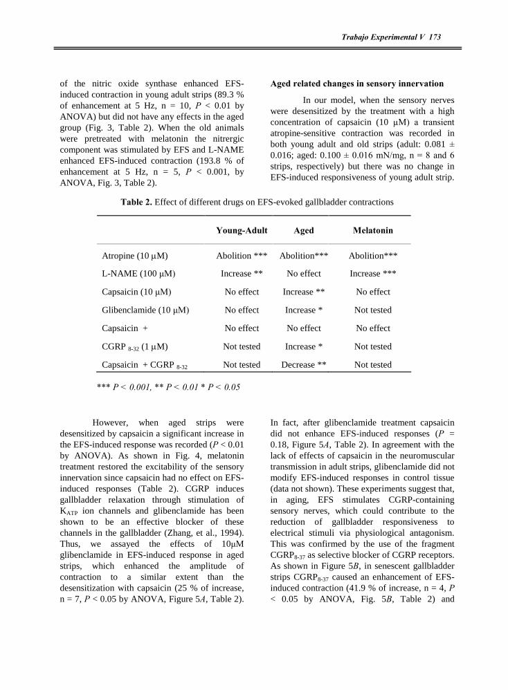
Trabajo Experimental V 173
of the nitric oxide synthase enhanced EFS-induced contraction in young adult strips (89.3 % of enhancement at 5 Hz, n = 10, P < 0.01 by ANOVA) but did not have any effects in the aged group (Fig. 3, Table 2). When the old animals were pretreated with melatonin the nitrergic component was stimulated by EFS and L-NAME enhanced EFS-induced contraction (193.8 % of enhancement at 5 Hz, n = 5, P < 0.001, by ANOVA, Fig. 3, Table 2).
Aged related changes in sensory innervation
In our model, when the sensory nerves were desensitized by the treatment with a high concentration of capsaicin (10 µM) a transient atropine-sensitive contraction was recorded in both young adult and old strips (adult: 0.081 ± 0.016; aged: 0.100 ± 0.016 mN/mg, n = 8 and 6 strips, respectively) but there was no change in EFS-induced responsiveness of young adult strip.
Table 2. Effect of different drugs on EFS-evoked gallbladder contractions
*** P < 0.001, ** P < 0.01 * P < 0.05
However, when aged strips were desensitized by capsaicin a significant increase in the EFS-induced response was recorded (P < 0.01 by ANOVA). As shown in Fig. 4, melatonin treatment restored the excitability of the sensory innervation since capsaicin had no effect on EFS-induced responses (Table 2). CGRP induces gallbladder relaxation through stimulation of KATP ion channels and glibenclamide has been shown to be an effective blocker of these channels in the gallbladder (Zhang, et al., 1994). Thus, we assayed the effects of 10ìM glibenclamide in EFS-induced response in aged strips, which enhanced the amplitude of contraction to a similar extent than the desensitization with capsaicin (25 % of increase, n = 7, P < 0.05 by ANOVA, Figure 5A, Table 2).
In fact, after glibenclamide treatment capsaicin did not enhance EFS-induced responses (P = 0.18, Figure 5A, Table 2). In agreement with the lack of effects of capsaicin in the neuromuscular transmission in adult strips, glibenclamide did not modify EFS-induced responses in control tissue (data not shown). These experiments suggest that, in aging, EFS stimulates CGRP-containing sensory nerves, which could contribute to the reduction of gallbladder responsiveness to electrical stimuli via physiological antagonism. This was confirmed by the use of the fragment CGRP8-37 as selective blocker of CGRP receptors. As shown in Figure 5B, in senescent gallbladder strips CGRP8-37 caused an enhancement of EFS-induced contraction (41.9 % of increase, n = 4, P < 0.05 by ANOVA, Fig. 5B, Table 2) and
Young-Adult Aged Melatonin
Atropine (10 M) Abolition *** Abolition*** Abolition***
L-NAME (100 ìM) Increase ** No effect Increase ***
Capsaicin (10 ìM) No effect Increase ** No effect
Glibenclamide (10 ìM) No effect Increase * Not tested
Capsaicin + Gliblenclamide
No effect No effect No effect
CGRP 8-32 (1 M) Not tested Increase * Not tested
Capsaicin + CGRP 8-32 Not tested Decrease ** Not tested

174 PJ Gómez-Pinilla
converted the effect of capsaicin to an inhibition of the contractile response (37 % of inhibition respect to CGRP8-37 alone, n = 4, P < 0.01 by ANOVA, Fig. 5B, Table 2).
Aged related changes in gallbladder contractility
To explore how aging affects Ca2+ influx-mediated contractions we activated voltage-activated calcium entry-induced by 60 mM KCl, which induced a sustained contraction that was significantly reduced (24.8 % reduction)
Fig. 5.- Ageing sensitizes CGRP containing fibers .A.- Original recordings of the effects of glibenclamide (10 µM) and CGRP8-37 in the absence and presence of capsaicin (10 µM )on the EFS (0.3 ms duration pulse, 5-40 Hz, 350 mA, for 10 sec every 3 min) applied to aged gallbladder strips. After EFS was performed in control conditions, strips were incubated for 30 min with glibenclamide or CGRP8-37 and EFS was repeated again. Then, strips were incubated for 30 min with glibenclamide or CGRP8-37 plus capsaicin and EFS was repeated again Vertical dotted lines indicate the timing of EFS. B.- Summary data of the frequency-response curves to EFS in the presence and absence of glibenclamide or CGRP8-37 and glibenclamide or CGRP8-37 plus capsaicin. Data are mean ± SEM. n = 7 and 4 strips for glibenclamide and capsaicin group, respectively. Note that both drugs have a stimulatory effect and after the treatment with glibenclamide or CGRP8-37 capsaicin does not increase EFS-induced contractions. P < 0.05 (by ANOVA) control vs glibenclamide and control vs CGRP8-37.
100 s
Glibenclamide (10 µM)
100 s
Glib + Capsaicin
1 m
N/m
g
CGRP8-37 (1 µM) CGRP8-37 + Capsaicin
1 m
N/m
g
Senescent
Senescent
0
1
2
ControlGlibenclamide (10 µM)Glibenclamide + capsaicin
Frequency (Hz)
0 10 20 30 400
1
2
Control
CGRP8-37 (1µ M)
CGRP8-37+ capsaicin
Ten
sion
(mN
/mg)
Ten
sion
(mN
/mg)
A
B
100 s
Glibenclamide (10 µM)
100 s
Glib + Capsaicin
1 m
N/m
g
CGRP8-37 (1 µM) CGRP8-37 + Capsaicin
1 m
N/m
g
Senescent
Senescent
0
1
2
ControlGlibenclamide (10 µM)Glibenclamide + capsaicin
Frequency (Hz)
0 10 20 30 400
1
2
Control
CGRP8-37 (1µ M)
CGRP8-37+ capsaicin
Ten
sion
(mN
/mg)
Ten
sion
(mN
/mg)
A
B

Trabajo Experimental V 175
when we tested gallbladder strips from aged animals and recovered by melatonin treatment (young adult, 3.77 ± 0.37; aged, 2.84 ± 0.26; melatonin: 4.24 ± 0.32 mN/mg, n = 26, 23 and 9 strips, respectively, P < 0.05). We have recently shown that capacitative calcium entry (CCE) is another influx route activating contraction in gallbladder smooth muscle (Morales, et al., 2004). As previously reported, following depletion of the stores by incubation of control gallbladder strips with 1 µM TPS in Ca2+-free medium for 30 min, reintroduction of extracellular Ca2+ induced a sustained contraction (3.69 ± 0.53 mN/mg, n = 12) that was significantly smaller in aged strips (2.68 ± 0.29 mN/mg, n = 9, P < 0.01) but similar in melatonin-treated animals (4.41 ± 0.39 mN/mg, n = 8).
To test whether aging affects Ca2+ release from sarcoplasmic reticulum, the main intracellular Ca2+ store in gallbladder smooth muscle (Morales, et al., 2005a), we used CCK as the tool of election. As was the case for Ca2+ influx-induced contractions, CCK-induced contraction was impaired in aged strips and melatonin treatment recovered contractility (young adult: 3.7 ± 0.38; aged: 2.48 ± 0.25; melatonin: 3.67 ± 0.52 mN/mg, n = 30, 23 and 9 strips, respectively, P < 0.01).
Aged related changes in calcium handling
The diminished contractile response to Ca2+ influx could be the result of the reduction in the calcium entry as consequence of aged-mediated calcium channel impairment. To test this possibility, we quantified [Ca2+]i in response to 60 mM KCl and to the protocol to activate capacitative calcium entry. As represented in Figs. 6A and B, sustained [Ca2+]i plateau due to Ca2+ entry from the extracellular medium was reduced in aged gallbladder smooth muscle cells (26.2 and 34.2 % reduction for KCl and CCE,
respectively, n = 49-18 cells, P < 0.05). Interestingly, melatonin did not have any effects on calcium influx (KCl: 0.111 ± 0.008 vs 0.115 ± 0.010 ÄF340/F380 n = 34 and 13 cells ; CCE: 0.063 ± 0.005 vs 0.067 ± 0.007 ÄF340/F380 n = 8 and 6 cells for aged and melatonin treated aged cells, respectively)
When we quantified [Ca2+]i in response to CCK challenge there was no change in the amplitude of Ca2+ transient peak indicative of intracellular Ca2+ release (n = 28 and 15 cells, P = 0.897, Fig. 6C). To confirm that Ca2+ release from stores through D-myo-inositol 1,4,5-triphosphate (IP3) and ryanodine channels remained unchanged in aging, we exposed the cells to 10 µM Bt3-Ins(1,4,5)P3-PM, a membrane permeable analogue of IP3, and to caffeine. Similar to CCK-induced Ca2+ transient, the peak responses to the IP3 analogue were not affected by aging (young adult, 0.426 ± 0.031; aged, 0.379 ± 0.028 ÄF340/F380, n = 13 and 7 cells, respectively, P = 0.272). When we used caffeine to induce Ca2+ release through ryanodine receptors we did not detect a difference in the Ca2+ transient in young adult vs. aged gallbladder smooth muscle cells (n = 31 and 36 cells, P = 0.412, Fig. 6D). Collectively, these results suggest that both Ca2+ release channels and calcium content in the stores are not affected by aging. The latter was confirmed by application of a low level of ionomycin in Ca2+ free solution to the cells, a treatment that releases the Ca2+ stores while bypassing channels and receptors. This treatment caused a similar increase in [Ca2+]i in both adult and aged cells (young adult, 0.188 ± 0.029; aged, 0.180 ± 0.015 ÄF340/F380, n = 14 and 24 cells, respectively, P = 0.821), indicating that the integrity of calcium stores is preserved in aging. Melatonin treatment did not have any effect on Ca2+ release mechanisms nor Ca2+ store content (data not shown).

176 PJ Gómez-Pinilla
Aged-related changes in contractile machinery Taken together, the results described above indicate that the aged impairment in contractility is, at least in part, the result of alteration in Ca2+ influx, but this physiological condition could also alter gallbladder contractility independently of the calcium signal. This possibility was tested by application of 1µM of ionomycin in presence of extracellular Ca2+ (at this concentration ionomycin raises [Ca2+]i independently of channels and receptors). Ionomycin caused similar elevation in adult and aged and
melatonin-treated cells (young adult: 0.355 ± 0.050; aged: 0.419 ± 0.052; melatonin: 0.376 ± 0.057 ÄF340/F380, n = 32, 30 and 16 cells, respectively, P > 0. 05), but induced much lower contraction in aged strips than in adult and melatonin-treated aged tissues (young adult: 4.23 ± 0.57; aged: 2.12 ± 0.201; melatonin: 5.39 ± 0.87 mN/mg, n = 7, 14 and 8 strips, respectively, P < 0.01 young adult vs aged), suggesting that ageing can also alter the contractile machinery independently of the calcium signal.
60 mM KCl
50 s
0.1
F34
0/F
380
KCl
adult senescent0.00
0.05
0.10
0.15
0.20
*
50 s
0.1
F34
0/F
380
2 mM Ca 2+
Ca 2+ free1 µM TPS
adultsenescent
CCE
adult senescent0.00
0.05
0.10
0.15
**
CCK
adult senescent0.0
0.1
0.2
0.3
0.4
0.5
Caffeine
adult senescent0.00
0.25
0.50
0.75
adultsenescent
0.1
F34
0/F
380
50 s
10 nM CCK
adultsenescent
0.1
F34
0/F
380
50 s
Caffeine (10 mM)
adultsenescent
A
C D
B
ÄF
340/
F38
0ÄF
340/
F38
0
ÄF
340
/F38
0ÄF
340/
F38
0
60 mM KCl
50 s
0.1
F34
0/F
380
KCl
adult senescent0.00
0.05
0.10
0.15
0.20
*
KCl
adult senescent0.00
0.05
0.10
0.15
0.20
*
50 s
0.1
F34
0/F
380
2 mM Ca 2+
Ca 2+ free1 µM TPS
adultsenescent
CCE
adult senescent0.00
0.05
0.10
0.15
**
CCE
adult senescent0.00
0.05
0.10
0.15
**
CCK
adult senescent0.0
0.1
0.2
0.3
0.4
0.5
CCK
adult senescent0.0
0.1
0.2
0.3
0.4
0.5
Caffeine
adult senescent0.00
0.25
0.50
0.75
adultsenescent
0.1
F34
0/F
380
50 s
10 nM CCK
adultsenescent
0.1
F34
0/F
380
50 s
Caffeine (10 mM)
adultsenescent
A
C D
B
ÄF
340/
F38
0ÄF
340/
F38
0
ÄF
340
/F38
0ÄF
340/
F38
0
Fig. 6. Melatonin restores gallbladder myogenic contractility A.- Representative original traces of changes in isometric tension in response to a depolarizing solution containing 60 mM KCl + in young adult (_____), aged (_ _ _
_) and aged treated with melatonin (. . . . . .) gallbladder strips. In B, gallbladder strips were treated with 1 µM thapsigargin (TPS) in Ca2+ free solution to deplete the stores. When indicated, the organ bath was perfused with a 2 mM Ca2+ K-H solution resulting in a sustained contraction that was reduced in aged strips (- - - - - ). In C contraction was induced by 10 nM CCK in all groups. Note that the response to all the stimuli was decreased in aged cells but melatonin treatment induced the recovery of the contractility. Traces are typical of 30-9 strips. Histograms show summary data tension (mN/mg) from experiments in the above described conditions (mean ± SEM). D.- Aged related changes in F actin content (expressed as arbitrary units) in young adult, aged and aged + melatonin gallbladder cells. Note the decrease in F-actin content in aged cells and the recovery when the aged animals were treated with melatonin. * P < 0.05, ** P < 0.01.

Trabajo Experimental V 177
To investigate one of the possible causes of this impaired contractility, we determined the total amount of the contractile protein F-actin labelling gallbladder smooth muscle cells with FITC-stained phalloidin. We found that aging resulted in a statistically significant decrease in the F- actin content that was recovered by melatonin treatment (young adult: 33.59 ± 0.86; aged: 9.92 ± 0.49; melatonin 28.95 ± 0.87, data as arbitrary units of fluorescence, n = 100, 63 and
92 cells, respectively, P < 0.05 young adult vs aged, Fig. 7D).
5-DISCUSSION
At a time when the world population is ageing at a rapid rate, there is an emergent need to understand the biology of aged GI tract and to translate this knowledge to therapeutics to improve GI function and quality of life of the elderly. The present study was conducted to
Fig. 7. Ageing reduces Ca2+ influx but not Ca2+ release from intracellular stores. A.- Representative original traces of changes in the fluorescence ratio in response to a depolarizing solution containing 60 mM KCl and 2 mM Ca2+ in young adult (_____) and aged (. . . . . ) cells . As observed, ageing reduced the plateau response. In B, fura-2 loaded cells were treated with 1 µM thapsigargin (TPS) in Ca2+ free solution to deplete the stores. When indicated, cells were perfused with a 2 mM Ca2+ HEPES solution resulting in a sustained [Ca2+]i increase that was reduced in aged cells (. . . .). In C and D Ca2+ release from intracellular stores was induced by 10 nM CCK (IP3-sensitive stores) (C) or caffeine (ryanodine-sensitive stores) (D). Note that the peak, indicative of Ca2+ release, was similar in both young adult and aged cells but the plateau, indicative of Ca2+ entry was reduced in senescent cells. Traces are typical of 45-15 cells from at least 4-5 independent experiments Histograms show summary data of ÄF340/F380 from experiments in the above described conditions (mean ± SEM). * P < 0.05, ** P < 0.01.
60 mM KCl
50 s
0.1
F34
0/F
380
KCl
adult senescent0.00
0.05
0.10
0.15
0.20
*
50 s
0.1
F34
0/F
380
2 mM Ca 2+
Ca 2+ free1 µM TPS
adultsenescent
CCE
adult senescent0.00
0.05
0.10
0.15
**
CCK
adult senescent0.0
0.1
0.2
0.3
0.4
0.5
Caffeine
adult senescent0.00
0.25
0.50
0.75
adultsenescent
0.1
F34
0/F
380
50 s
10 nM CCK
adultsenescent
0.1
F34
0/F
38
0
50 s
Caffeine (10 mM)
adultsenescent
A
C D
B
ÄF
340/
F38
0ÄF
34
0/F
380
ÄF
340/F
380
ÄF3
40/
F38
0
60 mM KCl
50 s
0.1
F34
0/F
380
KCl
adult senescent0.00
0.05
0.10
0.15
0.20
*
KCl
adult senescent0.00
0.05
0.10
0.15
0.20
*
50 s
0.1
F34
0/F
380
2 mM Ca 2+
Ca 2+ free1 µM TPS
adultsenescent
CCE
adult senescent0.00
0.05
0.10
0.15
**
CCE
adult senescent0.00
0.05
0.10
0.15
**
CCK
adult senescent0.0
0.1
0.2
0.3
0.4
0.5
CCK
adult senescent0.0
0.1
0.2
0.3
0.4
0.5
Caffeine
adult senescent0.00
0.25
0.50
0.75
adultsenescent
0.1
F34
0/F
380
50 s
10 nM CCK
adultsenescent
0.1
F34
0/F
38
0
50 s
Caffeine (10 mM)
adultsenescent
A
C D
B
ÄF
340/
F38
0ÄF
34
0/F
380
ÄF
340/F
380
ÄF3
40/
F38
0
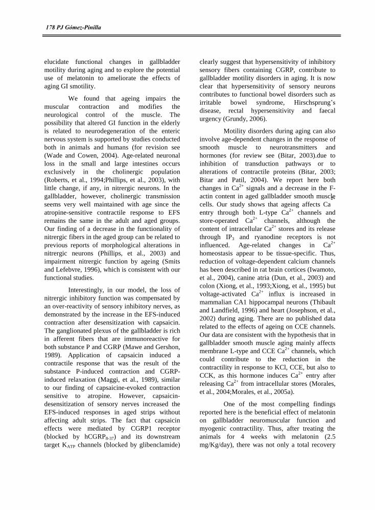
178 PJ Gómez-Pinilla
elucidate functional changes in gallbladder motility during aging and to explore the potential use of melatonin to ameliorate the effects of aging GI smotility.
We found that ageing impairs the muscular contraction and modifies the neurological control of the muscle. The possibility that altered GI function in the elderly is related to neurodegeneration of the enteric nervous system is supported by studies conducted both in animals and humans (for revision see (Wade and Cowen, 2004). Age-related neuronal loss in the small and large intestines occurs exclusively in the cholinergic population (Roberts, et al., 1994;Phillips, et al., 2003), with little change, if any, in nitrergic neurons. In the gallbladder, however, cholinergic transmission seems very well maintained with age since the atropine-sensitive contractile response to EFS remains the same in the adult and aged groups. Our finding of a decrease in the functionality of nitrergic fibers in the aged group can be related to previous reports of morphological alterations in nitrergic neurons (Phillips, et al., 2003) and impairment nitrergic function by ageing (Smits and Lefebvre, 1996), which is consistent with our functional studies.
Interestingly, in our model, the loss of nitrergic inhibitory function was compensated by an over-reactivity of sensory inhibitory nerves, as demonstrated by the increase in the EFS-induced contraction after desensitization with capsaicin. The ganglionated plexus of the gallbladder is rich in afferent fibers that are immunoreactive for both substance P and CGRP (Mawe and Gershon, 1989). Application of capsaicin induced a contractile response that was the result of the substance P-induced contraction and CGRP-induced relaxation (Maggi, et al., 1989), similar to our finding of capsaicine-evoked contraction sensitive to atropine. However, capsaicin-desensitization of sensory nerves increased the EFS-induced responses in aged strips without affecting adult strips. The fact that capsaicin effects were mediated by CGRP1 receptor (blocked by hCGRP8-37) and its downstream target KATP channels (blocked by glibenclamide)
clearly suggest that hypersensitivity of inhibitory sensory fibers containing CGRP, contribute to gallbladder motility disorders in aging. It is now clear that hypersensitivity of sensory neurons contributes to functional bowel disorders such as irritable bowel syndrome, Hirschsprung�s disease, rectal hypersensitivity and faecal urgency (Grundy, 2006).
Motility disorders during aging can also involve age-dependent changes in the response of smooth muscle to neurotransmitters and hormones (for review see (Bitar, 2003).due to inhibition of transduction pathways or to alterations of contractile proteins (Bitar, 2003; Bitar and Patil, 2004). We report here both changes in Ca2+ signals and a decrease in the F-actin content in aged gallbladder smooth muscle cells. Our study shows that ageing affects Ca
2+
entry through both L-type Ca2+ channels and store-operated Ca2+ channels, although the content of intracellular Ca2+ stores and its release through IP3 and ryanodine receptors is not influenced. Age-related changes in Ca2+ homeostasis appear to be tissue-specific. Thus, reduction of voltage-dependent calcium channels has been described in rat brain cortices (Iwamoto, et al., 2004), canine atria (Dun, et al., 2003) and colon (Xiong, et al., 1993;Xiong, et al., 1995) but voltage-activated Ca2+ influx is increased in mammalian CA1 hippocampal neurons (Thibault and Landfield, 1996) and heart (Josephson, et al., 2002) during aging. There are no published data related to the effects of ageing on CCE channels. Our data are consistent with the hypothesis that in gallbladder smooth muscle aging mainly affects membrane L-type and CCE Ca2+ channels, which could contribute to the reduction in the contractility in response to KCl, CCE, but also to CCK, as this hormone induces Ca2+ entry after releasing Ca2+ from intracellular stores (Morales, et al., 2004;Morales, et al., 2005a).
One of the most compelling findings reported here is the beneficial effect of melatonin on gallbladder neuromuscular function and myogenic contractility. Thus, after treating the animals for 4 weeks with melatonin (2.5 mg/Kg/day), there was not only a total recovery

Trabajo Experimental V 179
of the contractile response to Ca2+ influx activation and EFS, but also a reversion of the aged-induced changes in efferent and sensitive innervation, although melatonin did not restore the Ca2+ influx. The recovery of the contractility without changes in Ca2+ homeostasis can only be explained by effects of melatonin on Ca2+-insensitive contractility steps such as contractile proteins or Ca2+ sensitization of the contractile machinery. Whereas the former can be related to the increase in the F-actin content described in this study, the later is now under research in our laboratory.
Melatonin is a hormone that is secreted by the pineal gland following a circadian rhythm with minimal blood levels of melatonin during the daytime and maximal levels observed mostly during the middle of the night. Melatonin has special importance in the gastrointestinal tract since the enterochromaffin cells of the gut are the main source of extrapineal melatonin (Kvetnoy, et al., 2002). Metabolism of melatonin in liver appears to be the major metabolic pathway for its deactivation (Lane and Moss, 1985) but also active melatonin is secreted in bile and concentrated in the gallbladder (Tan, et al., 1999). Although it is well established that pineal melatonin decreases with age (Karasek, 2004), there is no information available regarding gastrointestinal melatonin. If gastrointestinal melatonin content also decreases with age, the hepatobiliary system in general, and the gallbladder in particular, would suffer the lack of melatonin protective effects. The restoration of gallbladder contractility in response to melatonin treatment in aged guinea pigs is consistent with this hypothesis.
Regarding the GI, there are numerous lines of evidence demonstrating the protective
and healing effects of melatonin in gastric ulcers and experimental colitis. Whereas a direct antioxidative effect, activation CO3H
- secretion by enterocytes, and release of endogenous prostaglandin mediates these protective effects, the healing effect was associated to production of endogenous NO and participation of sensory nerves (for review see (Reiter, et al., 2003). The effects of melatonin on symptoms of colitis are related to decrease of NO and PGE2 content, as the result of down-regulated expression of colonic iNOS and cycloxygenase-2 (Dong, et al., 2003) in addition to an enhancement of the immune response in the gastrointestinal tract (Mei, et al., 2002). The present study is the first report on the beneficial effects of melatonin treatment in aged gastrointestinal tract, but more studies are required to establish the cellular mechanisms through which melatonin exerts its effects in the gallbladder and the optimal pattern of melatonin treatment to ameliorate the effects of ageing. Both lines of research will elucidate the potential use of melatonin in elderly population.
In conclusion, our results indicate that ageing causes impairment of gallbladder function as consequence of neurally-mediated and neurally -independent mechanisms and support a possible beneficial effect of melatonin in gallbladder dysfunction related to ageing.
6-ACKNOWLEDGEMENTS
The authors thank Gary M Mawe for critical reading of the manuscript and to Purificación Delgado for technical assistance. Pedro J Gomez-Pinilla is recipient of Doctoral Fellowship from Junta de Extremadura
7-REFERENCES Bailey AJ (2001) Molecular mechanisms of ageing in connective tissues. Mech Ageing Dev 122:735-755.
Bitar KN (2003) Aging and neural control of the GI tract: V. Aging and gastrointestinal smooth muscle: from signal transduction to contractile proteins. Am J Physiol Gastrointest Liver Physiol 284:G1-G7.
180 PJ Gómez-Pinilla
Bitar KN and Patil SB (2004) Aging and gastrointestinal smooth muscle. Mech Ageing Dev 125:907-910.
Camilleri M, Lee JS, Viramontes B, Bharucha AE and Tangalos EG (2000) Insights into the pathophysiology and mechanisms of constipation, irritable bowel syndrome, and diverticulosis in older people. J Am Geriatr Soc 48:1142-1150.
Centers for Disease Control And Prevention (CDC) (2003) Trends in aging--United States and worldwide. MMWR Morb Mortal Wkly Rep 52:101-4, 106.
Dong WG, Mei Q, Yu JP, Xu JM, Xiang L and Xu Y (2003) Effects of melatonin on the expression of iNOS and COX-2 in rat models of colitis. World J Gastroenterol 9:1307-1311.
Dun W, Yagi T, Rosen MR and Boyden PA (2003) Calcium and potassium currents in cells from adult and aged canine right atria. Cardiovasc Res 58:526-534.
Ferrari AU, Radaelli A and Centola M (2003) Invited review: aging and the cardiovascular system. J Appl Physiol 95:2591-2597.
Gomes OA, de Souza RR and Liberti EA (1997) A preliminary investigation of the effects of aging on the nerve cell number in the myenteric ganglia of the human colon. Gerontology 43:210-217.
Grundy D (2006) Signalling the state of the digestive tract. Auton Neurosci 125:76-80.
Iwamoto M, Hagishita T, Shoji-Kasai Y, Ando S and Tanaka Y (2004) Age-related changes in the levels of voltage-dependent calcium channels and other synaptic proteins in rat brain cortices. Neurosci Lett 366:277-281.
Josephson IR, Guia A, Stern MD and Lakatta EG (2002) Alterations in properties of L-type Ca channels in aging rat heart. J Mol Cell Cardiol 34:297-308.
Karasek M (2004) Melatonin, human aging, and age-related diseases. Exp Gerontol 39:1723-1729.
Kvetnoy IM, Ingel IE, Kvetnaia TV, Malinovskaya NK, Rapoport SI, Raikhlin NT, Trofimov AV and Yuzhakov VV (2002) Gastrointestinal melatonin: cellular identification and biological role. Neuro Endocrinol Lett 23:121-132.
Lane EA and Moss HB (1985) Pharmacokinetics of melatonin in man: first pass hepatic metabolism. J Clin Endocrinol Metab 61:1214-1216.
Maggi CA, Santicioli P, Renzi D, Patacchini R, Surrenti C and Meli A (1989) Release of substance P- and calcitonin gene-related peptide-like immunoreactivity and motor response of the isolated guinea pig gallbladder to capsaicin. Gastroenterology 96:1093-1101.
Mawe GM and Gershon MD (1989) Structure, afferent innervation, and transmitter content of ganglia of the guinea pig gallbladder: relationship to the enteric nervous system. J Comp Neurol 283:374-390.
Mei Q, Yu JP, Xu JM, Wei W, Xiang L and Yue L (2002) Melatonin reduces colon immunological injury in rats by regulating activity of macrophages. Acta Pharmacol Sin 23:882-886.
Trabajo Experimental V 181
Morales S, Camello PJ, Alcon S, Salido GM, Mawe G and Pozo MJ (2004) Coactivation of capacitative calcium entry and L-type calcium channels in guinea pig gallbladder. Am J Physiol Gastrointest Liver Physiol 286:G1090-G1100.
Morales S, Camello PJ, Mawe GM and Pozo MJ (2005a) Characterization of intracellular Ca(2+) stores in gallbladder smooth muscle. Am J Physiol Gastrointest Liver Physiol 288:G507-G513.
Morales S, Camello PJ, Rosado JA, Mawe GM and Pozo MJ (2005b) Disruption of the filamentous actin cytoskeleton is necessary for the activation of capacitative calcium entry in naive smooth muscle cells. Cell Signal 17:635-645.
Nasr SZ and Ouslander JG (1998) Urinary incontinence in the elderly. Causes and treatment options. Drugs Aging 12:349-360.
Parkman HP, Pagano AP, Martin JS and Ryan JP (1997) Electric field stimulation-induced guinea pig gallbladder contractions: role of calcium channels in acetylcholine release. Dig Dis Sci 42:1919-1925.
Phillips RJ, Kieffer EJ and Powley TL (2003) Aging of the myenteric plexus: neuronal loss is specific to cholinergic neurons. Auton Neurosci 106:69-83.
Pozo MJ, Perez GJ, Nelson MT and Mawe GM (2002) Ca(2+) sparks and BK currents in gallbladder myocytes: role in CCK-induced response. Am J Physiol Gastrointest Liver Physiol 282:G165-G174.
Reiter RJ, Pablos MI, Agapito TT and Guerrero JM (1996) Melatonin in the context of the free radical theory of aging. Ann N Y Acad Sci 786:362-378.
Reiter RJ, Tan DX, Manchester LC and El Sawi MR (2002) Melatonin reduces oxidant damage and promotes mitochondrial respiration: implications for aging. Ann N Y Acad Sci 959:238-250.
Reiter RJ, Tan DX, Mayo JC, Sainz RM, Leon J and Bandyopadhyay D (2003) Neurally-mediated and neurally-independent beneficial actions of melatonin in the gastrointestinal tract. J Physiol Pharmacol 54 Suppl 4:113-125.
Roberts D, Gelperin D and Wiley JW (1994) Evidence for age-associated reduction in acetylcholine release and smooth muscle response in the rat colon. Am J Physiol 267:G515-G522.
Smits GJ and Lefebvre RA (1996) Influence of age on cholinergic and inhibitory nonadrenergic noncholinergic responses in the rat ileum. Eur J Pharmacol 303:79-86.
Sohal RS and Allen RG (1990) Oxidative stress as a causal factor in differentiation and aging: a unifying hypothesis. Exp Gerontol 25:499-522.
Tan D, Manchester LC, Reiter RJ, Qi W, Hanes MA and Farley NJ (1999) High physiological levels of melatonin in the bile of mammals. Life Sci 65:2523-2529.
Thibault O and Landfield PW (1996) Increase in single L-type calcium channels in hippocampal neurons during aging. Science 272:1017-1020.
182 PJ Gómez-Pinilla
Troulinaki K and Tavernarakis N (2005) Neurodegenerative conditions associated with ageing: a molecular interplay? Mech Ageing Dev 126:23-33.
Wade PR and Cowen T (2004) Neurodegeneration: a key factor in the ageing gut. Neurogastroenterol Motil 16 Suppl 1:19-23.
Winer N and Sowers JR (2004) Epidemiology of diabetes. J Clin Pharmacol 44:397-405.
Xiong Z, Sperelakis N, Noffsinger A and Fenoglio-Preiser C (1993) Changes in calcium channel current densities in rat colonic smooth muscle cells during development and aging. Am J Physiol 265:C617-C625.
Xiong Z, Sperelakis N, Noffsinger A and Fenoglio-Preiser C (1995) Ca2+ currents in human colonic smooth muscle cells. Am J Physiol 269:G378-G385.
Zhang L, Bonev AD, Nelson MT and Mawe GM (1994) Activation of ATP-sensitive potassium currents in guinea-pig gall-bladder smooth muscle by the neuropeptide CGRP. J Physiol 478 Pt 3:483-491.

Trabajo Experimental VI 183
Gut (Under Review)
EFFECT OF MELATONIN ON GALLBLADDER
NEUROMUSCULAR FUNCTION IN ACUTE CHOLECYSTITIS
Pedro J. Gomez-Pinilla, Pedro J. Camello and María J. Pozo*
Dept of Physiology, Nursing School,
University of Extremadura, 10071Cáceres, Spain
Running head: melatonin in acute cholecystitis
*Correspondence to:
María J. Pozo
Department of Physiology
Nursing School
Avda Universidad s/n
10071 Cáceres
Spain
E-mail: [email protected]
Key Words: innervation, EFS, experimental acute cholecystitis, melatonin, guinea pig
id6240015 pdfMachine by Broadgun Software - a great PDF writer! - a great PDF creator! - http://www.pdfmachine.com http://www.broadgun.com
184 PJ Gómez-Pinilla
CONTENTS
1- Abstract
2- Introduction
3- Material and methods
Experimental manoeuvres with animals
Melatonin administration
Functional studies
Solutions and drugs
Data analysis
4- Results
Effects of acute cholecystitis on gallbladder neuromuscular function
Effects of melatonin on neuromuscular function in acute cholecystitis
Melatonin effects on MDA and GSH content
Melatonin effects on Ca2+ homeostasis and contractility after bile duct deligation
5- Discussion
6- Acknowledgements
7- References
Abbreviations used in this paper: ACh, Acetylcholine; AC, acute cholecystitis; 2-APB, 2-aminoethoxydiphenylborane; [Ca2+]i, free cytosolic Ca2+ concentration; CCK, cholecystokinin; CCE, capacitative calcium entry; COX-2,cycloxygenase-2, ES, enzyme solution; GSH, reduced glutathione; K-HS, Krebs-Henseleit solution; MDA, Malondialdehyde; NO, nitric oxide; TPS, thapsigargin.

Trabajo Experimental VI 185
1-ABSTRACT
Gallbladder stasis is associated to experimental acute cholecystitis. Impaired contractility could be, at least in part, the result of inflammation-induced alterations in the neuromuscular function. This study was designed to determine the changes on gallbladder neurotransmission evoked by acute inflammation and to evaluate the protective effects of melatonin. Experimental acute cholecystitis was induced in guinea pigs by common bile duct ligation for two days. In a group of these animals, de-ligation of the bile duct was performed and after two days the neuromuscular function was evaluated. Electrical field stimulation (EFS, 5�40 Hz) was used to activate intrinsic nerves. The EFS-evoked isometric contractile responses were recorded and pharmacologically dissected to identify the chemical code of the stimulated nerves. EFS-induced gallbladder contraction was significantly lower in cholecystitic tissue. In addition, inflammation changed the pharmacological profile of these contractions that were insensitive to tetrodotoxin but sensitive to atropine and ù-conotoxin, indicating that acute cholecystitis affects action potential propagation in the intrinsic nerves. NO-mediated neurotransmission was reduced by inflammation which also increased the reactivity of sensitive afferent fibers. Melatonin treatment (2.5 and 30 mg/Kg) prevented qualitative changes in gallbladder neurotransmission, but did not improve EFS-induced contractility. However, the hormone recovered gallbladder neuromuscular function once the biliary obstruction was solved. These findings show for the first time the therapeutic potential of melatonin in the recovery of gallbladder neuromuscular function during acute cholecystitis.
2-INTRODUCTION
Gallbladder tone is mainly regulated by both myogenic mechanisms and neuro-hormonal inputs. Activation of nerves is an important event in the control of gallbladder
emptying. The neural control of gallbladder motility involves reflexes that include both efferent and afferent nerve fibers as well as the intrinsic plexus in the gallbladder wall (1). The gallbladder wall is innervated by three neural networks interconnected by nerve bundles. All gallbladder neurons are apparently cholinergic since they are immunoreactive for the essential biosynthetic enzyme for acetylcholine (ACh), choline acetyltransferase (2). The release of ACh from cholinergic neurons and its exogenous administration results in the contraction of the gallbladder smooth muscle, which depends on the activation of muscarinic receptors (3), Ca2+ influx and Ca2+ release from internal stores (4). In addition to its contractile properties, ACh has neuromodulatory functions promoting or inhibiting the release of other neurotransmitters (3). Cholinergic neurons co-express other neurotransmitters such as substance P, neuropeptide Y, somastotatin, vasoactive intestinal polypeptide, pituitary adenylate cyclase-activating polypeptide and nitric oxide (NO) (5). Afferent nerve fibers containing calcitonin gene-related peptide and tachykininis have also been described in the ganglionated plexus of the gallbladder (6).
Acute acalculous cholecystitis (AC) is a pathophysiological condition characterized by gallbladder inflammation in the absence of gallstones. Although its pathogenesis is unknown, gallbladder stasis is always present, probably as the result of the deleterious neural and muscular actions of inflammatory mediators such as reactive oxygen species and prostaglandins (7). In animal models, it has been described that cholecystitis reduces gallbladder contractile responses to agonists that act directly on smooth muscle cells (8;9) and also causes alterations in calcium signalling and contractile machinery (10). In addition, EFS-induced contractions are also impaired in inflamed gallbladder, mainly due to the reduction in the functionality of cholinergic nerves and the up-regulation of the inhibitory nitrergic component (11). The effect of
186 PJ Gómez-Pinilla
cholecystitis on afferent fibers has not yet been explored.
Melatonin, the main product of pineal gland in vertebrates, is a potent free radical scavenger and activates a broad group of antioxidant cellular mechanisms (12). These properties made melatonin efficacious against different diseases where oxidative stress is the main cause (13). Gastrointestinal tract is an important source of melatonin (14). The liver and the gallbladder are especially exposed to high levels of the hormone since metabolism of melatonin in liver appears to be the major pathway for its deactivation (15) but also active melatonin is secreted in bile and concentrated in the gallbladder (16). In the gastrointestinal tract, it has been reported that melatonin has a gastroprotective function (17) and therapeutic effects against malignance-associated to irritable bowel syndrome (18). In animal models of bile duct ligation, melatonin treatment reduced hepatic damage and oxidative stress due to its antioxidant and anti-inflammatory actions (19-21).
The aims of this study were to investigate the effects of acute cholecystitis in the neuromuscular transmission and to evaluate the impact of melatonin treatment. Our results indicate that melatonin is able to restore this important function in inflamed gallbladder, which can be of importance avoiding gallbladder failure in this pathological condition.
3-MATERIALS AND METHODS
Experimental manoeuvres with animals. Male guinea pigs, weighing 400 g to 600 g were purchased from Animal Care Center of the University of Extremadura where they were fed with a standard chow diet and water ad libitum under a 12-hour photoperiod. Acute acalculous cholecystitis was induced to animals by ligature of the common bile duct (CBDL) for 2 days, as previously described (10). This method has been previously shown to reproduce the pathological changes seen in human acute acalculous cholecystitis (8;9;11), and it was
approved by the Animal Care and Ethical Committees of the University of Extremadura. In brief, after anesthesia with ketamine hydrochloride (20 mg/Kg ip) and xylacine (5 mg/Kg ip) a laparotomy was performed and the distal end of the common bile duct was ligated. The surgical incision was then sutured and two days after the animals were sacrificed for tissue harvest. In a group of animals, two days after CBDL the common bile duct was de-ligated under anaesthesia with microsurgical scissors and two days after the animals were sacrificed. For both experimental models, a group of guinea pigs were sham operated, which included all the surgical steps except the common bile duct ligation.
Melatonin administration. Guinea pigs were treated orally with melatonin (2.5 or 30 mg/kg/day). Melatonin was dissolved in glucose solution (1.5 %) and placed in the oropharynx by a syringe. This treatment was applied daily at the same time, just before the light in the animal house was switched off (7:00 PM). Melatonin was administered 14 days before the sacrifice of the animals in both experimental groups.
Functional studies. At the adequate time, the animals were killed with deep halothane anaesthesia and cervical dislocation. Gallbladders were immediately placed in cold Krebs-Henseleit solution (K-HS; for composition, see Solutions and drugs) at pH 7.35. The gallbladder was opened from the end of the cystic duct to the base, and the mucosa was dissected away carefully. Later, the gallbladder was cut in longitudinal strips (measuring ~3 x 10 mm) and this were placed vertically in a 10-ml organ bath filled with Krebs-Henseleit solution maintained at 37°C and gassed with 95% O2-5% CO2. Isometric contractions were measured using force displacement transducers that were interfaced with a Macintosh computer using a MacLab hardware unit and software (ADInstruments, Colorado Springs, CO). The muscle strips were placed under an initial resting tension equivalent to a 1.5-g load and allowed to
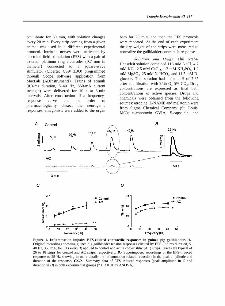
Trabajo Experimental VI 187
equilibrate for 60 min, with solution changes every 20 min. Every strip coming from a given animal was used in a different experimental protocol. Intrinsic nerves were activated by electrical field stimulation (EFS) with a pair of external platinum ring electrodes (0.7 mm in diameter) connected to a square-wave stimulator (Cibertec CS9/ 3BO) programmed through Scope software application from MacLab (ADInstruments). Trains of stimuli (0.3-ms duration, 5�40 Hz, 350-mA current strength) were delivered for 10 s at 3-min intervals. After construction of a frequency-response curve and in order to pharmacologically dissect the neurogenic responses, antagonists were added to the organ
bath for 20 min, and then the EFS protocols were repeated. At the end of each experiment the dry weight of the strips were measured to normalize the gallbladder contractile responses.
Solutions and Drugs. The Krebs-Henseleit solution contained 113 mM NaCl, 4.7 mM KCl, 2.5 mM CaCl2, 1.2 mM KH2PO4, 1.2 mM MgSO4, 25 mM NaHCO3, and 11.5 mM D-glucose. This solution had a final pH of 7.35 after equilibration with 95% O2-5% CO2. Drug concentrations are expressed as final bath concentrations of active species. Drugs and chemicals were obtained from the following sources: atropine, L-NAME and melatonin were from Sigma Chemical Company (St. Louis, MO); ù-conotoxin GVIA, E-capsaicin, and
1 m
N/m
g
3 min
A Control
AC
5 Hz10 Hz 15 Hz
25 Hz 40 Hz
50 s
B
DC
25 Hz
1 m
N/m
g
*0 10 20 30 40 50
0
1
2
3ControlAC
Frequency (Hz)
Ten
sion
(mN
/mg)
*
*
0 10 20 30 40 500
25
50
75ControlAC
Frequency (Hz)
Tim
e (s
)
1 m
N/m
g
3 min
A Control
AC
5 Hz10 Hz 15 Hz
25 Hz 40 Hz
50 s
B
DC
25 Hz
1 m
N/m
g
*0 10 20 30 40 50
0
1
2
3ControlAC
Frequency (Hz)
Ten
sion
(mN
/mg)
**0 10 20 30 40 50
0
1
2
3ControlAC
Frequency (Hz)
Ten
sion
(mN
/mg)
*
*
0 10 20 30 40 500
25
50
75ControlAC
Frequency (Hz)
Tim
e (s
)
*
0 10 20 30 40 500
25
50
75ControlAC
Frequency (Hz)
Tim
e (s
)
Figure 1. Inflammation impairs EFS-elicited contractile responses in guinea pig gallbladder. A.- Original recordings showing guinea pig gallbladder tension responses elicited by EFS (0.3 ms duration, 5-40 Hz, 350 mA, for 10 s every 3) applied to control and acute cholecistitic (AC) strips. Traces are typical of 26 to 18 strips for control and AC strips, respectively. B.- Superimposed recordings of the EFS-induced response to 25 Hz showing in more details the inflammation-related reduction in the peak amplitude and duration of the response. C&D.- Summary data of EFS induced-responses (peak amplitude in C and duration in D) in both experimental groups (* P < 0.01 by ANOVA).

188 PJ Gómez-Pinilla
tetrodotoxin citrate were from Tocris Cookson (Bristol,UK). Other chemicals used were of analytical grade from Panreac (Barcelona, Spain). Stock solutions of atropine, capsaicin and ù-conotoxin GVIA were prepared in DMSO. The solutions were diluted such that the final concentration of DMSO was ≤ 0.1% v/v. This concentration of DMSO did not have effects on gallbladder tone.
Data analysis. Results are expressed as means ± S.E.M. of n gallbladder strips from at least 6 different animals. Gallbladder tension is given in millinewtons per milligrams of tissue (mN/mg). Statistical differences between multiple groups or the effects of inhibitor treatments were tested using adequate analysis of variance (ANOVA). Differences were considered significant at P < 0.05.
4-RESULTS
Effects of acute cholecystitis on gallbladder neuromuscular function. EFS was used to stimulate the neuronal network in the gallbladder wall and the recording of isometric tension allowed us to evaluate the neuromuscular function. EFS evoked a
frequency-dependent contraction in control strips with amplitude of 0.98 ± 0.11 mN/mg and 2.23 ± 0.18 mN/mg when the strips were stimulated at 5 and 40 Hz, respectively (Fig. 1 A-C). The contractile response to EFS lasted for 42.66 ± 1.26 s and 55.51 ± 2.10 s at 5 and 40 Hz, respectively, which were longer than the duration of the stimuli (10 s, Fig. 1D). Similar results were obtained in strips from sham-operated animals (data not shown). As also shown in figure 1, when gallbladder strips from animals subjected to CBDL were electrically stimulated, a significant decrease in the EFS-induced contraction was recorded at all of the frequencies tested. The diminished response was reflected by reductions in both the amplitude and the duration of the contractions (P < 0.01, two-way ANOVA for both, Fig. 1C & D).
In order to determine the neural and myogenic components of the EFS-evoked contractions, the nerve Na+ channel inhibitor, tetrodotoxin (TTX) was used. In control strips, 1 µM of TTX abolished EFS-elicited responses (Fig. 2A). In inflamed strips, tetrodotoxin was no effective (Figure 2B), but when the strips
A
Control
B
* *
0 10 20 30 40 50-1
0
1
2
3ControlTTX
Frequency (Hz)
Ten
sion
(mN
/mg)
Acute Cholecystitis
* *
0 10 20 30 40 500.0
0.1
0.2
0.3
0.4
0.5ControlTTX-Conotoxin
Frequency (Hz)
Ten
sion
(mN
/mg)
A
Control
B
* *
0 10 20 30 40 50-1
0
1
2
3ControlTTXControlTTX
Frequency (Hz)
Ten
sion
(mN
/mg)
Acute Cholecystitis
* *
0 10 20 30 40 500.0
0.1
0.2
0.3
0.4
0.5ControlTTX-Conotoxin
ControlTTX-Conotoxin
Frequency (Hz)
Ten
sion
(mN
/mg)
Figure 2. AC induces a TTX-resistant gallbladder response to EFS. Effects of 1 µM TTX on EFS-elicited contractile response in control (A) and AC gallbladder strips (B). After EFS was performed in control conditions (solid lines) strips were incubated for 20 min with the antagonist and EFS was repeated again (dotted line). Note the lack of effects of TTX on inflamed tissue and the reduction in the response after incubation with 0.1 µM ù-conotoxin. Data are mean ± SEM. n = 7 and 6 strips for control and AC strips, respectively. (** P < 0.01 by ANOVA).

Trabajo Experimental VI 189
were co-incubated with tetrodotoxin (1 µM) plus ù-conotoxin GVIA (0.1 µM), a N-type calcium channel blocker, there was a reduction in the contractile response evoked by EFS (Fig. 2B). These results indicate that in inflamed gallbladder the transmission of the action potential along neural fibers is impaired and EFS stimulates neurotransmitters release directly from nervous terminals.
To elucidate the neurotransmitters involved in the EFS-induced contraction we tested several antagonists on this neural response. Under normal conditions ACh is the primary excitatory neurotransmitter in the gallbladder (22). In agreement with this, in control animals we found that 1 ìM atropine, a competitive cholinergic receptor blocker, reduced the EFS-elicited contractile response
by 73.00 ± 10.47 % at 5 Hz and 89.55 ± 8.02 % at 40 Hz (Fig. 3A), demonstrating that ACh released from cholinergic inputs is the main excitatory stimulus involved in EFS-induced contraction. The strips from cholecystitic animals were less sensitive to atropine blockade (58.38 ± 10.25 % of inhibition at 5 Hz and 28.60 ± 8.92 % of inhibition at 40 Hz, Fig. 3D), indicating that inflammation reduces the cholinergic participation in the neurogenic contractile gallbladder response.
The impact of inflammation on the contribution of NO was assayed by using the inhibitor of nitric oxide synthase, L-NAME, in both experimental groups, control and acute cholecystitic animals. L-NAME (100 ìM) enhanced EFS-induced contraction in strips from control guinea pigs, specially at the lowest
Control
Acute Cholecystitis
BA C
D E F
*
*
*
*
0 10 20 30 40 50-0.5
0.0
0.5
1.0
1.5
2.0
2.5
Control 1 M Atropine
Frequency (Hz)
Ten
sion
(mN
/mg)
0 10 20 30 40 500
1
2
3Control 100 M L-NAME
Frequency (Hz)
Ten
sion
(mN
/mg)
0 10 20 30 40 500
1
2
3Control 10 M Capsaicin
Frecuency (Hz)
Ten
sion
(mN
/mg)
0 10 20 30 40 500.0
0.1
0.2
0.3
0.4
0.5
Control 1 M Atropine
Frequency (Hz)
Ten
sion
(mN
/mg)
0 10 20 30 40 500.0
0.2
0.4
0.6Control 100 M L-NAME
Frequency (Hz)
Ten
sion
(mN
/mg)
0 10 20 30 40 500.0
0.1
0.2
0.3
0.4
0.5
Control 10 M Capsaicin
Frequency (Hz)T
ensi
on(m
N/m
g)
*
**
Figure 3. AC impairs the efferent innervation and increases the excitability of sensory contractile fibers Effects of 1 µM atropine, 100 µM of L-NAME and 10 µM of capsaicin on EFS-elicited contractile response in control (A, B, C) and AC gallbladder strips (D, E, F). After EFS was performed in control conditions (solid lines) strips were incubated for 20 min with the antagonist and EFS was repeated again (dotted line). Note the lack of effects of L-NAME on inflamed tissue and the reduction in the response after incubation with capsaicin. Data are mean ± SEM. n = 8-21 strips (* P < 0.05, ** P < 0.01 by ANOVA).
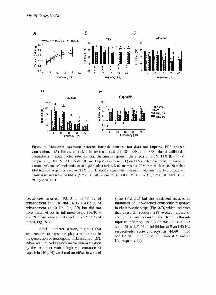
190 PJ Gómez-Pinilla
frequencies assayed (90.40 ± 11.48 % of enhancement at 5 Hz and 14.05 ± 4.62 % of enhancement at 40 Hz, Fig. 3B) but did not have much effect in inflamed strips (16.80 ± 9.70 % of increase at 5 Hz and 1.16 ± 5.54 % of increa, Fig. 3E).
Small diameter sensory neurons that are sensitive to capsaicin play a major role in the generation of neurogenic inflammation (23). When we induced sensory nerve desensitization by the treatment with a high concentration of capsaicin (10 µM) we found no effect in control
strips (Fig. 3C) but this treatment induced an inhibition of EFS-elicited contractile responses in cholecystitic strips (Fig. 3F), which indicates that capsaicin reduces EFS-evoked release of contractile neurotransmitters from afferents input in inflamed tissue (Control; -25.58 ± 7.78 and 4.61 ± 5.53 % of inhibition at 5 and 40 Hz, respectively; acute cholecystitis: 44.60 ± 7.01 and 62.70 ± 5.22 % of inhibition at 5 and 40 Hz, respectively).
A B C
D E
* * * * *
* * ** *
* * ** *
*
ä
ControlACMEL 2.5MEL 30
* * * *�� � �
0 10 20 30 40 500.0
0.1
0.2
0.3
0.4
0.5
AC MEL 2.5 MEL 30
Frequency (Hz)
Ten
sion
(mN
/mg)
5 10 15 25 40-50
0
50
100
150
Frequency (Hz)
Inhi
bitio
n(%
)
5 10 15 25 400
25
50
75
100
Frequency (Hz)
Inhi
bitio
n(%
)
5 10 15 25 400
25
50
75
100
125
Frequency (Hz)
Incr
ease
(%)
ä ää ä
5 10 15 25 40-50
-25
0
25
50
75
100
Frequency (Hz)
Inhi
bitio
n(%
)
ää
ä
ää
Atropine
L-NAME Capsaicin
TTX
�����
A B C
D E
* * * * *
* * ** *
* * ** *
*
ä
ControlACMEL 2.5MEL 30
* * * *�� � �
0 10 20 30 40 500.0
0.1
0.2
0.3
0.4
0.5
AC MEL 2.5 MEL 30
Frequency (Hz)
Ten
sion
(mN
/mg)
5 10 15 25 40-50
0
50
100
150
Frequency (Hz)
Inhi
bitio
n(%
)
5 10 15 25 400
25
50
75
100
Frequency (Hz)
Inhi
bitio
n(%
)
5 10 15 25 400
25
50
75
100
125
Frequency (Hz)
Incr
ease
(%)
ää ääää ää
5 10 15 25 40-50
-25
0
25
50
75
100
Frequency (Hz)
Inhi
bitio
n(%
)
ääää
ää
ääää
Atropine
L-NAME Capsaicin
TTX
�����
Figure 4. Melatonin treatment protects intrinsic neurons but does not improve EFS-induced contraction. (A) Effects of melatonin treatment (2.5 and 30 mg/Kg) on EFS-induced gallbladder contractions in acute cholecystitic animals. Histograms represent the effects of 1 µM TTX (B), 1 µM atropine (C), 100 µM of L-NAME (D) and 10 µM of capsaicin (E) on EFS-elicited contractile response in control, AC and AC melatonin-treated gallbladder strips. Data are mean ± SEM. n = 6-18 strips. Note that EFS-induced responses recover TTX and L-NAME sensitivity, whereas melatonin has less effects on cholinergic and sensitive fibers. (* P < 0.01 AC vs control �P < 0.05 MEL30 vs AC, ä P < 0.01 MEL 30 vs AC by ANOVA).

Trabajo Experimental VI 191
Effects of melatonin on neuromuscular function in acute cholecystitis We have previously reported that melatonin treatment was able to restore gallbladder neuromuscular function in aging (24). To check whether this hormone had beneficial effects in the alterations described above, we treated the animals with 2.5 and 30 mg/Kg melatonin (MEL 2.5; MEL 30) as described in the Material and Methods section. Under these conditions, none of melatonin doses used enhanced the amplitude of the contractile responses evoked by EFS
(Fig. 4A), but the contractions partially recovered the sensitivity to TTX (Fig. 4B). Although the treatment dose-dependently increased the inhibitory effects of atropine and decreased the inhibitory effects of capsaicin significantly at some frequencies (Fig 4C & E). However, melatonin was able to protect nitrergic nerves because when 100 µM of L-NAME was added to the organ bath, the EFS-evoked contractile responses were enhanced in similar proportions to those found in control strips (Fig. 4D).
0.5
mN
/mg
100 s
A
AC
DL
B C
Control
AC
DL + MEL 2.5
DL + MEL 30
0 10 20 30 40 500
1
2
3
Frequency (Hz)
Ten
sion
(mN
/mg)
0 10 20 30 40 500.0
0.1
0.2
0.3
0.4
0.5 AC
DL
Frequency (Hz)
Ten
sion
(mN
/mg)
**
**
0.5
mN
/mg
100 s
A
AC
DL
B C
Control
AC
DL + MEL 2.5
DL + MEL 30
Control
AC
DL + MEL 2.5
DL + MEL 30
0 10 20 30 40 500
1
2
3
Frequency (Hz)
Ten
sion
(mN
/mg)
0 10 20 30 40 500
1
2
3
Frequency (Hz)
Ten
sion
(mN
/mg)
0 10 20 30 40 500.0
0.1
0.2
0.3
0.4
0.5 AC
DL
Frequency (Hz)
Ten
sion
(mN
/mg)
**
**
Figure 5. Melatonin treatment improves the neurogenic contractile response worsened by deligation procedure. (A) Original recordings showing guinea pig gallbladder tension responses elicited by EFS (0.3 ms duration, 5-40 Hz, 350 mA, for 10 s every 3) applied to acute cholecistitic (AC) and de-ligated strips. Traces are typical of 16 and 17 strips for AC and de-ligated strips, respectively.. B.- Summary data of EFS induced-responses (peak amplitude) in both experimental groups (* P < 0.01 by ANOVA). C.- Effects of melatonin treatment (2.5 and 30 mg/kg) on EFS-elicited contractile response gallbladder strips from animals that underwent the de-ligation procedure. Data are mean ± SEM. (n = 16-28 strips, * P < 0.05 , ** P < 0.01 by ANOVA).

192 PJ Gómez-Pinilla
These results indicate that melatonin had some effects on inflammation of the gallbladder, which agrees with the normalization of MDA and GSH content we have found (Experimental Cap. VII), but there are some contractile disabilities that melatonin treatment can not restore. It has to be noted that in the model used in this study, the gallbladder suffers stretch as the result of bile duct ligation and the continuous bile output. Taking this into account, it would be difficult to see any improvement in the neuromuscular function keeping the bile duct ligated, which represents a remarkably extreme pathological condition. To solve this, after inducing AC, we performed de-ligation of bile duct and after two days we harvested the tissue and performed the EFS
experiments. In those animals that were not treated with melatonin the neuromuscular function worsened after de-ligation, as indicated by the reduction in the EFS-induced contraction (Fig 5A & B). Taking into account the small amplitude of these contractions we did not applied antagonists of the neurotransmitters to know the nature of this response.
Under these conditions, it was very noticeable the improvement that melatonin treatment induced in gallbladder neuromuscular function. As shown in figure 5C, melatonin increased the gallbladder neurogenic responses in a dose-dependent way. In the strips from animals treated with melatonin, the EFS-elicited responses recovered the sensitiveness to TTX (Fig. 6A) and atropine (Fig. 6B) present in
A
C D
Control ACAC DL + MEL 2.5DL + MEL 2.5 DL + MEL 30DL + MEL 30
B
ä
* * **
*
Atropine
ä ä ä ä
5 10 15 25 400
25
50
75
100
Frequency (Hz)
Inhi
bitio
n(%
)
B
ä
* * **
*
Atropine
ää ä ä ä
5 10 15 25 400
25
50
75
100
Frequency (Hz)
Inhi
bitio
n(%
)* *
** *
ä
ää ä
ä
L-NAME
5 10 15 25 400
20
40
60
80
100
120
Frequency (Hz)
Increase(%)
* **
* *
ä
ää ä
ä
L-NAME
5 10 15 25 400
20
40
60
80
100
120
Frequency (Hz)
Increase(%)
* * * * *
ä ä ä ä ä
TTX
5 10 15 25 40-50
0
50
100
150
Frequency (Hz)
Inhi
bitio
n(%
)
* * * * *
ä ä ä ä ä
TTX
5 10 15 25 40-50
0
50
100
150
Frequency (Hz)
Inhi
bitio
n(%
)
* * * **
ä ä ä ä ä
Capsaicin
5 10 15 25 40-50
-25
0
25
50
75
100
Frequency (Hz)
Inhi
bitio
n(%
) * * * **
ä ä ä ä ä
Capsaicin
5 10 15 25 40-50
-25
0
25
50
75
100
Frequency (Hz)
Inhi
bitio
n(%
)
Figure 6. Melatonin treatment normalizes the different neural components stimulated by EFS. Effects of 100 µM TTX (A), 1 µM atropine (B), 100 µM of L-NAME (C) and 10 µM of capsaicin (D) on EFS-elicited contractile responses in control, AC and melatonin treated gallbladder strips. After EFS was performed in control conditions strips were incubated for 20 min with the antagonist and EFS was repeated again of melatonin Melatonin (2.5 and 30 mg/Kg) was administered to animals that underwent the de-ligation protocol. Data are mean ± SEM. (n = 7-9 strips, * P < 0.01 vs control , ä P < 0.01 vs AC, by ANOVA).

Trabajo Experimental VI 193
control tissue. Although capsaicin (Fig. 6F) still induced a small inhibition of EFS-induced responses, the reduction was significantly smaller than that found in inflamed tissue and at the highest frequencies this effect was not different from that registered in control tissue
In this experimental group, 30 mg/Kg melatonin also re-established the sensitivity to L-NAME (Fig. 6D) but this was not the case for 2.5 mg/Kg melatonin, suggesting that the nitrergic function worsened by the de-ligation procedure.
5-DISCUSION
The current paper shows that the impairment in guinea pig gallbladder neurotransmission evoked by inflammation was associated to a decrease in the contribution of the efferent plexus and the up-regulation of sensory afferent fibers. In addition, melatonin treatment caused the improvement in the neurogenic contractile response and the normalization of the different neural components.
In agreement with previous reports (22;25), our results indicate that EFS evokes gallbladder response by the release of different inhibitory and excitatory neurotransmitters and that the contractile responses are frequency-dependent and last longer than the time of stimulation. Our data, in addition, show that the frequency dependence is maintained in inflammation, despite the big reduction observed in the amplitude of the contraction. The smaller contractile responses to EFS suggests the existence of an inflammation-induced impairment in the gallbladder intrinsic nerves, in agreement with previous results in human and animal models (11;26;27). However, the reduced gallbladder smooth muscle contractility to ACh found in cholecystitis (8;10;11;28) could also collaborate to the impaired neuromuscular function in inflamed tissue.
The most striking finding in our study was the lack of sensitivity to TTX shown by
cholecystitic strips, which could be explained by a myogenic origin of the EFS-induced contractions or by a direct release of neurotransmitter from nervous terminal. Whereas the former is not possible since the contractions were reduced by atropine, the latter was confirmed by the reduction obtained using ù-conotoxin GVIA, a blocker of N type calcium channel located in the presinaptic membrane whose activation is necessary for neurotransmitter release. These results suggest that inflammation evokes a functional denervation in the gallbladder that avoids the genesis or propagation of action potential through efferent fibres. Alterations in the properties and/or expression levels of voltage-dependent Na+ channels have been implicated in a variety of pathological states, including inflammation of the viscera (29-31). In this regard, the more common effect of inflammation on Na+ channels is the up-regulation of TTX-resistant slow (Nav1.8) (29;31). The use of transgenic animals, gene expression analysis and specific pharmacologic tools is necessary to explore the possibility of cholecystitis-evoked changes in Na+ channel type.
The neurotransmitters released from the intrinsic plexus were investigated in the present study by pharmacological dissection of EFS-induced gallbladder contractile responses. Classically, ACh released in response to EFS is the mainly excitatory component of the gallbladder contraction (22;25). Here, we show that in control conditions atropine abolished EFS-induced contraction, while in inflamed tissue it just reduced EFS-elicited contraction about 50 %, indicative of a functional denervation of the cholinergic component, similar to results previously described in inflamed gallbladder (11) and in other models of gastrointestinal inflammation, such as colitis (32)
Non adrenergic non cholinergic (NANC) neurotransmission in guinea pig gallbladder has been described more than a decade ago (33) and NO is the main NANC

194 PJ Gómez-Pinilla
neurotransmitter involved (34;35). Inflammation evokes a functional impairment in gallbladder nitrergic innervation as demonstrated by the lack of effects of L-NAME in cholecystic strips compared to control tissue. This result does not support the study from Parkman et al. (11), where L-NAME only had effect in inflamed tissue indicating that normal gallbladder does not release NO from the intrinsic plexus. This is in conflict with the presence of nitrergic nerves described in guinea pig gallbladder (1) and the functional data reported above. Functional nitrergic denervation associated to inflammation is a common finding in experimental colitis (36).
Neurotransmitters released from sensory nerves evoked contraction or relaxation of the gallbladder (37). Substance P, neurokinin A and neurokinin B induce contraction while calcitonin gene-related peptide (CGRP) evokes relaxation (6;37;38) In our study, sensory denervation with capsaicin had not effects in control conditions while it reduced EFS-elicited contractile response in inflammation, suggesting excitatory neurotransmitter release from sensory nerves in inflamed gallbladder. This finding can also explain the TTX resistant EFS-evoked contractile responses in cholecystitis since sensory neurons are rich in TTX-resistant slow (Nav1.8) and persistent (Nav1.9) type Na channel (39). The mayor participation of the sensory innervation is also a common finding in neurogenic inflammation (23) and airway diseases as asthma and chronic obstructive pulmonary disease (40). In the gallbladder, we have shown that aging, which is also related to increased oxidative stress, is associated to over-reactivity of sensory fibers (24).
The most important finding of our study is that melatonin has prophylactic effects on inflammation-induced impairment in gallbladder neuromuscular function. Thus, under melatonin treatment EFS-induced contractile response recovered the sensitiveness to TTX, indicating that melatonin protects the voltage-dependent Na+-channels involved in the
neural transmission of the action potential. Furthermore, the nitrergic innervation recovered its functionality and sensory fibers became less sensitive to EFS. However, melatonin itself did not improve the contractile response to EFS unless the obstruction of the bile duct was relieved. Under these conditions, melatonin reversed the impairment in contractility in a dose-dependent manner, and fully recovered the different neural components stimulated by EFS. It must be pointed out that 2.5 mg/Kg melatonin had no effects on the nitrergic innervation after deligation, although this treatment was efficacious increasing the participation of these inhibitory nerves with the bile duct ligated. De-ligation itself worsened gallbladder contractility, as consequence probably of an increase in oxidative stress insult due to reperfusion of the organ once the mechanical stretch disappeared. This is supported by the increase in the MDA levels indicative of lipidic peroxidation and oxidative stress injury found after de-ligation in our laboratory (Experimental Cap VIII). On this basis, it appears that nitrergic innervation is especially sensitive to the enhanced oxidative stress after de-ligation. In agreement with this, we have recently reported a minor participation of nitrergic nerves in neuromuscular transmission in aging and its recovery after melatonin treatment (24). Furthermore, melatonin has been shown to have neurally-mediated actions in the gut regulating either cholinergic, nitrergic and/or sensory innervation (41;42).
It is well accepted that melatonin administration at pharmacological doses decreases free radical formation and leads to a substantial recovery of the major antioxidant enzymes (43). Recent evidences have shown that melatonin has protective effects on liver and hepatic injury after extrahepatic bile duct ligation in rats (19-21). Additional to liver and hepatic damage, free radical accumulation associated to bile duct ligation has been implicated in the genesis of gallstone (44). In this regard antioxidant treatment with melatonin

Trabajo Experimental VI 195
not only reversed the increased oxidative stress, but also prevented gallstone formation (20). In our preparation, melatonin treatment was effective reducing MDA levels and increasing the endogenous antioxidant defence reduced gluthation (GSH) (Experimental Cap VIII), indicating that melatonin antioxidant effects can be responsible for the improvement in neuromuscular function. However, melatonin receptors have been described in the gallbladder, receptor-mediated effects can not been ruled out. The beneficial effects of melatonin against inflammation-associated neural damage in gallbladder run in parallels with an improvement in myogenic contractions (45). Collectively, these experimental evidences suggest a prophylactic and therapeutic role of melatonin in this pathological condition, a remarkable finding due to the inexistence of pharmacological treatment for acute cholecystitis.
In conclusion, the results obtained in the present study indicate that inflammation impairs gallbladder neuromuscular function as the result of changes in the neural inputs to smooth muscle. These changes can be summarized as a denervation of efferent nerves together with a hyperactivity of afferent fibers. Melatonin significantly ameliorated the inflammation-related changes in gallbladder neuromuscular transmission indicating its potential to combat inflammation-induced gallbladder damage.
6-ACKNOWLEDGEMENTS
The authors thank Rosario Moreno for technical assistance.Supported by Junta de Extremadura (2PR03A020) and Ministerio de Educacion y Ciencia (BFU 2004-0637). Pedro J Gómez-Pinilla is recipient of Doctoral Fellowship from Junta de Extremadura.
7-REFERENCE LIST
(1) Mawe GM, Saccone GT, Pozo MJ. Neural Control of the Gallbladder and Sphincter of Oddi. In: Johnson LR, Barret KE, Ghishan FK et al, editors. Physiology of the Gastrointestinal Tract. San Diego, CA: Elsevier Academic Press, 2006: 841-9.
(2) Mawe GM, Talmage EK, Cornbrooks EB et al. Innervation of the gallbladder: structure, neurochemical coding, and physiological properties of guinea pig gallbladder ganglia. Microsc Res Tech 1997;39(1):1-13.
(3) Parkman HP, Pagano AP, Ryan JP. Subtypes of muscarinic receptors regulating gallbladder cholinergic contractions. Am J Physiol 1999;276(5 Pt 1):G1243-G1250.
(4) Alcon S, Camello PJ, Garcia LJ et al. Activation of tyrosine kinase pathway by vanadate in gallbladder smooth muscle. Biochem Pharmacol 2000;59(9):1077-89.
(5) Talmage EK, Pouliot WA, Cornbrooks EB et al. Transmitter diversity in ganglion cells of the guinea pig gallbladder: an immunohistochemical study. J Comp Neurol 1992;317(1):45-56.
(6) Mawe GM, Gershon MD. Structure, afferent innervation, and transmitter content of ganglia of the guinea pig gallbladder: relationship to the enteric nervous system. J Comp Neurol 1989;283(3):374-90.
(7) Pozo MJ, Camello PJ, Mawe GM. Chemical mediators of gallbladder dysmotility. Curr Med Chem 2004;11(13):1801-12.

196 PJ Gómez-Pinilla
(8) Parkman HP, Bogar LJ, Bartula LL et al. Effect of experimental acalculous cholecystitis on gallbladder smooth muscle contractility. Dig Dis Sci 1999;44(11):2235-43.
(9) Xiao ZL, Chen Q, Biancani P et al. Abnormalities of gallbladder muscle associated with acute inflammation in guinea pigs. Am J Physiol Gastrointest Liver Physiol 2001;281(2):G490-G497.
(10) Gomez-Pinilla PJ, Morales S, Camello-Almaraz C et al. Changes in guinea pig gallbladder smooth muscle Ca2+ homeostasis by acute acalculous cholecystitis. Am J Physiol Gastrointest Liver Physiol 2006;290(1):G14-G22.
(11) Parkman HP, James AN, Bogar LJ et al. Effect of acalculous cholecystitis on gallbladder neuromuscular transmission and contractility. J Surg Res 2000;88(2):186-92.
(12) Tan DX, Reiter RJ, Manchester LC et al. Chemical and physical properties and potential mechanisms: melatonin as a broad spectrum antioxidant and free radical scavenger. Curr Top Med Chem 2002;2(2):181-97.
(13) Karasek M. Melatonin, human aging, and age-related diseases. Exp Gerontol 2004;39(11-12):1723-9.
(14) Kvetnoy IM, Ingel IE, Kvetnaia TV et al. Gastrointestinal melatonin: cellular identification and biological role. Neuro Endocrinol Lett 2002;23(2):121-32.
(15) Lane EA, Moss HB. Pharmacokinetics of melatonin in man: first pass hepatic metabolism. J Clin Endocrinol Metab 1985;61(6):1214-6.
(16) Tan D, Manchester LC, Reiter RJ et al. High physiological levels of melatonin in the bile of mammals. Life Sci 1999;65(23):2523-9.
(17) Konturek PC, Konturek SJ, Brzozowski T et al. Gastroprotective activity of melatonin and its precursor, L-tryptophan, against stress-induced and ischaemia-induced lesions is mediated by scavenge of oxygen radicals. Scand J Gastroenterol 1997;32(5):433-8.
(18) Head KA, Jurenka JS. Inflammatory bowel disease Part 1: ulcerative colitis--pathophysiology and conventional and alternative treatment options. Altern Med Rev 2003;8(3):247-83.
(19) Ohta Y, Imai Y, Matsura T et al. Successively postadministered melatonin prevents disruption of hepatic antioxidant status in rats with bile duct ligation. J Pineal Res 2005;39(4):367-74.
(20) Shiesh SC, Chen CY, Lin XZ et al. Melatonin prevents pigment gallstone formation induced by bile duct ligation in guinea pigs. Hepatology 2000;32(3):455-60.
(21) Esrefoglu M, Gul M, Emre MH et al. Protective effect of low dose of melatonin against cholestatic oxidative stress after common bile duct ligation in rats. World J Gastroenterol 2005;11(13):1951-6.
(22) Parkman HP, Pagano AP, Martin JS et al. Electric field stimulation-induced guinea pig gallbladder contractions: role of calcium channels in acetylcholine release. Dig Dis Sci 1997;42(9):1919-25.
(23) Sann H, Dux M, Schemann M et al. Neurogenic inflammation in the gastrointestinal tract of the rat. Neurosci Lett 1996;219(3):147-50.
(24) Gomez-Pinilla PJ, Camello-Almaraz C, Moreno R et al. Melatonin treatment reverts age-related changes in Guinea pig gallbladder neuromuscular transmission and contractility. J Pharmacol Exp Ther 2006;319(2):847-56.

Trabajo Experimental VI 197
(25) Yau WM, Youther ML. Modulation of gallbladder motility by intrinsic cholinergic neurons. Am J Physiol 1984;247(6 Pt 1):G662-G666.
(26) Merg AR, Kalinowski SE, Hinkhouse MM et al. Mechanisms of impaired gallbladder contractile response in chronic acalculous cholecystitis. J Gastrointest Surg 2002;6(3):432-7.
(27) McKirdy ML, Johnson CD, McKirdy HC. Inflammation impairs neurally mediated responses to electrical field stimulation in isolated strips of human gallbladder muscle. Dig Dis Sci 1994;39(10):2229-34.
(28) Cullen JJ, Maes EB, Aggrawal S et al. Effect of endotoxin on opossum gallbladder motility: a model of acalculous cholecystitis. Ann Surg 2000;232(2):202-7.
(29) Beyak MJ, Ramji N, Krol KM et al. Two TTX-resistant Na+ currents in mouse colonic dorsal root ganglia neurons and their role in colitis-induced hyperexcitability. Am J Physiol Gastrointest Liver Physiol 2004;287(4):G845-G855.
(30) Stewart T, Beyak MJ, Vanner S. Ileitis modulates potassium and sodium currents in guinea pig dorsal root ganglia sensory neurons. J Physiol 2003;552(Pt 3):797-807.
(31) Yoshimura N, Seki S, Novakovic SD et al. The involvement of the tetrodotoxin-resistant sodium channel Na(v)1.8 (PN3/SNS) in a rat model of visceral pain. J Neurosci 2001;21(21):8690-6.
(32) Blandizzi C, Fornai M, Colucci R et al. Altered prejunctional modulation of intestinal cholinergic and noradrenergic pathways by alpha2-adrenoceptors in the presence of experimental colitis. Br J Pharmacol 2003;139(2):309-20.
(33) Mourelle M, Guarner F, Molero X et al. Regulation of gall bladder motility by the arginine-nitric oxide pathway in guinea pigs. Gut 1993;34(7):911-5.
(34) Alcon S, Morales S, Camello PJ et al. Relaxation of canine gallbladder to nerve stimulation involves adrenergic and non-adrenergic non-cholinergic mechanisms. Neurogastroenterol Motil 2001;13(6):555-66.
(35) McKirdy ML, McKirdy HC, Johnson CD. Non-adrenergic non-cholinergic inhibitory innervation shown by electrical field stimulation of isolated strips of human gall bladder muscle. Gut 1994;35(3):412-6.
(36) Sung TS, La JH, Kim TW et al. Alteration of nitrergic neuromuscular transmission as a result of acute experimental colitis in rat. J Vet Sci 2006;7(2):143-50.
(37) Maggi CA, Santicioli P, Renzi D et al. Release of substance P- and calcitonin gene-related peptide-like immunoreactivity and motor response of the isolated guinea pig gallbladder to capsaicin. Gastroenterology 1989;96(4):1093-101.
(38) Kline LW, Pang PK. Calcitonin gene-related peptide relaxes cholecystokinin-induced tension in Iguana iguana gallbladder strips. Gen Comp Endocrinol 1994;95(3):381-6.
(39) Herzog RI, Cummins TR, Waxman SG. Persistent TTX-resistant Na+ current affects resting potential and response to depolarization in simulated spinal sensory neurons. J Neurophysiol 2001;86(3):1351-64.
(40) De Swert KO, Joos GF. Extending the understanding of sensory neuropeptides. Eur J Pharmacol 2006;533(1-3):171-81.
198 PJ Gómez-Pinilla
(41) Reiter RJ, Tan DX, Mayo JC et al. Neurally-mediated and neurally-independent beneficial actions of melatonin in the gastrointestinal tract. J Physiol Pharmacol 2003;54 Suppl 4:113-25.
(42) Barajas-Lopez C, Peres AL, Espinosa-Luna R et al. Melatonin modulates cholinergic transmission by blocking nicotinic channels in the guinea-pig submucous plexus. Eur J Pharmacol 1996;312(3):319-25.
(43) Reiter RJ. Cytoprotective properties of melatonin: presumed association with oxidative damage and aging. Nutrition 1998;14(9):691-6.
(44) Eder MI, Miquel JF, Jongst D et al. Reactive oxygen metabolites promote cholesterol crystal formation in model bile: role of lipid peroxidation. Free Radic Biol Med 1996;20(5):743-9.
(45) Gomez-Pinilla PJ, Camello-Almaraz C, Moreno R et al. Melatonin improves acute cholecistitis-induced gallbladder motility disordes. Neurogastroenterol Motil 2006;18:789.

Trabajo Experimental VII 199
American Journal of physiology Gastrointestinal and Liver Physiology (Accepted 12 August 2005)
CHANGES IN GUINEA PIG GALLBLADDER SMOOTH MUSCLE CA2+ HOMEOSTASIS BY ACUTE ACALCULOUS CHOLECYSTITIS
Pedro J. Gomez-Pinilla, Sara Morales, Cristina Camello-Almaraz, Rosario Moreno, *María J. Pozo, and Pedro J. Camello
Deptartment of Physiology, Nursing School,
University of Extremadura, (10071) Cáceres, Spain
Running head: Ca2+ homeostasis in cholecystitis
*Correspondence to:
Maria J. Pozo
Department of Physiology
Nursing School
Avda Universidad s/n
10071 Cáceres
Spain
Telephone: + 34-927-257450
Fax: + 34-927-257451
E-mail: [email protected]
Key Words: smooth muscle, contractility, calcium stores, calcium channels, F actin
id6836546 pdfMachine by Broadgun Software - a great PDF writer! - a great PDF creator! - http://www.pdfmachine.com http://www.broadgun.com

200 -PJ Gómez-Pinilla
CONTENTS
1- Abstract
2- Introduction
3- Material and methods
Model of acute acalculous cholecystitis
Cell isolation
Cell loading and [Ca2+]i determination
Contraction recording of guinea pig gallbladder smooth muscle strips
F-actin Content Measurement
Solutions and drugs
Quantification and statistics
4- Results
Cholecystitis-induced changes in Ca2+ release from intracellular stores
Cholecystitis-induced changes in Ca2+ influx
Cholecystitis-induced changes in gallbladder contractility
5- Discussion
6- Acknowledgements
7- Grants
8- References

Trabajo Experimental VII 201
1-ABSTRACT
Impaired smooth muscle contractility is a hallmark of acute acalculous cholecystitis. While free cytosolic Ca2+ ([Ca2+]i) is a critical step in smooth muscle contraction, possible alterations in Ca2+ homeostasis by cholecystitis have not been elucidated. Our aim was to elucidate changes in the Ca2+ signalling pathways induced by this gallbladder dysfunction. [Ca2+]i was determined by epifluorescence microscopy in fura-2 loaded isolated gallbladder smooth muscle cells, and isometric tension was recorded from gallbladder muscle strips. F-actin content was quantified by confocal microscopy. Ca2+ responses to the InsP3 mobilizing agonist, CCK and to caffeine, an activator of the ryanodine receptors, were impaired in cholecystitic cells. This impairment was not due to a decrease in the size of the releasable pool. Inflammation also inhibited Ca2+ influx through L-type Ca2+ channels, as well as capacitative calcium entry induced by depletion of intracellular Ca2+ pools. In addition, the pharmacological phenotype of these channels was altered in cholecystitic cells. Inflammation impaired contractility further than Ca2+ signal attenuation, which could be related to the decrease in F actin that was detected in cholecystitic smooth muscle cells. These findings indicate that cholecystitis decreases both Ca2+ release and Ca2+ influx in gallbladder smooth muscle, but a loss in the sensitivity of the contractile machinery to Ca2+ may also be responsible for the impairment in gallbladder contractility.
2-INTRODUCTION
The key events that modulate smooth muscle contraction include increases in the cytosolic calcium concentration ([Ca2+]i) and phosphorylation of the regulatory light chains of myosin II by myosin light chain kinase (32). Changes in [Ca2+]i are regulated through the control of Ca2+ transport mechanisms across plasma and intracellular store membranes (4). Due to the wide spectrum of calcium regulatory actions, alterations of [Ca2+]i signals can have
deep impact on cell function. Thus, Ca2+ deregulation has been reported to participate in conditions as cell death (21) or inflammation (42).
However, information regarding the nature of changes of [Ca2+]i signals during inflammatory processes in smooth muscle is rather scarce. In gastrointestinal smooth muscle there is evidence that inflammation attenuates calcium signals through changes in plasma membrane ion channel expression (11) and regulation (12). By contrast, there are conflicting reports on the effects of inflammation on Ca2+ release from intracellular stores (7; 26; 29).
Acute inflammation in the absence of gallstones, a pathological condition commonly referred to as acute acalculous cholecystitis, is an increasingly prevalent complication among individuals in the intensive care unit and in patients without predisposing illness (2). The pathogenesis of this disease is unclear. It has been speculated that impaired muscle contractility is secondary to inflammation and may play a role in the clinic pathology of acute acalculous cholecystitis (8; 23; 24). Previous reports using functional methods have described that, in gallbladder smooth muscle, the main targets for inflammation are ion channels and G protein coupled receptors of the plasma membrane, while intracellular pathways (InsP3 receptors, PKC, G proteins) are not affected (43). However, the effects of acute acalculous cholecystitis on gallbladder smooth muscle calcium signals have not previously been tested. The aim of this study was to explore the alterations of calcium signals during cholecystitis.
3-MATERIALS AND METHODS
Model of acute acalculous cholecystitis. Acute acalculous cholecystitis was induced in male guinea pigs by ligation of the common bile duct for 2 days. This method has been previously shown to reproduce the pathological changes seen in human AAC (20; 23; 24; 43; 44) and it was approved by the

202 -PJ Gómez-Pinilla
Ethical Committee of University of Extremadura. A laparotomy was performed under anaesthesia with ketamine hydrochloride (20 mg/kg ip) and xylacine (5 mg/kg ip). Then,
the distal end of the common bile duct was
ligated (4-0 silk) at its junction with the duodenum using minimal manipulation of the bile duct and no manipulation of the gallbladder. Under these conditions, there was no interruption of the blood supply to the gallbladder. The surgical incision was then sutured. A group of control animals were sham operated, which included all the surgical steps except for the common bile duct ligation. After
the operative procedures, the animals were housed separately and they were provided with food and water ad libitum. Two days later the animals were euthanized with deep halothane anesthesia and cervical dislocation for tissue
harvest. Gallbladders were immediately placed in cold Krebs-Henseleit solution (K-HS; for composition see Solutions and drugs) at pH 7.35. The gallbladder was opened from the end of the cystic duct to the base and the mucosa was carefully dissected away.
Cell isolation. Gallbladder smooth muscle cells were dissociated enzymatically using a previously described method (25). Briefly, after preparing the tissue as indicated above, the gallbladder was cut into small pieces and incubated for 34 min at 37°C in enzyme solution (ES, for composition see Solutions and drugs) supplemented with 1 mg/ml BSA, 1 mg/ml papain, and 1 mg/ml dithioerythritol. Next, the tissue was transferred to fresh ES containing 1 mg/ml BSA, 1 mg/ml collagenase, and 100 µM CaCl2 and incubated for 9 min at 37°C. The tissue was then washed three times using ES, and the single smooth muscle cells were isolated by several passages of the tissue pieces through the tip of a fire-polished glass Pasteur pipette. The resultant cell suspension was kept in ES at 4°C until use, generally within 6 h. Cell viability was routinely checked by trypan blue staining of cells (85 ± 5 % and 83 ± 7 % of viability for control and cholecystitic cell suspensions, respectively). All
experiments involving isolated cells were performed at room temperature (22°C).
Cell loading and [Ca2+]i determination. [Ca2+]i was determined by epifluorescence microscopy using the fluorescent ratiometric Ca2+ indicator fura 2. Isolated cells were loaded with 4 µM fura 2-AM at room temperature for 25 min. An aliquot of cell suspension was placed in an experimental chamber made with a glass poly-D-lysine treated coverslip (0.17 mm thick) filled with Na+-HEPES solution (for composition see Solutions and drugs) and mounted on the stage of an inverted microscope (Eclipse TE2000-S; Nikon). After cell sedimentation, a gravity-fed system was used to perfuse the chamber with Na+-HEPES solution in the absence or presence of experimental agents. Cells were illuminated at 340 and 380 nm by a computer-controlled monochromator (Optoscan, Cairn Research) at 0.3�1 cycles/s, and the emitted fluorescence was selected by a 510/40-nm band-pass filter. The emitted fluorescence images were captured with a cooled digital charge-coupled device camera (ORCAII-ER; Hamamatsu Photonics) and recorded using dedicated software (Metafluor, Universal Imaging). The ratio of fluorescence at 340 nm to fluorescence at 380 nm (F340/F380) was calculated pixel by pixel and used to indicate the changes in [Ca2+]i. A calibration of the ratio for [Ca2+]i was not performed in view of the many uncertainties related to the binding properties of fura 2 with Ca2+ inside of smooth muscle cells.
Contraction recording of guinea pig gallbladder strips. Gallbladder strips (measuring 3 x 10 mm) were mounted vertically in a 10-ml organ bath filled with K-HS maintained at 37°C and gassed with 95% O2-5% CO2. Isometric contractions were measured using force displacement transducers that were interfaced with a Macintosh computer using a MacLab hardware unit and software (ADInstruments, Colorado Spring, CO). The muscle strips were placed under an initial resting tension equivalent to 1.5 g load and
Trabajo Experimental VII 203
allowed to equilibrate for 60 min, with solution changes every 20 min. Every strip coming from the same animal was used in a different experimental protocol. The direct effects of CCK, ionomycin or KCl on gallbladder tone were studied by addition of these agents to the organ bath.
F-actin Content Measurement. The F-actin content of control and cholecystitic gallbladder smooth muscle cells was determined according to a previously published procedure (17). Briefly, samples of cell suspensions (200 µl) were placed in Na+-HEPES solution and quickly transferred to 200 µl ice-cold 3% (w/v) formaldehyde in phosphate-buffered saline solution (PBS; for composition see Solution and drugs) for 10 min. Fixed cells were permeabilised by incubation for 10 min with 0.025 % (v/v) Nonidet P-40 detergent dissolved in PBS. Cells were then incubated for 30 min with fluorescein isothiocyanate-labeled phalloidin (FITC-phalloidin; 1 µM) in PBS solution supplemented with 0.5 % (w/v) bovine serum albumin (BSA). After incubation, the cells were collected by centrifugation for 2 min at 10000 x g and resuspended in PBS solution. Staining of actin filaments was measured using a confocal laser-scanning system (model MRC-1024, Bio-Rad) with excitation wavelength of 488 nm and emission at 515 nm. The cellular F-actin content was quantified as arbitrary units of fluorescence using the ImageJ software.
Solutions and drugs. The K-HS contained (in mM): 113 NaCl, 4.7 KCl, 2.5 CaCl2, 1.2 KH2PO4, 1.2 MgSO4, 25 NaHCO3, and 11.5 D-glucose. This solution had a final pH of 7.35 after equilibration with 95% O2-5% CO2. The ES used to disperse cells was made up of (in mM): 10 HEPES, 55 NaCl, 5.6 KCl, 80 sodium glutamate, 2 MgCl2, and 10 D-glucose, with pH adjusted to 7.3 with NaOH. The Na+-HEPES solution contained (in mM): 10 HEPES, 140 NaCl, 4.7 KCl, 2 CaCl2, 2
MgCl2, and 10 D-glucose, with pH adjusted to 7.3 with NaOH. The Ca2+-free Na+-HEPES solution was prepared by substituting EGTA (1mM) for CaCl2. The PBS solution used in F-actin studies contained (in mM): NaCl 137, KCl 2.7, Na2HPO4 5.62, NaH2PO4 1.09 and KH2PO4 1.47 with pH adjusted to 7.2. Drug concentrations are expressed as final bath concentrations of active species. Drugs and chemicals were obtained from the following sources: (±)BayK8644, caffeine, CCK-(26�33) (CCK-8) sulfated, 1,4-dithio-DLthreitol, thapsigargin, FITC-phalloidin, nitrendipine, pinacidil and trypan blue were from Sigma Chemical (St. Louis, MO); 2-aminoethoxydiphenylborane (2-APB) from Tocris (Bristol, UK); 2,3,6-Tri-O-Butyryl-myo-Inositol-1,4,5-trisphosphate-Hexakis (propionoxymethyl) Ester (Bt3-Ins(1,4,5)P3-PM) was from SiChem (Bremen, Germany), fura 2-AM was from Molecular Probes (Molecular Probes Europe, Leiden, Netherlands); collagenase was from Fluka (Madrid, Spain); and papain was from Worthington Biochemical (Lakewood, NJ). Other chemicals used were of analytical grade from Panreac (Barcelona, Spain). Stock solutions of fura 2-AM, thapsigargin, ionomycin, pinacidil and 2-APB were prepared in DMSO, and (±)BayK8644, nitrendipine and FITC-phalloidin were prepared in ethanol. The solutions were diluted such that the final concentrations of DMSO or ethanol were ≤0.1% vol/vol. These concentrations of solvents did not interfere with fura 2 fluorescence.
Quantification and statistics. Results are expressed as means ± SEM of n cells or gallbladder strips. All results from [Ca2+]i determinations are given as ÄF340/F380. Gallbladder tension is given in millinewtons (mN)/mg of tissue. Statistical differences between means were determined by Student�s t-test. Differences were considered significant at P < 0.05.

204 -PJ Gómez-Pinilla
4-RESULTS
To characterize Ca2+ dynamics in cholecystitis, we challenged gallbladder smooth muscle cells with different types of stimuli: CCK, the natural agonist for gallbladder contraction, and caffeine, an activator of ryanodine receptors (RyR), were used to release calcium from intracellular stores, while potassium chloride rich medium and activation of capacitative calcium entry were used to generate Ca2+ signals via plasma membrane Ca2+ entry.
Cholecystitis-induced changes in Ca2+ release from intracellular stores. CCK (10 nM) induced a fast [Ca2+]i response comprising an initial rise due to Ca2+ release from internal stores (15) and a subsequent plateau, slightly higher than resting [Ca2+]i values (Fig. 1A),
which is entirely dependent on influx of extracellular calcium (15; 16). As shown in Fig. 1A, cholecystitis decreased the initial peak by almost 50% (control: 0.36 ± 0.05 ÄF340/F380, cholecystitis: 0.180 ± 0.044 ÄF340/F380, n = 36 and14 cells from 5 and 3 animals, respectively, P < 0.05). To more directly test whether Ca2+ release through InsP3 receptor was impaired by inflammation, we used 10 µM Bt3-Ins(1,4,5)P3-PM, a membrane permeable analogue of InsP3
(27), in a Ca2+ free medium, which caused a Ca2+ transient similar to that reached after CCK treatment (0.426 ± 0.030 ÄF340/F380, n = 13 cells from 3 animals, Fig. 1B) without the sustained plateau as Ca2+ influx. This transient was significantly reduced in smooth muscle cells from cholecystitic animals (0.203 ± 0.028 ÄF340/F380, n = 15 cells from 3 animals, P < 0.001, Fig. 1B). Caffeine, at mM concentration, releases Ca2+ via RyR in several muscle types,
0.1
F34
0/F
380
10 nM CCK
10 nM CCK
A B
Ca2+ free
Ca2+ free
10 µM InsP3
10 µM InsP3
50 s
C
50 nM Ionomycin
50 s
Ca2+ free
D
Ca2+ free
50 nM Ionomycin
10 mM Caffeine
10 mM Caffeine
0.1
F34
0/F
380
0.1
F34
0/F
380
0.1
F34
0/F
380
50 s 50 s
0.1
F34
0/F
380
10 nM CCK
10 nM CCK
A B
Ca2+ free
Ca2+ free
10 µM InsP3
10 µM InsP3
50 s
C
50 nM Ionomycin
50 s
Ca2+ free
D
Ca2+ free
50 nM Ionomycin
10 mM Caffeine
10 mM Caffeine
0.1
F34
0/F
380
0.1
F34
0/F
380
0.1
F34
0/F
380
50 s 50 s
Figure 1. Cholecystitis reduces transient [Ca2+]i responses to store depletion but does not change Ca2+ releasable pool size. Representative [Ca2+]i responses to CCK 10 nM (A), 10 µM Bt3-Ins(1,4,5)P3 (B) and 10 mM caffeine (C) in fura-loaded GBSM cells from control (solid line) and bile duct ligated (dotted line) animals. Low concentration of ionomycin (50 nM) was used to estimate Ca2+ content of intracellular stores (D). Gallbladder smooth muscle cells were treated with a Ca2+ free solution during 45 seconds before application of ionomycin. The resulting transient in Ca2+ is due to Ca2+ release from intracellular stores. No difference was observed between control and AAC cells. Traces are typical of 8 to 36 cells from 3-5 animals.

Trabajo Experimental VII 205
including gallbladder smooth muscle (16). Similar to CCK, application of caffeine induced a fast [Ca2+]i peak that returned immediately to the resting value (Fig. 1C). As was the case for CCK, inflammation induced a significant inhibition of caffeine-evoked peak (control: 0.465 ± 0.034 ÄF340/F380, cholecystitis: 0.208 ± 0.031 ÄF340/F380, n = 11 and 26 cells from 4 and 3 animals, respectively, P < 0.05, Fig. 1C).
Taken together, the results described above suggest that cholecystitis may reduce the Ca2+ pool that is mobilized by InsP3 and RyR agonists. This impairment could be due to a decrease in the Ca2+ store content or to a reduction in InsP3R and RyR sensitivity. To discriminate between these possibilities we applied a low level of ionomycin in Ca2+ free solution, a treatment which releases the Ca2+ stores while bypassing channels and receptors (18). Fig. 1D shows that [Ca2+]i transients induced by 50 nM of ionomycin in control and inflamed cells were similar (control: 0.188 ± 0.029 ÄF340/F380, cholecystitis: 0.181 ± 0.017 ÄF340/F380, n = 14 and 8 cells from 3 animals for both groups, not significant), indicating that the amount of Ca2+ in the stores was not affected by inflammation.
60 mM KCl
60 mM KCl
60 mM KCl
60 mM KCl
50 s0.
1F
340/
F38
00.
1F
340/
F38
0
50 s
50 s
60 mM KCl
60 mM KCl
0.1
F3
40/F
380
1 µM Nitrendipine
10 µM Pinacidil 10 µM Pinacidil
1 µM Nitrendipine
A
B
C
60 mM KCl
60 mM KCl
60 mM KCl
60 mM KCl
50 s0.
1F
340/
F38
00.
1F
340/
F38
0
50 s
50 s
60 mM KCl
60 mM KCl
0.1
F3
40/F
380
1 µM Nitrendipine
10 µM Pinacidil 10 µM Pinacidil
1 µM Nitrendipine
A
B
C
Figure 2. Cholecystitis reduces Ca2+ influx through L-type Ca2+ channels. (A) [Ca2+]i signal in response to a depolarizing solution containing 60 mM KCl and 2 mM Ca2+ in control (solid line) and inflamed (dotted line) cells . As observed, cholecystitis reduced the plateau response. In B and C, cells were pre-treated with the inhibitor of L-type Ca2+ channels nitrendipine (1 µM) and the KATP potassium channel opener pinacidil (10 µM). Note the lack of effect of these inhibitors on cholecystitic cells. Traces are typical of 7-36 cells from 3-6 animals.

206 -PJ Gómez-Pinilla
Cholecystitis-induced changes in Ca2+ influx. In gallbladder smooth muscle and other excitable tissues, Ca2+ entry plays a major role in Ca2+ signals. In the case of the gallbladder, extracellular Ca2+ mainly enters via L-type voltage operated Ca2+ channels (1; 28; 30).
Therefore, we explored how cholecystitis affects voltage-activated calcium entry. Application of a depolarizing medium containing 60 mM KCl induced a sustained [Ca2+]i increase (0.28 ± 0.03 ÄF340/F380, n = 13 cells from 6 animals, Fig. 2A) that was sensitive
Ca2+ free
Ca2+
200 s
100 µM 2-APB
1 µM Nitrendipine
Ca2+
0.1
F34
0/F
380
Ca2+ free
Ca2+ Ca2+
200 s
0.1
F34
0/F
380
Ca2+ free
Ca2+
Ca2+Ca2+ free
Ca2+ Ca2+
100 µM 2-APB
Ca2+ freeCa2+
Ca2+
1 µM Nitrendipine
Ca2+ free
Ca2+ Ca2+
2-APB +Nitrendipine
Ca2+ free
Ca2+
Ca2+
2-APB + Nitrendipine
Ca2+ free
Ca2+ Ca2+
0.1
F34
0/F
380
0.1
F34
0/F
380
200 s
200 s
1 µM Tps1 µM Tps
1 µM Tps1 µM Tps
1 µM Tps
1 µM Tps
1 µM Tps
1 µM Tps
A
B
C
D
Ca2+ free
Ca2+
200 s
100 µM 2-APB
1 µM Nitrendipine
Ca2+
0.1
F34
0/F
380
Ca2+ free
Ca2+ Ca2+
200 s
0.1
F34
0/F
380
Ca2+ free
Ca2+
Ca2+Ca2+ free
Ca2+ Ca2+
100 µM 2-APB
Ca2+ freeCa2+
Ca2+
1 µM Nitrendipine
Ca2+ free
Ca2+ Ca2+
2-APB +Nitrendipine
Ca2+ free
Ca2+
Ca2+
2-APB + Nitrendipine
Ca2+ free
Ca2+ Ca2+
0.1
F34
0/F
380
0.1
F34
0/F
380
200 s
200 s
1 µM Tps1 µM Tps
1 µM Tps1 µM Tps
1 µM Tps
1 µM Tps
1 µM Tps
1 µM Tps
A
B
C
D
Figure 3 Capacitative calcium entry is impaired in gallbladder smooth muscle cells from animals with acute cholecystitis. (A) After application of a Ca2+ free medium with the Ca2+ pump inhibitor thapsigargin (1 µM), 2 mM external Ca2+ was restored to initiate Ca2+ entry, evidenced by a fast plateau [Ca2+]i. Another similar capacitative plateau was then obtained upon Ca2+ removal and readmission. As illustrated in the upper traces, capacitative calcium entry was decreased in inflamed cells (dotted line). B, C, D, Show changes in [Ca2+]i under the same protocol but after application of 100 µM 2-APB, 1 µM nitrendipine and 2-APB plus nitrendipine in control (solid lines) and cholecystitic (dotted lines) cells before the second entry episode. None of these treatments blocked capacitative entry in cholecystitis but they evoked a significant reduction in control cells. Traces are typical of 8 - 49 cells from 3-11 animals.

Trabajo Experimental VII 207
to the L-type channel blocker, nitrendipine (1 µM) (0.014 ± 0.002 ÄF340/F380, 95 % inhibition, n = 13 cells from 3 animals, P <0.001, Fig. 2B). As expected, when cells were hyperpolarized by the treatment with the ATP-sensitive K+ channel opener pinacidil (10 µM) KCl-induced influx was almost abolished (0.031 ± 0.005 ÄF340/F380, 89 % inhibition, n = 14 cells from 3 animals, P < 0.001, Fig. 2C), indicating that voltage operated calcium channels were activated by KCl. Inflammation decreased the KCl-evoked Ca2+ response to 57 % of control values (0.12 ± 0.01 ÄF340/F380, n = 8 cells from 5 animals, P < 0.05, Fig. 2A). This reduction parallels the changes in the phenotype and/or functional state of L-type Ca2+ channels as indicated by the lack of effect of nitrendipine (0.088 ± 0.01 ÄF340/F380, n = 12 cells from 3 animals, Fig. 2B) or pinacidil (0.086 ± 0.01 ÄF340/F380, n = 7 cells from 3 animals, Fig. 2C) on the KCl-evoked [Ca2+]i signal in inflamed cells. To specifically activate L-type Ca2+ channels we used 1 µM (±)BayK8644, a dihydropyridine that stabilizes the L-type Ca2+ channel in a gating mode with long channels openings and shorts closings thus activating selectively Ca2+ influx through these channels (9). Inflammation reduced (±)BayK8644-induced Ca2+ influx to 59 % of control values (control: 0.192 ± 0.007 ÄF340/F380, cholecystitis: 0.102 ± 0.011 ÄF340/F380, n = 17 and 12 cells from 3 animals, respectively, P < 0.001). Similar to its effect on KCl-induced Ca2+ influx, nitrendipine did not modify (±)BayK8644-induced Ca2+ increase in inflamed cells but reduced by 83 % this response in control cells (control: 0.033 ± 0.003 ÄF340/F380, cholecystitis: 0.101 ± 0.014 ÄF340/F380, n = 7 cells from 3 animals in each group).
In addition to working via L-type Ca2+ channels, we have recently demonstrated that capacitative calcium entry also mediates [Ca2+]i signals in response to Ca2+ mobilization from intracellular stores (15), supporting the sustained phase of [Ca2+]i signals in response to agonists in gallbladder smooth muscle cells. This mechanism is more complex in gallbladder
smooth muscle than in other systems because depletion of stores initiates Ca2+ influx through two pathways: voltage-independent �classical� capacitative calcium channels and co-activation of L-type Ca2+ channels (15). To activate this mechanism and test the effects of inflammation we followed the protocol shown in Fig. 3A where cells were pretreated with 1 µM thapsigargin, a specific inhibitor of the Ca2+ ATPase of the stores (35), during 30 minutes in a Ca2+ -free medium to fully deplete the stores. The reintroduction of Ca2+ in the bath resulted in a sustained [Ca2+]i plateau due to Ca2+ entry from the extracellular medium which is frequently used as index of capacitative calcium entry (6; 15; 17). This protocol induces a persistent activation of capacitative calcium entry and allows repeated measurements of calcium entry upon restoration of extracellular [Ca2+] (Fig. 3A). Capacitative calcium entry was clearly impaired by inflammation (control: 0.095 ± 0.005 ÄF340/F380, cholecystitis: 0.062 ± 0.005 ÄF340/F380, n = 17 and 49 cells from 8 and 11 animals, respectively, P < 0.05, Fig. 3A). In addition, the pharmacology of the remaining capacitative calcium entry was also altered in inflamed cells. Thus, while in normal cells capacitative calcium entry was sensitive to the well recognized blocker of �classical� capacitative calcium channels, 2-APB (100 µM) and to nitrendipine (51 and 57 % of reduction, respectively), the residual capacitative calcium entry present in cholecystitic cells was insensitive to both of these compounds (0.069 ± 0.012 ÄF340/F380 vs 0.078 ± 0.007 and 0.062 ± 0.009 vs 0.068 ± 0.016 for nitrendipine and 2-APB, respectively, n = 8-11 cells from 3-6 animals, Fig. 3B and C). This was also the case when nitrendipine and 2-APB were applied together, a condition that almost blocked capacitative calcium entry in control cells (89 % of inhibition, Fig. 3D). However, in cholecystitis nitrendipine- and 2-APB-insensitive capacitative calcium entry was blocked by gadolinium (52 % inhibition, 0.059 ± 0.008 ÄF340/F380 vs 0.031 ± 0.007 ÄF340/F38, n = 10 cells from 3 animals, P < 0.05), a trivalent cation widely used to block capacitative

208 -PJ Gómez-Pinilla
channels in different cellular types including excitable tissues (10; 31; 36).
Taken together, our results show that both Ca2+ release from the stores and Ca2+ influx from extracellular medium are impaired by acute cholecystitis. Given that inflammation could alter fura-2 behaviour due to changes in intracellular parameters (e.g. viscosity, pH,�), we compared the ratio of fura-2 fluorescence in response to a high level of ionomycin (1 µM) in presence of extracellular Ca2+. At this concentration ionomycin acts as a Ca2+ ionophore, thus [Ca2+]i is elevated independently of channels and receptors. Fig. 4 shows that the sustained plateau evoked by this treatment was not affected in inflamed cells (control: 0.356 ± 0.050 ÄF340/F380, cholecystitis: 0.306 ± 0.075 ÄF340/F380, n = 32 and 18 cells from 5 and 3 animals, respectively, no significant) ruling out the possibility that cholecystitis introduces an artefact in [Ca2+]i determinations.
Cholecystitis-induced changes in gallbladder contractility. [Ca2+]i is a major determinant of smooth muscle contraction; therefore, we investigated the effect of cholecystitis on gallbladder contractility to
evaluate the association between impairment of [Ca2+]i signals and contractile deficit in inflammation. Fig. 5A-C show that, similar to [Ca2+]i signals, the contraction induced by CCK, KCl and capacitative calcium entry were also impaired, although to a greater extent than their corresponding Ca2+ signals, which could indicate a decrease in the Ca2+ sensitivity of contractile machinery. In order to check this, we assayed the contractile response to ionomycin since, as described above, inflammation did not change ionomycin-induced [Ca2+]i signal. However, the same [Ca2+]i elevation induced much lower contraction in cholecystitic strips than in control tissue (control: 3.12 ± 0.57 mN/mg, cholecystitis: 0.72 ± 0.157 mN/mg, n = 7 and 8 strips from 4 and 5 animals, respectively, P <0.001, Fig. 5D), which reveals that this disorder can also alter the contractile machinery independently of the calcium signal. To investigate one of the possible causes of this impairment in contractility, we determined the total amount of the contractile protein F-actin labelling gallbladder smooth muscle cells with FITC-stained phalloidin. We found that cholecystitis induced a statistically significant decrease in the actin content (control: 38.93 ±
0.2
F34
0/F
30
50 s
1 µM ionomicyn 1 µM ionomicyn0.
2 F
340/
F30
50 s
1 µM ionomicyn 1 µM ionomicyn
Figure 4. Cholecystitis does not induce artefacts in Ca2+ determination. Control (solid line) and inflammed (dotted line) cells were treated with 1 µM ionomycin in 2 mM Ca2+ medium to induce a sustained [Ca2+]i increase independent of channels and receptors activation. No difference was observed in response to cholecystitis. Traces are representative of 32 and 18 cells from 5 and 3 animals for control and cholecystitis, respectively.

Trabajo Experimental VII 209
1.93, cholecystitis: 29.57 ± 3.78, data as arbitrary units of fluorescence, n = 16 and 13 cells from 5 and 3 animals, respectively, P < 0.05).
5-DISCUSSION
We present here data showing that acute acalculous cholecystitis evoked by bile duct ligation induces loss of Ca2+ signals in response to physiological and pharmacological stimuli, involving a down-regulation of Ca2+ release and Ca2+ entry mechanisms.
Bile duct ligation is a commonly used model of acute acalculous cholecystitis (23; 43). This disease is accompanied by a loss of contractile response to agonists and intrinsic nerve stimulation and gallbladder stasis (23; 24). Our results showing that cholecystitis impairs both contraction and [Ca2+]i signals evoked by CCK and depolarization could explain, at least in part, this loss of contractility. The contractile impairment was detected not only in response to agonists and depolarization, but also when calcium increase was achieved by pharmacological release of intracellular Ca2+ stores or by ionophore treatment. However, the contractile impairment likely involves additional mechanisms, as evidenced by the decrease of high concentration ionomycin-evoked contraction. A possibility is that deregulation of calcium signals or some inflammatory mediators alter the expression of proteins involved in contraction, as suggested by the reduction in F actin in cholecystitic cells.
10 nM CCK10 nM CCK
3 m
N/m
g
100 s
60 mM KCl60 mM KCl
100 s
250 s
Ca2+ free
Ca2+
1 µM Tps Ca2+ free
Ca2+
1 µM Tps
3 m
N/m
g3
mN
/mg
3 m
N/m
g
100 s
1 µM ionomicyn1 µM ionomicyn
A
B
C
D
10 nM CCK10 nM CCK
3 m
N/m
g
100 s
60 mM KCl60 mM KCl
100 s
250 s
Ca2+ free
Ca2+
1 µM Tps Ca2+ free
Ca2+
1 µM Tps
3 m
N/m
g3
mN
/mg
3 m
N/m
g
100 s
1 µM ionomicyn1 µM ionomicyn
A
B
C
D
Figure 5. Cholecystitis induces a general impairment in gallbladder contractile response. Gallbladder smooth muscle strips from control and cholecystitic animals were challenged with 10 nM CCK (A), 60 mM KCl (B), capacitative calcium entry protocol (C) or 1 µM ionomycin in Ca2+ containing solution (D), and isometric contraction was recorded. Inflammation induced a clear impairment in the contraction for all the treatments. Traces are representative of 37-49 strips from 4-8 animals.

210 -PJ Gómez-Pinilla
Impairment of calcium signals during inflammation occurs at least at two levels: decrease of Ca2+ release from pools and decrease of Ca2+ influx through store-operated channels and voltage-operated L channels. In the case of stores, our data points to a decrease in RyR and/or InsP3R sensitivity to its agonists since caffeine and the membrane permeable analogue of InsP3, Bt3-Ins(1,4,5)P3-PM, released less calcium from intracellular stores in cholecystitic cells. Given the similar effects of low concentrations of ionomycin in control and inflamed cells, a reduction in the Ca2+ content of the stores can not explain the impairment in Ca2+ release. Since the assayed agonists act directly on the RyR and InsP3R and we measure directly the released Ca2+, alterations in the plasma membrane or G-proteins could not be responsible for the reduced response to caffeine or to the membrane permeable analogue of InsP3. It has been previously suggested that the muscle defect associated with acute inflammation in the gallbladder is located in the plasma membrane with the signal transduction distal to the membrane receptors unaffected (43). In that study, similar concentration-response curves were obtained when the contractile effect of InsP3 was assayed in control and cholecystitic gallbladder smooth muscle cells, which could suggest that Ca2+ release through InsP3 receptors was totally preserved in inflammatory conditions. However, our study clearly demonstrates that the release of calcium through InsP3R stimulated by either CCK or Bt3-Ins(1,4,5)P3-PM is decreased in inflammation. Although the reduction in Bt3-Ins(1,4,5)P3-PM is only explained by alterations in the InsP3R, as discussed above, the impaired CCK-mediated [Ca2+]i release could also reflect concurrent damage occurring at any place between receptor binding and Ca2+ release mechanisms.
KCl-induced Ca2+ influx in control gallbladder smooth muscle cells is sensitive to nitrendipine blockade, which means that KCl activates mainly L-type Ca2+ channels in the gallbladder. The reduction in KCl-induced Ca2+
transient by cholecystitis could be the result of the decrease in the L-type channel expression as described in inflamed circular smooth muscle of the canine colon (14) or by changes in the channel structure. However, the most striking feature of inflammation-evoked calcium imbalance is the change of functional and pharmacological profiles of plasma membrane calcium entry pathways. Cholecystitis induced not only a loss of voltage-operated L-type Ca2+ influx, as judged by decrease of KCl- and BayK8644-induced [Ca2+]i response, but also a change in the identity or at least the functional state of the channels activated, as evidenced by the insensitivity to nitrendipine. Two different classes of high-voltage activated Ca2+ channels can be distinguished pharmacologically: L-type Ca2+ channels which are highly sensitive to dihydropyridine agonists and antagonists and non-L-type channels (P-type, Q-type, N-type and R-type). The latter, which play a prominent role for fast neurotransmitter release in neurons, do not display dihydropyridine sensitivity but are selectively blocked by different peptide enzymes (5). This difference in dihydropyridine sensitivity is the result of different á1 subunit isoforms which, together with accessory subunits such as á2- ä and â form the Ca2+ channel complexes. The isoforms á1S, á1C from Cav1.1 and Cav1.2 L-type channels (expressed in skeletal and cardiac/ smooth muscles, respectively) present a dihydropyridine binding domain that links with high affinity the dihydropyridine agonists and antagonists to promote or block current through the channels. The structural determinants of agonists and antagonists activity is the orientation of the pseudoaxial aryl group in the molecule. Enantiomers having an up-orientated pseudoaxial aryl group are Ca2+ channels blockers whereas down-orientation results in channel activation (agonists) (for review see (34). In addition, individual aminoacid residues that form the dihydropyridine pocket are required for agonist and antagonist activity (41). The fact the BayK8644 was able to induce Ca2+ influx in cholecystitic cells indicates that L-type Ca2+ channels are still functional in

Trabajo Experimental VII 211
inflammation, although they are activated by the dihydropyridine agonist to a less extent than in control cells (similar to KCl-induced Ca2+ influx). The loss of antagonist sensitiveness could be explained by inflammation-mediated alterations in these specific amino acid residues that renders agonist activity. Another possible explanation for the lack of nitredipine effect is that, in inflammation, Cav1.2 L-type Ca2+ channels change to the Cav1.3 subtype which, being expressed in many of the cells that express Cav 1.2, are less sensitive to dihydropyridine antagonists and require weak depolarization to activate (13). This could explain that in the presence of pinacidil, which counteracts the KCl-induced depolarization, we still observed KCl-mediated Ca2+ influx in inflamed cells. However, it could also be possible that inflammation induced impairment in KATP channels and the consequent pinacidil-induced hyperpolarization. Thus, in the presence of pinacidil KCl-induced depolarization would be the same as just in the presence of KCl. Changes in L-type channels can be of particular importance for contraction, given that these channels are not only essential for initiation of contraction in response to neurotransmitters as ACh, but they also participate in the maintenance of contraction once the stores have been depleted by CCK (15). The change in the pharmacological phenotype of L-type calcium channels in smooth muscle induced by inflammation would have important therapeutic implications.
Besides voltage-activated L-type Ca2+ channels, the capacitative calcium route is also altered by cholecystitis. At present, the best molecular candidates for capacitative calcium channels are the TRP proteins (so-called because of their homology with the Transient Receptor Potential protein that underlies phototransduction in Drosophila) (37). In this regard, much attention has been focussed on the canonical TRP (TRPC) subfamily. Data from our laboratory indicate that at least TRPC1 serves as capacitative channel in gallbladder smooth muscle cells, and that the transcription
and expression of TRPC1-4 is modulated by Ca2+ dependent CaMKII/calcineurin-nuclear factor of activated T cells pathways (S. Morales, P.J. Camello and M.J.Pozo, unpublished observations). The low level of capacitative calcium entry displayed by cholecystitic cells is likely to be the result of the loss of these channels or changes in the subtype of TRP channels, as we observed a reduction in mRNA TRPC transcription and protein expression during cholecystitis (S. Morales, P.J. Camello and M.J.Pozo, unpublished observations). In addition to this, inflammation also alters the nitrendipine-sensitive component of capacitative calcium entry in agreement with the alteration of KCl- and BayK8644-induced Ca2+ influx. The capacitative entry remaining in cholecystitic cells is sensitive to Gd3+. Although Gd3+ has been frequently used to block �classical� capacitative calcium entry (10; 31; 36), it has also been described that this trivalent metal ion can block low- and high-voltage-activated calcium channels (3). This broad spectrum of blocking multiple channels isoforms could be responsible for the success of Gd3+ on blocking capacitative calcium entry when the possible isoforms or the functional state of the channels are different under inflammatory stress.
Taken together, it is clear that desensitization of calcium pools and changes in the functional status of plasma membrane calcium channels account for the extensive loss of calcium signals and contraction observed in cholecystitis. However, it must be noted that the inflammation somehow alters contraction of cells independently of [Ca2+]i signal, as evidenced by the reduction of the contractile response to ionomycin. Here, we show for the first time that inflammation can alter the amount of contractile proteins such as actin, which could be responsible for the reduction in the sensitivity of the contractile machinery to Ca2+. This finding could, at least in part, explain the dramatic loss of contractile response compared to the impairment in Ca2+ homeostasis. In agreement with our results,

212 -PJ Gómez-Pinilla
fluorescent staining for the actin cytoskeleton showed distortion and fragmentation in the intestinal epithelium and brush border of a murine dextran sodium sulfate (DSS)-induced colitis model, which improved by antioxidant treatment (22). It has been recently reported that the content of cytoskeletal protein markers specific for smooth muscle (including actin) were not modified in primary and secondary cultures grown from control and cholecystitic guinea pig gallbladders (19). However, in that study the analysis of cytoskeletal proteins was performed after at least 7�10 days (primary culture) or 10�14 days (secondary culture) in culture media, an environment far from the in situ inflammatory conditions, which could reverse possible alterations caused by inflammation. In fact, we have previously described in guinea pig smooth muscle short term changes in F-actin (7-8 minutes) (17).
Inflammation-induced alterations of other mechanisms that mediate Ca2+ sensitization such as RhoA/Rho kinase pathway-mediated inhibition of myosin light-chain phosphatase (33) could also be responsible for the impairment in the contractile response.
During cholecystitis, gallbladder stasis can cause an increase in bile acid concentration that frequently precedes onset of the inflammation.(38) GBSM cells are sensitive to
hydrophobic bile acids, which increase in the pathogenesis of most of acute cholecystitis cases (43; 44). Since during bile duct ligation GBSM cells are undoubtedly exposed to high concentrations of bile acids, proceeding from the lumen and from the vascular compartment, it is very likely that these acids can exert their deleterious effects on smooth muscle physiology (44). In fact, recent reports have shown that hydrophobic bile acids can activate cationic conductances and calcium signals in pancreatic acinar cells (39; 40). This could lead to a chronic state of depolarization, which can easily account for the genetic and functional changes observed in our experimental conditions. Additional studies are needed to determine the role of bile acids in gallbladder smooth muscle Ca2+ homeostasis.
6-ACKNOWLEDGEMENTS
The authors thank Gary M Mawe for critical reading of the manuscript and to Purificación Delgado for technical assistance.
7-GRANTS
Supported by Ministerio de Educacion y Ciencia (SAF 2001-0295, BFU 2004-0637) and Junta de Extremadura (2PR03A020). Pedro J Gómez-Pinilla is recipient of Doctoral Fellowship from Junta de Extremadura.
8-REFERENCES
1. Alcon S, Morales S, Camello PJ and Pozo MJ. Tyrosine kinases regulate basal tone and responses to
agonists in the guinea pig gallbladder through modulation of Ca2+ mobilization. Anal Pharmacol 2: 48-56, 2001.
2. Barie PS and Eachempati SR. Acute acalculous cholecystitis. Curr Gastroenterol Rep 5: 302-309, 2003.
3. Beedle AM, Hamid J and Zamponi GW. Inhibition of transiently expressed low- and high-voltage-activated calcium channels by trivalent metal cations. J Membr Biol 187: 225-238, 2002.
4. Berridge MJ, Lipp P and Bootman MD. The versatility and universality of calcium signalling. Nat Rev Mol Cell Biol 1: 11-21, 2000.
5. Birnbaumer L, Campbell KP, Catterall WA, Harpold MM, Hofmann F, Horne WA, Mori Y, Schwartz A, Snutch TP, Tanabe T and . The naming of voltage-gated calcium channels. Neuron 13: 505-506, 1994.

Trabajo Experimental VII 213
6. Camello C, Pariente JA, Salido GM and Camello PJ. Sequential activation of different Ca2+ entry pathways upon cholinergic stimulation in mouse pancreatic acinar cells. J Physiol 516 ( Pt 2): 399-408, 1999.
7. Cao W, Harnett KM, Cheng L, Kirber MT, Behar J and Biancani P. H2O2: a mediator of esophagitis-induced damage to calcium-release mechanisms in cat lower esophageal sphincter. Am J Physiol Gastrointest Liver Physiol 288: G1170-G1178, 2005.
8. Cullen JJ, Maes EB, Aggrawal S, Conklin JL, Ephgrave KS and Mitros FA. Effect of endotoxin on opossum gallbladder motility: a model of acalculous cholecystitis. Ann Surg 232: 202-207, 2000.
9. Hess P, Lansman JB and Tsien RW. Different modes of Ca channel gating behaviour favoured by dihydropyridine Ca agonists and antagonists. Nature 311: 538-544, 1984.
10. Hunton DL, Zou L, Pang Y and Marchase RB. Adult rat cardiomyocytes exhibit capacitative calcium entry. Am J Physiol Heart Circ Physiol 286: H1124-H1132, 2004.
11. Jin X, Malykhina AP, Lupu F and Akbarali HI. Altered gene expression and increased bursting activity of colonic smooth muscle ATP-sensitive K+ channels in experimental colitis. Am J Physiol Gastrointest Liver Physiol 287: G274-G285, 2004.
12. Kang M, Morsy N, Jin X, Lupu F and Akbarali HI. Protein and gene expression of Ca(2+) channel isoforms in murine colon: effect of inflammation. Pflugers Arch 2004.
13. Lipscombe D, Helton TD and Xu W. L-type calcium channels: the low down. J Neurophysiol 92: 2633-2641, 2004.
14. Liu X, Rusch NJ, Striessnig J and Sarna SK. Down-regulation of L-type calcium channels in inflamed circular smooth muscle cells of the canine colon. Gastroenterology 120: 480-489, 2001.
15. Morales S, Camello PJ, Alcon S, Salido GM, Mawe G and Pozo MJ. Coactivation of capacitative calcium entry and L-type calcium channels in guinea pig gallbladder. Am J Physiol Gastrointest Liver Physiol 286: G1090-G1100, 2004.
16. Morales S, Camello PJ, Mawe GM and Pozo MJ. Characterization of intracellular Ca2+ stores in gallbladder smooth muscle. Am J Physiol Gastrointest Liver Physiol 288: G507-G513, 2005.
17. Morales S, Camello PJ, Rosado JA, Mawe GM and Pozo MJ. Disruption of the filamentous actin cytoskeleton is necessary for the activation of capacitative calcium entry in naive smooth muscle cells. Cell Signal 17: 635-645, 2005.
18. Muallem S, Pandol SJ and Beeker TG. Hormone-evoked calcium release from intracellular stores is a quantal process. J Biol Chem 264: 205-212, 1989.
19. Myers SI, Bartula LL, Colvin MP, Parkman HP, Braverman AA and Ruggieri MR. Bile duct ligation induced acute inflammation up-regulates cyclooxygenase-2 content and PGE(2) release in guinea pig gallbladder smooth muscle cell cultures. Prostaglandins Leukot Essent Fatty Acids 72: 327-333, 2005.
20. Myers SI, Haley-Russell D, Parks L and Husband K. Common bile duct ligation in rabbit: a new model of acute cholecystitis description of histology and bile analysis. J Surg Res 45: 556-564, 1988.
21. Nicholls DG. Mitochondrial dysfunction and glutamate excitotoxicity studied in primary neuronal cultures. Curr Mol Med 4: 149-177, 2004.
22. Oz HS, Chen TS, McClain CJ and de Villiers WJ. Antioxidants as novel therapy in a murine model of colitis. J Nutr Biochem 16: 297-304, 2005.
23. Parkman HP, Bogar LJ, Bartula LL, Pagano AP, Thomas RM and Myers SI. Effect of experimental acalculous cholecystitis on gallbladder smooth muscle contractility. Dig Dis Sci 44: 2235-2243, 1999.
24. Parkman HP, James AN, Bogar LJ, Bartula LL, Thomas RM, Ryan JP and Myers SI. Effect of acalculous cholecystitis on gallbladder neuromuscular transmission and contractility. J Surg Res 88: 186-192, 2000.

214 -PJ Gómez-Pinilla
25. Pozo MJ, Perez GJ, Nelson MT and Mawe GM. Ca(2+) sparks and BK currents in gallbladder myocytes: role in CCK-induced response. Am J Physiol Gastrointest Liver Physiol 282: G165-G174, 2002.
26. Rich H, Sohn UD, Behar J, Kim N and Biancani P. Experimental esophagitis affects intracellular calcium stores in the cat lower esophageal sphincter. Am J Physiol 272: G1523-G1529, 1997.
27. Schultz C. Prodrugs of biologically active phosphate esters. Bioorg Med Chem 11: 885-898, 2003. 28. Shaffer EA, Bomzon A, Lax H and Davison JS. The source of calcium for CCK-induced contraction
of the guinea-pig gall bladder. Regul Pept 37: 15-26, 1992. 29. Shi XZ and Sarna SK. Impairment of Ca(2+) mobilization in circular muscle cells of the inflamed
colon. Am J Physiol Gastrointest Liver Physiol 278: G234-G242, 2000. 30. Shimada T. Voltage-dependent calcium channel current in isolated gallbladder smooth muscle cells of
guinea pig. Am J Physiol 264: G1066-G1076, 1993. 31. Shlykov SG, Yang M, Alcorn JL and Sanborn BM. Capacitative cation entry in human myometrial
cells and augmentation by hTrpC3 overexpression. Biol Reprod 69: 647-655, 2003. 32. Somlyo AP and Somlyo AV. Signal transduction and regulation in smooth muscle. Nature 372: 231-
236, 1994. 33. Somlyo AP and Somlyo AV. Ca2+ sensitivity of smooth muscle and nonmuscle myosin II: modulated
by G proteins, kinases, and myosin phosphatase. Physiol Rev 83: 1325-1358, 2003. 34. Striessnig J. Pharmacology, structure and function of cardiac L-type Ca(2+) channels. Cell Physiol
Biochem 9: 242-269, 1999. 35. Thastrup O. Role of Ca2(+)-ATPases in regulation of cellular Ca2+ signalling, as studied with the
selective microsomal Ca2(+)-ATPase inhibitor, thapsigargin. Agents Actions 29: 8-15, 1990. 36. Trebak M, Bird GS, McKay RR and Putney JW, Jr. Comparison of human TRPC3 channels in
receptor-activated and store-operated modes. Differential sensitivity to channel blockers suggests fundamental differences in channel composition. J Biol Chem 277: 21617-21623, 2002.
37. Vazquez G, Wedel BJ, Aziz O, Trebak M and Putney JW, Jr. The mammalian TRPC cation channels. Biochim Biophys Acta 1742: 21-36, 2004.
38. Velanovich V. Biliary dyskinesia and biliary crystals: a prospective study. Am Surg 63: 69-74, 1997. 39. Voronina SG, Barrow SL, Gerasimenko OV, Petersen OH and Tepikin AV. Effects of
secretagogues and bile acids on mitochondrial membrane potential of pancreatic acinar cells: comparison of different modes of evaluating DeltaPsim. J Biol Chem 279: 27327-27338, 2004.
40. Voronina SG, Gryshchenko OV, Gerasimenko OV, Green AK, Petersen OH and Tepikin AV. Bile acids induce a cationic current, depolarizing pancreatic acinar cells and increasing the intracellular Na+ concentration. J Biol Chem 280: 1764-1770, 2005.
41. Wappl E, Mitterdorfer J, Glossmann H and Striessnig J. Mechanism of dihydropyridine interaction with critical binding residues of L-type Ca2+ channel alpha 1 subunits. J Biol Chem 276: 12730-12735, 2001.
42. Ward JB, Sutton R, Jenkins SA and Petersen OH. Progressive disruption of acinar cell calcium signaling is an early feature of cerulein-induced pancreatitis in mice. Gastroenterology 111: 481-491, 1996.
43. Xiao ZL, Chen Q, Biancani P and Behar J. Abnormalities of gallbladder muscle associated with acute inflammation in guinea pigs. Am J Physiol Gastrointest Liver Physiol 281: G490-G497, 2001.
44. Xiao ZL, Rho AK, Biancani P and Behar J. Effects of bile acids on the muscle functions of guinea pig gallbladder. Am J Physiol Gastrointest Liver Physiol 283: G87-G94, 2002.

Trabajo Experimental VIII 215
Juornal of Pinneal Research (Under Review)
PROTECTIVE EFFECT OF MELATONIN ON CALCIUM HOMEOSTASIS
AND CONTRACTILITY IN ACUTE CHOLECYSTITIS
Pedro J. Gomez-Pinilla, Pedro J. Camello and María J. Pozo*
Dept of Physiology, Nursing School,
University of Extremadura, 10071Cáceres, Spain
Running head: melatonin in acute cholecystitis
*Correspondence to:
María J. Pozo
Department of Physiology
Nursing School
Avda Universidad s/n
10071 Cáceres
Spain
E-mail: [email protected]
Key Words: calcium homeostasis, contractility, experimental acute cholecystitis, melatonin, guinea pig
id7535343 pdfMachine by Broadgun Software - a great PDF writer! - a great PDF creator! - http://www.pdfmachine.com http://www.broadgun.com

216 PJ Gómez-Pinilla
CONTENTS
1- Abstract
2- Introduction
3- Material and methods
Model of acute acalculous cholecystitis
Cell isolation
Cell loading and [Ca2+]i determination
Contraction recording of guinea pig gallbladder strips
Malondialdehyde (MDA) and glutathione(GSH) assays
Western blot analysis for COX-2
Solutions and drugs
Quantification and statistics
4- Results
Effects of melatonin on Ca2+ homeostatic mechanisms in AC
Effects of melatonin on AC-impaired contractility
Melatonin effects on MDA and GSH content
Melatonin effects on Ca2+ homeostasis and contractility after bile duct deligation
5- Discussion
6- Acknowledgements
7- Reference list
Abbreviations used in this paper: ACh, Acetylcholine; AC, acute cholecystitis; 2-APB, 2-aminoethoxydiphenylborane; [Ca2+]i, free cytosolic Ca2+ concentration; CCK, cholecystokinin; CCE, capacitative calcium entry; COX-2,cycloxygenase-2, ES, enzyme solution; GSH, reduced glutathione; K-HS, Krebs-Henseleit solution; MDA, Malondialdehyde; NO, nitric oxide; TPS, thapsigargin.

Trabajo Experimental VIII 217
1-ABSTRACT
Background & Aims: Impaired Ca2+ homeostasis and smooth muscle contractility coexist in acute cholecystitis (AC) leading to gallbladder dysfunction. There is not a pharmacological treatment for this pathological condition. Our aim was to evaluate the effects of melatonin treatment on Ca2+ signalling pathways and contractility altered by cholecystitis. Methods: [Ca2+]i was determined by epifluorescence microscopy in fura-2 loaded isolated gallbladder smooth muscle cells, and isometric tension was recorded from gallbladder muscle strips. Malondialdehyde (MDA) and reduced glutathione (GSH) contents were determined by spectrophotometry and cycloxygenase-2 (COX-2) expression content was quantified by western blot. Melatonin was tested in two experimental groups, one of which underwent common bile duct ligation for 2 days and another that was later de-ligated for 2 days Results: Inflammation-induced impairment of Ca2+ responses to cholecystokinin (CCK) and caffeine were recovered by melatonin treatment (30 mg/Kg). This treatment also ameliorated the detrimental effects of AC in Ca2+ influx through both L-type and capacitative Ca2+channels, and it was effective preserving the pharmacological phenotype of these channels. Despite its effects on Ca2+ homeostasis, melatonin did not improve contractility. After de-ligation, Ca2+ influx and contractility were still impaired, but both were recovered by melatonin. These effects of melatonin were associated to a reduction of MDA levels, an increase in GSH content and a decrease in COX-2 expression. Conclusion: These findings indicate that melatonin restores Ca2+ homeostasis during AC and resolves inflammation. In addition, this hormone helps in the subsequent recovery of functionality.
2-INTRODUCTION
Acute inflammation in the absence of gallstones, a pathological condition commonly referred to as acalculous cholecystitis (AC), is an increasingly prevalent complication among
individuals in the intensive care unit and in patients without predisposing illness.1 The pathogenesis of this disease is a paradigm of complexity. Ischemia and reperfusion injury, or the effects of eicosanoid pro-inflammatory mediators, appear to be the central mechanisms, but bile stasis, opioids therapy, positive-presure ventilation and total parenteral nutrition have all been implicated.2
It has been speculated that impaired muscle contractility is secondary to inflammation and may play a role in the clinic pathology of AC.3-5 In experimental animal models, AC is accompanied by a loss of contractile response to agonists and intrinsic nerve stimulation (reviewed in6). We have recently shown that impaired contractility is associated to reduced Ca2+ signals in this pathological condition.7 In this regard, cholecystitis impaired in parallel both contraction and [Ca2+]i signals evoked by cholecystokinin (CCK) and depolarization but also when calcium increase was achieved by pharmacological release of intracellular Ca2+ stores. The most striking feature of inflammation-evoked calcium imbalance was the change of functional and pharmacological profiles of plasma membrane calcium entry pathways, which would have important therapeutic implications.
Early diagnosis and treatment is essential to avoid the high rates of morbidity and mortality associated to AC.8 The treatment involves gallbladder percutaneous or surgical drainage or laparoscopic or open cholecystectomy.9, 10 However, there is not any pharmacological treatment to improve cholecystitis-induced gallbladder dysfunction or to prevent it and its associated risks. Recently, melatonin, the main hormone of the pineal gland was proposed as a protective agent against macromolecular destruction associated with inflammation. The protective effects of melatonin could be related to the ability of the hormone to synchronize circadian rhythms and thereby to reduce biological stress, as well as to its direct free radical scavenging activity and its

218 PJ Gómez-Pinilla
indirect antioxidants properties.11 Melatonin has special importance in the gastrointestinal tract since the enterochromaffin cells of the gut are the main source of extrapineal melatonin.12 Metabolism of melatonin in liver appears to be the major metabolic pathway for its deactivation13 but also active melatonin is secreted in bile and concentrated in the gallbladder14 where it reaches concentrations that are 10-40 times higher than in the gastrointestinal mucosa.
In the current study we explored the effects of melatonin on Ca2+ homeostasis and contractility in AC. These results show for the first time that melatonin was able to protect the gallbladder from the effects of AC on Ca2+ homeostasis mechanisms and also improved the contractility of gallbladder smooth muscle, indicating the potential therapeutic benefits of melatonin treatment in gallbladder inflammation.
3-MATERIALS AND METHODS
Model of acute acalculous cholecystitis. Acute acalculous cholecystitis was induced in male guinea pigs by ligation of the common bile duct for 2 days. This method has been previously shown to reproduce the pathological changes seen in human AC5, 15 and it was approved by the Ethical Committee of University of Extremadura. A laparotomy was performed under anaesthesia with ketamine hydrochloride (20 mg/kg i.p.) and xylacine (5 mg/kg i.p.). Then, the distal end of the common bile duct was ligated (4-0 silk) at its junction with the duodenum using minimal manipulation of the bile duct and no manipulation of the gallbladder. Under these conditions, there was no interruption of the blood supply to the gallbladder. The surgical incision was then sutured. A group of control animals were sham operated, which included all the surgical steps except for the common bile duct ligation. After
the operative procedures, the animals were housed separately and they were provided with food and water ad libitum. Two days later the animals were euthanized with deep halothane
anesthesia and cervical dislocation for tissue
harvest. To allow the resolution of inflammation de-ligation of the bile duct was performed in a group of animals and after two days the animals were sacrificed. Some animals were orally treated with melatonin (2.5 or 30 mg/Kg/day). This treatment was applied daily at the same time, just before the light in the animal house was switched off (7 p.m.). The treatment started 14 days before the euthanasia of the animals.
Gallbladders were immediately placed in cold Krebs-Henseleit solution (K-HS; for composition see Solutions and drugs) at pH 7.35. The gallbladder was opened from the end of the cystic duct to the base and the mucosa was carefully dissected away.
Cell isolation. Gallbladder smooth muscle cells were dissociated enzymatically using a previously described method.16 Briefly, after preparing the tissue as indicated above, the gallbladder was cut into small pieces and incubated for 34 min at 37°C in enzyme solution (ES, for composition see Solutions and drugs) supplemented with 1 mg/ml BSA, 1 mg/ml papain, and 1 mg/ml dithioerythritol. Next, the tissue was transferred to fresh ES containing 1 mg/ml BSA, 1 mg/ml collagenase, and 100 µM CaCl2 and incubated for 9 min at 37°C. Single smooth muscle cells were isolated by several passages of the tissue pieces through the tip of a fire-polished glass Pasteur pipette. The resultant cell suspension was kept in ES at 4°C until use, generally within 6 h. All experiments involving isolated cells were performed at room temperature (22°C).
Cell loading and [Ca2+]i determination. [Ca2+]i was determined by epifluorescence microscopy using the fluorescent ratiometric Ca2+ indicator fura 2. Isolated cells were loaded with 4 µM fura 2-AM at room temperature for 25 min. An aliquot of cell suspension was placed in an experimental chamber made with a glass poly-D-lysine treated coverslip (0.17 mm thick) filled with Na+-HEPES solution (for

Trabajo Experimental VIII 219
composition see Solutions and drugs) and mounted on the stage of an inverted microscope (Eclipse TE2000-S; Nikon). After cell sedimentation, a gravity-fed system was used to perfuse the chamber with Na+-HEPES solution in the absence or presence of experimental agents. Cells were illuminated at 340 and 380 nm by a computer-controlled monochromator (Optoscan, Cairn Research) at 0.3�1 cycles/s, and the emitted fluorescence was selected by a 510/40-nm band-pass filter. The emitted fluorescence images were captured with a cooled digital charge-coupled device camera (ORCAII-ER; Hamamatsu Photonics) and recorded using dedicated software (Metafluor, Universal Imaging). The ratio of fluorescence at 340 nm to fluorescence at 380 nm (F340/F380) was calculated pixel by pixel and used to indicate the changes in [Ca2+]i. A calibration of the ratio for [Ca2+]i was not performed in view of the many uncertainties related to the binding properties of fura 2 with Ca2+ inside of smooth muscle cells.
Contraction recording of guinea pig gallbladder strips. Gallbladder strips (measuring 3 x 10 mm) were mounted vertically in a 10-ml organ bath filled with K-HS maintained at 37°C and gassed with 95% O2-5% CO2. Isometric contractions were measured using force displacement transducers that were interfaced with a Macintosh computer using a MacLab hardware unit and software (ADInstruments, Colorado Spring, CO). The muscle strips were placed under an initial resting tension equivalent to 1.5 g load and allowed to equilibrate for 60 min, with solution changes every 20 min. Every strip coming from the same animal was used in a different experimental protocol. The direct effects of CCK, ionomycin or KCl on gallbladder tone were studied by addition of these agents to the organ bath.
Malondialdehyde (MDA) and glutathione(GSH)assays.Gallbladder fragments
of about 10 mg were homogenized in a cold phosphate buffer at a proportion 1/5 (w/v), using a homogenizer (Ika-Werke, Staufen, Germany) for two minutes and centriguted at 10000 rpm for 15 minutes at 4º C. Later, the pellet was rejected, and a small aliquot of the homogenate was reserved to protein determination with a commercial kit from sigma (TPRO-562). The rest of homogenate was treated with cold perchloric acid (7% vol/vol) to eliminated proteins and kept at -80º C until determination. MDA level, indicative of lipidic peroxidation, was determined based on colorimetric Recknagel´s methods. The samples were incubated with 0.4 % of tiobarbituric acid at 80º C for 20 minutes and later the sample absorbance at 550 nm was measured. Glutathione determination was carried out as Hissin and Hilf method. In brief, samples were incubated with 0.005 % of ortophtaldehide under darkness at room temperature for 45 minutes and the fluorescent complex formed, indicative of reduced glutathione level, has a maximal of excitation at 350 nm and a maximal of emission at 425 nm. All results from MDA and GSH determinations are given as nmol/mg of protein.
Western blot analysis for COX-2. Gallbladder smooth muscle was homogenized in lysis solution (for composition see Solutions) using a homogenizer (Ika-Werke, Staufen, Germany) and then sonicated for 5 sec. Lysates were centrifuged at 10,000 g for 15 min at 4ºC to remove nuclei and unlysed cells and the protein concentration was measured.. Protein extracts (200 µg) were heat-denaturalized at 95ºC for 5 min with DTT, electrophoresed on 7.5 % polyacrylamide-SDS gels and then transferred to a nitrocellulose membrane. Membranes were blocked for 1 h at room Tª using 10% bovine serum albumin (BSA) and incubated overnight at 4ºC with affinity-purified polyclonal antibodies for COX-2 (1:500, BD Bioscience). A mouse anti-á tubulin monoclonal antibody (1:1000, Santa Cruz
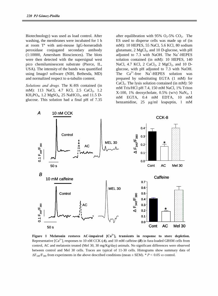
220 PJ Gómez-Pinilla
Biotechnology) was used as load control. After washing, the membranes were incubated for 1 h at room Tª with anti-mouse IgG-horseradish peroxidase conjugated secondary antibody (1:10000, Amersham Biosciences). The blots were then detected with the supersignal west pico chemiluminescent substrate (Pierce, IL, USA). The intensity of the bands was quantified using ImageJ software (NIH, Bethesda, MD) and normalized respect to á-tubulin content.
Solutions and drugs. The K-HS contained (in mM): 113 NaCl, 4.7 KCl, 2.5 CaCl2, 1.2 KH2PO4, 1.2 MgSO4, 25 NaHCO3, and 11.5 D-glucose. This solution had a final pH of 7.35
after equilibration with 95% O2-5% CO2. The ES used to disperse cells was made up of (in mM): 10 HEPES, 55 NaCl, 5.6 KCl, 80 sodium glutamate, 2 MgCl2, and 10 D-glucose, with pH adjusted to 7.3 with NaOH. The Na+-HEPES solution contained (in mM): 10 HEPES, 140 NaCl, 4.7 KCl, 2 CaCl2, 2 MgCl2, and 10 D-glucose, with pH adjusted to 7.3 with NaOH. The Ca2+-free Na+-HEPES solution was prepared by substituting EGTA (1 mM) for CaCl2. The lysis solution contained (in mM): 50 mM Tris/HCl pH 7.4, 150 mM NaCl, 1% Triton X-100, 1% deoxycholate, 0.5% (w/v) NaN3, 1 mM EGTA, 0.4 mM EDTA, 10 mM benzamidine, 25 µg/ml leupeptin, 1 mM
CCK-8
0.0
0.1
0.2
0.3
0.4
Cont AC Mel 30
ÄF
340/
F38
0
*AC
0.1
F34
0/F
380
10 nM CCK
50 s
Control
MEL 30
ÄF
340/
F38
0
Caffeine
0.0
0.1
0.2
0.3
0.4
0.5
0.6
0.7
Cont AC Mel 30
A
B10 mM caffeine
0.1
F34
0/F
380
50 s
AC
ControlMEL 30
*
CCK-8
0.0
0.1
0.2
0.3
0.4
Cont AC Mel 30
ÄF
340/
F38
0
*AC
0.1
F34
0/F
380
10 nM CCK
50 s
Control
MEL 30
ÄF
340/
F38
0
Caffeine
0.0
0.1
0.2
0.3
0.4
0.5
0.6
0.7
Cont AC Mel 30
Caffeine
0.0
0.1
0.2
0.3
0.4
0.5
0.6
0.7
Cont AC Mel 30
A
B10 mM caffeine
0.1
F34
0/F
380
50 s
AC
ControlMEL 30
*
Figure 1 Melatonin restores AC-impaired [Ca2+]i transients in response to store depletion. Representative [Ca2+]i responses to 10 nM CCK (A), and 10 mM caffeine (B) in fura-loaded GBSM cells from control, AC and melatonin treated (Mel 30, 30 mg/Kg/day) animals. No significant differences were observed between control and Mel 30 cells. Traces are typical of 11-30 cells. Histograms show summary data of ÄF340/F380 from experiments in the above described conditions (mean ± SEM). * P < 0.05 vs control.

Trabajo Experimental VIII 221
phenylmethylsulfonyl fluoride (PMSF). The phosphate buffer for MDA and GSH quantifications was (in nM): 20 NaCl, 2.7 KCl, 16 Na2HPO4, 4 NaH2PO4 with pH adjusted to 7.4. Drug concentrations are expressed as final bath concentrations of active species. Drugs and chemicals were obtained from the following sources: Acetylcholine (ACh), (±)BayK8644, caffeine, CCK-(26�33) sulfated, ionomycin, 1,4-dithio-DLthreitol, thapsigargin, nitrendipine and pinacidil were from Sigma Chemical (St. Louis, MO); 2-aminoethoxydiphenylborane (2-APB) from Tocris (Bristol, UK); fura 2-AM was from Molecular Probes (Molecular Probes
Europe, Leiden, Netherlands); collagenase was from Fluka (Madrid, Spain); and papain was from Worthington Biochemical (Lakewood, NJ). Other chemicals used were of analytical grade from Panreac (Barcelona, Spain). Stock solutions of fura 2-AM, thapsigargin, ionomycin, pinacidil and 2-APB were prepared in DMSO, and (±)BayK8644 and nitrendipine were prepared in ethanol. The solutions were diluted such that the final concentrations of DMSO or ethanol were ≤0.1% vol/vol. These concentrations of solvents did not interfere with fura 2 fluorescence.
20 sCCE
0.00
0.05
0.10
0.15
Cont AC Mel 30
ÄF
340/
F38
0
10 nM KCl
Cont AC Mel 30
60 mM KCl
0.0
0.1
0.2
0.3
ÄF
340/F
380
0.1
F34
0/F
380
AC
Control
MEL 30
*
*
A
B
50 s
0.05
F34
0/F
380
Ca2+ freeCa2+
1 µM TPS
ControlAC
MEL 30
20 sCCE
0.00
0.05
0.10
0.15
Cont AC Mel 30
ÄF
340/
F38
0
10 nM KCl
Cont AC Mel 30
60 mM KCl
0.0
0.1
0.2
0.3
ÄF
340/F
380
60 mM KCl
0.0
0.1
0.2
0.3
ÄF
340/F
380
0.1
F34
0/F
380
AC
Control
MEL 30
*
*
A
B
50 s
0.05
F34
0/F
380
Ca2+ freeCa2+
1 µM TPS
ControlAC
MEL 30
Figure 2. Melatonin restores AC-induced reduction in Ca2+ influx. (A) [Ca2+]i signal in response to a depolarizing solution containing 60 mM KCl and 2 mM Ca2+ in control, AC and Mel 30 cells . In B, after application of a Ca2+ free medium with the Ca2+ pump inhibitor thapsigargin (1 µM), 2 mM external Ca2+ was restored to initiate Ca2+ entry, evidenced by a fast plateau [Ca2+]i. Traces are typical of 7-36 cells. Histograms show summary data of ÄF340/F380 from experiments in the above described conditions (mean ± SEM). * P < 0.05 vs control.

222 PJ Gómez-Pinilla
Quantification and statistics. Results are expressed as means ± SEM of n cells or gallbladder strips. All results from [Ca2+]i determinations are given as ÄF340/F380. Gallbladder tension is given in millinewtons (mN)/mg of tissue. Statistical differences between means were determined by Student�s t-test. Differences were considered significant at P < 0.05.
4-RESULTS
We have recently shown that gallbladder inflammation decreases both Ca2+ release from intracellular stores and Ca2+ influx, which resulted in diminished gallbladder contractility.7 To characterize melatonin effects on Ca2+ homeostasis in cholecystitis, we challenged gallbladder smooth muscle cells from the ligated and de-ligated groups with different types of stimuli to release calcium from intracellular stores (CCK and caffeine) and to activate plasma membrane Ca2+ entry (KCl and depletion of intracellular Ca2+ stores). We also tested the effects of the hormone in the contractile response to the stimuli, the oxidative stress status and COX-2 expression.
Effects of melatonin on Ca2+ homeostatic mechanisms in AC. As shown in Fig. 1A, melatonin treatment (30 mg/Kg) was able to reverse the AC-induced reduction in 10 nM CCK-evoked Ca2+ response. This dose recovered both the fast initial rise due to Ca2+ release from internal stores and the subsequent plateau, which is entirely dependent on influx of extracellular calcium. Melatonin also preserved intracellular Ca2+ release through ryanodine receptors stimulated by caffeine, which was impaired by AC (Figure 1B).
When we explored Ca2+ influx through L-type voltage operated Ca2+ channels by application of a depolarizing medium containing 60 mM KCl, we found a decrease in the response of inflamed cells, as previously reported 7. In animals treated with melatonin, KCl-induced Ca2+ entry was almost normal (Figure 2A). Similar effects were obtained when L-type Ca2+ channels were selectively activated
with (±) BayK8644 (control: 0.179 ± 0.009 ÄF340/F380; AC: 0.108 ± 0.009 ÄF340/F380; Mel 30: 0.213 ± 0.027 ÄF340/F380, n = 19, 13 and 11 cells, P < 0.001 AC vs Mel 30).
A special feature of depolarization-induced Ca2+ entry in AC is the insensitivity to the blockade with dihydropyridines such as nitrendipine,7 which suggests inflammation-induced changes in the functional state or the pharmacological profile of L-type Ca2+ channels. When animals were treated with melatonin, KCl-induced Ca2+ influx was sensitive to 1 µM nitrendipine and to the ATP-sensitive K+ channel blocker pinacidil (10 µM) (88% and 75 % inhibition for nitrendipine and pinacidil, respectively, n = 12 and 9 cells; P < 0.001, Table 1), indicating that melatonin protects these channels from inflammation-associated damage.
We have recently demonstrated that capacitative Ca2+ entry (CCE) in response to intracellular Ca2+ stores is present in the gallbladder17 and that this mechanism was reduced in cholecystitic cells18. In this work we have also tested the effects of melatonin in CCE in AC. In order to do that, we activated CCE by pre-treatment of cells with thapsigargin (TPS) in Ca2+ free medium for 30 min and later reintroduction of Ca2+ in the bath. As shown in Figure 2B, melatonin treatment was effective in the recovery of CCE. In addition, changes in the functionality CCE channels induced by AC, which made them insensitive to both nitrendipine and 2-APB (100 µM), were absent under melatonin treatment. Thus, CCE was inhibited by both nitrendipine and 2-APB to the same extent than in control cells (61 %, 68 % and 80% of inhibition for nitrendipine, 2-APB, and both, respectively, n = 10, 15 nd 15 cells, P < 0.01, Table 2).
We also assayed a smaller dose of melatonin (2.5 mg/Kg), which recovered CCK- and caffeine-induced Ca2+ release (CCK: 0.249 ± 0.028 ÄF340/F380; caffeine: 0.393 ± 0.038 ÄF340/F380, n = 15 and 17 cells, not significant vs control), did not improve KCl-induced Ca2+

Trabajo Experimental VIII 223
influx (KCl: 0.09 ± 0.006 ÄF340/F380; n = 17cells P < 0.01), but recovered the pharmacological profile of calcium entry (data not shown), indicating that this dose is less effective in the treatment of AC.
Effects of melatonin on AC-impaired contractility. Taken together, our results show that both Ca2+ release from the stores and Ca2+ influx from extracellular medium, impaired by acute cholecystitis, were normalized under high dose melatonin treatment. Given that the increase in [Ca2+]i is determinant for activation of myosin light chain kinase leading to smooth muscle contraction,19 we would expect the recovery of the contractile responses to both Ca2+ release and Ca2+ influx manoeuvres after melatonin treatment. However, as shown in Figure 3, gallbladder strips from melatonin-
treated AC guinea pigs responded to 10 nM CCK and 60 mM KCl with similar or smaller contractions than untreated AC gallbladder strips. This contractile impairment was also evident when melatonin treated strips were exposed to 10 µM ACh, CCE activating protocol (Fig 3 B) or even to 1 µM ionomicin, which causes Ca2+ increases independently from activation of receptors and channels (control: 3.11 ± 0.57 mN/mg, AC: 0.72 ± 0.15 mN/mg, Mel 30: 0.94 ± 0.16 mN/mg; n = 8 - 4 strips, P > 0.05 AC vs Mel 30). Contractility was also impaired when animals were treated with 2.5 mg/Kg melatonin.
Melatonin effects on MDA and GSH content. The levels of MDA in the gallbladder tissue were measured in order to assess lipid peroxidation in AC. MDA levels were found to
1 m
N/m
g
60 mM KCl
Control AC ML 300
1
2
3
4
5
mN
/mg
10 nM CCK
AC
60 s
Control
MEL 30
60 mM KCl
60 s
1 m
N/m
g
ACControl
MEL 30
CCK-8
0
1
2
3
4
5
mN
/mg
ACh
0
1
2
3
4
CCE
0
1
2
3
4
5
Control AC ML 30 Control AC ML 30 Control AC ML 30
A
B
** *** * * *
mN
/mg
mN
/mg
1 m
N/m
g
60 mM KCl
Control AC ML 300
1
2
3
4
5
mN
/mg
10 nM CCK
AC
60 s
Control
MEL 30
10 nM CCK
AC
60 s
Control
MEL 30
60 mM KCl
60 s
1 m
N/m
g
ACControl
MEL 30
CCK-8
0
1
2
3
4
5
mN
/mg
ACh
0
1
2
3
4
CCE
0
1
2
3
4
5
Control AC ML 30 Control AC ML 30 Control AC ML 30
A
B
** *** * * *
mN
/mg
mN
/mg
Figure 3 AC-impaired contractility is not improved by melatonin treatment. A.- Representative original traces of changes in isometric tension in response to 10 nM CCK and a depolarizing solution containing 60 mM KCl in control, AC and Mel 30 gallbladder strips. B.- Histograms show summary data of contractile responses to 10 nM CCK, 10 ìM ACh, 60 mM KCl and CCE protocol (mean ± SEM) in the three experimental groups. N = 8-14 strips, * P < 0.001 vs control.

224 PJ Gómez-Pinilla
be higher in AC group compared to control tissue (control: 0.51 ± 0.07 nmol/mg protein; AC: 1.39 ± 0.26 nmol/mg protein, n = 5 and 4 animals , P < 0.05). In the group of animals treated with melatonin (30 mg /Kg) MDA content was similar to that of control group (0.72 ± 0.10 nmol/mg protein, n = 3 , P > 0.05 vs control). Melatonin was also effective in increasing the content of reduced glutathione (control: 11.31 ± 1.04 nmol/mg protein; Mel 30: 15.81 ± 1.02 nmol/mg protein, n = 5 and 3, P < 0.05) that was reduced in AC (5.64 ± 0.62 nmol/mg protein, P < 0.0001 vs control ).
Melatonin effects on Ca2+ homeostasis and contractility after bile duct deligation. The results above suggest that although melatonin resolves inflammation there are some contractile disabilities that melatonin treatment can not restore. It has to be noted that in the model used in this study, the gallbladder suffers stretch as the result of bile duct ligation and the continuous bile output. Taking this into account, it would be difficult to see any improvement in motility keeping the bile duct ligated. To solve this, after inducing AC, we performed de-ligation of bile duct and after two days we harvested the tissue and tested Ca2+
20 s
10 nM CCK
ACMEL 30
DL
20 s
10 mM caffeine
0.1
F34
0/F
380
0.1
F34
0/F
380 AC
MEL 30DL
CCK-8
0.0
0.2
0.4
0.6
0.8
Caffeine
0.0
0.2
0.4
0.6
0.8
ÄF
340/
F38
0
AC Mel 30DL
AC Mel 30DL
A
B
ÄF
340/
F38
0*
*
**
ä
20 s
10 nM CCK
ACMEL 30
DL
20 s
10 mM caffeine
0.1
F34
0/F
380
0.1
F34
0/F
380 AC
MEL 30DL
CCK-8
0.0
0.2
0.4
0.6
0.8
Caffeine
0.0
0.2
0.4
0.6
0.8
ÄF
340/
F38
0
AC Mel 30DL
AC Mel 30DL
A
B
ÄF
340/
F38
0*
*
**
ä
Figure 4. De-ligation normalizes AC-impaired [Ca2+]i transients in response to store depletion. Representative [Ca2+]i responses to 10 nM CCK (A), and 10 mM caffeine (B) in fura-loaded GBSM cells from AC, de-ligated (DL) and melatonin-treated DL (30 mg/Kg/day) animals. Note that melatonin treatment induces an additional recovery of CCK-induced Ca2+ release. Traces are typical of 18-21 cells. Histograms show summary data of ÄF340/F380 from experiments in the above described conditions (mean ± SEM). * P < 0.01 vs AC, ä P < 0.05 vs DL.

Trabajo Experimental VIII 225
homeostasis and contractility to check for the effects of melatonin on the recovery of gallbladder function.
Ca2+ release from intracellular stores (through IP3 and ryanodine receptors stimulated by CCK and caffeine, respectively) was restored just with the de-ligation procedure and this recovery was maintained after 30 mg/Kg melatonin treatment (Fig 4). However, Ca2+ influx activated either by depolarization (60 mM KCl) or by store depletion (following TPS treatment and Ca2+ reposition) was still impaired after de-ligation (Fig 5) and this Ca2+
entry was not sensitive to nitrendipine, pinacidil or 2-APB (Table 1). Melatonin was able to recover both the magnitude and the pharmacologic profile of Ca2+ influx (Figure 5, Table 1).
The effects of melatonin on Ca2+ homeostasis after de-ligation correlates with a decrease in lipidic peroxidation as indicated by the reduction in MDA content and the increase in GSH (MDA: 0.87 ± 0.08 nmol/mg protein, P < 0.05 vs AC, GSH: 10.03 ± 0.73 nmol/mg protein, P < 0.05 vs AC, n = 5 animals ).
60 mM KCl
40 s
0.05
F34
0/F
380
ACMEL 30
DL
KCl
0.00
0.05
0.10
0.15
0.20
0.25
ÄF
340/
F38
0
CCE
0.00
0.05
0.10
0.15
0.20
ÄF
340/
F38
0
*
*
AC Mel 30DL
AC Mel 30DL
A
300 s
0.02
5F
340/
F38
0
AC MEL 30DL
Ca2+ free Ca2+
1 µM TPSB
ä
ä
60 mM KCl
40 s
0.05
F34
0/F
380
ACMEL 30
DL
KCl
0.00
0.05
0.10
0.15
0.20
0.25
ÄF
340/
F38
0
CCE
0.00
0.05
0.10
0.15
0.20
ÄF
340/
F38
0
*
*
AC Mel 30DL
AC Mel 30DL
A
300 s
0.02
5F
340/
F38
0
AC MEL 30DL
Ca2+ free Ca2+
1 µM TPSB
ä
ä
Figure 5. Melatonin restores Ca2+ influx after de-ligation. (A) [Ca2+]i signal in response to a depolarizing solution containing 60 mM KCl and 2 mM Ca2+ in AC, deligated (DL) and melatonin-treated DL cells. In B, after application of a Ca2+ free medium with the Ca2+ pump inhibitor thapsigargin (1 µM), 2 mM external Ca2+ was restored to initiate Ca2+ entry, evidenced by a fast plateau [Ca2+]i. Traces are typical of 14-20 cells. Histograms show summary data of ÄF340/F380 from experiments in the above described conditions (mean ± SEM). * P < 0.01 vs AC, ä P < 0.01 vs DL

226 PJ Gómez-Pinilla
However, de-ligation was associated to an increase in GSH content compared to that in AC group, and elevated MDA levels (GSH: 10.97 ± 0.52 nmol/mg protein, P < 0.05 vs AC; MDA: 1.75 ± 0.08 nmol/mg protein, P < 0.05 vs AC, n = 5 animals). As inflammation of gastrointestinal tract is associated with an increase in COX-2 expression,20 we quantified the expression of this enzyme in response to AC, de-ligation and melatonin treatment. As observed in Figure 6A and B, the expression in control tissue is almost undetectable, but it increases after AC, the levels are still high after two days of de-ligation and melatonin drives the expression to control levels.
When we tested the contractility of gallbladder strips after de-ligation, we found that contractions in response to almost every tested stimulus were the same or even smaller than in AC strips (Figure 7). The fact that the response to CCK was impaired (Figures 7A & B), in spite of the recovery of CCK-induced Ca2+ release, suggests that Ca2+ desensitization of the contractile apparatus occurs. This was supported by the impairment in ionomicin-induced contraction (control: 3.72 ± 0.45 mN/mg, AC: 1.39 ± 0.15 mN/mg, DL: 0.67 ±
0.14 mN/Kg, Mel 30 + DL: 2.10 ± 0.22 mN/mg; n = 12 - 6 strips, P < 0.05 AC vs Mel 30 + DL and control vs Mel 30 + DL), which caused a similar [Ca2+]i increase in all experimental groups. Interestingly, the contractility of melatonin treated gallbladder strips improved, especially when ACh was used as agonist (Figure 7B). The contractile response to activation of Ca2+ influx (either by depolarization or CCE) was also higher in melatonin treated animals (Figure 7B). All together this suggests that melatonin, at high doses, affects either Ca2+ dependent and independent mechanisms to improve contraction and recover gallbladder functionality.
5-DISCUSION
We present here data showing that melatonin protects against acute cholecystitis-induced alterations of Ca2+ homeostatic mechanisms, normalizes markers of oxidative stress and recovers gallbladder contractility shortly after inflammation, which is of importance for a fast restoration of gallbladder functionality in this pathological state.
75 kDa
55 kDa
Fol
din
crea
se(C
OX
-2/ á
-tub
ulin
)
0
1
2
Control
AC Mel 30 + DLDL
COX-2
á-Tubulin
ACMel 3
0
+ DLDL
Cont
A B
*
**
ä
75 kDa
55 kDa
Fol
din
crea
se(C
OX
-2/ á
-tub
ulin
)
0
1
2
Control
AC Mel 30 + DLDL
COX-2
á-Tubulin
ACMel 3
0
+ DLDL
Cont
A B
*
**
ä
Figure 6.- Melatonin treatment normalizes COX-2 expression A.- Typical example of western blot analysis of COX-2 and á-tubulin protein expression in gallbladder smooth muscle from control, AC, de-ligated (DL) and melatonin-treated DL (30 mg/Kg/day) animals. á-tubulin content was used as loading control. B.- Summary data are expressed as fold increases respect to control tissue (represented in the figure as a dotted line). Data are means ± SE of 4-6 experiments. *P < 0.05 vs control, ä P < 0.01 vs AC and DL.

Trabajo Experimental VIII 227
AC is a clinical condition characterized by inflammation of the gallbladder tissue.3 Important functional consequences of AC includes a loss of contractile response to agonists and intrinsic nerve stimulation and gallbladder stasis,5, 21 mainly due to a deregulation of calcium signals but also to changes in contractile proteins such as F actin.7
Inflammation is related to overproduction of oxygen-derived free radicals (especially, superoxide and hydroxyl radical), which leads to considerable oxidative stress.22 In the gastrointestinal tract, where nitric oxide
(NO) is an important neurotransmitter mediating relaxation,23 peroxynitrite production has been found to mediate deleterious effects in inflammation.24 Reactive oxygen species and peroxynitrite induce cellular injury via several mechanisms including peroxidation of membrane lipids.25 Many studies have reported that there is a correlation between the intensity of biliary tract obstruction and increased liver free radical generation, as well as a direct correlation between the level of oxidative stress and the biochemical markers of liver injury.26-30 In addition, it has been established that deleterious accumulation of lipid peroxides is correlated with marked impairment of soluble
AC
MEL 30 + DL
10 nM CCK
DL
60 mM KCl
60 s
1 m
N/m
g
60 s
1 m
N/m
g
CCE
0
1
2
3
4
KCl
0
1
2
3
AChCCK
mN
/mg
mN
/mg
mN
/mg
mN
/mg
AC
MEL 30 + DLDL
** **
A B
C
0
1
2
3
4
**
AC Mel 30 + DLDL AC Mel 30 + DLDL AC Mel 30 + DLDL0
1
2
3
4
AC Mel 30 + DLDL
*ä
AC
MEL 30 + DL
10 nM CCK
DL
60 mM KCl
60 s
1 m
N/m
g
60 s
1 m
N/m
g
CCE
0
1
2
3
4
KCl
0
1
2
3
AChCCK
mN
/mg
mN
/mg
mN
/mg
mN
/mg
AC
MEL 30 + DLDL
** **
A B
C
0
1
2
3
4
**
AC Mel 30 + DLDL AC Mel 30 + DLDL AC Mel 30 + DLDL0
1
2
3
4
AC Mel 30 + DLDL
*ä
Figure 7.- Melatonin treatment recovers gallbladder contractility after de-ligation. A.- Representative original traces of changes in isometric tension in response to 10 nM CCK and a depolarizing solution containing 60 mM KCl + in AC, deliagated (DL) and melatonin-treated DL gallbladder strips. B.- Histograms show summary data of contractile response to 10 nM CCK, 10 ìM ACh, 60 mM KCl and CCE protocol (mean ± SEM) in the three experimental groups. N = 8-29 strips, * P < 0.05 vs AC, ä P < 0.01 vs DL, ** P < 0.001 vs AC and DL.

228 PJ Gómez-Pinilla
antioxidant defence mechanisms.31 In this study, we demonstrate that bile duct ligation also induces an increase in lipid peroxidation in gallbladder smooth muscle represented by high levels of MDA as well as by the depletion of the ubiquotus antioxidant gluthation. Increased MDA levels in gallbladder smooth muscle in response to H2O2 exposure has been previously reported,32 which agrees with the increase in H2O2 and MDA content described in gallbladder inflammation.33
The most important finding in the present study is that melatonin dose-dependently protected gallbladder smooth muscle Ca2+ homeostasis from damage caused by the AC protocol, being 30 mg/kg more effective than 2.5 mg/Kg. The protective effect of melatonin was accompanied by a significant decrease in MDA levels and an increase in GSH content, suggesting that melatonin counteracts the increase in AC-induced lipid peroxidation and preserves the intracellular signalling related to Ca2+ homeostasis. The protective effects of melatonin in the digestive system have been demonstrated in various pathological conditions related to inflammation such as acute pancreatitis,34 various forms of gastritis35 and colitis.36 The mechanism of this protection by melatonin has been mainly attributed to its antioxidant properties and its ability to attenuate lipid membrane peroxidation and related damage of cell membranes. Thus, melatonin functions as a direct free radical scavenger of the highly toxic hydroxyl radical, the cytotoxic O2
. and the peroxyl radical produced during peroxidation of lipids.22 Melatonin has also been shown to scavenge peroxynitrite anion,
which agrees with its inhibitory effects on peroxynitrite-evoked contraction reported by our group in gallbladder smooth muscle.37 Besides its free radical-scavenging actions, melatonin also protects the cells acting as an inducer of antioxidant enzymes such as superoxide dismutase, catalase, and glutathion peroxidase.38
In our study, melatonin protects Ca2+ homoestatic mechanisms located both in the plasma and sarcoplasmic membranes, which is consistent with the lipophilicity and easy entry of melatonin into cells protecting the subcellular compartments. However, the fact that 2.5 mg/Kg melatonin was able to protect sarcoplasmic Ca2+ channels but did not afford protection to Ca2+ channels located in plasma membrane, similar to the de-ligation manoeuvre, indicates that plasma membrane suffers more damage in acute cholecystitis.
It is noteworthy that the protection provided by melatonin was not completely effective against AC-induced contractility impairment unless the obstruction of the bile duct was relieved. In this regard, it has to be noted that melatonin protected cells from oxidative injury and lipid peroxidation as demonstrated by the levels of MDA and GSH, as well as by the normalization of Ca2+ homeostasis. However, melatonin did not protect against the mechanical modifications of the gallbladder wall as consequence of the distension, which translates into contractility impairment. In addition, persistent alterations in GI function are commonly observed after resolution of intestinal inflammation.39
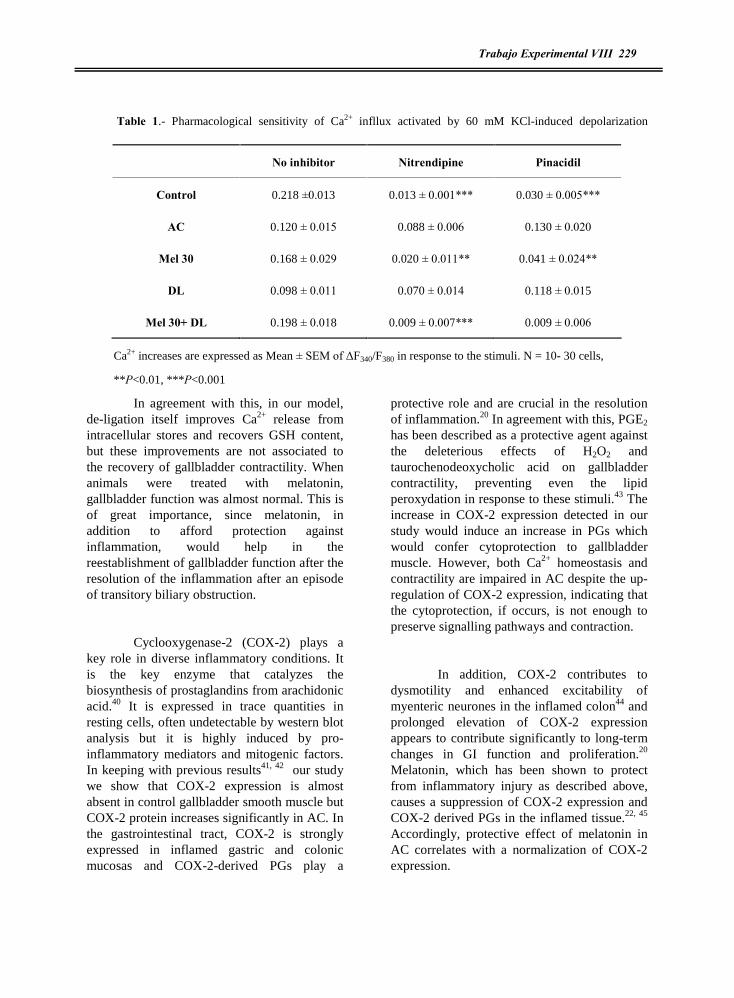
Trabajo Experimental VIII 229
Table 1.- Pharmacological sensitivity of Ca2+ infllux activated by 60 mM KCl-induced depolarization
Ca2+ increases are expressed as Mean ± SEM of ÄF340/F380 in response to the stimuli. N = 10- 30 cells,
**P<0.01, ***P<0.001
In agreement with this, in our model, de-ligation itself improves Ca2+ release from intracellular stores and recovers GSH content, but these improvements are not associated to the recovery of gallbladder contractility. When animals were treated with melatonin, gallbladder function was almost normal. This is of great importance, since melatonin, in addition to afford protection against inflammation, would help in the reestablishment of gallbladder function after the resolution of the inflammation after an episode of transitory biliary obstruction.
Cyclooxygenase-2 (COX-2) plays a key role in diverse inflammatory conditions. It is the key enzyme that catalyzes the biosynthesis of prostaglandins from arachidonic acid.40 It is expressed in trace quantities in resting cells, often undetectable by western blot analysis but it is highly induced by pro-inflammatory mediators and mitogenic factors. In keeping with previous results41, 42 our study we show that COX-2 expression is almost absent in control gallbladder smooth muscle but COX-2 protein increases significantly in AC. In the gastrointestinal tract, COX-2 is strongly expressed in inflamed gastric and colonic mucosas and COX-2-derived PGs play a
protective role and are crucial in the resolution of inflammation.20 In agreement with this, PGE2 has been described as a protective agent against the deleterious effects of H2O2 and taurochenodeoxycholic acid on gallbladder contractility, preventing even the lipid peroxydation in response to these stimuli.43 The increase in COX-2 expression detected in our study would induce an increase in PGs which would confer cytoprotection to gallbladder muscle. However, both Ca2+ homeostasis and contractility are impaired in AC despite the up-regulation of COX-2 expression, indicating that the cytoprotection, if occurs, is not enough to preserve signalling pathways and contraction.
In addition, COX-2 contributes to dysmotility and enhanced excitability of myenteric neurones in the inflamed colon44 and prolonged elevation of COX-2 expression appears to contribute significantly to long-term changes in GI function and proliferation.20 Melatonin, which has been shown to protect from inflammatory injury as described above, causes a suppression of COX-2 expression and COX-2 derived PGs in the inflamed tissue.22, 45 Accordingly, protective effect of melatonin in AC correlates with a normalization of COX-2 expression.
No inhibitor Nitrendipine Pinacidil
Control 0.218 ±0.013 0.013 ± 0.001*** 0.030 ± 0.005***
AC 0.120 ± 0.015 0.088 ± 0.006 0.130 ± 0.020
Mel 30 0.168 ± 0.029 0.020 ± 0.011** 0.041 ± 0.024**
DL 0.098 ± 0.011 0.070 ± 0.014 0.118 ± 0.015
Mel 30+ DL 0.198 ± 0.018 0.009 ± 0.007*** 0.009 ± 0.006
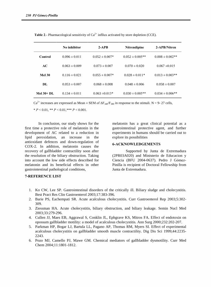
230 PJ Gómez-Pinilla
Table 2.- Pharmacological sensitivity of Ca2+ infllux activated by store depletion (CCE).
Ca2+ increases are expressed as Mean ± SEM of ÄF340/F380 in response to the stimuli. N = 9- 27 cells,
* P < 0.01, ** P < 0.01,*** P < 0.001.
In conclusion, our study shows for the first time a protective role of melatonin in the development of AC related to a reduction in lipid peroxidation, an increase in the antioxidant defences and down-regulation of COX-2. In addition, melatonin causes the recovery of gallbladder contractility soon after the resolution of the biliary obstruction. Taking into account the low side effects described for melatonin and its beneficial effects in other gastrointestinal pathological conditions,
melatonin has a great clinical potential as a gastrointestinal protective agent, and further experiments in humans should be carried out to explore its possibilities
6-ACKNOWLEDGEMENTS
Supported by Junta de Extremadura (2PR03A020) and Ministerio de Educacion y Ciencia (BFU 2004-0637). Pedro J Gómez-Pinilla is recipient of Doctoral Fellowship from Junta de Extremadura.
7-REFERENCE LIST
1. Ko CW, Lee SP. Gastrointestinal disorders of the critically ill. Biliary sludge and cholecystitis. Best Pract Res Clin Gastroenterol 2003;17:383-396.
2. Barie PS, Eachempati SR. Acute acalculous cholecystitis. Curr Gastroenterol Rep 2003;5:302-309.
3. Ziessman HA. Acute cholecystitis, biliary obstruction, and biliary leakage. Semin Nucl Med 2003;33:279-296.
4. Cullen JJ, Maes EB, Aggrawal S, Conklin JL, Ephgrave KS, Mitros FA. Effect of endotoxin on opossum gallbladder motility: a model of acalculous cholecystitis. Ann Surg 2000;232:202-207.
5. Parkman HP, Bogar LJ, Bartula LL, Pagano AP, Thomas RM, Myers SI. Effect of experimental acalculous cholecystitis on gallbladder smooth muscle contractility. Dig Dis Sci 1999;44:2235-2243.
6. Pozo MJ, Camello PJ, Mawe GM. Chemical mediators of gallbladder dysmotility. Curr Med Chem 2004;11:1801-1812.
No inhibitor 2-APB Nitrendipine 2-APB/Nitren
Control 0.096 ± 0.011 0.052 ± 0.007* 0.052 ± 0.005** 0.008 ± 0.002**
AC 0.063 ± 0.009 0.073 ± 0.007 0.070 ± 0.020 0.067 ±0.015
Mel 30 0.116 ± 0.021 0.055 ± 0.007* 0.028 ± 0.011* 0.013 ± 0.003**
DL 0.053 ± 0.007 0.068 ± 0.008 0.048 ± 0.006 0.058 ± 0.007
Mel 30+ DL 0.134 ± 0.011 0.063 ±0.013* 0.030 ± 0.005** 0.034 ± 0.006**

Trabajo Experimental VIII 231
7. Gomez-Pinilla PJ, Morales S, Camello-Almaraz C, Moreno R, Pozo MJ, Camello PJ. Changes in guinea pig gallbladder smooth muscle Ca2+ homeostasis by acute acalculous cholecystitis. Am J Physiol Gastrointest Liver Physiol 2006;290:G14-G22.
8. Kalliafas S, Ziegler DW, Flancbaum L, Choban PS. Acute acalculous cholecystitis: incidence, risk factors, diagnosis, and outcome. Am Surg 1998;64:471-475.
9. Patel M, Miedema BW, James MA, Marshall JB. Percutaneous cholecystostomy is an effective treatment for high-risk patients with acute cholecystitis. Am Surg 2000;66:33-37.
10. Babb RR. Acute acalculous cholecystitis. A review. J Clin Gastroenterol 1992;15:238-241. 11. Reiter RJ, Tan DX, Manchester LC, El Sawi MR. Melatonin reduces oxidant damage and
promotes mitochondrial respiration: implications for aging. Ann N Y Acad Sci 2002;959:238-250. 12. Kvetnoy IM, Ingel IE, Kvetnaia TV, Malinovskaya NK, Rapoport SI, Raikhlin NT, Trofimov AV,
Yuzhakov VV. Gastrointestinal melatonin: cellular identification and biological role. Neuro Endocrinol Lett 2002;23:121-132.
13. Lane EA, Moss HB. Pharmacokinetics of melatonin in man: first pass hepatic metabolism. J Clin Endocrinol Metab 1985;61:1214-1216.
14. Tan D, Manchester LC, Reiter RJ, Qi W, Hanes MA, Farley NJ. High physiological levels of melatonin in the bile of mammals. Life Sci 1999;65:2523-2529.
15. Myers SI, Haley-Russell D, Parks L, Husband K. Common bile duct ligation in rabbit: a new model of acute cholecystitis description of histology and bile analysis. J Surg Res 1988;45:556-564.
16. Pozo MJ, Perez GJ, Nelson MT, Mawe GM. Ca(2+) sparks and BK currents in gallbladder myocytes: role in CCK-induced response. Am J Physiol Gastrointest Liver Physiol 2002;282:G165-G174.
17. Morales S, Camello PJ, Alcon S, Salido GM, Mawe G, Pozo MJ. Coactivation of capacitative calcium entry and L-type calcium channels in guinea pig gallbladder. Am J Physiol Gastrointest Liver Physiol 2004;286:G1090-G1100.
18. Xiao ZL, Chen Q, Biancani P, Behar J. Abnormalities of gallbladder muscle associated with acute inflammation in guinea pigs. Am J Physiol Gastrointest Liver Physiol 2001;281:G490-G497.
19. Somlyo AP, Somlyo AV. Signal transduction and regulation in smooth muscle. Nature 1994;372:231-236.
20. Wallace JL. COX-2: a pivotal enzyme in mucosal protection and resolution of inflammation. ScientificWorldJournal 2006;6:577-588.
21. Parkman HP, James AN, Bogar LJ, Bartula LL, Thomas RM, Ryan JP, Myers SI. Effect of acalculous cholecystitis on gallbladder neuromuscular transmission and contractility. J Surg Res 2000;88:186-192.
22. Cuzzocrea S, Reiter RJ. Pharmacological action of melatonin in shock, inflammation and ischemia/reperfusion injury. Eur J Pharmacol 2001;426:1-10.
23. Sanders KM, Ward SM. Nitric oxide as a mediator of nonadrenergic noncholinergic neurotransmission. Am J Physiol 1992;262:G379-G392.
24. Miller MJ, Sandoval M. Nitric Oxide. III. A molecular prelude to intestinal inflammation. Am J Physiol 1999;276:G795-G799.
25. Cochrane CG. Mechanisms of oxidant injury of cells. Mol Aspects Med 1991;12:137-147. 26. Baron V, Muriel P. Role of glutathione, lipid peroxidation and antioxidants on acute bile-duct
obstruction in the rat. Biochim Biophys Acta 1999;1472:173-180. 27. Pastor A, Collado PS, Almar M, Gonzalez-Gallego J. Antioxidant enzyme status in biliary
obstructed rats: effects of N-acetylcysteine. J Hepatol 1997;27:363-370. 28. Orellana M, Rodrigo R, Thielemann L, Guajardo V. Bile duct ligation and oxidative stress in the
rat: effects in liver and kidney. Comp Biochem Physiol C Toxicol Pharmacol 2000;126:105-111.

232 PJ Gómez-Pinilla
29. Ohta Y, Kongo M, Kishikawa T. Melatonin exerts a therapeutic effect on cholestatic liver injury in rats with bile duct ligation. J Pineal Res 2003;34:119-126.
30. Esrefoglu M, Gul M, Emre MH, Polat A, Selimoglu MA. Protective effect of low dose of melatonin against cholestatic oxidative stress after common bile duct ligation in rats. World J Gastroenterol 2005;11:1951-1956.
31. Engin A, Bozkurt BS, Altan N, Memis L, Bukan N. Nitric oxide-mediated liver injury in the presence of experimental bile duct obstruction. World J Surg 2003;27:253-255.
32. Xiao ZL, Andrada MJ, Biancani P, Behar J. Reactive oxygen species (H(2)O(2)): effects on the gallbladder muscle of guinea pigs. Am J Physiol Gastrointest Liver Physiol 2002;282:G300-G306.
33. Xiao ZL, Chen Q, Biancani P, Behar J. Abnormalities of gallbladder muscle associated with acute inflammation in guinea pigs. Am J Physiol Gastrointest Liver Physiol 2001;281:G490-G497.
34. Qi W, Tan DX, Reiter RJ, Kim SJ, Manchester LC, Cabrera J, Sainz RM, Mayo JC. Melatonin reduces lipid peroxidation and tissue edema in cerulein-induced acute pancreatitis in rats. Dig Dis Sci 1999;44:2257-2262.
35. Jaworek J, Brzozowski T, Konturek SJ. Melatonin as an organoprotector in the stomach and the pancreas. J Pineal Res 2005;38:73-83.
36. Marquez E, Sanchez-Fidalgo S, Calvo JR, la de Lastra CA, Motilva V. Acutely administered melatonin is beneficial while chronic melatonin treatment aggravates the evolution of TNBS-induced colitis. J Pineal Res 2006;40:48-55.
37. Alcon S, Morales S, Camello PJ, Hemming JM, Jennings L, Mawe GM, Pozo MJ. A redox-based mechanism for the contractile and relaxing effects of NO in the guinea-pig gall bladder. J Physiol 2001;532:793-810.
38. Reiter RJ, Tan DX, Osuna C, Gitto E. Actions of melatonin in the reduction of oxidative stress. A review. J Biomed Sci 2000;7:444-458.
39. Lomax AE, O'hara JR, Hyland NP, Mawe GM, Sharkey KA. Persistent alterations to enteric neural signalling in guinea pig colon following resolution of colitis. Am J Physiol Gastrointest Liver Physiol 2006.
40. Vane JR, Bakhle YS, Botting RM. Cyclooxygenases 1 and 2. Annu Rev Pharmacol Toxicol 1998;38:97-120.
41. Myers SI, Bartula LL, Colvin MP, Parkman HP. Cholecystokinin (CCK) down regulates PGE2 and PGI2 release in inflamed Guinea pig gallbladder smooth muscle cell cultures. Prostaglandins Leukot Essent Fatty Acids 2005;73:121-126.
42. Myers SI, Bartula LL, Colvin MP, Parkman HP, Braverman AA, Ruggieri MR. Bile duct ligation induced acute inflammation up-regulates cyclooxygenase-2 content and PGE2 release in guinea pig gallbladder smooth muscle cell cultures. Prostaglandins Leukot Essent Fatty Acids 2005;72:327-333.
43. Xiao ZL, Biancani P, Behar J. Role of PGE2 on gallbladder muscle cytoprotection of guinea pigs. Am J Physiol Gastrointest Liver Physiol 2004;286:G82-G88.
44. Linden DR, Sharkey KA, Ho W, Mawe GM. Cyclooxygenase-2 contributes to dysmotility and enhanced excitability of myenteric AH neurones in the inflamed guinea pig distal colon. J Physiol 2004;557:191-205.
45. Mayo JC, Sainz RM, Tan DX, Hardeland R, Leon J, Rodriguez C, Reiter RJ. Anti-inflammatory actions of melatonin and its metabolites, N1-acetyl-N2-formyl-5-methoxykynuramine (AFMK) and N1-acetyl-5-methoxykynuramine (AMK), in macrophages. J Neuroimmunol 2005;165:139-149.

6. Discusión.

Discusión general 235
Nuestro estudio muestra que el envejecimiento altera la cinética de la vejiga urinaria de cobaya, produciendo dos cambios fundamentales y aparentemente contradictorios: aumento en la excitabilidad de la vejiga durante la fase de llenado y disminución en la contracción del detrusor durante la micción. El aumento de actividad está indicado por la reducción de la capacidad de la vejiga, acortamiento del intervalo de micción y contracciones no asociadas a micción. La disminución en la respuesta contráctil del detrusor produce una menor presión de vaciado y conduce a la aparición de volumen de reserva. Estas dos modificaciones funcionales de la vejiga envejecida reflejan en parte el comportamiento clínico observado en humanos en las alteraciones de la micción asociadas a la edad: hiperactividad y pérdida de contracción del detrusor, así como diversas formas de incontinencia.
Hay que observar que, si bien es posible que la vejiga envejecida presente cambios en la matriz extracelular con hipotéticas consecuencias mecánicas, nuestro estudio cistométrico descarta que el detrusor envejecido tenga alteraciones mecánicas importantes, dado que no se alteran la complianza de la vejiga ni la presión basal. Esto indica que los efectos de la edad sobre la función vesical deben explicarse principalmente por alteraciones de los mecanismos contráctiles y/o de control.
Los datos presentados en esta memoria pueden explicar razonablemente la citada disminución de la contracción del detrusor. Por un lado hemos observado que la función neuromuscular está alterada en el envejecimiento, lo que se debe tanto a una clara disminución funcional de la inervación excitadora motora purinérgica y aferente, como a un aumento de la inervación inhibidora (nitrérgica y adrenérgica) (Cap II). Es de notar que los cambios que el envejecimiento produce en la función del plexo intrínseco incluyen cambios en la modulación colinégica de otras fibras nerviosas (aparece inhibición de terminales purinérgicas excitadoras y desaparece la inhibición de fibras relajantes presente en individuos jóvenes) que recuerda los conocidos cambios en las redes sinápticas de la corteza cerebral observados en el envejecimiento.
Junto a estos cambios neurales, el envejecimiento actúa directamente en las células del detrusor inhibiendo la actividad de la vía de sensibilización al calcio, gracias a una reducción de la activación de las proteínas CPI-17 y MYPT1 y a una menor expresión de esas proteínas y de sus proteínas activadoras (PKC, ROCK,�). Esto explica que la respuesta contráctil del músculo detrusor a diversos estímulos (Cap III) coexista con un paradójico incremento en la magnitud de las respuestas de calcio a esos mismos estímulos. Aunque nuestros resultados sobre la homeostasis del calcio pueden explicar este incremento (aumentos del depósito movilizable de calcio y de la sensibilidad de los canales de entrada de calcio en el citosol, junto a disminución en la velocidad de extrusión de calcio desde el citosol, (Cap IV)), el significado y mecanismo de estos cambios es difícil de explicar, aunque una alteración debida a estrés oxidativo crónico asociado al envejecimiento es muy probable, debido a la conocida sensibilidad de los diversos transportadores del ión calcio frente a oxidantes. La discrepancia entre señales de calcio y nivel de contracción también puede reflejar el hecho de que las señales de calcio son solamente un componente iniciador del mecanismo contráctil.
Dado que algunas teorías del envejecimiento incluyen como factor causal cambios en la homeostasis del calcio, nuestro hallazgo de una mayor amplitud de la señal producida por diversos estímulos y un enlentecimiento de la subsiguiente recuperación podría ser significativa, ya que se sabe que cambios en los parámetros de amplitud y frecuencia de las señales repetitivas de calcio controlan la expresión genética, que como hemos mostrado en esta memoria, está modificada para proteínas muy importantes en la señalización intracelular (Cap III).
En la reducción de la contracción en la vejiga envejecida también podría colaborar el enlentecimiento de los mecanismos de extrusión de calcio (Cap IV), ya que un retraso en la recuperación
id17328406 pdfMachine by Broadgun Software - a great PDF writer! - a great PDF creator! - http://www.pdfmachine.com http://www.broadgun.com

236 PJ Gómez-Pinilla
de los niveles de calcio puede reducir la excitabilidad celular al actuar sobre los diversos canales de K+ presentes en este tipo celular.
En nuestras condiciones experimentales la melatonina fue capaz de recuperar la funcionalidad del tejido vesical en individuos envejecidos, revirtiendo la mayor parte de los parámetros afectados por la edad. Es probable que este efecto se deba a su capacidad antioxidante, tanto directa como indirecta, ya que redujo el incremento que la edad provocaba en los índices de estrés oxidativo, si bien nuestros resultados tan sólo permiten establecer firmemente una correlación, sin demostrar causalidad.
Acerca de la aparición de hiper-excitabilidad vesical, hay factores tanto mecánicos como funcionales que podrían explicarla. La mayor rigidez de la vejiga envejecida, así como la presencia de un volumen residual, podrían facilitar que la vejiga se contrajera antes en individuos envejecidos, aunque como hemos comentado antes, la complianza no está alterada en este grupo. Desde el punto de vista funcional disponemos de dos posibles explicaciones: la aparición de contracciones gigantes en la zona del trígono, y la alteración de los nervios aferentes sensitivos. Este último mecanismo, puesto de manifiesto en el capítulo II, permitiría que la vejiga presentase mayor actividad a pesar de tener una menor capacidad de contracción. Dado que en última instancia la actividad del detrusor está regulada por un arco reflejo que incluye nervios extravesicales y médula espinal, cualquier alteración en el componente aferente puede resultar en cambios en el patrón final de excitación del efector. Además, en nuestro laboratorio hemos detectado en el trígono de individuos envejecidos la existencia de contracciones gigantes espontáneas (datos no incluidos en esta memoria) y de una mayor sensibilidad a la activación del plexo nervioso. Este patrón es plenamente compatible con los rasgos de mayor excitabilidad en presencia de una menor capacidad de contracción del detrusor: la mayor actividad del trígono produciría contracciones no asociadas a la micción que, cuando se transmitieran al resto de la vejiga inducirían su vaciamiento con una menor presión debido al debilitamiento del detrusor.
Para determinar si el proceso de envejecimiento tenía las mismas características en diferentes tipos de músculo o si por el contrario había aspectos singulares relacionados con la localización y función del órgano, analizamos las alteraciones que el envejecimiento produce en la vesícula biliar (Cap V), órgano en el que, a pesar de tener su epitelio importantes funciones reabsortivas, la disposición de las fibras musculares, función de reservorio así como el control por el plexo nervioso intrínseco son similares a los de la vejiga urinaria. Al igual que ocurría en el otro tejido objeto de estudio, el envejecimiento provocaba una reducción en la capacidad contráctil y alteraciones en la inervación intrínseca, dando lugar a una función neuromuscular alterada que probablemente causará disfunciones en el vaciamiento en respuesta a la ingesta de alimentos o durante los complejos motores migradores típicos del tracto digestivo. El análisis más detallado de la inervación nos indicó una reducción en el plexo nitrérgico inhibidor, totalmente opuesto a lo que ocurría en la vejiga urinaria, y una elevación en la sensibilidad de fibras sensitivas de carácter inhibidor liberadoras de CGRP, que tampoco coincide con lo que describimos en la vejiga. A nivel de la célula muscular sí que eran coincidentes los resultados en cuanto a la hipotonía en respuesta a los principales agonistas y la activación de la entrada de calcio mediada por despolarización o por vaciamiento de depósitos. En este tipo muscular la menor contractilidad estaba asociada a una disminución de la [Ca2+]c y no a una sobrecarga como ocurría en la vejiga. Sin embargo, debe destacarse que la señalización del calcio sólo se afecta en lo que respecta a la entrada del ión desde el espacio extracelular, manteniéndose la movilización del Ca2+ en respuesta a CCK y cafeína que promueven la liberación de Ca2+ desde el depósito a través de canales/receptores de IP3 y rianodina, respectivamente. Estos resultados indican la posibilidad de que los canales iónicos situados en la membrana de las células musculares de la vesícula biliar sean sensibles el envejecimiento mientras los localizados en los depósitos mostrarían una mayor resistencia al deterioro o daño asociado al envejecimiento. El hecho de que la respuesta contráctil a la CCK sea inferior en el envejecimiento, a

Discusión general 237
pesar de movilizar este agente la misma cantidad de Ca2+, sugiere que, además de la contracción dependiente de calcio, se afectan procesos contráctiles independientes de Ca2+ y relacionados con la sensibilidad de la maquinaria contráctil al ión. Mientras que la posibilidad de una reducción en los mecanismos o vías de sensibilidad al Ca2+ se está explorando actualmente en nuestro laboratorio, en esta memoria hemos demostrado que se produce una reducción en la cantidad de F-actina presente en las células musculares, lo que ocasionaría en sí mismo una reducción en la capacidad contráctil de estas células.
Al igual que ocurría en la vejiga urinaria la administración de melatonina a animales envejecidos mejoraba espectacularmente la capacidad contráctil de la vesícula biliar, tanto en lo que respecta a la contracción miógena como a la de tipo neurógeno. En cuanto a la homeostasis del Ca2+, la melatonina no fue capaz de modificar el perfil de entrada de Ca2+ al citosol, indicando su incapacidad de recuperar la funcionalidad de los canales de Ca2+ del plasmalema. El hecho de inducir una mejora en la contractilidad sin afectar la [Ca2+]c sugiere que la melatonina puede modificar mecanismos de sensibilización al Ca2+, tal y como apuntan los resultados obtenidos recientemente en nuestro laboratorio y la recuperación en el contenido de F-actina que presentamos en esta memoria.
Según los resultados que acabamos de comentar, es evidente que el envejecimiento de la vejiga urinaria y vesícula biliar, a pesar de producir globalmente una pérdida de contractilidad, tiene aspectos diferenciados en cada tejido. Debe tenerse en cuenta que a pesar de que la función de ambos órganos es almacenar/expeler fluidos, ya encontramos singularidades en la función neuromuscular de ambos músculos en animales adultos. Así, las fibras inhibidoras nitrérgicas se estimulan y ejercen efectos relajantes en la vesícula biliar mientras que en la vejiga se encuentran �frenadas� bajo modulación colinérgica (al igual que ocurre con las adrenérgicas inhibidoras). Teniendo en cuenta que en el caso de la vejiga urinaria una contracción no siempre va acompañada de una evacuación, ya que ésta depende también del estado de otros componentes como los esfínteres interno y externo, uretra, y distintos componentes neurales del reflejo de micción, es posible que este órgano necesite de mecanismos complejos de neuromodulación ausentes en el otro divertículo. Es llamativo también el que la vejiga urinaria sufra una mayor pérdida de contractilidad (60 % frente a 30%) y mayores alteraciones en la homeostasis del Ca2+ por el envejecimiento que la vesícula biliar. Esto puede estar relacionado con una mayor protección antioxidante en la vesícula biliar por ser el hígado el principal órgano metabólico y detoxificante, haciendo necesario el incremento del nivel de protección en sus células y estructuras anejas como la vesícula biliar. Esto se pone de manifiesto en las medidas de GSH realizadas en ambos tejidos y presentadas en los trabajos experimentales II y VIII donde encontramos que en el animal adulto los niveles de GSH de la vesícula biliar son dos veces superiores a los detectados en la vejiga urinaria, siendo los niveles de MDA, indicativos de peroxidación lipídica, similares en ambos tejidos. Además, teniendo en cuenta que la melatonina se excreta por vía hepática y siendo el tracto digestivo la fuente más importante de melatonina extrapineal, la exigua melatonina remanente en el animal envejecido podría conferirle mayor protección a este órgano.
Los trabajos VI-VIII de esta memoria tienen como objetivo determinar si la melatonina tiene efectos beneficiosos sobre el músculo liso en situaciones patológicas asociadas a un aumento considerable del estrés oxidativo como es la inflamación. Utilizamos un modelo experimental de colecistitis acalculosa aguda por tratarse de una patología con un índice de mortalidad elevado y para la que no existe tratamiento farmacológico curativo o preventivo. La posibilidad de encontrar un tratamiento preventivo es de gran importancia teniendo en cuenta su alta prevalencia en enfermos hospitalizados en unidades de cuidados intensivos.
La colecistitis acalculosa aguda, que según nuestros resultados cursa con un incremento en la peroxidación lipídica como demuestran el incremento en los niveles de MDA y la reducción de la

238 PJ Gómez-Pinilla
defensa antioxidante GSH, provocó un gran deterioro en la capacidad contráctil tanto neurógena como miógena. Con respecto al plexo nervioso intrínseco, los canales de Na+ voltaje-dependientes son especialmente sensibles a la inflamación, volviéndose inactivos o insensibles al bloqueo por TTX, por lo que la respuesta contráctil a la EFS, cuyo origen neurógeno lo demuestra el hecho de ser bloqueada por atropina y ù-conotoxina, se debe a la estimulación de las terminales nerviosas del tejido inflamado. Al igual que ocurría en el envejecimiento, las fibras nitrérgicas son especialmente sensibles al estrés oxidativo y pierden su funcionalidad. Por el contrario, las fibras sensitivas excitadoras se vuelven más excitables y colaboran en la contracción neurógena. En la vesícula envejecida las fibras sensitivas de carácter inhibidor eran las que aumentaban su sensibilidad, por lo que es posible que estas fibras inhibidoras tengan un umbral de excitación mas bajo siendo excitadas por bajos niveles de estrés oxidativo mientras que cuando el daño oxidativo aumenta también se excitan las de naturaleza excitadora, que bien por ser mayoritarias o liberar proporcionalmente más neurotransmisor, se vuelven dominantes (Cap VI).
En la disfunción neuromuscular descrita también participa la menor respuesta contráctil de carácter miógeno que se describe en los capítulos VII y VIII de esta memoria. Todos los estímulos ensayados (CCK, acetilcolina, activación de entrada de Ca2+ por despolarización o vaciamiento de los depósitos) produjeron menor respuesta, siendo ésta significativamente inferior a la producida en el animal envejecido, lo que concuerda con la mayor agresión oxidante que se produce durante el proceso de inflamación. En células aisladas encontramos una gran afectación de los mecanismos de movilización de Ca2+, con deterioro no sólo de los canales de Ca2+ localizados en la membrana, sino también de los situados en los depósitos y sensibles a IP3 y rianodina, lo que indica que si el estrés oxidativo es suficientemente fuerte también ocurre en la vesícula biliar un deterioro de todos los elementos que orquestan la homeostasis del calcio. Es muy destacable el cambio en la sensibilidad farmacológica de los canales situados en la membrana plasmática, que se vuelven insensibles a sus antagonistas específicos (Cap VII y VIII).
Todas las alteraciones relacionadas con los mecanismos homeostáticos del calcio mejoraban considerablemente con el tratamiento con melatonina, al igual que lo hacían los parámetros relacionados con el estrés oxidativo y la sensibilidad farmacológica de las contracciones neurógenas. Sin embargo, las respuestas contráctiles a agonistas y a la estimulación eléctrica del plexo nervioso continuaban afectadas. Debe considerarse que en nuestro modelo se produce un estiramiento continuo de la vesícula como consecuencia del paso de bilis al árbol biliar extrahepático, de modo que los efectos mecánicos de este estiramiento persisten a pesar de haberse paliado la inflamación. Para evitar que el estiramiento ocultara los efectos de la melatonina en la contractilidad, sometimos al animal a una nueva intervención quirúrgica en la que se desligaba el conducto cístico y tras dos días se sacrificaba el animal. Tras realizar esta maniobra, comprobamos que debido probablemente a la reperfusión del órgano previamente estirado, los parámetros de estrés oxidativo aumentaban y la contractilidad estaba aún más deteriorada. Sin embargo, en animales tratados con melatonina se producía una recuperación notable de la contractilidad que coincidía con una disminución en la expresión de COX-2, enzima ligada a los procesos inflamatorios del tracto digestivo.
En conjunto nuestros resultados demuestran que el tratamiento con melatonina es efectivo en la recuperación del músculo liso y plexo nervioso de la vejiga urinaria y vesícula biliar sometidos a un ligero estrés oxidativo mantenido en el tiempo como ocurre en el envejecimiento, pero también ejerce efectos beneficiosos en estados de mayor agresión oxidativa como la inflamación aguda. Consideramos que estos resultados tienen gran relevancia clínica ya que sientan las bases para la utilización terapéutica de la melatonina en condiciones fisiopatológicas para las que no existe un tratamiento eficaz.

7. Conclusiones.


Conclusiones 241
1.- El envejecimiento produce en la vejiga urinaria una disfunción en la micción caracterizada por un aumento en la excitabilidad de la vejiga durante la fase de llenado y una disminución en la contracción del detrusor durante la fase de vaciamiento. Estos cambios en el modelo �in vivo� pueden explicarse por cambios en la excitabilidad y neuromodulación del plexo nervioso intrínseco, así como por la reducción en la capacidad contráctil miógena. Esta última está causada por una disminución en los mecanismos contráctiles de sensibilización al calcio, ya que la contractilidad deteriorada se acompaña de un incremento en la señal de Ca2+ debido, al menos en parte, a una reducción en los mecanismos de extrusión del ion. 2.- El envejecimiento también altera la contractilidad de la vesícula biliar afectando tanto al componente neurógeno como al miógeno de la respuesta contráctil del tejido, si bien el deterioro es inferior al que se produce en la vejiga urinaria. En el plexo nervioso pierden su funcionalidad los nervios nitrérgicos, y la falta de señal relajante eferente se acompaña de una hipersensibilidad de fibras sensitivas inhibitorias. A nivel del músculo liso, el envejecimiento reduce el proceso de entrada de Ca2+ al interior celular, lo que se traduce en una menor respuesta contráctil a los agonistas que estimulan dicha entrada. Además, el menor contenido en F-actina que se detecta en las células envejecidas puede colaborar activamente en la pérdida de contractilidad. 3.- El tratamiento de los animales envejecidos con melatonina durante un mes produce en ambos órganos una recuperación de la funcionalidad y la normalización de la práctica totalidad de los mecanismos afectados. El hecho de que el efecto beneficioso de la melatonina se correlacione con una disminución en los parámetros de estrés oxidativo y la profusión de mecanismos restablecidos, apoyan la hipótesis de que los cambios asociados al envejecimiento están causados por un incremento gradual del estrés oxidativo al que se ven sometidos los distintos componentes celulares de ambos tejidos. 4.- La inflamación aguda de la vesícula biliar produce un gran deterioro en la función contráctil. Al igual que ocurría en el envejecimiento, las fibras nitrégicas pierden su funcionalidad pero el estrés oxidativo aumentado provoca una hipersensibilidad de las fibras sensitivas excitatorias. Esta mayor agresión oxidativa se ve reflejada en un mayor deterioro de la homeostasis del calcio, que se acompaña incluso de un cambio en el perfil farmacológico de los canales de entrada del ión y una reducción en los procesos de liberación de calcio desde depósitos. Esto, junto con una disminución de la cantidad de F-actina muscular, provoca una dramática pérdida en la contractilidad del músculo. 5.- El tratamiento con melatonina previo a la inducción de la colecistitis tiene efectos preventivos tanto del deterioro del plexo nervioso como de la homeostasis del calcio en las células musculares, que se acompañan de una recuperación en la capacidad contráctil una vez resuelta la obstrucción biliar. Estos efectos se correlacionan con una reducción en el estrés oxidativo y en la expresión de COX-2, enzima relacionada con la inflamación del tracto digestivo. 6.- La melatonina puede constituir una herramienta terapeútica eficaz en la prevención de alteraciones vesicales y vesiculares relacionadas con un incremento del estrés oxidativo como el que se produce en el envejecimiento y/o inflamación.
id18050890 pdfMachine by Broadgun Software - a great PDF writer! - a great PDF creator! - http://www.pdfmachine.com http://www.broadgun.com

242 PJ Gómez-Pinilla
Todos tenemos la felicidad dentro. Ya sabes dónde está: no hay que buscar más,
sólo eliminar los obstáculos que nos impiden llegar a ella. Eso sí, exige creatividad.
Lou Marinoff, �El ABC de la felicidad� (Ediciones B)


Nadie comprende la realidad, así que no se
preocupe � esto es sólo el camino hacia ella.
Roger Penrose
�El camino a la realidad, una guía
completa de las leyes del universo�
(Ediciones Debate)