Edinburgh Research Explorer · Studies of human cases of self-inflicted poisoning suggest that ......
28
Edinburgh Research Explorer Protein tyrosine adduct in humans self-poisoned by chlorpyrifos Citation for published version: Li, B, Eyer, P, Eddleston, M, Jiang, W, Schopfer, LM & Lockridge, O 2013, 'Protein tyrosine adduct in humans self-poisoned by chlorpyrifos' Toxicology and Applied Pharmacology, vol 269, no. 3, pp. 215-225. DOI: 10.1016/j.taap.2013.03.021 Digital Object Identifier (DOI): 10.1016/j.taap.2013.03.021 Link: Link to publication record in Edinburgh Research Explorer Document Version: Peer reviewed version Published In: Toxicology and Applied Pharmacology Publisher Rights Statement: Published in final edited form as: Toxicol Appl Pharmacol. Jun 15, 2013; 269(3): 215–225. General rights Copyright for the publications made accessible via the Edinburgh Research Explorer is retained by the author(s) and / or other copyright owners and it is a condition of accessing these publications that users recognise and abide by the legal requirements associated with these rights. Take down policy The University of Edinburgh has made every reasonable effort to ensure that Edinburgh Research Explorer content complies with UK legislation. If you believe that the public display of this file breaches copyright please contact [email protected] providing details, and we will remove access to the work immediately and investigate your claim. Download date: 26. Jun. 2018
Transcript of Edinburgh Research Explorer · Studies of human cases of self-inflicted poisoning suggest that ......

Edinburgh Research Explorer
Protein tyrosine adduct in humans self-poisoned by chlorpyrifos
Citation for published version:Li, B, Eyer, P, Eddleston, M, Jiang, W, Schopfer, LM & Lockridge, O 2013, 'Protein tyrosine adduct inhumans self-poisoned by chlorpyrifos' Toxicology and Applied Pharmacology, vol 269, no. 3, pp. 215-225.DOI: 10.1016/j.taap.2013.03.021
Digital Object Identifier (DOI):10.1016/j.taap.2013.03.021
Link:Link to publication record in Edinburgh Research Explorer
Document Version:Peer reviewed version
Published In:Toxicology and Applied Pharmacology
Publisher Rights Statement:Published in final edited form as:Toxicol Appl Pharmacol. Jun 15, 2013; 269(3): 215–225.
General rightsCopyright for the publications made accessible via the Edinburgh Research Explorer is retained by the author(s)and / or other copyright owners and it is a condition of accessing these publications that users recognise andabide by the legal requirements associated with these rights.
Take down policyThe University of Edinburgh has made every reasonable effort to ensure that Edinburgh Research Explorercontent complies with UK legislation. If you believe that the public display of this file breaches copyright pleasecontact [email protected] providing details, and we will remove access to the work immediately andinvestigate your claim.
Download date: 26. Jun. 2018

Protein tyrosine adduct in humans self-poisoned by chlorpyrifos
Bin Lia, Peter Eyerb, Michael Eddlestonc, Wei Jianga, Lawrence M. Schopfera, and OksanaLockridgea,*
Bin Li: [email protected]; Peter Eyer: [email protected]; Michael Eddleston: [email protected]; WeiJiang: [email protected]; Lawrence M. Schopfer: [email protected]; Oksana Lockridge: [email protected] Institute, University of Nebraska Medical Center, Omaha, NE 68198-5950 USAbWalther-Straub-Institut Für Pharmakologie und Toxikologie, Ludwig-Maximilians-UniversitätMünchen, 80336 München, GermanycClinical Pharmacology Unit, University of Edinburgh, Edinburgh, UK
AbstractStudies of human cases of self-inflicted poisoning suggest that chlorpyrifos oxon reacts not onlywith acetylcholinesterase and butyrylcholinesterase but also with other blood proteins. A favoredcandidate is albumin because in vitro and animal studies have identified tyrosine 411 of albuminas a site covalently modified by organophosphorus poisons. Our goal was to test this proposal inhumans by determining whether plasma from humans poisoned by chlorpyrifos has adducts ontyrosine. Plasma samples from 5 self-poisoned humans were drawn at various time intervals afteringestion of chlorpyrifos for a total of 34 samples. All 34 samples were analyzed for plasma levelsof chlorpyrifos and chlorpyrifos oxon (CPO) as a function of time post-ingestion. Eleven sampleswere analyzed for the presence of diethoxyphosphorylated tyrosine by mass spectrometry. Sixsamples yielded diethoxyphosphorylated tyrosine in pronase digests. Blood collected as late as 5days after chlorpyrifos ingestion was positive for CPO-tyrosine, consistent with the 20-day half-life of albumin. High plasma CPO levels did not predict detectable levels of CPO-tyrosine. CPO-tyrosine was identified in pralidoxime treated patients as well as in patients not treated withpralidoxime, indicating that pralidoxime does not reverse CPO binding to tyrosine in humans.Plasma butyrylcholinesterase was a more sensitive biomarker of exposure than adducts ontyrosine. In conclusion, chlorpyrifos oxon makes a stable covalent adduct on the tyrosine residueof blood proteins in humans who ingested chlorpyrifos.
Keywordschlorpyrifos; diethoxyphosphorylated tyrosine; butyrylcholinesterase; albumin; poisoned patients;mass spectrometry
© 2013 Elsevier Inc. All rights reserved.*Corresponding author: Oksana Lockridge, Eppley Institute, University of Nebraska Medical Center, Omaha, NE 68198-5950 USA,phone 402 559 6032, FAX 402 559 4651, [email protected].
Conflict of Interest StatementThe authors declare no conflict of interest.
Publisher's Disclaimer: This is a PDF file of an unedited manuscript that has been accepted for publication. As a service to ourcustomers we are providing this early version of the manuscript. The manuscript will undergo copyediting, typesetting, and review ofthe resulting proof before it is published in its final citable form. Please note that during the production process errors may bediscovered which could affect the content, and all legal disclaimers that apply to the journal pertain.
NIH Public AccessAuthor ManuscriptToxicol Appl Pharmacol. Author manuscript; available in PMC 2014 June 15.
Published in final edited form as:Toxicol Appl Pharmacol. 2013 June 15; 269(3): 215–225. doi:10.1016/j.taap.2013.03.021.
NIH
-PA Author Manuscript
NIH
-PA Author Manuscript
NIH
-PA Author Manuscript

IntroductionChlorpyrifos (CPF) is an organophosphorus agent widely used in agriculture as a pesticide.It is regarded as a relatively safe agent because low doses cause no obvious symptoms inhumans (Du et al., 2011). However, deliberate overdose in self-poisoning can be lethal(Eddleston et al., 2005). Acute toxicity from CPF is due to inhibition of acetylcholinesteraseby chlorpyrifos oxon (CPO), the metabolically activated form of CPF. Plasmabutyrylcholinesterase (BChE) is also inhibited by CPO, but inhibition of BChE has noadverse effects (Albers et al., 2004). The relative safety of chlorpyrifos stems from the factthat toxicity is manifested only after its metabolic conversion to chlorpyrifos oxon bycytochrome P450 enzymes (Sams et al., 2000). The toxic chlorpyrifos oxon (CPO) can alsobe produced by oxidation of CPF with hypochlorous acid during disinfection of drinkingwater with chlorine (Duirk and Collette, 2006).
In the course of their studies on self-inflicted chlorpyrifos poisoning Eyer et al. (2009) foundthat the reaction of AChE with excess CPO, in the presence of diluted plasma, did not go tocompletion (Eyer et al., 2009). They reasoned that CPO was subject to competingirreversible reactions with proteins in plasma. The possibility that CPO was beinghydrolyzed by paraoxonase was ruled out on the basis of complete inhibition of paraoxonaseactivity by EDTA in the blood collection tube. Scavenging by butyrylcholinesterase wasruled out because BChE was completely inhibited prior to initiation of the reaction. Theyconcluded that the most likely remaining candidate for competition was serum albumin. It isknown that tyrosine 411 of human albumin is modified by organophosphates in general andby chlorpyrifos oxon in particular (Means and Wu, 1979; Williams et al., 2007; Li et al.,2010d; van der Schans et al., 2012). To test the hypothesis that proteins in addition to AChEand BChE are modified by chlorpyrifos exposure, we examined the blood of human subjectswho poisoned themselves with chlorpyrifos.
Figure 1 shows the structures of CPF and CPO, the metabolic conversion of CPF to CPO byP450 enzymes and structures of the tyrosine 411 adducts expected from the reaction ofserum albumin with either CPF or CPO. The predominant human P450 enzyme in theconversion of CPF to CPO is CYP2B6 (Choi et al., 2006; Foxenberg et al., 2007) thoughCYP3A4, CYP1A2 and others also have a role (Buratti et al., 2003).
Previous mass spectrometry analysis of the reaction of CPO and CPF with serum albuminwas performed either in vitro (with plasma or pure serum albumin) or in laboratory animals(Li et al., 2007; Ding et al., 2008; Noort et al., 2009; Jiang et al., 2010). A recent case studyof two humans poisoned by chlorpyrifos used multiple reaction monitoring to identify CPOand CPF tyrosine adducts in patient plasma (van der Schans et al., 2012). Our primary goalwas to determine whether we could detect products from the reaction of CPO with tyrosinein human plasma, in vivo, at concentrations of CPO in the nanomolar range, in patients whosurvived self-poisoning. Concentrations of CPO and CPF in plasma were measured as afunction of time after ingestion of CPF to assess the pharmacokinetics of CPF in relation toadduct formation.
Materials and MethodsMaterials
Chlorpyrifos oxon 98% pure (catalog # MET-674B) and chlorpyrifos 99% pure (catalog #PS-674) were from ChemService Inc. (West Chester, PA, USA). The following were fromSigma-Aldrich, St. Louis, MO, USA. Pronase XIV (catalog # P5147). Pronase wasdissolved in 50 mM NH4HCO3 at a concentration of 10 mg/ml and stored at −20°C. Humanserum albumin (essentially fatty acid free, catalog # 05418). 2, 5-Dihydroxybenzoic acid
Li et al. Page 2
Toxicol Appl Pharmacol. Author manuscript; available in PMC 2014 June 15.
NIH
-PA Author Manuscript
NIH
-PA Author Manuscript
NIH
-PA Author Manuscript

(Fluka; catalog # 85707) 10 mg/ml was dissolved in 50% acetonitrile, 0.3% trifluoroaceticacid and stored at −20°C. Alpha-cyano-4-hydroxycinnamic acid (Sigma catalog # 70990) 10mg/ml in 50% acetonitrile, 1% trifluoroacetic acid was stored at −20°C. DNA sequencinggrade acetonitrile (catalog # BP1170-4) was from Fisher Scientific (Pittsburgh, PA, USA).
Human plasmaPlasma from five patients who attempted suicide by ingesting chlorpyrifos was collectedusing EDTA as anticoagulant and kindly provided by Dr. Peter Eyer and Dr. MichaelEddleston. Plasma was from patients who were enrolled in two randomized controlledstudies (RCT1: ISRCTN02920054 and RCT2: ISRCTN55264358) in Sri Lankan hospitalsin Anuradhapura and Polonnaruwa (Eddleston et al., 2005). The clinical studies aimed todetermine whether the three most common organophosphorus insecticides used for self-poisoning in Sri Lanka differ in the clinical features and severity of poisoning they cause.The blood samples available for the present work were from 5 males who were 30 years oldon average at the time they ingested chlorpyrifos. Ethics approval was obtained from theFaculty of Medicine Ethics Committees at the University of Colombo and the University ofPeradeniya, and from the Oxfordshire Clinical Research Ethics Committee. Writteninformed consent was taken from each patient or the relatives, in their own language.Multiple plasma samples from a given individual were collected at timed intervals post-ingestion. Control human plasma samples were taken from outdated units from the NebraskaMedical Center Blood Bank.
Measurement of chlorpyrifos (CPF) and chlorpyrifos oxon (CPO) in patient plasmaCPF was extracted from plasma with hexane and analyzed by HPLC as described (Eyer etal., 2009). CPO was extracted from plasma with n-pentane and quantified in an enzyme-based assay (Heilmair et al., 2008).
Butyrylcholinesterase activityPlasma samples were tested for BChE activity with 1 mM butyrylthiocholine iodide usingthe Ellman assay (Ellman et al., 1961) in 0.1 M potassium phosphate pH 7.0 at 25°C in aGilford spectrophotometer interfaced to a MacLab 200 (ADInstruments Pty Ltd., CastleHill, Australia). Units of activity, expressed as micromoles per min, were calculated fromthe increase in absorbance at 412 nm using the extinction coefficient 13,600 M−1cm−1.
In vitro treatment of human serum albumin and human plasma with chlorpyrifos oxon(CPO) and chlorpyrifos (CPF)
A 1 mg/ml (0.015 mM) human albumin solution in 50 mM NH4HCO3 pH 8.1 was incubatedwith 0.75 mM CPO or CPF at 37°C overnight. One ml of human plasma from a volunteerblood donor was incubated with 1 mM CPO or CPF at 37°C overnight. Samples weredialyzed against 50 mM NH4HCO3 pH 8.1 to remove excess reagents, because pronase isinhibited by organophosphorus toxicants.
Pronase digestionUntreated albumin, untreated plasma, CPO-treated albumin, CPF-treated albumin, CPO-treated plasma, CPF-treated plasma, and patient plasma samples (150–300 μl each) werediluted to 500 μl with 50 mM NH4HCO3 pH 8.1 and digested with 100 μl of 10 mg/mlpronase type XIV at 37°C overnight (Williams et al., 2007; Read et al., 2010).
Offline HPLC purification of CPO and CPF labeled tyrosinePronase digested samples were purified by HPLC (Waters LC 625 system, Milford, MA,USA) on a Phenomenex Prodigy, 5μ C18 column, 100 × 4.6 mm, eluted with a 60-min
Li et al. Page 3
Toxicol Appl Pharmacol. Author manuscript; available in PMC 2014 June 15.
NIH
-PA Author Manuscript
NIH
-PA Author Manuscript
NIH
-PA Author Manuscript

gradient starting at 0.1% trifluoroacetic acid in water and ending at 60% acetonitrile, 0.09%trifluoroacetic acid, at a flow rate of 1ml/min. One ml fractions were collected and reducedto 20–30 μl in a vacuum centrifuge in preparation for screening by MALDI TOF massspectrometry. The CPO-labeled tyrosine eluted between 17–20 % acetonitrile. The CPF-labeled tyrosine eluted between 27–28 % acetonitrile.
Pepsin digestion of CPO-albumin and CPF-albuminA 10 μl aliquot of a 1 mg/ml solution of CPO-albumin or CPF-albumin was acidified byaddition of 10 μl of 1% trifluoroacetic acid and digested with 2 μl of 1 mg/ml pepsin for 2 hat 37° C. Pepsin-digested albumin was diluted 100-fold with water. One μl was spotted ontoan Opti-TOF plate, air dried, overlaid with 1 μl of α-cyano-4-hydroxycinnamic acid and airdried again for MALDI TOF analysis.
Matrix-assisted laser desorption ionization time-of-flight (MALDI –TOF) mass spectrometryFor purposes of screening the HPLC fractions from the pronase digestions of CPO- andCPF-labeled tyrosine, a 1 μl aliquot from each concentrated HPLC fraction (20–30 μl total)was spotted on a 384-well Opti-TOF plate (cat. no. 1016491, Applied Biosystems, FosterCity, CA, USA), air dried, overlaid with 1 μl of 2,5-dihydroxybenzoic acid or α-cyano-4-hydroxycinnamic acid, and air dried again. MALDI mass spectra were acquired on aMALDI-TOF-TOF 4800 mass spectrometer (Applied Biosystems, Framingham, MA, USA)in positive reflector mode. Data collection was controlled by 4000 Series Data Explorersoftware (version 3.5). Tandem mass spectrometry (MSMS) fragmentation spectra ofselected parent ions were obtained in positive mode using post source decay, supplementedwith collision induced dissociation.
CPO adds diethoxyphosphate to the hydroxyl group of tyrosine giving a mass for thecomplex of 318.3 Da in positive mode. CPF adds diethoxy phosphorothiolate to the tyrosinegiving a mass for the complex of 334.1 Da.
LTQ-Orbitrap mass spectrometryHPLC fractions from the pronase digests that were determined to contain a mass at 318.3 Daby MALDI mass spectrometry were dried in a vacuum centrifuge, dissolved in 50 μl of0.1% formic acid, and further analyzed by liquid chromatography tandem mass spectrometry(LC-MSMS) on the LTQ-Orbitrap XL ETD mass spectrometer (Thermo Scientific Inc., SanJose, CA, USA). Diluted pepsin digests were acidified with formic acid to 0.1% and alsoanalyzed in the LTQ-Orbitrap mass spectrometer using the settings described in previouspublications (Liyasova et al., 2012; Biberoglu et al., 2013)
QTRAP 4000 mass spectrometryPepsin digests of albumin treated with CPO or CPF were diluted 100-fold into 0.1% formicacid and analyzed on a QTRAP 4000 tandem quadrupole, linear ion trap mass spectrometer(Applied Biosystems, Framingham, MA, USA) using electrospray ionization. Datacollection was controlled by Analyst software (version 1.5). Liquid chromatography tandemmass spectrometry (LC–MSMS) was performed on 4 pmol of sample in a 2-μl volume.Details of the methods are described in a previous publication (Jiang et al., 2010).
TripleTOF 5600 mass spectrometryData acquisition was performed with a Triple-TOF 5600 mass spectrometer (ABI Sciex,Framingham, MA) fitted with a Nanospray III source (AB SCIEX, Framingham, MA) and aPico Tip emitter (# FS360-20-10-N-5-C12, New Objectives, Woburn, MA). The ion sprayvoltage was 2400 V, declustering potential 70 V, curtain gas 30 psi, nebulizer gas 7 psi, and
Li et al. Page 4
Toxicol Appl Pharmacol. Author manuscript; available in PMC 2014 June 15.
NIH
-PA Author Manuscript
NIH
-PA Author Manuscript
NIH
-PA Author Manuscript

interface heater temperature 150 °C. Mass spectra were taken in positive reflector mode overa mass range of 400–1800 m/z. Four time bins were summed for each scan at a pulserfrequency value of 14 kHz, through monitoring of the 40 GHz multichannel TDC detectorwith 4-anode/channel detection. Product ion scans were collected using information directedacquisition for mass spectra exceeding a threshold of 100 counts per second with charge-states of +2 to +5. Product ion scans for the same parent ion mass were taken twice and thenthat mass was excluded for 6 seconds. A maximum of 50 parent ions were subjected tofragmentation per cycle. Product ion scans were taken using collision induced dissociationwith nitrogen as collision gas, rolling collision energy, over a range of 100 to 1800 m/z.
A splitless Ultra 1D Plus ultra high pressure chromatography system (Eksigent, Dublin, CA)was coupled to the Triple-TOF via a cHiPLC Nanoflex microchip column (Eksigent,Dublin, CA). The Nanoflex system uses a replaceable microfluidic trap column andseparation column packed with ChromXP C18 (3 μm, 120 Å particles; 200 μm × 0.5 mmtrap; 75 μm × 15 cm separation). Solvents were water/acetonitrile/formic acid (A:100/0/0.1%, B: 0/100/0.1%). Picomole amounts of sample in a 5 μl volume were loaded.Trapping and desalting were carried out at 2 μl/min for 15 minutes with 100% mobile phaseA. Separation was obtained with a linear gradient 5%A/95%B to 70%A/30% B over 60 minat a flow rate of 0.3 μl/min.
Data were analyzed manually using the Analyst 1.6 Swath software (AB SCIEX,Framingham, MA), or by database search using Mascot v1.9 (Matrix Science, London,U.K.) (Perkins et al., 1999). Manual analysis was aided by Protein Prospector v5.10.9(prospector.ucsf.edu/prospector/mshome.htm) and Proteomics Toolkit(db.systemsbiology.net:8080/proteomicToolkit/FragIonServlet.html).
ResultsPositive controls treated with CPO or CPF in vitro
The pronase digestion method was introduced by the Porton Down laboratory for detectionof nerve agent-tyrosine adducts (Williams et al., 2007; Read et al., 2010). We adapted thismethod for detection of CPO and CPF-tyrosine adducts. Samples were digested withpronase and purified by HPLC. The CPO-labeled tyrosine eluted between 17 and 20minutes, whereas the CPF-labeled tyrosine eluted between 27 and 28 minutes. The CPO-labeled tyrosine has a diethoxyphosphate group covalently linked to the hydroxyl group onthe tyrosine sidechain. The positively charged adduct ionized in the MALDI-TOF massspectrometer with three different masses: 318.04, 340.03, and 356.04 m/z. See Figure 2A.All three ions are diethoxyphosphorylated-tyrosine; the 340.03 ion includes the mass of asodium ion, while the 356.04 ion includes the mass of a potassium ion. Figure 2B shows thatcontrol human plasma not treated with CPO, but digested with pronase and purified byHPLC, has no masses that correspond to diethoxyphosphorylated-tyrosine. Furthermore,Figure 2B shows that the 2.5-dihydroxybenzoic acid matrix does not have ions in this massrange. It was important to use an HPLC grade of acetonitrile when preparing the 2,5-dihydroxybenzoic acid matrix because technical grade acetonitrile had a peak at 318 m/z.Comparison of Figures 2A and 2C shows the same three masses fordiethoxyphosphorylated-tyrosine from CPO-treated human albumin and human plasma.
CPF made a covalent adduct on tyrosine in human albumin (Figure 3A) and in humanplasma (Figure 3C). The mass of diethoxythiophosphorylated-tyrosine was 334.1 m/z, withadditional peaks at 356 for the adduct plus a sodium ion, and at 372 for the adduct plus apotassium ion. Table 1 summarizes the parent ion masses for adducts on tyrosine.
Li et al. Page 5
Toxicol Appl Pharmacol. Author manuscript; available in PMC 2014 June 15.
NIH
-PA Author Manuscript
NIH
-PA Author Manuscript
NIH
-PA Author Manuscript

Fragmentation of the 318 m/z and 334 m/z parent ionsThe 318 m/z parent ion, purified from CPO-treated human plasma, was analyzed by LC-MSMS in the LTQ-Orbitrap mass spectrometer. Figure 4A shows the structure of thediethoxyphosphorylated-tyrosine parent ion and the fragment ions that support this structure.Amino acids typically fragment by losing HCOOH (46 Da) and ammonia (17 Da). The272.1 Da fragment corresponds to loss of HCOOH (46 Da) from the parent ion, while the254.7 Da fragment results from subsequent loss of NH3 (17 Da). Diethoxyphosphorylatedadducts typically fragment by losing an ethylene group. Loss of 28 Da from the 272.1 Daion yields the 244.2 Da mass, which corresponds to loss of C2H4 ethylene. Subsequent lossof NH3 yields the 227.2 Da ion. Similarly, loss of 28 Da from the parent ion at 318.3 Dayields the 290 Da ion. The ion at 164.3 Da is the tyrosine immonium ion without thephosphate group. A similar MSMS spectrum was obtained for parent ion 318.3 from CPO-treated human albumin. The masses of these fragment ions are consistent withdiethoxyphosphorylated tyrosine.
In Figure 4B fragmentation of the 334.1 m/z parent ion from CPF-treated human plasmayielded ions corresponding to loss of HCOOH (46 Da) to produce the 288.1 m/z ion and lossof C2H4 (28 Da) to produce the 260.1 m/z ion. Noort et al. have a fragmentation spectrumfor parent ion 334 m/z isolated from parathion-treated human albumin (Noort et al., 2009)that is similar to Figure 4B. These results support 334.1 m/z as diethoxythiophosphorylatedtyrosine.
Tyrosine 411 of albumin is modified by CPO and CPFThough several tyrosines of albumin could react with CPO (Ding et al., 2008), the mostreactive is tyrosine 411. To confirm the identity of the CPO and CPF modified tyrosine, theCPO-albumin and CPF-albumin samples were digested with pepsin and analyzed byMALDI-TOF mass spectrometry. Figure 5A shows control human albumin peptidesVRY411TKKVPQVSTPTL at 1716.8 m/z and LVRY411TKKVPQVSTPTL at 1829.9 m/z.These masses shifted by 136 Da to 1852.8 and 1965.9 m/z in CPO-treated albumin (Figure5B), corresponding to diethoxyphosphorylation of the peptide. Similarly, the 1716.8 and1829.9 m/z masses shifted by 152 Da to 1868.8 and 1981.9 m/z in CPF-treated albumin(Figure 5C), corresponding to diethoxythiophosphorylation of the peptide.
The albumin residue modified by CPO was identified by fragmentation of the doublycharged parent ion. Figure 6A shows the MSMS spectrum of the control, unmodifiedpeptide VRY411TKKVPQVSTPTL. The doubly charged parent ion 859.6 m/z yields ionswhose masses support the sequence. Figure 6B shows the MSMS spectrum of the samepeptide modified by CPO on Tyr 411. The peptide sequence was identified from the massesof the b-ion series (b2 through b10 with associated a-ions and b-ions minus ammonia orminus water) plus a y-ion sequence (y2 through y4) and an internal fragment (PQ). Themass interval between b2 and b3 (299.2 Da) is consistent with a dehydro mass of tyrosine(163 Da) and an added mass of diethoxyphosphate (136 Da), thus defining tyrosine 411 asthe labeled residue. Masses of b3 through b10 are consistent with an added mass of 136 onTyr 411.
Figure 7A shows the MSMS spectrum of LVRY411TKKVPQVSTPTL, the control,unmodified peptide from human albumin. The masses of the y-, b-, and a- ions support thesequence. Figure 7B shows the MSMS spectrum of the same peptide modified by CPF onTyr 411. The identity of the modified residue is based on the mass interval between b3 andb5, which shows that either Tyr 411 or Thr 412 carries an added mass of 152 Da fromdiethoxythiophosphate. An adduct on Thr 412 of albumin has never been detected, but
Li et al. Page 6
Toxicol Appl Pharmacol. Author manuscript; available in PMC 2014 June 15.
NIH
-PA Author Manuscript
NIH
-PA Author Manuscript
NIH
-PA Author Manuscript

adducts on Tyr 411 are known, leading to the conclusion that Tyr 411 is the modifiedresidue.
Patient plasma samples, mass spectrometry and BChE activityPlasma from 5 patients who intentionally poisoned themselves by ingesting chlorpyrifos wastested for the presence of adducts on tyrosine. Thirty-four samples collected at various timesafter ingestion of CPF were available, but only samples that had a volume of at least 150 μlwere tested because the assay required a minimum of 150 μl plasma. We searched the massspectral data for diethoxyphosphorylated–tyrosine from CPO, with masses of 318, 340 and356 Da and for diethoxythiophosphorylated-tyrosine from CPF with masses of 334, 356 and372 Da. Positive identification required not only the correct mass in the MS spectrum butalso a corroborating MSMS fragmentation spectrum of that mass. A representative MALDI-TOF mass spectrum for a CPO-labeled sample is shown in Figure 8 for patient P2284 forblood collected 52 hours after the patient ingested CPF. Masses at 318, 340 and 356 m/z areconsistent with the presence of diethoxyphosphorylated-tyrosine in the patient’s plasma. TheMSMS spectrum in Figure 9 for patient N344, at the 26 hour time point, confirms theidentity of the 318 m/z ion as CPO-Tyr. These results support the proposal that chlorpyrifosoxon makes an adduct on a protein tyrosine in vivo.
The CPO-tyrosine adduct of mass 318 Da contains two ethoxy groups on the phosphorusatom. Adducts with a single ethoxy group were not found. This confirms previous reportsthat organophosphorus adducts on tyrosine do not age (Williams et al., 2007; Li et al.,2008).
Weak signals for masses corresponding to CPF-tyrosine (at 334, 356, and 372 Da) weredetected in MALDI TOF spectra from some patient samples, but the signal intensities weretoo low to confirm their identity by fragmentation analysis.
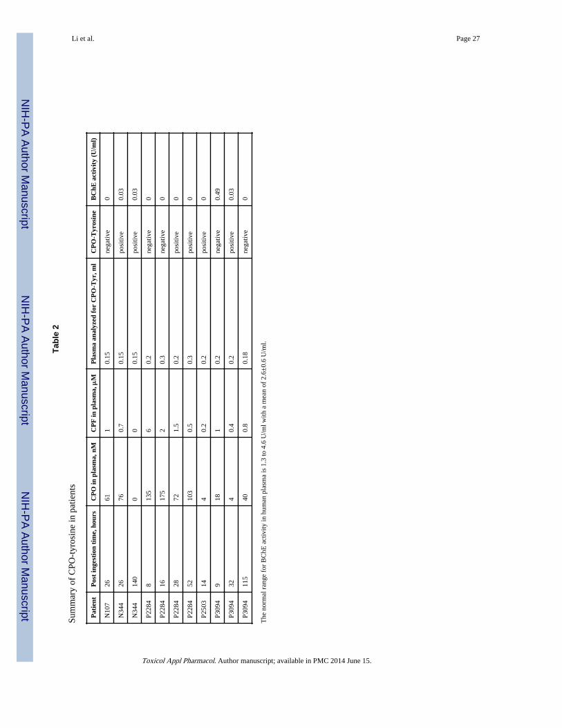
Table 2 summarizes patient results. Four of the 5 patients were positive for the presence ofCPO-tyrosine, but their positive status depended on the time elapsed between ingestion ofCPF and blood collection. Patient N344 was positive for plasma collected after 26 and 140hours post ingestion. Patient P2284 was negative for plasma collected 8 and 16 hours postingestion, but positive after 28 and 52 hours. Patient P2503 was positive for plasmacollected 14 hours post ingestion. Patient P3094 was negative at 9 hours, positive at 32hours, and negative at 115 hours post ingestion.
Plasma butyrylcholinesterase (BChE) activity was measured because BChE is readilyinhibited by CPO (Amitai et al., 1998), but poorly inhibited by CPF, the reaction of CPOwith BChE being orders of magnitude faster. Thus, inhibition of plasma BChE activityreflects the presence of CPO in the circulation, a fact confirmed by finding detectableamounts of CPO in plasma. Table 2 shows that BChE activity in all samples wasundetectable or very low (0.03–0.49 U/ml). Normal BChE activity in our laboratory rangesbetween 1.3 and 4.6 U/ml with a mean of 2.6±0.6 U/ml. The low BChE activity in patientplasma suggests that enough time had elapsed between ingestion of the poison and the blooddraw to allow a significant amount of the CPF to be converted to CPO. However the level ofBChE inhibition did not always correlate with the appearance of CPO-tyrosine. Forexample, patient N107 had undetectable BChE activity and 61 nM CPO in plasma at 26hours post ingestion but was negative for CPO-Tyr. The negative CPO-Tyr finding is notexplained by absence of CPO. On the other hand, the activity of 0.49 U/ml for patient P3094and the negative CPO-Tyr status at 9 hours post ingestion, could indicate a level of CPO (18nM) too low to give measurable amounts of tyrosine adduct at that time interval. A similarargument can be made for patient P2284 at 8 and 16 hours post ingestion. It follows that
Li et al. Page 7
Toxicol Appl Pharmacol. Author manuscript; available in PMC 2014 June 15.
NIH
-PA Author Manuscript
NIH
-PA Author Manuscript
NIH
-PA Author Manuscript

CPO-tyrosine is not as reliable a measure of exposure to CPF/CPO as is inhibition of BChEactivity or direct measurement of CPO in plasma.
Time course of CPF and CPO levels in patient plasmaBlood was drawn from poisoned patients at various time intervals following ingestion ofCPF. The blood samples were analyzed for the presence of intact CPF and CPO. Figure 10shows that both CPF and CPO were recovered from plasma. CPF concentrations werehigher than CPO concentrations at all time points (note that the scales are micromolar forCPF and nanomolar for CPO). The concentration of CPF declined with time, whereas theconcentration of CPO increased before declining, consistent with metabolic activation ofCPF to CPO. Peak CPO concentrations in plasma were achieved 3 h post ingestion in patientP2503, 15 to 50 h post ingestion in patients N107, N344, and P2284, and 80 h in patientP3094.
Samples for most of the time points in Figure 10 were not analyzed for adducts on tyrosine.A large portion of the patient plasma was consumed in assays to quantify CPF and CPO,leaving sufficient volume in only 11 samples for analysis of adducts on tyrosine. Six ofthese 11 samples were positive for CPO-tyrosine and 5 were negative. (See Table 2).Positive samples are marked with a filled triangle in Figure 10, while negative samples aremarked with an X. Five of the positive samples were taken 24 hours or longer after ingestionof CPF. Three of the negative samples were taken less than 24 hours after ingestion of CPF.This relative time dependence most likely reflects the time needed to convert a sufficientamount of CPF into CPO. This argument was validated for samples P3094 and P2284 wherethe CPO-tyrosine adduct was absent at early time points but present at time points after 24hours. Insufficient data exist for the other samples to address this issue.
The presence of CPO-tyrosine in a sample did not correlate well with the quantity of CPO inplasma. In three of the positive samples the CPO concentration was between 72 and 103 nMbut two samples contained only 4 nM CPO, and for one there was no measurable CPO, seeTable 2. The latter sample was taken 140 hours after ingestion and can be rationalized asloss of CPO from the circulation with retention of the protein containing the CPO-tyrosine.However, the samples containing only 4 nM CPO were taken at short times after ingestion(14 and 32 hours). The most extreme example of a positive reading was for patient P2503who was positive at 14 hours, even though this patient’s CPO plasma level was only 4 nM(Table 2 and Figure 10). Four of the five samples that proved to be negative for CPO-tyrosine had CPO concentrations of 40 to 175 nM. It is unclear why no tyrosine adduct wasdetected in these cases.
Since albumin is the most likely protein source of CPO-tyrosine, and since albumin has ahalf-life in the circulation of 20 days, it was expected that a positive sample would continueto be positive for several days. This expectation was fulfilled in patient N344 whose plasmawas positive at 26 hours and 140 hours. However, it was not fulfilled in patient P3094,whose plasma was positive at 32 hours, but negative at 115 hours.
Pralidoxime treatment does not reverse binding of CPO to tyrosinePralidoxime is routinely given to organophosphate-poisoned patients to reverse covalentbinding of toxicants to the active site serine of acetylcholinesterase. This treatment restoresacetylcholinesterase activity if the adducts have not aged, that is, if both alkyl groups areretained on the phosphorus atom. The adducts on tyrosine in the present report have notaged. The question arose whether pralidoxime would release the diethoxyphosphate adductfrom tyrosine. To address this question we tested plasma from patients treated and nottreated with pralidoxime.
Li et al. Page 8
Toxicol Appl Pharmacol. Author manuscript; available in PMC 2014 June 15.
NIH
-PA Author Manuscript
NIH
-PA Author Manuscript
NIH
-PA Author Manuscript

Patients N107 and N344 were treated with bolus injections of pralidoxime on admission tothe hospital, again after 12 h, and then daily for one week. Patient P3094 was continuouslyinfused with pralidoxime for 9 days. Patients P2284 and P2503 received no pralidoxime.Examination of Table 2 and Figure 10 reveals that CPO-tyrosine status was unrelated topralidoxime treatment. CPO-tyrosine was identified in pralidoxime treated patients N334and P3094 as well as in patients not treated with pralidoxime (P2284 and P2503). It isconcluded that pralidoxime does not reverse CPO binding to tyrosine in humans.
Nerve agent intoxicated guinea pigs treated with the oximes P2S, HI-6 or toxogonin andnerve agent intoxicated marmosets treated with HI-6 were found to retain adducts ontyrosine (Williams et al., 2007; Read et al., 2010), supporting the conclusion thatorganophosphorus adducts on tyrosine are not degraded by therapy with oximes.
DiscussionPronase digested human plasma from 5 patients who poisoned themselves by ingestingchlorpyrifos contained a tyrosine-diethylphosphate adduct. This demonstrates that tyrosineon one or more plasma proteins is irreversibly modified by the organophosphorus toxicant,chlorpyrifos oxon. The modification takes place in vivo in humans poisoned by chlorpyrifoswhere the chlorpyrifos oxon concentrations are in the nanomolar range. Since albumin is themost abundant protein in plasma, and since we have demonstrated that tyrosine 411 ofhuman albumin reacts with chlorpyrifos oxon, it can be surmised that albumin is a targetprotein for chlorpyrifos oxon in plasma. Though albumin is a highly likely target, otherproteins could also be modified. In vitro experiments have demonstrated that OP (CPO,dichlorvos, diisopropylfluorophosphate, FP-biotin, sarin, and soman) can covalently modifytyrosine on at least 12 proteins including transferrin, keratin, and tubulin (Schopfer et al.,2010).
Detection of organophosphorus adducts on protein tyrosine in the blood of humans is a newapproach to analyzing OP exposure. The classical approach focuses on inhibition ofacetylcholinesterase and butyrylcholinesterase activity, and on mass spectrometry of adductson the active site serine of these enzymes. The literature contains only two previouspublications on OP adducts on protein tyrosine in humans (Li et al., 2010d; van der Schanset al., 2012). The subjects had poisoned themselves with dichlorvos or chlorpyrifos (Li et al.,2010d; van der Schans et al., 2012). Two dichlorvos-poisoned subjects haddimethoxyphosphorylated albumin and monomethoxyphosphorylated butyrylcholinesterasein their plasma. The modification sites were identified by mass spectrometry as tyrosine 411in albumin and serine 198 in BChE (Li et al., 2010b; Li et al., 2010d). Mass spectrometryanalysis of plasma from one subject poisoned by chlorpyrifos identified two types of adductson Tyr 411 of albumin: O,O-diethyl phosphorothio and O,O-diethylphosphoro adducts, inother words diethoxythiophosphate and diethoxyphosphate adducts on Tyr 411 of albumin(van der Schans et al., 2012). The same subject was found to have the agedmonoethylphosphate adduct on serine 198 of butyrylcholinesterase (van der Schans et al.,2012).
Assessment of organophosphorus adducts on tyrosine has several advantages. 1) Adductscan be identified many days after exposure because albumin has a half-life of 20 days inhumans. 2) Adducts on tyrosine can be informative regarding the identity of theorganophosphorus poison, because adducts on tyrosine do not lose an alkyl group duringaging. For example, exposure to sarin can be discriminated from exposure to soman(Williams et al., 2007). 3) Adducts on tyrosine are not degraded by treatment with an oxime(Read et al., 2010).
Li et al. Page 9
Toxicol Appl Pharmacol. Author manuscript; available in PMC 2014 June 15.
NIH
-PA Author Manuscript
NIH
-PA Author Manuscript
NIH
-PA Author Manuscript

Individual variation in metabolic transformation of CPF to CPOPeak CPO concentrations in plasma were achieved 3 h post ingestion in patient P2503, 15 to50 h post ingestion in patients N107, N344, and P2284, and 80 h in patient P3094. Thesedifferences could be explained by individual genetic differences in the enzymes involved inmetabolic transformation of CPF into CPO and the subsequent degradation of CPO (Eyer etal., 2009). Pertinent enzymes include cytochromes P450, paraoxonase 1, glucuronidetransferase, and glutathione transferase (Choi et al., 2006).
Butyrylcholinesterase is a more sensitive biomarker of OP exposure than albuminButyrylcholinesterase is very sensitive to inhibition by OP. Humans exposed to low doses ofOP are expected to have detectable adducts on BChE, but not on albumin. This expectationcomes from studies comparing reaction rates of OP with purified BChE and purifiedalbumin. For example, the rate of reaction of soman with BChE is 500-fold faster than withalbumin (Li et al., 2008). Chlorpyrifos adducts on BChE were detected in blood samples thathad no detectable adducts on albumin (Li et al., 2010a; Li et al., 2010c). However, low doseOP exposure in humans has not yet been documented by mass spectrometry analysis ofadducts on BChE. To date only high dose exposures have been confirmed by massspectrometry of adducts on plasma BChE. The first high dose exposure identified the sarin-adduct on BChE in the blood of patients intoxicated by sarin released in the Tokyo subway(Fidder et al., 2002). A second study identified adducts on BChE in the blood of patientswho deliberately poisoned themselves with dichlorvos, chlorpyrifos, or the carbamateAldicarb (Li et al., 2010a). A third study identified adducts on BChE in patients poisoned bydiazinon or chlorpyrifos (van der Schans et al., 2012). The technology for measuring lowdose exposure is available, but has not yet been applied (Sporty et al., 2010). Though BChEis the most sensitive biomarker of OP exposure, the goal of the present work was todocument that proteins in addition to BChE and AChE are modified by OP exposure.
Low dose OP exposure detected in urineAnalysis of OP metabolites in urine, specific to a given OP, is one of the most commonlyused means to estimate OP exposure (Aprea et al., 2000; Lu et al., 2001; Hernandez et al.,2002; Kupfermann et al., 2004; Riches et al., 2005; Dulaurent et al., 2006; Mawhinney et al.,2007; Barr et al., 2011). Methods to detect and quantify metabolites of chlorpyrifos,parathion, malathion, diazinon, sarin, soman, VX, Russian VX, and cyclosarin have beendeveloped for urine samples. A disadvantage of analyzing urine samples is that metabolitesare rapidly excreted, more than 90% being excreted within 48–72 h (Riches et al., 2005).Another disadvantage is that metabolites in urine do not allow one to distinguish betweenexposure to intact OP and OP hydrolysis products formed in the environment before foodwas ingested. OP compounds are unstable in aqueous solution, degrading to inactive,nontoxic hydrolysis products within hours. Finding metabolites in urine does not prove thatthe subject was exposed to intact OP. However, finding adducts on proteins in blood doesprove exposure to live, intact OP.
SignificanceToxicologists have long recognized that proteins in addition to acetylcholinesterase andbutyrylcholinesterase are modified by organophosphorus compounds (Moser, 1995; Pope,1999; Richards et al., 2000). Toxic signs in animals depend on the identity of theorganophosphorus poison. For example, a low dose of fenthion decreases motor activity inrats 86%, but does not alter the tail-pinch response, whereas a low dose of parathion doesnot affect motor activity but does decrease the tail-pinch response (Moser, 1995).Furthermore, toxic signs do not correlate with degree of acetylcholinesterase inhibition. Ratswhose acetylcholinesterase activity is inhibited to the same degree by parathion and
Li et al. Page 10
Toxicol Appl Pharmacol. Author manuscript; available in PMC 2014 June 15.
NIH
-PA Author Manuscript
NIH
-PA Author Manuscript
NIH
-PA Author Manuscript

chlorpyrifos have more severe toxicity from parathion than from chlorpyrifos (Pope, 1999).Toxic symptoms in people are not always accompanied by acetylcholinesterase inhibition.Workers who manufacture quinalphos have significantly lower than normal scores formemory, learning ability, vigilance, and motor response, though their bloodacetylcholinesterase activity levels are normal (Srivastava et al., 2000). These observationshave led to the conclusion that organophosphorus compounds react with toxicologicallyrelevant proteins in addition to the cholinesterases. Our finding that organophosphoruspoisons covalently modify tyrosine in humans suggests that as-yet-unidentified,toxicologically-relevant proteins may also be modified on tyrosine, and that some of theeffects of organophosphate poisoning may be due to modification of such unknown proteins.
AcknowledgmentsThis work was supported by National Institutes of Health grant [P30CA036727] to the Eppley Cancer Center,directed by Kenneth Cowan; the Wellcome Trust grant [GR063560 to ME]; and Centers for Disease Control andPrevention contract [200-2012-M-53381 to OL]. ME is a Scottish Senior Clinical Research Fellow (Scottish ChiefScientist Office/Scottish Funding Council) and Lister Research Prize Fellow (Lister Institute for PreventativeMedicine). Mass spectra were obtained with the support of the Mass Spectrometry and Proteomics core facility atthe University of Nebraska Medical Center.
Abbreviations
AChE acetylcholinesterase
BChE butyrylcholinesterase
CPO chlorpyrifos oxon
CPF chlorpyrifos
HPLC high performance liquid chromatography
MALDI-TOF matrix assisted laser desorption ionization –time of flight
MS mass spectrum
MSMS tandem mass spectrum
OP organophosphorus compound
ReferencesAlbers JW, Garabrant DH, Schweitzer SJ, Garrison RP, Richardson RJ, Berent S. The effects of
occupational exposure to chlorpyrifos on the peripheral nervous system: a prospective cohort study.Occup Environ Med. 2004; 61:201–211. [PubMed: 14985514]
Amitai G, Moorad D, Adani R, Doctor BP. Inhibition of acetylcholinesterase and butyrylcholinesteraseby chlorpyrifos-oxon. Biochem Pharmacol. 1998; 56:293–299. [PubMed: 9744565]
Aprea C, Strambi M, Novelli MT, Lunghini L, Bozzi N. Biologic monitoring of exposure toorganophosphorus pesticides in 195 Italian children. Environ Health Perspect. 2000; 108:521–525.[PubMed: 10856025]
Barr DB, Wong LY, Bravo R, Weerasekera G, Odetokun M, Restrepo P, Kim DG, Fernandez C,Whitehead RD Jr, Perez J, Gallegos M, Williams BL, Needham LL. Urinary concentrations ofdialkylphosphate metabolites of organophosphorus pesticides: National Health and NutritionExamination Survey 1999–2004. Int J Environ Res Public Health. 2011; 8:3063–3098. [PubMed:21909292]
Biberoglu K, Schopfer LM, Saxena A, Tacal O, Lockridge O. Polyproline tetramer organizing peptidesin fetal bovine serum acetylcholinesterase. Biochim Biophys Acta. 2013; 1834:745–753. [PubMed:23352838]
Li et al. Page 11
Toxicol Appl Pharmacol. Author manuscript; available in PMC 2014 June 15.
NIH
-PA Author Manuscript
NIH
-PA Author Manuscript
NIH
-PA Author Manuscript

Buratti FM, Volpe MT, Meneguz A, Vittozzi L, Testai E. CYP-specific bioactivation of fourorganophosphorothioate pesticides by human liver microsomes. Toxicol Appl Pharmacol. 2003;186:143–154. [PubMed: 12620367]
Choi K, Joo H, Rose RL, Hodgson E. Metabolism of chlorpyrifos and chlorpyrifos oxon by humanhepatocytes. J Biochem Mol Toxicol. 2006; 20:279–291. [PubMed: 17163483]
Ding SJ, Carr J, Carlson JE, Tong L, Xue W, Li Y, Schopfer LM, Li B, Nachon F, Asojo O,Thompson CM, Hinrichs SH, Masson P, Lockridge O. Five tyrosines and two serines in humanalbumin are labeled by the organophosphorus agent FP-biotin. Chem Res Toxicol. 2008; 21:1787–1794. [PubMed: 18707141]
Du D, Wang J, Wang L, Lu D, Smith JN, Timchalk C, Lin Y. Magnetic electrochemical sensingplatform for biomonitoring of exposure to organophosphorus pesticides and nerve agents based onsimultaneous measurement of total enzyme amount and enzyme activity. Anal Chem. 2011;83:3770–3777. [PubMed: 21462919]
Duirk SE, Collette TW. Degradation of chlorpyrifos in aqueous chlorine solutions: pathways, kinetics,and modeling. Environ Sci Technol. 2006; 40:546–551. [PubMed: 16468401]
Dulaurent S, Saint-Marcoux F, Marquet P, Lachatre G. Simultaneous determination of sixdialkylphosphates in urine by liquid chromatography tandem mass spectrometry. J Chromatogr BAnalyt Technol Biomed Life Sci. 2006; 831:223–229.
Eddleston M, Eyer P, Worek F, Mohamed F, Senarathna L, von Meyer L, Juszczak E, Hittarage A,Azhar S, Dissanayake W, Sheriff MH, Szinicz L, Dawson AH, Buckley NA. Differences betweenorganophosphorus insecticides in human self-poisoning: a prospective cohort study. Lancet. 2005;366:1452–1459. [PubMed: 16243090]
Ellman GL, Courtney KD, Andres V Jr, Feather-Stone RM. A new and rapid colorimetricdetermination of acetylcholinesterase activity. Biochem Pharmacol. 1961; 7:88–95. [PubMed:13726518]
Eyer F, Roberts DM, Buckley NA, Eddleston M, Thiermann H, Worek F, Eyer P. Extreme variabilityin the formation of chlorpyrifos oxon (CPO) in patients poisoned by chlorpyrifos (CPF). BiochemPharmacol. 2009; 78:531–537. [PubMed: 19433070]
Fidder A, Hulst AG, Noort D, de Ruiter R, van der Schans MJ, Benschop HP, Langenberg JP.Retrospective detection of exposure to organophosphorus anti-cholinesterases: mass spectrometricanalysis of phosphylated human butyrylcholinesterase. Chem Res Toxicol. 2002; 15:582–590.[PubMed: 11952345]
Foxenberg RJ, McGarrigle BP, Knaak JB, Kostyniak PJ, Olson JR. Human hepatic cytochrome p450-specific metabolism of parathion and chlorpyrifos. Drug Metab Dispos. 2007; 35:189–193.[PubMed: 17079358]
Heilmair R, Eyer F, Eyer P. Enzyme-based assay for quantification of chlorpyrifos oxon in humanplasma. Toxicol Lett. 2008; 181:19–24. [PubMed: 18655824]
Hernandez F, Sancho JV, Pozo OJ. Direct determination of alkyl phosphates in human urine by liquidchromatography/electrospray tandem mass spectrometry. Rapid Commun Mass Spectrom. 2002;16:1766–1773. [PubMed: 12207365]
Jiang W, Duysen EG, Hansen H, Shlyakhtenko L, Schopfer LM, Lockridge O. Mice treated withchlorpyrifos or chlorpyrifos oxon have organophosphorylated tubulin in brain and disruptedmicrotubule structures, suggesting a role for tubulin in neurotoxicity associated with exposure toorganophosphorus agents. Tox Sci. 2010; 115:183–193.
Kupfermann N, Schmoldt A, Steinhart H. Rapid and sensitive quantitative analysis of alkyl phosphatesin urine after organophosphate poisoning. J Anal Toxicol. 2004; 28:242–248. [PubMed:15189674]
Li B, Nachon F, Froment MT, Verdier L, Debouzy JC, Brasme B, Gillon E, Schopfer LM, LockridgeO, Masson P. Binding and hydrolysis of soman by human serum albumin. Chem Res Toxicol.2008; 21:421–431. [PubMed: 18163544]
Li B, Ricordel I, Schopfer LM, Baud F, Megarbane B, Masson P, Lockridge O. Dichlorvos,chlorpyrifos oxon and Aldicarb adducts of butyrylcholinesterase, detected by mass spectrometry inhuman plasma following deliberate overdose. J Appl Toxicol. 2010a; 30:559–565. [PubMed:20809544]
Li et al. Page 12
Toxicol Appl Pharmacol. Author manuscript; available in PMC 2014 June 15.
NIH
-PA Author Manuscript
NIH
-PA Author Manuscript
NIH
-PA Author Manuscript

Li B, Ricordel I, Schopfer LM, Baud F, Megarbane B, Masson P, Lockridge O. Dichlorvos,chlorpyrifos oxon, and aldicarb adducts of butyrylcholinesterase detected by mass spectrometry inhuman plasma following deliberate overdose. J Appl Toxicol. 2010b; 30:559–565. [PubMed:20809544]
Li B, Ricordel I, Schopfer LM, Baud F, Megarbane B, Nachon F, Masson P, Lockridge O. Detectionof adduct on tyrosine 411 of albumin in humans poisoned by dichlorvos. Toxicol Sci. 2010c;116:23–31. [PubMed: 20395308]
Li B, Ricordel I, Schopfer LM, Baud F, Megarbane B, Nachon F, Masson P, Lockridge O. Detectionof adducts on tyrosine 411 of albumin in humans poisoned by dichlorvos. Toxicol Sci. 2010d;116:23–31. [PubMed: 20395308]
Li B, Schopfer LM, Hinrichs SH, Masson P, Lockridge O. Matrix-assisted laser desorption/ionizationtime-of-flight mass spectrometry assay for organophosphorus toxicants bound to human albuminat Tyr411. Anal Biochem. 2007; 361:263–272. [PubMed: 17188226]
Liyasova MS, Schopfer LM, Lockridge O. Cresyl saligenin phosphate, an organophosphorus toxicant,makes covalent adducts with histidine, lysine, and tyrosine residues of human serum albumin.Chem Res Toxicol. 2012; 25:1752–1761. [PubMed: 22793878]
Lu C, Knutson DE, Fisker-Andersen J, Fenske RA. Biological monitoring survey of organophosphoruspesticide exposure among pre-school children in the Seattle metropolitan area. Environ HealthPerspect. 2001; 109:299–303. [PubMed: 11333193]
Mawhinney DB, Hamelin EI, Fraser R, Silva SS, Pavlopoulos AJ, Kobelski RJ. The determination oforganophosphonate nerve agent metabolites in human urine by hydrophilic interaction liquidchromatography tandem mass spectrometry. J Chromatogr B Analyt Technol Biomed Life Sci.2007; 852:235–243.
Means GE, Wu HL. The reactive tyrosine residue of human serum albumin: characterization of itsreaction with diisopropylfluorophosphate. Arch Biochem Biophys. 1979; 194:526–530. [PubMed:443818]
Moser VC. Comparisons of the acute effects of cholinesterase inhibitors using a neurobehavioralscreening battery in rats. Neurotoxicol Teratol. 1995; 17:617–625. [PubMed: 8747743]
Noort D, Hulst AG, van Zuylen A, van Rijssel E, van der Schans MJ. Covalent binding oforganophosphorothioates to albumin: a new perspective for OP-pesticide biomonitoring? ArchToxicol. 2009; 83:1031–1036. [PubMed: 19575182]
Perkins DN, Pappin DJ, Creasy DM, Cottrell JS. Probability-based protein identification by searchingsequence databases using mass spectrometry data. Electrophoresis. 1999; 20:3551–3567.[PubMed: 10612281]
Pope CN. Organophosphorus pesticides: do they all have the same mechanism of toxicity? J ToxicolEnviron Health B Crit Rev. 1999; 2:161–181. [PubMed: 10230392]
Read RW, Riches JR, Stevens JA, Stubbs SJ, Black RM. Biomarkers of organophosphorus nerve agentexposure: comparison of phosphylated butyrylcholinesterase and phosphylated albumin afteroxime therapy. Arch Toxicol. 2010; 84:25–36. [PubMed: 19862504]
Richards PG, Johnson MK, Ray DE. Identification of acylpeptide hydrolase as a sensitive site forreaction with organophosphorus compounds and a potential target for cognitive enhancing drugs.Mol Pharmacol. 2000; 58:577–583. [PubMed: 10953051]
Riches J, Morton I, Read RW, Black RM. The trace analysis of alkyl alkylphosphonic acids in urineusing gas chromatography-ion trap negative ion tandem mass spectrometry. J Chromatogr BAnalyt Technol Biomed Life Sci. 2005; 816:251–258.
Sams C, Mason HJ, Rawbone R. Evidence for the activation of organophosphate pesticides bycytochromes P450 3A4 and 2D6 in human liver microsomes. Toxicol Lett. 2000; 116:217–221.[PubMed: 10996483]
Schopfer LM, Grigoryan H, Li B, Nachon F, Masson P, Lockridge O. Mass spectral characterizationof organophosphate-labeled, tyrosine-containing peptides: characteristic mass fragments and a newbinding motif for organophosphates. J Chromatogr B Analyt Technol Biomed Life Sci. 2010;878:1297–1311.
Sporty JL, Lemire SW, Jakubowski EM, Renner JA, Evans RA, Williams RF, Schmidt JG, van derSchans MJ, Noort D, Johnson RC. Immunomagnetic separation and quantification of
Li et al. Page 13
Toxicol Appl Pharmacol. Author manuscript; available in PMC 2014 June 15.
NIH
-PA Author Manuscript
NIH
-PA Author Manuscript
NIH
-PA Author Manuscript

butyrylcholinesterase nerve agent adducts in human serum. Anal Chem. 2010; 82:6593–6600.[PubMed: 20617824]
Srivastava AK, Gupta BN, Bihari V, Mathur N, Srivastava LP, Pangtey BS, Bharti RS, Kumar P.Clinical, biochemical and neurobehavioural studies of workers engaged in the manufacture ofquinalphos. Food Chem Toxicol. 2000; 38:65–69. [PubMed: 10685015]
van der Schans MJ, Hulst AG, van der Riet-van Oeveren D, Noort D, Benschop HP, Dishovsky C.New tools in diagnosis and biomonitoring of intoxications with organophosphorothioates: Casestudies with chlorpyrifos and diazinon. Chem Biol Interact. 2012
Williams NH, Harrison JM, Read RW, Black RM. Phosphylated tyrosine in albumin as a biomarker ofexposure to organophosphorus nerve agents. Arch Toxicol. 2007; 81:627–639. [PubMed:17345062]
Li et al. Page 14
Toxicol Appl Pharmacol. Author manuscript; available in PMC 2014 June 15.
NIH
-PA Author Manuscript
NIH
-PA Author Manuscript
NIH
-PA Author Manuscript

Highlights
• Chlorpyrifos-poisoned patients have adducts on protein tyrosine
• Diethoxyphosphate-tyrosine does not lose an alkyl group
• Proteins in addition to AChE and BChE are modified by organophosphates
Li et al. Page 15
Toxicol Appl Pharmacol. Author manuscript; available in PMC 2014 June 15.
NIH
-PA Author Manuscript
NIH
-PA Author Manuscript
NIH
-PA Author Manuscript

Figure 1.Chlorpyrifos is metabolically activated to chlorpyrifos oxon by cytochrome P450 enzymes.Chlorpyrifos and chlorpyrifos oxon react covalently with Tyr 411 of albumin to makeadducts with added masses of 152 Da and 136 Da, respectively. The reaction withchlorpyrifos is much slower than that with chlorpyrifos oxon. Adducts on albumin arestable. They do not spontaneously dissociate nor do they lose an alkyl group from theorganophosphorus moiety. Loss of an alkyl group is commonly observed withorganophosphorylated cholinesterases and is called aging.
Li et al. Page 16
Toxicol Appl Pharmacol. Author manuscript; available in PMC 2014 June 15.
NIH
-PA Author Manuscript
NIH
-PA Author Manuscript
NIH
-PA Author Manuscript

Fig. 2.MALDI-TOF mass spectra of diethoxyphosphorylated-tyrosine isolated from CPO treatedhuman albumin and CPO treated human plasma. CPO treated albumin (panel A), controlhuman plasma (panel B), and CPO treated human plasma (panel C) were digested withpronase to release individual amino acids. The digest components were separated by HPLCand analyzed by mass spectrometry using 2,5-dihydroxybenzoic acid matrix.
Li et al. Page 17
Toxicol Appl Pharmacol. Author manuscript; available in PMC 2014 June 15.
NIH
-PA Author Manuscript
NIH
-PA Author Manuscript
NIH
-PA Author Manuscript

Figure 3.MALDI-TOF mass spectra of diethoxythiophosphorylated-tyrosine isolated from CPFtreated human albumin and CPF treated human plasma. CPF treated albumin (panel A),control human plasma (panel B), and CPF treated human plasma (panel C) were digestedwith pronase to release individual amino acids. The digest components were separated byHPLC and analyzed by mass spectrometry using 2,5-dihydroxybenzoic acid matrix.
Li et al. Page 18
Toxicol Appl Pharmacol. Author manuscript; available in PMC 2014 June 15.
NIH
-PA Author Manuscript
NIH
-PA Author Manuscript
NIH
-PA Author Manuscript

Figure 4.LC-MSMS analysis of CPO-tyrosine and CPF-tyrosine. Panel A) Fragmentation of parention 318 m/z, corresponding to CPO-tyrosine, causes loss of 46 Da and further loss of 28 Da.Panel B) Fragmentation of parent ion 334.1 m/z, corresponding to CPF-tyrosine, causes lossof 46 Da and further loss of 28 Da. The samples were prepared from CPO- and CPF-treatedalbumin.
Li et al. Page 19
Toxicol Appl Pharmacol. Author manuscript; available in PMC 2014 June 15.
NIH
-PA Author Manuscript
NIH
-PA Author Manuscript
NIH
-PA Author Manuscript

Figure 5.MALDI-TOF mass spectra of pepsin digested CPO-albumin and CPF-albumin prepared invitro. The digests were diluted and analyzed using α-cyano-4-hydroxycinnamic acid matrix.The accession number for human albumin is gi:122920512 in the NCBI database.
Li et al. Page 20
Toxicol Appl Pharmacol. Author manuscript; available in PMC 2014 June 15.
NIH
-PA Author Manuscript
NIH
-PA Author Manuscript
NIH
-PA Author Manuscript

Figure 6.QTrap MSMS spectra of peptic peptides of human albumin. Panel A) control humanalbumin. Panel B) CPO-albumin prepared in vitro. Tyrosine 411 is the modified residue.
Li et al. Page 21
Toxicol Appl Pharmacol. Author manuscript; available in PMC 2014 June 15.
NIH
-PA Author Manuscript
NIH
-PA Author Manuscript
NIH
-PA Author Manuscript

Figure 7.Triple-TOF 5600 spectra of peptic peptides of human albumin. Panel A) control humanalbumin. Panel B) CPF-albumin prepared in vitro. Tyrosine 411 is the modified residue.
Li et al. Page 22
Toxicol Appl Pharmacol. Author manuscript; available in PMC 2014 June 15.
NIH
-PA Author Manuscript
NIH
-PA Author Manuscript
NIH
-PA Author Manuscript

Fig. 8.MADLI-TOF mass spectrum identifying diethoxyphosphorylated-tyrosine in plasma from achlorpyrifos-poisoned patient (P2284, 52 hour time point). Plasma (200 μl) was digestedwith pronase and the digestion mixture separated by HPLC, before mass spectral analysisusing 2,5-dihydroxybenzoic acid matrix. The 318.08 Da peak is consistent with CPO labeledTyr. The 340.07 and 356.06 Da peaks correspond to the sodium and potassium adducts ofCPO labeled tyrosine.
Li et al. Page 23
Toxicol Appl Pharmacol. Author manuscript; available in PMC 2014 June 15.
NIH
-PA Author Manuscript
NIH
-PA Author Manuscript
NIH
-PA Author Manuscript

Fig. 9.LTQ-Orbitrap MSMS spectrum of CPO labeled tyrosine from the plasma of a chlorpyrifospoisoned patient (N344, 26 hour time point). The patient sample has characteristic ions at272 and 255 Da from amino acid fragmentation, and characteristic ions at 290 and 244 Dafor fragmentation of diethoxyphosphorylated-tyrosine. The ion at 301.2 Da has lost NH3from the parent ion at 318.2 m/z. The parent ion eluted from the Picofrit BioBasics C18column at 46 minutes.
Li et al. Page 24
Toxicol Appl Pharmacol. Author manuscript; available in PMC 2014 June 15.
NIH
-PA Author Manuscript
NIH
-PA Author Manuscript
NIH
-PA Author Manuscript

Figure 10.Time course of CPF and CPO in patient plasma. Plasma was withdrawn from 5 chlorpyrifos-poisoned patients at the indicated times after ingestion of chlorpyrifos. Times ranged from 1to 222 hours. CPF and CPO were extracted from plasma and quantified. The scale for CPOis nanomolar, while that for CPF is micromolar. The area under the curve (AUC) is for CPOin nM x h. CPO-tyrosine adducts were not quantified but are simply indicated as present orabsent. Samples that were positive for CPO-tyrosine are indicated by a filled triangle ▲.Samples that were negative for CPO-tyrosine are indicated by X.
Li et al. Page 25
Toxicol Appl Pharmacol. Author manuscript; available in PMC 2014 June 15.
NIH
-PA Author Manuscript
NIH
-PA Author Manuscript
NIH
-PA Author Manuscript

NIH
-PA Author Manuscript
NIH
-PA Author Manuscript
NIH
-PA Author Manuscript
Li et al. Page 26
Table 1
Parent ion masses of CPO and CPF adducts on tyrosine
Adduct Mass + H+ Mass + Na+ Mass + K+ Figure number*
CPO-tyrosine 318.0 340.0 356.0 2A, 2C
CPF-tyrosine 334.1 356.0 372.0 3A, 3C
*Figures in the manuscript that show these adducts in albumin and plasma samples treated with CPO or CPF in vitro.
Toxicol Appl Pharmacol. Author manuscript; available in PMC 2014 June 15.
NIH
-PA Author Manuscript
NIH
-PA Author Manuscript
NIH
-PA Author Manuscript
Li et al. Page 27
Tabl
e 2
Sum
mar
y of
CPO
-tyr
osin
e in
pat
ient
s
Pat
ient
Pos
t in
gest
ion
tim
e, h
ours
CP
O in
pla
sma,
nM
CP
F in
pla
sma,
μM
Pla
sma
anal
yzed
for
CP
O-T
yr, m
lC
PO
-Tyr
osin
eB
ChE
act
ivit
y (U
/ml)
N10
726
611
0.15
nega
tive
0
N34
426
760.
70.
15po
sitiv
e0.
03
N34
414
00
00.
15po
sitiv
e0.
03
P228
48
135
60.
2ne
gativ
e0
P228
416
175
20.
3ne
gativ
e0
P228
428
721.
50.
2po
sitiv
e0
P228
452
103
0.5
0.3
posi
tive
0
P250
314
40.
20.
2po
sitiv
e0
P309
49
181
0.2
nega
tive
0.49
P309
432
40.
40.
2po
sitiv
e0.
03
P309
411
540
0.8
0.18
nega
tive
0
The
nor
mal
ran
ge f
or B
ChE
act
ivity
in h
uman
pla
sma
is 1
.3 to
4.6
U/m
l with
a m
ean
of 2
.6±
0.6
U/m
l.
Toxicol Appl Pharmacol. Author manuscript; available in PMC 2014 June 15.