Edinburgh Research ExplorerARTICLE Single human B cell-derived monoclonal anti-Candida antibodies...
17
Edinburgh Research Explorer Single human B cell-derived monoclonal anti- Candida antibodies enhance phagocytosis and protect against disseminated candidiasis Citation for published version: Rudkin, F, Raziunaite, I, Workman, H, Essono, S, Belmonte, R, MacCallum, DM, Johnson, EM, Silva, LM, Palma, AS, Feizi, T, Jensen, A, Erwig, LP & Gow, NAR 2018, 'Single human B cell-derived monoclonal anti- Candida antibodies enhance phagocytosis and protect against disseminated candidiasis', Nature Communications, vol. 9, no. 1, 5288 (2018). https://doi.org/10.1038/s41467-018-07738-1 Digital Object Identifier (DOI): 10.1038/s41467-018-07738-1 Link: Link to publication record in Edinburgh Research Explorer Document Version: Publisher's PDF, also known as Version of record Published In: Nature Communications General rights Copyright for the publications made accessible via the Edinburgh Research Explorer is retained by the author(s) and / or other copyright owners and it is a condition of accessing these publications that users recognise and abide by the legal requirements associated with these rights. Take down policy The University of Edinburgh has made every reasonable effort to ensure that Edinburgh Research Explorer content complies with UK legislation. If you believe that the public display of this file breaches copyright please contact [email protected] providing details, and we will remove access to the work immediately and investigate your claim. Download date: 01. Mar. 2020
Transcript of Edinburgh Research ExplorerARTICLE Single human B cell-derived monoclonal anti-Candida antibodies...

Edinburgh Research Explorer
Single human B cell-derived monoclonal anti- Candidaantibodies enhance phagocytosis and protect againstdisseminated candidiasis
Citation for published version:Rudkin, F, Raziunaite, I, Workman, H, Essono, S, Belmonte, R, MacCallum, DM, Johnson, EM, Silva, LM,Palma, AS, Feizi, T, Jensen, A, Erwig, LP & Gow, NAR 2018, 'Single human B cell-derived monoclonal anti-Candida antibodies enhance phagocytosis and protect against disseminated candidiasis', NatureCommunications, vol. 9, no. 1, 5288 (2018). https://doi.org/10.1038/s41467-018-07738-1
Digital Object Identifier (DOI):10.1038/s41467-018-07738-1
Link:Link to publication record in Edinburgh Research Explorer
Document Version:Publisher's PDF, also known as Version of record
Published In:Nature Communications
General rightsCopyright for the publications made accessible via the Edinburgh Research Explorer is retained by the author(s)and / or other copyright owners and it is a condition of accessing these publications that users recognise andabide by the legal requirements associated with these rights.
Take down policyThe University of Edinburgh has made every reasonable effort to ensure that Edinburgh Research Explorercontent complies with UK legislation. If you believe that the public display of this file breaches copyright pleasecontact [email protected] providing details, and we will remove access to the work immediately andinvestigate your claim.
Download date: 01. Mar. 2020

ARTICLE
Single human B cell-derived monoclonal anti-Candida antibodies enhance phagocytosis andprotect against disseminated candidiasisFiona M. Rudkin1, Ingrida Raziunaite2, Hillary Workman3, Sosthene Essono4, Rodrigo Belmonte5,
Donna M. MacCallum 1, Elizabeth M. Johnson6, Lisete M. Silva7, Angelina S. Palma8, Ten Feizi7, Allan Jensen9,
Lars P. Erwig10 & Neil A.R. Gow11
The high global burden of over one million annual lethal fungal infections reflects a lack of
protective vaccines, late diagnosis and inadequate chemotherapy. Here, we have generated a
unique set of fully human anti-Candida monoclonal antibodies (mAbs) with diagnostic and
therapeutic potential by expressing recombinant antibodies from genes cloned from the B
cells of patients suffering from candidiasis. Single class switched memory B cells isolated
from donors serum-positive for anti-Candida IgG were differentiated in vitro and screened
against recombinant Candida albicans Hyr1 cell wall protein and whole fungal cell wall pre-
parations. Antibody genes from Candida-reactive B cell cultures were cloned and expressed in
Expi293F human embryonic kidney cells to generate a panel of human recombinant anti-
Candida mAbs that demonstrate morphology-specific, high avidity binding to the cell wall.
The species-specific and pan-Candida mAbs generated through this technology display
favourable properties for diagnostics, strong opsono-phagocytic activity of macrophages
in vitro, and protection in a murine model of disseminated candidiasis.
https://doi.org/10.1038/s41467-018-07738-1 OPEN
1Medical Research Council Centre for Medical Mycology at the University of Aberdeen, Aberdeen Fungal Group, Institute of Medical Sciences, Foresterhill,Aberdeen AB25 2ZD, UK. 2 Division of Infection and Immunity, The Roslin Institute and Royal (Dick) School of Veterinary Studies, University of Edinburgh,Edinburgh EH25 9RG, UK. 3 Global Biotherapeutic Technologies, Pfizer Inc., 610 Main Street, Kendall Square, Cambridge, MA 02139, USA. 4HiFiBiO, 325Vassar Street, Cambridge, MA 02139, USA. 5MSD Animal Health Innovation AS, Thormøhlensgate 55, N-5006 Bergen, Norway. 6 National Infection Service,PHE South West Laboratory, Science Quarter, Southmead Hospital, Bristol BS10 5NB, UK. 7 Glycosciences Laboratory, Department of Medicine, ImperialCollege London, Du Cane Road, London W12 0NN, UK. 8 UCIBIO-REQUIMTE, Department of Chemistry, Faculty of Science and Technology, NOVAUniversity of Lisbon, Lisbon 1099-085, Portugal. 9 H. Lundbeck, Ottiliavej 9, 2500 Valby, Denmark. 10 Galvani Bioelectronics, 980 Great West Road,Brentford TW8 9GS, UK. 11 School of Biosciences, University of Exeter, Geoffrey Pope Building, Exeter EX4 4QD, UK. Correspondence and requests formaterials should be addressed to N.A.R.G. (email: [email protected])
NATURE COMMUNICATIONS | (2018) 9:5288 | https://doi.org/10.1038/s41467-018-07738-1 | www.nature.com/naturecommunications 1
1234
5678
90():,;

Fungi cause approximately 1.5 million lethal infections eachyear—as many as tuberculosis or HIV, and more thanmalaria or breast or prostate cancer1. Of these fungal dis-
eases, Candida species collectively account for the majority ofserious fungal infections and represent the fourth leading cause ofhealthcare-associated infections in the United States1,2. Candidaalbicans is the most commonly isolated species and represents themost prevalent fungal opportunistic pathogen worldwide3.Impairment of host immunity, due to trauma, pharmacological orsurgical intervention, or alteration in the natural microbiota,determines the frequency and severity of disease4. Late diagnosisof invasive candidiasis using ‘gold standard’ blood culturemethodologies along with limitations in the versatility, accuracyand widespread availability of inexpensive and rapid diagnostictests contribute to the poor prognosis and high mortality ratesassociated with septicaemia and invasive fungal disease5–7. Tomake inroads into these high disease burdens and mortality fig-ures, better diagnostics, antifungal drugs, immunotherapies andfungal vaccines are urgently required.
Pooled immunoglobulin from serum was one of the first widelyavailable treatments for microbial infections. For example,hyperimmune human serum immunoglobulin has been used totreat a number of infections including cytomegalovirus, hepatitisA and B virus rabies and measles8–10. In recent years, monoclonalantibodies (mAbs) have become some of the world’s bestsellingdrugs, with global sales forecast to reach approximately $125billion by 202011. To date, the majority of these mAbs have beenlicensed for the treatment of cancer and autoimmunediseases12,13, but the revolution in applied mAb research has yetto be focussed on mycotic infections. There is currently only onemAb approved for the treatment of an infectious disease (Synagis;respiratory syncytial virus)14. Advances have been made in recentyears to generate mAbs to viral and bacterial targets andantibody–antibiotic conjugates have also been explored as noveltherapeutics against intracellular bacterial pathogens15–18. Pro-tective mAbs for clinically relevant fungi have now been reportedbut these are almost exclusively murine in origin, and generatedvia hybridoma technology10,19–24. Fully human antibodies wouldrepresent highly valuable reagents to explore future immu-notherapies targeting medical mycoses.
Increased mAb research in the field of mycotic disease has alsoled to progress in mAb-based diagnostics including the Asper-gillus-specific mAb JF5 for the detection of invasive pulmonaryaspergillosis, a Candida albicans germ tube mAb (CAGTA) fordeep-seated Candida infection and a new cryptococcal antigendipstick test25–27. Assays detecting the pan-fungal marker β-glucan have been a valuable addition to the armamentarium, butfor Candida-specific diagnosis, the application of MALDI-TOFMS (matrix-assisted laser desorption ionisation–time-of-flightmass spectrometry) and the introduction of the T2Candida paneltest utilising miniaturised magnetic resonance technology toidentify clinically relevant species of Candida have beenimportant28,29. However, inexpensive, sensitive and specificpoint-of-care diagnostics that can accurately detect the majorhuman fungal pathogens are urgently required to inform ther-apeutic strategies.
There are currently no vaccines for the prevention of fungalinfection in the clinic, although experimental vaccines based onfungal cell wall targets are in development30–32. NDV-3, a vaccinebased on a recombinant fragment of the Als3 cell wall adhesin,has now completed Phase II clinical trials where it demonstratedsafety and a reduction in the frequency of symptomatic episodesin women suffering from recurrent vulvovaginal candidiasis33–36.This vaccine also demonstrates cross-kingdom protection againstStaphylococcus aureus due to structural homology of Als3 withsurface adhesin/invasin molecules of S. aureus37. A pre-clinical
Candida-specific vaccine based on the recombinant N-terminalfragment of C. albicans Hyr1 protein demonstrated efficacy in amurine model of disseminated candidiasis, and more recentlycross-kingdom protection against the bacterial pathogen Acine-tobacter baumannii through structural homology to cell A. bau-mannii surface proteins38–40. These experimental vaccines arebased on neutralising and/or protective antibodies that may bedeployed in prophylactic or pre-emptive therapies.
Methods and approaches for the production of mAbs fordiagnostic and/or therapeutic use have diversified dramatically inrecent years. Early mAbs were mainly of murine origin but wereimmunogenic in the human host41,42. Today, the majority ofmAbs used clinically are chimeric, humanised or fully humanIgG1 mAbs generated through hybridoma cell lines12. Combi-natorial display technologies using phage or yeast have also beenvaluable in generating fully human mAbs43,44 but these oftenrequire a period of in vitro affinity maturation and produce mAbswith randomised heavy and light chain pairings. Recently,retention of native VH and VL pairings through direct amplifi-cation of individual VH and VL chain domain genes from in vitroexpanded single human B cells has led to the generation of fullyhuman mAbs with increased safety, immunotolerance, efficacyand relevance to human disease in areas where current treatmentsare suboptimal45–48.
In the present study we address the urgent requirement forimproved diagnostics, treatments and vaccines for fungal infec-tion, by generating bespoke recombinant human antibodies fromsingle human B cells. Human antibody encoding variable domain(V) genes targeting Candida epitopes were cloned from single Bcells derived from donors who had recovered from mucosalCandida infections to generate a panel of fully human recombi-nant IgG1 mAbs that display a range of specific binding profilesto pathogenic Candida species. The mAbs demonstrate opsono-phagocytic activity in vitro against C. albicans and the emergingmultidrug-resistant pathogen Candida auris, and therapeuticefficacy in vivo in a murine model of disseminated candidiasis.They are also effective in a number of diagnostic formats inrecognising Candida antigens. This highlights the translationalvalue of this technology for developing fungal diagnostics andtherapeutics, and exploring anti-Candida vaccine development.
ResultsFully human recombinant anti-Candida mAbs derived fromsingle B cells. Fully human recombinant mAbs were generated bydirect amplification of VH and VL genes from single B cells inwhich the native antibody heavy and light chain pairing remainintact, thus preserving the original target specificity and diseaserelevance45 (Fig. 1). Through screening of in vitro activated classswitch memory (CSM) B-cell repertoires, mAbs were isolated thatrecognised a recombinant C. albicans-specific protein antigen—the hyphal cell wall protein Hyr149, or C. albicans whole cell wallpreparations. Hyr1 was selected following pre-clinical data that arecombinant N-terminal Hyr1 fragment conferred protection in amurine model of disseminated candidiasis38,39. Whole cell wallpreparations were used to generate mAbs to a variety of antigenswith a range of reactivities and clinical uses.
To enhance the likelihood of isolating Candida-specificantibodies, CSM B cells were isolated from the blood ofindividuals who had recovered from a superficial Candidainfection (mostly vaginitis) within a year of sampling. Donorswere selected from a panel of volunteers and the levels of target-specific circulating IgG in the donor plasma was assessed viaenzyme-linked immunosorbent assay (ELISA). In the screen, thedonors displaying the greatest IgG activity against Hyr1 (donor23) and C. albicans whole cells (donor 85) (Fig. 2a–c) were used
ARTICLE NATURE COMMUNICATIONS | https://doi.org/10.1038/s41467-018-07738-1
2 NATURE COMMUNICATIONS | (2018) 9:5288 | https://doi.org/10.1038/s41467-018-07738-1 | www.nature.com/naturecommunications

to provide the source of B cells for the generation of mAbs.Approximately 80,000–150,000 CSM B cells were plated out at ≤5cells/well and activated with a cocktail of cytokines andsupplements to promote differentiation to plasmablast or plasmacells associated with secretion of IgG into the culturesupernatant47,50–52. IgG was detected by ELISA-based high-throughput screening of culture supernatants against targetantigens. Typically, 0.05% wells per screen were positive (OD>4× background). Non-specific hits were identified and elimi-nated by ELISA screening against two unrelated proteins—human serum albumin (HSA) and human embryonic kidney cell(HEK) nuclear antigen. Antigen-positive activated CSM B cellswere lysed and used for VH, Vκ-Cκ and Vλ-Cλ geneamplification via reverse transcription polymerase chain reaction(RT-PCR) and nested PCR (representative images shown inSupplementary Figures 3a, b). VH, Vκ-Cκ and Vλ-Cλ genes weresubcloned into a mammalian expression vector and correspond-ing heavy and light chains originating from the same hit well wereco-transfected into Expi293F cells for small-scale recombinantwhole IgG1 expression. Recombinant mAbs that demonstratedbinding to the original target were selected for large-scaleexpression and then purified via affinity-based fast protein liquidchromatography (FPLC) using a protein A resin before qualitycontrol (QC) checking via analytical mass spectrometry, analy-tical size exclusion chromatography (SEC) and sodium dodecylsulphate–polyacrylamide gel electrophoresis (SDS-PAGE) gelanalysis under non-reducing and reducing conditions (represen-tative images shown in Supplementary Figures 3 c-f respectively).In total, 17 purified recombinant IgG1 mAbs were generated. Fiveof these bound to purified Hyr1 protein and were split into fourclusters defined by their VH CDR3 amino acid sequence. Theremaining 12 mAbs bound to C. albicans whole cells and weresplit into 7 clusters based on their VH CDR3 amino acidsequences (Supplementary Table 6).
Target-specific binding. Purified anti-Hyr1 mAbs AB120,AB121, AB122 and AB123 demonstrated strong binding to pur-ified recombinant Hyr1 with half-maximal effective concentration(EC50) values between 50 and 100 ng/ml (Fig. 2d). AB124 boundto Hyr1 with lower affinity with an EC50 value of 1050 ng/ml. No
binding to HSA and HEK nuclear antigen was observed (Sup-plementary Figure 4a and 4b). The majority of purified anti-whole cell mAbs bound C. albicans yeast cells with high affinitywith EC50 values between 3 and 30 ng/ml (Fig. 2e, f). AB134 andAB135, which share high amino acid sequence homology,demonstrated a lower degree of binding with EC50 values of 1060and 220 ng/ml respectively to yeast cells and 684 and 69 ng/ml tohyphal cells (Fig. 2f). Binding avidity for most antibodiesscreened against yeast and hyphal cell walls was C. albicansmorphology dependent (Fig. 2g, h). EC50 values were used here todemonstrate the variability in anti-whole cell mAbs binding to C.albicans cell surface antigens. As before, no binding of anti-wholecell mAbs to HSA or HEK nuclear antigen was observed, con-firming that their specificity for fungal cells was retainedthroughout the cloning process (Supplementary Figure 4c-f).Therefore, this methodology generated a panel of mAbs whichbound to a variety of fungal-specific cell targets.
mAbs bind to C. albicans cell surface antigens. Anti-Hyr1 mAbswere used to stain the C. albicans cell surface by indirectimmunostaining. The anti-Hyr1 mAbs bound only to hyphae,and not the parental yeast cells of germ tubes (Fig. 3a). Anti-Hyr1mAbs did not bind to hyphae of a hyr1Δ/hyr1Δ null mutant andbinding was restored in a mutant that was transformed with asingle reintegrated copy of HYR1 (Fig. 3a). These antibodies didnot stain hyphae of C. dubliniensis—a closely related species to C.albicans that lacks the HYR1 gene.
Immunofluorescent and transmission electron microscopy(TEM)-immunogold staining using anti-whole cell mAbs demon-strated specific and distinct binding patterns to C. albicans cells(Fig. 3b). AB126 and AB131 bound both yeast and hyphae butexhibited very little binding to the mother yeast cells. AB127bound to mother yeast cells and hyphal apices, but not to thetrunk of growing hyphae. AB118, AB119, AB140 and AB135bound to antigens widely expressed on both C. albicans yeast andhyphae. Therefore, collectively the panel of mAbs detected arange of both morphology-specific and morphology-independentepitopes located in the inner cell wall.
C. albicans cells were treated enzymatically with proteinase K,endoglycosidase H (endo-H), Jack Bean α-mannosidase and
Isolate andactivate CSM B
cells fromrecovered patients
Amplify VH and VLdomain genes from
positive wells via RT-PCR and nested PCR
Clone into mammalianexpression vector and
sequence
Express and purifyrecombinant mAbs
Assess binding andfunctionality
Screen activated B-cellsupernatants against C.
albicans targetantigens/live C. albicans
VH
CLVL
Heavy chain
Light chain
VH
CH
CLVL
Fig. 1 Generation of human monoclonal antibodies from single B cells. Class switched memory B cells were isolated from individuals and microcultured inactivating media to induce differentiation into plasmblast/plasma cells to promote IgG secretion for screening against target antigens. VH and VL genesfrom B-cell cultures positive for the target were recovered by RT-PCR and cloned into a mammalian expression vector for expression as full-length humanIgG1 followed by standard IgG purification via fast protein liquid chromatography. Following QC, recombinant mAbs were assessed for functional activityin vitro and in vivo. VH: heavy chain variable domain, VL: light chain variable domain, CH: heavy chain constant domain, CL: light chain constant domain
NATURE COMMUNICATIONS | https://doi.org/10.1038/s41467-018-07738-1 ARTICLE
NATURE COMMUNICATIONS | (2018) 9:5288 | https://doi.org/10.1038/s41467-018-07738-1 | www.nature.com/naturecommunications 3

zymolyase 20T and then assessed for mAb binding. Followingendo-H and α-mannosidase treatment, binding of AB119 wasdecreased, suggesting that the target epitope for this mAbcontains a core N-mannan structure but not outer chain manno-oligosaccharides (Supplementary Figure 5a). Binding of AB135was reduced following treatment of cells with zymolyase 20T,suggesting that this mAb targets a Candida-specific β-glucan-associated epitope (Supplementary Figure 5b). Proteinase Ktreatment reduced binding of AB120 (anti-Hyr1) but not
anti-whole cell mAbs to C. albicans, confirming that AB120recognises a protein or proteoglycan epitope (SupplementaryFigure 5c). Some anti-whole cell mAbs demonstrated increasedfluorescence after enzymatic treatment, suggesting that theirepitopes might be located deeper in the cell wall.
No cross-reactivity to other fungal, bacterial, plant or mam-malian glycans. The mAb with the lowest EC50 value from each
0
0.5
1
1.5
2
2.5
OD
450
OD
450
OD
450
OD
450
OD
450
3a e
b f
c g
d h
0
1
2
3
OD
450
0
1
2
3
OD
450
0
1
2
3
OD
450
0
1
2
3
0.00001 0.001 0.1(Purified IgG) μg/ml
C. albicans yeast
C. albicans hyphae
(Purified IgG) μg/ml
(Purified IgG) μg/ml
Recombinant Hyr1
10 1000 0.000001 0.0001 0.01(Purified IgG) μg/ml
1 100
0.000001 0.0001 0.01(Purified IgG) μg/ml
1 100
0.000001 0.0001 0.01
(Purified IgG) μg/ml
1 100
0.000001 0.0001 0.01
(Purified IgG) μg/ml
1 100
0
0.5
1
1.5
2
2.5
3
0.00001 0.001 0.1 10 1000
0
0.5
1
1.5
2
2.5
2.0
1.5
1.0
0.5
0.0
3
0.00001 0.001 0.1 10 1000
(Purified IgG) μg/ml
0.000001 0.0001 0.1 1 100
AB120
AB121
AB122
AB123
AB124
IgG1 control
161
85
52
23
IVIG
Anti-KLH
161
85
52
23
IVIG
Anti-KLH
161
AB118
AB119
AB126
AB127
AB129
AB131
IgG1 control
AB118
AB119
AB126
AB127
AB129
AB131
IgG1 control
AB132
AB133
AB134
AB135
AB139
AB140
IgG1 control
AB132
AB133
AB134
AB135
AB139
AB140
IgG1 control
85
52
23
IVIG
Anti-KLH
Fig. 2 Screen of donor circulating IgG and binding of purified anti-Candida mAbs to target antigens. Purified IgG isolated from donor serum was screenedagainst C. albicans yeast cell wall extract (a), C. albicans hyphal cell wall extract (b), and recombinant Hyr1 protein fragment (c) via ELISA. Values representmeans (n= 2). d Purified anti-Hyr1 mAbs AB120-AB124 binding to purified recombinant Hyr1. e, g Purified cell wall mAbs AB118-AB131 binding to C.albicans yeast and hyphal cells respectively. Binding of the remaining cell wall mAbs AB132-AB140 to C. albicans yeast and hyphal cells via ELISA is shown inf and h respectively. Values represent mean ± SEM (n= 2–4)
ARTICLE NATURE COMMUNICATIONS | https://doi.org/10.1038/s41467-018-07738-1
4 NATURE COMMUNICATIONS | (2018) 9:5288 | https://doi.org/10.1038/s41467-018-07738-1 | www.nature.com/naturecommunications

major clonal cluster was chosen as a representative for furtherscreening against polysaccharides and glycoproteins that containoligosaccharide sequences found in fungal cell walls (Supple-mentary Table 7) and against mammalian-type N-glycans (Sup-plementary Table 8) in an immobilised glycan microarrayformat53. Glycan microarray studies were conducted in
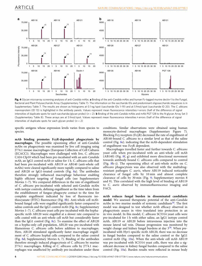
accordance with the guidelines as published by the MIRAGEinitiative54. A MIRAGE Glycan Microarray Document is includedas a Supplementary Note in Supplementary Information. All anti-Candida mAbs tested (except the protein-specific anti-Hyr1 mAbAB121) demonstrated strong binding to C. albicans mannopro-tein (ID 13 in N-Glycan Array Set 3) and no cross-reactivity toother fungal-type, bacterial or plant oligosaccharides included inthe array. The mammalian β-glucan receptor dectin-1, which wasincluded for reference in the analyses, showed the predictedhighly specific binding to fungal β1,3-glucans (Fig. 4a and Sup-plementary Table 7). Additionally, there was negligible or nobinding to the mammalian-type N-glycans tested (Fig. 4b andSupplementary Table 8). In these analyses the broadly neu-tralising anti-HIV mAb PGT 128, known to recognise Man9- andMan8-high-mannose N-glycans55, was used as a reference. Theresults further demonstrated the specificity of the anti-CandidamAbs for Candida-associated polysaccharide epitope(s).
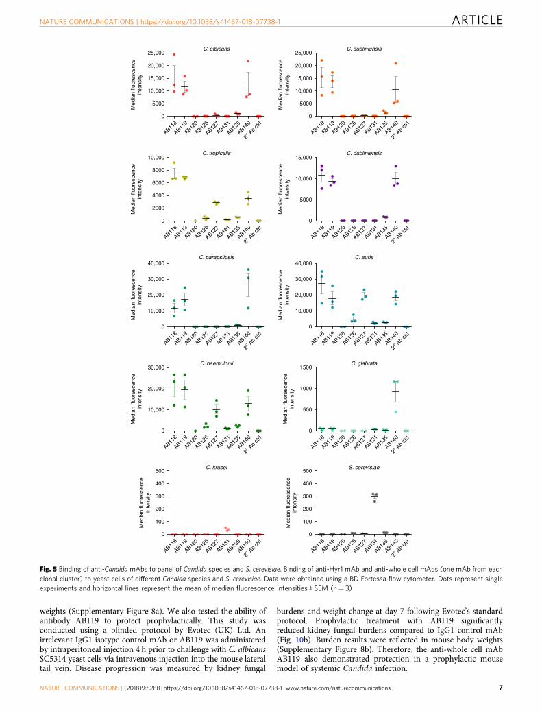
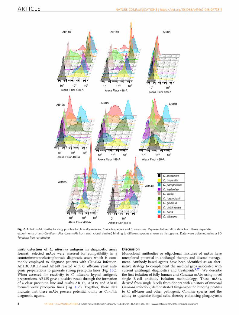
mAbs demonstrate distinct binding profiles to other patho-genic fungi. When assessing anti-whole cell mAb binding toyeast cells of other pathogenic Candida species via fluorescence-activated cell sorting (FACS), AB118, AB119, AB135 and AB140(representing mAbs from four clonal clusters) bound avidly to themost closely related Candida species C. dubliniensis, C. tropicalis,C. parapsilosis and C. lusitaniae and the emerging pathogen C.auris. AB140 also demonstrated specific binding to the moredistantly related C. glabrata. mAbs which bound to epitopes morehighly expressed on C. albicans hyphae such as AB126, AB127and AB131 (representing three clonal clusters) demonstratedoverall lower levels of binding to the phylogenetically closestrelatives of C. albicans (Figs. 5, 6). Interestingly, AB131 alsodemonstrated low levels of binding to C. krusei and Sacchar-omyces cerevisiae (Figs. 5, 6). Immunofluorescent imaging wascarried out to assess patterns of mAb binding in hypha-inducingconditions. Here, AB126, AB127 and AB131 demonstratedincreased levels of binding to most species and in particular C.albicans, commensurate with the predominantly hyphal-associated expression of their target epitopes (Fig. 7b). Toassess for pan-fungal binding activity, anti-whole cell mAbs weretested against species from the other three major human fungalpathogens—Aspergillus fumigatus, Cryptococcus neoformans,Cryptococcus gattii and Pneumocystis carinii. No binding to anyof these species was observed (Fig. 7b).
Commensurate with the C. albicans-specific nature of HYR1,anti-Hyr1 mAbs bound only to C. albicans hyphae and not to C.albicans yeast cells or any of the other Candida species testedincluding C. dubliniensis which lacks the HYR1 gene (Figs. 5, 6,7a). Therefore, the anti-Hyr1 mAbs were C. albicans specific andthe majority of anti-whole cell mAbs demonstrated a variety ofbinding patterns to wild-type C. albicans and other pathogenicCandida species, but not to more distantly related fungalpathogens, indicating that they target a range of Candida-
Fig. 3 Visualising anti-Candida mAbs binding to C. albicans cells.a Representative immunofluorescent images from three separateexperiments with anti-Hyr1 mAb AB120 against WT CAI4-CIp10, Hyr1 nullmutant and a Hyr1 reintegrant strain. b Also shown are immunofluorescentimages and corresponding immunogold localisation of anti-whole cell mAbsbinding to C. albicans yeast and hyphal cell walls. A fluorescently conjugatedsecondary goat anti-human IgG antibody was used to detect anti-CandidamAb binding. Fluorescent images are representative images from threeseparate experiments. Transmission electron microscopy (TEM) imagesshow representative cells from at least one experiment. Scale barsrepresent 4 µm on immunofluorescent images and 100 nm on TEM images
Flu
ores
cenc
eD
ICΔhyr1WT Δhyr1 + HYR1a
C. albicansyeast
C. albicanshyphae
AB
126
AB
131
AB
127
AB
119
AB
140
AB
135
AB
118
Ctr
l mA
b
DIC Fluorescenceb
NATURE COMMUNICATIONS | https://doi.org/10.1038/s41467-018-07738-1 ARTICLE
NATURE COMMUNICATIONS | (2018) 9:5288 | https://doi.org/10.1038/s41467-018-07738-1 | www.nature.com/naturecommunications 5
specific antigens whose expression levels varies from species tospecies.
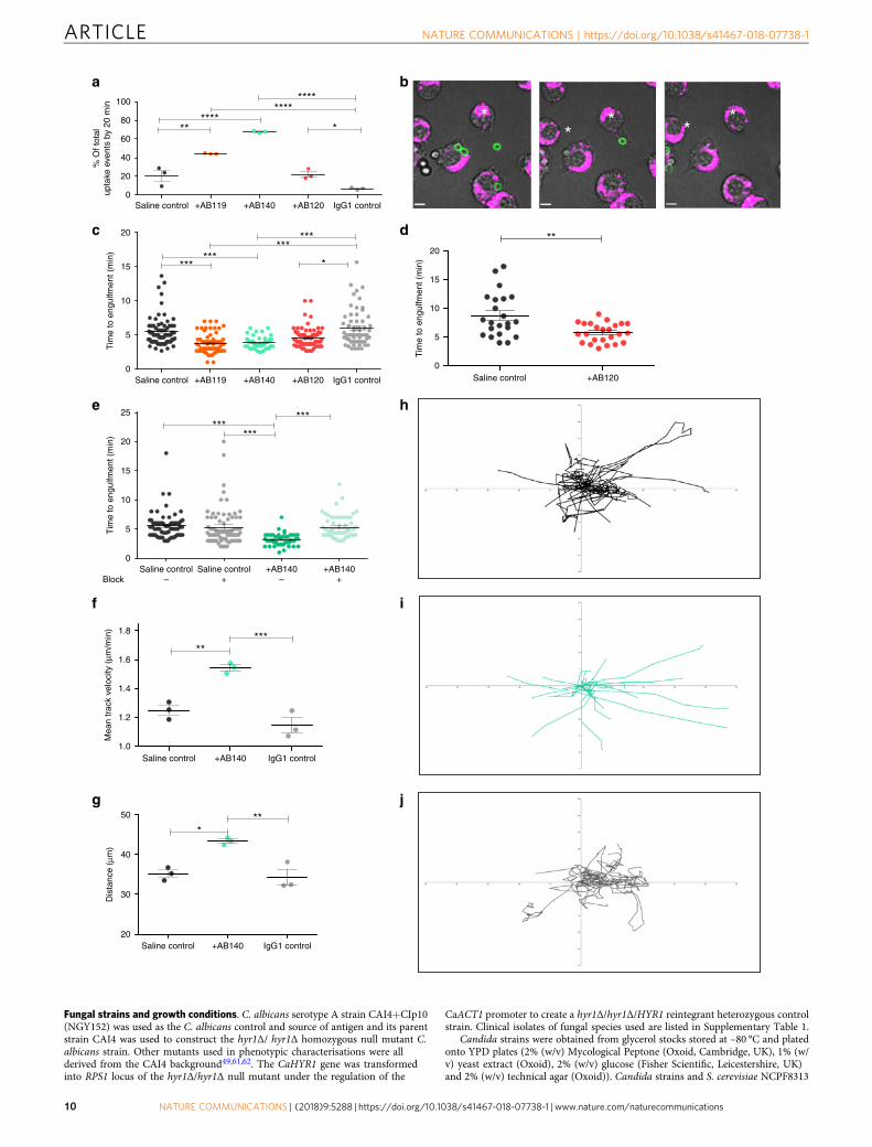
mAb binding promotes FcγR-dependent phagocytosis bymacrophages. The possible opsonising effect of anti-CandidamAbs on phagocytosis was examined by live cell imaging usingJ774.1 mouse macrophages (European Collection of Cell Cultures(ECACC)). Macrophages were challenged with live, C. albicansCAI4-CIp10 which had been pre-incubated with an anti-CandidamAb, an IgG1 control mAb or saline for 1 h. C. albicans cells thathad been pre-incubated with AB119 or AB140 (anti-whole cellmAbs) were cleared significantly more rapidly compared to salineand AB120 or IgG1-treated controls (Fig. 8a). The antibodiestherefore strongly influenced macrophage behaviour enablinghighly efficient targeting of fungal cells (see SupplementaryMovies 1–7). We compared differences in the rate of engulfmentof C. albicans pre-incubated with selected anti-Candida mAbswith isotype controls, defining engulfment as the time taken fromthe establishment of fungus–phagocyte contact to the time forcomplete engulfment indicated by loss of fluorescein iso-thiocyanate (FITC) fluorescence (Fig. 8b). Anti-whole cell mAb-bound fungal cells were engulfed significantly faster compared tosaline controls and the IgG1 control mAb (Fig. 8c; SupplementaryMovies 1–7). C. albicans yeast cells pre-incubated with the hypha-specific mAb AB120 were engulfed at a slower rate compared tocells coated with an anti-whole cell mAb but considerably fasterthan the IgG1 control (Fig. 8c). To examine the effect of AB120on a hypha-only cell population, AB120 was pre-incubated withfilamentous C. albicans cells before addition to macrophages.Here, AB120 stimulated significantly faster macrophage engulf-ment of C. albicans hyphal cells compared to the saline control(Fig. 8d; Supplementary Movies 6, 7). Binding of the antibodiestherefore strongly induced phagocytosis of C. albicans by murineJ774.1 macrophages. Killing of C. albicans cells by J774.1 mac-rophages was unaffected by antibody pre-incubation under these
conditions. Similar observations were obtained using humanmonocyte-derived macrophages (Supplementary Figure 7).Blocking Fcγ receptors (FcγR) decreased the rate of engulfment ofAB140-bound C. albicans to a similar level as that of the salinecontrol (Fig. 8e). indicating that the mAb-dependent stimulationof engulfment was FcγR dependent.
Macrophages travelled faster and further towards C. albicansyeast cells when pre-incubated with an anti-whole cell mAb(AB140) (Fig. 8f, g) and exhibited more directional movementtowards antibody-bound C. albicans cells compared to control(Fig. 8h–j). The opsonising effect of anti-whole mAbs on C.albicans phagocytosis was also observed with the multidrug-resistant pathogen C. auris, where AB119 induced noticeableclearance of fungal cells by 10 min and almost completeclearance of cells by 30 min (Fig. 9; Supplementary movies 8and 9). This correlated with the high level of binding of AB119to C. auris observed by immunofluorescence imaging andFACS.
mAb reduces fungal burden in disseminated candidiasismodel. We assessed therapeutic potential of the anti-CandidamAbs in two murine models of systemic candidiasis56. The firstmodel was designed to test whether mAb efficacy observed inphagocytosis assays in vitro translated into protection in anin vivo model. In this model, C. albicans SC5314 yeast cells werepre-incubated for 1 h with either saline, an IgG1 isotype controlmAb, AB119 or AB120 before intravenous injection into themouse lateral tail vein. Disease progression was monitored byweight change and kidney fungal burdens at day 356. When pre-incubated with Hyr1-specific mAb AB120, there was no decreasein fungal burden compared to the saline control or the IgG1control mAb (Fig. 10a). When the anti-whole cell mAb AB119was pre-incubated with SC5314 yeast cells, there was also a sig-nificant decrease in kidney fungal burden compared to the salinecontrol (Fig. 10a). Burden results were reflected in mouse body
1
70,000AB118a b
13
13
13
13
13 13
13
AB119 AB121 AB118 AB119 AB121
AB126 AB127 AB131 AB126 AB127 AB131
AB135 AB140 Dectin-1 AB135 AB140 PGT 128
35,000
0
9000
4500
0
9000
4500
0
9000
4500
0
70,000
Flu
ores
cenc
e in
tens
ity
35,000
0
70,000
35,000
0
5 10 15 20 1 5 10 15 20 1 5 10 15 20 1 5 10 15 20 25 30 35 1 5 10 15 20 25 30 35 1 5 10 15 20 25 30 35
1 5 10 15 20 1 5 10 15 20 1 5 10 15 20 1 5 10 15 20 25 30 35 1 5 10 15 20 25 30 35 1 5 10 15 20 25 30 35
1 5 10 15 20 1 5 10
Saccharides Glycans
15 20 1 5 10 15 20 1 5 10 15 20 25 30 35 1 5 10 15 20 25 30 35 1 5 10 15 20 25 30 35
Fig. 4 Glycan microarray screening analyses of anti-CandidamAbs. a Binding of the anti-CandidamAbs and human Fc-tagged murine dectin-1 to the Fungal,Bacterial and Plant Polysaccharide Array (Supplementary Table 7). The information on the saccharide IDs and predominant oligosaccharide sequences is inSupplementary Table 7. The results are shown as histograms at 0.1 ng/spot (saccharide IDs 1–19) and at 5 fmol/spot (saccharide ID 20). The C. albicansmannoprotein (ID 13) is highlighted in the antibody panels. Values represent mean fluorescence intensities ± errors (half of the difference of signalintensities of duplicate spots for each saccharide/glycan probe) (n= 2). b Binding of the anti-CandidamAbs and mAb PGT 128 to the N-glycan Array Set 3(Supplementary Table 8). These arrays are at 5 fmol/spot. Values represent mean fluorescence intensities ± errors (half of the difference of signalintensities of duplicate spots for each glycan probe) (n= 2)
ARTICLE NATURE COMMUNICATIONS | https://doi.org/10.1038/s41467-018-07738-1
6 NATURE COMMUNICATIONS | (2018) 9:5288 | https://doi.org/10.1038/s41467-018-07738-1 | www.nature.com/naturecommunications
weights (Supplementary Figure 8a). We also tested the ability ofantibody AB119 to protect prophylactically. This study wasconducted using a blinded protocol by Evotec (UK) Ltd. Anirrelevant IgG1 isotype control mAb or AB119 was administeredby intraperitoneal injection 4 h prior to challenge with C. albicansSC5314 yeast cells via intravenous injection into the mouse lateraltail vein. Disease progression was measured by kidney fungal
burdens and weight change at day 7 following Evotec’s standardprotocol. Prophylactic treatment with AB119 significantlyreduced kidney fungal burdens compared to IgG1 control mAb(Fig. 10b). Burden results were reflected in mouse body weights(Supplementary Figure 8b). Therefore, the anti-whole cell mAbAB119 also demonstrated protection in a prophylactic mousemodel of systemic Candida infection.
25,000C. albicans C. dubliniensis
C. tropicalis
C. parapsilosis C. auris
C. haemulonii C. glabrata
C. krusei S. cerevisiae
Med
ian
fluor
esce
nce
inte
nsity
20,000
15,000
10,000
5000
0
10,000
Med
ian
fluor
esce
nce
inte
nsity
8000
6000
4000
2000
0
40,000
Med
ian
fluor
esce
nce
inte
nsity
30,000
20,000
10,000
0
40,000
Med
ian
fluor
esce
nce
inte
nsity
30,000
20,000
10,000
0
30,000
Med
ian
fluor
esce
nce
inte
nsity
20,000
10,000
0
500
Med
ian
fluor
esce
nce
inte
nsity
400
300
200
100
0
AB118
AB119
AB120
AB126
AB135
AB140
2° A
b ctr
l
AB127
AB131
25,000
Med
ian
fluor
esce
nce
inte
nsity
20,000
15,000
10,000
5000
0
AB118
AB119
AB120
AB126
AB135
AB140
2° A
b ctr
l
AB127
AB131
C. dubliniensis
Med
ian
fluor
esce
nce
inte
nsity
15,000
10,000
5000
0
AB118
AB119
AB120
AB126
AB135
AB140
2° A
b ctr
l
AB127
AB131
AB118
AB119
AB120
AB126
AB135
AB140
2° A
b ctr
l
AB127
AB131
1500
Med
ian
fluor
esce
nce
inte
nsity
1000
500
0
AB118
AB119
AB120
AB126
AB135
AB140
2° A
b ctr
l
AB127
AB131
500
400
300
200
100
Med
ian
fluor
esce
nce
inte
nsity
0
AB118
AB119
AB120
AB126
AB135
AB140
2° A
b ctr
l
AB127
AB131
AB118
AB119
AB120
AB126
AB135
AB140
2° A
b ctr
l
AB127
AB131
AB118
AB119
AB120
AB126
AB135
AB140
2° A
b ctr
l
AB127
AB131
AB118
AB119
AB120
AB126
AB135
AB140
2° A
b ctr
l
AB127
AB131
AB118
AB119
AB120
AB126
AB135
AB140
2° A
b ctr
l
AB127
AB131
Fig. 5 Binding of anti-Candida mAbs to panel of Candida species and S. cerevisiae. Binding of anti-Hyr1 mAb and anti-whole cell mAbs (one mAb from eachclonal cluster) to yeast cells of different Candida species and S. cerevisiae. Data were obtained using a BD Fortessa flow cytometer. Dots represent singleexperiments and horizontal lines represent the mean of median fluorescence intensities ± SEM (n= 3)
NATURE COMMUNICATIONS | https://doi.org/10.1038/s41467-018-07738-1 ARTICLE
NATURE COMMUNICATIONS | (2018) 9:5288 | https://doi.org/10.1038/s41467-018-07738-1 | www.nature.com/naturecommunications 7
mAb detection of C. albicans antigens in diagnostic assayformat. Selected mAbs were assessed for compatibility in acounterimmunoelectrophoresis diagnostic assay which is com-monly employed to diagnose patients with Candida infection.AB118, AB119 and AB140 reacted with C. albicans yeast anti-genic preparations to generate strong precipitin lines (Fig. 10c).When assessed for reactivity to C. albicans hyphal antigenicpreparations, AB135 gave a positive result through the formationof a clear precipitin line and mAbs AB118, AB119 and AB140formed weak precipitin lines (Fig. 10d). Together, these dataindicate that these mAbs possess potential utility as Candidadiagnostic agents.
DiscussionMonoclonal antibodies or oligoclonal mixtures of mAbs haveunexplored potential in antifungal therapy and disease manage-ment. Antibody-based agents have been identified as an alter-native strategy to complement the medical gaps associated withcurrent antifungal diagnostics and treatments9,57. We describethe first isolation of fully human anti-Candida mAbs using novelsingle B-cell antibody isolation methodology. These mAbs,derived from single B cells from donors with a history of mucosalCandida infection, demonstrated fungal-specific binding profilesto C. albicans and other pathogenic Candida species and theability to opsonise fungal cells, thereby enhancing phagocytosis
AB140
AB127AB126
101 103 105
Alexa Fluor 488-A
AB118
S. cerevisiae
C. tropicalis
C. parapsilosis
C. lusitaniae
C. krusei
C. haemulonii
C. glabrata
C. dubliniensis
C. auris
C. albicans
101 103 105
Alexa Fluor 488-A
AB119
101 104
Alexa Fluor 488-A
AB120
101 103 105
Alexa Fluor 488-A101 103 105
Alexa Fluor 488-A
AB131
101 103 105
Alexa Fluor 488-A
AB135
101 103
Alexa Fluor 488-A
101 103 105
Alexa Fluor 488-A
Fig. 6 Anti-Candida mAbs binding profiles to clinically relevant Candida species and S. cerevisiae. Representative FACS data from three separateexperiments of anti-Candida mAbs (one mAb from each clonal cluster) binding to different species shown as histograms. Data were obtained using a BDFortessa flow cytometer
ARTICLE NATURE COMMUNICATIONS | https://doi.org/10.1038/s41467-018-07738-1
8 NATURE COMMUNICATIONS | (2018) 9:5288 | https://doi.org/10.1038/s41467-018-07738-1 | www.nature.com/naturecommunications

and exhibiting protection in a murine model of disseminatedcandidiasis. The antibodies also exhibited potential in the gen-eration of Candida diagnostic assays.
We generated 17 fully human recombinant anti-CandidamAbs through the direct cloning of VH and VL antibody genesencoded by memory B cells and elicited naturally in vivo inresponse to a Candida infection. This technology circumvents theconsiderable expense of humanising antibodies isolated fromrodents or other sources. Furthermore, the recombinant affinitymatured human mAbs generated are less likely to be immuno-genic and, importantly, their native antibody heavy and lightchain natural pairings remain intact, thus preserving their origi-nal epitope specificity as presented during the course of diseaseand their biochemical properties48. IgG1 was selected as thescaffold of choice because it is the most common isotype oftherapeutic antibodies in the clinic and is the best characterised interms of drug development12,58,59.
Twelve of the mAbs generated bound to C. albicans whole celland 5 bound to recombinant purified Hyr1 protein—a proteinthat is important for C. albicans resistance to phagocytosis and iscurrently in development as an experimental vaccine38,39. Anti-bodies with species-specific and pan-species activity both havetranslational potential as diagnostics and therapeutics6,7. There-fore, we validated that both highly specific and cross-reactiveantibodies can be generated using the same methodology. Theanti-Hyr1 mAbs bound only to C. albicans hyphae. Most of theanti-whole cell mAbs bound to the closely related pathogens C.dubliniensis, C. tropicalis, C. parapsilosis and C. auris. There wasless or no binding to the more evolutionarily distant species C.glabrata and C. krusei. When assessed in a routinely employeddiagnostic assay, these mAbs were able to react with Candidaantigenic preparations. Therefore, the novel technology employedhere can be utilised to generate species-specific and/or pan-Candida mAbs with considerable potential in antifungal drugdiscovery and diagnostics.
Many therapeutic mAbs exert their protective effects throughopsonising cells for phagocytosis60. We observed that C. albicansyeast and hyphal cells treated with anti-whole cell mAbs or anti-Hyr1 mAbs were phagocytosed more quickly compared to non-opsonised cells, and that this was FcγR dependent. Furthermore,macrophages migrated further, faster and more directionallytowards opsonised C. albicans cells and this contributed to the
rapid clearance of fungal cells. Our antibodies could also promoteC. auris phagocytosis, demonstrating the potential of these mAbsin targeting difficult-to-treat drug-resistant pathogens. Opsono-phagocytic activity was reflected in the observed protective effectof selected antibodies in a 3-day murine model of systemicinfection. Humanised or fully human IgG1 mAbs make up themajority of antibody therapeutics used clinically and the IgG1isotype has been routinely tested pre-clinically in murine modelsof disease. Human IgG1 also binds to all activating mFcγRs with asimilar profile to the most potent IgG isotype in mice, mIgG2a,validating the use of mouse models to assess Fc-mediated effectsof hIgG1 mAbs58. We utilised a 3-day mouse model of dis-seminated candidiasis56 to assess the efficacy of anti-CandidamAbs in vivo and observed a significant decrease in kidney fungalburden when C. albicans was pre-incubated with an anti-wholecell mAb. We then established that the same anti-whole cell mAbdemonstrated protection in a clinically relevant 7-day prophy-lactic treatment model of disseminated candidiasis.
This study describes the first generation of fully human mAbsto a fungal pathogen cloned from genes from single B cells. ThesemAbs included those with specific pan-Candida and species-specific properties that have utility in the antifungal diagnosticand therapeutic sectors. We used C. albicans as the screeningtarget but this technology has broad potential in a wide range ofother clinical and biological contexts. The characteristics of themAbs are suitable for use singly or in multiplex formats to createnovel polyvalent diagnostic tests. As therapeutics they could beused to direct the immune system to destroy fungal pathogens ina similar approach to cancer immunotherapy, or by targetingtoxic molecules to specific microbial or cellular targets. They alsohave potential in exploring novel antifungal vaccines through theidentification of protective antigens.
MethodsEthics statement. Human blood samples were taken from donors according to thelocal guidelines and regulations, approved by the Pfizer Institutional Review Boardand/or College Ethics Review Board of the University of Aberdeen (CERB/2012/11/676). Informed consent was obtained from all human participants. Animalexperiments were approved by the University of Aberdeen Animal Welfare andEthical Review Body (AWERB) and were carried out under UK Home OfficeProject Licence PPL60/4135 and conformed to the European Union Directive2010/63/EU on the Protection of Animals Used for Scientific Purposes. All animalexperiments conducted by Evotec Ltd (UK) were performed under UK HomeOffice Licence PA67E0BAA, and with local ethical committee clearance.
mAb C. albicans C. dubliniensis C. tropicalis C. parapsilosis C. lusitaniae
C. albicans C. dubliniensis C. tropicalis C. parapsilosis C. lusitaniae C. auris C. haemulonii C. glabrata C. krusei A. fumigatus C. neoformans C. gattii P. carinii
C. glabrata C. krusei
AB120
a
bmAb
AB118
AB119
AB126
AB127
AB131
AB135
AB140
High binding No binding
AB121
AB122
AB123
Fig. 7 Heat-map of anti-Candida mAbs binding to Candida and other pathogenic fungi. Immunofluorescence microscopy analysis of a anti-Hyr1 mAbs(AB120-AB123) and b cell wall mAbs (one from each clonal cluster) binding to C. albicans and other clinically relevant fungal species under hyphal inducingconditions. Red (high binding) to yellow (no binding)
NATURE COMMUNICATIONS | https://doi.org/10.1038/s41467-018-07738-1 ARTICLE
NATURE COMMUNICATIONS | (2018) 9:5288 | https://doi.org/10.1038/s41467-018-07738-1 | www.nature.com/naturecommunications 9
Fungal strains and growth conditions. C. albicans serotype A strain CAI4+CIp10(NGY152) was used as the C. albicans control and source of antigen and its parentstrain CAI4 was used to construct the hyr1Δ/ hyr1Δ homozygous null mutant C.albicans strain. Other mutants used in phenotypic characterisations were allderived from the CAI4 background49,61,62. The CaHYR1 gene was transformedinto RPS1 locus of the hyr1Δ/hyr1Δ null mutant under the regulation of the
CaACT1 promoter to create a hyr1Δ/hyr1Δ/HYR1 reintegrant heterozygous controlstrain. Clinical isolates of fungal species used are listed in Supplementary Table 1.
Candida strains were obtained from glycerol stocks stored at –80 °C and platedonto YPD plates (2% (w/v) Mycological Peptone (Oxoid, Cambridge, UK), 1% (w/v) yeast extract (Oxoid), 2% (w/v) glucose (Fisher Scientific, Leicestershire, UK)and 2% (w/v) technical agar (Oxoid)). Candida strains and S. cerevisiae NCPF8313
100
a b
c d
e h
f i
g j
**
***
******
***
***
**
***
******
**
* **
*** **
*
********
****
*
% O
f tot
alup
take
eve
nts
by 2
0 m
inT
ime
to e
ngul
fmen
t (m
in)
Tim
e to
eng
ulfm
ent (
min
)M
ean
trac
k ve
loci
ty (
μm/m
in)
Dis
tanc
e (μ
m)
80
60
40
20
0
0
20
15
10
5
Tim
e to
eng
ulfm
ent (
min
)
0
20
15
10
5
0
1.0
20
30
40
50
1.2
1.4
1.6
1.8
20
25
15
10
5
Saline control +AB119 +AB140 +AB120 IgG1 control
Saline control +AB119 Saline control +AB120+AB140 +AB120
Saline control Saline control +AB140 +AB140–Block + – +
IgG1 control
Saline control +AB140 IgG1 control
Saline control +AB140 IgG1 control
ARTICLE NATURE COMMUNICATIONS | https://doi.org/10.1038/s41467-018-07738-1
10 NATURE COMMUNICATIONS | (2018) 9:5288 | https://doi.org/10.1038/s41467-018-07738-1 | www.nature.com/naturecommunications
were grown in YPD (see above without the technical agar) except to prepare theinoculum for in vivo experiments where strains were grown in NGY medium (0.1%(w/v) Neopeptone (BD Biosciences), 0.4% (w/v) glucose (Fisher Scientific) and0.1% (w/v) yeast extract (Oxoid). Aspergillus fumigatus clinical isolate V05–27 wascultured on Potato Dextrose Agar slants for 7 days before the spores were harvestedby gentle shaking with sterile 0.1% Tween-20 in phosphate-buffered saline (PBS).Harvested spores were purified, counted and resuspended at a concentration of 1 ×10⁸ spores/ml. Swollen spores were generated by incubation in RPMI media for 4 hat 37 °C. Cryptococcus neoformans KN99α and Cryptococcus gattii R265 weregrown in YPD overnight, washed in PBS and 1 × 10⁷ cells were added to 6 ml RPMI+10% foetal calf serum (FCS) in 6-well plates. Plates were incubated at 37 °C+5%CO2 for 5 days to induce capsule formation. Harvested cells were washed in PBS.Rat lung tissue isolates of Pneumocystis carinii M167–6 were washed in PBS andimmunostained.
Generation of recombinant Hyr1 N-terminus fragment protein. A recombinantN-terminal fragment of C. albicans Hyr1 (amino acids 63 to 350, SupplementaryFigure 1a) incorporating an N-terminal 6xHis tag was expressed in HEK293F cellsand purified by nickel-based affinity chromatography using a NTA superflowcolumn (QIAGEN, USA). Fractions containing recombinant Hyr1 were pooled andfurther purified via Analytical Superdex 200 gel filtration chromatography (GEHealthcare, USA) in PBS. QC of the recombinant protein via SDS-PAGE gelanalysis and analytical SEC confirmed a protein of a predicted mass of 32 kDa(Supplementary Figure 1b, c).
PBMC isolation and purification of circulating IgG from donor plasma. EDTA-containing peripheral venous blood was collected from individuals who hadrecovered from a Candida infection within the last year. Separation of peripheralblood mononuclear cells (PBMCs) and plasma from whole blood was carried outvia density gradient separation using Accuspin System-Histopaque-1077 kits(Sigma-Aldrich). PBMCs were washed three times in PBS, resuspended at a con-centration of 1 × 10⁷ cells/ml in R10 media (RPMI 1640 (Gibco, Life Technologies),10% FCS, 1 mM sodium pyruvate (Sigma), 10 mM HEPES (Gibco, Life Technol-ogies), 4 mM L-glutamine (Sigma), 1× penicillin/streptomycin (Sigma)) containingadditional 10% FCS and 10% dimethyl sulphoxide before storing in liquid nitrogen.IgG was purified from donor plasma using VivaPure MaxiPrepG Spin columns(Sartorius Stedman). Concentration of eluted IgG was measured by absorbance at280 nm.
Circulating antigen-specific IgG titres. Circulating IgG was screened againsttarget antigens via ELISA to identify donors for subsequent CSM B-cell isolationand activation. NUNC 384-well Maxisorp microtitre plates (Sigma-Aldrich) werecoated with extracted C. albicans cell walls or C. albicans overnight culture (wholecell) or 1 µg/ml recombinant Hyr1 protein antigen in PBS. Following incubation at4 °C overnight, wells were washed three times with PBS+0.05% Tween and thenblocked with PBS+0.05% Tween+0.5% bovine serum albumin (BSA) for 1 h atroom temperature. After three washes, titrated purified IgG or IVIG in block bufferwas added in duplicate, and plates were incubated for 2 h at room temperature.Wells were washed three times before addition of goat anti-human IgG, horse-radish peroxidase conjugated (Thermo Scientific # 31413) secondary antibody at1:5000 dilution in blocking buffer. Following incubation for 45 min at roomtemperature, wells were washed and stained using tetramethylbenzidine (TMB)(Thermo Scientific). Plates were incubated for 5 min before addition of 0.18Msulphuric acid and then read at an absorbance of 450 nm. Labstats software inMicrosoft Excel was used to generate concentration-response curves for EC50
determination and donor selection for subsequent CSM B-cell isolation andactivation.
Isolation and activation of CSM B cells. Approximately 5 × 10⁷ PBMCs werethawed and diluted 1:10 in pre-warmed R10 medium and treated with benzonasenuclease HC, purity >99% (Novagen) at 1:10,000. PBMCs were washed twice inPBS before final resuspension in pre-warmed R10 medium. CSM B cells wereisolated from PBMCs by depletion of non-target cells using a Switched Memory Bcell isolation kit with Pre-Separation Filters and LS columns on a MACS Separator(MACS Miltenyi Biotec). To activate CSM B cells and promote antibody secretion,R10 medium was supplemented with a cocktail of antibodies and cytokines tomake complete R10 medium. CSM B cells were resuspended in complete R10medium at 56 cells/ml and then plated out in 90 μl aliquots (5 cells/well) inThermoFisher Matrix 384-well plates using a Biomek FX (Beckman Coulter). Cellswere incubated at 37 °C, 5% CO2 for 7 days. On day 7, 30 μl/well of supernatantwas replaced with 30 μl fresh complete R10. On day 13, all supernatants wereharvested and screened against recombinant N-terminus Hyr1 protein antigen, C.albicans extracted cell walls or C. albicans whole cells from overnight cultures byELISA. B-cell activation and culturing was monitored by measuring IgG1 con-centrations in B-cell supernatants at day 7 and day 13.
Screening of IgG in B-cell supernatant against target antigens via ELISA. Thesame ELISA protocol was employed for the screening of B-cell supernatants againsttarget antigens with the exception that neat B-cell supernatant was added in theplace of titrated purified IgG or IVIG. Positive hits were defined as wells with anOD450 reading >4× background. B cells in ‘positive hit’ wells were lysed in buffer(1 ml DEPC-treated H2O (Life Technologies), 10 µl 1 M Tris pH 8, 25 µl RNAsinPlus RNAse Inhibitor (Promega)) and stored at –80 °C.
cDNA synthesis and PCR1. A schematic of the cloning protocol is shown inSupplementary Figure 2. Primers used for the RT-PCR reaction were based onthose used by Smith et al.46 and are listed in Supplementary Tables 2–5. To ensureall possible VH germline families were captured during the amplification, fourforward primers specific to the leader sequences encompassing the different humanVH germline families (VH1–7) were used in combination with two reverse primers—both in the human IgCH1 region. For the RT-PCR of human Vκ-Cκ genes, threeforward primers specific to the leader sequences for the different human Vκgermline families (Vκ1–4) were used with a reverse primer specific to the humankappa constant region (Cκ) and two further reverse primers which were specific tothe C- and N-terminal ends of the 3’ untranslated region (UTR). To capture therepertoire of human Vλ genes, seven forward primers capturing the leadersequences for the different human Vλ germline families (Vλ1–8) were used withtwo reverse primers complementary to the C- and N-terminal ends of the 3’ UTRalong with another reverse primer specific to the human lambda constant region(Cλ). Amplification of the variable domain of human Ig heavy chain genes (VH),the variable and constant domains of human Ig kappa light chain genes (Vκ-Cκ)and the variable and constant domains of human Ig lambda light chain genes (Vλ-Cλ) was performed in separate reactions.
Prior to complementary DNA (cDNA) synthesis, B-cell lysates were thawed anddiluted 1:5, 1:15 and 1:25 in nuclease-free H2O (Life Technologies) before additionof oligo-dT20 (50 µM) (Invitrogen, Life Technologies) and incubation at 70 °C for5 min.
Reverse transcription and the first PCR reaction (RT-PCR) were performedsequentially in 96-well PCR plates using the QIAGEN OneStep RT-PCR kit,with gene-specific forward and reverse primer mixes (10 µM) and neat or diluted(1:5, 1:15, 1:25) B-cell lysate as the template.
Amplification of VH, Vκ-Cκ and Vλ-Cλ genes: nested PCR reaction. Primersused for the nested PCR reaction were again based on those used by Smith et al.46.A total of 27 forward primers specific for the VH framework 1 (FW1) sequencewere used together with two reverse primers specific for the framework 4 (FW4)
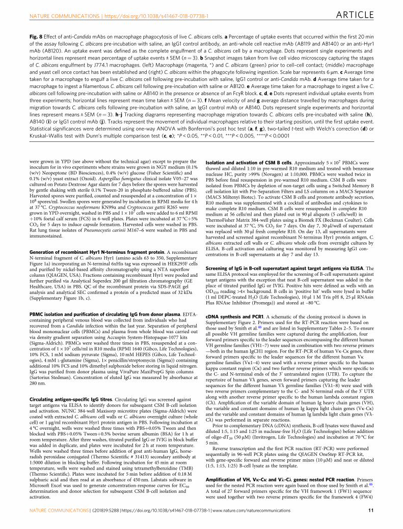
Fig. 8 Effect of anti-CandidamAbs on macrophage phagocytosis of live C. albicans cells. a Percentage of uptake events that occurred within the first 20minof the assay following C. albicans pre-incubation with saline, an IgG1 control antibody, an anti-whole cell reactive mAb (AB119 and AB140) or an anti-Hyr1mAb (AB120). An uptake event was defined as the complete engulfment of a C. albicans cell by a macrophage. Dots represent single experiments andhorizontal lines represent mean percentage of uptake events ± SEM (n= 3). b Snapshot images taken from live cell video microscopy capturing the stagesof C. albicans engulfment by J774.1 macrophages. (left) Macrophage (magenta, *) and C. albicans (green) prior to cell–cell contact; (middle) macrophageand yeast cell once contact has been established and (right) C. albicans within the phagocyte following ingestion. Scale bar represents 6 µm. c Average timetaken for a macrophage to engulf a live C. albicans cell following pre-incubation with saline, IgG1 control or anti-Candida mAb. d Average time taken for amacrophage to ingest a filamentous C. albicans cell following pre-incubation with saline or AB120. e Average time taken for a macrophage to ingest a live C.albicans cell following pre-incubation with saline or AB140 in the presence or absence of an FcγR block. c, d, e Dots represent individual uptake events fromthree experiments; horizontal lines represent mean time taken ± SEM (n= 3). f Mean velocity of and g average distance travelled by macrophages duringmigration towards C. albicans cells following pre-incubation with saline, an IgG1 control mAb or AB140. Dots represent single experiments and horizontallines represent means ± SEM (n= 3). h–j Tracking diagrams representing macrophage migration towards C. albicans cells pre-incubated with saline (h),AB140 (i) or IgG1 control mAb (j). Tracks represent the movement of individual macrophages relative to their starting position, until the first uptake event.Statistical significances were determined using one-way ANOVA with Bonferroni’s post hoc test (a, f, g), two-tailed t-test with Welch’s correction (d) orKruskal–Wallis test with Dunn’s multiple comparison test (c, e); *P < 0.05, **P < 0.01, ***P < 0.005, ****P < 0.0001
NATURE COMMUNICATIONS | https://doi.org/10.1038/s41467-018-07738-1 ARTICLE
NATURE COMMUNICATIONS | (2018) 9:5288 | https://doi.org/10.1038/s41467-018-07738-1 | www.nature.com/naturecommunications 11
region of the VH gene. For nested PCR of the Vκ-Cκ gene, a mixture of 18 forwardprimers specific for human Vκ FW1 sequence were used with a reverse primerspecific to the human kappa constant region 3’ end. For amplification of the Vλ-Cλgene, a mixture of 31 forward primers specific for human Vλ FW1 sequences wereused together with a reverse primer that was placed at the 3’ end of the humanlambda constant region. All of these primers contained 15 bp extensions whichwere complementary to the target downstream pTT5 expression vector. NestedPCR reactions were carried out using Platinum PCR SuperMix High Fidelity(Invitrogen, Life Technologies), nested gene-specific forward (10 µM) and reverse(10 µM) primer mixes and cDNA template from the RT-PCR reaction. PCRamplifications of VH genes, Vκ-Cκ genes and Vλ-Cλ genes were carried out inseparate reactions and then stored on ice. After the nested PCR reaction, sampleswere analysed via agarose gel electrophoresis and positive hits identified and takenforward for downstream In-Fusion cloning with pTT5 mammalian expressionvector.
pTT5 mammalian expression vector preparation. pTT5 was the expressionvector used for recombinant mAb expression (licensed from the National ResearchCouncil of Canada (NRCC))63. pTT5 vector plasmid containing an IgG1 heavychain gene in the multiple cloning site was linearised by double digestion usingFastDigest Restriction enzymes (Thermo Scientific) in separate reactions to facil-itate generation of HC and LC backbones for subsequent cloning of VH and Vκ-Cκor Vλ-Cλ genes. HC and LC backbone DNA was run on a 1% agarose gel andbands were excised from the gel and purified using the QIAquick Gel Extraction kit(QIAGEN). DNA was quantified on a NanoVue Plus Spectrophotometer (GEHealthcare). The 3’- and 5’-termini of the linearised plasmids were depho-sphorylated using FastAP Thermosensitive Alkaline phosphatase (Thermo Scien-tific) to prevent vector self-ligation. Reaction mixtures were cleaned using theMinElute Reaction Cleanup Kit (QIAGEN) and then run on 1% agarose gels.Bands corresponding to dephosphorylated HC and LC backbones were excisedfrom the gel and purified using the QIAQuick Gel Extraction kit (QIAGEN) asabove. Dephosphorylated linearised vector DNA was quantified on a NanoVuePlus spectrophotometer (GE Healthcare).
In-Fusion cloning. The In-Fusion HD Cloning Kit (Clontech, USA) was used toclone the VH, Vκ-Cκ and Vλ-Cλ genes into the pTT5 mammalian expressionvector before transformation of Stellar Competent cells in a 96-well plate format(Clontech). Transformed cells were recovered in SOC medium (Clontech) withshaking at 37 °C for 45–60 min before plating out onto LB agar plates (1% (w/v)tryptone, 0.5% (w/v) yeast extract, 1% (w/v) NaCl, 1.5% (w/v) agar) containing 100µg/ml ampicillin. Single colonies picked for inoculation of 2xTY media containing100 µg/ml ampicillin in a Greiner deep well, 96-well plate (Sigma). Plasmid
miniprep DNA was isolated from cultures in 96-well microtitre plates and anepMotion® 5075 laboratory robot (Eppendorf, Germany) and stored at –20 °C untilrequired for mammalian transfections following sequence analysis of com-plementary determining region (CDR) diversity and comparison to germlinesequences.
Small- and large-scale expression of recombinant mAbs. A file containing allpossible VH and Vκ/Vλ combinations resulting from the original hit wells from theprimary ELISA screen was generated. Automated mixing of native HC and LCDNA pairing combinations (1.5 µg of HC plasmid DNA and 1.5 µg of LC plasmidDNA) was facilitated using a Hamilton Microlab® Starline liquid handling platform(Life Science robotics, Hamilton Robotics). Transient transfections of 3 ml ofcultured Expi293F HEK cells (Gibco, USA) was at a density of 2.5 × 10⁶ cells/ml in24-well tissue culture plates using the Expifectamine 293 Transfection kit (LifeTechnologies, USA). Expi293F cells were maintained in sterile Expi293 expressionmedia (Invitrogen) without antibiotics at 37 °C, with 7% CO2, 120 rpm shaking.For downstream large-scale transfections, DNA was prepared using a QIAGENPlasmid Maxi Kit (QIAGEN, USA) with typical yields of 1.5 µg/µl. Large-scaletransfections were carried out using 100 µg of total DNA (50 µg of HC plasmidDNA and 50 µg LC plasmid DNA) and a 100 ml of suspension cultured Expi293Fcells (Life Technologies). Supernatants were harvested on day 6 and recombinantmAb expression was quantified using anti-human IgG Fc sensors on an Octet QKe
(ForteBio, CA, USA). Following upscaling, recombinant mAb expression wasquantified with an Octet before purification via affinity-based FPLC using HiTrapProtein A HP columns on an ÄKTA (GE Healthcare). mAbs were eluted in 20 mMcitric acid, 150 nM NaCl (pH 2.5) before neutralisation with 1M Tris buffer (pH 8)and then dialysis in PBS overnight. IgG concentration was quantified on aNanoVue Spectrophotometer (GE Healthcare).
QC of recombinant mAbs. Purified recombinant mAbs were checked via SDS-PAGE gel analysis using 4–12% Bis-Tris SDS-PAGE gels under reducing and non-reducing conditions, analytical SEC and analytical mass spectrometry. Confirma-tion of binding to original target antigen was carried out via ELISA using theprotocol described for the circulating antigen-specific IgG screen.
Immunofluorescence imaging of mAbs binding to fungal cells. Single coloniesof Candida were inoculated into 10 ml YPD medium and incubated at 30 °C, 200rpm overnight. Cultures were diluted 1:1333 in milliQ water and then adhered on apoly-L-lysine-coated glass slide (Thermo Scientific, Menzel-Gläser) for 30 min. Toinduce filamentation, cells were incubated in pre-warmed RPMI+10% FCS at 37 °Cfor 90 min to 2 h (this step omitted for staining of yeast cells). For A. fumigatus,hyphal growth was induced from swollen conidia grown in RPMI+10% FCS. Other
Anti-whole cell mAb
IgG1 control mAb
T = 10 min T = 30 min
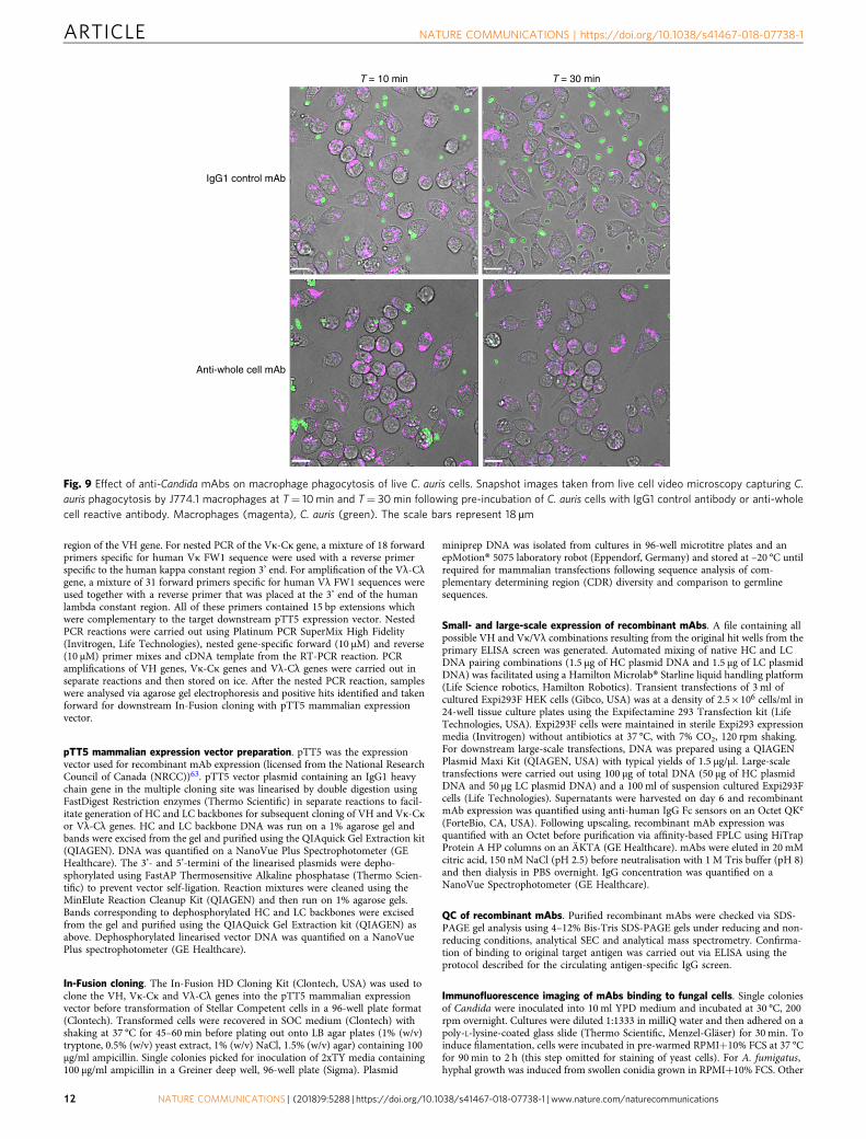
Fig. 9 Effect of anti-Candida mAbs on macrophage phagocytosis of live C. auris cells. Snapshot images taken from live cell video microscopy capturing C.auris phagocytosis by J774.1 macrophages at T= 10 min and T= 30min following pre-incubation of C. auris cells with IgG1 control antibody or anti-wholecell reactive antibody. Macrophages (magenta), C. auris (green). The scale bars represent 18 µm
ARTICLE NATURE COMMUNICATIONS | https://doi.org/10.1038/s41467-018-07738-1
12 NATURE COMMUNICATIONS | (2018) 9:5288 | https://doi.org/10.1038/s41467-018-07738-1 | www.nature.com/naturecommunications

fungal cells were prepared as described in fungal strains and growth conditions. Allfungal cells were then washed in Dulbecco’s phosphate-buffered saline (DPBS) andfixed with 4% paraformaldehyde, washed and blocked with 1.5% normal goatserum (Life Technologies) before staining with an anti-Candida mAb at 1–10 µg/ml for 1 h at room temperature. After three washes with PBS, cells were stainedwith Alexa Fluor® 488 goat anti-human IgG antibody (Life Technologies #A11013)at a 1:400 dilution and incubated at room temperature for 1 h prior to imaging inthree dimensions (3D) on an UltraVIEW® VoX spinning disk confocal microscope(Nikon, Surrey, UK).
High-pressure freezing of C. albicans cell samples. C. albicans yeast and hyphalcell samples were prepared by high-pressure freezing using an EMPACT2 high-pressure freezer and rapid transport system (Leica Microsystems Ltd., MiltonKeynes, UK). Using a Leica EMAFS2, cells were freeze-substituted in acetone+1%(w/v) OsO4 before embedding in Spurr’s resin and polymerising at 60 °C for 48 h.A Diatome diamond knife on a Leica UC6 ultramicrotome was used to cutultrathin sections which were then mounted onto nickel grids.
Immunogold labelling of samples for transmission electron microscopy. Sec-tions on nickel grids were blocked in blocking buffer (PBS+1% (w/v) BSA and0.5% (v/v) Tween20) for 20 min before incubation in incubation buffer (PBS+0.1%(w/v) BSA) for 5 min times 3. Sections were then incubated with anti-CandidamAb (5 µg/ml) for 90 min before incubation in incubation buffer for 5 min, for atotal of 6 times. mAb binding was detected by incubation with Protein A con-jugated to 10 nm gold (Aurion) (diluted 1:40 in incubation buffer) for 60 minbefore another six, 5 min washes, in incubation buffer, followed by three, 5 minwashes in PBS and three, 5 min washes in water. Sections were then stained withuranyl acetate for 1 min before three, 2 min washes, in water and left to dry. TEMimages were taken using a JEM-1400 Plus using an AMT UltraVUE camera.
Enzymatic modification of C. albicans cell wall. For proteinase K treatment,single colonies of Candida were inoculated into 10 ml YPD medium and incubatedat 30 °C, 200 rpm overnight. Cultures were diluted in milliQ water and then
adhered on poly-L-lysine-coated glass slides. To induce filamentation, cells wereincubated in pre-warmed RPMI+10% FCS at 37 °C for 90 min to 2 h. Slides werewashed with DPBS and cells were treated with 50 μg/ml proteinase K at 37 °C for 1h. For Endo-H, α(1–2,3.6)-mannosidase from Jack Bean and Zymolyase 20 Ttreatments, C. albicans overnight yeast cells were washed and resuspended inDPBS. Filamentous cells were induced as above. Cells were washed in DPBS andresuspended in Glycobuffer and Endoglycosidase H (10 U/μl; NEB), 20 mMsodium acetate with 0.4 mM Zn2+ pH 5 and α-(1–2,3,6)-mannosidase or 20 mMPIPES with 2M Sorbitol pH 6.5 and Zymolyase 20T (50 U/g wet cells; MPBIO) at37 °C for 2 h. Control cells were kept in reaction buffers without enzymes. Cellswere then washed in DPBS and fixed with 4% paraformaldehyde, washed andblocked with 1.5% normal goat serum (Life Technologies) before staining with ananti-Candida mAb at 1 µg/ml for 1 h at room temperature. After 3 washes withDPBS, cells were stained with Alexa Fluor® 488 goat anti-human IgG antibody(Life Technologies) at a 1:400 dilution and incubated at room temperature for 1 hprior to imaging in 3D on an UltraVIEW® VoX spinning disk confocal microscope(Nikon, Surrey, UK).
Preparation of human monocyte-derived macrophages. Human macrophageswere derived from monocytes isolated from the blood of healthy volunteers.PBMCs were resuspended in Dulbecco’s modified Eagle’s medium (DMEM)(Lonza, Slough, UK) supplemented with 200 U/ml penicillin/streptomycin anti-biotics (Invitrogen, Paisley, UK) and 2 mM L-glutamine (Invitrogen, Paisley, UK).Serum isolated from blood was heat inactivated for 20 min at 56 °C. PBMCs wereseeded at 6 × 10⁵ in 300 µl/well supplemented DMEM containing 10% autologoushuman serum, onto an 8-well glass-based imaging dish (Ibidi, Munich, Germany)and incubated at 37 °C with 5% CO2 for 1 h 45 min to facilitate monocyteadherence to the glass surface. Floating lymphocytes in the supernatant wereaspirated and the same volume of fresh pre-warmed supplemented DMEM con-taining 10% autologous human serum added to the well. Cells were incubated at 37°C, 5% CO2 for 7 days with media changed on days 3 and 664. Cells were used inimaging experiments on day 7. Supplemented DMEM was replaced with pre-warmed supplemented CO2-independent media containing 1 µM LysoTracker RedDND-99 (Invitrogen) immediately prior to phagocytosis experiments.
7
6 ** *
Log
CF
U/g
tiss
ue5
4
7
6
Log
CF
U/g
tiss
ue
5
4
3
2
CommercialAB
c
a
d
b
AB119
CommercialAB
AB135
mAbYeast
supernatant mAbYeast crude
extract mAbHyphal
supernatant mAbHyphal crude
extract
Saline
cont
rol
IgG1
cont
rol
Anti-w
hole
cell m
Ab
IgG1
cont
rol
Anti-w
hole
cell m
Ab
Anti-h
yr1
mAb
Fig. 10 Assessment of anti-Candida mAbs in murine models of disseminated candidiasis and a routine diagnostic assay. a C. albicans SC5314 was pre-incubated with saline, IgG1 control, anti-whole cell mAb (AB119) or anti-Hyr1 mAb (AB120) and then injected i.v. into the tail vein of female BALB/c mice(n= 6 per group). Kidney fungal burdens from each group were determined on day 3 post infection. b IgG1 control or anti-whole cell mAb (AB119) wasadministered i.p. 4 h prior to injection of C. albicans SC5314 i.v. into the lateral tail vein of male CD1 mice (n= 10 per group). Kidney fungal burdens fromeach group were determined on day 7 post infection. Dots represent individual animals and horizontal lines represent mean; statistical significance wasdetermined by two-tailed t-test; *P < 0.05, **P < 0.01. Purified anti-Candida mAbs react with yeast (c) and hyphal (d) antigenic preparations in acounterimmunoelectrophoresis assay routinely employed to diagnose Candida infection. Precipitin lines between wells indicate a positive reaction
NATURE COMMUNICATIONS | https://doi.org/10.1038/s41467-018-07738-1 ARTICLE
NATURE COMMUNICATIONS | (2018) 9:5288 | https://doi.org/10.1038/s41467-018-07738-1 | www.nature.com/naturecommunications 13

Glycan microarray screening analysis. For microarray screening analysis, twomicroarray platforms were used: (1) an array, designated ‘Fungal, Bacterial andPlant Polysaccharide Array’ featuring 19 saccharides (polysaccharides or glyco-proteins) derived from fungi, bacteria and plants and one lipid-linked neoglyco-lipid (NGL) probe derived from the hexasaccharide of β1,4-linked N-acetylglucosamine (GlcNAc) (representative of fungal chitin), and (2) an array of38 sequence-defined NGL probes of N-glycans (majority of mammalian-type)(Supplementary Table 8) designated ‘N-glycan Array Set 3’, described previously65.The polysaccharides and glycoprotein antigens included in the Fungal, Bacterialand Plant Polysaccharide Array are listed in Supplementary Table 7. The glucanpolysaccharides (IDs 1–11) have been described previously66. The mannan from S.cerevisiae (ID 12) was purchased from Sigma. Purified Candida albicans N-linkedmannoprotein preparation (ID 13)67 was a kind gift from David Williams (EastTennessee State University); the purified Aspergillus fumigatus mannoproteinpreparation (ID 14)25 was a kind gift from Christopher Thornton (University ofExeter). The antigen preparations from Mycobacterium smegmatis and M. tuber-culosis (IDs 15–18) were obtained from the National Institutes of Health (NIH)Biodefense and Emerging Infections Research Resources Repository (BEI Resour-ces) and were described previously68. The glucurono-xylomannan from Tremellafuciformis (ID 19) was purchased from Elicityl. For construction of the microarraysthe saccharides and the NGL probes were immobilised noncovalently onnitrocellulose-coated glass slides, following established protocols53,66. Poly-saccharides and glycoproteins were taken up in water, with the exception of cur-dlan polysaccharide that was solubilised using mild alkaline solution (50 mMNaOH) and glucurono-xylomannan solubilised in 150 mM NaCl. The microarrayswere probed with the anti-Candida mAbs, following described protocols53,66. Inbrief, after blocking with 10 mM HEPES-buffered saline (pH 7.4), 150 mM NaCl, 5mM CaCl2 (referred to as HBS) containing 1% w/v BSA (Sigma) and 0.02% v/vCasein (Pierce), the microarrays were overlaid with the mAbs diluted to a finalconcentration of 10 µg/ml (for the Fungal, Bacterial and Plant PolysaccharideArray) and at 50 µg/ml (for the N-glycan Array Set 3) in the blocking solution.Binding was detected using biotinylated anti-human IgG (Vector, 5 μg/ml) fol-lowed by Alexa Fluor-647-labelled streptavidin (Molecular Probes, 1 μg/ml), dilu-ted in the blocking solution. The mAb PGT 128 was a kind gift of Katie J. Doores(King’s College London, UK) and was analysed at 50 µg/ml55. Fc-dectin-1 waskindly provided by Gordon Brown (University of Aberdeen, UK) and was analysedat 20 µg/ml in the blocking solution 0.5% v/v casein (Pierce) in HBS66. Analyseswere performed at ambient temperature. Microarray data analysis was performedusing dedicated software developed by Mark Stoll of the GlycosciencesLaboratory69.
FACS. Candida and S. cerevisiae colonies were grown in YPD medium andincubated at 30 °C, 200 rpm overnight. Cells were washed twice in 1× PBS, countedand then centrifuged and fixed in 4% paraformaldehyde for 45 min. Cells were thenwashed twice in 1× PBS to remove residual paraformaldehyde before 2.5 × 10⁶
cells/well were added into V-bottomed 96-well tissue culture plates. Anti-CandidamAb was then added to wells at 1 µg/ml in FACS buffer (1× PBS, 1% FBS, 0.5 mMEDTA) and incubated for 45 min. Cells were washed once with FACS buffer andthen stained with 5 µg/ml Alexa Fluor® 488 goat anti-human IgG antibody (LifeTechnologies) for 30 min at room temperature. Stained cells were then washedtwice in FACS buffer before final resuspension in FACS buffer and storage in thedark at 4 °C. Samples were analysed using a BD Fortessa flow cytometer where10,000 events were acquired for each sample from 3 independent experiments.Median fluorescence intensity was calculated for each sample using FloJov.10 software. The gating strategy employed is shown in Supplementary Figure 6.
Preparation of J774.1 mouse macrophages. J774.1 macrophages (ECACC, HPA,Salisbury, UK) were maintained in tissue culture flasks in DMEM (Lonza) sup-plemented with 10% (v/v) FCS (Biosera, Ringmer, UK), 200 U/ml penicillin/streptomycin antibiotics (Invitrogen) and 2 mM L-glutamine (Invitrogen) andincubated at 37 °C, 5% CO2. For phagocytosis assays, macrophages were seeded insupplemented DMEM at a density of 1 × 10⁵ cells/well in an 8-well glass-basedimaging dish (Ibidi) and incubated overnight at 37 °C, 5% CO2. Immediately priorto phagocytosis experiments, supplemented DMEM was replaced with pre-warmedsupplemented CO2-independent media (Gibco) containing 1 µM LysoTracker RedDND-99 (Invitrogen).
Preparation of FITC-stained C. albicans. C. albicans colonies were grown in YPDmedium and incubated at 30 °C, 200 rpm overnight. Live C. albicans cells werestained for 10 min at room temperature in the dark with 10 mg/ml FITC (Sigma,Dorset, UK) in 0.05 M carbonate-bicarbonate buffer (pH 9.6) (BDH Chemicals,VWR International, Leicestershire, UK). Following 10 min of incubation, in pha-gocytosis assays using C. albicans FITC-labelled yeast, the cells were washed threetimes in 1× PBS to remove any residual FITC and finally resuspended in 1× PBS or1× PBS containing purified anti-Candida mAb at 1–50 µg/ml. For assays wherefilamentous C. albicans cells were added to immune cells, these were washed andresuspended in supplemented CO2-independent media with or without anti-CandidamAb at 1–50 µg/ml and incubated at 37 °C with gentle shaking for 45 min.
Live cell video microscopy phagocytosis assays. Phagocytosis assays wereperformed using a standard protocol with modifications64,70,71. Following pre-incubation with/without 50 µg/ml anti-Candida mAb, live FITC-stained wild-typeC. albicans (CA14-CIp10) yeast or hyphal cells were added to LysoTracker RedDND-99-stained J774.1 murine macrophages in 8-well glass-based imaging dish(Ibidi) at a multiplicity of infection of 3. For experiments investigating the effect ofa mFcγR blocker on the phagocytosis of C. albicans pre-incubated with 5 µg/mlanti-Candida whole cell mAb (AB140), J774.1 macrophages were incubated with10 µg/ml purified rat anti-mouse CD16/CD32 mAb (BD Pharmingen, BD Bios-ciences #553142) for 5 min at room temperature before addition of C. albicanscells. Video microscopy was performed using an UltraVIEW® VoX spinning diskconfocal microscope (Nikon) in a 37 °C chamber and images were captured at 1min intervals over a 3 h period. At least three independent experiments wereperformed for each antibody. Twenty-five macrophages were selected at randomfrom each experiment and analysed individually at 1 min intervals over a 3 h periodusing Volocity 6.3 imaging analysis software (Improvision, PerkinElmer, Coventry,UK). Measurements included C. albicans uptake—defined as the number of C.albicans cells taken up by an individual phagocyte over the 3 h period—and C.albicans rate of engulfment—defined as the time point at which cell–cell contactwas established until the time point at which C. albicans was fully engulfed. Finally,Volocity 6.3 imaging analysis software was used to measure the distance travelled,directionality and velocity of macrophages at 1 min intervals until the first uptakeevent to provide a detailed overview of macrophage migration towards C. albicanscells. Mean values and standard errors of the mean were calculated. One-wayanalysis of variance (ANOVA) followed by Bonferroni multiple comparison tests,Kruskal–Wallis test with Dunn’s multiple comparisons test or unpaired, two-tailedt-tests with Welch’s correction were used to determine statistical significance.
Proof of concept in 3-day systemic candidiasis infection model. A 3-day modelof disseminated candidiasis was employed to assess the efficacy of anti-CandidamAbs in vivo in immunocompetent 6–8-week-old female BALB/c mice (Envigo,UK)56. To determine the minimum group size required to detect a statistical dif-ference in outcomes (P < 0.05), power analyses were carried out using data gen-erated from other experiments using the same model (power= 0.8). Mice wererandomly assigned to groups (n= 6) and were housed in individually ventilatedcages (IVC), with food and water provided ad libitum. Each cage was randomlyassigned a treatment, and investigators were not blinded when assessing outcome.Endotoxin levels in the purified recombinant mAb samples were determined withthe Limulus Amebocyte Lysate QCL-1000 test (Lonza) to ensure that endotoxinwas <0.6 EU/ml before in vivo administration. On day 0, ~3.2 × 10⁵ C. albicansSC5314 yeast cells were pre-incubated with either sterile saline, 1.5 mg/ml purifiedIgG1 isotype control mAb, 1.5 mg/ml purified anti-Candida mAb AB119 or 1.5mg/ml purified anti-Hyr1 mAb AB120 in 700 µl. Following 60 min of incubation atroom temperature, mice were intravenously (i.v.) infected with 100 µl via the lateraltail vein. This is the equivalent of approximately 1.6 × 10⁴ colony-forming units(CFUs)/g per mouse, treated with 7.5 mg/kg purified antibody. Mice were mon-itored and weighed at least once per day from day 0 up to and including day 3. Onday 3, animals were culled by cervical dislocation and the kidneys harvested forquantitative analysis of fungal burden.
Proof of concept in prophylactic treatment model of systemic candidiasis.This prophylactic treatment model was conducted by Evotec Ltd (UK). In thismodel, 7–8-week-old immunocompetent male CD1 mice (Charles River) wererandomly assigned to groups (n= 10) and housed in IVC, with food and waterprovided ad libitum. Mice were injected i.v. via the lateral tail vein with 2.13 × 10⁵
C. albicans SC5314 per mouse in 200 µl. At 4 h prior to challenge, each mousereceived 1 mg of IgG1 control or AB119 antibody by intraperitoneal (i.p.) injectionin 200 µl. Mice were monitored and weighed at least once per day from day 0 up toand including day 7. On day 7, animals were killed by administration of pento-barbitone overdose and the kidneys harvested for quantitative analysis of fungalburden.
Analysis of disease progression. Kidneys were homogenised and serial dilutionsplated on Sabouraud dextrose agar plates (1% mycological peptone (w/v), 4%glucose (w/v), 2% agar (w/v)) and incubated at 35 °C overnight. Fungal burdenswere expressed as log CFUs/g of infected organ. Weight change was measured andexpressed as a percentage of mouse starting weight. Statistical analyses of fungalburdens were carried out using unpaired two-tailed t-test. An F-test was used toshow there was no statistical significance in variance within the groups (P= 0.1353and P= 0.0817 for the 3-day and 7-day models, respectively). All statistical ana-lyses were carried out using GraphPad Prism 5.04 software.
Counterimmunoelectrophoresis. Agar gels were prepared (Veronal buffer+0.5%(w/v) purified agar+0.5% (w/v) LSA agarose+0.05% (w/v) sodium azide, pH 8.2)and wells were cut out using a cutter. Into one column of wells, 10 µl of neat anti-Candida mAb was added. The same volume of antigen (crude C. albicans yeast orhyphal preparation (following glass bead disruption of cells and 1 min cen-trifugation at 15,200 × g to generate disrupted cell wall/glass bead slurry and cellsupernatant antigenic preparations)) was added to the second column of wells and
ARTICLE NATURE COMMUNICATIONS | https://doi.org/10.1038/s41467-018-07738-1
14 NATURE COMMUNICATIONS | (2018) 9:5288 | https://doi.org/10.1038/s41467-018-07738-1 | www.nature.com/naturecommunications

gels were placed into an electrophoresis tank containing veronal buffer. Gels wereoriented so that the antibody wells were lined up alongside the anode and theantigen wells alongside the cathode due to antibody migration towards the cathodevia electroendosmosis and antigen migration towards the anode due to lowerisoelectric points than the buffer pH. The gels were run at 100 V for 90 min beforeremoval and immersion in saline-trisodium citrate overnight. The following day,the gels were rinsed with water and covered with moistened filter paper and left todry in an oven for 2 h. Once dried, the filter paper was moistened and removed andthe gels put back into the oven for a further 15 min to dry completely. Gels werethen immersed in Buffalo Black solution (0.05% (v/v) Buffalo Black, 50% (v/v)distilled water, 40% (v/v) methylated spirit, 10% (v/v) acetic acid) for 10 min beforedestaining in 45% (v/v) industrial methylated spirits, 10% (v/v) acetic acid, 45% (v/v) distilled water for 10 min. Gels were then dried and examined for the formationof precipitin lines.
Statistical analysis. Statistical analyses of data were carried out using GraphPadPrism 5.04. Results are expressed as mean ± SEM. When comparing two groups,unpaired two-tailed t-tests were performed with Welch’s correction when appro-priate. For comparison of more than two groups, one-way ANOVA with Bon-ferroni’s post hoc comparisons or the Kruskal–Wallis test with Dunn’s multiplecomparisons test was performed.
Data availabilityAll relevant data are available from the authors upon request.
Received: 30 June 2016 Accepted: 13 November 2018
References
1. Brown, G. D. et al. Hidden killers: human fungal infections. Sci. Transl. Med.4, 165rv13 (2012).
2. Lockhart, S. R. Current epidemiology of Candida infection. Clin. Microbiol.Newsl. 36, 131–136 (2014).
3. Kim, J. & Sudbery, P. Candida albicans, a major human fungal pathogen. J.Microbiol. 49, 171–177 (2011).
4. Gow, N. A. R., Van De Veerdonk, F. L., Brown, A. J. P. & Netea, M. G.Candida albicans morphogenesis and host defence: Discriminating invasionfrom colonization. Nat. Rev. Microbiol. 10, 112–122 (2012).
5. Ellepola, A. N. B. & Morrison, C. J. Laboratory diagnosis of invasivecandidiasis. J. Microbiol. 43, 65–84 (2005).
6. Ostrosky-Zeichner, L. Invasive mycoses: diagnostic challenges. Am. J. Med.125, S14–S24 (2012).
7. Perfect, J. R. Fungal diagnosis: how do we do it and can we do better? Curr.Med. Res. Opin. 29, 3–11 (2013).
8. Berry, J. D. & Gaudet, R. G. Antibodies in infectious diseases: polyclonals,monoclonals and niche biotechnology. N. Biotechnol. 28, 489–501 (2011).
9. Saylor, C., Dadachova, E. & Casadevall, A. Monoclonal antibody-basedtherapies for microbial diseases. Vaccine 27, G38–G46 (2009).
10. Casadevall, A. & Pirofski, L. A. Immunoglobulins in defense, pathogenesis,and therapy of fungal diseases. Cell Host Microbe 11, 447–456 (2012).
11. Ecker, D. M., Jones, S. D. & Levine, H. L. The therapeutic monoclonalantibody market. Mabs 7, 9–14 (2015).
12. Strohl, W. R. Antibody discovery: sourcing of monoclonal antibody variabledomains. Curr. Drug Discov. Technol. 11, 3–19 (2014).
13. Yoon, S., Kim, Y. S., Shim, H. & Chung, J. Current perspectives on therapeuticantibodies. Biotechnol. Bioprocess Eng. 15, 709–715 (2010).
14. Connor, E. M. Palivizumab, a humanized respiratory syncytial virusmonoclonal antibody, reduces hospitalization from respiratory syncytial virusinfection in high-risk infants. Pediatrics 102, 531–537 (1998).
15. Migone, T. S. et al. Raxibacumab for the treatment of inhalational anthrax. N.Engl. J. Med. 361, 135–144 (2009).
16. Wrammert, J. et al. Broadly cross-reactive antibodies dominate the human Bcell response against 2009 pandemic H1N1 influenza virus infection. J. Exp.Med. 208, 181–193 (2011).
17. Liao, H. X. et al. Initial antibodies binding to HIV-1 gp41 in acutely infectedsubjects are polyreactive and highly mutated. J. Exp. Med. 208, 2237–2249(2011).
18. Lehar, S. M. et al. Novel antibody-antibiotic conjugate eliminates intracellularS. aureus. Nature 527, 323–328 (2015).
19. Dromer, F., Salamero, J., Contrepois, A., Carbon, C. & Yeni, P. Production,characterization, and antibody specificity of a mouse monoclonal antibodyreactive with Cryptococcus neoformans capsular polysaccharide. Infect.Immun. 55, 742–748 (1987).
20. Gigliotti, F. & Hughes, W. T. Passive immunoprophylaxis with specificmonoclonal antibody confers partial protection against Pneumocystis cariniipneumonitis in animal models. J. Clin. Invest. 81, 1666–1668 (1988).
21. Brena, S. et al. Fungicidal monoclonal antibody C7 interferes with ironacquisition in Candida albicans. Antimicrob. Agents Chemother. 55,3156–3163 (2011).
22. Moragues, M. D. et al. A monoclonal antibody directed against a Candidaalbicans cell wall mannoprotein exerts three anti-C. albicans activities. Infect.Immun. 71, 5273–5279 (2003).
23. Torosantucci, A. et al. Protection by anti-ß-glucan antibodies is associatedwith restricted ß-1,3 glucan binding specificity and inhibition of fungal growthand adherence. PLoS One 4, e5392 (2009).
24. Xin, H. & Cutler, J. E. Vaccine and monoclonal antibody that enhance mouseresistance to candidiasis. Clin. Vaccin. Immunol. 18, 1656–1667 (2011).
25. Thornton, C. R. Development of an immunochromatographic lateral-flowdevice for rapid serodiagnosis of invasive aspergillosis. Clin. Vaccin. Immunol.15, 1095–1105 (2008).
26. Martínez-Jiménez, M. C. et al. Potential role of Candida albicans germ tubeantibody in the diagnosis of deep-seated candidemia.Med. Mycol. 52, 270–275(2014).
27. Jarvis, J. N. et al. Evaluation of a novel point-of-care cryptococcal antigen teston serum, plasma, and urine from patients with HIV-associated cryptococcalmeningitis. Clin. Infect. Dis. 53, 1019–1023 (2011).
28. Arendrup, M. C., Posteraro, B., Sanguinetti, M. & Guinea, J. The state-of-the-art mycology laboratory: Visions of the future. Curr. Fungal Infect. Rep. 9,37–51 (2015).
29. Kullberg, B. J. & Arendrup, M. C. Invasive candidiasis. N. Engl. J. Med. 373,1445–1456 (2015).
30. Cassone, A. Development of vaccines for Candida albicans: fighting a skilledtransformer. Nat. Rev. Microbiol. 11, 884–891 (2013).
31. De Bernardis, F. et al. A virosomal vaccine against candidal vaginitis:immunogenicity, efficacy and safety profile in animal models. Vaccine 30,4490–4498 (2012).
32. Pietrella, D. et al. A ß-glucan-conjugate vaccine and anti-ß-glucan antibodiesare effective against murine vaginal candidiasis as assessed by a novel in vivoimaging technique. Vaccine 28, 1717–1725 (2010).
33. Ibrahim, A. S. et al. NDV-3 protects mice from vulvovaginal candidiasisthrough T- and B-cell immune response. Vaccine 31, 5549–5556 (2013).
34. Schmidt, C. S. et al. NDV-3, a recombinant alum-adjuvanted vaccine forCandida and Staphylococcus aureus, is safe and immunogenic in healthyadults. Vaccine 30, 7594–7600 (2012).
35. Spellberg, B. J. et al. Efficacy of the anti-Candida rAls3p-N or rAls1p-Nvaccines against disseminated and mucosal candidiasis. J. Infect. Dis. 194,256–260 (2006).
36. Edwards, J. E. et al. A fungal immunotherapeutic vaccine (NDV-3A) fortreatment of recurrent vulvovaginal candidiasis-a phase 2 randomized,double-blind, placebo-controlled trial. Clin. Infect. Dis. 66, 1928–1936 (2018).
37. Yeaman, M. R. et al. Mechanisms of NDV-3 vaccine efficacy in MRSA skinversus invasive infection. Proc. Natl Acad. Sci. USA 111, E5555–E5563 (2014).
38. Luo, G., Ibrahim, A. S., French, S. W., Edwards, J. E. Jr. & Fu, Y. Active andpassive immunization with rHyr1p-N protects mice against hematogenouslydisseminated candidiasis. PLoS One 6, e25909 (2011).
39. Luo, G. et al. Candida albicans Hyr1p confers resistance to neutrophil killingand is a potential vaccine target. J. Infect. Dis. 201, 1718–1728 (2010).
40. Uppuluri, P. et al. The Hyr1 protein from the fungus Candida albicans is across kingdom immunotherapeutic target for Acinetobacter bacterial infection.PLoS Pathog. 14, e1007056 (2018).
41. Chames, P., Van Regenmortel, M., Weiss, E. & Baty, D. Therapeuticantibodies: successes, limitations and hopes for the future. Br. J. Pharmacol.157, 220–233 (2009).
42. Carter, P. J. Potent antibody therapeutics by design. Nat. Rev. Immunol. 6,343–357 (2006).
43. Hoogenboom, H. R. Selecting and screening recombinant antibody libraries.Nat. Biotechnol. 23, 1105–1116 (2005).
44. Winter, G., Griffiths, A. D., Hawkins, R. E. & Hoogenboom, H. R. Makingantibodies by phage display technology. Annu. Rev. Immunol. 12, 433–455(1994).
45. Wilson, P. C. & Andrews, S. F. Tools to therapeutically harness the humanantibody response. Nat. Rev. Immunol. 12, 709–719 (2012).
46. Smith, K. et al. Rapid generation of fully human monoclonal antibodiesspecific to a vaccinating antigen. Nat. Protoc. 4, 372–384 (2009).
47. Huang, J. et al. Isolation of human monoclonal antibodies from peripheralblood B cells. Nat. Protoc. 8, 1907–1915 (2013).
48. Tiller, T. Single B cell antibody technologies. N. Biotechnol. 28, 453–457 (2011).49. Bailey, D. A., Feldmann, P. J. F., Bovey, M., Gow, N. A. R. & Brown, A. J. P.
The Candida albicans HYR1 gene, which is activated in response to hyphaldevelopment, belongs to a gene family encoding yeast cell wall proteins. J.Bacteriol. 178, 5353–5360 (1996).
NATURE COMMUNICATIONS | https://doi.org/10.1038/s41467-018-07738-1 ARTICLE
NATURE COMMUNICATIONS | (2018) 9:5288 | https://doi.org/10.1038/s41467-018-07738-1 | www.nature.com/naturecommunications 15
50. Good, K. L., Bryant, V. L. & Tangye, S. G. Kinetics of human B cell behaviorand amplification of proliferative responses following stimulation with IL-21.J. Immunol. 177, 5236–5247 (2006).
51. Zotos, D. et al. IL-21 regulates germinal center B cell differentiation andproliferation through a B cell-intrinsic mechanism. J. Exp. Med. 207, 365–378(2010).
52. Geffroy-Luseau, A. et al. TLR9 ligand induces the generation of CD20+plasmablasts and plasma cells from CD27+ memory B-cells. Front.Immunol. 2, 83 (2011).
53. Liu, Y. et al. Neoglycolipid-based oligosaccharide microarray system:preparation of NGLs and their noncovalent immobilization on nitrocellulose-coated glass slides for microarray analyses. Methods Mol. Biol. 808, 117–136(2012).
54. Liu, Y. et al. The minimum information required for a glycomics experiment(MIRAGE) project: improving the standards for reporting glycan microarray-based data. Glycobiology 27, 280–284 (2017).
55. Pejchal, R. et al. A potent and broad neutralizing antibody recognizesand penetrates the HIV glycan shield. Science 334, 1097–1103(2011).
56. MacCallum, D. M. et al. Genetic dissection of azole resistance mechanisms inCandida albicans and their validation in a mouse model of disseminatedinfection. Antimicrob. Agents Chemother. 54, 1476–1483 (2010).
57. Beyda, N. D., Regen, S., Lewis, R. E. & Garey, K. W. Immunomodulatoryagents as adjunctive therapy for the treatment of resistant Candida species.Curr. Fungal Infect. Rep. 7, 119–125 (2013).
58. Overdijk, M. B. et al. Crosstalk between human IgG isotypes and murineeffector cells. J. Immunol. 189, 3430–3438 (2012).
59. Salfeld, J. G. Isotype selection in antibody engineering. Nat. Biotechnol. 25,1369–1372 (2007).
60. Casadevall, A., Dadachova, E. & Pirofski, L. A. Passive antibody therapy forinfectious diseases. Nat. Rev. Microbiol. 2, 695–703 (2004).
61. Brand, A., MacCallum, D. M., Brown, A. J. P., Gow, N. A. R. & Odds, F. C.Ectopic expression of URA3 can influence the virulence phenotypes andproteome of Candida albicans but can be overcome by targeted reintegrationof URA3 at the RPS10 locus. Eukaryot. Cell 3, 900–909 (2004).
62. Fonzi, W. A. & Irwin, M. Y. Isogenic strain construction and gene mapping inCandida albicans. Genetics 134, 717–728 (1993).
63. Durocher, Y., Perret, S. & Kamen, A. High-level and high-throughputrecombinant protein production by transient transfection of suspension-growing human 293-EBNA1 cells. Nucleic Acids Res. 30, E9 (2002).
64. McPhillips, K. A. & Erwig, L. P. Assessment of apoptotic cell phagocytosis bymacrophages. Methods Mol. Biol. 559, 247–256 (2009).
65. Falkowska, E. et al. Broadly neutralizing HIV antibodies define a glycan-dependent epitope on the prefusion conformation of gp41 on cleavedenvelope trimers. Immunity 40, 657–668 (2014).
66. Palma, A. S. et al. Unravelling glucan recognition systems by glycomemicroarrays using the designer approach and mass spectrometry. Mol. Cell.Proteom. 14, 974–988 (2015).
67. Lowman, D. W. et al. Mannan structural complexity is decreased whenCandida albicans is cultivated in blood or serum at physiological temperature.Carbohydr. Res. 346, 2752–2759 (2011).
68. Hanashima, S. et al. Defining the interaction of human soluble lectin ZG16pand Mycobacterial phosphatidylinositol mannosides. Chembiochem 16,1502–1511 (2015).
69. Stoll, M. S. & Feizi, T. Software tools Proc. of the Beilstein Symposium onGlyco-Bioinformatics (ed. Kettner, C.) 123–140 (Beilstein Institute for theAdvancement of Chemical Sciences, Frankfurt, 2009).
70. Lewis, L. E. et al. Stage specific assessment of Candida albicans phagocytosisby macrophages identifies cell wall composition and morphogenesis as keydeterminants. PLoS Pathog. 8, e1002578 (2012).
71. Rudkin, F. M. et al. Altered dynamics of Candida albicans phagocytosis bymacrophages and PMNs when both phagocyte subsets are present. MBio 4,e00810–e00813 (2013).
AcknowledgementsWe thank the BBSRC, SULSA BioSKAPE and Pfizer Inc. for funding for a studentship forF.M.R. and the Wellcome Trust (086827, 075470, 099215, 099197 and 101873) and aWellcome Trust ISSF award (105625), MRC CiC (MC_PC_14114) and MRC Centre forMedical Mycology and University of Aberdeen for funding and a Wellcome TrustStrategic Award (097377) and a Wellcome Trust grant 099197MA to T.F. and FCTInvestigator IF/00033/2012 and PTDC/QUI-QUI/112537/2009 to A.S.P. We thank IanBroadbent, Angus McDonald and Ron Gladue for constructive discussions; Chris Bostonand Amanda Fitzgerald for advice on antibody expression and purification; Ed Lavallieand Wayne Stochaj for design and expression of the recombinant Hyr1; Louise Walkerfor high-pressure freezing of samples for TEM analysis; Jeanette Wagener for endotoxintesting of mAbs for in vivo experiments; Yan Liu of the Glycosciences laboratory forinsight in the analysis with N-glycan array; Rebecca Hall and Mark Gresnigt for pro-viding fungal strains; Andrew Limper and Theodore J. Kottom for providing Pneumo-cystis infected lung tissue extracts; David Williams for C. albicans mannoprotein;Christopher Thornton for A. fumigatus mannoprotein; Katie J. Doores for mAb PGT128; and Gordon Brown for the murine Fc-Dectin-1. We are grateful to Lucinda Wight,Debbie Wilkinson and Kevin MacKenzie in the Microscopy and Histology Core Facility(Aberdeen University) and Raif Yuecel in the Iain Fraser Cytometry Centre (AberdeenUniversity) for their expert help with microscopy and cytometry experiments. We arealso grateful to the staff at the University of Aberdeen Medical Research Facility forassistance with in vivo experiments and members of the Glycosciences Laboratory fortheir support of the Carbohydrate Microarray Facility.
Author contributionsF.M.R., N.A.R.G., L.P.E. and A.J. designed research. F.M.R., N.A.R.G., L.P.E., A.J., H.W.,S.E. and E.M.J. conceived and designed experiments. F.M.R., I.R., R.B., D.M.M., L.M.S.,A.S.P. and T.F. carried out experiments and analysed data. F.M.R., N.A.R.G., L.P.E., A.J.,L.M.S., A.S.P. and T.F. contributed to writing of the manuscript. N.A.R.G., L.P.E. and A.J.supervised the project. All authors reviewed the manuscript.
Additional informationSupplementary Information accompanies this paper at https://doi.org/10.1038/s41467-018-07738-1.
Competing interests: F.M.R., N.A.R.G., L.P.E. and A.J. are listed as inventors in a relatedpatent application (Antibody molecules and uses thereof, PCT/GB2016/050577). Theremaining authors declare no competing interests.
Reprints and permission information is available online at http://npg.nature.com/reprintsandpermissions/
Publisher’s note: Springer Nature remains neutral with regard to jurisdictional claims inpublished maps and institutional affiliations.
Open Access This article is licensed under a Creative CommonsAttribution 4.0 International License, which permits use, sharing,
adaptation, distribution and reproduction in any medium or format, as long as you giveappropriate credit to the original author(s) and the source, provide a link to the CreativeCommons license, and indicate if changes were made. The images or other third partymaterial in this article are included in the article’s Creative Commons license, unlessindicated otherwise in a credit line to the material. If material is not included in thearticle’s Creative Commons license and your intended use is not permitted by statutoryregulation or exceeds the permitted use, you will need to obtain permission directly fromthe copyright holder. To view a copy of this license, visit http://creativecommons.org/licenses/by/4.0/.
© The Author(s) 2018
ARTICLE NATURE COMMUNICATIONS | https://doi.org/10.1038/s41467-018-07738-1
16 NATURE COMMUNICATIONS | (2018) 9:5288 | https://doi.org/10.1038/s41467-018-07738-1 | www.nature.com/naturecommunications