Edelfosine Lipid Nanoparticles overcome MDR in K-562 ... et al..pdfmechanisms of ET and ET-LN in...
25
1 Edelfosine Lipid Nanoparticles overcome MDR in K-562 leukemia cells by caspase- independent mechanism María Ángela Aznar‡-Beatriz Lasa-Saracíbar‡, Maria J. Blanco-Prieto* Department of Pharmacy and Pharmaceutical Technology, School of Pharmacy, University of Navarra, Pamplona, Spain ‡ MA Aznar and B Lasa-Saracibar contributed equally to the work *Corresponding author Dr. María J. Blanco-Prieto, Department of Pharmaceutics and Pharmaceutical Technology, School of Pharmacy, University of Navarra, C/Irunlarrea 1, E-31080 Pamplona, Spain, Office phone: + 34 948 425 600 ext. 6519, Fax: + 34 948 425 649, e-mail: [email protected]
Transcript of Edelfosine Lipid Nanoparticles overcome MDR in K-562 ... et al..pdfmechanisms of ET and ET-LN in...

1
Edelfosine Lipid Nanoparticles overcome MDR in K-562 leukemia cells by caspase-
independent mechanism
María Ángela Aznar‡-Beatriz Lasa-Saracíbar‡, Maria J. Blanco-Prieto*
Department of Pharmacy and Pharmaceutical Technology, School of Pharmacy, University of Navarra, Pamplona, Spain
‡ MA Aznar and B Lasa-Saracibar contributed equally to the work
*Corresponding author
Dr. María J. Blanco-Prieto, Department of Pharmaceutics and Pharmaceutical Technology, School of Pharmacy, University of Navarra, C/Irunlarrea 1, E-31080 Pamplona, Spain, Office phone: + 34 948 425 600 ext. 6519, Fax: + 34 948 425 649, e-mail: [email protected]

2
ABSTRACT GRAPHIC
Graphic abstract. Proposed mechanisms of uptake of edelfosine (ET) and lipid nanoparticles containing ET (ET-LN) in HL-60 and K-562 leukemia cells

3
ABSTRACT
The anti-tumor ether lipid edelfosine is the prototype of a novel generation of promising anticancer drugs that has been shown to be an effective anti-tumor agent in numerous malignancies. However, several cancer types display resistance to different anti-tumor compounds due to multi-drug resistance (MDR), which is a major drawback in anticancer therapy.
The leukemic cell line K-562 shows resistance to edelfosine, which can be overcome by the use of nanotechnology. The present paper describes the rate and mechanism of internalization of free and nano-encapsulated edelfosine. The molecular mechanisms underlying this cell death is described in the present paper by characterization of several molecules implied in the apoptotic and autophagic pathways (PARP, LC3IIB, Caspases-3, -9 and -7) and the pattern of expression is compared with cell induction in a sensitive cell line HL-60.
The results showed different internalization patterns in both cells. Clathrin and lipid raft- mediated endocytosis were observable in edelfosine uptake whereas these mechanism were not visible in the uptake of lipid nanoparticles which might suffer phagocytosis and macropinocytosis. Both treatments endorsed caspase-mediated apoptosis in HL-60 cells but this cell death was not observed in K-562 cells. Moreover, an important increase in autophagic vesicles was visible in K-562 cells. Thus, this mechanism might be implicated in the overcoming of K-562 resistance to the treatment by lipid nanoparticles.

4
KEY WORDS
Apoptosis, autophagy, edelfosine, endocytosis, leukemia, lipid nanoparticles

5
INTRODUCTION
The native characteristics of each tumor enable it to be sensitive or refractory to anti-tumor agents, and these are the main cause of treatment failure in several cancer types 1-5. In the case of chronic myeloid leukemia (CML), the introduction of Imatinib mesylate has transformed the preferred treatment. However, patients rarely achieve complete recovery due to inherent imatinib-resistant leukemic cells, with characteristics of immature progenitors6.
Therapeutic systems based on the nanometric scale, or nanomedicines, are currently at the cutting edge. Among all nanomedicines, lipid nanoparticles (LN) have been shown to be effective vehicles for overcoming multi-drug resistance (MDR) in cancer cells 7. Edelfosine (ET) is an anti-tumor drug of the family of alkylphospholipids (ALPs) with proven anti-tumor efficacy 8, 9. Previous studies developed by our research group have shown that LN containing ET (ET-LN) are as effective as the free drug and prevent severe side-effects of ET such as hemolysis and gastrointestinal toxicity 10, 11. Furthermore, ET-LN are able to overcome MDR in leukemia 10 and breast cancer cells 12. These results might be explained by the different mechanisms of entry of the free and the encapsulated drug into the cell. Different mechanisms of drug incorporation could influence the intracellular concentration of ET or might change its intracellular trafficking, and eventually promote cell death. Despite the growing interest in research in the field of LN, the cellular uptake mechanism of these nanosystems has not yet been clarified and it seems to be dependent on the nature of the nanoparticles and the cell type 13. Hence, this research focused on the uptake mechanisms of ET and ET-LN in HL-60 and K-562 leukemia cells. In addition, the molecular mechanisms implicated in cell death upon internalization of both the drug and the ET-LN in the cells was examined.
LN interact with the cell plasmatic membrane and may be delivered to different intracellular compartments upon internalization. The uptake mechanism might entail a location of the nanoparticles different from that of the free drug. Mammalian cells follow different uptake pathways that promote the delivery of the cargos into subcellular compartments. Nanoparticles might enter into the cell either by endocytosis or, to a lesser extent, by passive transport 14, 15. Endocytic pathways are typically classified into two subtypes: phagocytosis and pinocytosis. Phagocytosis is characteristic of specialized cells (with Fc receptors and complement receptors) such as macrophages, monocytes, neutrophils and dendritic cells, whereas pinocytosis occurs in all kind of mammalian cells.
ET uptake by cancer cells has been shown to be dependent on the cell type 16. Raft-mediated endocytosis seems to be the major entry mechanism in leukemia cells, whereas an energy-dependent mechanism involving a lipid transporter/translocase (flippase) has been found in carcinoma cells 17-19. Flippases are membrane proteins that translocate phospholipids from one leaflet of the bilayer to the opposing leaflet in order to assure the assembly and maintenance of the lipid bilayer structure of cellular membranes 20. Phospholipid flip-flop in plasmatic membrane of eukaryotic cells is highly regulated and
Con formato: Color de fuente:Automático
Con formato: Color de fuente:Automático
Código de campo cambiado
Con formato: Color de fuente:Automático
Con formato: Color de fuente:Automático
Con formato: Color de fuente:Automático
Con formato: Color de fuente:Automático
Con formato: Color de fuente:Automático
Con formato: Color de fuente:Automático
Con formato: Color de fuente:Automático
Con formato: Color de fuente:Automático
Con formato: Color de fuente:Automático
Con formato: Color de fuente:Automático
Con formato: Color de fuente:Automático
Con formato: Color de fuente:Automático
Con formato: Color de fuente:Automático
Con formato: Color de fuente:Automático
Código de campo cambiado
Con formato: Color de fuente:Automático
Con formato: Color de fuente:Automático
Con formato: Color de fuente:Automático
Código de campo cambiado
Con formato: Color de fuente:Automático
Con formato: Color de fuente:Automático
Con formato: Color de fuente:Automático

6
energy-dependent. Lipid translocation from the outer to inner membrane leaflet promotes endocytic vesicle formation and accelerates endocytosis by stabilization of the vesicles with coat proteins such as clathrin CDE 20.
The uptake mechanism of a toxic compound into a cell is thought to influence the pathway of cell demise. In general, cell death can be achieved by different intracellular mechanisms (apoptosis, necrosis, autophagic cell death). Thus, the diverse cell death mechanisms can be distinguished according to the cell’s morphological and molecular features 21, 22.
Several studies have described apoptosis induction in response to drug-loaded nanoparticles in cancer cell lines 10, 12, 23, pointing towards the potential of lipid nanoparticles as anticancer agents 7. On the other hand, autophagy and autophagic cell death are reported to be induced as a response to different classes of nanoparticles such as quantum dots, gold nanoparticles, and iron oxide nanoparticles 24-27. However, the role of LN in autophagic cell death induction remains unclear.
It has been previously demonstrated that ET-LN induces stronger cell death than the free drug in several leukemic cell lines, overcoming the resistance of K-562 to ET 10, a cell line that presents inherent resistance to several compounds 6, 28, 29. Besides, previous flow cytometry studies revealed a cell death induction in response to the ET-LN treatment that was caused by apoptosis activation in the sensitive cell line HL-60 but not in K-562 10. Hence, in the present study these findings were examined at molecular level in order to further characterize cell demise induction in response to ET-LN and its possible relation to drug uptake.
EXPERIMENTAL SECTION
1.1.Chemicals
ET was purchased from APOINTECH (Salamanca, Spain). Precirol® ATO 5 was a gift from Gattefossé (France). Tween® 80 was purchased from Roig Pharma (Barcelona, Spain). Chloroform was from Panreac (Madrid, Spain), formic acid 99% for mass spectroscopy was obtained from Fluka (Barcelona, Spain), and methanol was purchased from Merck (Barcelona, Spain). Ultra-purified water was used throughout and all solvents employed for the chromatographic analysis were of analytical grade; all other chemicals were of reagent grade and used without further purification. Amicon Ultra-15 10,000 MWCO centrifugal filter devices were purchased from Millipore (Cork, Ireland). RPMI 1640 culture media, Heat-inactivated Fetal Bovine Serum (FBS), Glutamax, MEM Non-Essential Amino Acids and Penicillin/Streptomycin antibiotics were purchased from Life Technologies, (Barcelona, Spain). M-PER Mammalian Protein Extraction Reagent was purchased from Thermo Fisher Scientific (Madrid, Spain). Protease inhibitor cocktail was from Roche (Madrid, Spain). Genistein was purchased from LC Laboratories (Massachusetts, USA). Chlorpromazine, Methyl-β-cyclodextrin, Triton-X-100, DTT and Phosphate-buffered saline (PBS; 10 mM phosphate, 0.9% NaCl) were obtained from Sigma Aldrich Quimica (Madrid, Spain). The antibodies anti-PARP1 (9542), anti-caspase-3 (9662), anti-caspase-7 (9492), anti-caspase-9 (9508), and anti-LC3 I/II (4108) were
Con formato: Color de fuente:Automático
Código de campo cambiado
Con formato: Color de fuente:Automático
Con formato: Color de fuente:Automático
Con formato: Color de fuente:Automático
Código de campo cambiado
Con formato: Color de fuente:Automático
Con formato: Color de fuente:Automático
Con formato: Color de fuente:Automático
Con formato: Color de fuente:Automático
Con formato: Color de fuente:Automático

7
purchased from Cell Signaling (Izasa, Barcelona, Spain), and anti-β-actin antibody was from Sigma Aldrich (Madrid, Spain).
1.2.Preparation and characterization of lipid nanoparticles
LN were prepared by the hot homogenization method consisting of high shear homogenization and ultrasonication 11. ET (30 mg) and Precirol® (300 mg) were melted at approximately 5ºC above the melting point of the lipid (60ºC). A 2% Tween® 80 aqueous solution (10 mL) previously heated at the same temperature was added and dispersed in the molten lipid with the help of a Microson™ ultrasonic cell disruptor (NY, USA) and an Ultraturrax® (IKA-Werke, Germany). The emulsion was removed from the heat and placed in an ice bath to obtain LN by lipid solidification. Then, the LN suspension was centrifuged and washed twice with distilled water. Afterwards, 150 % (w/w of lipid weight) trehalose was added as cryoprotectant agent to the LN suspension, which was then kept at -80ºC and freeze-dried to obtain a nanoparticulate powder. Particle size and polydispersity index (PDI) were evaluated by photon correlation spectroscopy (PCS) using a Zetasizer Nano (Malvern Instruments, UK). Surface charge was measured using the same Zetasizer Nano equipment combined with laser Doppler velocimetry. ET loading determination was carried out after ET extraction from LN by a previously validated ultra-high-performance liquid chromatography tandem mass spectrometry (UHPLC-MS/MS) method 30.
1.3.Cell culture
Human cell lines HL-60 and K-562 (American Type Culture Collection, Manassas, VA, USA) were cultured at 5 x 10⁵ cells/ml in RPMI supplemented with 20% (v/v) FBS, 100 units/mL penicillin and 100 µg/mL streptomycin at 37ºC in a humidified incubator supplemented with 5% carbon dioxide. Cells were split 1:3-1:5 every 3-4 days.
1.4.Study of endocytic pathways in leukemia cells: Quantification of internalized ET by
UPLC-MS/MS
HL-60 and K-562 cells were incubated with the different treatments: i) free ET; ii) ET-LN at a dose equivalent to 5 µg/ml (9.55 µM) of the free drug. For the study of the involvement of energy in the endocytosis, cells were incubated for 3 h at 37ºC or at 4ºC. For the inhibition of the internalization pathways, cells were pre-incubated with medium (control), Genistein (200 µM, 120 minutes), Methyl-β-cyclodextrin (5 mM, 60 minutes) and Chlorpromazine (30 µM, 60 minutes). Afterwards, cells were washed three times with PBS, treatments were added and cells were incubated for 5 h. Next, cells were harvested, washed three times with PBS and lysed. The total amount of proteins per sample was quantified using the Bradford assay and internalized ET was quantified by UPLC-MS/MS 30.
1.5.Study of cell death mechanisms
In brief, 4106 cells were grown in the presence of ET, non-loaded LN (Blank-LN) and ET-LN in 25 cm2 culture flasks at 37ºC. According to the IC50 of both cell lines determined in our previous studies 10, 5 and 10 µg/mL (9.55 and 19.1 µM) of ET or equivalent concentrations of Blank-LN and ET-LN were selected for HL-60 and K-562 respectively. Afterwards, cells were collected to perform Western blot analysis.
Con formato: Color de fuente:Automático
Con formato: Color de fuente:Automático
Con formato: Color de fuente:Automático
Con formato: Color de fuente:Automático
Con formato: Color de fuente:Automático

8
Cells treated only with culture medium served as negative control for the experiment and cultures grown with EBSS starving medium and normal culture medium containing 1 µM of staurosporine served as positive controls for autophagy and apoptosis experiments respectively.
1.6.Western Blot Analysis
Cells were collected by centrifugation at 1500 rpm for 5 min, and were washed in PBS followed by detergent lysis (1% TRITON X-100, 1mM DTT), containing protease inhibitor cocktail. Protein concentration was determined by BRADFORD protein assay (Bio-Rad, Madrid, Spain).
Equal protein amounts of each sample were resolved in 15% SDS polyacylamide gel for LC3 I/II detection, and 12% SDS polyacrylamide gel for the detection of the rest of proteins studied. Afterwards proteins were transferred to polyvinylidene difluoride membranes, washed with Tris buffered saline containing Tween (TBST) and blocked 1 h at RT with TBST containing 5% nonfat dry milk (TBSTM). Eventually, proteins of interest were detected by incubating overnight at 4ºC overnight in TBSTM with the following primary antibody dilutions: anti-PARP1 (1:2000), anti-caspase-3 (1:2000), anti-caspase-7 (1:5000), caspase-9 (1:5000), and LC3 I/II (1:5000); as loading control, membranes were then incubated with anti-β-actin antibody (1:10000). Afterward, membranes were incubated with the corresponding anti-mouse or anti-rabbit secondary antibody (Sigma Aldrich, Madrid, Spain) in a 1:5000 dilution for 1 h at room temperature. Proteins were visualized by using enhanced chemiluminescence detection reagents (Amersham Biosciences, Barcelona, Spain). Band intensities were detected and quantified using a GE Healthcare ImageQuant ECL system with IQuant Capture ECL software (GE Healthcare, Madrid, Spain). Experiments were performed in triplicate.
For apoptosis detection, cells were collected at 24, 48 and 72 h after adding treatments and for autophagy detection, immunoblot of LC3 I/II was performed in samples of 24 and 48 h. LC3 I refers to the unconjugated form of LC3 protein and LC3 II is the form of the protein which is present in autophagosomal membranes. Both forms differ in molecular weight (14 and 12 KDa for LC3 I and LC3 II respectively), which allows its detection by western blot. Besides, LC3 II is rapidly degraded by lysosomal activity in autophagosomes. Thus lysosome degradation was blocked by adding 40mM of the lysosomotropic chemical NH4Cl, a V-ATPase-independent neutralyzer of lysosomal pH to the culture medium 4 h before collecting cells 31, 32.
LC3 protein conversion was used as a marker of autophagy induction 33. Autophagy was measured by LC3 immunoblotting and quantification of LC3 II/LC3 I ratio as previously described 34.
1.7.Statistical analysis
Data are presented as a mean of three or more independent experiments, with error bars indicating the standard deviation. Statistical comparisons were performed by analysis of variance, and further post-hoc testing was conducted using the statistical software GraphPad Prism 5 (GraphPad Software, Inc., San Diego, CA, USA). Groups that are
Con formato: Color de fuente:Automático
Con formato: Color de fuente:Automático

9
significantly different from control are indicated in the figures as *p < 0.05, **p < 0.01; ***p < 0.001.

10
RESULTS AND DISCUSSION
1.1.Lipid Nanoparticle characterization
The hot homogenization method consisting of high shear homogenization and ultrasonication provided LN with a size of 127.89 ± 9.95 nm and negative surface charge (-28.42 ± 1.39). ET-LN loading was 22.677 ± 2.262 µg ET/mg of formulation. For in vitro experiments, ET-LN were resuspended at a maximum concentration of 440 µg/ml in cell culture media (the maximum quantity of ET-LN that can be resuspended in aqueous media is 12 mg/ml). LN size was evaluated in order to ensure that ET-LN were not aggregated.
1.2.Uptake of ET and ET-LN, an energy-dependent mechanism?
Phospholipids can be internalized in cells by a passive transport consisting of a spontaneous or a facilitated (mediated by flippase) trans-bilayer movement from the outer to the inner leaflet of the cell membrane, by an active transport mediated either by a translocator protein (ATP-dependent flippase), or via an endocytic mechanism 16. These mechanisms seem to be present in cell lines to a greater or lesser depending on the cell type. To assess the importance of endocytosis in ET and ET-LN uptake, leukemic cells were incubated with both treatments at 4ºC and at 37ºC. Results showed that ET and ET-LN uptake was energy-dependent and, therefore, it was inhibited at 4ºC (Fig. 1).
Figure 11. Graphic representation of edelfosine (ET) and lipid nanoparticles containing ET (ET-LN) internalization in HL-60 and K-562 cells after 3 h of incubation at different temperatures (4ºC and 37ºC). Values are means of triplicates ± SD. *P < 0.05; **P < 0.01; ***P < 0.001. One-way ANOVA (Bonferroni post-test).
Con formato: Color de fuente:Automático
Con formato: Color de fuente:Automático
Código de campo cambiado

11
Relative to ET uptake, Fig. 1 shows that ET was internalized in a larger quantity (1.9 times higher) in HL-60 leukemia cells than in K-562 cells in normal culture conditions (37ºC). ET uptake was decreased when cells were incubated at 4ºC in both cell lines, confirming that an energy-dependent mechanism was involved in this uptake. Conversely, low temperature did not entirely prevent ET internalization, suggesting the implication of an energy-independent mechanism (passive transport) that seemed to be independent of the cell line as there were no significant differences in ET uptake in both cells lines at 4ºC. These results demonstrated that there might be a similar passive entry of ET in both cell lines and that, therefore, the differences in ET internalization in HL-60 and K-562 cells were due to the entry of the drug into the cells by an energy-dependent mechanism that predominates in HL-60 leukemia cells.
Similarly, LN internalization also seemed to involve passive and active transport (endocytosis) 13. A higher internalization of drug (statistically significant in case of HL-60 cells) was detected when cells were incubated at 37ºC. Besides, ET-LN were also incorporated in a similar rate by both cell lines at 4ºC. Previous studies have described the internalization of nanoparticles from 4 to 600 nm by passive membrane penetration in red blood cells 14, 35. Although large nanoparticles may produce local membrane deformation with subsequent hemolysis, Zhao et al. 35 showed that nanoparticles of 100 nm do not disturb erythrocytes’ cell membrane. Considering that the diameter of ET-LN was around 100 nm, this uptake mechanism might be involved in ET-LN uptake. To our knowledge, there are no studies referring to LN passive transport in cells and this should therefore be studied further. The results obtained also indicated that encapsulating the drug in LN did not enhance its internalization in comparison to the free drug in either leukemia cell line. Encapsulated ET was internalized to a lesser extent by HL-60 cells than free drug, whereas it did not show any differences with respect to the free drug in K-562 cells. Besides, ET-LN were incorporated at similar rates by both cell lines.
1.3.Effect of endocytosis inhibitors in ET and ET-LN uptake
ET uptake mechanisms seem to be dependent on the cell line. Lipid raft internalization is common in leukemic cells whereas spontaneous flipping (to a lesser extent) or an energy-dependent mechanism which involves a lipid transporter/translocase (flippase) is found in other cancer cells 16. In this sense, recent studies carried out by Rui Chen et al. 19 reported the involvement of the transmembrane protein subunit CD50a (TMEM30a) in the endocytosis of ET in mammalian cells. They demonstrated that ET endocytosis is an energy-dependent process mediated by TMEM30a in complex with a transmembrane phospholipid flippase (P4-ATPase).
To get more insights into the mechanisms involved in ET and ET-LN uptake, cells were pre-incubated with different endocytosis inhibitors: chlorpromazine, methyl beta cyclodextrin (MβCD) and genistein. Afterwards, cells were grown in medium containing either the free or nanoencapsulated drug (5 µg/ml; 9.55 µM) and internalized drug was quantified and normalized with respect to the total amount of protein.
Fig. 2 shows the results of the internalization of ET and ET-LN after pre-incubation of the cells with the inhibitors. Concerning the ET uptake, the internalization of the free drug in HL-60 cells was reduced by pre-incubating the cells with MβCD and Chlorpromazine
Con formato: Color de fuente:Automático
Con formato: Color de fuente:Automático
Con formato: Color de fuente:Automático
Con formato: Color de fuente:Automático
Con formato: Color de fuente:Automático
Con formato: Color de fuente:Automático
Código de campo cambiado
Con formato: Color de fuente:Automático
Con formato: Color de fuente:Automático
Con formato: Color de fuente:Automático
Con formato: Color de fuente:Automático
Con formato: Color de fuente:Automático
Con formato: Color de fuente:Automático
Con formato: Color de fuente:Automático
Con formato: Color de fuente:Automático
Con formato: Color de fuente:Automático
12
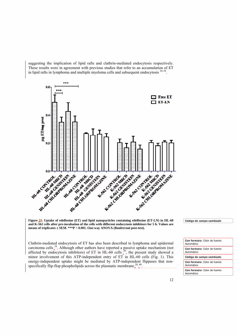
suggesting the implication of lipid rafts and clathrin-mediated endocytosis respectively. These results were in agreement with previous studies that refer to an accumulation of ET in lipid rafts in lymphoma and multiple myeloma cells and subsequent endocytosis 36-38.
Figure 22. Uptake of edelfosine (ET) and lipid nanoparticles containing edelfosine (ET-LN) in HL-60 and K-562 cells after pre-incubation of the cells with different endocytosis inhibitors for 5 h. Values are means of triplicates ± SEM. ***P < 0.001. One-way ANOVA (Bonferroni post-test).
Clathrin-mediated endocytosis of ET has also been described in lymphoma and epidermal carcinoma cells 18. Although other authors have reported a passive uptake mechanism (not affected by endocytosis inhibitors) of ET in HL-60 cells 39, the present study showed a minor involvement of this ATP-independent entry of ET in HL-60 cells (Fig. 1). This energy-independent uptake might be mediated by ATP-independent flippases that non-specifically flip-flop phospholipids across the plasmatic membrane 20, 40.
Código de campo cambiado
Con formato: Color de fuente:Automático
Con formato: Color de fuente:Automático
Código de campo cambiado
Con formato: Color de fuente:Automático
Con formato: Color de fuente:Automático

13
Regarding ET-resistant cell line K-562, the inhibition of endocytosis mechanisms induced a slight but non-significant decrease in ET internalization suggesting a minor role of CME and lipid rafts endocytosis in this cell line. Therefore, ET uptake mechanisms such as spontaneous flipping from the outer to the inner leaflet 16 might be responsible for the free drug uptake in K-562 resistant cells.
Blocking CvME with genistein did not induce significant differences in free or encapsulated ET internalization in any of the two cell lines. Moreover, endocytosis inhibitors had no apparent effect in the internalization of ET-LN. Even though endocytosis of LN by caveolin 41 and clathrin 42 has been reported by other authors, we did not detect LN internalization via these routes in the present study. This might be due to a lack of caveolae protein in the plasmatic membrane of these cells, since, as stated by some authors, the expression and/or distribution of this protein might be dependent on the activation and/or maturation state of immune cells 43. Caveolae-mediated pathway should, therefore, not be discarded in other cancer cells.
On the other hand, Guilleron et al 34 have recently showed that LN can be internalized via CME followed by macropinocytosis, suggesting that macropinocytosis could be the major endocytic process involved in LN uptake, after being activated after LN uptake by CME. Although we did not observe a CME of LN in this study, Guilleron et al. stated that the implication of CME in LN endocytosis accounted for less than 1% of total endocytic nanoparticles. Therefore, CME might also be occurring in our experiments although it was not possible to detect it. Macropinocytosis might be, therefore, the major endocytosis mechanism involved in ET-LN internalization. Nevertheless, phagocytosis might be also a reliable endocytic mechanism in these cells; indeed, HL-60 and K-562 are reported to express Fc receptors (FcR) 44, a heterogeneous group of cell membrane receptors involved in phagocytosis due to its binding to immunoglobulins (Ig). In addition, LN can be opsonized by several opsonic factors such as IgG 45 and, therefore, ET-LN might be being phagocytized by HL-60 cells. This phagocytic activity might be more relevant in case of HL-60 cells due to their neutrophilic origin 46. Nevertheless, it might be also feasible in K-562 cells; indeed, this cell line is characterized by its multipotential profile and its capacity to spontaneously differentiate into recognizable progenitors of the erythrocytic, granulocytic and monocytic series 47.
1.4.K-562 from HL-60 exhibit different molecular mechanisms that contribute to cell
death in response to ET and ET-LN treatments
As has been previously described by flow cytometry studies, both K-562 and HL-60 cell lines presented massive cell demise when grown in presence of ET-LN, and therefore the viability of cells was significantly reduced. In the case of the ET-sensitive HL-60 cell line, comparable levels of cell death were induced with the nanoencapsulated and the free drug. However, the IC50 of K-562 cell line was considerably reduced with the treatment with encapsulated drug, which inhibited its proliferation from the first 24 h of incubation at a ET-LN dose equivalent to 10 µg/ml of free drug 10. This reversion of resistance to ET in K-562 could be attributed to an increased intracellular ET concentration after nanoparticle uptake or to an enhanced cell death mechanism due to the different intracellular localization of the drug. Higher intracellular accumulation of the drug with the ET-LN was discarded on
Con formato: Color de fuente:Automático
Con formato: Color de fuente:Automático
Con formato: Color de fuente:Automático
Con formato: Color de fuente:Automático
Con formato: Color de fuente:Automático
Con formato: Color de fuente:Automático
Con formato: Color de fuente:Automático
Con formato: Color de fuente:Automático
Con formato: Color de fuente:Automático
Con formato: Color de fuente:Automático
Con formato: Color de fuente:Automático
Con formato: Color de fuente:Automático
Con formato: Color de fuente:Automático

14
the basis of the above results and, thus, different sensitivity to the treatments might depend on the death mechanisms triggered by both treatments.
To gain further insights into mechanisms for apoptosis induction, caspase activation was studied at protein level in both cell lines. Thus, cells were grown in medium containing ET or ET-LN (5 and 10 µg/mL of ET or equivalent concentrations of Blank-LN and ET-LN for HL-60 and K-562 respectively) at several time points and proteins were subsequently obtained from cell lysates. Western blots were performed to detect effector caspase activation, which would entail apoptosis induction. Additionally, one of the first identified substrates of caspases, PARP1 was inspected. The processing of both effector caspases -7 and -3 and their substrate, PARP1 was detected in ET- and ET-LN-treated HL-60 cells (Fig. 3), thereby indicating the same apoptotic cell induction of both treatment groups. These results might be explained due to the fact that HL-60 is ET-sensitive. Besides, the encapsulated drug might be rapidly internalized in HL-60 and released into the cell. Due to its sensitivity, low concentrations of ET might be enough to induce apoptosis. The abovementioned uptake studies indicate that ET may be internalized by other mechanisms apart from lipid rafts and that those mechanisms may be different from those involved in the uptake of ET-LN.
Figure 33. Characterization of caspase activation status of HL-60 and K-562 after incubation with medium, free edelfosine (ET) and drug-loaded LN (ET-LN) at a dose equivalent to 5 and 10 µg/mL of ET respectively. Caspase activation was inspected by western blot. Protein extracts of cells grown in starvation EBSS medium (Starv 3h), Staurosporine (STS), untreated control cells (C) and unloaded nanoparticles (Blank-LN) were included as controls.
Código de campo cambiado
15
On the other hand, K-562 did not display caspase-mediated apoptosis when treated with either ET or drug-loaded LN, as processed forms of inductor caspase-9 and effector caspases-3 and -7 were not detected in protein extracts (Fig. 3).
The K-562 cell line was derived from a chronic myelogenous leukemia (CML) blast crisis patient 48 and therefore it presents several characteristics of this stage of the disease, such as hyperproliferation and apoptosis resistance. For that reason, the resistance of K-562 to apoptosis induction has been extensively reported 49-52. This resistance may be attributed to Bcr-Abl expression. Besides, low ganglioside levels in cell membrane and ERK/MAPK overactivation among others may have an important role in the resistance of this cell line 53,
54. These molecular particularities would explain the absence of caspase-mediated cell death in K-562 in response to the treatments.
The abovementioned results indicate that HL-60 cell line induced caspase-mediated cell death in response to free and ET-LN treatments whereas K-562 exhibited an enhanced resistance in inducing the processing and activation of pro-caspases and therefore in inducing apoptosis via caspase activation. These findings confirm our previous observations using flow cytometry that showed absence of caspase activation in K-562 cells after treatment with either ET or ET-LN 10. Therefore, mechanisms different from caspase activation may have a role in cell induction in K-562 cell line.
1.5.Increase of autophagy-associated LC3 II protein in response to ET and ET-LN
treatments.
We have previously hypothesized that this different induction of cell death by ET-LN might be related to a distinct entry route of the drug into the cell when it is encapsulated 10, causing a different intracellular location of ET. In the above uptake studies we showed that ET endocytosis in K-562 cells was low, suggesting the major implication of passive flip-flop of ET in these cells. This uptake would promote direct delivery of the drug in the cytoplasm whereas the encapsulated ET intracellular traffic might be mediated by macropinosomes and phagosomes. Then, vesicles might: i) directly fuse with lysosomes or be engulfed by into double-membrane vesicles called autophagosomes which will later fuse with lysosomes. In addition, the aforementioned absence of caspase activation would indicate that K-562 cells might undergo a caspase-independent cell death mechanism. Hence, autophagic cell death could have a role in the cell demise induced by ET-LN in K-562 cells. To gain a better insight, LC3 I and LC3 II protein levels were inspected via western blot and subsequent band quantification. LC3 is a ubiquitin-like protein that can be detected unconjugated (LC3 I) or associated with autophagosomal membranes (LC3 II). Thus, LC3 II levels are augmented during autophagy. Accordingly, an increase in the ratio of LC3 II to LC3 I reflects the accumulation of autophagic vesicles in cells, and therefore autophagy induction.
For that reason, the status of autophagy was inspected in order to detect an increase in LC3 II/LC3 I ratio (Fig. 4). In both cell lines, the ratio increased in all the groups after 24 h of treatment. This could be explained as a response to cell stress produced due to the change of the culture conditions (that is, the presence of the nanoparticles or the free drug in the cellular media) 55. At 48 h after treatment, Blank-LN control group recovered similar
Con formato: Color de fuente:Automático
Con formato: Color de fuente:Automático
Código de campo cambiado
Con formato: Color de fuente:Automático
Con formato: Color de fuente:Automático
Con formato: Color de fuente:Automático
Con formato: Color de fuente:Automático
Con formato: Color de fuente:Automático
Con formato: Color de fuente:Automático

16
expression levels to the untreated control whereas those of the treated cells rose. This increase reached statistical significance at 48 h in K-562 cells, indicating an increase of LC3 II compared to LC3 I in nanoparticle-treated cells, which in turn entails a higher presence of autophagosomes in treated cells 10.
Figure 44. LC3 protein expression in HL-60 and K-562 cells after 24 and 48 h of treatment with edelfosine (ET) and lipid nanoparticles containing edelfosine (ET-LN) at a dose equivalent to 5 and 10 µg/mL of ET respectively. LC3I and LC3II were detected by western blot. Protein extracts of cells grown in starvation EBSS medium (Starv 3h), Staurosporine (STS), untreated control cells (C) and unloaded nanoparticles (Blank-LN) were included as controls. Graphs depict the fold increase of LC3 II/LC3 I ratio relative to untreated control cells. Values are means of triplicates ± SEM. **P<0.01 vs. Blank-LN control by one-way ANOVA (Bonferroni post-test).
Hence, both K-562 ET and ET-LN-treated cells presented high levels of LC3 II/LC3 I at 48 h, although cultures treated with encapsulated drug underwent a strong cell death induction not detected with the free drug.
The observed autophagosome induction may be due to a deleterious overactivation of defense mechanism caused by the treatment or might indicate that autophagic cell death could be responsible for the cytotoxicity of the LN. Autophagy activation may stand for a cellular attempt to cope with stress induced by cytotoxic agents as several anticancer
Con formato: Color de fuente:Automático
Código de campo cambiado

17
therapies induce the accumulation of autophagosomes in tumor cell lines in vitro 56, 57. Mishima and colleagues have described the excessive autophagy in K-562, associated with treatment of 12-O-tetradecanoyl-phorbol-13-acetate (TPA); and after notable autophagic degradation, the cells finally underwent autophagic cell death, suggesting that autophagy regulated two mechanisms in K-562 cells: both the cell survival system and autophagic cell death 58. In that sense, the observed autophagy induction in K-562 cell line might be a reflection of an stress situation that the cell undergoes in presence of either the nanoencapsulated and the free drug. In addition, ET could be internalized in K-562 by a passive flip-flop mechanism although a minor implication of CME and lipid raft endocytosis could be also involved while phagocytosis and macropinocytosis might be the preferential mechanisms implied in ET-LN uptake in these cells. Therefore, ET might be mainly delivered to the cytoplasm whereas ET-LN might suffer macroendocytic vacuole trafficking. These differences in uptake in both treatments could entail different subcellular locations of ET in K-562 cells, and therefore different effects on subcellular machinery. In addition, LN could be recycled into the cell, as previously reported 59. ET hydrolysis by lysosome fusion in autolysosomes may be counteracted in ET-LN due to its encapsulation, and therefore ET might be protected from lysosomal degradation. Afterwards, permeases would facilitate the release of the resulting products into the cytoplasm and organelles, were ET-LN would exert its toxic effects in intracellular membranes (i.e disruption of autophagosome, ER or Golgi membranes), inducing cell death or inducing permeability transition and/or rupture of the lysosomal membrane 60, provoking the release of lysosomal enzymes to the cytoplasm due to an increase in the lysosomal membrane permeability, that has been reported to trigger apoptotic cell death. Thus, the aforementioned equilibrium between cell life and stress-induced demise could have been broken towards cell death in ET-LN treated cells due to the different toxic mechanisms exerted by ET when it is incorporated in the nanoparticles. In such situations, the cells could not cope with the stress and would be driven to cell death.
On the other hand, ET-LN would alter the autophagic machinery in cells. Ma et al. 61 reported that gold nanoparticles induced an accumulation of autophagic vacuoles through lysosomal impairment. The authors described that autophagosome accumulation was induced by a blockage of the autophagic flux rather than induction of autophagy. Such interference with lysosomal function might also eventually lead to cell death and would also explain the results observed with K-562.
In addition, many efforts are being made to further understand the crosstalk between necrosis, autophagy and apoptosis 62, 63. For this reason, the novel autophagic induction described in the present paper in response to ET-LN treatment could induce a caspase-independent cell death mechanism (i.e caspase independent apoptosis or necrosis). To our knowledge this study represents the first report on the possible role of autophagy in the cellular response to LN.
CONCLUSION
The data presented above provide evidence that ET and ET-LN intracellular incorporation is prompted by different uptake mechanisms. Endocytic (HL-60) and facilitated diffusion
Con formato: Color de fuente:Automático
Con formato: Color de fuente:Automático
Con formato: Color de fuente:Automático
Con formato: Color de fuente:Automático
Código de campo cambiado
Con formato: Color de fuente:Automático
Con formato: Color de fuente:Automático

18
(HL-60 and K-562) appear to be major uptake mechanisms in free drug uptake, whereas, in case of ET-LN, passive diffusion, phagocytosis and macropinocytosis are the most likely uptake mechanisms. LN do not enhance the intracellular concentration of the drug in both leukemic cell lines despite the different uptake mechanism of ET-LN. Besides, both treatments activate caspase-mediated cell death in the ET-sensitive cell line HL-60, while conversely in K-562 caspases were not activated. Moreover, an important increase in lipidated LC3 II was detected after both treatments, pointing towards an increase of autophagic vesicles in K-562 cells. Importantly, as ET-LN overcome the resistance of K-562 cells, autophagic cell death could be involved in the cell demise process caused by the toxic effects of ET when it is incorporated in the nanoparticles at a subcellular location different from the free drug.
19
REFERENCES
(1) Burris HA 3rd1, Moore MJ, Andersen J, Green MR, Rothenberg ML, Modiano MR, Cripps MC, Portenoy RK, Storniolo AM, Tarassoff P, Nelson R, Dorr FA, Stephens CD, Von Hoff DD. Improvements in survival and clinical benefit with gemcitabine as first-line therapy for patients with advanced pancreas cancer: a randomized trial J. Clin. Oncol.1997, 15: 2403–2413
(2) Merl et al. 2010 ASCO Annual Meeting”. Chicago, IL, USA. June 4–8, 2010, JOP, vol. 11, pp. 317–320
(3) Liu QH1, Zhang J, Zhao CY, Yu DH, Bu HJ, Chen Y, Ni CY, Zhu MH. Surviving cells after treatment with gemcitabine or 5-fluorouracil for the study of de novo resistance of pancreatic cancer Cancer Lett. 2012, 314, 119-25
(4) Gonen N1, Assaraf YG. Antifolates in cancer therapy: structure, activity and mechanisms of drug resistance. Drug Resist Updat. 2012, 15, 183-21
(5) Meena AS1, Sharma A, Kumari R, Mohammad N, Singh SV, Bhat MK. Inherent and acquired resistance to paclitaxel in hepatocellular carcinoma: molecular events involved. PLoS One. 2013, 8, e61524
(6) Redner RL. Why doesn't imatinib cure chronic myeloid leukemia? Oncologist. 2010, 15, 182-6
(7) Lasa-Saracibar, B.; Estella-Hermoso de Mendoza, A.; Guada, M.; Dios-Vieitez, C.; Blanco-Prieto, M. J. Lipid nanoparticles for cancer therapy: state of the art and future prospects Expert Opin Drug Deliv. 2012, 9, 1245-61.
(8) Gajate, C.; Mollinedo, F. The antitumor ether lipid ET-18-OCH(3) induces apoptosis through translocation and capping of Fas/CD95 into membrane rafts in human leukemic cells Blood. 2001, 98, 3860-3.
(9) Castro, B. M.; Fedorov, A.; Hornillos, V.; Delgado, J.; Acuna, A. U.; Mollinedo, F.; Prieto, M. Edelfosine and Miltefosine Effects on Lipid Raft Properties: Membrane Biophysics in Cell death by Anti-Tumor Lipids. J Phys Chem B, 2013, 117, 7929–7940.
(10) Lasa-Saracíbar, B; Estella-Hermoso de Mendoza, A; Mollinedo, F; Odero, MD; Blanco-Prieto, M. J. Edelfosine lipid nanosystems overcome drug resistance in leukemic cell lines. Cancer Lett., 2013, 334, 302-310.
(11) Estella-Hermoso de Mendoza, A.; Blanco-Prieto, M. J.; Campanero, M.A., Mollinedo, F.; Villa-Pulgarín, J., Varela, R. Development and use of lipidic nanoparticles loaded with edelfosine and ether phospholipids in antitumoral and antiparasitic therapy. P201130433. 2011.
(12) Aznar, M. A.; Lasa-Saracibar, B.; Estella-Hermoso de Mendoza, A.; Blanco-Prieto, M. J. Efficacy of edelfosine lipid nanoparticles in breast cancer cells Int J Pharm. 2013, 454, 720-6.
Con formato: Color de fuente:Automático
Código de campo cambiado
Con formato: Color de fuente:Automático
Con formato: Color de fuente:Automático
Con formato: Color de fuente:Automático

20
(13) Treuel, L.; Jiang, X.; Nienhaus, G. U. New views on cellular uptake and trafficking of manufactured nanoparticles J R Soc Interface. 2013, 10, 20120939.
(14) Wang, T.; Bai, J.; Jiang, X.; Nienhaus, G. U. Cellular uptake of nanoparticles by membrane penetration: a study combining confocal microscopy with FTIR spectroelectrochemistry ACS Nano. 2012, 6, 1251-9.
(15) Hillaireau, H.; Couvreur, P. Nanocarriers' entry into the cell: relevance to drug delivery Cell Mol Life Sci. 2009, 66, 2873-96.
(16) van Blitterswijk, W. J.; Verheij, M. Anticancer mechanisms and clinical application of alkylphospholipids Biochim Biophys Acta. 2013, 1831, 663-74.
(17) Small, G. W.; Strum, J. C.; Daniel, L. W. Characterization of an HL-60 cell variant resistant to the antineoplastic ether lipid 1-O-octadecyl-2-O-methyl-rac-glycero-3-phosphocholine Lipids. 1997, 32, 715-23.
(18) Vink, S. R.; van der Luit, A. H.; Klarenbeek, J. B.; Verheij, M.; van Blitterswijk, W. J. Lipid rafts and metabolic energy differentially determine uptake of anti-cancer alkylphospholipids in lymphoma versus carcinoma cells Biochem Pharmacol. 2007, 74, 1456-65.
(19) Chen, R.; Brady, E.; McIntyre, T. M. Human TMEM30a promotes uptake of antitumor and bioactive choline phospholipids into mammalian cells J Immunol. 2011, 186, 3215-25.
(20) Pomorski, T.; Menon, A. K. Lipid flippases and their biological functions Cell Mol Life Sci. 2006, 63, 2908-21.
(21) Galluzzi, L.; Vitale, I.; Abrams, J. M.; Alnemri, E. S.; Baehrecke, E. H.; Blagosklonny, M. V.; Dawson, T. M.; Dawson, V. L.; El-Deiry, W. S.; Fulda, S.; Gottlieb, E.; Green, D. R.; Hengartner, M. O.; Kepp, O.; Knight, R. A.; Kumar, S.; Lipton, S. A.; Lu, X.; Madeo, F.; Malorni, W.; Mehlen, P.; Nunez, G.; Peter, M. E.; Piacentini, M.; Rubinsztein, D. C.; Shi, Y.; Simon, H. U.; Vandenabeele, P.; White, E.; Yuan, J.; Zhivotovsky, B.; Melino, G.; Kroemer, G. Molecular definitions of cell death subroutines: recommendations of the Nomenclature Committee on Cell Death 2012 Cell Death Differ. 2012, 19, 107-20.
(22) Kroemer, G.; Galluzzi, L.; Vandenabeele, P.; Abrams, J.; Alnemri, E. S.; Baehrecke, E. H.; Blagosklonny, M. V.; El-Deiry, W. S.; Golstein, P.; Green, D. R.; Hengartner, M.; Knight, R. A.; Kumar, S.; Lipton, S. A.; Malorni, W.; Nunez, G.; Peter, M. E.; Tschopp, J.; Yuan, J.; Piacentini, M.; Zhivotovsky, B.; Melino, G. Classification of cell death: recommendations of the Nomenclature Committee on Cell Death 2009 Cell Death Differ. 2009, 16, 3-11.
(23) Wang, P.; Zhang, L.; Peng, H.; Li, Y.; Xiong, J.; Xu, Z. The formulation and delivery of curcumin with solid lipid nanoparticles for the treatment of on non-small cell lung cancer both in vitro and in vivo Mater Sci Eng C Mater Biol Appl. 2013, 33, 4802-4808.
(24) Andon, F. T.; Fadeel, B. Programmed cell death: molecular mechanisms and implications for safety assessment of nanomaterials Acc Chem Res. 2013, 46, 733-42.
21
(25) Halamoda Kenzaoui, B.; Chapuis Bernasconi, C.; Guney-Ayra, S.; Juillerat-Jeanneret, L. Induction of oxidative stress, lysosome activation and autophagy by nanoparticles in human brain-derived endothelial cells Biochem J. 2012, 441, 813-21.
(26) Li, H.; Li, Y.; Jiao, J.; Hu, H. M. Alpha-alumina nanoparticles induce efficient autophagy-dependent cross-presentation and potent antitumour response Nat Nanotechnol. 2011, 6, 645-50.
(27) Yu, K. N.; Yoon, T. J.; Minai-Tehrani, A.; Kim, J. E.; Park, S. J.; Jeong, M. S.; Ha, S. W.; Lee, J. K.; Kim, J. S.; Cho, M. H. Zinc oxide nanoparticle induced autophagic cell death and mitochondrial damage via reactive oxygen species generation Toxicol In Vitro. 2013, 27, 1187-95.
(28) Damiano JS1, Hazlehurst LA, Dalton WS. Cell adhesion-mediated drug resistance (CAM-DR) protects the K-562 chronic myelogenous leukemia cell line from apoptosis induced by BCR/ABL inhibition, cytotoxic drugs, and gamma-irradiation. Leukemia. 2001 15, 1232-9
(29) Huang M1, Wang Y, Collins M, Graves LM. CPEC induces erythroid differentiation of human myeloid leukemia K-562 cells through CTP depletion and p38 MAP kinase. Leukemia. 2004 18, 1857-63.
(30) Estella-Hermoso de Mendoza, A.; Campanero, M. A.; Mollinedo, F.; Blanco-Prieto, M. J. Comparative study of A HPLC-MS assay versus an UHPLC-MS/MS for anti-tumoral alkyl lysophospholipid edelfosine determination in both biological samples and in lipid nanoparticulate systems J Chromatogr B Analyt Technol Biomed Life Sci. 2009, 877, 4035-41.
(31) Rubinsztein, D. C.; Cuervo, A. M.; Ravikumar, B.; Sarkar, S.; Korolchuk, V.; Kaushik, S.; Klionsky, D. J. In search of an "autophagomometer" Autophagy. 2009, 5, 585-9.
(32) Seglen, P. O.; Reith, A. Ammonia inhibition of protein degradation in isolated rat hepatocytes. Quantitative ultrastructural alterations in the lysosomal system Exp Cell Res. 1976, 100, 276-80.
(33) Mizushima, N.; Levine, B. Autophagy in mammalian development and differentiation Nat Cell Biol. 2010, 12, 823-30.
(34) He, B.; Lin, P.; Jia, Z.; Du, W.; Qu, W.; Yuan, L.; Dai, W.; Zhang, H.; Wang, X.; Wang, J.; Zhang, X.; Zhang, Q. The transport mechanisms of polymer nanoparticles in Caco-2 epithelial cells Biomaterials. 2013, 34, 6082-98.
(35) Zhao, Y.; Sun, X.; Zhang, G.; Trewyn, B. G.; Slowing, II; Lin, V. S. Interaction of mesoporous silica nanoparticles with human red blood cell membranes: size and surface effects ACS Nano. 2011, 5, 1366-75.
(36) van der Luit, A. H.; Vink, S. R.; Klarenbeek, J. B.; Perrissoud, D.; Solary, E.; Verheij, M.; van Blitterswijk, W. J. A new class of anticancer alkylphospholipids uses lipid rafts as membrane gateways to induce apoptosis in lymphoma cells Mol Cancer Ther. 2007, 6, 2337-45.
Con formato: Color de fuente:Automático
Con formato: Color de fuente:Automático
Con formato: Color de fuente:Automático

22
(37) Mollinedo, F.; de la Iglesia-Vicente, J.; Gajate, C.; Estella-Hermoso de Mendoza, A.; Villa-Pulgarin, J. A.; Campanero, M. A.; Blanco-Prieto, M. J. Lipid raft-targeted therapy in multiple myeloma Oncogene. 2010, 29, 3748-57.
(38) Gajate, C.; Gonzalez-Camacho, F.; Mollinedo, F. Involvement of raft aggregates enriched in Fas/CD95 death-inducing signaling complex in the antileukemic action of edelfosine in Jurkat cells PLoS One. 2009, 4, e5044.
(39) Tsutsumi, T.; Tokumura, A.; Kitazawa, S. Undifferentiated HL-60 cells internalize an antitumor alkyl ether phospholipid more rapidly than resistant K-562 cells Biochim Biophys Acta. 1998, 1390, 73-84.
(40) Gummadi, S. N.; Kumar, K. S. The mystery of phospholipid flip-flop in biogenic membranes Cell Mol Biol Lett. 2005, 10, 101-21.
(41) Sahay, G.; Alakhova, D. Y.; Kabanov, A. V. Endocytosis of nanomedicines J Control Release. 2010, 145, 182-95.
(42) Gilleron, J.; Querbes, W.; Zeigerer, A.; Borodovsky, A.; Marsico, G.; Schubert, U.; Manygoats, K.; Seifert, S.; Andree, C.; Stoter, M.; Epstein-Barash, H.; Zhang, L.; Koteliansky, V.; Fitzgerald, K.; Fava, E.; Bickle, M.; Kalaidzidis, Y.; Akinc, A.; Maier, M.; Zerial, M. Image-based analysis of lipid nanoparticle-mediated siRNA delivery, intracellular trafficking and endosomal escape Nat Biotechnol. 2013, 31, 638-46.
(43) Harris, J.; Werling, D.; Hope, J. C.; Taylor, G.; Howard, C. J. Caveolae and caveolin in immune cells: distribution and functions Trends Immunol. 2002, 23, 158-64.
(44) van de Winkel, J. G.; Anderson, C. L. Biology of human immunoglobulin G Fc receptors J Leukoc Biol. 1991, 49, 511-24.
(45) Uner, M.; Yener, G. Importance of solid lipid nanoparticles (SLN) in various administration routes and future perspectives Int J Nanomedicine. 2007, 2, 289-300.
(46) Gallagher, R.; Collins, S.; Trujillo, J.; McCredie, K.; Ahearn, M.; Tsai, S.; Metzgar, R.; Aulakh, G.; Ting, R.; Ruscetti, F.; Gallo, R. Characterization of the continuous, differentiating myeloid cell line (HL-60) from a patient with acute promyelocytic leukemia Blood. 1979, 54, 713-33.
(47) Lozzio, B. B.; Lozzio, C. B.; Bamberger, E. G.; Feliu, A. S. A multipotential leukemia cell line (K-562) of human origin Proc Soc Exp Biol Med. 1981, 166, 546-50.
(48) Lozzio, B. B.; Lozzio, C. B. Properties of the K-562 cell line derived from a patient with chronic myeloid leukemia Int J Cancer. 1977, 19, 136.
(49) Chang, M. P.; Bramhall, J.; Graves, S.; Bonavida, B.; Wisnieski, B. J. Internucleosomal DNA cleavage precedes diphtheria toxin-induced cytolysis. Evidence that cell lysis is not a simple consequence of translation inhibition J Biol Chem. 1989, 264, 15261-7.
(50) Kaufmann, S. H.; Desnoyers, S.; Ottaviano, Y.; Davidson, N. E.; Poirier, G. G. Specific proteolytic cleavage of poly(ADP-ribose) polymerase: an early marker of chemotherapy-induced apoptosis Cancer Res. 1993, 53, 3976-85.
(51) Martins, L. M.; Mesner, P. W.; Kottke, T. J.; Basi, G. S.; Sinha, S.; Tung, J. S.; Svingen, P. A.; Madden, B. J.; Takahashi, A.; McCormick, D. J.; Earnshaw, W. C.;

23
Kaufmann, S. H. Comparison of caspase activation and subcellular localization in HL-60 and K-562 cells undergoing etoposide-induced apoptosis Blood. 1997, 90, 4283-96.
(52) McGahon, A.; Bissonnette, R.; Schmitt, M.; Cotter, K. M.; Green, D. R.; Cotter, T. G. BCR-ABL maintains resistance of chronic myelogenous leukemia cells to apoptotic cell death Blood. 1994, 83, 1179-87.
(53) Tringali, C.; Lupo, B.; Cirillo, F.; Papini, N.; Anastasia, L.; Lamorte, G.; Colombi, P.; Bresciani, R.; Monti, E.; Tettamanti, G.; Venerando, B. Silencing of membrane-associated sialidase Neu3 diminishes apoptosis resistance and triggers megakaryocytic differentiation of chronic myeloid leukemic cells K-562 through the increase of ganglioside GM3 Cell Death Differ. 2009, 16, 164-74.
(54) Kang, C. D.; Yoo, S. D.; Hwang, B. W.; Kim, K. W.; Kim, D. W.; Kim, C. M.; Kim, S. H.; Chung, B. S. The inhibition of ERK/MAPK not the activation of JNK/SAPK is primarily required to induce apoptosis in chronic myelogenous leukemic K-562 cells Leuk Res. 2000, 24, 527-34.
(55) Kroemer, G.; Marino, G.; Levine, B. Autophagy and the integrated stress response Mol Cell. 2010, 40, 280-93.
(56) Kroemer, G.; Levine, B. Autophagic cell death: the story of a misnomer Nat Rev Mol Cell Biol. 2008, 9, 1004-10.
(57) Maiuri, M. C.; Zalckvar, E.; Kimchi, A.; Kroemer, G. Self-eating and self-killing: crosstalk between autophagy and apoptosis Nat Rev Mol Cell Biol. 2007, 8, 741-52.
(58) Mishima Y1, Terui Y, Mishima Y, Taniyama A, Kuniyoshi R, Takizawa T, Kimura S, Ozawa K, Hatake K Autophagy and autophagic cell death are next targets for elimination of the resistance to tyrosine kinase inhibitors. Cancer Sci. 2008, 11, 2200-8.
(59) Sahay G1, Querbes W, Alabi C, Eltoukhy A, Sarkar S, Zurenko C, Karagiannis E, Love K, Chen D, Zoncu R, Buganim Y, Schroeder A, Langer R, Anderson DG. Efficiency of siRNA delivery by lipid nanoparticles is limited by endocytic recycling. Nat Biotechnol. 2013, 7, 653-8.
(60) Ohkuma S, Poole B. Fluorescence probe measurement of the intralysosomal pH in living cells and the perturbation of pH by various agents. Proc Natl Acad Sci U S A. 1978 7, 3327-31.
(61) Ma, X.; Wu, Y.; Jin, S.; Tian, Y.; Zhang, X.; Zhao, Y.; Yu, L.; Liang, X. J. Gold nanoparticles induce autophagosome accumulation through size-dependent nanoparticle uptake and lysosome impairment ACS Nano. 2011, 5, 8629-39.
(62) Nikoletopoulou, V.; Markaki, M.; Palikaras, K.; Tavernarakis, N. Crosstalk between apoptosis, necrosis and autophagy. Biochim Biophys Acta, 2013. doi: 10.1016/j.bbamcr.2013.06.001
(63) Strasser, A.; Cory, S.; Adams, J. M. Deciphering the rules of programmed cell death to improve therapy of cancer and other diseases EMBO J. 2011, 30, 3667-83.
Con formato: Color de fuente:Automático
Con formato: Color de fuente:Automático

24

25
CONFLICT OF INTEREST DISCOSURE
The authors have no other relevant affiliations or financial involvement with any organization or entity with a financial interest in or financial conflict with the subject matter or materials discussed in the manuscript apart from those disclosed.
AKNOWLEDGEMENTS
This work has been carried out in the framework of the COST Action TD1004. Financial support from Caja Navarra Foundation, University of Navarra (FUN), the Government of Navarra, Department of Health (ref: 63/09, ‘Ortiz de Landázuri’ fellowship) and the Spanish Ministry of Science and Innovation (SAF2010-15547 are acknowledged. B. Lasa-Saracibar is supported by the research grant from “Asociación de Amigos de la Universidad de Navarra”.
AUTHORS CONTRIBUTIONS
The manuscript was written through contributions of all authors. All authors have given approval to the final version of the manuscript. ‡These authors contributed equally.


![Monoclonal Antibody-Based Therapies for Myasthenia Gravis...562 S. Alabbad et al. Eculizumabisahumanized,chimericmonoclonalanti-bodythattargetscomplementprotein5(C5)[45].Eculi-zumabinhibitstheC5convertaseandtherebylimitsthe](https://static.fdocuments.us/doc/165x107/61374f1e0ad5d2067648899e/monoclonal-antibody-based-therapies-for-myasthenia-gravis-562-s-alabbad-et.jpg)
![[XLS] · Web view2957 2836 260577 263245 1226 381 2415000 92 562 2850356 2524827 137 562 4032654 2230516 330 562 2600004 2604606 346 562 2852792 367 562 9219536032 9258046774 531 562](https://static.fdocuments.us/doc/165x107/5aa8f7477f8b9a95188c374d/xls-view2957-2836-260577-263245-1226-381-2415000-92-562-2850356-2524827-137-562.jpg)


![562 IEEE TRANSACTIONS ON AUTOMATIC CONTROL, VOL. 51, …mn/...TAC_dynamic_active_contours_for_visual_tr… · tions to autonomous driving.2 See Sinopoli et al. [10] and Sharp et al.](https://static.fdocuments.us/doc/165x107/5f1f9dff4303091c4c0feee7/562-ieee-transactions-on-automatic-control-vol-51-mntacdynamicactivecontoursforvisualtr.jpg)
