
Ectopic Pregnancy - fogsi.org
12
FOGSI GCPR ON ECTOPIC PREGNANCY FOGSI President Dr Alpesh Gandhi | Version 01 27th September, 2020 Editors Dr Alpesh Gandhi Dr Suchitra Pandit Dr Parikshit Tank Co-ordinators Dr Mansi Medhekar Dr Chinmay Umarjee
Transcript of Ectopic Pregnancy - fogsi.org

FOGSI GCPRON
ECTOPIC PREGNANCYFOGSI President
Dr Alpesh Gandhi
|Version 01 27th September, 2020
Editors
Dr Alpesh Gandhi
Dr Suchitra Pandit
Dr Parikshit Tank
Co-ordinators
Dr Mansi Medhekar
Dr Chinmay Umarjee

Editors:
Dr Alpesh Gandhi
Dr Suchitra Pandit
Dr Parikshit Tank
FOGSI GCPRON
Ectopic Pregnancy
Coordinators:
Dr Mansi Medhekar
Dr Chinmay Umarjee
Dr Anita Singh
Dr Ashok Kumar
Dr Chandrakant Sanklecha
Dr Manish Machave
Dr Mehul Damani
Dr Mitra Saxena
Dr Pragnesh Shah
Dr Ramesh B
Dr Sarita Bhalerao
Dr Sunil Shah
Dr Sushma Baxi
Dr Suvarna Khadilkar
Experts
Dr Parul Kotdawala
FOGSI GCPRON
Ectopic Pregnancy
FOGSI President
Dr Alpesh Gandhi

Introduction:Ectopic pregnancy is the leading cause of maternal death in early pregnancy. Ectopic pregnancy is defined as the implantation of a fertilized egg outside the uterine cavity. The most common ectopic site of implantation (97%) is the fallopian tube. The remaining 3% of ectopic pregnancies are implanted in the cervix, ovary, peritoneal cavity, or uterine scars. A growing ectopic pregnancy in any location can cause the tissue to become vascular, friable and eventually rupture resulting in internal bleeding. This situation can be life threatening and needs to be treated as medical emergency.
Risk factors: · Pelvic inflammatory disease including pelvic tuberculosis· Previous ectopic pregnancy· Pregnancy which has occurred with an Intrauterine Device· Tubal surgeries (ligations, reconstructions, re-implantations)· Sexually transmitted diseases· Smoking · Infertility, ovulation induction and ART procedures However, the majority of women with an ectopic pregnancy have no identifiable risk factor.
Incidence:The incidence of ectopic pregnancy among all pregnancies is about 0.25-2.0% (1).
Indian studies have found a incidence of ectopic pregnancies ranging from 1-2%.(2). However, there is a marked disparity in case fatality rates. Some studies have shown no mortality,whereas, it is as high as 3.5% in others.(3). This is a reflection of the variable quality and infrastructure of care available in the country across various settings. In India there is high prevalence of pelvic tuberculosis. Pelvic tuberculosis has been identified as an important etiological factor. In one study, genital tuberculosis was found in 13.2% of all cases of ectopic pregnancy.(4)
Clinical presentation Ectopic pregnancy shouldbe suspected in any woman with child bearing age presenting to the clinic or casualty with symptoms such as amenorrhea, abdominal pain and vaginal bleeding (5). The presentation may sometimes be dominated with the complaint or fainting, collapse, breathlessness, or dizziness. Uncommon symptoms include diarrhoea, pain in the shoulder, rectal pressure, urinary symptoms, and anaemia.
The range of presentations correspondingly produces a variety of features on examination. For a small, undisturbed ectopic pregnancy, the physical examination could be normal. In these situations, the diagnosis is based on investigations. On the other hand, with late presentations, there could be a disturbance of the vital signs and features of shock may be present including tachycardia, tachypnea, hypotension, and rarely bradycardia. Abdominal examination may reveal guarding, rigidity and tenderness. There may be cervical motion tenderness, adnexal tenderness or fullness in the adnexae and pouch of Douglas.

The presence of abdominal signs with altered vital parameters suggests a hemoperitoneum and mandates urgent resuscitation and management at a centre with appropriate facilities for surgery and blood transfusion.
Diagnostic testsDiagnostic tests for ectopic pregnancy include a urine pregnancy tests, Serum beta hcG and ultrasound. Increased clinical suspicion combined with these tests plays a very important role in management and outcome.
The instant result of a urine pregnancy test is a useful pointer for the clinician to suspect an ectopic pregnancy. The easy availability, low cost and reliability of this test should be utilized. The test is a useful triage tool for clinicians to rule out a pregnancy when the clinical situation is not clear such as a patient who is not sure of dates, does not remember or is in a state of shock and the history cannot be elicited.
Laboratory testsA single laboratory value of beta hCG should not be used to diagnose the location of a pregnancy. The typical level in a healthy pregnancy on the day of the missed period is 50 to 100 IU/L. In a healthy intrauterine pregnancy, levels of serum �-hCG will double every 1.4 to 2.1 days and peak between 50000 and 100000 IU/L at 8 to 10 weeks of pregnancy. Compared to the pattern observed in healthy intrauterine pregnancies, the rate of increase between two serum�-hCG levels done 48 hours apart is slower (<50% increase) in unhealthy pregnancies (those destined to miscarry or in ectopic locations)(6,7). There are, however exceptions to the rule. Viable pregnancies may have a slow rise in serum levels and ectopic pregnancies which are viable may mimic a healthy intrauterine pregnancy in terms of serum �-hCGlevels(8). Also, interpreting levels and rates of rise are complicated in situations where the pregnancy is a result of assisted reproduction or a multiple pregnancy. Taking into view these considerations, paired serum �-hCG levels though useful, should be used in conjunction with transvaginal ultrasound to establish a diagnosis.This approach does not have a role in the diagnosis of caesarean scar and heterotropic pregnancy pregnancy.
The initial serum �-hCGlevel is a key prognostic indicator for the success of conservative management (expectant and medical) in cases of ultrasound visualized ectopic pregnancies in all locations.
A serum b-hCG level may not be available at short notice in all care settings. Depending on the clinical situation, pregnancy can be confirmed by a urine pregnancy test and one may proceed with emergency care as appropriate using clinical judgement. A single serum b-hCG should be carried out at diagnosis to help with management.
Progesterone levels are not useful for the diagnosis of an ectopic and maybe used in the prognostication of pregnancy of unknown location.
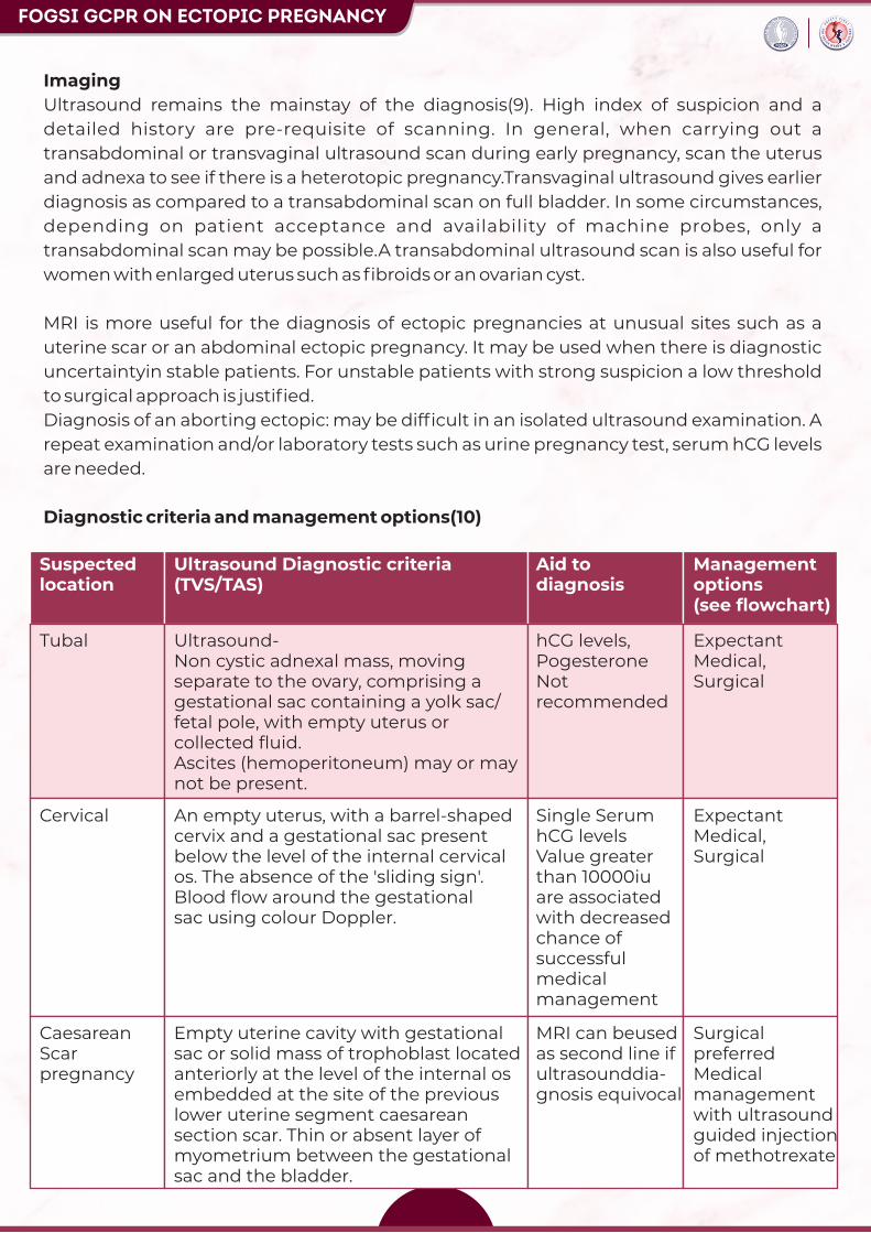
ImagingUltrasound remains the mainstay of the diagnosis(9). High index of suspicion and a detailed history are pre-requisite of scanning. In general, when carrying out a transabdominal or transvaginal ultrasound scan during early pregnancy, scan the uterus and adnexa to see if there is a heterotopic pregnancy.Transvaginal ultrasound gives earlier diagnosis as compared to a transabdominal scan on full bladder. In some circumstances, depending on patient acceptance and availability of machine probes, only a transabdominal scan may be possible.A transabdominal ultrasound scan is also useful for women with enlarged uterus such as fibroids or an ovarian cyst.
MRI is more useful for the diagnosis of ectopic pregnancies at unusual sites such as a uterine scar or an abdominal ectopic pregnancy. It may be used when there is diagnostic uncertaintyin stable patients. For unstable patients with strong suspicion a low threshold to surgical approach is justified. Diagnosis of an aborting ectopic: may be difficult in an isolated ultrasound examination. A repeat examination and/or laboratory tests such as urine pregnancy test, serum hCG levels are needed.
Diagnostic criteria and management options(10)
Suspected location
Ultrasound Diagnostic criteria(TVS/TAS)
Aid to diagnosis
Management options (see flowchart)
Tubal Ultrasound- Non cystic adnexal mass, moving separate to the ovary, comprising a gestational sac containing a yolk sac/ fetal pole, with empty uterus or collected fluid. Ascites (hemoperitoneum) may or may not be present.
hCG levels,PogesteroneNot recommended
ExpectantMedical,Surgical
Cervical An empty uterus, with a barrel-shaped cervix and a gestational sac present below the level of the internal cervical os. The absence of the 'sliding sign'. Blood flow around the gestational sac using colour Doppler.
Single Serum hCG levelsValue greaterthan 10000iu are associated with decreasedchance of successful medical management
ExpectantMedical,Surgical
Caesarean Scarpregnancy
Empty uterine cavity with gestational sac or solid mass of trophoblast locatedanteriorly at the level of the internal os embedded at the site of the previous lower uterine segment caesarean section scar. Thin or absent layer of myometrium between the gestational sac and the bladder.
MRI can beusedas second line ifultrasounddia-gnosis equivocal
Surgical preferredMedical management with ultrasound guided injectionof methotrexate

Evidence of prominent trophoblastic/placental circulation on Doppler examination. Empty endocervical canal.
in the gestatio-nal sac. Intramuscular methotrexate may be used for very early presentations
Interstitial Empty uterine cavity with products of conception / gestational sac located laterally in the interstitial (intramural) part of the tube and surrounded by less than 5 mm of myometrium in all imaging planes.Presence of the 'interstitial line sign'.
3D ultrasound or MRI to distinguish between angular and early intrauterine pregnancy.
Medical Surgical
Cornualrudimentary horn
Visualization of a single interstitial portion of fallopian tube in the main uterine body and gestational sac/products of conception seen mobile and separate from the uterus and completely surrounded by myometrium.Vascular pedicle adjoining the gestational sac to the unicornuate uterus.
3D ultrasound or MRI to distinguish between angular and early intrauterine pregnancy.
Medical Surgical
Ovarian No specific agreed criteria Findings suggestive:Empty uterusWide echogenic ring with an internal anechoic area on ovaryNegative sliding organ sign- not possible to separate cysticstructure from ovary on gentle probe palpationCorpus luteum identified separate fromthe ovary Presence of free fluid around the mass may represent ruptured ovarian ectopic
Single serumhCG
Surgical
Heterotropic Ultrasound presence of an intrauterine pregnancy with a coexisting ectopic pregnancy
Persistant painand rising hCG levels following miscarriage or termination of pregnancy
SurgicalMedical/ Expectantif non-viable intrauterine pregnancy
Abdominal Ectopic
Absence of an intrauterine gestational sac Absence of both an evident dilated tube and a complex adnexal mass. A gestational cavity surrounded by loops of bowel and separated from them by peritoneum.
MRI, hCG levels, SurgicalMedical/ Expectantif non-viable intrauterine pregnancy

A wide mobility similar to fluctuation of the sac, particularly evident with pressure of the transvaginal probe toward the posterior cul-de-sac.
Pregnancy of unknown location
Scan cannot demonstrate a pregnancyhowever hCG levels confirm the same.
Serial hCG, scanning, highindex of suspicion. SOS Surgery.
Expectant for confirmation of location. Persistent PUL can be offered medical or surgical treatment.
MEDICAL MANAGEMENT CRITERIA
Pregnancy confirmed:Tubal, scar or
cervical ectopic.
No symptoms, haemodynamically stable, scan documents ectopic
pregnancy.
Ultrasound criteria- Mean GSD<35mm
Absent cardiac activity, no signs of rupture. Beta-hCG<1500 or
1500-5000*
Medical management
YES
YES
Criteria not meet
NO Surgery- Laparoscopy if facilitites are available. Prefer GA for unstable patients.
Clinically unstable or criteria for expectant or medical managementnot met
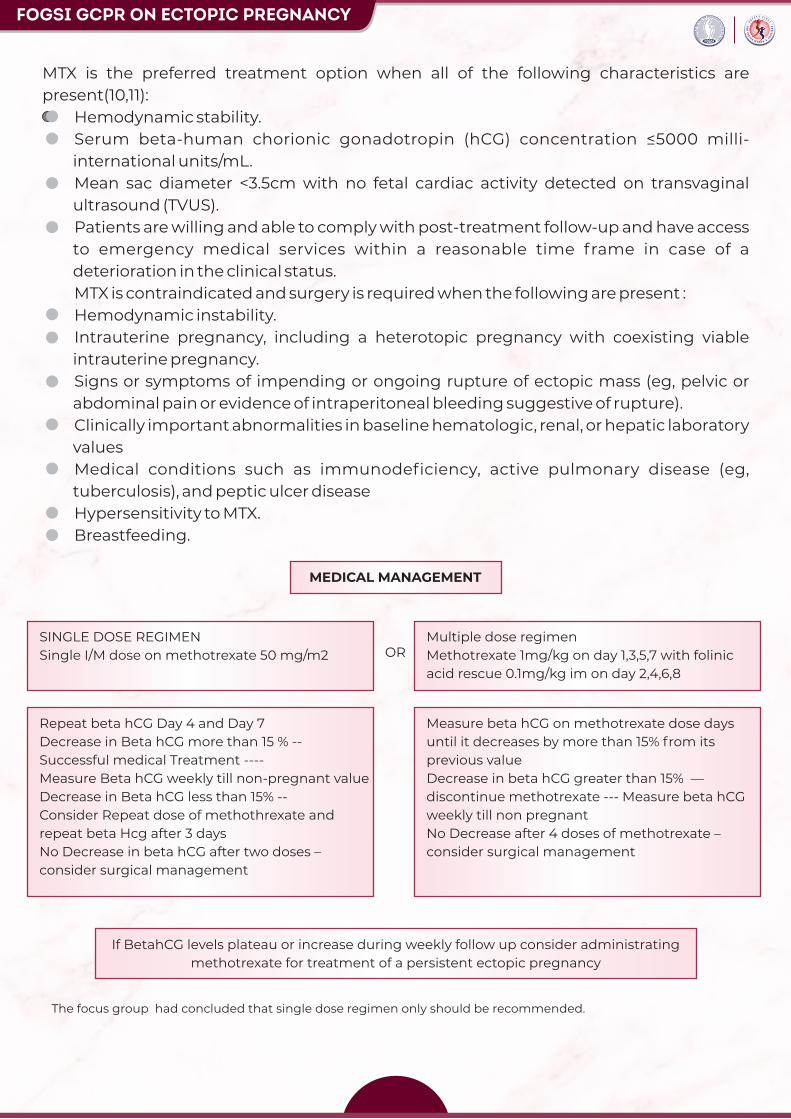
MTX is the preferred treatment option when all of the following characteristics are present(10,11):
Hemodynamic stability.Serum beta-human chorionic gonadotropin (hCG) concentration ≤5000 milli-international units/mL.Mean sac diameter <3.5cm with no fetal cardiac activity detected on transvaginal ultrasound (TVUS). Patients are willing and able to comply with post-treatment follow-up and have access to emergency medical services within a reasonable time frame in case of a deterioration in the clinical status.MTX is contraindicated and surgery is required when the following are present :Hemodynamic instability.Intrauterine pregnancy, including a heterotopic pregnancy with coexisting viable intrauterine pregnancy. Signs or symptoms of impending or ongoing rupture of ectopic mass (eg, pelvic or abdominal pain or evidence of intraperitoneal bleeding suggestive of rupture).Clinically important abnormalities in baseline hematologic, renal, or hepatic laboratory values Medical conditions such as immunodeficiency, active pulmonary disease (eg, tuberculosis), and peptic ulcer disease Hypersensitivity to MTX.Breastfeeding.
MEDICAL MANAGEMENT
ORSINGLE DOSE REGIMENSingle I/M dose on methotrexate 50 mg/m2
Multiple dose regimen Methotrexate 1mg/kg on day 1,3,5,7 with folinic acid rescue 0.1mg/kg im on day 2,4,6,8
Repeat beta hCG Day 4 and Day 7Decrease in Beta hCG more than 15 % -- Successful medical Treatment ---- Measure Beta hCG weekly till non-pregnant valueDecrease in Beta hCG less than 15% -- Consider Repeat dose of methothrexate and repeat beta Hcg after 3 daysNo Decrease in beta hCG after two doses – consider surgical management
Measure beta hCG on methotrexate dose days until it decreases by more than 15% from its previous valueDecrease in beta hCG greater than 15% —discontinue methotrexate --- Measure beta hCG weekly till non pregnantNo Decrease after 4 doses of methotrexate – consider surgical management
If BetahCG levels plateau or increase during weekly follow up consider administrating methotrexate for treatment of a persistent ectopic pregnancy
The focus group had concluded that single dose regimen only should be recommended.
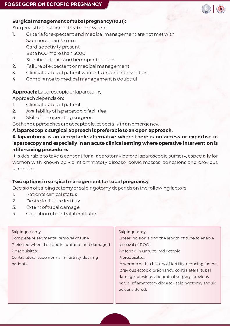
Surgical management of tubal pregnancy(10,11):Surgery isthe first line of treatment when:1. Criteria for expectant and medical management are not met with· Sac more than 35 mm· Cardiac activity present· Beta hCG more than 5000· Significant pain and hemoperitoneum2. Failure of expectant or medical management3. Clinical status of patient warrants urgent intervention4. Compliance to medical management is doubtful
Approach: Laparoscopic or laparotomyApproach depends on:1. Clinical status of patient2. Availability of laparoscopic facilities3. Skill of the operating surgeonBoth the approaches are acceptable, especially in an emergency. A laparoscopic surgical approach is preferable to an open approach. A laparotomy is an acceptable alternative where there is no access or expertise in laparoscopy and especially in an acute clinical setting where operative intervention is a life-saving procedure.It is desirable to take a consent for a laparotomy before laparoscopic surgery, especially for women with known pelvic inflammatory disease, pelvic masses, adhesions and previous surgeries.
Two options in surgical management for tubal pregnancyDecision of salpingectomy or salpingotomy depends on the following factors1. Patients clinical status2. Desire for future fertility3. Extent of tubal damage4. Condition of contralateral tube
Salpingectomy
Complete or segmental removal of tube
Preferred when the tube is ruptured and damaged
Prerequisites:
Contralateral tube normal in fertility-desiring
patients
Salpingotomy
Linear incision along the length of tube to enable
removal of POCs
Preferred in unruptured ectopic
Prerequisites:
In women with a history of fertility-reducing factors
(previous ectopic pregnancy, contralateral tubal
damage, previous abdominal surgery, previous
pelvic inflammatory disease), salpingotomy should
be considered.
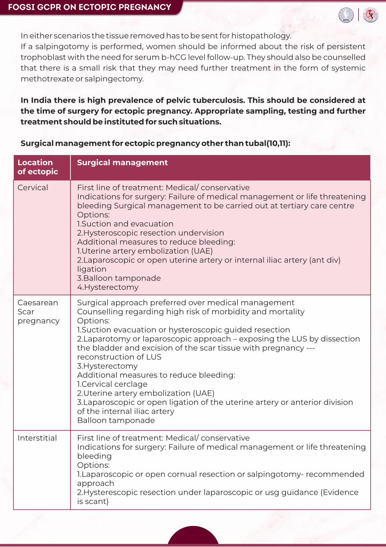
In either scenarios the tissue removed has to be sent for histopathology.If a salpingotomy is performed, women should be informed about the risk of persistent trophoblast with the need for serum b-hCG level follow-up. They should also be counselled that there is a small risk that they may need further treatment in the form of systemic methotrexate or salpingectomy.
In India there is high prevalence of pelvic tuberculosis. This should be considered at the time of surgery for ectopic pregnancy. Appropriate sampling, testing and further treatment should be instituted for such situations.
Surgical management for ectopic pregnancy other than tubal(10,11):
Location of ectopic
Surgical management
Cervical First line of treatment: Medical/ conservativeIndications for surgery: Failure of medical management or life threateningbleeding Surgical management to be carried out at tertiary care centreOptions:1. Suction and evacuation2. Hysteroscopic resection undervisionAdditional measures to reduce bleeding:1. Uterine artery embolization (UAE)2. Laparoscopic or open uterine artery or internal iliac artery (ant div) ligation3. Balloon tamponade4. Hysterectomy
Caesarean Scar pregnancy
Surgical approach preferred over medical managementCounselling regarding high risk of morbidity and mortalityOptions:1. Suction evacuation or hysteroscopic guided resection 2. Laparotomy or laparoscopic approach – exposing the LUS by dissection the bladder and excision of the scar tissue with pregnancy --- reconstruction of LUS3. HysterectomyAdditional measures to reduce bleeding:1. Cervical cerclage2. Uterine artery embolization (UAE)3. Laparoscopic or open ligation of the uterine artery or anterior division of the internal iliac arteryBalloon tamponade
Interstitial First line of treatment: Medical/ conservativeIndications for surgery: Failure of medical management or life threatening bleedingOptions:1. Laparoscopic or open cornual resection or salpingotomy- recommended approach2. Hysterescopic resection under laparoscopic or usg guidance (Evidence is scant)
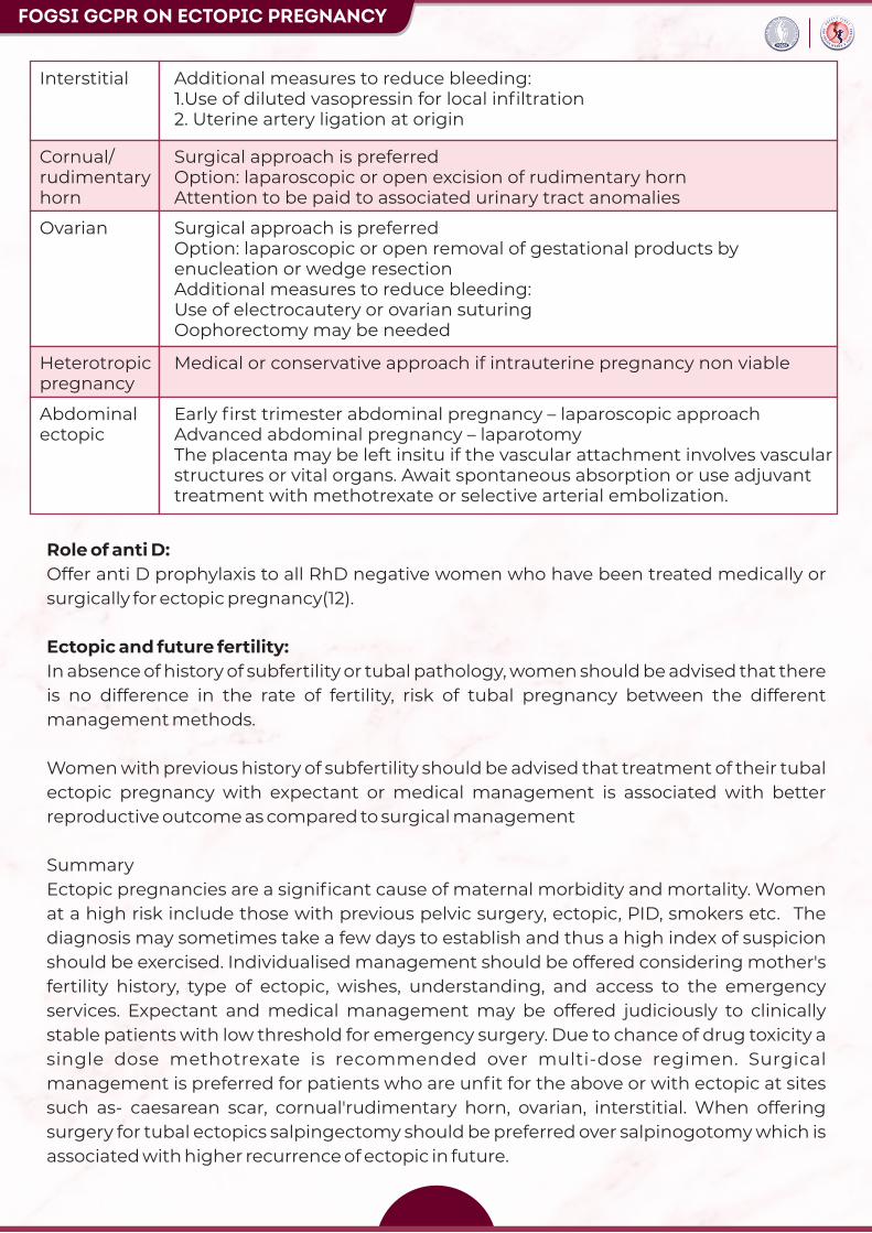
Interstitial Additional measures to reduce bleeding:1. Use of diluted vasopressin for local infiltration2. Uterine artery ligation at origin
Cornual/ rudimentaryhorn
Surgical approach is preferredOption: laparoscopic or open excision of rudimentary hornAttention to be paid to associated urinary tract anomalies
Ovarian Surgical approach is preferredOption: laparoscopic or open removal of gestational products by enucleation or wedge resection Additional measures to reduce bleeding:Use of electrocautery or ovarian suturingOophorectomy may be needed
Heterotropicpregnancy
Medical or conservative approach if intrauterine pregnancy non viable
Abdominalectopic
Early first trimester abdominal pregnancy – laparoscopic approachAdvanced abdominal pregnancy – laparotomyThe placenta may be left insitu if the vascular attachment involves vascularstructures or vital organs. Await spontaneous absorption or use adjuvant treatment with methotrexate or selective arterial embolization.
Role of anti D:Offer anti D prophylaxis to all RhD negative women who have been treated medically or surgically for ectopic pregnancy(12).
Ectopic and future fertility:In absence of history of subfertility or tubal pathology, women should be advised that there is no difference in the rate of fertility, risk of tubal pregnancy between the different management methods.
Women with previous history of subfertility should be advised that treatment of their tubal ectopic pregnancy with expectant or medical management is associated with better reproductive outcome as compared to surgical management
SummaryEctopic pregnancies are a significant cause of maternal morbidity and mortality. Women at a high risk include those with previous pelvic surgery, ectopic, PID, smokers etc. The diagnosis may sometimes take a few days to establish and thus a high index of suspicion should be exercised. Individualised management should be offered considering mother's fertility history, type of ectopic, wishes, understanding, and access to the emergency services. Expectant and medical management may be offered judiciously to clinically stable patients with low threshold for emergency surgery. Due to chance of drug toxicity a single dose methotrexate is recommended over multi-dose regimen. Surgical management is preferred for patients who are unfit for the above or with ectopic at sites such as- caesarean scar, cornual'rudimentary horn, ovarian, interstitial. When offering surgery for tubal ectopics salpingectomy should be preferred over salpinogotomy which is associated with higher recurrence of ectopic in future.

Routine endometrial curettage to document Arias Stella reaction is NOT recommended. Anti-D should be offered to Rh negative mothers.
References:1. Stulberg DB, Cain LR, Dahlquist I, Lauderdale DS. Ectopic pregnancy rates and racial disparities in the
Medicaid population, 2004-2008. FertilSteril2014; 102:1671. 2. S Tahmina et al. Clinical analysis of ectopic pregnancies in a tertiary care centre in southern India: A six
year retrospective study. J Clin Diagn Res 2016 Oct; 10(10)3. Changing epidemiology of maternal mortality in rural India: time to reset strategies for MDG-5. Shah P,
Shah S, Kutty RV, Modi D. Trop Med Int Health 2014; 19(5):568-575.4. Sharma JB, Naha M, Kumar S, Roy KK, Singh N, Arora R. Genital tuberculosis: an important cause of
ectopic pregnancy in India. Indian J Tuberc. 2014 Oct;61(4):312-75. Della-Giustina D, Denny M. Ectopic pregnancy. Emerg Med Clin North Am 2003;21:565-84.6. Barnhart KT. Clinical Practice. Ectopic Pregnancy. N Engl J Med 2009;361:379-87.7. Tank PD. Early Pregnancy Complications. In: Arias' Practical Guide to High Risk Pregnancy and Delivery:
a South Asian Perspective. Eds: Arias F, Bhide AG, Arulkumaran S, Damania K, Daftary SN. Elsevier India, New Delhi; 2019.
8. Morse CB, Sammel MD, Shaunik A, et al. Performance of human chorionic gonadotropin curves in women at risk for ectopic pregnancy: exceptions to the rules. Fertility and Sterility. 2012;97(1):101-106.
9. Gurel S. Sarikaya B, Gurel K, Akata D. Role of sonography in the diagnosis of Ectopic Pregnancy. J Clin Ultrasound. 2007;35:509-517.
10. RCOG. Diagnosis and Management of Ectopic Pregnancy: Green-top Guideline no 21, Nov 201611. ACOG Practice Bulletin No. 191: Tubal Ectopic Pregnancy: February 2018 - Volume 131 - Issue 2 - p e65-
e7712. . 12. BCSH guideline for the use of anti‐D immunoglobulin for the prevention of haemolytic disease of the
fetus and newborn H. Qureshi 2014