Economics Brain imaging- fMRI and beyond - oecd.org€¦ · Brain imaging- fMRI and beyond....
16
Restricted © Siemens Healthcare GmbH, 2016 The State of Mind in Economics Brain imaging- fMRI and beyond Christine Boehm, PhD MD October 30, 2017
Transcript of Economics Brain imaging- fMRI and beyond - oecd.org€¦ · Brain imaging- fMRI and beyond....

Restricted © Siemens Healthcare GmbH, 2016
The State of Mind in Economics Brain imaging- fMRI and beyond
Christine Boehm, PhD MD October 30, 2017
HC DI MR SI Page 2 | Confidential © Siemens Healthcare GmbH, 2016
> €1 bn R&D spent
75 countries
with direct presence
> 209,000 patients
every hour2
Biggest supplier
of medtech infrastructure
World market leader
in most businesses
46,000 employees
12,500 granted patents
globally
> 70% of critical clinical
decisions are influenced by the
type of technology we provide1
€13 bn revenue
Access for
1.08 bn people in developing
countries2
Who We Are
1 AdvaMedDX, “A Policy Primer on Diagnostics”, June 2011, page 3 2 Siemens AG, “Sustainable healthcare strategy - Indicators in fiscal 2014”, page 3-4

HC DI MR SI Page 3 | Confidential © Siemens Healthcare GmbH, 2016
Our innovations – 120 years track record
3 1) Product availability may vary from country to country and is subject to varying regulatory requirements.
Enterprise Services
Advanced Therapies
Molecular Diagnostics
Digital Health
Services
1896 Industrially manufactured X-ray appliance for medical diagnostics
1956 CLINISTIX − dry chemistry testing for glucose in urine
1957 Fully automated discrete chemistry analyzer for whole blood or serum
1967 First real-time ultrasound scanner
1982 First acridinium
ester based chemilumin-
escence immuno-assays
1983 First Siemens MRI scanner
1999 First intuitive medical IT platform from Siemens
2005 First Dual Source CT scanner
2008 Robotic-assisted angiography system
2011 First integrated, simultaneous whole-body MRI and PET
2014 Cloud-based network: teamplay
1975 First Siemens CT scanner
1998 First Siemens
track-based laboratory
automation system
2001 First PET/CT system from
Siemens
2006 Diagnostic
analyzer integrating four technologies in
one system
2009 Multi-modality
3D imaging network
2008 Digital radiography, wireless flat panel detector
2014 “Free
breathing”CT scanning with
dual X-ray sources & detectors
2012 Wireless transducers for ultrasound
2015 First Twin Robotic X-ray scanner for enhanced patient care and productivity
2017 Lab diagnostics solution for immunoassay and clinical chemistry: AtellicaTM Solution1
2015 Wide-angle image acquisition breast
tomosynthesis
2016 Liquid biopsy
Future
Costs
Outcomes
1901 Nobel prize
winners (Physics + Medicine)
Röntgen Von Behring
HC DI MR SI Page 4 | Confidential © Siemens Healthcare GmbH, 2016
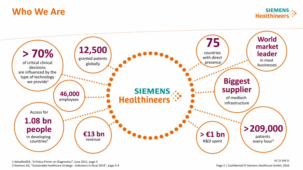
Ultrasound
Advanced Therapies
Diagnostic Imaging
Laboratory Diagnostics
Point of Care
Services
Computed Tomography, Magnetic Resonance Imaging, Molecular Imaging, Radiography & Fluoroscopy, Imaging IT
Cardiology, Interventional Radiology, Radiation Oncology, Surgery
Cardiology, Radiology
Immunoassay, Chemistry, Hematology, Hemostasis, Specialty Testing, Automation, IT and Services, Molecular Diagnostics1
Blood Gas, Diabetes Urinalysis, Coagulation, Cardiology
System Services, Education, Enterprise Services, Digital Services
Undisputed market leader in diagnostic imaging
Empowering innovative therapy concepts
Versatility and functionality across clinical questions
Delivering clinical and workflow excellence
Lab-accurate, actionable, timely results at the point of care
Transformative services to maximize opportunities and minimize risks
1) Incubated within Business Function Strategy & Innovation 2) Image courtesy Diagnostic Imaging: CMRR, Minneapolis, MGH, Boston
Image courtesy Advanced Therapies: IHU Strasbourg, France
Engineering success – With broadest and deepest portfolio
4
We support
to raise …
clinical excellence
financial profitability
operational efficiency
HC SI MR SI Page 5 | Restricted © Siemens Healthcare GmbH, 2016
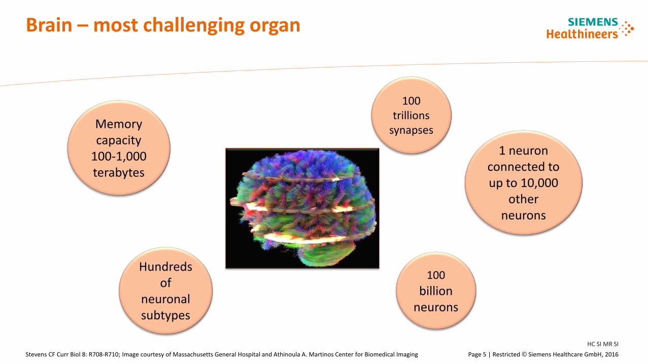
Brain – most challenging organ
Stevens CF Curr Biol 8: R708-R710; Image courtesy of Massachusetts General Hospital and Athinoula A. Martinos Center for Biomedical Imaging
100 billion
neurons
100 trillions
synapses
Hundreds of
neuronal subtypes
1 neuron connected to up to 10,000
other neurons
Memory capacity
100-1,000 terabytes
HC SI MR SI Page 6 | Restricted © Siemens Healthcare GmbH, 2016
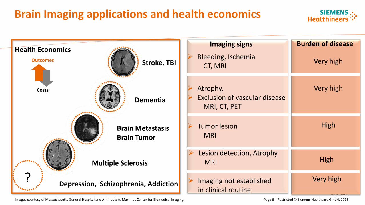
Brain Imaging applications and health economics
Images courtesy of Massachusetts General Hospital and Athinoula A. Martinos Center for Biomedical Imaging
Burden of disease
Atrophy, Exclusion of vascular disease
MRI, CT, PET
Very high
Tumor lesion MRI
High
Lesion detection, Atrophy MRI High
Imaging not established in clinical routine
Very high
Imaging signs
Costs
Outcomes
Depression, Schizophrenia, Addiction
Multiple Sclerosis
Brain Metastasis Brain Tumor
Dementia
Stroke, TBI
Health Economics Very high
Bleeding, Ischemia CT, MRI

HC SI MR SI Page 7 | Restricted © Siemens Healthcare GmbH, 2016
Magnetic Resonance Imaging (MRI)
Non-invasive imaging technology Magnetic moment of proton of
hydrogen atoms Scanner consists of a strong
magnet (1,5 T; 3 T; 7 T) with radio transmitter and receiver
Uses magnetism and radio waves to produce body images
Produces series of slices of-tissue contrast images based on signal intensities („T1, T2 contrast“)
Siemens Patient information about an MRI scan
HC SI MR SI Page 8 | Restricted © Siemens Healthcare GmbH, 2016
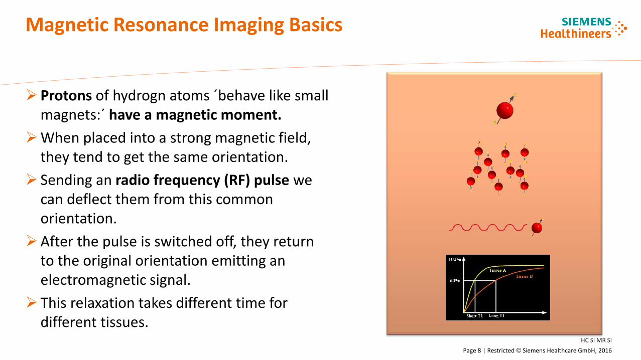
Magnetic Resonance Imaging Basics
Protons of hydrogn atoms ´behave like small magnets:´ have a magnetic moment.
When placed into a strong magnetic field, they tend to get the same orientation.
Sending an radio frequency (RF) pulse we can deflect them from this common orientation.
After the pulse is switched off, they return to the original orientation emitting an electromagnetic signal.
This relaxation takes different time for different tissues.
HC SI MR SI Page 9 | Restricted © Siemens Healthcare GmbH, 2016
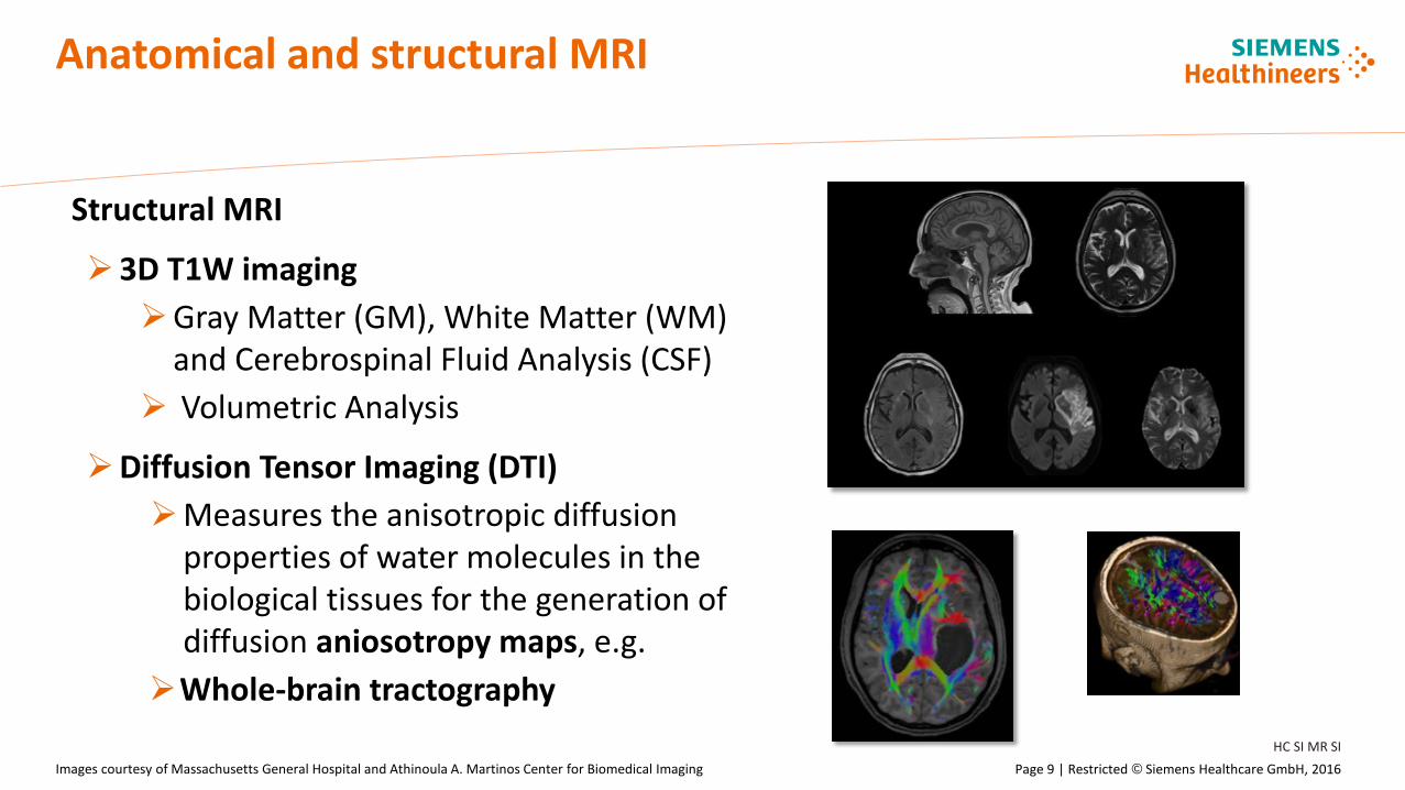
Anatomical and structural MRI
Structural MRI
3D T1W imaging Gray Matter (GM), White Matter (WM)
and Cerebrospinal Fluid Analysis (CSF) Volumetric Analysis
Diffusion Tensor Imaging (DTI) Measures the anisotropic diffusion
properties of water molecules in the biological tissues for the generation of diffusion aniosotropy maps, e.g.
Whole-brain tractography
Images courtesy of Massachusetts General Hospital and Athinoula A. Martinos Center for Biomedical Imaging
HC SI MR SI Page 10 | Restricted © Siemens Healthcare GmbH, 2016
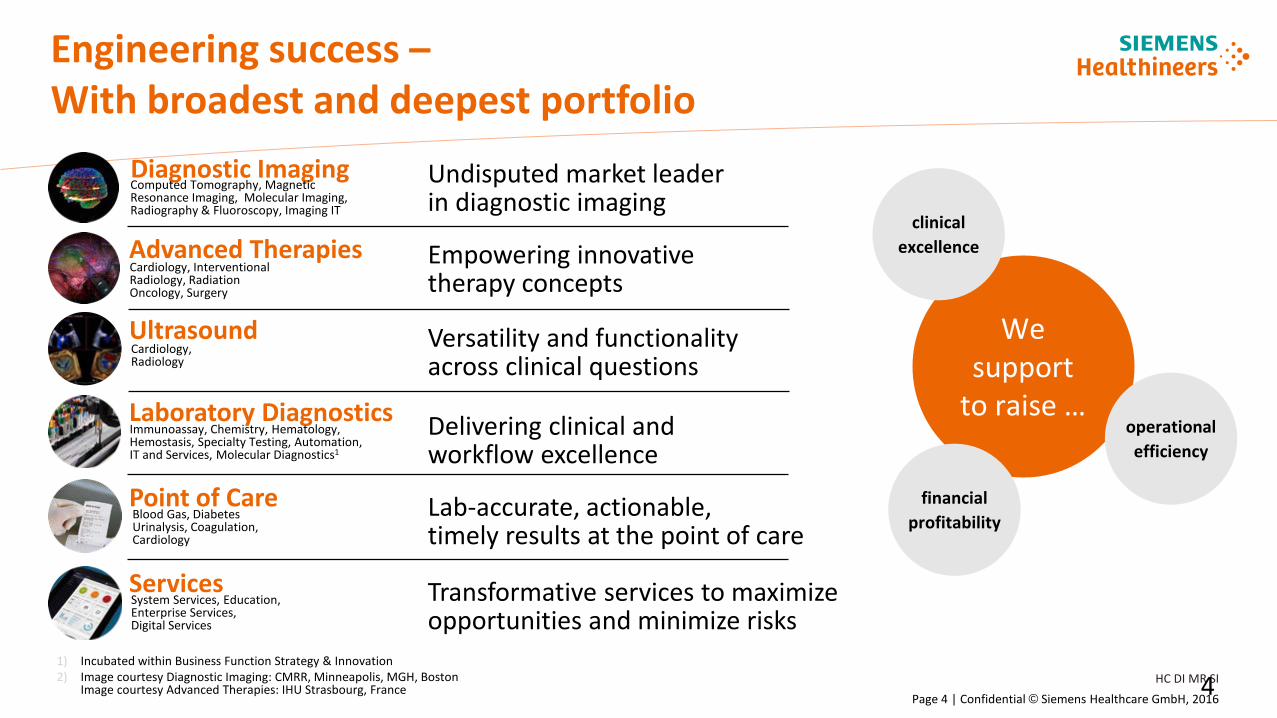
Functional MRI (fMRI)
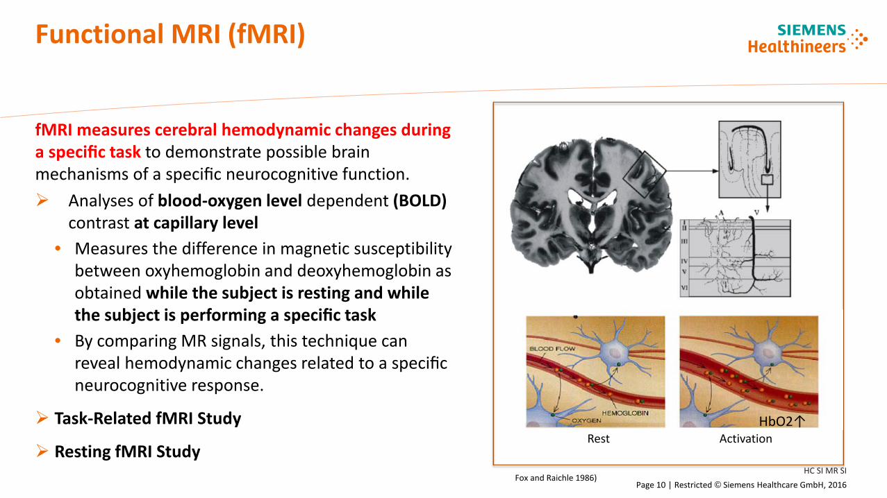
fMRI measures cerebral hemodynamic changes during a specific task to demonstrate possible brain mechanisms of a specific neurocognitive function. Analyses of blood-oxygen level dependent (BOLD)
contrast at capillary level • Measures the difference in magnetic susceptibility
between oxyhemoglobin and deoxyhemoglobin as obtained while the subject is resting and while the subject is performing a specific task
• By comparing MR signals, this technique can reveal hemodynamic changes related to a specific neurocognitive response.
Task-Related fMRI Study
Resting fMRI Study Rest Activation
HbO2↑
Fox and Raichle 1986)
HC SI MR SI Page 11 | Restricted © Siemens Healthcare GmbH, 2016
Task fMRI
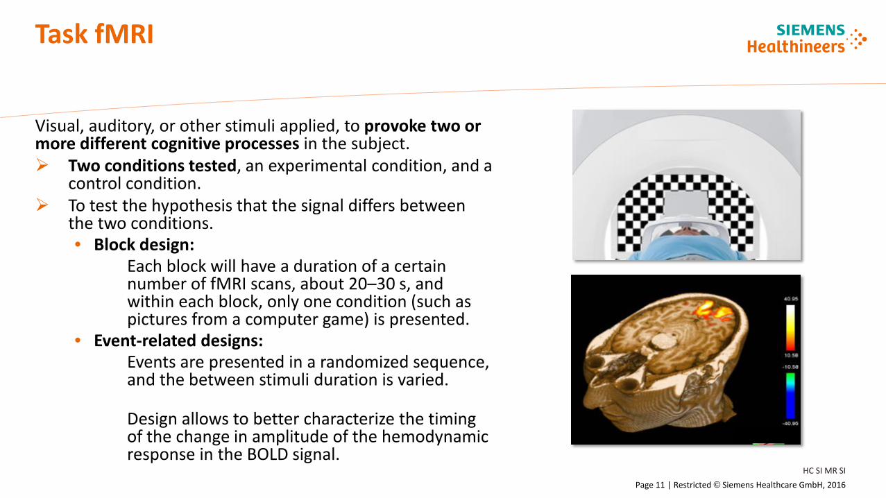
Visual, auditory, or other stimuli applied, to provoke two or more different cognitive processes in the subject. Two conditions tested, an experimental condition, and a
control condition. To test the hypothesis that the signal differs between
the two conditions. • Block design:
Each block will have a duration of a certain number of fMRI scans, about 20–30 s, and within each block, only one condition (such as pictures from a computer game) is presented.
• Event-related designs: Events are presented in a randomized sequence, and the between stimuli duration is varied. Design allows to better characterize the timing of the change in amplitude of the hemodynamic response in the BOLD signal.

HC SI MR SI Page 12 | Restricted © Siemens Healthcare GmbH, 2016
Resting state fMRI
Investigation of neural circuits which exhibit spontaneous activity at rest. These slow-frequency fluctuations are temporally
correlated within spatially distinct but functionally related networks and represent specific patterns of synchronous activity.
Evaluation of resting-state functional connectivity provides an opportunity to characterize distributed circuit normalities and abnormalities in neuropsychiatric illnesses.
However, the interpretation of this connectivity is difficult. For example, both lower connectivity and over connectivity were suggested to indicate the impaired function.
syngo.MR Neuro 3D Images courtesy of Dr Andreas Bartsch, Radiologie Bamberg, Depts of Neuroradiology, Univesities of Heidelberg and Wuerzberg, Germany, Oxford Centre of Functional MRI of the Brain (FMRIB), University of Oxford, UK
HC SI MR SI Page 13 | Restricted © Siemens Healthcare GmbH, 2016
Internet addiction
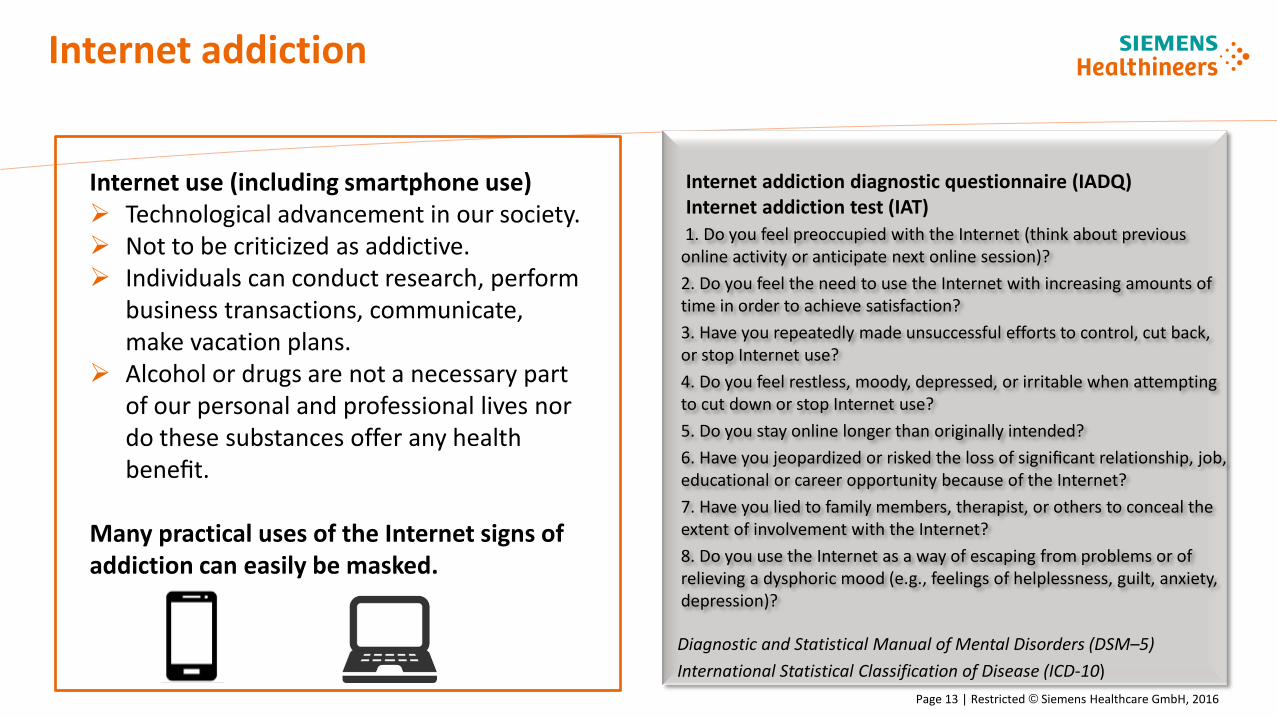
Diagnostic and Statistical Manual of Mental Disorders (DSM–5) International Statistical Classification of Disease (ICD-10)
1. Do you feel preoccupied with the Internet (think about previous online activity or anticipate next online session)? 2. Do you feel the need to use the Internet with increasing amounts of time in order to achieve satisfaction? 3. Have you repeatedly made unsuccessful efforts to control, cut back, or stop Internet use? 4. Do you feel restless, moody, depressed, or irritable when attempting to cut down or stop Internet use? 5. Do you stay online longer than originally intended? 6. Have you jeopardized or risked the loss of significant relationship, job, educational or career opportunity because of the Internet? 7. Have you lied to family members, therapist, or others to conceal the extent of involvement with the Internet? 8. Do you use the Internet as a way of escaping from problems or of relieving a dysphoric mood (e.g., feelings of helplessness, guilt, anxiety, depression)?
Internet addiction diagnostic questionnaire (IADQ) Internet addiction test (IAT)
Internet use (including smartphone use) Technological advancement in our society. Not to be criticized as addictive. Individuals can conduct research, perform
business transactions, communicate, make vacation plans.
Alcohol or drugs are not a necessary part of our personal and professional lives nor do these substances offer any health benefit.
Many practical uses of the Internet signs of addiction can easily be masked.

HC SI MR SI Page 14 | Restricted © Siemens Healthcare GmbH, 2016
Some results form fMRI studies on internet addiction
• Brain regions involved in IA identified by fMRI, but number of subjects limited. • Neuroanatomical changes involving prefrontal cortex, thalamus, and other brain regions. • The pattern of IAD-related structural differences in the brain resemble, to some extent, those changes
observed in substance addiction. • Controversy exists among the scientific community regarding whether IAD constitutes a standalone
illness. • Online gaming requires good cognitive function and decision-making, particularly under risk, unlike
substance addiction which result in a deficit in cognitive function or risk decision. • To be considered:
Well defined study hypothesis and study design Heterogeneity of the subjects (variety in online activities, age, difference in disease stage and chronicity)
Structural and functional MRI data in combination with clinical data Sources of physiological bias heart rate, respiration, head motion influencing fMRI
Many ways to analyze resting fMRI data. Internet Addiction, 2nd Ed, Eds C. Montag, M. Reuters, Springer
HC SI MR SI Page 15 | Restricted © Siemens Healthcare GmbH, 2016
Additional applications and technologies for brain imaging
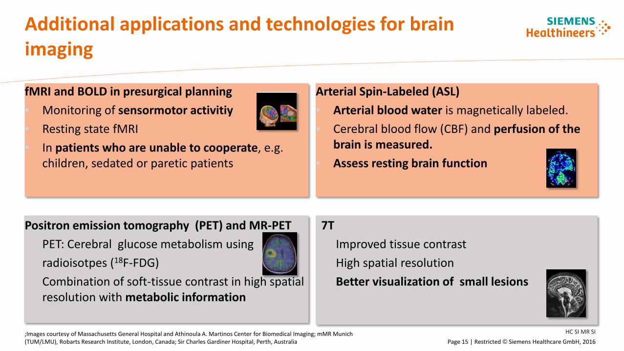
fMRI and BOLD in presurgical planning • Monitoring of sensormotor activitiy • Resting state fMRI • In patients who are unable to cooperate, e.g.
children, sedated or paretic patients
Arterial Spin-Labeled (ASL) • Arterial blood water is magnetically labeled. • Cerebral blood flow (CBF) and perfusion of the
brain is measured. • Assess resting brain function
;Images courtesy of Massachusetts General Hospital and Athinoula A. Martinos Center for Biomedical Imaging; mMR Munich (TUM/LMU), Robarts Research Institute, London, Canada; Sir Charles Gardiner Hospital, Perth, Australia
Positron emission tomography (PET) and MR-PET • PET: Cerebral glucose metabolism using • radioisotpes (18F-FDG) • Combination of soft-tissue contrast in high spatial
resolution with metabolic information
7T • Improved tissue contrast • High spatial resolution Better visualization of small lesions
Engineering success. Pioneering healthcare. Together.
Restricted © Siemens Healthcare GmbH, 2016
Now’s our time
to inspire the future of healthcare