ECG in +Pericarditis and BER.pptx
29
ECG in *carditis and BER The faces of ECG series
-
Upload
smoggindakrak -
Category
Documents
-
view
33 -
download
3
description
Features of ECG in Pericarditis and Benign Early Repolarisation
Transcript of ECG in +Pericarditis and BER.pptx

ECG in *carditis and
BERThe faces of ECG series

Objectives
• Mechanism of changes in pericarditis
• ECG in pericarditis
• Pericardial effusion
• Mechanism of changes in BER
• ECG in BER

A Touch of Anatomy

Case ExampleA 49yo male. Found collapsed at homeby a friend. Has been unwell for the lastfortnight - has been short of breath, chest pain.

Case ExampleA 49yo male. Found collapsed at home by a friend. Has been unwell for the last fortnight - has been short of breath, chest pain.

Case ExampleA 49yo male. Found collapsed at home by a friend. Has been unwell for the last fortnight - has been short of breath, chest pain. A double trouble.
Pericardial Abnormalities
Acute pericarditis

Pericardial Abnormalities
Acute pericarditis
ST/T ratio in V6 >0.25 - pericarditis
ST T
V6

Pericardial Abnormalities
ECG stages of acute pericarditisSTAGE CHANGES ON ECG
I Diffuse concave STE with concordance of TW;
STD in aVR or V1;
PR-segment depression; low voltage;
Absence of reciprocal ST segment changes
II ST segments return to baseline;
TW flattening
III TW inversion
IV Gradual resolution
aVR

Pericardial Abnormalities
- Classic four stage presentation is rare- ST elevation is small- Dynamic ST changes are usually absent- Normal SR or tachycardia are most common
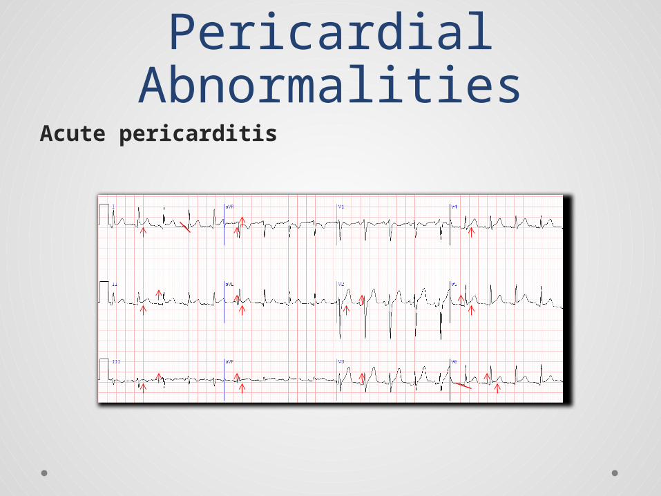
Case Example35yo male brought in ED afteran altercation at a communitygathering with a stab wound to upper abdomen. He is pale, thrashing on the gurney. HR 108bpm, sBP107mmHg. ChestUS showed no pneumothorax.

Case Example35yo male brought in ED after an altercation at a community gathering with a stab wound to upper abdomen….

Case Example49yo woman with metastatic breast cancer. She is pale with cool periph-eries, sBP 95mmHg, HR 109bpm.

Case Example49yo woman with metastatic breast cancer. She is pale with cool peripheries, sBP 95mmHg, HR 109bpm.

Pericardial Abnormalities
Pericardial Effusion- Low QRS and T wave voltage
- Electrical alternans- QRS complex- QRS-T- P-QRS-T
- PEA- Sinus tachycardia

Pericardial Abnormalities
Pericardial effusion- ECG is often normal. NO ECG finding is diagnostic.- Reduction in ECG voltage compared to previous
ECG is very suggestive.- P-QRS-T alternans is highly suggestive of cardiac
tamponade.

Case ExampleYoung man with chest painafter weight lifting.

Case ExampleYoung woman found collapsed near a night club.

Benign Early Repolarization
Features- ST segment elevation- Concave ST segment- Concordant, large TW- Widespread STE- Temporal stability

Case ExampleYoung woman found collapsed near a night club.
Isoelectric PR

Case ExampleYoung man with chest painafter weight lifting.
Isoelectric PR

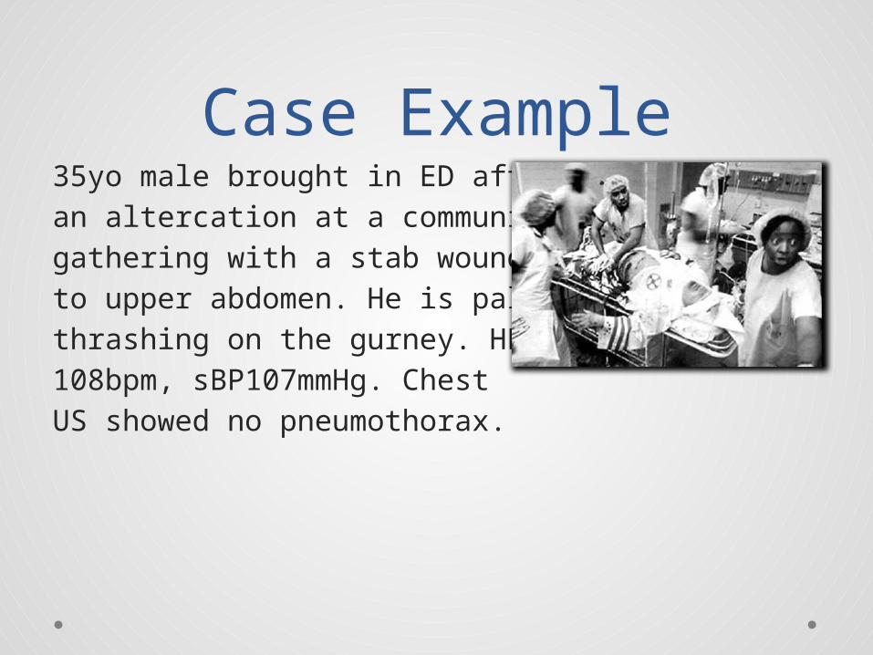
BER v Pericarditis v AMI
?

BER v Pericarditis v AMI
Notched J point
Concave ST

BER v Pericarditis v AMI
Notched J pointST elevation
PR depression
Concave ST
BER v Pericarditis v AMI
Notched J pointST elevation
Convex ST
PR depression
Concave ST

The End