EBV Tegument Protein BNRF1 Disrupts DAXX-ATRX to Activate ... · EBV Tegument Protein BNRF1...
12
EBV Tegument Protein BNRF1 Disrupts DAXX-ATRX to Activate Viral Early Gene Transcription Kevin Tsai 1,2 , Nadezhda Thikmyanova 1 , Jason A. Wojcechowskyj 2 , Henri-Jacques Delecluse 3 , Paul M. Lieberman 1 * 1 The Wistar Institute, Philadelphia, Pennsylvania, United States of America, 2 Cell and Molecular Biology Program, The University of Pennsylvania, School of Medicine, Philadelphia, Pennsylvania, United States of America, 3 German Cancer Research Center, ATV-F100, Heidelberg, Germany Abstract Productive infection by herpesviruses involve the disabling of host-cell intrinsic defenses by viral encoded tegument proteins. Epstein-Barr Virus (EBV) typically establishes a non-productive, latent infection and it remains unclear how it confronts the host-cell intrinsic defenses that restrict viral gene expression. Here, we show that the EBV major tegument protein BNRF1 targets host-cell intrinsic defense proteins and promotes viral early gene activation. Specifically, we demonstrate that BNRF1 interacts with the host nuclear protein Daxx at PML nuclear bodies (PML-NBs) and disrupts the formation of the Daxx-ATRX chromatin remodeling complex. We mapped the Daxx interaction domain on BNRF1, and show that this domain is important for supporting EBV primary infection. Through reverse transcription PCR and infection assays, we show that BNRF1 supports viral gene expression upon early infection, and that this function is dependent on the Daxx- interaction domain. Lastly, we show that knockdown of Daxx and ATRX induces reactivation of EBV from latently infected lymphoblastoid cell lines (LCLs), suggesting that Daxx and ATRX play a role in the regulation of viral chromatin. Taken together, our data demonstrate an important role of BNRF1 in supporting EBV early infection by interacting with Daxx and ATRX; and suggest that tegument disruption of PML-NB-associated antiviral resistances is a universal requirement for herpesvirus infection in the nucleus. Citation: Tsai K, Thikmyanova N, Wojcechowskyj JA, Delecluse H-J, Lieberman PM (2011) EBV Tegument Protein BNRF1 Disrupts DAXX-ATRX to Activate Viral Early Gene Transcription. PLoS Pathog 7(11): e1002376. doi:10.1371/journal.ppat.1002376 Editor: Bill Sugden, University of Wisconsin-Madison, United States of America Received December 20, 2010; Accepted September 28, 2011; Published November 10, 2011 Copyright: ß 2011 Tsai et al. This is an open-access article, free of all copyright, and may be freely reproduced, distributed, transmitted, modified, built upon, or otherwise used by anyone for any lawful purpose. The work is made available under the Creative Commons CC0 public domain dedication. Funding: This work was funded by grants NIH RO1 CA 085678 to PML and to the Wistar institute Cancer Center (5 P30 CA 010815-41). K. Tsai is a trainee on the University of Pennsylvania Training Grant in Tumor Virology (T32-CA115299). The funders had no role in study design, data collection and analysis, decision to publish, or preparation of the manuscript. Competing Interests: The authors have declared that no competing interests exist. * E-mail: [email protected] Introduction Epstein-Barr virus (EBV) is a member of the human gamma- herpesvirus subfamily that infects over 90% of the global adult population [1,2]. EBV preferentially establishes latent infection in B-lymphocytes but can also infect epithelial cells [3,4]. EBV primary infection is one of the main causes of infectious mononucleosis (IM); while EBV latent infection is associated with multiple malignancies such as nasopharyngeal carcinoma, Burkitt’s lymphoma, and Hodgkin’s lymphoma [3,4]. Furthermore, EBV is responsible for the majority of lymphoproliferative diseases associated with AIDS and immunosuppression following organ transplant [5]. Like all herpesviruses, EBV exists in a dynamic balance between productive and latent infection. The factors that regulate the fate decisions for lytic reactivation from latency have been investigated in some detail, but relatively little is known about the fate regulation during the earliest stages of primary infection. Upon entry into the nuclear compartment, herpesvirus DNA genomes must confront several intrinsic anti-viral resistances that restrict viral gene expression and replication. One prominent nuclear structure involved in antiviral resistances is the PML nuclear body (PML-NB), also referred to as nuclear domain 10 (ND10). PML-NBs are nucleoplasmic protein aggregates mainly consisting of (but not limited to) the components PML, Sp100, Daxx, and ATRX [6,7]. The size and abundance of PML-NB is interferon inducible [8,9,10], and over-expression of the PML protein represses viral infection [11]. PML-NB is the nuclear localization site of many DNA viruses, including Herpes Simplex virus (HSV-1), Human Cytomegalovirus (HCMV) and Adenovirus (Ad5) [12,13]. These viruses then modify the morphology and/or protein composition of PML-NBs shortly after infection [12,14]. The mechanism of PML-NB-mediated antiviral repression is not clearly determined. PML, Sp100, and Daxx are all associated with transcription repression, and this function may act on viral genomes [15]. Daxx can act as a transcription co-repressor of many cellular transcription factors [16,17,18,19], and forms repressive transcrip- tion complexes with histone deacetylases (HDACs) [20,21] and DNA methyltransferase I (DNMT I) [22,23]. Daxx has been shown to induce heterochromatin markers on the HCMV genome and repress viral gene expression in a HDAC dependent manor [24,25]. Daxx also forms a chromatin-remodeling complex with ATRX [26] and both can form a repression complex at heterochromatin [27]. Furthermore, RNA interference (RNAi) studies have shown that knockdown of Daxx or ATRX can result in a higher infection level of HCMV [28,29,30] and also relieve the infection defect of mutant HSV deficient in disrupting PML-NB [31]. Herpesviruses confront intrinsic anti-viral resistances immediate- ly upon entering the host cell nucleus, and therefore must counteract these resistances at the earliest possible time points to PLoS Pathogens | www.plospathogens.org 1 November 2011 | Volume 7 | Issue 11 | e1002376
Transcript of EBV Tegument Protein BNRF1 Disrupts DAXX-ATRX to Activate ... · EBV Tegument Protein BNRF1...
EBV Tegument Protein BNRF1 Disrupts DAXX-ATRX toActivate Viral Early Gene TranscriptionKevin Tsai1,2, Nadezhda Thikmyanova1, Jason A. Wojcechowskyj2, Henri-Jacques Delecluse3, Paul M.
Lieberman1*
1 The Wistar Institute, Philadelphia, Pennsylvania, United States of America, 2 Cell and Molecular Biology Program, The University of Pennsylvania, School of Medicine,
Philadelphia, Pennsylvania, United States of America, 3 German Cancer Research Center, ATV-F100, Heidelberg, Germany
Abstract
Productive infection by herpesviruses involve the disabling of host-cell intrinsic defenses by viral encoded tegumentproteins. Epstein-Barr Virus (EBV) typically establishes a non-productive, latent infection and it remains unclear how itconfronts the host-cell intrinsic defenses that restrict viral gene expression. Here, we show that the EBV major tegumentprotein BNRF1 targets host-cell intrinsic defense proteins and promotes viral early gene activation. Specifically, wedemonstrate that BNRF1 interacts with the host nuclear protein Daxx at PML nuclear bodies (PML-NBs) and disrupts theformation of the Daxx-ATRX chromatin remodeling complex. We mapped the Daxx interaction domain on BNRF1, and showthat this domain is important for supporting EBV primary infection. Through reverse transcription PCR and infection assays,we show that BNRF1 supports viral gene expression upon early infection, and that this function is dependent on the Daxx-interaction domain. Lastly, we show that knockdown of Daxx and ATRX induces reactivation of EBV from latently infectedlymphoblastoid cell lines (LCLs), suggesting that Daxx and ATRX play a role in the regulation of viral chromatin. Takentogether, our data demonstrate an important role of BNRF1 in supporting EBV early infection by interacting with Daxx andATRX; and suggest that tegument disruption of PML-NB-associated antiviral resistances is a universal requirement forherpesvirus infection in the nucleus.
Citation: Tsai K, Thikmyanova N, Wojcechowskyj JA, Delecluse H-J, Lieberman PM (2011) EBV Tegument Protein BNRF1 Disrupts DAXX-ATRX to Activate ViralEarly Gene Transcription. PLoS Pathog 7(11): e1002376. doi:10.1371/journal.ppat.1002376
Editor: Bill Sugden, University of Wisconsin-Madison, United States of America
Received December 20, 2010; Accepted September 28, 2011; Published November 10, 2011
Copyright: � 2011 Tsai et al. This is an open-access article, free of all copyright, and may be freely reproduced, distributed, transmitted, modified, built upon, orotherwise used by anyone for any lawful purpose. The work is made available under the Creative Commons CC0 public domain dedication.
Funding: This work was funded by grants NIH RO1 CA 085678 to PML and to the Wistar institute Cancer Center (5 P30 CA 010815-41). K. Tsai is a trainee on theUniversity of Pennsylvania Training Grant in Tumor Virology (T32-CA115299). The funders had no role in study design, data collection and analysis, decision topublish, or preparation of the manuscript.
Competing Interests: The authors have declared that no competing interests exist.
* E-mail: [email protected]
Introduction
Epstein-Barr virus (EBV) is a member of the human gamma-
herpesvirus subfamily that infects over 90% of the global adult
population [1,2]. EBV preferentially establishes latent infection in
B-lymphocytes but can also infect epithelial cells [3,4]. EBV
primary infection is one of the main causes of infectious
mononucleosis (IM); while EBV latent infection is associated with
multiple malignancies such as nasopharyngeal carcinoma, Burkitt’s
lymphoma, and Hodgkin’s lymphoma [3,4]. Furthermore, EBV is
responsible for the majority of lymphoproliferative diseases
associated with AIDS and immunosuppression following organ
transplant [5]. Like all herpesviruses, EBV exists in a dynamic
balance between productive and latent infection. The factors that
regulate the fate decisions for lytic reactivation from latency have
been investigated in some detail, but relatively little is known about
the fate regulation during the earliest stages of primary infection.
Upon entry into the nuclear compartment, herpesvirus DNA
genomes must confront several intrinsic anti-viral resistances that
restrict viral gene expression and replication. One prominent
nuclear structure involved in antiviral resistances is the PML
nuclear body (PML-NB), also referred to as nuclear domain 10
(ND10). PML-NBs are nucleoplasmic protein aggregates mainly
consisting of (but not limited to) the components PML, Sp100,
Daxx, and ATRX [6,7]. The size and abundance of PML-NB is
interferon inducible [8,9,10], and over-expression of the PML
protein represses viral infection [11]. PML-NB is the nuclear
localization site of many DNA viruses, including Herpes Simplex
virus (HSV-1), Human Cytomegalovirus (HCMV) and Adenovirus
(Ad5) [12,13]. These viruses then modify the morphology and/or
protein composition of PML-NBs shortly after infection [12,14].
The mechanism of PML-NB-mediated antiviral repression is not
clearly determined. PML, Sp100, and Daxx are all associated with
transcription repression, and this function may act on viral genomes
[15]. Daxx can act as a transcription co-repressor of many cellular
transcription factors [16,17,18,19], and forms repressive transcrip-
tion complexes with histone deacetylases (HDACs) [20,21] and
DNA methyltransferase I (DNMT I) [22,23]. Daxx has been shown
to induce heterochromatin markers on the HCMV genome and
repress viral gene expression in a HDAC dependent manor [24,25].
Daxx also forms a chromatin-remodeling complex with ATRX [26]
and both can form a repression complex at heterochromatin [27].
Furthermore, RNA interference (RNAi) studies have shown that
knockdown of Daxx or ATRX can result in a higher infection level
of HCMV [28,29,30] and also relieve the infection defect of mutant
HSV deficient in disrupting PML-NB [31].
Herpesviruses confront intrinsic anti-viral resistances immediate-
ly upon entering the host cell nucleus, and therefore must
counteract these resistances at the earliest possible time points to
PLoS Pathogens | www.plospathogens.org 1 November 2011 | Volume 7 | Issue 11 | e1002376

initiate viral gene expression. Herpesvirus tegument proteins, which
are pre-packaged and delivered with the infectious virion, are
strategically positioned to counteract the intrinsic anti-viral defenses
and support the early steps of infection [32]. Both alpha- and beta-
herpesviruses encode tegument proteins that regulate early events
during lytic replication, including the disruption of the PML-NBs.
HSV-1 immediate early gene ICP0, disrupts PML-NB structure by
degrading the core component PML [33,34,35] and eliminating
SUMO-modified Sp100 [36]; while HCMV tegument protein pp71
displaces ATRX and subsequently degrades Daxx [24,30]. Both
ICP0-deficient HSV-1 and pp71-deficient HCMV mutants are
deficient in infection, where viral gene expression is shutdown,
resulting in a dormant viral genome [29,37,38]. Interestingly, it has
been reported that disruption of PML-NB by ICP0 is mediated by de
novo synthesized ICP0, instead of tegument delivered ICP0 protein,
suggesting that this event is coordinated with early viral gene
activation or, perhaps, reactivation from latent infection [35]. We
have previously shown that EBV genomes localize to and then
disrupt PML-NB during lytic replication; while latent EBV
episomes are segregated away from PML-NBs during latency
[39]. EBV regulatory proteins, including the lytic cycle immediate
early gene Zta (also referred to as BZLF1, ZEBRA, and Z), and
latency associated EBNA1 and EBNA-LP, have been implicated in
PML-NB interactions [40,41,42]. However, it remains unclear if
PML-NBs regulate early events associated with viral gene
expression upon EBV nuclear entry, and if an EBV tegument
protein modulates this intrinsic defense.
The EBV major tegument protein BNRF1 is one of the most
abundant tegument proteins in the virion [43] and is essential for
the establishment of viral latent infection [44], yet its function is
largely unknown. BNRF1 homologues are present in all
gammaherpesviruses but absent in the alpha- and beta- herpes-
virus subfamilies. All BNRF1 orthologues share regions homolo-
gous to the cellular enzymes Phosphoribosylformylglycineamide
Amidotransferase (FGARAT) and Aminoimidazole ribonucleotide
(AIR) synthetase, ATP-dependent enzymes in the 4th and 5th steps
of the purine de novo biosynthesis pathway. However, no enzymatic
activity has been found in any BNRF1 orthologues. In a knockout
study, transfected BNRF1-deficient EBV genomes can reactivate
from latency, produce morphologically normal virions, and the
progeny can enter cells with little observed defects [44]. Yet, upon
infection of B cells the mutant virus showed a 20-fold lower
expression of a viral latency associated gene EBNA2 and failed to
induce B cell transformation [44]. This suggests an important role
of BNRF1 in supporting early infection. Furthermore, the BNRF1
orthologue encoded by murine herpes virus 68 (MHV68),
tegument protein ORF75c, induces PML degradation and is
essential for initiation of viral gene expression [45,46].
Here, we demonstrate that EBV BNRF1 is a novel PML-NB-
interacting viral protein, and that this interaction is important for
supporting EBV primary infection. We first show that Daxx is a
primary cellular interaction partner of BNRF1. BNRF1 co-
localizes with Daxx at PML-NB foci while disrupting the Daxx-
ATRX complex. Furthermore, we identify a novel Daxx
interaction domain on BNRF1. This domain is essential for
BNRF1 to interact with Daxx, localize to PML-NB, and displace
ATRX from Daxx. We then show that BNRF1 supports EBV
primary infection and promotes the expression of viral genes soon
after viral genomes enter the cell, and that the Daxx interaction
domain contributes to these functions. Lastly, we show that
knockdown of either Daxx or ATRX results in disruption of viral
latency, suggesting that Daxx and ATRX play a role in the
restriction of viral gene expression. Our study suggests that EBV
tegument protein BNRF1 disassemble the Daxx-ATRX antiviral
resistance complex to enable viral gene expression after cell
invasion, and likely regulate the chromatin organization for the
establishment of latent infection.
Results
BNRF1 interacts with cellular protein DaxxTo characterize the biological properties of the EBV major
tegument protein BNRF1, we took a proteomic approach to
screen for potential cellular interaction partners. BNRF1 was
cloned into a 3x FLAG tag expression vector under the control of
a CMV promoter. 293T cells were then stably transfected with
either FLAG-vector or FLAG-tagged BNRF1. Nuclear extracts
from stable cell lines were subject to immunopurification (IP) with
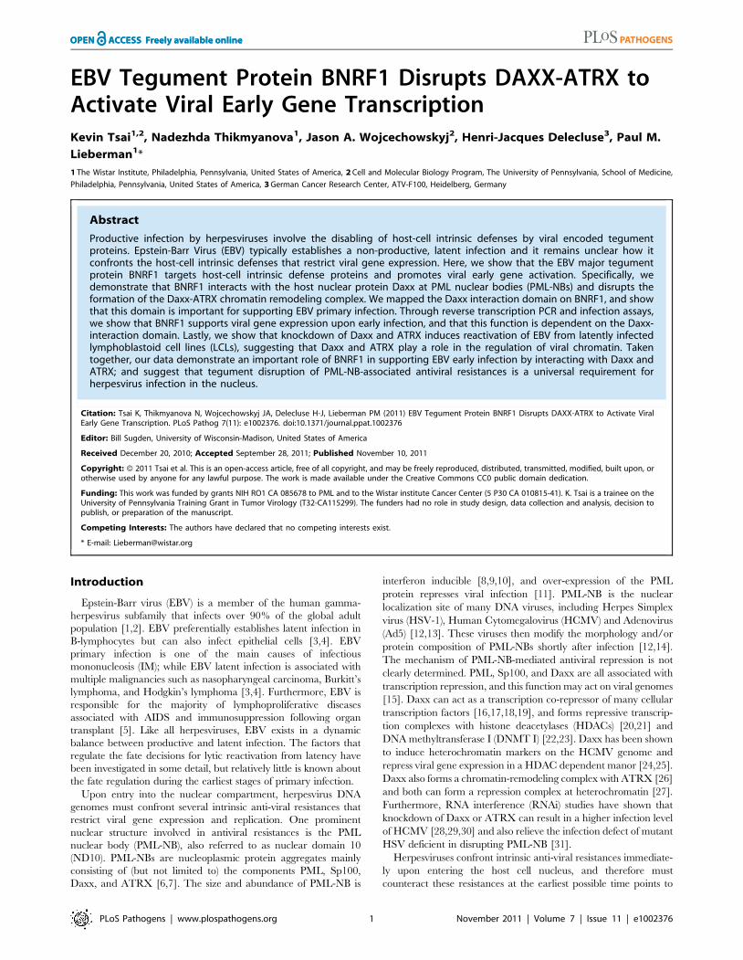
a FLAG antibody, and then analyzed by SDS-PAGE (Fig. 1A).
Bands unique to the BNRF1 lane (B) were cut out and analyzed by
liquid chromatography-tandem mass spectrometry (LC/MS/MS).
The major identified species was BNRF1, but substoichiometric
proteins enriched in the BNRF1 IP were also identified, including
Daxx, nucleophosmin (NPM1), and PARP1 (Fig. 1C). We
subsequently confirmed in BNRF1 transiently transfected 293T
cells that Daxx co-precipitates with BNRF1 (Fig. 1B) by both
FLAG pull-down and the Daxx reverse pull-down, indicating a
stable in vivo interaction between BNRF1 and Daxx. Neither
PARP1 nor NPM1 interaction with BNRF1 could be validated by
subsequent co-IPs (data not shown), we therefore focused our
efforts on characterizing the interaction with Daxx.
BNRF1 utilizes a novel Daxx interaction domainTo further characterize the interaction between BNRF1 and
Daxx, we introduced serial deletions on the FLAG-BNRF1
expression plasmid. We first made five deletion constructs of
BNRF1, sequentially deleting regions coding for 300 amino acids
(Fig. 2A, constructs d1 through d5). We then performed IPs with
either control IgG, aFLAG, or aDaxx on lysates of cells
transfected with the BNRF1 deletion constructs. Daxx co-
precipitated in the FLAG IP for all of the BNRF1 mutants with
the exception of the BNRF1 300–600 aa deletion mutant (d2)
(Fig. 2B, middle panels). Similarly, all of the FLAG-BNRF1
mutants, with the exception of d2, co-precipitated with Daxx IP
(Fig. 2B, right panels). Since d2 was expressed and recovered by
FLAG IP to similar levels as other BNRF1 mutants capable of
Author Summary
Persistent infection by Epstein-Barr virus (EBV) is associatedwith a variety of diseases, including lymphoid andepithelial tumors. Despite a wealth of information on themechanism of viral persistence, relatively little is knownabout the early steps of EBV infection and viral geneactivation. Host cells actively mount resistances againstviral infection, which viruses need to overcome to invadethe cell. We have found that among the proteins packagedin the EBV viral particle, BNRF1 plays an important role ofcounteracting cellular defenses. We show that EBV proteinBNRF1 binds to the cellular protein Daxx and disassemblesthe Daxx-ATRX complex, where both Daxx and ATRX arecellular proteins known to inhibit viral gene expression. Wealso confirm that BNRF1 can promote expression of earlyviral genes, and that Daxx-binding by BNRF1 is required forthis function. Finally, we demonstrate that Daxx and ATRXrepress viral gene expression during latency. We concludethat BNRF1 disassembles cellular antiviral defense machin-ery to promote expression of viral genes in the host cell.
Daxx-ATRX Disruption by EBV Tegument BNRF1
PLoS Pathogens | www.plospathogens.org 2 November 2011 | Volume 7 | Issue 11 | e1002376
interacting with Daxx, we conclude that a putative Daxx-
interaction domain is located in the region between 300-600aa
of BNRF1.
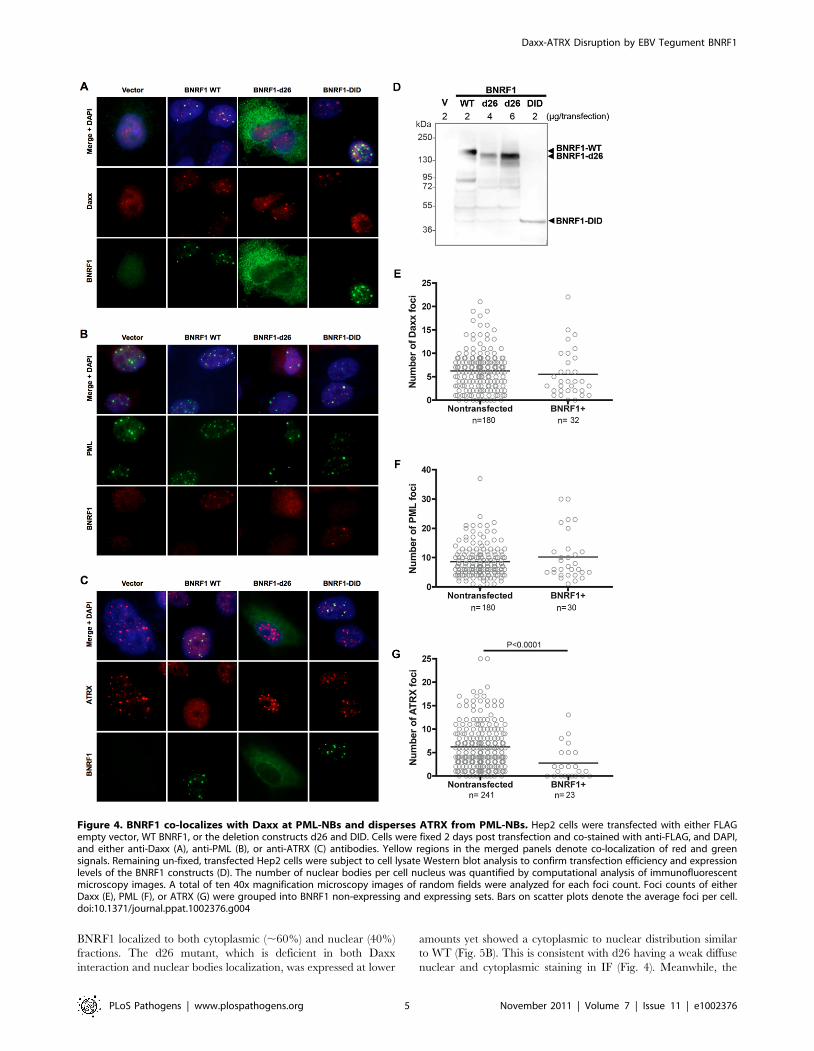
We then further made six serial deletions of 60 amino acids in
the 300–600 aa region (Fig. 3A, constructs d21 through d26) to
narrow down the suspected Daxx-interaction domain to a smaller
region. After a subsequent round of IP pull-downs, we found that
all BNRF1 deletions, with the exception of d21, were defective in
binding Daxx (Fig. 3B), suggesting that the 360–600 aa region of
BNRF1 is responsible for interaction with Daxx. To determine if
this region was sufficient for interaction with Daxx, we expressed
only the 300–600 aa region in the FLAG-expression vector
(Fig. 3A, construct DID) and performed IP pull-downs. We found
that this region bound Daxx as efficiently as WT-BNRF1, in both
FLAG IP and in the reverse IP with anti-Daxx antibody (Fig. 3C).
Notably, we failed to find sequence homology of this Daxx
interaction domain with any known protein motif, and this
domain is also distinct from the FGARAT and AIR synthetase
homology regions. These findings suggests that BNRF1 utilizes a
previously unknown motif to bind Daxx, and that the Daxx
interaction domain (300–600 aa) may contain a complex protein
fold sensitive to smaller truncation deletions.
BNRF1 disrupts the Daxx-ATRX chromatin remodelingcomplex
Daxx forms a chromatin remodeling complex with ATRX [26]
and ATRX has been implicated in the transcriptional repression
of both HSV-1 and HCMV during the early steps of infection
[30]. Moreover, both HSV-1 and HCMV utilize viral encoded
proteins that disrupt the interaction between Daxx and ATRX
[30,31]. To determine if BNRF1 also disrupted the interaction
between Daxx and ATRX, we assayed the effect of WT and
mutant BNRF1 proteins on the co-IP of Daxx with ATRX. We
observed that WT BNRF1 disrupted the interaction between
Daxx and ATRX (Fig. 3C, 2nd panel from top, right). However,
deletion mutants d22 and d26, which fail to interact with Daxx,
did not disrupt ATRX binding in Daxx IP assays (Fig. 3C, 2nd
panel from top, right). Interestingly, the Daxx interaction domain
by itself (DID), which binds Daxx efficiently, could only partially
disrupt ATRX binding. This suggests that Daxx binding by
BNRF1 is necessary, but not sufficient for the disruption of ATRX
with Daxx. We also found no evidence that BNRF1 co-IPs with
PML (Fig. 3C, 3rd panel from top).
To determine whether any other domains of BNRF1 contribute
to the disruption of ATRX from Daxx, we assayed FLAG-BNRF1
IPs for ATRX binding using the set of larger BNRF1 deletions
examined in Figure 2 (Fig. 3D). We found that WT BNRF1 did
not co-IP with ATRX, although it efficiently pulled down Daxx.
The BNRF1 d2 mutant failed to pull down Daxx or ATRX, as
expected. In contrast, the BNRF1 d3 and d4 mutants, which
disrupts most of the FGARAT and AIR synthetase homology
regions, efficiently pulled down both ATRX and Daxx. The d1
and d5 truncations, which lie outside of the FGARAT and AIR
synthase homology regions, pulled down only Daxx but not
ATRX, suggesting it efficiently disrupted the ATRX-Daxx
interaction similar to WT. These data suggest that the FGARAT
and AIR synthetase homology regions of BNRF1 may contribute
to the disruption of ATRX-Daxx complex.
BNRF1 co-localizes with Daxx to PML nuclear bodies anddisperses ATRX from nuclear bodies, in a Daxx interactiondomain-dependent manner
Daxx is a prominent component of PML nuclear bodies [47],
and Daxx localization at these nuclear bodies are disrupted by
viral proteins of both HSV-1 and HCMV [24,34]. Thus, it is
Figure 1. BNRF1 binds the cellular protein Daxx. (A) Colloidalblue stained SDS-PAGE of FLAG-immunoprecipitated BNRF1 andinteracting partners. 293T cells were stably transfected with emptyFLAG vector (V) or FLAG-tagged BNRF1 (B). Cell lysates were subject toImmunoprecipitation (IP) by anti-FLAG antibodies, then analyzed bySDS-PAGE. Bands unique to lane B were cut out and identified by LC/MS/MS. (B) IP confirmation of BNRF1/Daxx interaction. 293T cells weretransiently transfected with empty vector (V) or wild-type BNRF1 (B).Cells harvested two days post-transfection were subject to IP with non-specific IgG, anti-FLAG or anti-Daxx antibodies, and analyzed byWestern blot (WB) with anti-FLAG or anti-Daxx antibodies. (C) Summaryof LC/MS/MS data from FLAG-BNRF1 purification. Genebank accessionnumber (GI), percent of peptide coverage, number of peptidesidentified, and protein name are indicated.doi:10.1371/journal.ppat.1002376.g001
Figure 2. Mapping the Daxx-interaction domain on BNRF1, andthe effect of BNRF1 on the Daxx-ATRX complex. (A) Diagram ofwild-type BNRF1 (WT) and mutation constructs with 300 aa deletions(d1-d5). Dark gray block denotes the amino-terminal FLAG tag. Lightgray blocks denote regions with sequence homology to the cellularenzymes Aminoimidazole ribonucleotide synthetase (AIR_S) and Type 1glutamine amidotransferase (GATase1, an enzymatic domain ofFGARAT), as identified by the National Center for BiotechnologyInformation (NCBI) conserved domain search. (B) IP pull down analysisof the BNRF1 deletion constructs. 293T cells were either transfectedwith empty FLAG vector (V), the BNRF1 constructs WT, or mutants d1-d5. Cell lysates of transfected cells were then subject to IP pull-downswith non-specific IgG, anti-FLAG, or anti-Daxx- antibodies, then Westernblots were probed for Daxx (top panels), and FLAG-tagged proteins(lower panels). Input is shown for each mutant in the left most panels.doi:10.1371/journal.ppat.1002376.g002
Daxx-ATRX Disruption by EBV Tegument BNRF1
PLoS Pathogens | www.plospathogens.org 3 November 2011 | Volume 7 | Issue 11 | e1002376

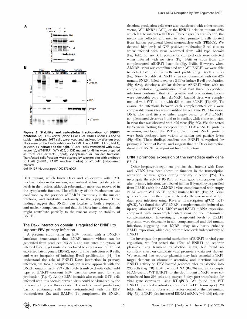
important to investigate the sub-cellular location of BNRF1-Daxx
interaction, and check if BNRF1 disrupts Daxx localization to the
nuclear bodies. For immunofluorescence (IF) microscopy studies,
we selected Hep2 carcinoma cell lines because of their larger size
and prominent PML nuclear bodies, and their common use in
many previous studies with herpesvirus protein interactions with
PML-NBs. Hep2 cells were transiently transfected with empty
FLAG vector (V) or BNRF1 constructs WT, d26, or DID. Cells
were then fixed two days post transfection and subject to IF
staining. We found that WT BNRF1 partially co-localized with
nuclear foci containing Daxx (Fig. 4A, S1A and Table S1), PML
(Fig. 4B, S1B and Table S1), and Sp100 (Fig. S1D), suggesting that
BNRF1 interacts with Daxx at the PML nuclear bodies. We also
noticed that DID itself is sufficient for localizing to PML nuclear
bodies, while the d26 deletion mutant showed a weak dispersed
pattern in the cell (Fig. 4A and B, S1 and Table S1). The co-
localizations were also confirmed by line scan analysis, where the
BNRF1 WT and DID intensity peaks overlap with Daxx and PML
peaks (Fig. S2). To ensure that the diffuse pattern of the d26
mutant is not due to deficient protein expression, the same set of
transfected cells as used for IF were also assayed by Western blot
for total expression levels of BNRF1 proteins (Fig. 4D). We found
that d26 protein was expressed at levels similar to that of WT,
despite its diffuse staining in IF studies, confirming its protein
expression in the cells used in our microscopy study. These
findings indicate that the interaction with Daxx is necessary and
sufficient for BNRF1 to localize to the nuclear bodies.
To understand the BNRF1 disruption of Daxx-ATRX complex
in a sub-cellular spatial context, we also examined ATRX by IF in
BNRF1-transfected Hep2 cells (Fig. 4C and S1C). Again, we
found ATRX foci co-localizing with WT BNRF1 and DID but not
d26, which is also confirmed by line scan analysis (Fig. S2C).
However, we also found a substantial reduction in ATRX foci
intensity when cells were transfected with WT BNRF1, but no
apparent reduction when transfected with d26 or DID-mutants
(Fig. 4C). The failure of DID to disperse ATRX is consistent with
its only partial disruption of ATRX from Daxx IP (Fig. 3C).
Quantification of Daxx (Fig. 4E), PML (Fig. 4F), and ATRX
(Fig. 4G) nuclear foci in BNRF1-expressing cells compared to non-
expressing cells revealed that BNRF1-expressing cells contain a
significantly lower (p,0.0001) average number of ATRX nuclear
foci than non-expressing cells (Fig. 4G). In contrast, we found no
significant difference in the number of Daxx (Fig. 4E) and PML
nuclear foci (Fig. 4F) in BNRF1 transfected cells. Taken together,
these results suggest that BNRF1 not only disrupts the Daxx-
ATRX complex, but also actively disperses ATRX away from
nuclear bodies.
HSV-1 ICP0 and HCMV pp71 each induce the degradation of
PML and Daxx proteins respectively, yet we did not observe any
evidence of this with BNRF1 in our microscopy studies. To
investigate the potential degradation of PML, Daxx and ATRX
proteins by BNRF1, we examined the stability of these proteins in
BNRF1 stably transfected cells (Fig. 5A). 293T cells stably
transfected with control vector (clone C) or WT BNRF1 (stable
transfection clones 3 and 9) were lysed and subject to Western blot
analysis. We found no evidence of degradation or gross post-
translational modification of PML, Daxx, nor ATRX in BNRF1-
expressing cell lines. We also analyzed the protein stability of PML
and Daxx in Hep2 cells transiently transfected with control vector,
WT BNRF1 or d26, and again found no evidence of BNRF1-
induced protein degradation (Fig. S3). This suggests that BNRF1
does not mimic the protein degradation function of HCMV pp71
or HSV-1 ICP0, but rather, disrupts Daxx-ATRX interactions
through alternative mechanisms.
The diffuse distribution of the BNRF1-d26 mutant raised the
question of whether the Daxx interaction domain of BNRF1
correlated with nuclear localization. To test this, we utilized
biochemical fractionation methods to isolate nuclear and cyto-
plasmic proteins from 293T cells (Fig. 5B). We found that WT
Figure 3. The Daxx-interaction domain on BNRF1 is locatedbetween sites 360-600 aa, and BNRF1 disrupts Daxx-ATRXbinding. (A) Diagram of BNRF1 mutation constructs with 60 aadeletions (d21–d25) within the 360–600 aa Daxx-interaction domain(DID) and the DID only. Blocks in the diagram drown as Fig. 2A. (B) IPanalysis of BNRF1 60 aa deletion constructs. 293T cells were transfectedwith various BNRF1 expression vector constructs, and cell lysates weresubject to IP pull downs with non-specific IgG and anti-FLAGantibodies, then Western blots were probed for Daxx (top panel), orFLAG-tagged proteins (lower panel). (C) IP analysis of 293T cellstransfected with vector control (V), WT BNRF1 (WT), BNRF1 deletionmutants d22, d26, or DID. Input, IgG control IP, FLAG IP, or Daxx IP(panels left to right) as indicated above each panel. Western blots of IPswere probed with antibody to Daxx, ATRX, PML, FLAG, or Actin, asindicated to the left of each panel. (D) IP Western of 293T cellstransfected with vector control (V), WT-BNRF1 (WT), or BNRF1 deletionmutants d1, d2, d3, d4, or d5. FLAG-IPs were analyzed by Western blotwith antibodies to ATRX (top panel) Daxx (middle panel), or FLAG(lower panel).doi:10.1371/journal.ppat.1002376.g003
Daxx-ATRX Disruption by EBV Tegument BNRF1
PLoS Pathogens | www.plospathogens.org 4 November 2011 | Volume 7 | Issue 11 | e1002376
BNRF1 localized to both cytoplasmic (,60%) and nuclear (40%)
fractions. The d26 mutant, which is deficient in both Daxx
interaction and nuclear bodies localization, was expressed at lower
amounts yet showed a cytoplasmic to nuclear distribution similar
to WT (Fig. 5B). This is consistent with d26 having a weak diffuse
nuclear and cytoplasmic staining in IF (Fig. 4). Meanwhile, the
Figure 4. BNRF1 co-localizes with Daxx at PML-NBs and disperses ATRX from PML-NBs. Hep2 cells were transfected with either FLAGempty vector, WT BNRF1, or the deletion constructs d26 and DID. Cells were fixed 2 days post transfection and co-stained with anti-FLAG, and DAPI,and either anti-Daxx (A), anti-PML (B), or anti-ATRX (C) antibodies. Yellow regions in the merged panels denote co-localization of red and greensignals. Remaining un-fixed, transfected Hep2 cells were subject to cell lysate Western blot analysis to confirm transfection efficiency and expressionlevels of the BNRF1 constructs (D). The number of nuclear bodies per cell nucleus was quantified by computational analysis of immunofluorescentmicroscopy images. A total of ten 40x magnification microscopy images of random fields were analyzed for each foci count. Foci counts of eitherDaxx (E), PML (F), or ATRX (G) were grouped into BNRF1 non-expressing and expressing sets. Bars on scatter plots denote the average foci per cell.doi:10.1371/journal.ppat.1002376.g004
Daxx-ATRX Disruption by EBV Tegument BNRF1
PLoS Pathogens | www.plospathogens.org 5 November 2011 | Volume 7 | Issue 11 | e1002376
DID mutant, which binds Daxx and co-localizes with PML
nuclear bodies in the nucleus, was isolated at low, yet detectable
levels in the nucleus; although substantially more was recovered in
the cytoplasmic fraction. The efficiency of the fractionation was
confirmed by the presence of PARP1 exclusively in the nuclear
fractions, and a-tubulin exclusively in the cytoplasm. These
findings suggest that BNRF1 can localize to both cytoplasmic
and nuclear compartments, and that the Daxx interaction domain
might contribute partially to the nuclear entry or stability of
BNRF1.
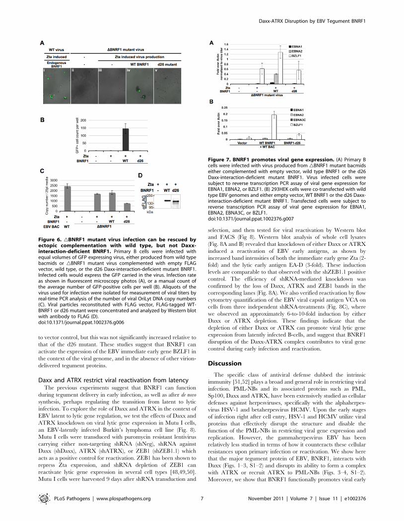
The Daxx interaction domain is required for BNRF1 tosupport EBV primary infection
A previous study using an EBV bacmid with a BNRF1-
knockout demonstrated that BNRF1-mutant virions can be
generated from producer 293 cells and can enter the cytosol of
infected B-cells; yet mutant virus failed to express one of the first
expressed latent genes, EBNA2, upon primary infection of B cells,
and were incapable of inducing B-cell proliferation [44]. To
understand the role of BNRF1-Daxx interaction in primary
infection, we took a complementation rescue approach with the
BNRF1-mutant virus. 293 cells stably transfected with either wild
type or BNRF1-knockout EBV bacmids were used for virus
production (Fig. 6). As the EBV bacmids also encode GFP, cells
infected with this bacmid-derived virus could be visualized by the
presence of green fluorescence. To induce viral production,
bacmid containing cells were co-transfected with the EBV
transactivator Zta and BALF4. To complement for BNRF1
deletion, production cells were also transfected with either control
vector, WT BNRF1 (WT), or the BNRF1 deletion mutant (d26)
which fails to interact with Daxx. Three days after transfection, the
media was collected and used to infect primary B cells isolated
from human peripheral blood mononuclear cells (PBMCs). We
detected high-levels of GFP positive proliferating B-cell clusters
when infected with virus generated from wild type bacmid
(Fig. 6Ai), but no GFP positive or clumped cells were detected
when infected with no virus (Fig. 6Aii) or virus from un-
complemented DBNRF1 bacmids (Fig. 6Aiii). However, when
DBNRF1 virus was complimented with WT BNRF1 we were able
to detect GFP positive cells and proliferating B-cell clusters
(Fig. 6Aiv). Notably, DBNRF1 virus complimented with the d26
mutant BNRF1 failed to express GFP or induce B cell proliferation
(Fig. 6Av), showing a similar defect as DBNRF1 virus with no
complementation. Quantification of at least three independent
infections confirmed that GFP positive and proliferating B-cells
were detectable only when DBNRF1 bacmid virus was comple-
mented with WT, but not with d26 mutant BNRF1 (Fig. 6B). To
ensure the infections between each complemented virus were
comparable, virus titer was quantified by real time PCR for virion
DNA. The viral titers of either empty vector or WT BNRF1
complemented virus was found to be similar, while some reduction
in virus titer was observed with d26 virus (Fig. 6C). We also tested
by Western blotting for incorporation of FLAG-BNRF1 proteins
in virions, and found that WT and d26 mutant BNRF1 proteins
were both packaged into virions to similar per particle levels
(Fig. 6D). These findings confirm that BNRF1 is required for
primary infection of B-cells, and suggests that the Daxx interaction
domain of BNRF1 is important for this function.
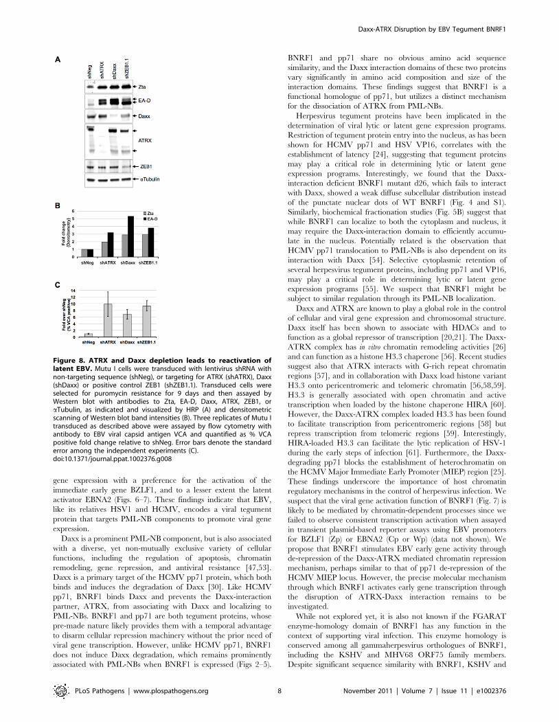
BNRF1 promotes expression of the immediate early geneBZLF1
Other herpesvirus tegument proteins that interact with Daxx
and ATRX have been shown to function in the transcription
activation of viral genes during primary infection [25]. To
investigate the role of BNRF1 on viral gene transcription early
after primary infection, we infected human B-lymphocytes purified
from PBMCs with the DBNRF1 virus complemented with empty
FLAG-vector, WT BNRF1 or d26 mutant BNRF1 (Fig. 7A). Viral
gene expression in these newly infected cells was assayed at four
days post infection using Reverse Transcription qPCR (RT-
qPCR). We found that WT BNRF1 complementation induced an
up-regulation of EBNA1, EBNA2 and BZLF1 mRNA expression
compared with non-complemented virus or the d26-mutant
complementation. Interestingly, background levels of BZLF1
expression were detectable in non-complemented and d26 mutant
infections, suggesting that BNRF1 may only partly enhance
BZLF1 expression, which can occur at low levels independently of
BNRF1.
To investigate the potential mechanism of BNRF1 in viral gene
regulation, we first tested the effect of BNRF1 on reporter
plasmids using transient transfection assays, but found no
consistent effect on candidate viral promoters (data not shown).
We reasoned that reporter plasmids may lack essential BNRF1
target elements or chromatin assembly, and therefore assayed
BNRF1 activity on EBV bacmid genomes after transfection into
293 cells (Fig. 7B). EBV bacmid DNA (Bac36) and either empty
FLAG-vector, WT BNRF1, or the d26 mutant BNRF1 were co-
transfected into 293 cells and assayed 3 days post transfection for
viral gene expression using RT-qPCR. We found that WT
BNRF1 promoted a robust expression of BZLF1 transcripts (,20
fold), which was not observed in vector control or the d26 mutant
(Fig. 7B). BNRF1 also increased EBNA2 mRNA (,3 fold) relative
Figure 5. Stability and subcellular fractionation of BNRF1proteins. (A) FLAG vector (clone C) or FLAG-BNRF1 (clones 3 and 9)stably transfected 293T cells were lysed and analyzed by Western blot.Blots were probed with antibodies to PML, Daxx, ATRX, FLAG (BNRF1),or Actin, as indicated to the right. (B) 293T cells transfected with FLAGvector (V), WT-BNRF1 (WT), d26, or DID mutant for 48 hrs were preparedas total cell extracts (input), cytoplasmic or nuclear fractions.Transfected cells fractions were assayed by Western blot with antibodyto FLAG (BNRF1), PARP1 (nuclear marker) or aTubulin (cytoplasmicmarker).doi:10.1371/journal.ppat.1002376.g005
Daxx-ATRX Disruption by EBV Tegument BNRF1
PLoS Pathogens | www.plospathogens.org 6 November 2011 | Volume 7 | Issue 11 | e1002376
to vector control, but this was not significantly increased relative to
that of the d26 mutant. These studies suggest that BNRF1 can
activate the expression of the EBV immediate early gene BZLF1 in
the context of the viral genome, and in the absence of other virion-
delivered tegument proteins.
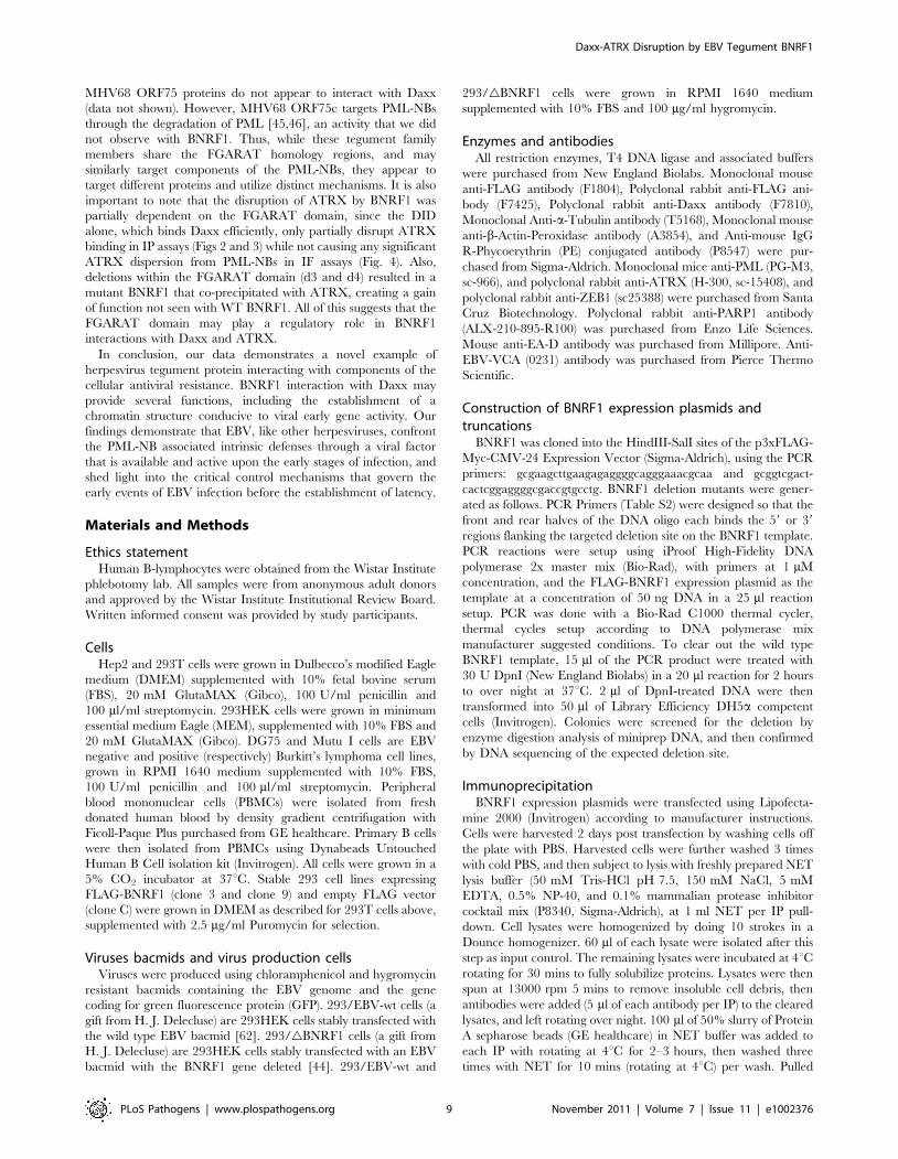
Daxx and ATRX restrict viral reactivation from latencyThe previous experiments suggest that BNRF1 can function
during tegument delivery in early infection, as well as after de novo
synthesis, perhaps regulating the transition from latent to lytic
infection. To explore the role of Daxx and ATRX in the context of
EBV latent to lytic gene regulation, we test the effects of Daxx and
ATRX knockdown on viral lytic gene expression in Mutu I cells,
an EBV-latently infected Burkitt’s lymphoma cell line (Fig. 8).
Mutu I cells were transduced with puromycin resistant lentivirus
carrying either non-targeting shRNA (shNeg), shRNA against
Daxx (shDaxx), ATRX (shATRX), or ZEB1 (shZEB1.1) which
acts as a positive control for reactivation. ZEB1 has been shown to
repress Zta expression, and shRNA depletion of ZEB1 can
reactivate lytic gene expression in several cell types [48,49,50].
Mutu I cells were harvested 9 days after shRNA transduction and
selection, and then tested for viral reactivation by Western blot
and FACS (Fig 8). Western blot analysis of whole cell lysates
(Fig. 8A and B) revealed that knockdown of either Daxx or ATRX
induced a reactivation of EBV early antigens, as shown by
increased band intensities of both the immediate early gene Zta (2-
fold) and the lytic early antigen EA-D (3-fold). These induction
levels are comparable to that observed with the shZEB1.1 positive
control. The efficiency of shRNA-mediated knockdown was
confirmed by the loss of Daxx, ATRX and ZEB1 bands in the
corresponding lanes (Fig. 8A). We also verified reactivation by flow
cytometry quantification of the EBV viral capsid antigen VCA on
cells from three independent shRNA-treatments (Fig. 8C), where
we observed an approximately 6-to-10-fold induction by either
Daxx or ATRX depletion. These findings indicate that the
depletion of either Daxx or ATRX can promote viral lytic gene
expression from latently infected B-cells, and suggest that BNRF1
disruption of the Daxx-ATRX complex contributes to viral gene
control during early infection and reactivation.
Discussion
The specific class of antiviral defense dubbed the intrinsic
immunity [51,52] plays a broad and general role in restricting viral
infection. PML-NBs and its associated proteins such as PML,
Sp100, Daxx and ATRX, have been extensively studied as cellular
defenses against herpesviruses, specifically with the alphaherpes-
virus HSV-1 and betaherpesvirus HCMV. Upon the early stages
of infection right after cell entry, HSV-1 and HCMV utilize viral
proteins that effectively disrupt the structure and disable the
function of the PML-NBs in restricting viral gene expression and
replication. However, the gammaherpesvirus EBV has been
relatively less studied in terms of how it counteracts these cellular
resistances upon primary infection or reactivation. We show here
that the major tegument protein of EBV, BNRF1, interacts with
Daxx (Figs. 1–3, S1–2) and disrupts its ability to form a complex
with ATRX or recruit ATRX to PML-NBs (Figs. 3–4, S1–2).
Moreover, we show that BNRF1 functionally promotes viral early
Figure 6. gBNRF1 mutant virus infection can be rescued byectopic complementation with wild type, but not Daxx-interaction-deficient BNRF1. Primary B cells were infected withequal volumes of GFP expressing virus, either produced from wild typebacmids or gBNRF1 mutant virus complemented with empty FLAGvector, wild type, or the d26 Daxx-interaction-deficient mutant BNRF1.Infected cells would express the GFP carried in the virus. Infection rateas shown in fluorescent microscopy photos (A), or a manual count ofthe average number of GFP-positive cells per well (B). Aliquots of thevirus used for infection were isolated for measurement of viral titers byreal-time PCR analysis of the number of viral OriLyt DNA copy numbers(C). Viral particles reconstituted with FLAG vector, FLAG-tagged WT-BNRF1 or d26 mutant were concentrated and analyzed by Western blotwith antibody to FLAG (D).doi:10.1371/journal.ppat.1002376.g006
Figure 7. BNRF1 promotes viral gene expression. (A) Primary Bcells were infected with virus produced from gBNRF1 mutant bacmidseither complemented with empty vector, wild type BNRF1 or the d26Daxx-interaction-deficient mutant BNRF1. Virus infected cells weresubject to reverse transcription PCR assay of viral gene expression forEBNA1, EBNA2, or BZLF1. (B) 293HEK cells were co-transfected with wildtype EBV genomes and either empty vector, WT BNRF1 or the d26 Daxx-interaction-deficient mutant BNRF1. Transfected cells were subject toreverse transcription PCR assay of viral gene expression for EBNA1,EBNA2, EBNA3C, or BZLF1.doi:10.1371/journal.ppat.1002376.g007
Daxx-ATRX Disruption by EBV Tegument BNRF1
PLoS Pathogens | www.plospathogens.org 7 November 2011 | Volume 7 | Issue 11 | e1002376
gene expression with a preference for the activation of the
immediate early gene BZLF1, and to a lesser extent the latent
activator EBNA2 (Figs. 6–7). These findings indicate that EBV,
like its relatives HSV1 and HCMV, encodes a viral tegument
protein that targets PML-NB components to promote viral gene
expression.
Daxx is a prominent PML-NB component, but is also associated
with a diverse, yet non-mutually exclusive variety of cellular
functions, including the regulation of apoptosis, chromatin
remodeling, gene repression, and antiviral resistance [47,53].
Daxx is a primary target of the HCMV pp71 protein, which both
binds and induces the degradation of Daxx [30]. Like HCMV
pp71, BNRF1 binds Daxx and prevents the Daxx-interaction
partner, ATRX, from associating with Daxx and localizing to
PML-NBs. BNRF1 and pp71 are both tegument proteins, whose
pre-made nature likely provides them with a temporal advantage
to disarm cellular repression machinery without the prior need of
viral gene transcription. However, unlike HCMV pp71, BNRF1
does not induce Daxx degradation, which remains prominently
associated with PML-NBs when BNRF1 is expressed (Figs 2–5).
BNRF1 and pp71 share no obvious amino acid sequence
similarity, and the Daxx interaction domains of these two proteins
vary significantly in amino acid composition and size of the
interaction domains. These findings suggest that BNRF1 is a
functional homologue of pp71, but utilizes a distinct mechanism
for the dissociation of ATRX from PML-NBs.
Herpesvirus tegument proteins have been implicated in the
determination of viral lytic or latent gene expression programs.
Restriction of tegument protein entry into the nucleus, as has been
shown for HCMV pp71 and HSV VP16, correlates with the
establishment of latency [24], suggesting that tegument proteins
may play a critical role in determining lytic or latent gene
expression programs. Interestingly, we found that the Daxx-
interaction deficient BNRF1 mutant d26, which fails to interact
with Daxx, showed a weak diffuse subcellular distribution instead
of the punctate nuclear dots of WT BNRF1 (Fig. 4 and S1).
Similarly, biochemical fractionation studies (Fig. 5B) suggest that
while BNRF1 can localize to both the cytoplasm and nucleus, it
may require the Daxx-interaction domain to efficiently accumu-
late in the nucleus. Potentially related is the observation that
HCMV pp71 translocation to PML-NBs is also dependent on its
interaction with Daxx [54]. Selective cytoplasmic retention of
several herpesvirus tegument proteins, including pp71 and VP16,
may play a critical role in determining lytic or latent gene
expression programs [55]. We suspect that BNRF1 might be
subject to similar regulation through its PML-NB localization.
Daxx and ATRX are known to play a global role in the control
of cellular and viral gene expression and chromosomal structure.
Daxx itself has been shown to associate with HDACs and to
function as a global repressor of transcription [20,21]. The Daxx-
ATRX complex has in vitro chromatin remodeling activities [26]
and can function as a histone H3.3 chaperone [56]. Recent studies
suggest also that ATRX interacts with G-rich repeat chromatin
regions [57], and in collaboration with Daxx load histone variant
H3.3 onto pericentromeric and telomeric chromatin [56,58,59].
H3.3 is generally associated with open chromatin and active
transcription when loaded by the histone chaperone HIRA [60].
However, the Daxx-ATRX complex loaded H3.3 has been found
to facilitate transcription from pericentromeric regions [58] but
repress transcription from telomeric regions [59]. Interestingly,
HIRA-loaded H3.3 can facilitate the lytic replication of HSV-1
during the early steps of infection [61]. Furthermore, the Daxx-
degrading pp71 blocks the establishment of heterochromatin on
the HCMV Major Immediate Early Promoter (MIEP) region [25].
These findings underscore the importance of host chromatin
regulatory mechanisms in the control of herpesvirus infection. We
suspect that the viral gene activation function of BNRF1 (Fig. 7) is
likely to be mediated by chromatin-dependent processes since we
failed to observe consistent transcription activation when assayed
in transient plasmid-based reporter assays using EBV promoters
for BZLF1 (Zp) or EBNA2 (Cp or Wp) (data not shown). We
propose that BNRF1 stimulates EBV early gene activity through
de-repression of the Daxx-ATRX mediated chromatin repression
mechanism, perhaps similar to that of pp71 de-repression of the
HCMV MIEP locus. However, the precise molecular mechanism
through which BNRF1 activates early gene transcription through
the disruption of ATRX-Daxx interaction remains to be
investigated.
While not explored yet, it is also not known if the FGARAT
enzyme-homology domain of BNRF1 has any function in the
context of supporting viral infection. This enzyme homology is
conserved among all gammaherpesvirus orthologues of BNRF1,
including the KSHV and MHV68 ORF75 family members.
Despite significant sequence similarity with BNRF1, KSHV and
Figure 8. ATRX and Daxx depletion leads to reactivation oflatent EBV. Mutu I cells were transduced with lentivirus shRNA withnon-targeting sequence (shNeg), or targeting for ATRX (shATRX), Daxx(shDaxx) or positive control ZEB1 (shZEB1.1). Transduced cells wereselected for puromycin resistance for 9 days and then assayed byWestern blot with antibodies to Zta, EA-D, Daxx, ATRX, ZEB1, oraTubulin, as indicated and visualized by HRP (A) and densitometricscanning of Western blot band intensities (B). Three replicates of Mutu Itransduced as described above were assayed by flow cytometry withantibody to EBV viral capsid antigen VCA and quantified as % VCApositive fold change relative to shNeg. Error bars denote the standarderror among the independent experiments (C).doi:10.1371/journal.ppat.1002376.g008
Daxx-ATRX Disruption by EBV Tegument BNRF1
PLoS Pathogens | www.plospathogens.org 8 November 2011 | Volume 7 | Issue 11 | e1002376
MHV68 ORF75 proteins do not appear to interact with Daxx
(data not shown). However, MHV68 ORF75c targets PML-NBs
through the degradation of PML [45,46], an activity that we did
not observe with BNRF1. Thus, while these tegument family
members share the FGARAT homology regions, and may
similarly target components of the PML-NBs, they appear to
target different proteins and utilize distinct mechanisms. It is also
important to note that the disruption of ATRX by BNRF1 was
partially dependent on the FGARAT domain, since the DID
alone, which binds Daxx efficiently, only partially disrupt ATRX
binding in IP assays (Figs 2 and 3) while not causing any significant
ATRX dispersion from PML-NBs in IF assays (Fig. 4). Also,
deletions within the FGARAT domain (d3 and d4) resulted in a
mutant BNRF1 that co-precipitated with ATRX, creating a gain
of function not seen with WT BNRF1. All of this suggests that the
FGARAT domain may play a regulatory role in BNRF1
interactions with Daxx and ATRX.
In conclusion, our data demonstrates a novel example of
herpesvirus tegument protein interacting with components of the
cellular antiviral resistance. BNRF1 interaction with Daxx may
provide several functions, including the establishment of a
chromatin structure conducive to viral early gene activity. Our
findings demonstrate that EBV, like other herpesviruses, confront
the PML-NB associated intrinsic defenses through a viral factor
that is available and active upon the early stages of infection, and
shed light into the critical control mechanisms that govern the
early events of EBV infection before the establishment of latency.
Materials and Methods
Ethics statementHuman B-lymphocytes were obtained from the Wistar Institute
phlebotomy lab. All samples were from anonymous adult donors
and approved by the Wistar Institute Institutional Review Board.
Written informed consent was provided by study participants.
CellsHep2 and 293T cells were grown in Dulbecco’s modified Eagle
medium (DMEM) supplemented with 10% fetal bovine serum
(FBS), 20 mM GlutaMAX (Gibco), 100 U/ml penicillin and
100 ml/ml streptomycin. 293HEK cells were grown in minimum
essential medium Eagle (MEM), supplemented with 10% FBS and
20 mM GlutaMAX (Gibco). DG75 and Mutu I cells are EBV
negative and positive (respectively) Burkitt’s lymphoma cell lines,
grown in RPMI 1640 medium supplemented with 10% FBS,
100 U/ml penicillin and 100 ml/ml streptomycin. Peripheral
blood mononuclear cells (PBMCs) were isolated from fresh
donated human blood by density gradient centrifugation with
Ficoll-Paque Plus purchased from GE healthcare. Primary B cells
were then isolated from PBMCs using Dynabeads Untouched
Human B Cell isolation kit (Invitrogen). All cells were grown in a
5% CO2 incubator at 37uC. Stable 293 cell lines expressing
FLAG-BNRF1 (clone 3 and clone 9) and empty FLAG vector
(clone C) were grown in DMEM as described for 293T cells above,
supplemented with 2.5 mg/ml Puromycin for selection.
Viruses bacmids and virus production cellsViruses were produced using chloramphenicol and hygromycin
resistant bacmids containing the EBV genome and the gene
coding for green fluorescence protein (GFP). 293/EBV-wt cells (a
gift from H. J. Delecluse) are 293HEK cells stably transfected with
the wild type EBV bacmid [62]. 293/gBNRF1 cells (a gift from
H. J. Delecluse) are 293HEK cells stably transfected with an EBV
bacmid with the BNRF1 gene deleted [44]. 293/EBV-wt and
293/gBNRF1 cells were grown in RPMI 1640 medium
supplemented with 10% FBS and 100 mg/ml hygromycin.
Enzymes and antibodiesAll restriction enzymes, T4 DNA ligase and associated buffers
were purchased from New England Biolabs. Monoclonal mouse
anti-FLAG antibody (F1804), Polyclonal rabbit anti-FLAG ani-
body (F7425), Polyclonal rabbit anti-Daxx antibody (F7810),
Monoclonal Anti-a-Tubulin antibody (T5168), Monoclonal mouse
anti-b-Actin-Peroxidase antibody (A3854), and Anti-mouse IgG
R-Phycoerythrin (PE) conjugated antibody (P8547) were pur-
chased from Sigma-Aldrich. Monoclonal mice anti-PML (PG-M3,
sc-966), and polyclonal rabbit anti-ATRX (H-300, sc-15408), and
polyclonal rabbit anti-ZEB1 (sc25388) were purchased from Santa
Cruz Biotechnology. Polyclonal rabbit anti-PARP1 antibody
(ALX-210-895-R100) was purchased from Enzo Life Sciences.
Mouse anti-EA-D antibody was purchased from Millipore. Anti-
EBV-VCA (0231) antibody was purchased from Pierce Thermo
Scientific.
Construction of BNRF1 expression plasmids andtruncations
BNRF1 was cloned into the HindIII-SalI sites of the p3xFLAG-
Myc-CMV-24 Expression Vector (Sigma-Aldrich), using the PCR
primers: gcgaagcttgaagagaggggcagggaaacgcaa and gcggtcgact-
cactcggaggggcgaccgtgcctg. BNRF1 deletion mutants were gener-
ated as follows. PCR Primers (Table S2) were designed so that the
front and rear halves of the DNA oligo each binds the 59 or 39
regions flanking the targeted deletion site on the BNRF1 template.
PCR reactions were setup using iProof High-Fidelity DNA
polymerase 2x master mix (Bio-Rad), with primers at 1 mM
concentration, and the FLAG-BNRF1 expression plasmid as the
template at a concentration of 50 ng DNA in a 25 ml reaction
setup. PCR was done with a Bio-Rad C1000 thermal cycler,
thermal cycles setup according to DNA polymerase mix
manufacturer suggested conditions. To clear out the wild type
BNRF1 template, 15 ml of the PCR product were treated with
30 U DpnI (New England Biolabs) in a 20 ml reaction for 2 hours
to over night at 37uC. 2 ml of DpnI-treated DNA were then
transformed into 50 ml of Library Efficiency DH5a competent
cells (Invitrogen). Colonies were screened for the deletion by
enzyme digestion analysis of miniprep DNA, and then confirmed
by DNA sequencing of the expected deletion site.
ImmunoprecipitationBNRF1 expression plasmids were transfected using Lipofecta-
mine 2000 (Invitrogen) according to manufacturer instructions.
Cells were harvested 2 days post transfection by washing cells off
the plate with PBS. Harvested cells were further washed 3 times
with cold PBS, and then subject to lysis with freshly prepared NET
lysis buffer (50 mM Tris-HCl pH 7.5, 150 mM NaCl, 5 mM
EDTA, 0.5% NP-40, and 0.1% mammalian protease inhibitor
cocktail mix (P8340, Sigma-Aldrich), at 1 ml NET per IP pull-
down. Cell lysates were homogenized by doing 10 strokes in a
Dounce homogenizer. 60 ml of each lysate were isolated after this
step as input control. The remaining lysates were incubated at 4uCrotating for 30 mins to fully solubilize proteins. Lysates were then
spun at 13000 rpm 5 mins to remove insoluble cell debris, then
antibodies were added (5 ml of each antibody per IP) to the cleared
lysates, and left rotating over night. 100 ml of 50% slurry of Protein
A sepharose beads (GE healthcare) in NET buffer was added to
each IP with rotating at 4uC for 2–3 hours, then washed three
times with NET for 10 mins (rotating at 4uC) per wash. Pulled
Daxx-ATRX Disruption by EBV Tegument BNRF1
PLoS Pathogens | www.plospathogens.org 9 November 2011 | Volume 7 | Issue 11 | e1002376

down proteins were released by adding 50 ml 2x Laemmli buffer
(100 mM Tris-Cl pH 6.8, 4% SDS, 0.2% Bromophenol Blue,
20% Glycerol), and boiling for 10 mins at 100uC. The resulting
samples (excluding beads) were then loaded directly into protein
gels and subject to Western blot analysis.
For mass spectrometry identification of BNRF1 associated
proteins, FLAG-BNRF1 expressing and FLAG-vector control
stable cell lines were generated as mentioned above. Nuclear
extracts from 56107 cells were subject to immunopurification with
anti-FLAG Sepharose beads (A2220, Sigma-Aldrich) followed
extensive washing with NET buffer, and FLAG peptide elution.
Eluted protein was subject to precipitation with 10% trichlor-
oacetic acid (TCA) followed by SDS-PAGE and colloidal blue
staining. Sections of the gel with enriched polypeptides were
subject to LC/MS/MS at the Wistar Proteomics Facility.
Immunofluorescence microscopyHep2 cells were transfected with BNRF1 expression plasmids
using Lipofectamine 2000 (Invitrogen) according to the manufac-
turer’s instructions, transfected cells were then reseeded at
2.76104 cells/well in 24 well plates containing microscope
coverslips 5 hours post transfection. 2 days post transfection,
coverslips with cells attached were harvested, fixed with 1%
paraformaldehyde at room temperature for 15 mins, then
permeablized with 0.3% Triton-X 100. Coverslips were then
stained with the first antibodies over night at 4uC. First antibody
dilutions used were as follows: mouse anti-FLAG at 1:20000,
rabbit anti-Daxx at 1:5000, rabbit anti-FLAG at 1:5000, mouse
anti-PML at 1:250, rabbit anti-ATRX at 1:250, all antibodies
diluted in PBS. Second antibody stainings were carried out for 1
hour at room temperature with the red-fluorescent Alexafluor594
goat anti-rabbit antibody and green Alexafluor488 goat anti-
mouse antibody (both from Invitrogen) each at 1/800 dilution in
PBS. Coverslips were washed twice in PBS for 5 mins between
each of the above treatments. Cell nuclei were stained briefly with
DAPI (diluted to a final concentration of 0.167 mg/ml in PBS) for
2 mins, then washed with PBS, 70% EtOH, then 100% EtOH to
wash out residual salts. Coverslips were air-dried briefly, and then
mounted onto microscope slides with Vectasheld mounting media
(Vector Laboratories). Mounted slides were examined under a
Nikon E600 upright microscope with a 100x oil objective. Photos
for nuclear body quantification were took using a 40x objective to
maximize the number of cells in each photo while retaining a clear
view of PML bodies.
Nuclear body quantificationMicroscopy photos were analyzed using ImagePro Plus 6.2
software (Media Cybernetics). Photos were pre-processed by
subtracting out the background intensity using the operation
function (with a value of 230), and passing through a flatten filter
(a value of 10). A morphological ‘top hat’ filter was then applied to
emphasize points or grains brighter then the background. The
number of nuclear bodies in each cell nucleus was counted by
quantifying the object numbers after applying the signal intensity
threshold/segmentation tool to select the nuclear bodies as objects.
Cell boundaries were defined by the outline from DAPI channel
photos of the same field, while omitting all cells on the border of
the image border. Resulting quantification numbers were then
analyzed using Prism 4 software (Graph Pad Software), statistical
analysis did by Mann-Whitney U non-parametric, unpaired t test.
Subcellular fractionation assay293T cells were transfected in 10 cm plates with 2 mg
expression plasmids of either empty FLAG vector, WT-BNRF1,
BNRF1-DID, or 6 mg of BNRF1-d26. Transfection was carried
out using 10 ml Lipofectamine 2000 (Invitrogen) per transfection,
following manufacturer instructions. Cells were harvested 24 hrs
post transfection. 1/6 of cells isolated as input control. The rest of
the cell pellets were fractionated with the Fermentas ProteoJET
Cytoplasmic and Nuclear Protein Extraction Kit (K0311). The
resulting cytoplasmic and nuclear fractions, along with the input
samples, were analyzed by Western blot.
BNRF1 complementation virus infection assayTo induce lytic virus production, 293/EBV-wt and 293/
gBNRF1 cells were transfected in 10 cm plates with expression
plasmids of 1.75 mg BALF4, 3.25 mg BZLF1 or cDNA3 empty
vector, and 3 mg of either empty FLAG vector or 3 mg BNRF1 or
7.5 mg BNRF1-d26. Transfection was carried out using 15 ml
Lipofectamine 2000 (Invitrogen) per transfection, following
manufacturer instructions. The media of virus production cells
were harvested 3 days post transfection, filtered through 0.45 mm
filters, and added directly to freshly isolated primary B cells. B cells
in virus containing media were centrifuged for 1200 rpm 90 mins
at 25uC to enhance infection.
For measuring infection by GFP levels, infected B cells were
treated with 1 mM Sodium Butyrate and 20 ng/ml TPA 3 days
post transfection to enhance GFP expression, and the number of
GFP positive cells in each well were counted manually under a
Nikon TE2000 microscope using a 20x objective.
For measuring virus gene expression in infected B cells, cells
were collected 4 days post transfection, and total RNA was
purified using Trizol (Invitrogen). The resulting RNA was then
subject to DNase 1 treatment at 2 U/50 ml, 1 hour at 37uC, then
DNase was heat inactivated by adding a final concentration of
5 mM EDTA and incubated at 70uC for 10 mins. cDNA was
synthesized using the Super Script III first strand synthesis system
reverse-transcription kit (Invitrogen). The resulting cDNA was
then subject to real time PCR analysis by DCt method and
normalized to viral titers, measured as described below. Real time
PCR primers used are listed in Table S3.
To measure the amount of complemented BNRF1 protein that
were packaged into virions, gBNRF1 virions complemented with
WT-BNRF1, BNRF1-d26, or un-complemented, were produced
as mentioned above. 100 ml of the harvested and filtered virus-
containing media were isolated for viral titer measurement as
described below. The rest of the virus-containing media were
concentrated by loading the media above a 5 ml layer of 22.5%
sucrose in PBS, then centrifuged at 27000 rpm (,100,000 g) 4uCfor 1 hour in a SureSpin 630 Rotor (Thermo Scientific) with a
Sorvall WX 100 Ultra ultracentrifuge. The resulting virus pellet
was then resuspended in PBS, and analyzed by Western blot.
Protein gel loading volumes were normalized according to viral
titers to ensure equal amounts of virion protein in each well.
Virus titer measure by real time PCR detection of DNAcopy number
Viral DNA in media was extracted as described by C. Busse et
al. [63]. Virus-containing media were treated with 5 U/50 ml of
DNase I (New England Biolabs) for 1 hour at 37uC. DNase was
then deactivated by adding EDTA to a final concentration of
5 mM, followed by 10 mins heat inactivation at 70uC. Samples
were then mixed 1:1 with 0.1 mg/ml of proteinase K in water, and
incubated at 50uC for 1 hour, followed by 20 mins of heat
inactivation at 75uC. The released viral DNA was measured by
real time PCR analysis, using a serial dilution series of Namalwa
cell lysate as the standard curve, which contain two copies of
integrated EBV genome per Namalwa cell. EBV genomes were
Daxx-ATRX Disruption by EBV Tegument BNRF1
PLoS Pathogens | www.plospathogens.org 10 November 2011 | Volume 7 | Issue 11 | e1002376
detected using primers specific to the OriLyt region: 59-
CGTCTTACTGCCCAGCCTACTC-39 (OriLyt-fwd), 59- AGT-
GGGAGGGCAGGAAATG-39 (OriLyt-rev).
Virus gene expression assay on bacmid/BNRF1co-transfected cells
Wild type EBV genome bacmids were prepared from 2.5 mls
overnight LB culture using the Bacmax DNA purification kit
(Epicentre). 293HEK cells were seeded (2.3 million cells per plate)
the previous day in 10 cm plates, and transfected with 1.5 mg freshly
prepared bacmids along with 0.5 mg of either empty FLAG vector,
BNRF1, or BNRF1-d26 mutant. Transfection was carried out using
Effectene transfection reagents (Qiagen), following manufacturer
instructions. Cells were harvested three days post transfection, total
RNA was purified using Trizol (Invitrogen), and then subject to DNase
1 treatment at 2 U/50 ml, 1 hour at 37uC. DNase was heat inactivated
by adding a final concentration of 5 mM EDTA and incubated at
70uC for 10 mins. cDNA was then synthesized using the Super Script
III first strand synthesis system reverse-transcription kit (Invitrogen).
The resulting cDNA was then subject to real time PCR analysis byDCt
method. Real time PCR primers used are listed in Table S3.
shRNA-mediated knockdown of genesshNeg (pLKO-shNeg), shDaxx (pLKO-shDaxx-2) and shATRX
(pLKO-shATRX90) constructs in lentivirus production plasmid
backbones were generous gifts from Roger Everett. shNeg (sequence
TTATCGCGCATATCACGCG) was designed to poorly target the
E.coli DNA polymerase and extensively screened to ensure that it
does not affect human nor viral transcripts. Use of shDaxx and
shATRX was previously described else where [30,31]. shZEB1.1 was
obtained from the TRC library (Sigma, Inc), with targeting sequence
GCAACAATACAAGAGGTTAAACTCGAGTTTAACCTCTT-
GTATTGTTGC). Mutu I cells were infected with lentiviruses
carrying pLKO.1-puro vectors by spin-infection at 400 g for 45
minutes at room temperature. The pellets were resuspended in fresh
medium and left growing overnight. The RPMI medium was
replaced each day, with 2.5 ug/ml Puromycin added for selection for
lentivirus transduced cells. The cells were collected after 9 days of
puromycin selection, and subject to Flow cytometry quantification of
EBV viral capsid antigen positive cells, and Western blot analysis.
Supporting Information
Figure S1 BNRF1 co-localizes with Daxx at PML-NBsand disperses ATRX from PML-NBs. Hep2 cells were
transfected with either FLAG empty vector, WT BNRF1, or the
deletion constructs d26 (or d2 in panel D) and DID. Cells were
fixed 2 days post transfection and co-stained with anti-FLAG, and
DAPI, and either anti-Daxx (A), anti-PML (B), anti-ATRX (C), or
anti-Sp100 (AB1380, Chemicon International, used at 1/800
dilution in PBS) antibodies. Yellow regions in the merged panels
denote co-localization of red and green signals.
(TIF)
Figure S2 Signal intensity profiles analysis of BNRF1co-localization with PML nuclear bodies. Color channel
merged panels from Fig. 4 were subject to line scan signal intensity
analysis as [64]. Signal intensity plots of red and green channels
were plotted bellow each photo, where the x-axis runs from left to
right along the yellow line drawn across several nuclear foci in
each photo. Overlaps of BNRF1 signals with Daxx (A), PML (B),
and ATRX (C) are analyzed.
(TIF)
Figure S3 Time-course study of the effects of BNRF1 onPML and Daxx protein stability. Hep2 cells were transfected
with vector control, WT BNRF1, or BNRF1-d26 mutant
expression vectors. Total cell lysates were analysed by Western
blot at 0, 12, 24, or 48 hrs post-transfection. Western blots were
probed with antibodies to PML, Daxx, FLAG (BNRF1), or Actin,
as indicated to the right.
(TIF)
Table S1 Percentage of BNRF1 foci overlap with Daxxor PML foci, as observed by IF microscopy.
(XLS)
Table S2 Primers used in site-directed mutagenesis ofBNRF1 deletion constructs.
(XLS)
Table S3 Primers used for real time PCR detection ofviral gene expression.
(XLS)
Acknowledgments
We thank the Wistar Institute Cancer Center Core Facilities for
Proteomics, Microscopy, Flow Cytometry, and Genomics. We would like
to express our gratitude for the generous gifts of Roger Everett. We also
acknowledge the generous gifts and advice of Dmitri Negorev and Gerd
Maul, and dedicate this manuscript to the memory of Gerd Maul.
Furthermore, we thank Susan Janicki and Ilona Rasalska-Metcalf for help
with Line Scan analysis, and members of the Lieberman lab, especially
Andreas Wiedmer, Zhong Deng, and Italo Tempera for technical and
intellectual advice.
Author Contributions
Conceived and designed the experiments: KT NT PML. Performed the
experiments: KT NT JAW. Analyzed the data: KT PML. Contributed
reagents/materials/analysis tools: HJD. Wrote the paper: KT PML.
References
1. Cohen JI (2000) Epstein-Barr virus infection. N Engl J Med 343: 481–492.
2. Rickinson AB, Kieff, E (2007) Epstein-Barr Virus. Fields BN, Knipe DM,
Howley PM, eds. Philadelphia: Wolters Kluwer Health/Lippincott Williams &
Wilkins. 2 v. (xix, 3091, 3086 p.) p.
3. Young LS, Rickinson AB (2004) Epstein-Barr virus: 40 years on. Nat Rev
Cancer 4: 757–768.
4. Kieff E (2007) Epstein-Barr Virus and its replication. Fields BN, Knipe DM,
Howley PM, eds. Philadelphia: Wolters Kluwer Health/Lippincott Williams &
Wilkins. 2 v. (xix, 3091, 3086 p.) p.
5. Gottschalk S, Rooney CM, Heslop HE (2005) Post-Transplant Lymphoprolif-
erative Disorders. Annu Rev Med 56: 29–44.
6. Bernardi R, Pandolfi PP (2007) Structure, dynamics and functions of
promyelocytic leukaemia nuclear bodies. Nat Rev Mol Cell Biol 8: 1006–1016.
7. Everett RD, Chelbi-Alix MK (2007) PML and PML nuclear bodies: implications
in antiviral defence. Biochimie 89: 819–830.
8. Chelbi-Alix MK, Pelicano L, Quignon F, Koken MH, Venturini L, et al. (1995)
Induction of the PML protein by interferons in normal and APL cells. Leukemia
9: 2027–2033.
9. Grotzinger T, Jensen K, Will H (1996) The interferon (IFN)-stimulated gene
Sp100 promoter contains an IFN-gamma activation site and an imperfect IFN-
stimulated response element which mediate type I IFN inducibility. J Biol Chem
271: 25253–25260.
10. Lavau C, Marchio A, Fagioli M, Jansen J, Falini B, et al. (1995) The acute
promyelocytic leukaemia-associated PML gene is induced by interferon.
Oncogene 11: 871–876.
11. Chelbi-Alix MK, Quignon F, Pelicano L, Koken MH, de The H (1998)
Resistance to virus infection conferred by the interferon-induced promyelocytic
leukemia protein. J Virol 72: 1043–1051.
12. Ishov AM, Maul GG (1996) The periphery of nuclear domain 10 (ND10) as site
of DNA virus deposition. J Cell Biol 134: 815–826.
Daxx-ATRX Disruption by EBV Tegument BNRF1
PLoS Pathogens | www.plospathogens.org 11 November 2011 | Volume 7 | Issue 11 | e1002376

13. Korioth F, Maul GG, Plachter B, Stamminger T, Frey J (1996) The nuclear
domain 10 (ND10) is disrupted by the human cytomegalovirus gene productIE1. Exp Cell Res 229: 155–158.
14. Hensel GM, Meyer HH, Buchmann I, Pommerehne D, Schmolke S, et al.
(1996) Intracellular localization and expression of the human cytomegalovirusmatrix phosphoprotein pp71 (ppUL82): evidence for its translocation into the
nucleus. J Gen Virol 77(Pt 12): 3087–3097.15. Zhong S, Salomoni P, Pandolfi PP (2000) The transcriptional role of PML and
the nuclear body. Nat Cell Biol 2: E85–90.
16. Lehembre F, Muller S, Pandolfi PP, Dejean A (2001) Regulation of Pax3transcriptional activity by SUMO-1-modified PML. Oncogene 20: 1–9.
17. Emelyanov AV, Kovac CR, Sepulveda MA, Birshtein BK (2002) Theinteraction of Pax5 (BSAP) with Daxx can result in transcriptional activation
in B cells. J Biol Chem 277: 11156–11164.18. Li R, Pei H, Watson DK, Papas TS (2000) EAP1/Daxx interacts with ETS1 and
represses transcriptional activation of ETS1 target genes. Oncogene 19:
745–753.19. Michaelson JS, Leder P (2003) RNAi reveals anti-apoptotic and transcriptionally
repressive activities of DAXX. J Cell Sci 116: 345–352.20. Li H, Leo C, Zhu J, Wu X, O’Neil J, et al. (2000) Sequestration and inhibition of
Daxx-mediated transcriptional repression by PML. Mol Cell Biol 20:
1784–1796.21. Hollenbach AD, McPherson CJ, Mientjes EJ, Iyengar R, Grosveld G (2002)
Daxx and histone deacetylase II associate with chromatin through an interactionwith core histones and the chromatin-associated protein Dek. J Cell Sci 115:
3319–3330.22. Michaelson JS, Bader D, Kuo F, Kozak C, Leder P (1999) Loss of Daxx, a
promiscuously interacting protein, results in extensive apoptosis in early mouse
development. Genes Dev 13: 1918–1923.23. Muromoto R, Sugiyama K, Takachi A, Imoto S, Sato N, et al. (2004) Physical
and functional interactions between Daxx and DNA methyltransferase 1-associated protein, DMAP1. J Immunol 172: 2985–2993.
24. Saffert RT, Kalejta RF (2006) Inactivating a cellular intrinsic immune defense
mediated by Daxx is the mechanism through which the human cytomegaloviruspp71 protein stimulates viral immediate-early gene expression. J Virol 80:
3863–3871.25. Woodhall DL, Groves IJ, Reeves MB, Wilkinson G, Sinclair JH (2006) Human
Daxx-mediated repression of human cytomegalovirus gene expression correlateswith a repressive chromatin structure around the major immediate early
promoter. J Biol Chem 281: 37652–37660.
26. Xue Y, Gibbons R, Yan Z, Yang D, McDowell TL, et al. (2003) The ATRXsyndrome protein forms a chromatin-remodeling complex with Daxx and
localizes in promyelocytic leukemia nuclear bodies. Proc Natl Acad Sci U S A100: 10635–10640.
27. Ishov AM, Vladimirova OV, Maul GG (2004) Heterochromatin and ND10 are
cell-cycle regulated and phosphorylation-dependent alternate nuclear sites of thetranscription repressor Daxx and SWI/SNF protein ATRX. J Cell Sci 117:
3807–3820.28. Tavalai N, Papior P, Rechter S, Stamminger T (2008) Nuclear domain 10
components promyelocytic leukemia protein and hDaxx independentlycontribute to an intrinsic antiviral defense against human cytomegalovirus
infection. J Virol 82: 126–137.
29. Cantrell SR, Bresnahan WA (2006) Human cytomegalovirus (HCMV) UL82gene product (pp71) relieves hDaxx-mediated repression of HCMV replication.
J Virol 80: 6188–6191.30. Lukashchuk V, McFarlane S, Everett RD, Preston CM (2008) Human
cytomegalovirus protein pp71 displaces the chromatin-associated factor ATRX
from nuclear domain 10 at early stages of infection. J Virol 82: 12543–12554.31. Lukashchuk V, Everett RD (2010) Regulation of ICP0-null mutant herpes
simplex virus type 1 infection by ND10 components ATRX and hDaxx. J Virol84: 4026–4040.
32. Smibert CA, Popova B, Xiao P, Capone JP, Smiley JR (1994) Herpes simplex
virus VP16 forms a complex with the virion host shutoff protein vhs. J Virol 68:2339–2346.
33. Boutell C, Sadis S, Everett RD (2002) Herpes simplex virus type 1 immediate-early protein ICP0 and is isolated RING finger domain act as ubiquitin E3
ligases in vitro. J Virol 76: 841–850.34. Everett RD, Freemont P, Saitoh H, Dasso M, Orr A, et al. (1998) The disruption
of ND10 during herpes simplex virus infection correlates with the Vmw110- and
proteasome-dependent loss of several PML isoforms. J Virol 72: 6581–6591.35. Maul GG, Guldner HH, Spivack JG (1993) Modification of discrete nuclear
domains induced by herpes simplex virus type 1 immediate early gene 1 product(ICP0). J Gen Virol 74(Pt 12): 2679–2690.
36. Everett RD, Rechter S, Papior P, Tavalai N, Stamminger T, et al. (2006) PML
contributes to a cellular mechanism of repression of herpes simplex virus type 1infection that is inactivated by ICP0. J Virol 80: 7995–8005.
37. Preston CM, Nicholl MJ (1997) Repression of gene expression upon infection ofcells with herpes simplex virus type 1 mutants impaired for immediate-early
protein synthesis. J Virol 71: 7807–7813.
38. Stow EC, Stow ND (1989) Complementation of a herpes simplex virus type 1Vmw110 deletion mutant by human cytomegalovirus. J Gen Virol 70(Pt 3):
695–704.
39. Bell P, Lieberman PM, Maul GG (2000) Lytic but not latent replication ofepstein-barr virus is associated with PML and induces sequential release of
nuclear domain 10 proteins. J Virol 74: 11800–11810.
40. Adamson AL, Kenney S (2001) Epstein-barr virus immediate-early protein
BZLF1 is SUMO-1 modified and disrupts promyelocytic leukemia bodies. J Virol
75: 2388–2399.
41. Sivachandran N, Cao JY, Frappier L (2010) Epstein-Barr virus nuclear antigen 1
Hijacks the host kinase CK2 to disrupt PML nuclear bodies. J Virol 84:11113–11123.
42. Ling PD, Peng RS, Nakajima A, Yu JH, Tan J, et al. (2005) Mediation of
Epstein-Barr virus EBNA-LP transcriptional coactivation by Sp100. EMBO J24: 3565–3575.
43. Johannsen E, Luftig M, Chase MR, Weicksel S, Cahir-McFarland E, et al.
(2004) Proteins of purified Epstein-Barr virus. Proc Natl Acad Sci U S A 101:16286–16291.
44. Feederle R, Neuhierl B, Baldwin G, Bannert H, Hub B, et al. (2006) Epstein-Barr virus BNRF1 protein allows efficient transfer from the endosomal
compartment to the nucleus of primary B lymphocytes. J Virol 80: 9435–9443.
45. Gaspar M, Gill MB, Losing JB, May JS, Stevenson PG (2008) Multiple functionsfor ORF75c in murid herpesvirus-4 infection. PLoS One 3: e2781.
46. Ling PD, Tan J, Sewatanon J, Peng R (2008) Murine gammaherpesvirus 68
open reading frame 75c tegument protein induces the degradation of PML andis essential for production of infectious virus. J Virol 82: 8000–8012.
47. Lindsay CR, Morozov VM, Ishov AM (2008) PML NBs (ND10) and Daxx: fromnuclear structure to protein function. Front Biosci 13: 7132–7142.
48. Kraus RJ, Perrigoue JG, Mertz JE (2003) ZEB negatively regulates the lytic-
switch BZLF1 gene promoter of Epstein-Barr virus. J Virol 77: 199–207.
49. Ellis AL, Wang Z, Yu X, Mertz JE (2010) Either ZEB1 or ZEB2/SIP1 can play
a central role in regulating the Epstein-Barr virus latent-lytic switch in a cell-
type-specific manner. J Virol 84: 6139–6152.
50. Yu X, Wang Z, Mertz JE (2007) ZEB1 regulates the latent-lytic switch in
infection by Epstein-Barr virus. PLoS Pathog 3: e194.
51. Bieniasz PD (2007) An intrinsic host defense against HIV-1 integration? J ClinInvest 117: 302–304.
52. Bieniasz PD (2004) Intrinsic immunity: a front-line defense against viral attack.Nat Immunol 5: 1109–1115.
53. Michaelson JS (2000) The Daxx enigma. Apoptosis 5: 217–220.
54. Hofmann H, Sindre H, Stamminger T (2002) Functional interaction betweenthe pp71 protein of human cytomegalovirus and the PML-interacting protein
human Daxx. J Virol 76: 5769–5783.
55. Kolb G, Kristie TM (2008) Association of the cellular coactivator HCF-1 withthe Golgi apparatus in sensory neurons. J Virol 82: 9555–9563.
56. Lewis PW, Elsaesser SJ, Noh KM, Stadler SC, Allis CD (2010) Daxx is an H3.3-
specific histone chaperone and cooperates with ATRX in replication-independent chromatin assembly at telomeres. Proc Natl Acad Sci U S A
107: 14075–14080.
57. Law MJ, Lower KM, Voon HP, Hughes JR, Garrick D, et al. (2010) ATR-X
syndrome protein targets tandem repeats and influences allele-specific expression
in a size-dependent manner. Cell 143: 367–378.
58. Drane P, Ouararhni K, Depaux A, Shuaib M, Hamiche A (2010) The death-
associated protein DAXX is a novel histone chaperone involved in thereplication-independent deposition of H3.3. Genes Dev 24: 1253–1265.
59. Goldberg AD, Banaszynski LA, Noh KM, Lewis PW, Elsaesser SJ, et al. (2010)
Distinct factors control histone variant H3.3 localization at specific genomicregions. Cell 140: 678–691.
60. Loyola A, Almouzni G (2007) Marking histone H3 variants: how, when and
why? Trends Biochem Sci 32: 425–433.
61. Placek BJ, Huang J, Kent JR, Dorsey J, Rice L, et al. (2009) The histone variant
H3.3 regulates gene expression during lytic infection with herpes simplex virustype 1. J Virol 83: 1416–1421.
62. Delecluse HJ, Hilsendegen T, Pich D, Zeidler R, Hammerschmidt W (1998)
Propagation and recovery of intact, infectious Epstein-Barr virus fromprokaryotic to human cells. Proc Natl Acad Sci U S A 95: 8245–8250.
63. Busse C, Feederle R, Schnolzer M, Behrends U, Mautner J, et al. (2009) Epstein-
Barr viruses that express a CD21 antibody provide evidence that gp350’sfunctions extend beyond B-cell surface binding. J Virol 84: 1139–1147.
64. Shanbhag NM, Rafalska-Metcalf IU, Balane-Bolivar C, Janicki SM,Greenberg RA (2010) ATM-dependent chromatin changes silence transcription
in cis to DNA double-strand breaks. Cell 141: 970–981.
Daxx-ATRX Disruption by EBV Tegument BNRF1
PLoS Pathogens | www.plospathogens.org 12 November 2011 | Volume 7 | Issue 11 | e1002376