Easy differentiation of Mycobacterium mucogenicum from other species of the Mycobacterium fortuitum...
7
ELSEVIER Journal of Chromatography B, 689 (1997) 341-347 JOURNAL OF CHROMATOGRAPHY B Easy differentiation of Mycobacterium mucogenicum from other species of the Mycobacteriumfortuitum complex by thin-layer and gas chromatography of fatty esters and alcohols Manuel Mufioz a, Esther Julizin a, Merq6 Garcia-Barcel6 b, Vicente Ausina b, Marina Luquin a'* "Departamento de Gengtica y Microbiolog[a, Unidad de Microbiologla, Facultad de Ciencias, Universidad Autdnoma de Barcelona, 08193 Bellaterra, Barcelona, Spain hServicio de Microbiolog{a, Hospital Universitario Germans Trias i Pujol, 08916 Badalona. Barcelona, Spain Received 20 March 1996; revised 11 July 1996; accepted 11 July 1996 Abstract The mycolate pattern of a recently recognized mycobacterial pathogen, Mycobacterium mucogenicum (formerly Mycobacterium chelonae-like organism), was established for the first time. The reference strains, together with 31 environmental and clinical isolates belonging to this species, were examined for their mycolate composition by thin-layer chromatography. All strains tested exhibited the same mycolate profile. Mycolates were identified as belonging to the type without additional oxygenated chemical groups (mycolate I) and the type with a dicarboxylic group (mycolate VI); the identification of the latter was reinforced by the presence of 2-octadecanol, as seen by gas-liquid capillary chromatography. This mycolate profile permits the clear differentiation of M. mucogenicum from other related species, as members of the Mycobacterium Jortuitum complex. This fact is especially important because strains of M. mucogenicum are very difficult to differentiate from other species of the M. fortuitum complex by means of conventional biochemical tests. Moreover, the characteristic mycolate profile exhibited by the strains of M. mucogenicum supports the recent proposal which considers them as members of a new species. Keywords: Mycobacterium mucogenicum; Mycobacteriumfortuitum complex; Mycolates; Mycolic acid methyl ester; Fatty acid methyl esters; Alcohols; 1. Introduction Mycobacterium mucogenicum is a group of rapid- ly growing non-pigmented mycobacteria common in potable water, more specifically, tap water. They were implicated in nosocomial outbreaks of peritoni- tis which occurred in 1976 and 1978 in Washington Corresponding author. State, involving the use of hospital-based automated chronic peritoneal dialysis machines [1]. M. mucogenicum was later identified as a cause of nosocomial infections associated with haemodialysis in Louisiana [2] and California [3]. In all cases M. mucogenicum was recovered from tap water used in the dialysis process. A collection of 87 sporadic clinical isolates of these organisms was recently characterized [4]. The majority of these isolates 0378-4347/97/$17.00 Copyright © 1997 Elsevier Science B.V. All rights reserved PII S0378-4347(96)00330- 1
-
Upload
manuel-munoz -
Category
Documents
-
view
212 -
download
0
Transcript of Easy differentiation of Mycobacterium mucogenicum from other species of the Mycobacterium fortuitum...

E L S E V I E R Journal of Chromatography B, 689 (1997) 341-347
JOURNAL OF CHROMATOGRAPHY B
Easy differentiation of Mycobacterium mucogenicum from other species of the Mycobacterium fortuitum complex by thin-layer and
gas chromatography of fatty esters and alcohols
M a n u e l Muf ioz a, Es the r Julizin a, Merq6 G a r c i a - B a r c e l 6 b, V i c e n t e A u s i n a b,
M a r i n a L u q u i n a'*
"Departamento de Gengtica y Microbiolog[a, Unidad de Microbiologla, Facultad de Ciencias, Universidad Autdnoma de Barcelona, 08193 Bellaterra, Barcelona, Spain
hServicio de Microbiolog{a, Hospital Universitario Germans Trias i Pujol, 08916 Badalona. Barcelona, Spain
Received 20 March 1996; revised 11 July 1996; accepted 11 July 1996
Abstract
The mycolate pattern of a recently recognized mycobacterial pathogen, Mycobacterium mucogenicum (formerly Mycobacterium chelonae-like organism), was established for the first time. The reference strains, together with 31 environmental and clinical isolates belonging to this species, were examined for their mycolate composition by thin-layer chromatography. All strains tested exhibited the same mycolate profile. Mycolates were identified as belonging to the type without additional oxygenated chemical groups (mycolate I) and the type with a dicarboxylic group (mycolate VI); the identification of the latter was reinforced by the presence of 2-octadecanol, as seen by gas-liquid capillary chromatography. This mycolate profile permits the clear differentiation of M. mucogenicum from other related species, as members of the Mycobacterium Jortuitum complex. This fact is especially important because strains of M. mucogenicum are very difficult to differentiate from other species of the M. fortuitum complex by means of conventional biochemical tests. Moreover, the characteristic mycolate profile exhibited by the strains of M. mucogenicum supports the recent proposal which considers them as members of a new species.
Keywords: Mycobacterium mucogenicum; Mycobacterium fortuitum complex; Mycolates; Mycolic acid methyl ester; Fatty acid methyl esters; Alcohols;
1. Introduct ion
Mycobacterium mucogenicum is a group of rapid- ly growing non-pigmented mycobacteria common in potable water, more specifically, tap water. They were implicated in nosocomial outbreaks of peritoni- tis which occurred in 1976 and 1978 in Washington
Corresponding author.
State, involving the use of hospital-based automated chronic peritoneal dialysis machines [1]. M. mucogenicum was later identified as a cause of nosocomial infections associated with haemodialysis in Louisiana [2] and California [3]. In all cases M. mucogenicum was recovered from tap water used in the dialysis process. A collection of 87 sporadic clinical isolates of these organisms was recently characterized [4]. The majority of these isolates
0378-4347/97/$17.00 Copyright © 1997 Elsevier Science B.V. All rights reserved PII S0378-4347(96)00330- 1
342 M. Mufioz et al. I J. Chromatogr. B 689 (1997) 341-347
(62%) were respiratory isolates and had no clinical significance, except for 2 strains isolated from pa- tients with AIDS. Among 33 non-respiratory isolates, 20 were clinically significant. Clinical diseases in- cluded postraumatic wound infections and catheter related sepsis, infections which are widely recog- nized as being caused by the other groups of rapidly growing pathogenic mycobacteria [5].
Biochemical analysis of the 87 M. mucogenicum isolates revealed that they were fairly homogeneous and had typical characteristics of members of the M. fortuitum complex. Members of this complex are defined as non-pigmented mycobacteria that grow within 7 days, show a positive 3-day arylsulfatase reaction, and grow on MacConkey agar without crystal violet at 28°C [6]. Most M. mucogenicum strains meet these inclusion criteria. As shown by Wallace et al. [4], routine biochemical features failed to clearly differentiate M. mucogenicum from Mycobacterium chelonae and Mycobacterium ab- scessus (two species of the M. fortuitum complex), and only two special tests (utilization of mannitol as the sole carbon source and susceptibility to cephalo- thin) helped to separate M. mucogenicum from M. chelonae and M. abscessus. The interest in clearly differentiating M. mucogenicum from the other two is due to the fact that M. mucogenicum strains show a characteristic susceptibility pattern, that is, they are more drug susceptible than M. chelonae and M. abscessus [7]. Mycolic acids are major lipid con- stituents in the cell wall of mycobacteria and related taxa. They are s-branched, [3~hydroxy fatty acids which are characterized in mycobacteria by very long chains (up to 90 atoms long) and by the presence of various functional groups in the longest (mero) chain [8,91.
Silicic acid thin-layer chromatography (TLC) is a simple and efficient method to separate mycolic acid methyl esters in accordance with the presence and nature of oxygenated chemical functions in their long chain. Depending on the nature of these functional groups, different chromatographic behaviours are observed when preparations are subjected to TLC. Seven types of mycolates have been recognized in this way; these mycolates are type I and II mycolates (long and short mycolates with no additional oxy- genated functions), type III (methoxymycolates), type IV (ketomycolates), type V (epoxymycolates), type VI
(dicarboxymycolates) and type VII (o~-I methoxy- mycolates) [8-10]. Mycolate TLC patterns for the various mycobacterial species have already been published either in catalogues or in previous studies [8,9,11-13]. The data obtained from these studies demonstrated that determination of mycolate patterns is useful in the differentiation of mycobacterial species, and that mycolate composition is a stable taxonomic characteristic, since the content in myco- lares (except for minor components) is common to all strains of a species. Only vaccine strains of Mycobacterium bovis bacillus Calmette-Gu6rin (BCG) produce a mycolate pattern that is devoid of type III mycolates, and is thus distinct from authenti- cated profiles containing type I, III and IV mycolates found in all M. bovis strains and other tubercle bacilli [9]. One consequence of these studies is the recommendation that the TLC pattern of mycolates be included in the description of new Mycobacterium species [ 14].
In this study, for the first time, TLC analysis was applied to strains of M. mucogenicum, to try to find an easy and reproducible method to differentiate this group of microorganisms from other related species.
Additional information, such as the presence of secondary alcohols related to dicarboxymycolates (type VI), and the composition in non-hydroxylated fatty acids, was obtained by gas-liquid capillary chromatography (GLC).
2. Experimental
2.1. Mycobacterial strains
Clinical and environmental isolates of M. mucogenicum (31 strains) were characterized by cultural, biochemical and physiological standard procedures [6,15,16]. In addition to these, three M. mucogenicum reference strains, ATCC 49649, ATCC 49650 v and ATCC 49651, were tested.
Also, for identification purposes, partial sequenc- ing of the 16S rRNA gene was performed in four M. mucogenicum isolates. The 16S rRNA gene was amplified by using two primers whose oligonucleo- tide sequences were as follows: 5' GAGAGTTTGATCCTGGCTCAG 3' corresponding to the E. coli 16S rRNA gene from positions 9 to 30,
M. Mu~oz et al. / J. Chromatogr. B 689 (1997) 341-347 343
used in combination with 5' TTTCAC- GAACAACGCGACAA 3' corresponding to E. coli 16S rRNA gene from positions 609 to 590. These primers allowed the amplification of partial 16S rRNA gene that yielded a DNA fragment containing the species-specific character for mycobacteria 117,18].
The nucleic acid sequence obtained was compared with more than 30 mycobacterial 16S rRNA gene sequences, including M. mucogenicum ATCC 49650 T, deposited in the Gene Bank and EMBL data base. The Gene Bank nucleotide accession number for a selected isolate (strain CR-51) is U19366.
Reference strains, M. fi)rtuitum ATCC 6841T and M. chelonae NCTC 946 v, were included for thin- layer and gas chromatography comparative purposes. Mycobacterium terrae ATCC 15755 T and Mycobac- terium xenopi ATCC 19250 v, two slow-growing mycobacteria which are distantly related to M. mucogenicum were used exclusively to provide a pattern for identification on TLC of dicarboxymyco- late (mycolate VI). Species of mycobacteria closely related to M, mucogenicum, and displaying dicarboxy- mycolates do not, in fact, exist [8,11,13]. We therefore chose M. terrae and M. xenopi for TLC comparative purposes. However, other species which do possess dicarboxymycolates but which are equally distant taxonomically from M. mucogenicum, could have been chosen for this purpose [8,11,13]. For isolation of lipid components, all strains were grown on plates of Sauton agar and incubated at 37°C. The incubation period was two weeks for M. mucogenicum, M. fi)rtuitum and M. chelonae, and six weeks for M. terrae and M. xenopi.
2.2. Extraction of lipids
A spadeful of bacteria (10 mg wet weight) was scraped from the surface of Sauton agar plates and treated with 2 ml of a methanol-benzene solution (8:2, v/v) containing 5% (w/v) potassium hydroxide in a screw-cap test tube (14× 120 mm) fitted with a Teflon-lined cap. The mixture was heated in a covered bath at 80°C for 8 h. After cooling at room temperature, the samples were acidified by the addition of 20% (v/v) sulfuric acid, and the lipids were extracted into diethyl ether. The ether extracts were washed with water until neutral, dried over
anhydrous sodium sulfate, filtered and concentrated by evaporation to dryness.
2.3. Preparation of methyl esters
Lipids were methylated with freshly prepared diazomethane (diazomethane is toxic and has to be handled under a hood). Diazomethane was prepared from a commercially available compound, N-Nitro- so-N-methylurea (Sigma, St. Louis, MO, USA). For this purpose, 2 g of N-Nitroso-N-methylurea were dissolved in a precooled solution containing 30 ml of diethyl ether, 2.4 g of KOH and 6 ml of distilled water. The mixture was agitated for 5 to 10 rain, and then the supernatant was removed and placed in a new tube cooled in ice containing potassium hy- droxide pellets. A 1-ml portion of this solution was poured into each tube containing the dried lipids, and methylation was achieved within 5 min. Diazometh- ane was then evaporated. Lipid extracts were re- suspended in 0.5 ml of n-hexane, and then 10 Ixl were applied to silica gel plates and 1 ILl of the same lipid extract was injected in a gas chromatograph as described below. In order to avoid handling diazo- methane, methyl esters of fatty acids can be obtained with anhydrous hydrogen chloride in methanol, or boron trifluoride in methanol [19].
2.4. TLC of mycolic acid methyl esters
Analytical one-dimensional TLC was performed using sheets of precoated silica gel 60 TLC plates (0.25 mm thick, 20x20 cm; DC-Fertigplatten Kieselgel 60; Merck, Darmstadt, Germany). A triple development with 100 ml of a monophasic mixture of n-hexane-ether (85:15, v/v) was performed. The separate components were revealed as dark blue spots by spraying with 10% (w/v) molybdophos- phoric acid (Merck) in ethanol followed by charring. Identification of mycolates on TLC was performed by comparison with the mycolate patterns of refer- ence strains.
2.5. GLC of cellular fatty acid methyl esters and alcohols
Fatty acid methyl esters and alcohols were ana- lyzed on a fused-silica capillary column (15 mX0.25
344 M. Mu~oz et al. / J. Chromatogr. B 689 (1997) 341-347
mm I.D.) with cross-linked methyl silicone (HP-1, Hewlett-Packard, Palo Alto, CA, USA) as the stationary phase; the column was inserted in a Hewlett-Packard 5890A gas chromatograph equipped with a flame ionization detector. The column was programmed at 175 to 300°C at 8°C/rain and maintained at 300°C for 15 rain. The injector and detector temperatures were 275 and 315°C, respec- tively. The carrier gas was helium with a flow-rate of approximately 1 ml/min, and the split ratio was approximately 1:50. The chromatograms were inte- grated by using a HP3396 Series I1 integrator. The peaks were identified by comparing retention times with authentic methyl ester and alcohol standards (Supelco and Sigma). The identities of some com- pounds were also confirmed by mass spectrometry. A Hewlett-Packard Model 5988A mass spectrometer interfaced to a Hewlett-Packard 5890A gas chromatograph was used.
3. Results and discussion
differed from those of all described species of mycobacteria, but was identical to M. mucogenicum ATCC 49650 v [18].
3.1. Mycolate patterns obtained by TLC
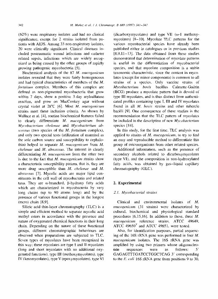
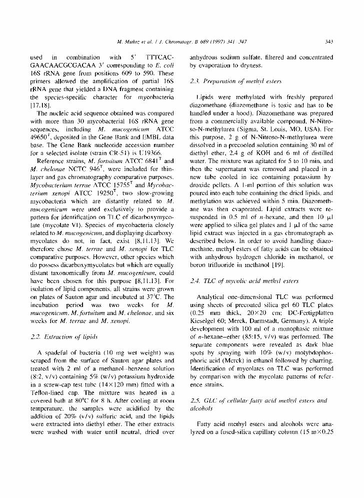
Reference strains and clinical and environmental isolates of M. mucogenicum showed the same pattern of mycolates, mycolate I (R F 0 . 4 8 ) and mycolate VI (g F 0.25) as reference strains M. xenopi and M. terrae (Figs. 1 and 2). This last type of mycolate was accompanied by secondary long-chain alcohols re- leased from dicarboxylic mycolate during saponifica- tion [8,9,20]. This mycolate composition was distinct
Clinical and environmental isolates (a total of 31 strains) grew in less than 7 days (100%), were non-pigmented (100%), gave a positive 3-day arylsulfatase reaction (100%) and grew on MacCon- key agar without crystal violet at 28°C (83%). Like M. chelonae, our strains were nitrate negative (80%), unable to grow in Lowenstein-Jensen containing 5% NaCI (100%) and utilized citrate as the sole carbon source (85%). Nevertheless, in contrast to M. chelonae and M. abscessus, isolates were susceptible to cephalothin (disk of 30 p~g) (94%) and grew on media containing mannitol as the sole carbon source (93%). The isolates were uniform in 49 other physiological, biochemical and growth characteris- tics [6,15,16]. Phenotypical characteristics shown by our isolates are identical to those described for M. mucogenicum strains [4].
The amplified region of the 16S rRNA gene was a 603 base pairs fragment containing a species-specific character for mycobacteria. Analysis of the signature sequence of the 16S rRNA gene, corresponding to the E. coli nucleotides at positions 175 to 202, which is unique for most established mycobacterial species [17,18], showed that the four selected M. mucogenicum isolates display a unique sequence that
,o e 0 o
va
v,|
I i b | , 1 2 3 4 $
Fig. 1. Thin-layer chromatography of methyl mycolates from M. mucogenicum ATCC 49650 r (lane 1), M. xenopi ATCC 19250 ~ (lane 2), M. terrae ATCC 15755 v (lane 3), M. fortuitum ATCC 6841 v (lane 4) and M. chelonae NCTC 946 v (lane 5). l, a- mycolates; II, e~'-mycolates; V, epoxymycolates: VI, dicarbo×y- mycolates.

M. Mutioz et al. / J. Chromatogr. B 689 (1997) 341-347 345
,o 0
v,! :
4 . o e
I 2 3 4 $ 6 7 8
Fig. 2. Thin-layer chromatography of methyl mycolates from M. mucogenicum ATCC 49650 ~ (lane l), ATCC 49649 (lane 2) and ATCC 49651 (lane 3). M. mucogenicum environmental and clinical isolates, CR-1 (lane 4), CR-2 (lane 5), CR-20 (lane 6). CR-46 (lane 7) and CR-51 (lane 8). I, c~-mycolates; VI, dicarboxymycolates.
from that of M. chelonae NCTC 946 v which con- tained only mycolates I and II (Rf 0.43), and from M. fortuitum ATCC 6841 v composed of mycolate I and mycolate V (Rf 0.34). In a previous study [10], we examined by TLC 102 other mycobacterial strains belonging to the four species which make up the M. fortuitum complex (28 M. chelonae, 6 M. abscessus, 42 M. fortuitum and 26 M. peregrinum). Mycolate V1 was not detected in any of these strains. All the strains belonging to M. chelonae and M. abscessus species showed a mycolate pattern composed of mycolates I and II. Likewise, M. fortuitum and M. peregrinum strains showed a pattern composed of mycolates I and V. In addition to these mycolates some strains of M. fortuitum and M. peregrinum
showed mycolates II and VII as minor compounds. These results agree with those obtained by other authors [8,9,1 1,13].
3.2. Analysis o f fat ty acid methyl esters and alcohols
Gas chromatograms from all strains tested re- vealed the presence of tetradecanoic (C 14:0)' hexade- cenoic (C~6:~), hexadecanoic (C~6:o), octadecenoic (C ~s:~ ), octadecanoic (C ~8:o), 10-methyloctadecanoic (TBS, tuberculostearic acid), docosanoic (C22:o) and tetracosanoic acids (C24:o) (Fig. 3). Gas chromato- grams of all the M. mucogenicum strains presented two additional peaks which were absent in M.

346 M. MuBoz et al. / J. Chromatogr. B 689 (1997) 341-347
A
I
I I
I
ha
B !
txt_~
C
?
Retent ion t i m e (rain)
2 4 6 n m Iz 14 16
Fig. 3. Gas chromatograms of fatty acid methyl esters and alcohols of M. fortuitum ATCC 6841T (A); M. chelonae NCTC 946 T (B) and M. mucogenicum ATCC 49650 T (C). The numbers above the peaks indicate the number of carbon atoms, followed by the number of double bonds. 10-M16:0, 10-methylhexadecanoate; 2-OH18:0, 2-octadecanol; TBS, tuberculostearate; FID, Flame ionization detector.
chelonae and M. fortuitum strains. These two com- pounds were identified by gas chromatography-mass spectrometry as 10-methylhexadecanoic acid (10-M Cl6:0 ) and 2-octadecanol (2-OH C18:0 ). As stated above, secondary long-chain alcohols (octadecanol, eicosanol and docosanol) were released from di- carboxymycolates during saponification. These al- cohols can easily be detected by GLC [20-24]. Thus, a secondary alcohol, 2-octadecanol, was detected in all the M. mucogenicum tested. As expected, sec- ondary alcohols were not detected in strains of M. chelonae and M. fi)rtuitum since these species, like M. abscessus and M. peregrinum, do not display dicarboxymycolates [8-10]. In previous extensive studies performed by us and other authors [22-27], appreciable amounts of 10-methylhexadecanoic acid were detected in only two photochromogenic species, Mycobacterium marinum and Mycobac- terium szulgai; this compound was not detected in any of the members of the M. fortuitum complex.
M. xenopi and M. terrae are two slow-growing mycobacteria which are distantly related to M. mucogenicum. Normally, chromatographic proce- dures are not used to differentiate these species from M. mucogenicum, since more simple observations such as growth rate, pigmentation and colonial morphology can accomplish this purpose. In the present study we used type strains of M. xenopi and M. terrae only to provide a pattern for identification on TLC of dicarboxylic mycolate. However, gas chromatograms of non-hydroxylated fatty acids and alcohols of M. xenopi and M. terrae could be used to discriminate between these species and M. mucogenicum since, as we reported previously [20], gas chromatograms of M. xenopi strains show two characteristic peaks corresponding to 2-docosanol (2-OH C22:0 ) and hexacosanoic acid (C26:o) which are absent in gas chromatograms of M. mucogenicum strains. On the other hand, M. terrae strains are devoid of 10-M C1~:o [23], a compound present in all the M. mucogenicum strains investigated.
Our study showed that the determination of the presence of dicarboxymycolates (type VI) by TLC, or 2-octadecanol and 10-methylhexadecanoic acid by GLC, in the M. mucogenicum strains, allows the clear differentiation of these from other members of the M. fortuitum complex. TLC or GLC can both be used for differentiation of M. mucogenicum strains.

M. Mu~oz et al. / J. Chromatogr. B 689 (1997) 341-347 347
T L C is an easy and i n e x p e n s i v e t echn ique ; G L C
requi res appropr ia te e q u i p m e n t and ski l led person-
nel, bu t is rou t ine ly used in m a n y c l in ical mic ro -
b io logy labora tor ies to ident i fy d i f fe ren t bac ter ia l
g roups (anaerob ic bacter ia , n o n - f e r m e n t a t i v e g ram-
nega t ive bacter ia , Campylobacter, Helicobacter,
Legionella, etc...).
The m o l e c u l a r type o f myco la t e s c o n t a i n i n g M.
mucogenicum s t ra ins and the profi le o f n o n - h y d r o x y -
la ted fat ty acids and a lcohols h a v e been es t ab l i shed
for the first t ime in the p resen t inves t iga t ion . These
f indings inc rease the ava i l ab le data on one recent ly
r ecogn ized m y c o b a c t e r i a l pa thogen . These data sup-
port the c o n c l u s i o n of p rev ious pheno typ ic and
gene t ic s tudies [4,18], namely , tha t the M.
mucogenicum is a new species of the M, fortuitum
complex .
Acknowledgments
We are gra tefu l to M. A. L a n r e l l e for her he lpful
c o m m e n t s and sugges t ions t h r o u g h o u t this study.
References
[1] J.D. Band, J.I. Ward, D.W. Fraser, N.J. Peterson, V.A. Silcox, R.C. Good, P.R. Ostrey and J. Kennedy, J. Infect. Dis., 145 (1982) 9.
[2] G. Bolan, A.L. Reingold, L.A. Carson, V.A. Silcox, C.L. Woodley, ES. Hayes, A.W. Hightower, L. McFarland, J.W. Brown III, N.J. Petersen, M.S. Favero, R.C. Good and C.V. Broome, J. Infect. Dis., 152 (1985) 1013.
[3] P.W. Lowry, C.M. Beck-Sague, L.A. Bland, S.M. Aguero, M.J. Arduino, A.N. Minuth, R.A. Murray, J.M. Swenson and W.R. Jarvis, J. Infect. Dis., 161 (1990) 85.
14] R.J. Wallace, Jr., V.A. Silcox, M. Tsukamura, B.A. Brown, J.O. Kilburn, W.R. Butler and G. Onyi, J. Clin. Microbiol., 31 (1993) 3231.
[5] R.J. Wallace, Jr., Eur. J. Clin. Microbiol. Infect. Dis., 13 (1994) 953.
[6] A. Silcox, R.C. Good and M.M. Floyd, J. Clin. Microbiol., 14 (1981) 686.
[7] A. Brown, R.J. Wallace, Jr, G.O. Onyi, V.D. Rosas and R.J. Wallace III, Antimicrob. Agents Chemother., 36 (1992) 180.
[8] M. Daft& M.A. Lanrelle, C. Asselineau, V. Lrvy-Frrbauh and H. David, Ann. Microbiol. (Inst. Pasteur), 134B (1983) 241.
[9] D.E. Minnikin, S.M. Minnikin, J.H. Partlett, M. Goodfellow and M. Magnusson, Arch. Microbiol., 139 (1984) 225.
[10] M. Luquin, M.A. Lanrelle, V. Ausina, M. Garcfa-Barcel6, F. Belda, C. Alonso and G. Prats, Int. J. Syst. Bacteriol., 41 (1991b) 390.
[11] V. Ausina, M. Luquin, L. Margarit and G. Prats, in M. Casal (Editor), Mycobacteria of Clinical Interest, Elsevier, Am- sterdam, 1986, p. 70.
[12] H.E Hinrikson and G.E. Pfyffer, Med. Microbiol. Lett., 3 (1994) 49.
[13] D.E. Minnikin, S.M. Minnikin, J.H. Parlett and M. Goodfel- low, Zbl. Bakt. Hyg. A, 259 (1985) 446.
[14] V. L6vy-Frrbault and F. Portaels, Int. J. Syst. Bacteriol., 42 (1992) 315.
[15] M. Tsukamura, Tubercle, 48 (1967) 311. [16] A. Vestal, Publication (CDC), (1975) 76. [ 17] E Kirschner, B. Springer, U. Vogel, A. Meier, A. Wrede, M.
Kiekenbeck, F.C. Bange and E.C. Brttger, J. Clin. Mi- crobiol., 31 (1993) 2882.
[18] B. Springer, E.C. B/Sttger, P. Kirschner and R.J. Wallace, Int. J. Syst. Bacteriol., 45 (1995) 262.
[19] W. Christie, Lipid Analysis, Pergamon Press. Oxford, 1973, p. 85.
[20] M. Luquin, F. L6pez and V. Ausina, J. Clin. Microbiol., 27 (1989) 1403.
[21] M. Garcia-Barcel6, M. Luquin, F. Belda and V. Ausina, J. Chromatogr., 617 (1993) 299.
[22] L. Larsson, E. Jantzen and J. Johnson, Eur. J. Clin. Mi- crobiol., 4 (1985) 483.
[23] M. Luquin, V. Ausina, F. L6pez-Calahorra, F. Belda, M. Garcfa-Barcel6, C. Celma and G. Prats, J. Clin. Microbiol., 29 (1991a) 120.
[24] J.J. Parez, M. Fauville-Dufaux, J.L. Dossogne, E. de Hoff- mann and F. Pouthier, Eur. J. Clin. Microbiol. Infect. Dis., 13 (1994) 717.
[25] M.A. Lambert, C.W. Moss, V.A. Silcox and R.C. Good, J. Clin. Microbiol., 23 (1986) 731.
[26] EA. Tisdall, G.D. Roberts and J.E Anhalt, J. Clin. Mi- crobiol., 10 (1979) 506.
[27] EA. Tisdall, D.R. DeYoung, G.D. Roberts and J.P. Anhalt, J. Clin. Microbiol., 16 (1982) 400.