Early‐Life Gut Microbiome—The Importance of Maternal and ...
20
Invited Review Nutrition in Clinical Practice Volume 35 Number 3 June 2020 386–405 © 2020 American Society for Parenteral and Enteral Nutrition DOI: 10.1002/ncp.10490 wileyonlinelibrary.com Early-Life Gut Microbiome—The Importance of Maternal and Infant Factors in Its Establishment Fatemeh Ramezani Kapourchali, PhD 1 ; and Gail A. M. Cresci, PhD, RD, LD, CNSC 1,2,3 Abstract The early-life microbiome is gaining appreciation as a major influencer in human development and long-term health. Multiple factors are known to influence the initial colonization, development, and function of the neonatal gut microbiome. In addition, alterations in early-life gut microbial composition is associated with several chronic health conditions such as obesity, asthma, and allergies. In this review, we focus on both maternal and infant factors known to influence early-life gut colonization. Also reviewed is the important role of infant feeding, including evidence-based strategies for maternal and infant supplementation with the goal to protect and/or restore the infant gut microbiome. (Nutr Clin Pract. 2020;35:386–405) Keywords asthma; developmental disabilities; gastrointestinal microbiome; human milk; infant formula; microbiota; obesity; pediatrics; prebiotics Introduction With its dynamic composition and function, the gut micro- biome plays a key role in health and disease. 1 The first 3 years of life are crucial to the early establishment of the gut microbiome, which continues to develop throughout childhood into adolescence. 2,3 During early life, the gut microbial composition rapidly changes, largely because of the infant’s diet transitioning from milk to solid foods. 4-6 As initial intestinal colonization coincides with the develop- ment of the gut immune system, gut microbial disturbances during this crucial period can potentially lead to adverse health outcomes later in life. 1 In the past decade, research has focused on early gut microbial colonization, acquisition, maturation, and factors that may affect these processes. Illustrated in Figure 1 are multiple modifiable and nonmod- ifiable factors known to influence early infant gut coloniza- tion. This review aims to summarize the process of early colonization, discussing prenatal and postnatal determinant factors associated with neonatal health outcomes, as well as how maternal or infant supplementation with prebiotics and/or probiotics may target and protect the neonatal gut microbiome. The Infant Gut Microbiome Throughout the first few years of life, the complexity of the neonatal gut microbiome shifts from being dominated by bifidobacteria and Lactobacillus to becoming enriched in Bacteroides and Firmicutes, like that of an adult. 7 This coin- cides with increased functionality of the microbiome, with a gain in genes relevant for plant polysaccharide metabolism, which primes the infant microbiome even before solid foods are presented. 8 Following the introduction of solid foods, a sustained shift in gut microbial composition and diversity occurs, with an increase in Bacteroidetes. Short-chain fatty acids (SCFAs), generated by the fermentation of dietary fermentable fibers by the gut microbiota, increase with the From the 1 Department of Inflammation and Immunity, Cleveland Clinic, Cleveland, Ohio, USA; 2 Department of Pediatric Gastroenterology, Cleveland Clinic, Cleveland, Ohio, USA; and the 3 Center for Human Nutrition, Cleveland Clinic, Cleveland, Ohio, USA. Financial disclosure: Funding was received from the National Institute on Alcohol Abuse and Alcoholism (R00AA023266). Conflicts of interest: None declared. Received for publication November 15, 2019; accepted for publication March 10, 2020. This article originally appeared online on April 24, 2020. Podcast available Listen to a discussion of this manuscript with NCP Associate Editor Mary S. McCarthy, PhD, RN, CNSC, FAAN, and authors Gail A.M. Cresci, PhD, RD, LD, CNSC, and Fatemeh Ramezani Kapourchali, PhD. This and other NCP podcasts are available at: https:// onlinelibrary.wiley.com/page/journal/19412452/homepage/podcasts Corresponding Author: Gail A. M. Cresci, PhD, RD, LD, CNSC, Cleveland Clinic, 9500 Euclid Avenue NB20, Cleveland, OH 44195, USA. Email: [email protected]
Transcript of Early‐Life Gut Microbiome—The Importance of Maternal and ...

Invited Review
Nutrition in Clinical PracticeVolume 35 Number 3June 2020 386–405© 2020 American Society forParenteral and Enteral NutritionDOI: 10.1002/ncp.10490wileyonlinelibrary.com
Early-Life Gut Microbiome—The Importance ofMaternal and Infant Factors in Its Establishment
Fatemeh Ramezani Kapourchali, PhD1 ; and Gail A. M. Cresci, PhD, RD, LD,CNSC1,2,3
AbstractThe early-life microbiome is gaining appreciation as a major influencer in human development and long-term health. Multiplefactors are known to influence the initial colonization, development, and function of the neonatal gut microbiome. In addition,alterations in early-life gut microbial composition is associated with several chronic health conditions such as obesity, asthma, andallergies. In this review, we focus on both maternal and infant factors known to influence early-life gut colonization. Also reviewedis the important role of infant feeding, including evidence-based strategies for maternal and infant supplementation with the goalto protect and/or restore the infant gut microbiome. (Nutr Clin Pract. 2020;35:386–405)
Keywordsasthma; developmental disabilities; gastrointestinal microbiome; human milk; infant formula; microbiota; obesity; pediatrics;prebiotics
Introduction
With its dynamic composition and function, the gut micro-biome plays a key role in health and disease.1 The first 3years of life are crucial to the early establishment of thegut microbiome, which continues to develop throughoutchildhood into adolescence.2,3 During early life, the gutmicrobial composition rapidly changes, largely because ofthe infant’s diet transitioning from milk to solid foods.4-6
As initial intestinal colonization coincides with the develop-ment of the gut immune system, gut microbial disturbancesduring this crucial period can potentially lead to adversehealth outcomes later in life.1 In the past decade, researchhas focused on early gutmicrobial colonization, acquisition,maturation, and factors that may affect these processes.Illustrated in Figure 1 are multiple modifiable and nonmod-ifiable factors known to influence early infant gut coloniza-tion. This review aims to summarize the process of earlycolonization, discussing prenatal and postnatal determinantfactors associated with neonatal health outcomes, as wellas how maternal or infant supplementation with prebioticsand/or probiotics may target and protect the neonatal gutmicrobiome.
The Infant Gut Microbiome
Throughout the first few years of life, the complexity of theneonatal gut microbiome shifts from being dominated bybifidobacteria and Lactobacillus to becoming enriched inBacteroides andFirmicutes, like that of an adult.7 This coin-cides with increased functionality of the microbiome, with a
gain in genes relevant for plant polysaccharide metabolism,which primes the infant microbiome even before solid foodsare presented.8 Following the introduction of solid foods, asustained shift in gut microbial composition and diversityoccurs, with an increase in Bacteroidetes. Short-chain fattyacids (SCFAs), generated by the fermentation of dietaryfermentable fibers by the gut microbiota, increase with the
From the 1Department of Inflammation and Immunity, ClevelandClinic, Cleveland, Ohio, USA; 2Department of PediatricGastroenterology, Cleveland Clinic, Cleveland, Ohio, USA; and the3Center for Human Nutrition, Cleveland Clinic, Cleveland, Ohio,USA.
Financial disclosure: Funding was received from the NationalInstitute on Alcohol Abuse and Alcoholism (R00AA023266).
Conflicts of interest: None declared.
Received for publication November 15, 2019; accepted for publicationMarch 10, 2020.
This article originally appeared online on April 24, 2020.
Podcast availableListen to a discussion of this manuscript with NCP Associate EditorMary S. McCarthy, PhD, RN, CNSC, FAAN, and authors Gail A.M.Cresci, PhD, RD, LD, CNSC, and Fatemeh Ramezani Kapourchali,PhD. This and other NCP podcasts are available at: https://onlinelibrary.wiley.com/page/journal/19412452/homepage/podcasts
Corresponding Author:Gail A. M. Cresci, PhD, RD, LD, CNSC, Cleveland Clinic, 9500Euclid Avenue NB20, Cleveland, OH 44195, USA.Email: [email protected]
Kapourchali and Cresci 387
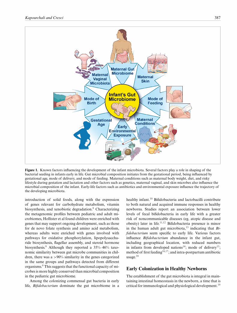
Figure 1. Known factors influencing the development of the infant microbiota. Several factors play a role in shaping of thebacterial seeding in infants early in life. Gut microbial composition initiates from the gestational period, being influenced bygestational age, mode of delivery, and mode of feeding. Maternal conditions such as maternal body weight, diet, and riskylifestyle during gestation and lactation and other factors such as genetics, maternal vaginal, and skin microbes also influence themicrobial composition of the infant. Early-life factors such as antibiotics and environmental exposure influence the trajectory ofthe developing microbiota.
introduction of solid foods, along with the expressionof genes relevant for carbohydrate metabolism, vitaminbiosynthesis, and xenobiotic degradation.8 Characterizingthe metagenomic profiles between pediatric and adult mi-crobiomes, Hollister et al found children were enriched withgenes that may support ongoing development, such as thosefor de novo folate synthesis and amino acid metabolism,whereas adults were enriched with genes involved withpathways for oxidative phosphorylation, lipopolysaccha-ride biosynthesis, flagellar assembly, and steroid hormonebiosynthesis.9 Although they reported a 35%–46% taxo-nomic similarity between gut microbe communities in chil-dren, there was a >90% similarity in the genes categorizedin the same groups and pathways detected from differentorganisms.9 This suggests that the functional capacity of mi-crobes is more highly conserved thanmicrobial compositionin the pediatric gut microbiome.
Among the colonizing commensal gut bacteria in earlylife, Bifidobacterium dominate the gut microbiome in a
healthy infant.10 Bifidobacteria and lactobacilli contributeto both natural and acquired immune responses in healthynewborns. Studies report an association between lowerlevels of fecal bifidobacteria in early life with a greaterrisk of noncommunicable diseases (eg, atopic disease andobesity) later in life.11,12 Bifidobacteria presence is minorin the human adult gut microbiota,13 indicating that Bi-fidobacterium seem specific to early life. Various factorsinfluence Bifidobacterium abundance in the infant gut,including geographical location, with reduced numbersin infants from developed nations14; mode of delivery15;method of first feeding16,17; and intra-postpartum antibioticusage.18
Early Colonization in Healthy Newborns
The establishment of the gut microbiota is integral in main-taining intestinal homeostasis in the newborn, a time that iscritical for immunological and physiological development.19

388 Nutrition in Clinical Practice 35(3)
Figure 2. Maternal and early-life factors and healthy-type neonatal microbiome. Neonatal gut bacterial colonization is affectedby maternal gut microbiome during gestation and lactation. Thus, healthy-type infant gut microbiomes are related to the healthymaternal gut, vaginal, and milk microbiome transferred during gestation, vaginal delivery, and breastfeeding.
The gut microbiota assists with essential nutrient syn-thesis and absorption,20,21 generates SCFAs that serve asan energy source for colonocytes,22-24 maintains the in-testinal mucosal barrier and protects against pathogenicbacteria and endotoxin translocation, stimulates immune-system maturation,25 provides anti-inflammatory signals tothe host,26 and influences infant growth.23,24 Disruption inthe gut microbiota (ie, gut dysbiosis) has been linked tonecrotizing enterocolitis, as well as some chronic diseases,including obesity, diabetes, inflammatory bowel disease,cancer, allergies, asthma,1 and neurological diseases associ-ated with the gut-brain axis.27
In Utero Colonization
Contrary to prior belief, the intrauterine environment isnot sterile.28 Nonpathogenic bacteria is detected in theplacenta, umbilical cord,29,30 and the meconium of thehealthy newborns, independent of birth mode of delivery,28
and its composition is associated with the gestational age.Lactobacillus and BifidobacteriumDNA have been detectedin infants delivered vaginally and by cesarean section(C-section), highlighting bacterial translocation from ma-ternal gut to placenta.29 Interestingly, with a healthy preg-nancy, the intrauterine bacteria appears to be similar tothat of the mother’s oral cavity.31 However, intrauterineinfection has been reported to be associated with a leakygut, confirmed by finding maternal gut microbes in the am-niotic fluid of women with premature membrane rupture.32
This suggests microbial translocation from a maternalleaky gut to the uterus and placenta. Animal experiments
with maternal provision of bacteria orally found the samebacteria in the placenta33 and meconium of the pupsdelivered by sterile C-section.28 These data demonstratemother-to-fetus transmission of bacteria during gestation.The placenta has been also shown to contain SCFA andtheir receptors,34 indicating a potential role of microbialby-product interactions during gestational development.Taken together, these findings suggest that initial small-scalecolonization occurs prior to birth. Potential mechanismsregarding the maternal-to-fetus microbiome interactionsand delivery outcomes remain to be elucidated.
Although increases in gut microbial density and diversityare suggested to be biologically determined,14 a range ofvarious factors influence the development of a healthyinfant microbiota, as illustrated in Figure 2. There is acommon universal pattern of early colonization of theinfant’s gut in vaginally born healthy children.14 Aerobicand facultative bacteria dominate the newborn’s gut, andin an age-dependent process, a reduction in oxygen enablesanaerobic bacterial growth,35 a process that is indepen-dent of diet.14 Gestational age affects infant gut bacterialcomposition. In a preterm infant, delayed intestinal bacte-rial colonization36 and an immature gastrointestinal tractmight increase susceptibility to infection.37 Geographicaldifferences appear to influence infant gut colonization. Themerged data published globally provide an overall view ofgeographical differences in the gut microbial compositionof infants. In a study reported by Korpela and de Vos,after adjusting for age, an American cohort had a relativelyhigh abundance of Bacteroides spp and enterobacteria, withsignificantly low levels of Bifidobacterium subsp during the

Kapourchali and Cresci 389
first 6 months of life.14 However, data from African, Asian,and Central European cohorts show high abundances ofbifidobacteria and lack of Clostridia during the first yearof life, implying a slow maturation pattern in the gutmicrobiota in children from these regions. Geographicaldifferences in both maternal and infant dietary practicesmay influence early gut colonization.14
Infant Mode of Delivery
Infant mode of delivery influences early-life gut microbialcomposition.38,39 Infants delivered vaginally are colonizedwith bacteria present in the maternal vagina,38,40 whereasthose delivered by C-section are colonized with bacteriasimilar to maternal skin and oral cavity.41,42 Compared withvaginally delivered infants, C-section–delivered infants arereported to have decreased α diversity,43 with a delayed andreduced colonization of Bacteroides persisting over time45
or being undetectable, indifferent to breastfeeding.6 Inter-estingly, a greater abundance of Clostridium difficile,44,45
bacilli, and enterobacteria are reported with C-sectiondelivery.14
Gut microbial differences associated with infant mode ofdelivery may disappear over time.41 However, more impor-tantly, early-life variances may be related to the incidenceof noncommunicable chronic diseases that appear later inlife.1 C-section–delivered infants are reportedly at a greaterrisk of developing asthma, obesity, and type 1 diabetes.46
A recent animal study reported a link between immune-system malfunction, microbial alterations, and increasedappetite in a mouse model.47 This study showed mice witha genetically impaired innate immune system exhibitedsignificant alterations in their gut microbial composition,which was correlated with hyperphagia, and clinical featuresof metabolic syndrome (eg, hyperlipidemia, hypertension,insulin resistance, and increased adiposity). Alterationsin immune system development and function as a resultof gut colonization differences between delivery modesdemonstrated in humans48 may partially explain C-section–associated incidence of noncommunicable diseases later inlife.49 Conversely, a recent study conducted by Ahlqvistet al revealed no significant clinical association betweenC-section and developing obesity later in life in youngadult men.50 Further longitudinal studies considering morematernal factors as well as gut microbiome differencesduring development are warranted to show the associationbetween mode of delivery and obesity later in life.
Infant Feeding Methods
Maternal milk. Among the factors affecting early coloniza-tion,mode of infant feeding is of high importance.Mother’sown milk is considered the gold standard for infant nutri-tion, as it meets the infant’s nutrition requirements during
Table 1. Bioactive Molecules in Maternal Milk.54,218,219
OligosaccharidesMicrobesImmunoglobulin AAntimicrobial peptides (lactoferrin, lysozyme,lactadherin, mucin)
AntioxidantsStem cellsGlycoconjugates
Table 2. Microbes Identified in Human Milk.61,62
StaphylococciStreptococciCorynebacteriaPropionibacteriaLactobacillus sppBifidobacterium spp
early life.51,52 Maternal milk contains all the nutrients andvitamins required for optimal infant development, includ-ing complex protein, fat, and carbohydrate.53 Additionally,maternal milk contains a myriad of biologically activemolecules that are critical and protective in early life54
(Table 1).
Maternal milk—Commensal microbes. Recent studiessupport that human milk is not sterile and is a primaryand continuous source of colonizing bacteria to the in-fant’s gut.55,56 Mother-to-child transmission studies, withand without culture consideration, support that bacterialtransfer from mother to infant occurs via breast milk.56-59
This was demonstrated by the presence of the same bac-terial strain identified in mother’s milk and their breastfedinfant’s stool.60 Pannaraj et al also showed that maternaltransfer of bacteria via breast milk has a greater impacton the newborn’s early colonization compared with areolarskin.56 Although the bacterial composition of human milkis low, with <3-log colony-forming units (CFU)/mL, it isphysiologically important.61,62 Following birth, breast milkmicrobiota is a main factor that drives the acquisition andevolution of the gut microbiota in early life. Breast milkcontributes significantly to the metabolism, development ofgut integrity, and maturation of the immune and neuroen-docrine systems.63-67
Breast milk contains a rich microbiota composed ofviable skin and non-skin gram-positive bacteria (Table 2).Streptococci (mitis and salivarius groups) and coagulase-negative staphylococci are among the dominant bacteriain both human milk59,68-70 and the feces of breastfedinfants.71-73 These microbes are potentially able to com-pete with the establishment of undesired pathogens (eg,

390 Nutrition in Clinical Practice 35(3)
Staphylococcus aureus) in the infant gut.74,75 Similarpathogen exclusion is noted through fermentation of the an-timicrobial compound glycerol monolaurate found in breastmilk.76 Propionibacterium acnes can prevent the growthof S aureus.77 Bifidobacterium and Lactobacillus spp inbreast milk are noted to activate immunoglobulin A (IgA)–producing plasma cells in the neonatal gut.61 It has beenhypothesized that Bifidobacterium control inflammationthrough mucosal host-microbe crosstalk.78 An associationwas shown between low levels of intestinal Bifidobacteriummicrobiota during infancy and an increased risk of atopylater in life.79-81 The original source of breast milk micro-biota is unclear.53 The entero-mammary pathway is onehypothesis that proposes a selective colonization of themammary gland by cells of the immune system.62 The sim-ilarity between the bacterial composition of maternal stooland breast milk supports this concept.82-84 Clinical studiesdemonstrating ingested probiotic strains were identified inmaternal breast milk further support this hypothesis.85,86
More information about the origin of milk microbes can befound in this review by Moossavi et al.87
The composition of human milk, including the structureof microbial community,53 produced by a healthy womanis personalized. Each mother’s own milk provides spe-cific requirements for their infant according to gestationalage, lactation stage, environmental exposures, geographicallocation,88-90 and daily breastfeeding practices.55,91 Gesta-tional age has been reported to influence the concentrationsof Bifidobacterium spp in maternal breast milk, with lowerlevels in preterm deliveries. However, the entire range of themicrobial groups detected in breast milk are similar withboth term and preterm deliveries.61 Therefore, maternalmilk is the preferred mode of feeding for all infants, bothterm and preterm.51,92-94
Maternal milk microbiota and infant mode of delivery.Infants born by C-section had increased total bacteriacounts, particularly Streptococcus spp,95 and reduced levelsof Bifidobacterium spp61 that persisted for up to 6 monthsafter lactation.88 A higher abundance of Staphylococcusreported in the gut bacteria of infants delivered byC-sectionhas been speculated to contribute to a higher incidence inmethicillin-resistant S aureus skin infections compared withthat in infants delivered vaginally.96 It has been suggestedthat maternal milk dysbiosis could exacerbate C-sectiondelivery–induced infant gut dysbiosis.97 Interestingly, breastmilk dysbiosis was only observed inmothers who underwentan elective C-section; the breast milk microbiota in motherswho underwent an emergency C-section was comparable tothat of mothers who delivered vaginally. Therefore, breastmilk microbial alterations might be attributed to the phys-iological stress and/or hormonal changes that occur withlabor or an emergency C-section.88 Vaginal delivery mightinduce intestinal permeability and enhance the bacterial
translocation from the gut to the mammary gland andbreast milk.97 A review by Neu et al found most infantsdelivered by C-section were not breastfed.46 Therefore, inaddition to a no-physiological initial colonization duringchildbirth, the absence of early nutrition support for theinfant microbiome due to delayed lactation, breast milkdysbiosis, or the lack of breastfeeding might contributeto a long-lasting dysbiosis in C-section–delivered infants.Further research investigating whether modulation of ma-ternal breast milk microbiota could affect the infant’s gutmicrobiome and their health in elective C-section–deliveredinfants, mimicking that of mothers who delivered vaginally,is warranted.
Maternal milk—Prebiotics. Human milk contains hu-man milk oligosaccharides (HMOs), a type of prebiotic.As an energy source for commensal gut microbes,52 HMOfermentation stimulates the growth of bifidobacteria, Lac-tobacillus, andBacteroideswithin the infant gastrointestinaltract.52 Furthermore, the abundance of specific bacterialtaxa in human milk, such as Staphylococcus species, mightincrease the levels of microbial by-products (eg, SCFAs)by fermenting HMOs.53 HMOs have also been shown toprevent neonatal diarrhea and respiratory tract infections.98
How maternal conditions influence HMOs in breast milk,microbial by-products, gut microbiome composition, andhealth outcomes is discussed later.
Donor human milk. Donor human milk is viewed as su-perior to infant formula for seriously ill infants, whenmaternal milk is unavailable or insufficient for appropri-ate infant growth.51,99-101 Donor human milk supports in-fant growth and development, including neurodevelopment,and protects against various diseases, including necrotizingenterocolitis.102,103 Milk donated to a milk bank is providedto fragile, hospitalized infants.Using questionnaires, humanmilk donors undergo screening regarding their lifestyle,disease, and risk factors, as well asmicrobiological screening(eg, HTLV-I and HTLV-II, syphilis, hepatitis B and C,human T-lymphotropic virus 2, human papillomavirus,herpes simplex virus types 1 and 2, Chlamydia trachomatis,Neisseria gonorrheae, or Trichomonas vaginalis), and thedonated milk undergoes prepasteurization and/or postpas-teurizationmicrobiological analysis.104 As recommended bythe American College of Pediatricians and the Centers forDisease Control, to further assure microbiological safety,donor human milk is pasteurized (56–62 °C for 30 min-utes) to destroy all non–spore-forming microbes that maycontaminate the milk via poor hygiene, from the extrac-tion devices during collection, or maternal transfer.105,106
Although pasteurization destroys the microbes in the donormilk, the biologically active components of the humanmilk (Table 1) are preserved.61 An approach to increasethe bacterial richness of pasteurized donor milk is by

Kapourchali and Cresci 391
personalization. This includes adding 10% of the infant’smaternal milk and incubating it for 4 hours to increasethe levels of some bacteria that were naturally found inthe maternal milk.68 Collecting the milk by breast pumpcan increase the bacterial count of milk compared withthat expressed manually.107 Maternal milk can be frozen at−20 °C for approximately 6 weeks without any adverseeffects on breast milk quality and bacteria abundance.107
Infant formula. Significant compositional distinction isfound between the gut microbiota of breastfed infantsand formula-fed infants. Earlier studies found that the gutof a breastfed infant was rich in gram-positive bacteria,acidophilic “Bacillus bifidus” (Bifidobacterium).109 In thefirst month of life, bifidobacteria and staphylococci pre-dominate in the intestine of breastfed infants, whereasenterococci, Coliform, and clostridia are rich in formula-fed infants.72 Grönlund et al (2007) reported that theconcentration of Bifidobacterium spp in breastfed infantscan increase up to 60%–90%of the total fecal microbiota.110
An enriched population of bifidobacteria competes withother species in the infant gut and produces nutrientsvital for early development, including sialic acid. Sialicacid (N-acetyl-neuraminic acid) is an essential nutrient foroptimal brain development and cognition.111,112 Ruhaaket al (2014) found the presence of sialylated oligosaccharidesin the blood of infants that might have originated fromthe hydrolysis of HMO.113 The gut microbiota in earlylife is composed of predominantly Lactobacillus, Staphy-lococcus, Megasphaera, and Actinobacteria in breastfedinfants, whereas Clostridiales and Proteobacteria are moreabundant in formula-fed infants. Formula feeding was alsoshown to enrich Atopobium and Bacteroides but reduceBifidobacterium.114 Feeding infants with formula reducedtotal gut bacterial numbers but increased gut microbialdiversity compared with breastfeeding. Lower gut microbialdiversity in breastfed infants is attributed to the uniqueHMO contained in breast milk, which may serve only alimited number of gut microbes.115 Breastfeeding was alsoshown to influence the oral bacteria in infants.
These data suggest that the prebiotics and probiotics inbreast milk may play a role in supporting infant health.Taken together, these data support the notion that addingthese components to infant formula might be an approachto improve health outcomes in formula-fed newborns thatdo not have access to human milk.
Maternal and Early-Life Factors InfluencingMaternal Milk Composition, EarlyColonization, and Neonatal Health Outcomes
Multiple factors are associated with maternal milk HMOand microbiota and with infant gut microbiota (see Fig-ure 1).116 However, in some cases, there is no explanation
for microbial changes in the infant gut, particularly thereduction in levels of Bifidobacterium.116 This highlights thepotential role of other factors, like maternal chronic condi-tions or risky lifestyle, that may influence the maternal gutmicrobiota,maternalmilk, and early infant gut colonization(Figure 3).
Maternal Obesity
Infant birth weight can vary between those born fromoverweight vs normal–body-weight mothers, and this trans-ference of maternal phenotype corresponds to the transferof the newborn’s gut microbiota.117 Intergenerational trans-mission of the obesogenic microbes hypothesis has beensupported by several studies.118,119 Obesity is characterizedwith an imbalance in the Firmicutes-to-Bacteroidetes ratioin the gut.120,121 Birth cohort studies reveal that infantsborn from overweight or obese mothers were abundantlycolonized with the bacterial genera belonging to the phylaFirmicutes, especially of the Lachnospiraceae family, andhad a greater risk of becoming overweight by 1–3 yearsof age.119 Experimental data in obese mice support thatLachnospiraceae may contribute to the development ofobesity122 and adipocyte inflammation123 and promotediabetes.124 Bacteroides colonization of the infant gut isassociated with reduced growth in early life.125 Some studieshave reported elevated levels of Bacteroides in the stoolfromboth obesemothers126 and infants whosemothers wereoverweight or obese.118 However, Santacruz et al reportedreduced numbers of Bacteroides, belonging to the phylumBacteroidetes, in overweight compared with normal-weightwomen.117 More studies are required to explain discrepan-cies in these data and their significance.
Levels of Staphylococcus,117 Enterobacteriaceae, andEscherichia coli have been reported to be elevated in over-weight compared with normal-weight pregnant women.117
Animal experiments suggest that higher levels of gram-negative bacteria in the gut are a result of endotoxemiaand inflammation induced by obesity.127 Excessive ges-tational weight gain, defined as >11.5 kg in overweightand >16 kg in normal-weight women, was associated withan expansion of Bacteroides,126 Enterobacteriaceae, andE coli and a reduction in Bifidobacterium and Akkerman-sia muciniphila.117 Thus, these data suggest that maternalgut microbiota might be a factor contributing to weightgain during pregnancy beyond the maternal nutrition.128
Moreover, a significant reduction in the community ofbacteria involved in metabolic signaling and energy regula-tion, including Enterococcus, Acinetobacter, Pseudomonas,and Hydrogenophilus, have been found in infants born tooverweight or obese mothers.118 Infant gut dysbiosis asso-ciated with maternal obesity has been shown to increase gutpermeability and directly initiate pathways of nonalcoholicfatty liver disease.129

392 Nutrition in Clinical Practice 35(3)
Figure 3. Maternal and early-life factors and dysbiosis in neonatal gut microbiome. Maternal gut and milk dysbiosis induced bymaternal conditions, such as maternal Western diet, obesity, alcohol, and tobacco use, can be vertically transmitted to offspring.Cesarean section (C-section), preterm delivery, and infant formula are other factors for imbalanced infant gut bacterialcommunity, especially Bifidobacterium. The potential role of selective supplement with prebiotics and probiotics in both mothersand infants makes them a good therapeutic strategy for infants at the risk of gut dysbiosis.
Maternal obesity–induced infant gut dysbiosis has beenreported to be more evident before 9 and 18 months130 andto differ by mode of delivery. In a study by Mueller et al,vertical transmission of higher levels of Bacteroides wasobserved in overweight pregnant women to their newbornsduring vaginal delivery. Reduced levels of Bacteroides wasdetected in the meconium soon after birth in infants deliv-ered by C-section.118 C-section–delivered infants from over-weight mothers are at higher risk of becoming overweightlater in life than infants born vaginally to overweight orobese mothers.119
Maternal weight status also might affect maternalmilk composition.131,132 Recent data associating the milkmetabolome with both maternal and infant obesity suggestthat obesity-related differences in human milk compositionmight contribute to early childhood obesity.133 Cabrera-Rubio et al detected higher total bacterial counts, expansionof Staphylococcus and Lactobacillus, and reduced levels ofBifidobacterium in themilk of obese comparedwith normal-weight women during the first 6 months of lactation.88 Theysuggested that the alterations in the microbial compositionof breast milk from an obese mother might be an additionalmechanism explaining the intensified obesity risk in infants
born to obese and overweight mothers. Limited studieshave investigated the role of maternal or infant probioticsupplementation on the breast milk as a means to mitigatethe obesity-related alterations.
Maternal Diet
Maternal diet has been shown to be linked to infant gutmicrobiota. Population-based human longitudinal cohortdata showed that a maternal high-fat diet altered earlybacterial colonization independent of maternal obesity.134
In association with a maternal high-fat diet, the neonatalmeconium microbiome varied with a significant relativedepletion in Bacteroides immediately post partum, whichpersisted until 6 weeks of age.134 Data from animal experi-ments also showed that a high-fat diet during gestation andpostweaning period caused significant gut dysbiosis earlyin life.135 This highlights the important role of a maternalhigh-fat diet rather than just maternal obesity per se inshaping the gut microbiota early in life.135 Maternal high-fat diet during gestation and lactation might also increasethe susceptibility to immune-mediated diseases and somemetabolic consequences for the offspring, partially through

Kapourchali and Cresci 393
alteration in responses to microbes.136 A study conductedby Val-Laillet et al demonstrated significant programmingeffects of a maternal Western diet during gestation andlactation on microbiota fermentation activity in sows. Thisresulted in a significant drop in fecal SCFA levels in bothsows and their piglets that persisted even after weaning.137
Thus, these data suggest maternal dietary factors modulateboth bacterial composition and their fermentation activityin the neonatal gut.
Limited data are available regarding the effects of otherfactors of a Western diet on maternal-infant gut dysbiosis,such as the influence of high sugar, sodium, and animalproteins. A combination of a high-fat/high-sugar diet ledto gut dysbiosis in mice138; and dietary intake of refinedsugars modulate the gut microbial composition to that ofan inflammatory-type microbiota.139 Since maternal milkmicrobiota is hypothesized to originate from maternal gutmicrobiota, the gut dysbiosis associated with the maternaldiet might be transferred to maternal milk and furtherexacerbate dysbiosis seen in the early gut microbiome inbreastfed infants. However, further studies are warranted toaddress the knowledge gaps regarding the role of maternalWestern diet and breast milk microbiota. In contrast, theMediterranean diet, which is enriched in fruits, vegetables,unsaturated fats, nuts, legumes, and whole grains, has beenlinked to improvements in the diversity and richness ofthe gut microbiota. This alteration in the gut microbiotamight be associated with a large number of health benefits,including the prevention of metabolic diseases and cogni-tive disorders.140 However, research investigating potentialbeneficial effects of a maternal Mediterranean diet duringgestation and lactation on the infant’s gut microbiota islacking.
Maternal and Infant Antibiotic Exposure
Antibiotic exposure during gestation or in infant early lifecan have short-term and long-term influences on the devel-oping infant gutmicrobiome.141 The antibiotic-induced per-turbation in gut microbiota of pregnant germ-free mice wastransmittable to their offspring’s gut. Although not directlyexposed to the antibiotic, the mouse pups had alterations intheir gut bacterial composition, which persisted for 21weeksand made them more susceptible to developing colitis.142
Persistence in maternal antibiotic-induced gut dysbiosiswas also associated with alterations in T cells’ functionsin mouse pups.143 Because microbial colonization in earlylife coincides with key neurodevelopment periods, it issuggested that antibiotic-induced perturbation in the infantgut microbiota might be linked to disruption in the gut-brain axis and potentially related to neurodevelopmentaldisorders, such as autism.144
Exposure to antibiotics is common in early life, with theaverage US child receiving 3 courses of antibiotics by 2
years of age.145 Antibiotic provision in the first year of lifeis associated with increased body weight146 and incidenceof inflammatory bowel disease147 and allergies.148,149 Theserelationships in antibiotic-treated infants are presumed tobe attributed to the antibiotics causing alterations in mi-crobiota assembly during early life.122 Antibiotic exposurethroughout early life reduced the levels of Lachnospiraceaespp and other Clostridiales within infant’s gut microbiota.Lachnospiraceae is known to produce butyrate and otherSCFA150 that regulate host immunity151,152 and controlbody weight.153
Maternal treatment with antibiotics can also affectthe breast milk microbiome. Intrapartum antibiotic expo-sure independently affects breast milk microbiota compo-sition 1 month after delivery97 and perturbs infant gutcolonization.154 Bifidobacterium has been reported to beuniquely detectable in the breast milk of mothers who didnot receive antibiotics compared with those that did.97
Maternal treatment with antibiotics during lactationreduced breast milk microbial community, including lac-tobacilli and bifidobacteria, and caused overgrowth ofmastitis-inducing opportunistic bacteria155,156 associatedwith lower bacterial diversity in breast milk.157,158 Manymothers cease breastfeeding early because of painful mas-titis. Combined reduction in breast milk microbial diversitywith early cessation of breastfeeding might lead to low in-testinal diversity in the first weeks of life, which is associatedwith necrotizing enterocolitis.159 Recent studies have shownthat some lactobacilli strains isolated from human milkhave been applied topically to treat or prevent mastitis,160,161
suggesting their potential involvement in mammary home-ostasis.
Other Prenatal Factors
Research is just beginning to investigate other maternalexposures on maternal and infant microbiomes. Maternalallergies may impact the microbiome in breast milk. A studyby Grönlund et al noted that significantly depleted levels ofBifidobacterium in the breast milk of mothers with allergieswere associated with the Bifidobacterium counts in theirinfants’ feces.110
Labrecque et al investigated the effects of alcohol and ar-tificial sweeteners on the gut microbiome during pregnancyin mice. They found that low to moderate levels of ethanolexposure in combination with artificial sweeteners reducedClostridium and Bacillus and increased Eubacterium levelsin the gut microbiota of pregnant mice compared withnonpregnant and control mice.162
Prenatal and postnatal exposure to environmental smokeincreased gut bacterial richness in infants, particularly theFirmicutes phylum, at 3 months of age and was associ-ated with a higher risk of overweight and obesity at 1–3years of age.163 Moreover, maternal smoking increased the
394 Nutrition in Clinical Practice 35(3)
abundance of Bacteroides and Staphylococcus at 6 monthsof age; and early-life exposure to environmental smokeincreased the levels of Ruminococcus and Akkermansia ininfant gut microbiota.163 Air pollution is also associatedwith immune, neurological, and metabolic disturbancesduring development that potentially can result in atopic dis-ease, obesity, and autism. Although it is hypothesized thatair pollution–associated diseases are related to disruptionsin the neonatal gut microbial composition, in particulardepletion of the phyla Firmicutes, further studies are neededto validate this theory.164
Jašarevi et al (2017) showed in mice that maternal stressin the first week of pregnancy caused lasting disruption infecal microbial diversity, community, and composition inthe vaginal microbiota during gestation and after birth.165
Maternal stress-induced vaginal microbial disruption high-lights the possibility for transmitting vaginal dysbiosis tooffspring during birth. Results from rodent and primatemodels also support the link between prenatal stress andoffspring intestinal microbiota.165-167 In a cohort study con-ducted by Zijlmans et al (2015), the microbiota compositionin vaginally born infants was associated with maternalprenatal stress, reported by mothers and evaluated by basalmaternal salivary cortisol levels.168 In this study, prenatalstress–exposed infant guts harbor significantly higher rel-ative levels of proteobacterial groups related to pathogensand lower relative levels of Lactobacillus and Bifidobac-terium. This alteration in the microbial pattern of maternalstress–exposed infants during pregnancy was associatedwith infant gastrointestinal symptoms and allergic reactionsas reported by their mothers.168
Gestational diabetes mellitus (GDM) is associated withaltered gut microbiota in pregnant women,169 including Bi-fidobacterium spp.170 Moreover, it has been shown that thisGDM-related dysbiosis can be vertically transmitted to theoffspring.171 Wang et al (2018) evaluated the microbial com-munities of oral, intestinal, and vaginal samples of pregnantwomen with GDM and oral, pharyngeal, meconium, andamniotic fluid samples of neonates, the majority of whomwere delivered via C-section. They found GDM-associateddysbiosis with maternal and neonatal gut bacteria at birth,as well as an association between the abundance of severalpredominant bacteria and values of oral glucose tolerancetests.171
Medical interventions such as chemotherapy reportedlyimpact the breast milk microbiome. A single case studyfound breast milk samples collected from a lactating womanundergoing a course of chemotherapy for Hodgkin’s lym-phoma contained reduced levels of genera Bifidobacterium,Eubacterium, Staphylococcus, and Cloacibacterium and ex-panded levels of Acinetobacter, Xanthomonadaceae, andStenotrophomonas.172 Eubacterium is a butyrate-producingbacteria173 that plays a critical role in infant health.174
Further larger cohort studies are warranted to examine the
chemotherapy-associated changes to lactating mothers andthe consequences for the microbiome and long-term healthof infants.
All these studies highlight the importance of early-lifemicrobiome formation and its relationship to maternalfactors, which offers a therapeutic approach by maternalintervention, therefore modulating initial microbial colo-nization to diminish the risk of adverse health outcomes.
Maternal and Early-Life Prebiotic andProbiotic Supplementation
Prebiotics
Prebiotics are complex polysaccharides that escapedigestion by the host and, upon reaching the distal gut,are fermented by and support the growth of commensalmicrobes.175 Whereas human milk is rich in prebiotics(HMO), bovine milk also contains oligosaccharides, someof which are structurally similar to HMOs. Simeoniet al (2016) conducted a randomized, double-blindedstudy comparing an infant formula with or withoutadded bovine milk–derived oligosaccharides, containinggalacto-oligosaccharides and 3′- and 6′-sialyllactose, andthe probiotic Bifidobacterium animalis subsp lactis strainCNCM I-3446 on tolerability and the ability to affect thegut microbiome. Breastfed infants served as a referencegroup.176 Compared with the nonsupplemented formula,the supplemented formula was well tolerated and supportedsimilar growth in newborns followed for 12 weeks. Thesupplemented formula stimulated a marked shift inendogenous Bifidobacterium (B longum, B breve, B bifidum,B pseudocatenulatum) and increased B lactis in the infantstool by 100-fold.176 Adding various types of prebiotics(eg, fructo-oligosaccharides and galacto-oligosaccharides)to infant formula has also been shown to stimulate thegrowth of bifidobacteria and lactobacilli in the infant’sgut to levels detected in breastfed infants.177 By inducinga fecal microbiota that closely resembles the microbiota ofbreastfed infants,178 prebiotic supplementation in infantsmay improve gut mucosal barrier, prevent enteric pathogeninfection and bacterial translocation,179,180 and ultimatelysupport infant growth.181 Both maternal and infantprebiotic supplementation have been reported in allergyprevention.182
Probiotics
A probiotic is defined as “a live micro-organism which,when administered in adequate amounts, confers a healthbenefit on the host.”183 Intervention with probiotics as ameans to maintain a healthy gut ecosystem in early lifehas gained popularity over the past 2 decades. In attemptsto mimic the composition of human milk, the design ofsome infant formulas includes the addition of probiotics.

Kapourchali and Cresci 395
Bifidobacterium and Lactobacillus species have been iso-lated from a healthy infant gut and added to the GRAS(generally recognized as safe) list by the Food and DrugAdministration. Bifidobacterium and Lactobacillus are thespecies added to infant formulations. However, comparedwith breast milk, probiotic-supplemented infant formulacontains a notably higher concentration of a limited num-ber of probiotic strains.61 Isolated from a healthy infantintestine, B longum subsp infantis (B infantis), B longumsubsp longum (B longum), B bifidum, and B breve have alarge repertoire of genes for the utilization of HMOs.184
Important to note is that not all probiotics are equally safe,and the effects demonstrated from one strain cannot beextrapolated to another strain, even if they belong to thesame species.185
Adding bifidobacteria strains to infant formula, frombirth to 12 months, does not seem to compensate fordifferences in microbiota composition observed betweenbreastfeeding and formula feeding in early life in full-terminfants.186 Moreover, bifidobacterial colonization in infantgut was not stable over time, because of competition withinthe ecosystem.186 However, recent data in preterm infantsshowed that compared with B animalis subsp lactis supple-ment,B infantis significantly increased the fecal bifidobacte-ria in formula-fed preterm infants.187 This might be becauseamong bifidobacterial strains, B infantis is the only one thathas the ability to consume HMOs because of its specificgenome sequence.188 Supplementation with Lactobacillusrhamnosus GG (LGG) into an extensively high casein-basedcommercially available formula, in infants at high risk forallergic manifestation associated with IgE-mediated cow’smilk allergy, reduced the incidence of other allergy mani-festations and improved the development of oral toleranceto cow’s milk allergy.189 This supplementation was moreeffective in combination with extensively high casein-basedthan with whey-based formula.190 The beneficial effectof LGG might be attributed to the alterations in strain-level bacterial community structure expanding butyrate-producing bacterial strains in food allergic infants.191
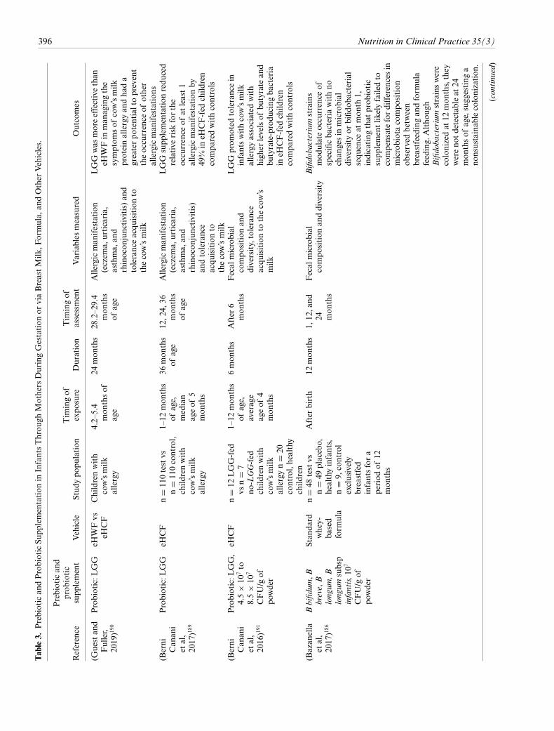
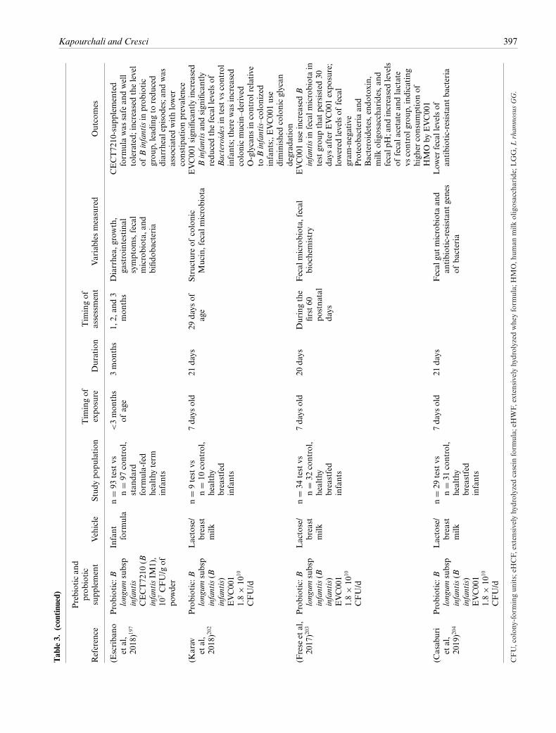
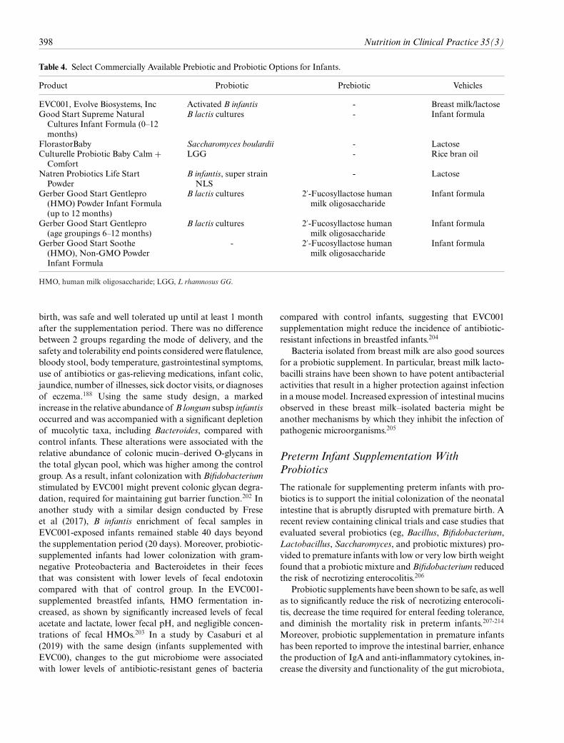
Probiotic provision can also reach the infant if providedto the mother during gestation and lactation or if addedseparately to breast milk or infant formula. This sectionsummarizes major studies involving maternal-to-infant mi-crobial transfer via maternal provision of prebiotics andprobiotics during gestation or lactation or via supplemen-tation of infant formula and other vehicles (see Table 3).Information pertaining to commercially available productscan be found in Table 4.
Maternal Probiotic Supplementation
Maternal supplementation with LGG (2 × 10⁹ CFU/d)during late pregnancy (30–36 weeks) showed colonizationwith LGG in the infant’s gut that was stable for up to 24
months in some cases. However, the role of breastfeeding onthe colonization proficiency of LGG was not reported.193
In contrast, LGG administration (1.8 × 10¹⁰ CFU/d),from 36 weeks of gestation until delivery, enhanced theintestinal colonization of Bifidobacterium species but notLGG in breastfed infants.194 Interestingly, the transfer ofLGG from mother to infant has been reported to establishmore diversity in Bifidobacterium in the infants’ gut.195
Administration of LGGduring gestation and then into theirinfants for 6monthswith amaternal family history of atopicdisease reduced the occurrence of atopic eczema by halfin supplemented infants compared with a placebo group.80
Therefore, it seems that factors such as the choice ofprobiotic strain and duration of supplementation affect thebeneficial effects of maternal probiotic supplementation.Maternal dietary supplementation with the Lactobacillusstrains during lactation lead to the isolation of such strainsin maternal milk.28,195 Furthermore, probiotic supplemen-tation only during gestation resulted in the appearance ofthe bacteria in the fecal samples of their breastfed neonates,even in those born by C-section and no exposure to thevaginal microbiome.192 This highlights the important roleof breast milk in the transfer of microbes to the infants.
Maternal Prebiotic Supplementation
In the study conducted by Paul et al,196 it has been shownthat maternal prebiotic supplementation with oligofructose,in the context of diet-induced obesity, increased circulat-ing concentrations of satiety hormones and the relativeabundance of Bifidobacterium spp in gut. These alterationswere accompanied by reduced gestational weight gain anda significant prevention in increased adiposity in bothdams and offspring at weaning, potentially impacting theirlifelong obesity risk.196
Term-Infant Supplementation With Probiotics
Provision of a B longum subsp infantis CECT7210–supplemented infant formula for 3 months was reportedto be safe and well tolerated and to significantly reducethe prevalence of diarrhea and constipation in healthyinfants.197 Exogenous administration of selected bifidobac-terial strains, alone or in combination with lactic acid bacte-ria, decreased the incidence of a variety of gastrointestinalor allergic conditions associated with delayed bifidobacte-rial colonization and/or a depleted bifidobacterial popula-tion in the infant’s gut.198 Data from in vitro studies revealthat B infantis stimulates anti-inflammatory and inhibitsproinflammatory cytokines in intestinal cells.199 Animalexperiments show that B infantis mitigates inflammation200
and prevents intestinal barrier dysfunction in a mousemodel of necrotizing enterocolitis.201 In breastfed infants,supplementation with 1.8–2.8 × 10¹⁰ CFU B longum subspinfantis EVC001 daily for 21 days, starting at day 7 after
396 Nutrition in Clinical Practice 35(3)Table3.
Prebiotican
dProbioticSu
pplementation
inInfantsThrou
ghMothers
DuringGestation
orviaBreastMilk
,Formula,
andOther
Vehicles.
Reference
Prebiotican
dprob
iotic
supp
lement
Vehicle
Stud
ypo
pulation
Tim
ingof
expo
sure
Duration
Tim
ingof
assessment
Variables
measured
Outcomes
(Guestan
dFuller,
2019)190
Probiotic:L
GG
eHWFvs
eHCF
Childrenwith
cow’smilk
allergy
4.2–5.4
mon
thsof
age
24mon
ths
28.2–2
9.4
mon
ths
ofage
Allergicman
ifestation
(eczem
a,urticaria,
asthma,
and
rhinocon
junctivitis)an
dtoleranceacqu
isitionto
thecow’smilk
LGG
was
moreeffectivethan
eHWFin
man
agingthe
symptom
sof
cow’smilk
proteinallergyan
dha
da
greaterpo
tentialtoprevent
theoccurrence
ofother
allergicman
ifestation
s(B
erni
Can
ani
etal,
2017)189
Probiotic:L
GG
eHCF
n=
110testvs
n=
110control,
child
renwith
cow’smilk
allergy
1–12
mon
ths
ofage,
median
ageof
5mon
ths
36mon
ths
ofage
12,2
4,36
mon
ths
ofage
Allergicman
ifestation
(eczem
a,urticaria,
asthma,
and
rhinocon
junctivitis)
andtolerance
acqu
isitionto
thecow’smilk
LGG
supp
lementation
redu
ced
relative
risk
forthe
occurrence
ofat
least1
allergicman
ifestation
by49
%in
eHCF-fed
child
ren
compa
redwithcontrols
(Berni
Can
ani
etal,
2016)191
Probiotic:L
GG,
4.5
×10
7to
8.5
×10
7
CFU/g
ofpo
wder
eHCF
n=
12LGG-fed
vsn
=7
no-LGG-fed
child
renwith
cow’smilk
allergyn
=20
control,healthy
child
ren
1–12
mon
ths
ofage,
average
ageof
4mon
ths
6mon
ths
After
6mon
ths
Fecal
microbial
compo
sition
and
diversity,tolerance
acqu
isitionto
thecow’s
milk
LGG
prom
oted
tolerancein
infantswithcow’smilk
allergyassociated
with
high
erlevelsof
butyrate
and
butyrate-produ
cing
bacteria
ineH
CF-fed
child
ren
compa
redwithcontrols
(Bazan
ella
etal,
2017)186
Bbifidum
,Bbreve,B
longum
,Blongum
subsp
infantis,1
07
CFU/g
ofpo
wder
Stan
dard
whey-
based
form
ula
n=
48testvs
n=
49placebo,
healthyinfants,
n=
9,control
exclusively
breastfed
infantsfora
period
of12
mon
ths
After
birth
12mon
ths
1,12
,and
24 mon
ths
Fecal
microbial
compo
sition
anddiversity
Bifidobacterium
strains
mod
ulateoccurrence
ofspecificba
cteria
withno
chan
gesin
microbial
diversityor
bifid
obacterial
sequ
ence
atmon
th1,
indicating
that
prob
iotic
supp
lementlik
elyfailedto
compensatefordifferencesin
microbiotacompo
sition
observed
betw
een
breastfeedingan
dform
ula
feeding.
Altho
ugh
Bifidobacterium
strainswere
colonizedat
12mon
ths,they
wereno
tdetectableat
24mon
thsof
age,sugg
esting
ano
nsustainablecolonization
.
(continued)
Kapourchali and Cresci 397
Table3.
(continued)
Reference
Prebiotican
dprob
iotic
supp
lement
Vehicle
Stud
ypo
pulation
Tim
ingof
expo
sure
Duration
Tim
ingof
assessment
Variables
measured
Outcomes
(Escriba
noet
al,
2018)197
Probiotic:B
longum
subsp
infantis
CECT72
10(B
infantisIM
1),
107CFU/g
ofpo
wder
Infant
form
ula
n=
93testvs
n=
97control,
stan
dard
form
ula-fed
healthyterm
infants
<3mon
ths
ofage
3mon
ths
1,2,
and3
mon
ths
Diarrhea,
grow
th,
gastrointestinal
symptom
s,fecal
microbiota,
and
bifid
obacteria
CECT72
10-sup
plem
ented
form
ulawas
safe
andwell
tolerated;
increasedthelevel
ofBinfantisin
prob
iotic
grou
p,lead
ingto
redu
ced
diarrhealepisodes;an
dwas
associated
withlower
constipa
tion
prevalence
(Karav
etal,
2018)202
Probiotic:B
longum
subsp
infantis(B
infantis)
EVC00
11.8
×10
10
CFU/d
Lactose/
breast
milk
n=
9testvs
n=
10control,
healthy
breastfed
infants
7da
ysold
21da
ys29
days
ofage
Structureof
colonic
Mucin,fecal
microbiota
EVC00
1sign
ificantlyincreased
Binfantisan
dsign
ificantly
redu
cedthefecallevelsof
Bacteroides
intestvs
control
infants;therewas
increased
colonicmucin–d
erived
O-glycans
incontrolrelative
toBinfantis–colon
ized
infants;,E
VC00
1use
diminishedcolonicglycan
degrad
ation
(Frese
etal,
2017)203
Probiotic:B
longum
subsp
infantis(B
infantis)
EVC00
11.8
×10
10
CFU/d
Lactose/
breast
milk
n=
34testvs
n=
32control,
healthy
breastfed
infants
7da
ysold
20da
ysDuringthe
first60
postna
tal
days
Fecal
microbiota,
fecal
biochemistry
EVC00
1useincreasedB
infantisin
fecalm
icrobiotain
testgrou
pthat
persisted30
days
afterEVC00
1expo
sure;
lowered
levelsof
fecal
gram
-negative
Proteob
acteriaan
dBacteroidetes,end
otox
in,
milk
oligosaccharides,a
ndfecalp
H;and
increasedlevels
offecala
cetate
andlactate
vscontrolg
roup
,ind
icating
high
erconsum
ptionof
HMO
byEVC001
(Casaburi
etal,
2019)204
Probiotic:B
longum
subsp
infantis(B
infantis)
EVC00
11.8
×10
10
CFU/d
Lactose/
breast
milk
n=
29testvs
n=
31control,
healthy
breastfed
infants
7da
ysold
21da
ysFecal
gutmicrobiotaan
dan
tibiotic-resistant
genes
ofba
cteria
Low
erfecallevelsof
antibiotic-resistant
bacteria
CFU,colon
y-form
ingun
its;eH
CF,
extensivelyhydrolyzed
casein
form
ula;
eHWF,
extensivelyhydrolyzed
wheyform
ula;
HMO,h
uman
milk
oligosaccharide;LGG,L
rham
nosusGG.
398 Nutrition in Clinical Practice 35(3)
Table 4. Select Commercially Available Prebiotic and Probiotic Options for Infants.
Product Probiotic Prebiotic Vehicles
EVC001, Evolve Biosystems, Inc Activated B infantis - Breast milk/lactoseGood Start Supreme Natural
Cultures Infant Formula (0–12months)
B lactis cultures - Infant formula
FlorastorBaby Saccharomyces boulardii - LactoseCulturelle Probiotic Baby Calm +
ComfortLGG - Rice bran oil
Natren Probiotics Life StartPowder
B infantis, super strainNLS
- Lactose
Gerber Good Start Gentlepro(HMO) Powder Infant Formula(up to 12 months)
B lactis cultures 2′-Fucosyllactose humanmilk oligosaccharide
Infant formula
Gerber Good Start Gentlepro(age groupings 6–12 months)
B lactis cultures 2′-Fucosyllactose humanmilk oligosaccharide
Infant formula
Gerber Good Start Soothe(HMO), Non-GMO PowderInfant Formula
- 2′-Fucosyllactose humanmilk oligosaccharide
Infant formula
HMO, human milk oligosaccharide; LGG, L rhamnosus GG.
birth, was safe and well tolerated up until at least 1 monthafter the supplementation period. There was no differencebetween 2 groups regarding the mode of delivery, and thesafety and tolerability end points considered were flatulence,bloody stool, body temperature, gastrointestinal symptoms,use of antibiotics or gas-relieving medications, infant colic,jaundice, number of illnesses, sick doctor visits, or diagnosesof eczema.188 Using the same study design, a markedincrease in the relative abundance of B longum subsp infantisoccurred and was accompanied with a significant depletionof mucolytic taxa, including Bacteroides, compared withcontrol infants. These alterations were associated with therelative abundance of colonic mucin–derived O-glycans inthe total glycan pool, which was higher among the controlgroup. As a result, infant colonization with Bifidobacteriumstimulated by EVC001 might prevent colonic glycan degra-dation, required for maintaining gut barrier function.202 Inanother study with a similar design conducted by Freseet al (2017), B infantis enrichment of fecal samples inEVC001-exposed infants remained stable 40 days beyondthe supplementation period (20 days). Moreover, probiotic-supplemented infants had lower colonization with gram-negative Proteobacteria and Bacteroidetes in their fecesthat was consistent with lower levels of fecal endotoxincompared with that of control group. In the EVC001-supplemented breastfed infants, HMO fermentation in-creased, as shown by significantly increased levels of fecalacetate and lactate, lower fecal pH, and negligible concen-trations of fecal HMOs.203 In a study by Casaburi et al(2019) with the same design (infants supplemented withEVC00), changes to the gut microbiome were associatedwith lower levels of antibiotic-resistant genes of bacteria
compared with control infants, suggesting that EVC001supplementation might reduce the incidence of antibiotic-resistant infections in breastfed infants.204
Bacteria isolated from breast milk are also good sourcesfor a probiotic supplement. In particular, breast milk lacto-bacilli strains have been shown to have potent antibacterialactivities that result in a higher protection against infectionin a mouse model. Increased expression of intestinal mucinsobserved in these breast milk–isolated bacteria might beanother mechanisms by which they inhibit the infection ofpathogenic microorganisms.205
Preterm Infant Supplementation WithProbiotics
The rationale for supplementing preterm infants with pro-biotics is to support the initial colonization of the neonatalintestine that is abruptly disrupted with premature birth. Arecent review containing clinical trials and case studies thatevaluated several probiotics (eg, Bacillus, Bifidobacterium,Lactobacillus, Saccharomyces, and probiotic mixtures) pro-vided to premature infants with low or very low birth weightfound that a probiotic mixture and Bifidobacterium reducedthe risk of necrotizing enterocolitis.206
Probiotic supplements have been shown to be safe, as wellas to significantly reduce the risk of necrotizing enterocoli-tis, decrease the time required for enteral feeding tolerance,and diminish the mortality risk in preterm infants.207-214
Moreover, probiotic supplementation in premature infantshas been reported to improve the intestinal barrier, enhancethe production of IgA and anti-inflammatory cytokines, in-crease the diversity and functionality of the gut microbiota,

Kapourchali and Cresci 399
and reduce pathological bacterial translocation.214 Al-though rare, there have been reports of fungal infection dueto probiotic contamination,215 and pharmaceutical stan-dards need to take into account howprobiotics are producedand maintained.211 There are challenges with successfulprobiotic supplementation in preterm infants. Because mostlow-birth-weight infants are exposed to antibiotics earlyin life, some were resistant to probiotic colonization; thus,prolonged probiotic therapy well beyond the period ofantibiotic exposure might be required.216
Other strategies to protect against necrotizing enterocol-itis is with oropharyngeal application with mother’s ownmilk to the preterm infant. Small studies support improvedimmune effects in infants that received 0.2 mL of their ownmother’s colostrum oropharyngeally every 2 hours for 48hours compared with controls. Supplemented infants hadelevated urinary lactoferrin level and achieved full enteralfeedings 10 days earlier than controls.217 Moreover, it hasbeen suggested that the donor milk can be individualizedby their own maternal milk, as mentioned earlier. Thedevelopment of human milk–like minimal or synthetic mi-crobiotas is also a novel approach for premature populationprotecting them against necrotizing enterocolitis and sepsisto increase the preterm viability at lower gestational age.61
Conclusion and Future Directions
Here, we summarize the current understanding of the initialcolonization and development of the early-life gut micro-biome. Our review of maternal and environmental factorsimpacting gut dysbiosis points to the need to be attentiveto these factors during gestation. The gut microbiome isan important factor in human growth and developmentand assists in establishing immune responses, supportingthat a balanced gut microbiome is crucial for optimalhealth. Emerging data demonstrating the importance ofearly-life gutmicrobiome development as a protective factoragainst gut dysbiosis–related diseases later in life supportthe rationale for targeted therapies to restore early-life gutmicrobiome.
Therefore, modifying infant early colonization or cor-recting early-life gut dysbiosis might be a potential strategyto support infant health later in life, which is highlighted inevidence-based interventions.
As this field is evolving with new technological advances,future investigations into strategies to modify maternal andinfant factors that contribute to early-life gut dysbiosisare needed. There is a gap in the literature regarding theinfluence of paternal gut dysbiosis with their offspring gutmicrobiome. With the rise in chronic health conditionsthat are now known to be associated with gut dysbiosis,understanding the impact of maternal-to-infant transfer ofdysbiotic microbes is imperative to avoid perpetuating thisepidemic to future generations.
Statement of Authorship
F. R. Kapourchali and G. A. M. Cresci equally contributed tothe conception and design of the research; F. R. Kapourchaliand G. A. M. Cresci contributed to acquisition, analysis, orinterpretation of the data; all authors drafted the manuscript,critically revised the manuscript, agree to be fully accountablefor ensuring the integrity and accuracy of the work, and readand approved the final manuscript.
References1. Young VB. The intestinal microbiota in health and disease.Curr Opin
Gastroenterol. 2012;28(1):63-69.2. Agans R, Rigsbee L, Kenche H, Michail S, Khamis HJ, Paliy O.
Distal gut microbiota of adolescent children is different from that ofadults. FEMSMicrobiol Ecol. 2011;77(2):404-412.
3. Ringel-Kulka T, Cheng J, Ringel Y, et al. Intestinal microbiota inhealthy U.S. young children and adults—a high throughput microar-ray analysis. PLoS ONE. 2013;8(5):e64315.
4. Favier CF, Vaughan EE, De Vos WM, Akkermans ADL. Molec-ular monitoring of succession of bacterial communities in humanneonates. Appl Environ Microbiol. 2002;68(1):219-226.
5. Moloney RD, Desbonnet L, Clarke G, Dinan TG, Cryan JF. Themicrobiome: stress, health and disease. Mamm Genome. 2014;25(1-2):49-74.
6. Azad MB, Konya T, Maughan H, et al. Gut microbiota of healthyCanadian infants: profiles by mode of delivery and infant diet at 4months. Can Med Assoc J. 2013;185(5):385-394.
7. Palmer C, Bik EM, DiGiulio DB, Relman DA, Brown PO. De-velopment of the human infant intestinal microbiota. PLoS Biol.2007;5(7):e177.
8. Koenig JE, Spor A, Scalfone N, et al. Succession of microbialconsortia in the developing infant gut microbiome. Proc Natl AcadSci U S A. 2011;108(suppl 1):4578-4585.
9. Hollister EB, Riehle K, Luna RA, et al. Structure and function ofthe healthy pre-adolescent pediatric gut microbiome. Microbiome.2015;3(1):36.
10. Benno Y, Mitsuoka T. Development of intestinal microflora inhumans and animals. Bifidobacteria Microflora. 1986;5(1):13-25.
11. Björkstén B, Sepp E, Julge K, Voor T, Mikelsaar M. Allergydevelopment and the intestinal microflora during the first year of life.J Allergy Clin Immunol. 2001;108(4):516-520.
12. Kalliomäki M, Carmen Collado M, Salminen S, Isolauri E. Earlydifferences in fecal microbiota composition in children may predictoverweight. Am J Clin Nutr. 2008;87(3):534-538.
13. Turroni F, Ribbera A, Foroni E, van SinderenD, VenturaM.Humangutmicrobiota and bifidobacteria: from composition to functionality.Antonie van Leeuwenhoek. 2008;94(1):35-50.
14. Korpela K, de Vos WM. Early life colonization of the human gut:microbes matter everywhere. Curr Opin Immunol. 2018;44:70-78.
15. Betrán AP, Ye J, Moller A-B, Zhang J, Gülmezoglu AM, TorloniMR. The increasing trend in caesarean section rates: global, re-gional and national estimates: 1990-2014. PLoS ONE. 2016;11(2):e0148343.
16. Henrick BM, Hutton AA, Palumbo MC, et al. Elevated fecal pHindicates a profound change in the breastfed infant gut microbiomedue to reduction of bifidobacterium over the past century. mSphere.2018;3(2):e00041-18.
17. Pokusaeva K, Fitzgerald GF, van Sinderen D. Carbohydratemetabolism in Bifidobacteria. Genes Nutr. 2011;6(3):285-306.
18. Duranti S, Lugli GA, Mancabelli L, et al. Prevalence of antibioticresistance genes among human gut-derived Bifidobacteria. ApplEnviron Microbiol. 2017;83(3):e02894-e02816.

400 Nutrition in Clinical Practice 35(3)
19. Koleva PT, Kim J-S, Scott JA, Kozyrskyj AL. Microbial program-ming of health and disease starts during fetal life. Birth Defect Res.2015;105(4):265-277.
20. Flint HJ, Scott KP, Duncan SH, Louis P, Forano E. Microbialdegradation of complex carbohydrates in the gut. Gut Microbes.2012;3(4):289-306.
21. LeBlanc JG, Milani C, de Giori GS, Sesma F, van Sinderen D, Ven-tura M. Bacteria as vitamin suppliers to their host: a gut microbiotaperspective. Curr Opin Biotechnol. 2013;24(2):160-168.
22. Blanton LV, Charbonneau MR, Salih T, et al. Gut bacteria thatprevent growth impairments transmitted by microbiota from mal-nourished children. Science. 2016;351(6275):aad3311-aad3311.
23. Charbonneau MR, O’Donnell D, Blanton LV, et al. Sialylated milkoligosaccharides promotemicrobiota-dependent growth inmodels ofinfant undernutrition. Cell. 2016;164(5):859-871.
24. Davis JCC, Lewis ZT, Krishnan S, et al. Growth and morbidity ofgambian infants are influenced by maternal milk oligosaccharidesand infant gut microbiota. Sci Rep. 2017;7(1):40466.
25. Round JL, Mazmanian SK. The gut microbiota shapes intestinalimmune responses during health and disease. Nat Rev Immunol.2009;9(5):313-323.
26. Gensollen T, Iyer SS, Kasper DL, Blumberg RS. How colonizationby microbiota in early life shapes the immune system. Science.2016;352(6285):539-544.
27. Neufeld KM, Kang N, Bienenstock J, Foster JA. Reduced anxiety-like behavior and central neurochemical change in germ-free mice.Neurogastroenterol Motil. 2011;23(3):255-e119.
28. Jiménez E, Marín ML, Martín R, et al. Is meconium from healthynewborns actually sterile? Res Microbiol. 2008;159(3):187-193.
29. Satokari R, Grönroos T, Laitinen K, Salminen S, Isolauri E. Bi-fidobacterium and Lactobacillus DNA in the human placenta. LettAppl Microbiol. 2009;48(1):8-12.
30. Steel JH,Malatos S, KenneaN, et al. Bacteria and inflammatory cellsin fetal membranes do not always cause preterm labor. Pediatr Res.2005;57(3):404-411.
31. Aagaard K, Ma J, Antony KM, Ganu R, Petrosino J, VersalovicJ. The placenta harbors a unique microbiome. Sci Transl Med.2014;6(237):237ra65-237ra65.
32. DiGiulio DB, Romero R, Kusanovic JP, et al. Prevalence anddiversity of microbes in the amniotic fluid, the fetal inflammatoryresponse, and pregnancy outcome in women with preterm pre-laborrupture of membranes. Am J Reprod Immunol. 2010;64(1):38-57.
33. Rautava S, Kainonen E, Salminen S, Isolauri E. Maternal pro-biotic supplementation during pregnancy and breast-feeding re-duces the risk of eczema in the infant. J Allergy Clin Immunol.2012;130(6):1355-1360.
34. Boro P, Kumaresan A, Singh AK, et al. Expression of shortchain fatty acid receptors and pro-inflammatory cytokines in utero-placental tissues is altered in cows developing retention of fetalmembranes. Placenta. 2014;35(7):455-460.
35. Albenberg L, Esipova TV, Judge CP, et al. Correlation betweenintraluminal oxygen gradient and radial partitioning of intestinalmicrobiota. Gastroenterology. 2014;147(5):1055-1063.e8.
36. Rougé C, Goldenberg O, Ferraris L, et al. Investigation of theintestinal microbiota in preterm infants using different methods.Anaerobe. 2010;16(4):362-370.
37. Shaw AG, Sim K, Randell P, et al. Late-onset bloodstream infectionand perturbed maturation of the gastrointestinal microbiota inpremature infants. PLoS ONE. 2015;10(7):e0132923.
38. Dominguez-Bello MG, Costello EK, Contreras M, et al. Deliverymode shapes the acquisition and structure of the initial microbiotaacross multiple body habitats in newborns. Proc Natl Acad Sci U SA. 2010;107(26):11971-11975.
39. Bokulich NA, Chung J, Battaglia T, et al. Antibiotics, birth mode,and diet shape microbiome maturation during early life. Sci TranslMed. 2016;8(343):343ra82-343ra82.
40. Mackie RI, Sghir A, Gaskins HR. Developmental microbialecology of the neonatal gastrointestinal tract. Am J Clin Nutr.1999;69(5):1035s-1045s.
41. Bäckhed F, Roswall J, Peng Y, et al. Dynamics and stabilization ofthe human gut microbiome during the first year of life. Cell HostMicrobe. 2015;17(6):852.
42. MacIntyre DA, Chandiramani M, Lee YS, et al. The vaginal micro-biome during pregnancy and the postpartum period in a Europeanpopulation. Sci Rep. 2015;5(1):8988.
43. Jakobsson HE, Abrahamsson TR, Jenmalm MC, et al. Decreasedgut microbiota diversity, delayed Bacteroidetes colonisation andreduced Th1 responses in infants delivered by Caesarean section.Gut.2014;63(4):559-566.
44. Adlerberth I, StrachanDP,Matricardi PM, et al. Gutmicrobiota anddevelopment of atopic eczema in 3 European birth cohorts. J AllergyClin Immunol. 2007;120(2):343-350.
45. Penders J, Thijs C, Vink C, et al. Factors influencing the compositionof the intestinalmicrobiota in early infancy.Pediatr. 2006;118(2):511-521.
46. Neu J, Rushing J. Cesarean versus vaginal delivery: long-term infantoutcomes and the hygiene hypothesis. Clin Perinatol. 2011;38(2):321-331.
47. Vijay-KumarM, Aitken JD, Carvalho FA, et al. Metabolic syndromeand altered gut microbiota in mice lacking toll-like receptor 5.Science. 2010;328(5975):228-231.
48. Rautava S, Luoto R, Salminen S, Isolauri E. Microbial contactduring pregnancy, intestinal colonization and human disease.NatRevGastroenterol Hepatol. 2012;9(10):565-576.
49. Sevelsted A, Stokholm J, Bonnelykke K, Bisgaard H. Cesareansection and chronic immune disorders. Pediatr. 2015;135(1):e92-e98.
50. Ahlqvist VH, Persson M, Magnusson C, Berglind D. Elective andnonelective cesarean section and obesity among young adult maleoffspring: a Swedish population–based cohort study. PLoS Med.2019;16(12):e1002996.
51. American Academy of Pediatrics [AAP]. Breastfeeding and the useof human milk. Pediatr. 2012;129(3):e827-e841.
52. Hennet T, Borsig L. Breastfed at Tiffany’s. Trends Biochem Sci.2016;41(6):508-518.
53. Andreas NJ, Kampmann B, Mehring Le-Doare K. Human breastmilk: a review on its composition and bioactivity. Early Hum Dev.2015;91(11):629-635.
54. Bode L, McGuire M, Rodriguez JM, et al. It’s alive: microbes andcells in human milk and their potential benefits to mother and infant.Adv Nutr. 2014;5(5):571-573.
55. Kumar H, du Toit E, Kulkarni A, et al. Distinct patterns in humanmilk microbiota and fatty acid profiles across specific geographiclocations. Front Microbiol. 2016;7:1619.
56. Pannaraj PS, Li F, Cerini C, et al. Association between breast milkbacterial communities and establishment and development of theinfant gut microbiome. JAMA Pediatr. 2017;171(7):647.
57. Asnicar F, Manara S, Zolfo M, et al. Studying vertical microbiometransmission from mothers to infants by strain-level metagenomicprofiling. mSystems. 2017;2(1):e00164-16.
58. Duranti S, Lugli GA, Mancabelli L, et al. Maternal inheritanceof bifidobacterial communities and bifidophages in infants throughvertical transmission.Microbiome. 2017;5(1):66.
59. Murphy K, Curley D, O’Callaghan TF, et al. The Composition ofhuman milk and infant faecal microbiota over the first three monthsof life: a Pilot Study. Sci Rep. 2017;7(1):40597.
Kapourchali and Cresci 401
60. Martín V, Maldonado-Barragán A, Moles L, et al. Sharing ofbacterial strains between breast milk and infant feces. J Hum Lact.2012;28(1):36-44.
61. Fernández L, Ruiz L, Jara J, Orgaz B, Rodríguez JM. Strategiesfor the preservation, restoration and modulation of the humanmilk microbiota. Implications for human milk banks and neonatalintensive care units. Front Microbiol. 2018;9:2676.
62. Khodayar-Pardo P, Mira-Pascual L, Collado MC, Martínez-CostaC. Impact of lactation stage, gestational age and mode of delivery onbreast milk microbiota. J Perinatol. 2014;34(8):599-605.
63. Fernández L, Langa S, Martín V, et al. The human milk microbiota:origin and potential roles in health and disease. Pharmacol Res.2013;69(1):1-10.
64. Jeurink PV, van Bergenhenegouwen J, Jiménez E, et al. Human milk:a source of more life than we imagine. Benef Microbes. 2013;4(1):17-30.
65. Jost T, Lacroix C, Braegger C, Chassard C. Impact of human milkbacteria and oligosaccharides on neonatal gut microbiota establish-ment and gut health. Nutr Rev. 2015;73(7):426-437.
66. Martino DJ, Currie H, Taylor A, Conway P, Prescott SL. Relation-ship between early intestinal colonization, mucosal immunoglobulinA production and systemic immune development. Clin Exp Allergy.2008;38(1):69-78.
67. McGuire MK, McGuire MA. Got bacteria? The astounding, yetnot-so-surprising, microbiome of humanmilk.Curr Opin Biotechnol.2017;44:63-68.
68. Cacho NT, Harrison NA, Parker LA, et al. Personalization of themicrobiota of donor human milk with mother’s own milk. FrontMicrobiol. 2017;8:1470.
69. Hunt KM, Preuss J, Nissan C, et al. Human milk oligosaccha-rides promote the growth of staphylococci. Appl Environ Microbiol.2012;78(14):4763-4770.
70. Jimenez E, Fernandez L,Maldonado A, et al. Oral administration ofLactobacillus strains isolated from breast milk as an alternative forthe treatment of infectious mastitis during Lactation. App EnvironMicrobiol. 2008;74(15):4650-4655.
71. Adlerberth I, Lindberg E, Åberg N, et al. Reduced enterobac-terial and increased Staphylococcal colonization of the infantilebowel: an effect of hygienic lifestyle? Pediatr Res. 2006;59(1):96-101.
72. Balmer SE, Wharton BA. Diet and faecal flora in the newborn:breast milk and infant formula. Arch Dis Child. 1989;64(12):1672-1677.
73. Jiménez E, Delgado S, Maldonado A, et al. Staphylococcus epider-midis: a differential trait of the fecal microbiota of breast-fed infants.BMCMicrobiol. 2008;8(1):143.
74. Iwase T, Uehara Y, Shinji H, et al. Staphylococcus epidermidisEsp inhibits Staphylococcus aureus biofilm formation and nasalcolonization. Nature. 2010;465(7296):346-349.
75. Park B, Iwase T, Liu GY. Intranasal application of S. epidermidisprevents colonization by methicillin-resistant Staphylococcus aureusin mice. PLoS ONE. 2011;6(10):e25880.
76. Schlievert PM,Kilgore SH, SeoKS, LeungDYM.Glycerolmonolau-rate contributes to the antimicrobial and anti-inflammatory activityof human milk. Sci Rep. 2019;9(1):14550.
77. Shu M, Wang Y, Yu J, et al. Fermentation of propionibacteriumacnes, a commensal bacterium in the human skin microbiome, asskin probiotics against Methicillin-Resistant Staphylococcus aureus.PLoS ONE. 2013;8(2):e55380.
78. LeBouder E, Rey-Nores JE, RabyA-C, et al.Modulation of neonatalmicrobial recognition: TLR-mediated innate immune responses arespecifically and differentially modulated by human milk. J Immunol.2006;176(6):3742-3752.
79. Stsepetova J, Sepp E, Julge K, Vaughan E,MikelsaarM, de VosWM.Molecularly assessed shifts of Bifidobacterium ssp. and less diversemicrobial communities are characteristic of 5-year-old allergic chil-dren. FEMS Immunol Med Microbiol. 2007;51(2):260-269.
80. Kalliomäki M, Kirjavainen P, Eerola E, Kero P, Salminen S, Iso-lauri E. Distinct patterns of neonatal gut microflora in infants inwhom atopy was and was not developing. J Allergy Clin Immunol.2001;107(1):129-134.
81. Suzuki S, Shimojo N, Tajiri Y, Kumemura M, Kohno Y. Differencesin the composition of intestinal Bifidobacterium species and thedevelopment of allergic diseases in infants in rural Japan. Clin ExpAllergy. 2007;37(4):506-511.
82. Jost T, Lacroix C, Braegger C, Chassard C. Assessment of bac-terial diversity in breast milk using culture-dependent and culture-independent approaches. Br J Nutr. 2013;110(7):1253-1262.
83. Musilova S, Modrackova N, Hermanova P, et al. Assessment of thesynbiotic properites of human milk oligosaccharides and Bifidobac-terium longum subsp. infantis in vitro and in humanised mice. BenefMicrobes. 2017;8(2):281-289.
84. Rodríguez JM. The origin of humanmilk bacteria: is there a bacterialentero-mammary pathway during late pregnancy and lactation? AdvNutr. 2014;5(6):779-784.
85. Abrahamsson TR, Sinkiewicz G, Jakobsson T, FredriksonM, Björk-stén B. Probiotic Lactobacilli in breast milk and infant stool in rela-tion to oral intake during the first year of life. J Pediatr GastroenterolNutri. 2009;49(3):349-354.
86. Perez PF, Dore J, Leclerc M, et al. Bacterial imprinting of theneonatal immune system: Lessons from maternal cells? Pediatr.2007;119(3):e724-e732.
87. Moossavi S, Azad MB. Origins of human milk microbiota: newevidence and arising questions. Gut Microbes. 2019:1-10.
88. Cabrera-Rubio R, Collado MC, Laitinen K, Salminen S, Isolauri E,Mira A. The human milk microbiome changes over lactation and isshaped by maternal weight and mode of delivery. Am J Clin Nutr.2012;96(3):544-551.
89. Fujita M, Roth E, Lo Y-J, Hurst C, Vollner J, Kendell A. In poorfamilies, mothers’ milk is richer for daughters than sons: a test ofTrivers-Willard hypothesis in agropastoral settlements in NorthernKenya. Am J Phys Anthropol. 2012;149(1):52-59.
90. Michaelsen KF, Skafte L, Badsberg JH, Jørgensen M. Variation inmacronutrients in human bank milk. J Pediatr Gastroenterol Nutr.1990;11(2):229-239.
91. Moossavi S, Sepehri S, Robertson B, et al. Composition and variationof the human milk microbiota are influenced by maternal and early-life factors. Cell Host & Microbe. 2019;25(2):324-335.e4.
92. Brown JVE, Walsh V, McGuire W. Formula versus maternal breastmilk for feeding preterm or low birth weight infants. CochraneDatabase Syst Rev. 2019;8:CD002972.
93. Chowning R, Radmacher P, Lewis S, Serke L, Pettit N, AdamkinDH. A retrospective analysis of the effect of human milk on preven-tion of necrotizing enterocolitis and postnatal growth. J Perinatol.2016;36(3):221-224.
94. Corpeleijn WE, Kouwenhoven SMP, Paap MC, et al. Intake of ownmother’s milk during the first days of life is associated with decreasedmorbidity and mortality in very low birth weight infants during thefirst 60 days of life. Neonatology. 2012;102(4):276-281.
95. Toscano M, De Grandi R, Peroni DG, et al. Impact of deliverymode on the colostrum microbiota composition. BMC Microbiol.2017;17(1):205.
96. Centers for Disease Prevention. Community-associated Methicillin-Resistant Staphylococcus aureus infection among healthy newborns:Chicago and Los Angeles County, 2004.MMWRMorbMortal WklyRep. 2006;55(8):329-332.

402 Nutrition in Clinical Practice 35(3)
97. Hermansson H, Kumar H, Collado MC, Salminen S, Isolauri E,Rautava S. Breast milk microbiota is shaped by mode of delivery andintrapartum antibiotic exposure. Front Nutr. 2019;6:4.
98. Morrow AL, Ruiz-Palacios GM, Altaye M, et al. Human milkoligosaccharide blood group epitopes and innate immune protectionagainst campylobacter and calicivirus diarrhea in breastfed infants.In: Pickering LK, Morrow AL, Ruiz-Palacios GM, Schanler RJ,eds. Protecting Infants through Human Milk. Vol 554. Boston, MA:Springer US; 2004:443-446.
99. DiLauro S, Unger S, Stone D, O’Connor DL. Human milk for illand medically compromised infants. JPEN J Parenter Enteral Nutr.2016;40(6):768-782.
100. ESPGHAN Committee on Nutrition Arslanoglu S, Corpeleijn W,Moro G, et al. Donor human milk for preterm infants. J PediatrGastroenterol Nutr. 2013;57(4):535-542.
101. Sisk P, Lambeth T, Rojas M, et al. Necrotizing enterocolitis andgrowth in preterm infants fed predominantly maternal milk, pasteur-ized donor milk, or preterm formula: a retrospective study. Amer JPerinatol. 2016;34(7):676-683.
102. Belfort MB, Anderson PJ, Nowak VA, et al. Breast milk feeding,brain development, and neurocognitive outcomes: a 7-year longitu-dinal study in infants born at less than 30 weeks’ gestation. J Pediatr.2016;177:133-139.e1.
103. Vohr BR, Poindexter BB, Dusick AM, et al. Persistent beneficialeffects of breast milk ingested in the neonatal intensive care unit onoutcomes of extremely low birth weight infants at 30 months of age.Pediatr. 2007;120(4):e953-e959.
104. Arslanoglu S, Bertino E, Tonetto P, et al. Guidelines for the estab-lishment and operation of a donor human milk bank. JMatern FetalNeonatal Med. 2010;23(suppl 2):1-20.
105. Hamilton Spence E, Huff M, Shattuck K, Vickers A, YunN, PaesslerS. Ebola virus and Marburg virus in human milk are inactivated byholder pasteurization. J Hum Lact. 2017;33(2):351-354.
106. Pfaender S, Vielle NJ, Ebert N, Steinmann E, Alves MP, Thiel V.Inactivation of Zika virus in human breast milk by prolonged storageor pasteurization. Virus Res. 2017;228:58-60.
107. MarínML, Arroyo R, Jiménez E, Gómez A, Fernández L, RodríguezJM. Cold storage of human milk: effect on its bacterial composition.J Pediatr Gastroenterol Nutr. 2009;49(3):343-348.
108. Valentine C, Morrow G, Reisinger A, Dingess K, Morrow A, RogersL. Lactational stage of pasteurized human donor milk contributes tonutrient limitations for infants. Nutrients. 2017;9(3):302.
109. Logan WR. The intestinal flora of infants and young children. JPathol. 1913;18(1):527-551.
110. Grönlund M-M, Gueimonde M, Laitinen K, et al. Maternal breast-milk and intestinal bifidobacteria guide the compositional develop-ment of the Bifidobacterium microbiota in infants at risk of allergicdisease. Clin Exp Allergy. 2007;37(12):1764-1772.
111. Meldrum SJ, D’Vaz N, Simmer K, Dunstan JA, Hird K, Prescott SL.Effects of high-dose fish oil supplementation during early infancy onneurodevelopment and language: a randomised controlled trial. Br JNutr. 2012;108(8):1443-1454.
112. Wang B. Molecular mechanism underlying sialic acid as an es-sential nutrient for brain development and cognition. Adv Nutr.2012;3(3):465S-472S.
113. Ruhaak LR, Stroble C, Underwood MA, Lebrilla CB. Detectionof milk oligosaccharides in plasma of infants. Anal Bioanal Chem.2014;406(24):5775-5784.
114. Bezirtzoglou E, Tsiotsias A, Welling GW. Microbiota profile in fecesof breast- and formula-fed newborns by using fluorescence in situhybridization (FISH). Anaerobe. 2011;17(6):478-482.
115. Marcobal A, Barboza M, Froehlich JW, et al. Consumption ofhuman milk oligosaccharides by gut-related microbes. J Agric FoodChem. 2010;58(9):5334-5340.
116. Tannock GW, Lee PS, Wong KH, Lawley B. Why don’t all infantshave bifidobacteria in their stool? Front Microbiol. 2016;7:834.
117. Santacruz A, Collado MC, García-Valdés L, et al. Gut microbiotacomposition is associated with body weight, weight gain and bio-chemical parameters in pregnant women. Br J Nutr. 2010;104(1):83-92.
118. Mueller NT, Shin H, Pizoni A, et al. Birth mode-dependent associa-tion between pre-pregnancy maternal weight status and the neonatalintestinal microbiome. Sci Rep. 2016;6(1):23133.
119. TunHM,BridgmanSL,ChariR, et al. Roles of birthmode and infantgut microbiota in intergenerational transmission of overweight andobesity from mother to offspring. JAMA Pediatr. 2018;172(4):368.
120. Kozyrskyj AL, Kalu R, Koleva PT, Bridgman SL. Fetal program-ming of overweight through the microbiome: boys are disproportion-ately affected. J Dev Orig Health Dis. 2016;7(1):25-34.
121. Kumari M, Kozyrskyj AL. Gut microbial metabolism defines hostmetabolism: an emerging perspective in obesity and allergic inflam-mation. Obes Rev. 2017;18(1):18-31.
122. Cho I, Yamanishi S, Cox L, et al. Antibiotics in early lifealter the murine colonic microbiome and adiposity. Nature.2012;488(7413):621-626.
123. Ravussin Y, Koren O, Spor A, et al. Responses of gut microbiotato diet composition and weight loss in lean and obese mice. Obesity.2012;20(4):738-747.
124. Kameyama K, Itoh K. Intestinal Colonization by a Lachnospiraceaebacterium contributes to the development of diabetes in obese mice.Microb Environ. 2014;29(4):427-430.
125. White RA, Bjørnholt JV, Baird DD, et al. Novel developmentalanalyses identify longitudinal patterns of early gut microbiota thataffect infant growth. PLoS Comput Biol. 2013;9(5):e1003042.
126. Collado MC, Isolauri E, Laitinen K, Salminen S. Distinct composi-tion of gut microbiota during pregnancy in overweight and normal-weight women. Am J Clin Nutr. 2008;88(4):894-899.
127. Cani PD, Neyrinck AM, Fava F, et al. Selective increases of bifi-dobacteria in gut microflora improve high-fat-diet-induced diabetesin mice through a mechanism associated with endotoxaemia. Dia-betologia. 2007;50(11):2374-2383.
128. Angelakis E, Merhej V, Raoult D. Related actions of probiotics andantibiotics on gut microbiota and weight modification. Lancet InfectDis. 2013;13(10):889-899.
129. Soderborg TK, Clark SE, Mulligan CE, et al. The gut microbiota ininfants of obese mothers increases inflammation and susceptibility toNAFLD. Nat Commun. 2018;9(1):4462.
130. Laursen MF, Andersen LBB, Michaelsen KF, et al. Infant gutmicrobiota development is driven by transition to family foodsindependent of maternal obesity. mSphere. 2016;1(1):e00069-15.
131. Fields DA, George B, Williams M, et al. Associations betweenhuman breast milk hormones and adipocytokines and infant growthand body composition in the first 6 months of life. Pediatr Obes.2017;12(S1):78-85.
132. Whitaker KM, Marino RC, Haapala JL, et al. Associations ofmaternal weight status before, during, and after pregnancy withinflammatory markers in breast milk. Obesity. 2017;25(12):2092-2099.
133. Isganaitis E, Venditti S, Matthews TJ, Lerin C, Demerath EW, FieldsDA.Maternal obesity and the human milk metabolome: associationswith infant body composition and postnatal weight gain. Am J ClinNutr. 2019;110(1):111-120.
134. Chu DM, Antony KM, Ma J, et al. The early infant gut microbiomevaries in association with a maternal high-fat diet. Genome Med.2016;8(1):77.
135. Ma J, Prince AL, Bader D, et al. High-fat maternal diet duringpregnancy persistently alters the offspring microbiome in a primatemodel. Nat Commun. 2014;5(1):3889.

Kapourchali and Cresci 403
136. Myles IA, Fontecilla NM, Janelsins BM, Vithayathil PJ, Segre JA,Datta SK. Parental dietary fat intake alters offspringmicrobiome andimmunity. J Immunol. 2013;191(6):3200-3209.
137. Val-Laillet D, Besson M, Guérin S, et al. A maternal Westerndiet during gestation and lactation modifies offspring’s microbiotaactivity, blood lipid levels, cognitive responses, and hippocampalneurogenesis in Yucatan pigs. FASEB J. 2017;31(5):2037-2049.
138. Martinez-MedinaM, Denizot J, Dreux N, et al. Western diet inducesdysbiosis with increasedE coli in CEABAC10mice, alters host barrierfunction favouring AIEC colonisation. Gut. 2014;63(1):116-124.
139. Spreadbury I. Comparison with ancestral diets suggests dense acel-lular carbohydrates promote an inflammatory microbiota, and maybe the primary dietary cause of leptin resistance and obesity.DMSO.2012;5:175-189.
140. Rinninella E, Cintoni M, Raoul P, et al. Food components anddietary habits: keys for a healthy gut microbiota composition. Nu-trients. 2019;11(10):2393.
141. Mbakwa CA, Scheres L, Penders J,MommersM, Thijs C, Arts ICW.Early life antibiotic exposure and weight development in children. JPediatr. 2016;176:105-113.e2.
142. Schulfer AF, Battaglia T, Alvarez Y, et al. Intergenerational transferof antibiotic-perturbed microbiota enhances colitis in susceptiblemice. Nat Microbiol. 2018;3(2):234-242.
143. Tormo-Badia N, Håkansson Å, Vasudevan K, Molin G, Ahrné S,Cilio CM. Antibiotic treatment of pregnant non-obese diabetic miceleads to altered gut microbiota and intestinal immunological changesin the offspring. Scand J Immunol. 2014;80(4):250-260.
144. Mezzelani A, Landini M, Facchiano F, et al. Environment, dysbiosis,immunity and sex-specific susceptibility: a translational hypothesisfor regressive autism pathogenesis. Nutr Neurosci. 2015;18(4):145-161.
145. Hicks LA, Taylor TH, Hunkler RJ. U.S. Outpatient antibioticprescribing, 2010. N Engl J Med. 2013;368(15):1461-1462.
146. AzadMB, Bridgman SL, BeckerAB,Kozyrskyj AL. Infant antibioticexposure and the development of childhood overweight and centraladiposity. Int J Obes. 2014;38(10):1290-1298.
147. Hviid A, Svanstrom H, Frisch M. Antibiotic use and inflammatorybowel diseases in childhood. Gut. 2011;60(1):49-54.
148. Netea SA, Messina NL, Curtis N. Early-life antibiotic exposure andchildhood food allergy: a systematic review. J Allergy Clin Immunol.2019;144(5):1445-1448.
149. Pitter G, Ludvigsson JF, Romor P, et al. Antibiotic exposure in thefirst year of life and later treated asthma, a population based birthcohort study of 143,000 children. Eur J Epidemiol. 2016;31(1):85-94.
150. Meehan CJ, Beiko RG. A phylogenomic view of ecological special-ization in the Lachnospiraceae, a family of digestive tract-associatedbacteria. Genome Biol Evol. 2014;6(3):703-713.
151. Chang PV, Hao L, Offermanns S, Medzhitov R. The micro-bial metabolite butyrate regulates intestinal macrophage functionvia histone deacetylase inhibition. Proc Natl Acad Sci USA.2014;111(6):2247-2252.
152. Smith PM, Howitt MR, Panikov N, et al. The microbial metabolites,short-chain fatty acids, regulate colonic Treg cell homeostasis. Sci-ence. 2013;341(6145):569-573.
153. Turnbaugh PJ, Hamady M, Yatsunenko T, et al. A core gut micro-biome in obese and lean twins. Nature. 2009;457(7228):480-484.
154. Turta O, Rautava S. Antibiotics, obesity and the link to microbes—what are we doing to our children? BMCMed. 2016;14(1):57.
155. Contreras GA, Rodríguez JM. Mastitis: comparative etiology andepidemiology. J Mammary Gland Biol Neoplasia. 2011;16(4):339-356.
156. Delgado S, García P, Fernández L, et al. Characterization of Staphy-lococcus aureus strains involved in human and bovine mastitis. FEMSImmunol Med Microbiol. 2011;62(2):225-235.
157. Jiménez E, de Andrés J, Manrique M, et al. Metagenomic analysisof milk of healthy and mastitis-suffering women. J Hum Lact.2015;31(3):406-415.
158. Patel SH, Vaidya YH, Patel RJ, Pandit RJ, Joshi CG, Kunjadiya AP.Culture independent assessment of human milk microbial commu-nity in lactational mastitis. Sci Rep. 2017;7(1):7804.
159. Mshvildadze M, Neu J, Shuster J, Theriaque D, Li N, Mai V.Intestinal microbial ecology in premature infants assessed with non–culture-based techniques. J Pediatr. 2010;156(1):20-25.
160. Fernández L, Cárdenas N, Arroyo R, et al. Prevention of infectiousmastitis by oral administration of lactobacillus salivarius PS2 duringlate pregnancy. Clin Infect Dis. 2016;62(5):568-573.
161. Hurtado JA,Maldonado-Lobón JA,Díaz-RoperoMP, et al. Oral ad-ministration to nursingwomen of lactobacillus fermentumCECT5716prevents lactational mastitis development: a randomized controlledtrial. Breastfeed Med. 2017;12(4):202-209.
162. Labrecque MT, Malone D, Caldwell KE, Allan AM. Impact ofethanol and saccharin on fecal microbiome in pregnant and non-pregnant mice. J Preg Child Health. 2015;2(5):1000193.
163. McLean C, Jun S, Kozyrskyj A. Impact of maternal smoking onthe infant gut microbiota and its association with child overweight:a scoping review.World J Pediatr. 2019;15(4):341-349.
164. Vallès Y, Francino MP. Air pollution, early life microbiome, anddevelopment. Curr Envir Health Rpt. 2018;5(4):512-521.
165. Jašarevic E, Howard CD, Misic AM, Beiting DP, Bale TL. Stressduring pregnancy alters temporal and spatial dynamics of the ma-ternal and offspring microbiome in a sex-specific manner. Sci Rep.2017;7(1):44182.
166. Bailey MT, Lubach GR, Coe CL. Prenatal stress alters bacterialcolonization of the gut in infant monkeys. J Pediatr GastroenterolNutr. 2004;38(4):414-421.
167. Golubeva AV, Crampton S, Desbonnet L, et al. Prenatal stress-induced alterations in major physiological systems correlate with gutmicrobiota composition in adulthood. Psychoneuroendocrinology.2015;60:58-74.
168. Zijlmans MAC, Korpela K, Riksen-Walraven JM, de Vos WM, deWeerth C. Maternal prenatal stress is associated with the infantintestinal microbiota. Psychoneuroendocrinology. 2015;53:233-245.
169. Ponzo V, Fedele D, Goitre I, et al. Diet-gut microbiota interactionsand gestational diabetesmellitus (GDM).Nutrients. 2019;11(2):E330.
170. Kuang Y-S, Lu J-H, Li S-H, et al. Connections between the hu-man gut microbiome and gestational diabetes mellitus. GigaScience.2017;6(8):1-12.
171. Wang J, Zheng J, Shi W, et al. Dysbiosis of maternal and neona-tal microbiota associated with gestational diabetes mellitus. Gut.2018;67(9):1614-1625.
172. Urbaniak C, McMillan A, Angelini M, et al. Effect of chemotherapyon the microbiota and metabolome of human milk, a case report.Microbiome. 2014;2(1):24.
173. Belenguer A, Duncan SH, Calder AG, et al. Two routes of metaboliccross-feeding between Bifidobacterium adolescentis and butyrate-producing anaerobes from the human gut. Appl Environ Microbiol.2006;72(5):3593-3599.
174. Cait A, Cardenas E, Dimitriu PA, et al. Reduced genetic potential forbutyrate fermentation in the gut microbiome of infants who developallergic sensitization. J Allergy Clin Immunol. 2019;144(6):1638-1647.e3.
175. Kullen M, Bettler J. The delivery of probiotics and prebiotics toinfants. CPD. 2005;11(1):55-74.
176. Simeoni U, Berger B, Junick J, et al. Gutmicrobiota analysis reveals amarked shift to bifidobacteria by a starter infant formula containinga synbiotic of bovine milk-derived oligosaccharides and Bifidobac-terium animalis subsp. lactis CNCM I-3446: infant gut microbiotamodulation by a synbiotic. Environ Microbiol. 2016;18(7):2185-2195.
404 Nutrition in Clinical Practice 35(3)
177. Underwood MA, Salzman NH, Bennett SH, et al. A randomizedplacebo-controlled comparison of 2 prebiotic/probiotic combina-tions in preterm infants: impact on weight gain, intestinal microbiota,and fecal short-chain fatty acids. J Pediatr Gastroenterol Nutr.2009;48(2):216-225.
178. Haarman M, Knol J. Quantitative real-time PCR assays to identifyand quantify fecal Bifidobacterium species in infants receiving aprebiotic infant formula. Appl Environ Microbiol. 2005;71(5):2318-2324.
179. Cherbut C, Michel C, Raison V, Kravtchenko T, Severine M. Acaciagum is a Bifidogenic dietary fibre with high digestive tolerance inhealthy humans.Microb Ecol Health Dis. 2003;15(1):43-50.
180. Guarner F. Studies with inulin-type fructans on intestinal infec-tions, permeability, and inflammation. J Nutr. 2007;137(11):2568S-2571S.
181. Mugambi MN, Musekiwa A, Lombard M, Young T, Blaauw R.Synbiotics, probiotics or prebiotics in infant formula for full terminfants: a systematic review. Nutr J. 2012;11(1):81.
182. Pretorius R, Prescott SL, Palmer DJ. Taking a prebiotic approachto early immunomodulation for allergy prevention. Expert Rev ClinImmunol. 2018;14(1):43-51.
183. FAO/WHO. Joint FAO/WHO Working Group Report on DraftingGuidelines for the Evaluation of Probiotics in Food. Toronto, ON:FAO/WHO; 2002.
184. Katayama T. Host-derived glycans serve as selected nutrients for thegut microbe: human milk oligosaccharides and bifidobacteria. BiosciBiotechnol Biochem. 2016;80(4):621-632.
185. McFarland LV, Evans CT, Goldstein EJC. Strain-specificity anddisease-specificity of probiotic efficacy: a systematic review andmeta-analysis. Front Med. 2018;5:124.
186. Bazanella M, Maier TV, Clavel T, et al. Randomized controlled trialon the impact of early-life intervention with bifidobacteria on thehealthy infant fecal microbiota and metabolome. Am J Clin Nutr.2017;106(5):1274-1286.
187. Underwood MA, Kalanetra KM, Bokulich NA, et al. A comparisonof two probiotic strains of Bifidobacteria in premature infants. JPediatr. 2013;163(6):1585-1591.e9.
188. Smilowitz JT,Moya J, BreckMA, et al. Safety and tolerability of Bifi-dobacterium longum subspecies infantis EVC001 supplementation inhealthy term breastfed infants: a phase I clinical trial. BMC Pediatr.2017;17(1):133.
189. Berni Canani R, Di Costanzo M, Bedogni G, et al. Extensivelyhydrolyzed casein formula containing Lactobacillus rhamnosus GGreduces the occurrence of other allergic manifestations in childrenwith cow’s milk allergy: 3-year randomized controlled trial. J AllergyClin Immunol. 2017;139(6):1906-1913.e4.
190. Guest JF, Fuller GW. Effectiveness of using an extensively hy-drolyzed casein formula supplemented with Lactobacillus rhamno-sus GG compared with an extensively hydrolysed whey formulain managing cow’s milk protein allergic infants. J Comp Eff Res.2019;8(15):1317-1326.
191. Berni Canani R, Sangwan N, Stefka AT, et al. Lactobacillusrhamnosus GG-supplemented formula expands butyrate-producingbacterial strains in food allergic infants. ISME J. 2016;10(3):742-750.
192. Schultz M, Göttl C, Young RJ, Iwen P, Vanderhoof JA. Administra-tion of oral probiotic bacteria to pregnant women causes temporaryinfantile colonization. J Pediat Gastroenterol Nutr. 2004;38(3):293-297.
193. Lahtinen SJ, Boyle RJ, Kivivuori S, et al. Prenatal probiotic adminis-tration can influence Bifidobacterium microbiota development in in-fants at high risk of allergy. J Allergy Clin Immunol. 2009;123(2):499-501.e8.
194. Gueimonde M, Sakata S, Kalliomäki M, Isolauri E, Benno Y,Salminen S. Effect of maternal consumption of Lactobacillus GGon transfer and establishment of fecal Bifidobacterial microbiota inneonates. J Pediatr Gastroenterol Nutr. 2006;42(2):166-170.
195. Arroyo R, Martín V, Maldonado A, Jiménez E, Fernández L,Rodríguez JM. Treatment of infectious mastitis during lactation:antibiotics versus oral administration of Lactobacilli isolated frombreast milk. Clin Infect Dis. 2010;50(12):1551-1558.
196. Paul HA, Bomhof MR, Vogel HJ, Reimer RA. Diet-induced changesin maternal gut microbiota and metabolomic profiles influence pro-gramming of offspring obesity risk in rats. Sci Rep. 2016;6(1):20683.
197. Escribano J, Ferré N, Gispert-Llaurado M, et al. Bifidobacteriumlongum subsp infantis CECT7210-supplemented formula reducesdiarrhea in healthy infants: a randomized controlled trial. PediatrRes. 2018;83(6):1120-1128.
198. Martin R, Jimenez E, Heilig H, et al. Isolation of bifidobacteriafrom breast milk and assessment of the bifidobacterial population byPCR-denaturing gradient gel electrophoresis and quantitative real-time PCR. Appl Environ Microbiol. 2009;75(4):965-969.
199. Chichlowski M, De Lartigue G, German JB, Raybould HE, MillsDA. Bifidobacteria isolated from infants and cultured on humanmilk oligosaccharides affect intestinal epithelial function. J PediatGastroenterol Nutr. 2012;55(3):321-327.
200. Underwood MA, Arriola J, Gerber CW, et al. Bifidobacteriumlongum subsp. infantis in experimental necrotizing enterocolitis: alter-ations in inflammation, innate immune response, and the microbiota.Pediatr Res. 2014;76(4):326-333.
201. Bergmann KR, Liu SXL, Tian R, et al. Bifidobacteria stabilizeclaudins at tight junctions and prevent intestinal barrier dysfunctionin mouse necrotizing enterocolitis. Am J Pathol. 2013;182(5):1595-1606.
202. Karav S, Casaburi G, Frese SA. Reduced colonic mucin degradationin breastfed infants colonized by Bifidobacterium longum subsp.infantis EVC001. FEBS Open Bio. 2018;8(10):1649-1657.
203. Frese SA, Hutton AA, Contreras LN, et al. Persistence of Supple-mented Bifidobacterium longum subsp. infantis EVC001 in BreastfedInfants. mSphere. 2017;2(6):e00501-17.
204. Casaburi G, Duar RM, Vance DP, et al. Early-life gut microbiomemodulation reduces the abundance of antibiotic-resistant bacteria.Antimicrob Resist Infect Control. 2019;8(1):131.
205. Olivares M, Diaz-Ropero MP, Martin R, Rodriguez JM, Xaus J.Antimicrobial potential of four Lactobacillus strains isolated frombreast milk. J Appl Microbiol. 2006;101(1):72-79.
206. Bi L, Yan B, Yang Q, Li M, Cui H. Which is the best probiotic treat-ment strategy to prevent the necrotizing enterocolitis in prematureinfants.Medicine. 2019;98(41):e17521.
207. AlFaleh K, Anabrees J, Bassler D, Al-Kharfi T. Probiotics forprevention of necrotizing enterocolitis in preterm infants. CochraneDatabase Syst Rev. 2011;3:CD005496.
208. AlFalehK,Anabrees J, BasslerD. Probiotics reduce the risk of necro-tizing enterocolitis in preterm infants: a meta-analysis. Neonatology.2010;97(2):93-99.
209. Athalye-JapeG,DeshpandeG,Rao S, Patole S. Benefits of probioticson enteral nutrition in preterm neonates: a systematic review. Am JClin Nutr. 2014;100(6):1508-1519.
210. Bonsante F, Iacobelli S, Gouyon J-B. Routine probiotic use in verypreterm infants: retrospective comparison of two cohorts. Amer JPerinatol. 2012;30(01):041-046.
211. Johnson-Henry KC, Abrahamsson TR, Wu RY, Sherman PM.Probiotics, prebiotics, and synbiotics for the prevention of necrotizingenterocolitis. Adv Nutr. 2016;7(5):928-937.
212. Srinivasjois R, Rao S, Patole S. Prebiotic supplementation in pretermneonates: updated systematic review and meta-analysis of ran-domised controlled trials. Clin Nutr. 2013;32(6):958-965.
Kapourchali and Cresci 405
213. Versalovic J. The humanmicrobiome and probiotics: implications forpediatrics. Ann Nutr Metab. 2013;63(s2):42-52.
214. Wang Q, Dong J, Zhu Y. Probiotic supplement reduces risk of necro-tizing enterocolitis and mortality in preterm very low-birth-weightinfants: an updatedmeta-analysis of 20 randomized, controlled trials.J Pediatr Surg. 2012;47(1):241-248.
215. Vallabhaneni S, Walker TA, Lockhart SR, et al. Notes from thefield: Fatal gastrointestinal mucormycosis in a premature infantassociated with a contaminated dietary supplement–Connecticut,2014.MMWRMorb Mortal Wkly Rep. 2015;64(6):155-156.
216. Agarwal R, SharmaN, Chaudhry R, et al. Effects of oral Lactobacil-lus GG on enteric microflora in low-birth-weight neonates. J PediatrGastroenterol and Nutr. 2003;36(3):397-402.
217. RodriguezNA,GroerMW,Zeller JM, et al. A randomized controlledtrial of the oropharyngeal administration of mother’s colostrum toextremely low birth weight infants in the first days of life. NeonatalIntensive Care. J Perinatol. 2011;24:31-35.
218. Lönnerdal B. Bioactive proteins in human milk: mechanisms ofaction. J Pediatr. 2010;156(2):S26-S30.
219. Walker A. Breast milk as the gold standard for protective nutrients. JPediatr. 2010;156(2):S3-S7.