Early Integration of Bilateral Touch in the Primary ... · These novel findings support the notion...
18
Early Integration of Bilateral Touch in the Primary Somatosensory Cortex Luigi Tame `, 1 * Francesco Pavani, 1,2 Christos Papadelis, 3 Alessandro Farne `, 4,5 and Christoph Braun 1,2,6,7 1 Center for Mind/Brain Sciences, University of Trento, Rovereto, Italy 2 Department of Psychology and Cognitive Sciences, University of Trento, Rovereto, Italy 3 Department of Newborn Medicine, Children’s Hospital Boston, Harvard Medical School, Boston, USA 4 INSERM U1028, CNRS UMR5292, Bron, France 5 Universit e Claude Bernard Lyon I, Lyon, France 6 MEG-Center, University of T€ ubingen, T€ ubingen, Germany 7 Werner Reichardt Centre for Integrative Neuroscience, University of T€ ubingen, T€ ubingen, Germany r r Abstract: Animal, as well as behavioural and neuroimaging studies in humans have documented inte- gration of bilateral tactile information at the level of primary somatosensory cortex (SI). However, it is still debated whether integration in SI occurs early or late during tactile processing, and whether it is somatotopically organized. To address both the spatial and temporal aspects of bilateral tactile process- ing we used magnetoencephalography in a tactile repetition-suppression paradigm. We examined somatosensory evoked-responses produced by probe stimuli preceded by an adaptor, as a function of the relative position of adaptor and probe (probe always at the left index finger; adaptor at the index or middle finger of the left or right hand) and as a function of the delay between adaptor and probe (0, 25, or 125 ms). Percentage of response-amplitude suppression was computed by comparing paired (adaptor 1 probe) with single stimulations of adaptor and probe. Results show that response suppres- sion varies differentially in SI and SII as a function of both spatial and temporal features of the stimuli. Remarkably, repetition suppression of SI activity emerged early in time, regardless of whether the adaptor stimulus was presented on the same and the opposite body side with respect to the probe. Contract grant sponsors: European Union’s Seventh Framework Programme for research [Marie Curie Individual Fellowship, (COFUND) European Commission/Provincia Autonoma di Trento], technological development and demonstration (to L.T.); Contract grant number: 40101908; Contract grant sponsor: ANR- 11-LABX-0042, ANR-10-IBHU-0003 and FRM-DMP20101120398, and the James S. McDonnell Scholar Award (to A.F.); Contract grant sponsors: Provincia Autonoma di Trento and Fondazione Cassa di Risparmio di Trento e Rovereto. This research was supported by the Werner Reichardt Centre for Integrative Neuroscience (CIN) at the University of T€ ubingen. The CIN is an Excellence Cluster funded by the Deutsche Forschungs- gemeinschaft (DFG) within the framework of the Excellence Initia- tive (EXC 307). Furthermore this research was supported as part of the Berstein Focus “Neurotechnology” by the German Ministry for Education and Research, Project BMBF #01GQ0831, #16SV5840. *Correspondence to: Luigi Tame `, Department of Psychological Sciences, Birkbeck, University of London, London WC1E 7HX, United Kingdom. E-Mail: [email protected] Received for publication 17 October 2014; Revised 30 November 2014; Accepted 1 December 2014. DOI: 10.1002/hbm.22719 Published online 16 December 2014 in Wiley Online Library (wileyonlinelibrary.com). r Human Brain Mapping 36:1506–1523 (2015) r V C 2014 Wiley Periodicals, Inc.
Transcript of Early Integration of Bilateral Touch in the Primary ... · These novel findings support the notion...

Early Integration of Bilateral Touch in thePrimary Somatosensory Cortex
Luigi Tame,1* Francesco Pavani,1,2 Christos Papadelis,3
Alessandro Farne,4,5 and Christoph Braun1,2,6,7
1Center for Mind/Brain Sciences, University of Trento, Rovereto, Italy2Department of Psychology and Cognitive Sciences, University of Trento,
Rovereto, Italy3Department of Newborn Medicine, Children’s Hospital Boston,
Harvard Medical School, Boston, USA4INSERM U1028, CNRS UMR5292, Bron, France
5Universit�e Claude Bernard Lyon I, Lyon, France6MEG-Center, University of T€ubingen, T€ubingen, Germany
7Werner Reichardt Centre for Integrative Neuroscience, University of T€ubingen, T€ubingen,Germany
r r
Abstract: Animal, as well as behavioural and neuroimaging studies in humans have documented inte-gration of bilateral tactile information at the level of primary somatosensory cortex (SI). However, it isstill debated whether integration in SI occurs early or late during tactile processing, and whether it issomatotopically organized. To address both the spatial and temporal aspects of bilateral tactile process-ing we used magnetoencephalography in a tactile repetition-suppression paradigm. We examinedsomatosensory evoked-responses produced by probe stimuli preceded by an adaptor, as a function ofthe relative position of adaptor and probe (probe always at the left index finger; adaptor at the indexor middle finger of the left or right hand) and as a function of the delay between adaptor and probe(0, 25, or 125 ms). Percentage of response-amplitude suppression was computed by comparing paired(adaptor 1 probe) with single stimulations of adaptor and probe. Results show that response suppres-sion varies differentially in SI and SII as a function of both spatial and temporal features of the stimuli.Remarkably, repetition suppression of SI activity emerged early in time, regardless of whether theadaptor stimulus was presented on the same and the opposite body side with respect to the probe.
Contract grant sponsors: European Union’s Seventh FrameworkProgramme for research [Marie Curie Individual Fellowship,(COFUND) European Commission/Provincia Autonoma diTrento], technological development and demonstration (to L.T.);Contract grant number: 40101908; Contract grant sponsor: ANR-11-LABX-0042, ANR-10-IBHU-0003 and FRM-DMP20101120398,and the James S. McDonnell Scholar Award (to A.F.); Contractgrant sponsors: Provincia Autonoma di Trento and FondazioneCassa di Risparmio di Trento e Rovereto.
This research was supported by the Werner Reichardt Centre forIntegrative Neuroscience (CIN) at the University of T€ubingen. TheCIN is an Excellence Cluster funded by the Deutsche Forschungs-gemeinschaft (DFG) within the framework of the Excellence Initia-
tive (EXC 307). Furthermore this research was supported as part ofthe Berstein Focus “Neurotechnology” by the German Ministry forEducation and Research, Project BMBF #01GQ0831, #16SV5840.*Correspondence to: Luigi Tame, Department of PsychologicalSciences, Birkbeck, University of London, London WC1E 7HX,United Kingdom. E-Mail: [email protected]
Received for publication 17 October 2014; Revised 30 November2014; Accepted 1 December 2014.
DOI: 10.1002/hbm.22719Published online 16 December 2014 in Wiley Online Library(wileyonlinelibrary.com).
r Human Brain Mapping 36:1506–1523 (2015) r
VC 2014 Wiley Periodicals, Inc.

These novel findings support the notion of an early and somatotopically organized inter-hemisphericintegration of tactile information in SI. Hum Brain Mapp 36:1506–1523, 2015. VC 2014 Wiley Periodicals, Inc.
Key words: SI; bilateral integration; touch at the fingers; MEG
r r
INTRODUCTION
Dexterous and well-coordinated bimanual motor tasksrequire early integration of tactile stimuli from the twosides of the body. This intuitive concept appears to be incontrast with the textbook notion of unilateral representa-tion of tactile input in primary somatosensory cortex (SI)[Nelson and Chen, 2008; Penfield and Boldrey, 1937]. Bilat-eral integration of tactile information is generally acceptedfor structures beyond SI, in particular the secondary soma-tosensory cortex (SII) [Eickhoff et al., 2010]. However,growing evidence in the last decade suggests that also SIcontributes to the integration of somatosensory inputsfrom the two sides of the body [Kanno et al., 2004; Tameet al., 2012; Tan et al., 2004; Tommerdahl et al., 2006]. Yet,whether SI involvement in the integration of bilateral tac-tile stimuli occurs early or late during tactile processingremains a matter of debate [Sutherland, 2006].
In animals, bilateral receptive fields have been found inthe monkey somatosensory area 2 [Iwamura et al., 2001,2002], that is considered to be the homologue of Brodmannarea 2 of human SI. Moreover, inter-hemispheric interac-tions have been revealed in SI of owl monkeys, withinarea 3b, for stimuli presented to both paws [Lipton et al.,2006; Reed et al., 2010, 2011]. In rats, SI integrates inputsfrom the contralateral and ipsilateral whisker pads [Shuleret al., 2001]. In humans, neuroimaging studies using mag-netoencephalography (MEG) and functional magnetic reso-nance imaging (fMRI) have documented neural activity inSI in response to tactile stimulation on the ipsilateral sideof the body [Hlushchuk and Hari, 2006; Tan et al., 2004;
Tommerdahl et al., 2006]. In a recent study, we examinedthe contribution of the somatosensory cortices to the spa-tial coding of touch at the fingers of the same or differenthands, taking advantage of fMRI repetition-suppression(RS) paradigm. It is commonly assumed that RS reflectsthe decrement of neuronal responses that results from therepeated presentation of a stimulus feature to which theneurons are selective [Grill-Spector and Malach, 2001; Kre-kelberg et al., 2006]. This physiological response was ini-tially described in single cell recordings [Gross et al., 1972;Li et al., 1993; Miller et al., 1991; Tanaka et al., 1991] andhas now been largely documented also using fMRI inhumans for different modalities such as audition [Belinand Zatorre, 2003; Dehaene-Lambertz et al., 2006], vision[Kourtzi and Kanwisher, 2000; Lingnau et al., 2009a,b;Mahon et al., 2007; Vuilleumier et al., 2002], touch [LiHegner et al., 2010; Tame et al., 2012] and cross-modalprocesses [Chong et al., 2008; Tian and Poeppel, 2013] (forreviews on the fMRI adaptation paradigm see Grill-Spectorand Malach, 2001; Grill-Spector et al., 2006; Henson, 2003).In accordance with this principle, in a previous fMRI studywe found that stimulating the index finger twice producedmore suppression (lower BOLD response) in the somato-sensory cortices than stimulating sequentially adjacent fin-gers (i.e., the middle finger followed by the index finger;[Tame et al., 2012]. Notably, this effect was present alsowhen pairs of tactile stimuli were delivered to oppositehands, suggesting that SI also can integrate signals originat-ing from the two sides of the body. Despite the prominentcontralateral response of SI, this approach has proven verysensitive in revealing the activity in the somatosensory cor-tices following bilateral tactile stimulation.
The low temporal resolution of fMRI, however, did notallow us to determine whether the integration of contralat-eral and ipsilateral tactile information occurred in SI atearly or late stages of tactile processing. Studies in animals[Iwamura et al., 2001, 2002; Shuler et al., 2001] and morerecent works in humans [Borchers et al., 2011; Kannoet al., 2004; Zhu et al., 2007], suggest that touch on the fin-gers from the two sides of the body might be integrated atan early stage of tactile information processing. Differentanatomical pathways as modelled in Figure 1, can mediatethis integration in SI [Sutherland, 2006]. A first possibility(direct ipsilateral projection) relies on projections from thereceptor surface to ipsilateral SI (iSI), that run in parallelto the ones targeting contralateral SI (cSI) [Kanno et al.,2003, 2004], and that are mediated by uncrossed afferentfibers [Noachtar et al., 1997]. A second possibility (transcal-losal projection) is that SI receives ipsilateral somatosensory
Abbreviations
cSI Contralateral SIcSII Contralateral SIIECD Equivalent current dipoleEEG ElectroencephalographyfMRI Functional magnetic resonanceGOF Goodness of fitISI Inter-stimulus intervaliSI Ipsilateral SIiSII Ipsilateral SIIITI Inter-trial intervalMEG MagnetoencephalographyMN Median nerveRS Repetition suppressionSEF Somatosensory evoked fieldSI Primary somatosensory cortexSII Secondary somatosensory cortex
r Early Integration of Bilateral Touch in SI r
r 1507 r
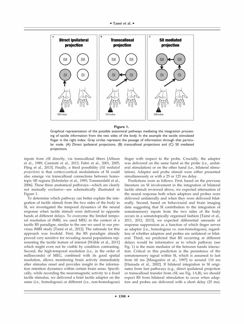
inputs from cSI directly, via transcallosal fibers [Allisonet al., 1989; Caminiti et al., 2013; Fabri et al., 2001, 2005;Fling et al., 2013]. Finally, a third possibility (SII mediatedprojection) is that cortico-cortical modulations of SI couldalso emerge via transcallosal connections between homo-topic SII regions [Schnitzler et al., 1995; Tommerdahl et al.,2006]. These three anatomical pathways—which are clearlynot mutually exclusive—are schematically illustrated inFigure 1.
To determine which pathway can better explain the inte-gration of tactile stimuli from the two sides of the body inSI, we investigated the temporal dynamics of the neuralresponse when tactile stimuli were delivered to oppositehands at different delays. To overcome the limited tempo-ral resolution of fMRI, we used MEG in the context of atactile RS paradigm, similar to the one we used in our pre-vious fMRI study [Tame et al., 2012]. The rationale for thisapproach was twofold. First, the RS paradigm alreadyproved very sensitive for revealing neural populations rep-resenting the tactile feature of interest [W€uhle et al., 2011]which might even not be visible by condition contrasting.Second, the high-temporal resolution (i.e., in the order ofmilliseconds) of MEG, combined with its good spatialresolution, allows monitoring brain activity immediatelyafter stimulus onset and provides insight in the informa-tion retention dynamics within certain brain areas. Specifi-cally, while recording the neuromagnetic activity to a fixedtactile stimulus, we delivered a brief tactile adaptor on thesame (i.e., homologous) or different (i.e., non-homologous)
finger with respect to the probe. Crucially, the adaptorwas delivered on the same hand as the probe (i.e., unilat-eral stimulation) or on the other hand (i.e., bilateral stimu-lation). Adaptor and probe stimuli were either presentedsimultaneously or with a 25 or 125 ms delay.
Predictions were as follows. First, based on the previousliterature on SI involvement in the integration of bilateraltactile stimuli reviewed above, we expected attenuation ofthe neural response both when adaptors and probes weredelivered unilaterally and when they were delivered bilat-erally. Second, based on behavioural and brain imagingdata suggesting that SI contribution to the integration ofsomatosensory inputs from the two sides of the bodyoccurs in a somatotopically organised fashion [Tame et al.,2011, 2012, 2013], we expected differential amounts ofresponse suppression as a function of which finger servesas adaptor (i.e., homologous vs. non-homologous), regard-less of whether adaptors and probes are unilateral or bilat-eral. Third, we predicted that RS occurring at differentdelays would be informative as to which pathway (seeFig. 1) is the main mediator of the between hands interac-tion. Critical in this prediction is the persistence of thesomatosensory signal within SI, which is assumed to lastfrom 60 ms [Mauguiere et al., 1997] to around 110 ms[Hamada et al., 2002]. If bilateral integration in SI origi-nates from fast pathways (e.g., direct ipsilateral projectionor transcallosal transfer from cSI; see Fig. 1A,B), we shouldexpect RS from bilateral stimulation to occur when adap-tors and probes are delivered with a short delay (25 ms).
Figure 1.
Graphical representation of the possible anatomical pathways mediating the integration process-
ing of tactile information from the two sides of the body. In the example the tactile stimulated
finger is the right index. Grey circles represent the passage of information through that particu-
lar node. (A) Direct ipsilateral projections, (B) transcallosal projections and (C) SII mediated
projections.
r Tame et al. r
r 1508 r

Instead, if bilateral integration in SI originates from aslower pathway (transcallosal connection between homo-topic SII regions; see Fig. 1C), RS should emerge onlywhen stimuli are delivered with a longer delay (125 ms).
MATERIAL AND METHODS
Participants
Twenty right-handed participants (mean age 5 27;SD 5 4; six females) participated in the study. Handednesswas inferred from participants’ self-reports. All partici-pants reported normal or corrected to normal vision. Fur-thermore, they reported no history of somatosensoryimpairment, psychiatric or neurological disorders, and nocurrent use of any psychoactive medications. Participantsgave their informed written consent before participating inthe study that was carried out according to 1964 Declara-tion of Helsinki (last update: Seoul, 2008). The study wasapproved by the ethical committee of the University ofTrento. Three participants were discarded from the analy-sis because of a too low activity in the probe-onlyconditions.
Stimulation
To study somatosensory evoked magnetic brain activity,participants were stimulated by short tactile pulses of skinindentation applied to the finger tips. One or two fingerswere stimulated at a time or in sequence. Tactile stimuliwere provided by a four channels piezo-electric stimulator(Quaerosys, Schotten, Germany, www.quaerosys.de) thatwas placed outside the shielded room hosting the MEGsystem. Tactile stimuli were set clearly above threshold tospecifically avoid extinction-like effects, reported so faronly under weak tactile stimulation [Farne et al., 2007;Jacobs et al., 2011]. In general, participants reported to per-ceive the double tactile stimulations in all conditions. Thecode for the stimulation sequence was downloaded fromthe control computer to the stimulator before the begin-ning of a trial. The output of the stimulation sequence wasinitiated by a trigger signal. The digitization rate of theoutput signal was 2 kHz. To avoid introduction of noiseinto the shielded room, the cables used to connect thestimulator outside the shielded room with the stimulatormodules attached to the subject’s hand were twisted and aseries of ferrite low-pass filters was applied. The four stim-ulation modules in contact with the participant’s fingerconsisted of a matrix of 2 3 5 rods (1 mm in diameter),poking from a flat surface of 4 3 8 mm2. The six centralrods of the matrix protruded for 12 (ms), producing aclearly perceivable skin indentation. The intensity of thetactile pulse was always set to the maximum level avail-able, except for 8% of the trials that served as catch trialsfor the behavioural task. Stimuli in catch trials were deliv-ered at half intensity. The stimulation modules were
attached to the finger pads of the middle and index fingerof either hand using Velcro tape, to ensure constant con-tact between the fingers and the stimulation modulesthroughout the experiment. Stimulation modules wereplaced on a woollen pad to attenuate any possible acousticnoise and to avoid any possible unwanted mechanicaltransfer of the vibrations between hands and fingers. Thehands were positioned in a comfortable posture on theparticipants’ legs. The index and middle fingers of eachhand rested on the stimulation modules fixed with theVelcro type.
During the experimental session, instructions were pre-sented by means of a video projector (Panasonic PT-D7700E). The projector was placed outside the shieldedroom and projected the verbal instructions onto a pellucidscreen placed in front of the participants (1.2 m). Instruc-tions were presented at the beginning of the experiment.To avoid eye movements, subjects were asked to gaze at afixation cross (a green cross of 4.3� of visual angle) thatwas presented during each run (see procedure for a com-plete description of the visual conditions) in the middle ofthe pellucid screen. The participants’ right thumb wasplaced on an optical response device measuring thechanges of the reflected infrared light that were inducedby lifting the thumb. The response signal was recordeddirectly by the MEG acquisition system. While stimuluspresentation was controlled by a stimulation computer,the response collection was achieved by the recording soft-ware of the MEG (ELEKTA Neuromag).
Throughout the experiment, white noise was presentedbinaurally using non-magnetic custom earphones con-nected to a computer (Dell T3400; Core Duo 2 Quad). Theacoustic noise signal was used to mask any sounds pro-duced by the operation of the tactile stimulation modules.The computer generating the noise was placed outside theshielded room.
Procedure
Participants sat in a sound-attenuating, magneticallyshielded room. Visual and tactile stimulations were pro-grammed using the in-house software “ASF” [Schwarz-bach, 2011], based on the MATLAB Psychtoolbox-3[Brainard, 1997] for Windows. The experiment comprised10 separate runs consisting of 240 trials each. In each run,the 15 experimental conditions were repeated 15 timesresulting in a total of 225 trials per run. Participants’ atten-tion towards the probe stimulus was checked in additional15 trials per run, yet excluded from further analyses. Atthe beginning of each trial a green fixation cross appearedat the middle of the pellucid screen, and remained visiblefor the entire duration of the trial. 500 ms after the appear-ance of the fixation cross, depending on the experimentalcondition, one or two consecutive or simultaneous tactilestimulations were delivered to the participants’ fingers,each lasting 12 ms (A, adaptor stimulus; P, probe stimulus,
r Early Integration of Bilateral Touch in SI r
r 1509 r

see Fig. 2). A and P occurred at either the same time, orseparated by an inter-stimulus interval (ISI) of 25 ms(short ISI) or 125 ms (long ISI). T was followed by aninter-trial interval (ITI) ranging randomly from 400 ms to2000 ms. Note that the second stimulus (probe) wasalways applied to the left index finger. The fifteen experi-mental conditions fell in four main classes: (1) sequentialpresentation of the adaptor and probe stimulus at theshort interval; (2) sequential presentation at the long inter-val; (3) simultaneous presentation and (4) single fingerstimulation. The first two classes included four differentstimulation conditions each as a function of whether thestimulation was repeated on the same or different fingers;and whether A and P were delivered to the same (unilat-eral stimulation) or the opposite hands (bilateral stimula-tion) (see Table I): (a) A and P were delivered on the samefinger of the same hand (unilateral stimulation) (i.e., leftindex finger, condition short/long Li-Li; Fig. 3a); (b) Aand P were delivered on non-homologous fingers of the
same hand (unilateral stimulation) (i.e., left middle andleft index fingers, condition short/long Lm-Li; Fig. 3b); (c)A and P were delivered on the homologous fingers ofopposite hands (bilateral stimulation) (i.e., right index andleft index fingers, condition short/long Ri-Li; Fig. 3c); (d)A and P were delivered on non-homologous fingers ofopposite hands (bilateral stimulation) (i.e., right middleand left index fingers, condition short/long Rm-Li; Fig.3d). The third class (i.e., simultaneous stimulation)involved only three of the previously described stimula-tion sequences. Simultaneous presentation of adaptor andprobe stimulus (i.e., Li-Li) could not be realized and wastherefore not included in this class. Finally, the fourth class(i.e., singles stimulation) included four conditions in whichindividual fingers where stimulated alone (see Table I):stimulation of the left index finger alone (condition Li;Fig. 3g); stimulation of the left middle finger alone (con-dition Lm; Fig. 3e); stimulation of the right index fingeralone (condition Ri; Fig. 3h); stimulation of the right
Figure 2.
Scheme of stimulation sequences. Pairs of short tactile stimuli (12 ms) were presented at differ-
ent ISIs (A) simultaneously (B) with an ISI of 25 ms or (C) with an ISI of 125 ms. The start of
the stimulation phase was indicated by a green fixation cross in the middle of the screen 500 ms
before the adaptor stimulus.
r Tame et al. r
r 1510 r

middle finger alone (condition Rm; Fig. 3f). The fifteenexperimental conditions were presented in a pseudo-random order. It was assured that the probability foreach experimental condition to be preceded by the samecondition or any different condition was fifty percent[Kourtzi and Kanwisher, 2000; Tame et al., 2012]. Thisprocedure ensured that each pair of stimuli forming thedifferent stimulation conditions had the same weight,avoiding that results in the response were affected by thefact that one condition occurred in a certain positionmore often than another.
To control for participants’ attention, a written questionappeared on the screen (“Did you feel the weak stimula-tion on your left index finger?”) in fifteen catch trials perrun, for a total of 150 trials in the whole experiment (on atotal of 2,550 trials). These catch trials were excluded fromthe analysis.
MEG Recording
Magnetic brain responses were recorded continuouslyfor 320 s per run with the 306-channels (204 first order pla-nar gradiometers, 102 magnetometers) whole-head MEGsystem (Elekta Neuromag Vectorview 306) in a magneticshielded room (AK3B, Vakuum Schmelze, Hanau, Ger-many). The continuous recording was digitized at 1 kHzusing a low-pass filter set at 330 Hz.
Analysis
Preprocessing
The continuous recording was filtered off-line with a 40Hz low-pass filter and a 1 Hz high-pass filter and seg-mented into trials of 650 ms. Each segment included theneuromagnetic responses evoked by the tactile stimulationsand a pre-stimulus baseline of 100 ms that was set beforethe first tactile stimulus of each trial. Somatosensory evokedfields (SEFs) were time-locked to the presentation of thefirst stimulus and averaged across trials and recordingblocks, separately for all stimulation conditions (see Fig. 3).The activity of the baseline defined as the time intervalranging from 0 to 50 ms before the onset of the first stimu-lus was subtracted from the whole trial. Participants withan activity in the probe alone stimulation condition lower
than 4.5 nAm were excluded from the analysis. Using thiscriterion, three participants were discarded.
Estimation of sources
Data analysis focused on the relative changes in neuro-magnetic responses of planar gradiometer sensors evokedby the second stimulus, the probe, which was always theleft index finger and which was either presented alone ortogether with an adaptor stimulus (1) simultaneously, (2)25 ms, or (3) 125 ms before the probe stimulus, respec-tively. To capture amplitude changes in SI and SII ipsi-and contralateral to the presentation of the probe stimulusan equivalent current dipole (ECD) approach was used.To compare source activities across all conditions, a fourdipoles model (SI and SII on both hemispheres) was fittedto the averaged evoked fields of the four single stimulationconditions, the index and middle finger stimulation ofboth hands. Since source location and source activity arestrongly related we refrained from estimating individualdipole source solutions for each of the single stimulus con-ditions. Variations in source localization for different stim-ulation conditions might confound the estimated sourceactivity and thus corrupt the comparison of the sourceactivities across different stimulation conditions. To havean adequate signal-to-noise ratio, the source estimationwas based on a total of 600 trials (150 per each of the sin-gle stimulation condition) (see Fig. 4a).
Two clear distinct components were observed in the SEF[Hari et al., 1984], one peaking at the latency rangebetween 20 and 60 ms (mean peak: 35 ms) and anotherfrom 60 and 90 ms (mean peak: 70 ms) in both hemi-spheres. Analysis of the peaks latency in SII revealed thatresponse was 6.7 ms faster in the contralateral comparedwith the ipsilateral hemisphere [t(16) 5 23.31, P 5 0.004).This delay is compatible with the somatosensory transfertime across the corpus callosum [Aboitiz et al., 1992; Cami-niti et al., 2013; Stancak et al., 2002] and corroborate thesuitability of our approach. ECDs were fitted at the peakof each component for each participant by using the BESAsoftware (BESA Research 5.3). Representations of indexand middle fingers of one hand were not distinguishedand were modeled as a common dipolar source. In thisway, we avoided incorrect assignments of activity to theindex and the middle finger to the two dipoles. Modelling
TABLE I. Stimulation conditions
(1) (2) (3) (4)
ConditionShort inter-stimulusinterval (25 ms)
Long inter-stimulusinterval (125 ms) Simultaneous Singles
(a) short Li-Li long Li-Li – Li(b) short Lm-Li long Lm-Li Lm-Li Lm(c) short Ri-Li long Ri-Li Ri-Li Ri(d) short Rm-Li long Rm-Li Rm-Li Rm
r Early Integration of Bilateral Touch in SI r
r 1511 r

the representation of both fingers with a common sourcewill introduce only minor fitting errors as shown by thegoodness of fit (GOF) values which are for all participantsand conditions better than 88% in SI and 82% in SII. More-over, using one dipole for both finger representations mayintroduce an error in the source activity estimate. How-ever, this error will be systematic, and will affect all condi-tions equally.
For dipole fitting, we used a spherical headmodel thatwas derived from participants’ headshapes and that wasdetermined before the MEG recording by using a Polhe-mus 3d-digitization system Fastrak digitizer (Polhemus,Colchester, VT). The anatomical correspondence of thedipole locations (Fig. 4b) was identified by using thecytoarchitectonic maps following a methodology wealready used successfully [Papadelis et al., 2011]. Figure 4cindicates the correspondence of ECDs with the cytoarchi-tectonic maps of BA3b (80% for right SI and 70% for leftSI) and OP1 (70% for right SII and 70% for left SII) for oneparticipant. Moreover, the SEFs for unilateral and bilateralstimulation conditions in the different delays and theircorrespondent topographic maps of a participant areshown in Figure 5.
Estimation of virtual channels
To estimate the activity of the dipole sources, which arereferred to as virtual channels cSI, iSI, contralateral SII(cSII) and ipsilateral SII (iSII), the leadfields (i.e., the matri-ces describing the relationship between the activity of a sin-gle dipole of unitary strength and the sensor levelactivation) of the four dipoles were calculated from thepositions and orientation of an ECD and the head modelfor each participant. The relation between the magneticfield and the source activity is given by the following equa-tion: B 5 LS 1 e, with B being an m 3 n matrix describingthe measured time course of magnetic activity (n samples)at m sensors. S is a k 3 n matrix containing the time courseof the k dipolar sources. L is an m 3 k lead field matrix ande reflects the noise activity which is assumed to be Gaussianas first approximation. Using linear regression analysis,activity of the virtual channel was estimated: S 5 (L0L)21
L0B, where ð:Þ0 denotes the transpose and ð:Þ21the inverseof a matrix. In total there were 1,020 waveforms corre-sponding to four sources, 15 conditions and 17 participants.
To assess the effect of the adaptor stimulus on the proc-essing of the probe stimulus, we subtracted the sourceactivity of the adaptor (i.e., activity derived from singlefinger stimulation) from the compound source activitiesevoked by the paired finger stimulation (adaptor 1 probe)for all double stimulation conditions. The probe stimulusevoked source activity for each stimulation condition wasagain baseline corrected by subtracting the mean activityof the 50 ms period before probe stimulus onset.
For statistical analyses, peak amplitudes and latencies ofthe ECDs’ source activities were defined. For right SI,which is contralateral to the application of the probe
Figure 3.
Schematic representation of paired (A) and single (B) stimulation
conditions. The empty circle represents the test stimulus, while
the colored circles represent the adaptor stimuli. (a) Stimulation
condition in which the left index was stimulated twice (Li1Li;
within hand stimulation); (b) Stimulation condition in which the
left index and middle fingers were stimulated (Lm1Li; within
hand stimulation); (c) Stimulation condition in which the two
index fingers were stimulated (Ri1Li; between hand stimula-
tion); (d) Stimulation condition in which the right middle and
left index fingers were stimulated (Rm1Li; between hand stimu-
lation); (e) Stimulation condition in which the left middle finger
was stimulated alone; (f) Stimulation condition in which the right
middle finger was stimulated alone; (g) Stimulation condition in
which the left index finger was stimulated alone; (h) Stimulation
condition in which the right index finger was stimulated alone.
r Tame et al. r
r 1512 r

Figure 4.
Schematic representation of the ECD approach used to define
the four ROIs in the contra- and ipsi-lateral S1 and S2. (a) SEFs
for the single finger stimulation (150 trials) of the left (left
panel) and right (right panel) index and middle fingers respec-
tively. Red and green arrows indicate the latencies at which the
ECDs were estimated. (b) The corresponding ECDs located at
SI and SII for the left (left panel) and right (right panel)
stimulation sides fitted at the peak of the first and second SEF
components. MRI slides are in neurological orientation. (c) The
correspondence of the two ECDs with the underlying cytoachi-
tectonic maps (Eickoff et al., 2006). Red and green ECDs are
overlaid on BA3b and OP1 maps respectively. (d) 3D represen-
tation of the four ECDs locations with respect to the partici-
pant’s anatomical scan.
r Early Integration of Bilateral Touch in SI r
r 1513 r

stimulus peaks were determined in a window between 20and 60 ms; for cSII the window ranged from 47 to 85 msand for iSII from 50 to 90 ms; since for iSI activation nopeaks were obvious and thus no peak parameters weredetermined.
Statistics
To quantify the amount of RS, dipole activity peak valuesfor each DS conditions Px were standardized by dividingthem by the peak values obtained for the left index fingeronly condition Pli (i.e., response to the probe). Data were pre-sented as percentage of suppression from index finger onlytrial (see Fig. 6). This normalisation procedure accounted forthe variability of the signal strength across participants.
rx5Pli2Px
Pli� 100
To examine whether the dipole activity (i.e., activity ofcSI, cSII, and iSII) was modulated as a function of the tactileDS conditions (different fingers and timing stimulation) wesubjected the strength of response suppression for the differ-ent adaptor conditions (Lm-Li, Li-Li, Rm-Li, and Ri-Li) toseparate repeated measures ANOVAs. For each dipole activ-ity (cSI, cSII, and iSII) two ANOVAs where computed, onefor the within hand and one for the between hands stimula-tion. The “within hand” ANOVAs used DELAY (short, long)and FINGER (homologous, non-homologous) as within factors,
whereas the “between hands” ANOVAs applied DELAY
(simultaneous, short, long) and FINGER (homologous, non-homologous) as within participant factors. To test whethertactile DS conditions produced suppression we further per-formed a series of one-tailed t-tests comparing the dipoleactivity resulting from the probe minus the DS conditions(i.e., probe–adaptor) for each of the sources (cSI, cSII, andiSII) against the activity in the condition in which the probestimulus was presented alone (i.e., probe only). Tukey HSDtest was used for all post-hoc comparisons.
RESULTS
Using a dipole source model we characterised the well-known stimulus-specific activation represented by activityin the cSI, cSII, and iSII (see Fig. 4), as reported by previousstudies [W€uhle et al., 2011]. During the subsequent steps ofthe analysis, the RS of the probe stimulus was determinedas a function of which adaptor stimuli preceded the probe.Figures 5 and 6 shows the response suppression (% reduc-tion in amplitude) for the different sources and for the dif-ferent timings of the adaptor with respect to the probestimulus (simultaneous, short: 25 ms, and long: 125 ms).
Primary Somatosensory Cortex
While cSI, with respect to the probe stimulus, revealed aclear response, no evoked signal was found for iSI.
Figure 5.
SEFs for the unilateral (left panels) and bilateral (right panels) stimulation of the single and double short
(30 ms) and long (125 ms) fingers stimulation. The corresponding topographic maps of the SEFs in the SI
are also shown aside. Green and black arrows indicate the latencies at which the maps were depicted.
r Tame et al. r
r 1514 r
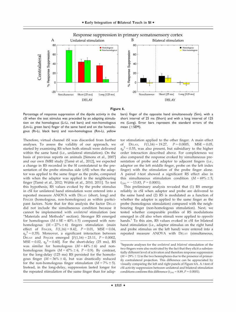
Therefore, virtual channel iSI was discarded from furtheranalyses. To assess the validity of our approach, westarted by examining RS when both stimuli were deliveredwithin the same hand (i.e., unilateral stimulation). On thebasis of previous reports on animals [Simons et al., 2007]and our own fMRI study [Tame et al., 2012], we expecteda change in RS recorded in the SI contralateral to the pre-sentation of the probe stimulus side (cSI) when the adap-tor was applied to the same finger as the probe, comparedwith when the adaptor was applied to the neighbouringfinger [Tame et al., 2012; W€uhle et al., 2010, 2011]. To testthis hypothesis, RS values evoked by the probe stimulusin cSI for unilateral hand stimulation were entered into arepeated measure ANOVA with DELAY (short, long) andFINGER (homologous, non-homologous) as within partici-pant factors. Note that for this analysis the factor DELAY
did not include the simultaneous condition because itcannot be implemented with unilateral stimulation (see“Materials and Methods” section). Stronger RS emergedfor homologous (M 6 SE 5 40% 6 5) compared with non-homologous (M 5 27% 6 6) fingers stimulation (maineffect of FINGER, F(1,16) 5 8.42, P 5 0.01, MSE 5 0.04,gp
2 5 0.35). Moreover, a significant interaction betweenDELAY and FINGER emerged [F(1,16) 5 23.11, P 5 0.0002,MSE 5 0.02, gp
2 5 0.60]. For the short-delay (25 ms), RSwas similar for homologous (M 5 44% 6 6) and non-homologous fingers (M 5 47% 6 6, P 5 0.9). By contrast,for the long-delay (125 ms) RS persisted for the homolo-gous finger (M 5 36% 6 4), but was drastically reducedfor the non-homologous finger stimulation (M 5 7% 6 5).Instead, in the long-delay, suppression lasted longer forthe repeated stimulation of the same finger than for adap-
tor stimulation applied to the other finger. A main effectof DELAY, F(1,16) 5 19.27, P 5 0.0005, MSE 5 0.05,gp
2 5 0.55, was also present, but subsidiary to the higherorder interaction described above. For completeness wealso compared the response evoked by simultaneous pre-sentation of probe and adaptor to adjacent fingers (i.e.,adaptor on the left middle finger, probe on the left indexfinger) with the stimulation of the probe finger alone.A paired t-test showed a significant RS effect also inthis simultaneous stimulation condition (M 5 69% 6 5;t(16) 5 213.83, P 5 0.0001).
This preliminary analysis revealed that (1) RS emergereliably in cSI when adaptor and probe are delivered tothe same hand and (2) RS is modulated as a function ofwhether the adaptor is applied to the same finger as theprobe (homologous stimulation) compared with the neigh-bouring finger (non-homologous stimulation). Next, wetested whether comparable profiles of RS modulationsemerged in cSI also when stimuli were applied to oppositehands.1 To this aim, RS values evoked in cSI for bilateralhand stimulation (i.e., adaptor stimulus on the right handand probe stimulus on the left hand) were entered into arepeated measure ANOVA with DELAY (simultaneous,
Figure 6.
Percentage of response suppression of the dipole activity in the
cSI when the test stimulus was preceded by an adapting stimula-
tion on the homologous (Li-Li, red bars) and non-homologous
(Lm-Li, green bars) finger of the same hand and on the homolo-
gous (Ri-Li, black bars) and non-homologous (Rm-Li, yellow
bars) finger of the opposite hand simultaneously (Sim), with a
short interval of 25 ms (Short) and with a long interval of 125
ms (Long). Error bars represent the standard errors of the
mean (6SEM).
1Separate analyses for the unilateral and bilateral stimulation of thetwo fingers were also motivated by the fact that they elicit a substan-tially different level of activation and therefore response suppression(M 5 29% 6 1) in the two hemispheres due to the presence of primar-ily contralateral projection. This difference can be appreciated byvisually comparing the left and right panels of Figure 6A. A t-test ofcSI activity suppression between unilateral and bilateral stimulationconditions confirms this difference (t(16) 5 8.89, P 5 0.0001).
r Early Integration of Bilateral Touch in SI r
r 1515 r
short, long) and FINGER (homologous, non-homologous) aswithin participant factors. When adaptor and probe stimu-lus were on opposite hands, results showed a finger-specific RS caused by significantly greater suppression inthe dipole activity for homologous (M 5 8% 6 6) comparedwith non-homologous (M 5 1% 6 6) fingers stimulation(main effect of FINGER, F(1,16) 5 9.79, P 5 0.006, MSE 5 0.01,gp
2 5 0.38; see Fig. 6). This indicates that finger homologybetween adaptor and probe matters also when the twostimuli were delivered to opposite sides of the body, atearly as well as later stages of tactile processing. More-over, RS was modulated by the temporal interval betweenadaptor and probe [main effect of DELAY, F(2,32) 5 7.54,P 5 0.002, MSE 5 0.02, gp
2 5 0.32]. The suppression associ-ated with the short-delay (M 5 12% 6 4) was strongerthan the one associated with the long-delay (M 5 0% 6 3;P 5 0.001) and tended to be stronger than the one associ-
ated with the simultaneous stimulation (M 5 4% 6 4;P 5 0.08). Although it might seem from the bar graphdepicted in Figure 6B that some degree of suppressionmight be present under simultaneous bilateral homolo-gous fingers stimulation, this did not turn out to be stat-istically significant (P 5 0.11). No other significant effectsor interactions were found (P> 0.5). These results indi-cate that despite the mainly contralateral representationof distal tactile information in SI there seems to be someearly interaction between hemispheres. The different sup-pression in the short and long-delay indicates the pres-ence of interactions at the very early stages of the corticalinformation process of tactile stimuli (i.e., 25ms). The dif-ferential effect as a function of stimulation of homolo-gous vs. non-homologous fingers indicates that thisinteraction reflects some features of somatotopicorganization.
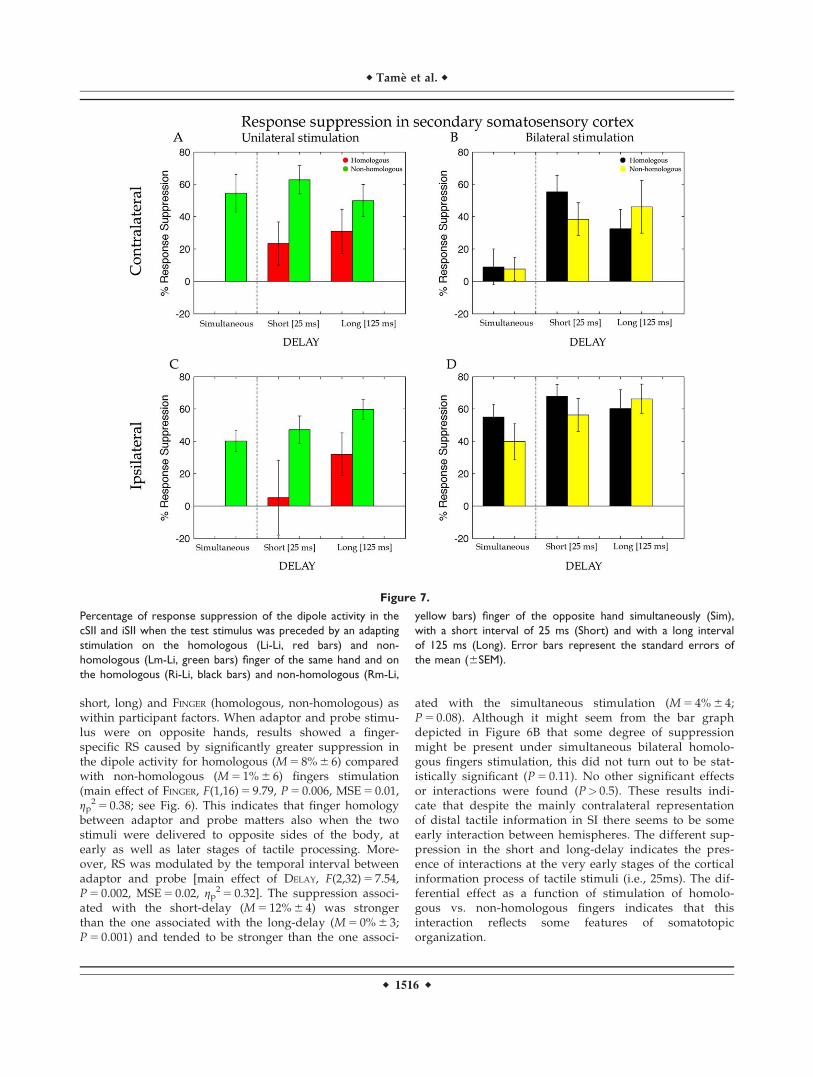
Figure 7.
Percentage of response suppression of the dipole activity in the
cSII and iSII when the test stimulus was preceded by an adapting
stimulation on the homologous (Li-Li, red bars) and non-
homologous (Lm-Li, green bars) finger of the same hand and on
the homologous (Ri-Li, black bars) and non-homologous (Rm-Li,
yellow bars) finger of the opposite hand simultaneously (Sim),
with a short interval of 25 ms (Short) and with a long interval
of 125 ms (Long). Error bars represent the standard errors of
the mean (6SEM).
r Tame et al. r
r 1516 r

Secondary Somatosensory Cortex (Contralateral tothe Side of the Probe Stimulus)
Figure 7 shows that the suppression of the dipole activ-ities for unilateral and bilateral stimulation of the hands inSII. Visual comparison of the left and right panels clearlyshows a similar profile for ipsilateral and cSII, unlike ourfindings in SI. This was confirmed by a t-test (see Fig. 7B;t(16) 5 21.13, P 5 0.9) indicating that strength of activity sup-pression was comparable for the two hemispheres at thelevel of SII.
The suppression of the dipole activity in SII under unilat-eral stimulation was entered into a repeated measureANOVA with DELAY (short, long) and FINGER (homologous,non-homologous) as within participant factors. Differentlyfrom cSI, the analysis showed weaker RS of the dipole activ-ity for the homologous (M 5 27% 6 16) compared with thenon-homologous (M 5 56% 6 11) fingers stimulation (maineffect of FINGER, F(1,16) 5 5.84, P 5 0.03, MSE 5 0.25,gp
2 5 0.27). Evidently, the finger dissociation between short-and long-delay that was found for cSI (see above) vanishedin SII and was replaced by a general reduction in thestrength of the suppression. No other significant maineffects or interactions were found (all ps> 0.2). As in SI, thesimultaneous stimulation of the index and middle finger atthe same hand showed a strong suppression effect(M 5 55% 6 12; t(16) 5 24.68, P 5 0.0001) compared with theprobe-only stimulation on paired t-test.
Effects of bilateral stimulation on RS measured in cSIIwere, as for SI, analysed by including three levels for thefactor DELAY (simultaneous, short and long) and FINGER
(homologous, non-homologous) as within participant fac-tors of a repeated measure ANOVA. This analysisrevealed a main effect of DELAY [F(2,32) 5 9.40, P 5 0.0006,MSE 5 0.15, gp
2 5 0.37) caused by a stronger suppressionof the dipole activity for the short- (M 5 47% 6 12) andlong- (M 5 39% 6 18) compared with the simultaneous(M 5 8% 6 12; ps 5 0.007) delay. With respect to cSI, herethere was an increase of the suppression for the long-delay that indicates a longer persistence of the effects ofbimanual stimulation in the cSII than for cSI. No othersmain effects or interactions resulted to be statisticallysignificant.
Secondary Somatosensory Cortex (Ipsilateral to theSide of the Probe Stimulus)
For SII ipsilateral to the probe stimulus a t-test revealedmore suppression for the bilateral compared with unilateralstimulation conditions [t(16) 5 22.53, P 5 0.022; see Fig. 7D).As for cSII source (Fig. 7A), also for iSII RS values for the uni-lateral stimulation were entered in a repeated measureANOVA with DELAY (short, long) and FINGER (homologous,non-homologous) as within participant factors. Unilateralstimulation produced weaker suppression for the homolo-gous (M 5 19% 6 23) compared with the non-homologous
(M 5 53% 6 9) fingers (main effect of FINGER, F(1,16) 5 6.74,P 5 0.02, MSE 5 0.31, gp
2 5 0.30). No others main effects orinteractions resulted to be statistically significant. A maineffect of DELAY was nearly significant F(1,16) 5 3.77, P 5 0.07,MSE 5 0.17, gp
2 5 0.19, showing a tendency toward a weakersuppression for the short- (M 5 26% 6 19) compared with thelong-delay (M 5 46% 6 12). Instead, analysis on the bilateralstimulation (Fig. 5B) revealed neither statistically significantmain effects nor interaction (all ps> 0.2). Compatible to theresults for cSI and cSII, the simultaneous stimulation of theprobe and its neighbour finger showed a strong suppressioneffect (M 5 40% 6 6; P 5 0.0001) compared with probe-onlystimulation.
DISCUSSION
We used a MEG based RS approach to clarify whethertouches delivered at both hands are integrated already inSI and whether this integration occurs according to“somatotopic” rules. We found reduction of the neuromag-netic activity in contralateral primary and bilateral second-ary somatosensory cortices (i.e., cSI, cSII, and iSII) whenthe adaptor preceded the probe, compared with when theprobe was presented alone. The within hand RS is consist-ent with previous works [Grill-Spector and Malach, 2001;Otsuru et al., 2011; Tame et al., 2012; Tanaka et al., 1991]showing a decrement of the neuronal response afterrepeated presentation of a stimulus feature to which neu-rons are selective. Importantly, also a between hands RSemerged when stimuli were presented on different sidesof the body. Furthermore, when the adaptor and probewere on different hands RS was somatotopically con-strained, as it was larger for stimulation of homologous ascompared with non-homologous fingers.
We also took advantage of the high-temporal resolutionof MEG to investigate the timing of the between handsinteractions. The second novel finding reported here is thatRS occurs in SI at short delays between adaptor and probe.RS evoked by bilateral stimulation emerged when adaptorand probe were separated by 25ms, but not when they wereseparated by 125ms (see Fig. 6B). Because the temporal inte-gration window is short in SI [Mauguiere et al., 1997] andlong in SII [W€uhle et al., 2011] any selective interaction forshort delays is more compatible with interactions occurringwithin SI, rather than top–down modulations via higherlevel processing. In agreement with our hypothesis, thisresult is compatible with the notion that somatosensoryinputs from opposite body sides can interact at early stagesof tactile processing most likely through transcallosal path-ways connecting SI in the two hemispheres (Fig. 1B).
Integration of Tactile Stimuli From Same andDifferent Sides of the Body
Similar to previous MEG studies, we found neuromag-netic activity in SI contralateral to the probe stimulus and
r Early Integration of Bilateral Touch in SI r
r 1517 r
in bilateral SII [Hari et al., 1993; Hoechstetter et al., 2001;Maldjian et al., 1999; W€uhle et al., 2011], whereas no activ-ity in the iSI was found, corroborating the notion of thecontralateral vocation of SI and the bilateral nature of SII.When adaptor and probe stimulus were delivered on thesame hand, unilateral stimulation response suppressionwas comparable in cSII and cSI. By contrast, when adaptorand probe were delivered on opposite hands, responsesuppression (i.e., our proxy of the interactions betweenadaptor and probe stimuli) was stronger in cSII comparedwith cSI. This result was largely expected and it likelyreflects the existence of denser bilateral afferent projectionsin SII compared with SI [Forss et al., 1995; Hari et al.,1993; Lin and Forss, 2002]. The crucial aspect of our find-ings, however, is the observation that when adaptor andprobe were on opposite hands, response suppression incSI was nonetheless present.
Bilateral interactions in SI are in line with previous neu-rophysiological findings in monkeys [Iwamura et al., 2001;Killackey et al., 1983; Lipton et al., 2006] and with behav-ioral [Braun et al., 2005; Craig, 1985; D’Amour and Harris,2013; Gescheider et al., 1970; Harrar et al., 2013; Harriset al., 2001; Nguyen et al., 2014; Sathian and Zangaladze,1997, 1998; Sherrick, 1964; Tame et al., 2011] and neuroi-maging studies in humans [Hlushchuk and Hari, 2006;Kakigi, 1986; Kakigi and Jones, 1985; Nihashi et al., 2005;Noachtar et al., 1997; Sutherland and Tang, 2006; Tameet al., 2012]. Previous findings from our laboratory [Tameet al., 2011, 2012, 2013] have recognised the ability of SI tointegrate inputs from the two sides of the body. However,the low temporal resolution of fMRI did not allow us todetermine the time course of the interactions between con-tralateral and ipsilateral tactile stimulation in SI and SII,thus limiting any definite conclusion about the pathway/flow of sensory information leading to the activationsobserved in SI in response to bilateral stimulation. In thepresent work, the high-temporal resolution of MEG com-bined with the RS paradigm sensitivity revealed that sup-pression in cSI in response to bilateral stimulation wasweak when adaptor and probe were delivered simultane-ously, emerged very clearly during the short-delay condi-tion, and vanished at the long delay. This temporal profileof the response pattern in cSI under bilateral stimulationreveals that, differently from previous reports [Chunget al., 2014; Jung et al., 2012], responses to bilateral touchcannot be entirely ascribed to higher stages of processing,such as SII, because the suppression occurs very early intime [i.e., short delay (25ms)]. In other words, very fastsuppression effects rule out the possibility that the activitywe registered in cSI is a mere reflection of top–down mod-ulation of cSI by well-known bilaterally organized higher-level brain areas. Instead, our data suggest that it is morelikely that tactile information from the stimulated bodyside reaches iSI via transcallosal connections already at thelevel of SI [Manzoni et al., 1989; Shuler et al., 2001; Tom-asch, 1954]. In this respect, a recent tractography study hasshown the presence of transcallosal fibre tracts connecting
homologous sensorimotor cortical regions [Fling et al.,2013]. In addition, earlier bilateral interactions at subcorti-cal and/or spinal cord level can be present, as shown byreports on patients that underwent callosotomy [Corballis,1994; Sergent, 1990]. A recent electroencephalography(EEG) study carried out by Ragert and coworkers (2011)suggested the existence of interactions between ipsilateraland cSI after median nerve (MN) stimulation in an intervalranging between 20 and 25 ms post stimulus [Ragert et al.,2011]. Moreover, in a study on healthy individuals Korve-noja and coworkers (1995) reported iSI activation, in fiveof the ten participants, after MN stimulation in a windowranging between 80 and 300 ms. The ipsilateral responsewas weaker compared with the contralateral one [Korve-noja et al., 1995]. Similarly, Zhu and coworkers (2007)studying the spatiotemporal integration of tactile informa-tion, using high-resolution MEG in a digit oddball para-digm, report early ipsilateral responses (i.e., 10 ms laterthan the contralateral response) in the anterior parietalfield (Zhu et al., 2007). However, in these previous reportson healthy subjects early ipsilateral activity was not consis-tently present in all participants. Moreover, Kanno andcoworkers (2004) in a study on two patients with a severeleft brain damage reported ECDs of ipsilateral responseson the central sulcus after right MN stimulation. Thedipole locations for right MN stimulation were adjacent tothe location of the N20m response to left MN stimulation[Kanno et al., 2004]. Recently, Nevalainen and coworkers(2012), studying adolescents with cerebral palsy withMEG, reported the presence of ipsilateral responses in SImore often than in the control groups under MN stimula-tion even though with longer latency compared with thecSI [Nevalainen et al., 2012]. Finally, a single case study ona brain-damaged patient provided indications for integra-tion of proprioceptive information from the two sides ofthe body also in the absence of SI in one of the two hemi-spheres [Borchers et al., 2011]. Although these findings onpatients are insightful, individuals with a brain damagetypically undergo some brain reorganization processesthat could account, at least in part, for atypical effects. Ourdata instead reveal interactions between stimuli deliveredto the two body sides detected as early as in SI of neuro-logically healthy controls.
The lack of any modulation in the neuromagnetic activ-ity when bilateral stimulation was delivered with a longerdelay (i.e., 125 ms) might be the consequence of the factthat the window for bilateral integration in SI is shorterthan the adopted delay. The maximal estimated perma-nence of the tactile signal in SI tends to be around 110 ms[Hamada et al., 2002], which is shorter than the 125 mslong-delay we adopted for comparative purposes. There-fore, our results provide further evidence, in addition tothe one just described above in healthy and pathologicalindividuals, in favour of an early integration of bilateralstimuli in SI under certain temporal constraints.
A further intriguing aspect of cSI response to bilateralstimulation is the modulation of response suppression as a
r Tame et al. r
r 1518 r

function of adaptor finger. In particular, RS in cSI inresponse to bilateral stimulation was larger when homolo-gous compared with non-homologous fingers were stimu-lated. This result extends the results of our previous fMRIstudy in which we have shown that repeated tactile stimu-lation of the same finger and homologous fingers of oppo-site hands produced an adaptation effect in the SI [Tameet al., 2012]. Similarly, in a series of behavioural studieswe have shown that masking effects vanish when homolo-gous fingers of opposite hands are stimulated [Tame et al.,2011, 2013]. This finger-specific pattern implies that notonly SI can respond to bilateral stimulation, but also that itdoes so in a somatotopically organised manner—whichconstitutes further evidence that the integration of bilateraltouch is occurring at the level of SI or earlier.
Implications for the Neural Mechanisms UnderlyingTactile Repetition Suppression
One unexpected finding of the present work emerged inthe profile of response suppression of cSI, when the adap-tor and probe were delivered to the same hand. Whilenon-homologous fingers stimulation produced smaller RSeffect at the long compared with the short delay, homolo-gous fingers stimulation produced comparable amount ofsuppression regardless of the delay (compare the two redbars in Fig. 6A). If RS response reflects a single underlyingneural mechanism, we should have observed identicalmodulations as a function of delay for both homologousand non-homologous finger stimulations. Because this wasclearly not the case, the hypothesis of a single mechanismmediating RS becomes unlikely. Instead, our data suggestthat different neural mechanisms can mediate RS whentactile stimulation repeats exactly on the same finger(homologous condition), compared with when tactile stim-ulation occurs on neighbouring fingers (non-homologouscondition). Below we discuss the possibility that RS effectsfor homologous fingers reflect neural adaptation, whereasRS effects for non-homologous fingers reflect lateralinhibition.
Previous works that measured RS to repeated tactilestimulation on the same body part have linked thisresponse to the phenomenon of neural adaptation (Otsuruet al., 2011; Tame et al., 2012; Tanaka et al., 1991). Thisinterpretation is in agreement with previous interpreta-tions of RS in other sensory modalities [Grill-Spector andMalach, 2001; Otsuru et al., 2011; Tame et al., 2012; Tanakaet al., 1991]. Of particular interest are two previous MEGstudies that examined reduction in the neuromagneticactivity following repeated stimulation of the same finger,when different delays between stimuli were applied[W€uhle et al., 2010, 2011]. W€uhle and coworkers (2011)found that when a tactile stimulus (i.e., probe) was pre-ceded by a near- or supra-threshold stimulus (i.e., adaptor)on the same finger, the dipole activity evoked by the probestimulus was reduced compared with when the probe
stimulus was presented alone. More interestingly, theadaptation effect was the same regardless of whether thedelay between the stimuli was 80 ms or 100ms (for similarresults see also W€uhle et al., 2010). Similarly, in the pres-ent work in the unilateral stimulation condition theresponse suppression was the same with comparativelyshorter and longer temporal delays (i.e., 25 ms and 125ms). These findings suggest that in SI the somatosensoryprocessing of repeated tactile stimulations of the same fin-ger starts to be integrated as early as 25 ms and this inte-gration can occur at least up to 125 ms. Instead, underbilateral stimulation the RS was no longer present at thelatter delay. Therefore, the adaptation effect indicates thatthe persistence of tactile signals in SI coming from thesame region of the skin is lasting longer (i.e., at least 125ms), as compared with when tactile stimuli are deliveredto a different region, even if the latter concerns the homol-ogous finger, but of opposite hands (i.e., at least 25 ms).
The suppression effect for repeated touches delivered todifferent fingers has been studied to a much lesser extent.To our knowledge, our previous fMRI report was the onlystudy that examined RS in non-homologous conditions[Tame et al., 2012]. The effect appears to be broadly con-sistent with a lateral inhibition phenomenon [Biermannet al., 1998; Cardini et al., 2011], first described by vonBekesy (1967) in his seminal book “Sensory Inhibition”, pre-cisely for the tactile modality [Von B�ek�esy, 1967; Mooreand Nelson, 1998; Zhu and Connors, 1999]. Lateral inhibi-tion is a neurophysiological effect determined by the firingof neurons responding to a certain region with a conse-quent reduction of the neuronal activity in the neighbour-ing regions [DiCarlo et al., 1998]. For instance, when aregion of the skin is stimulated (e.g., index finger), theneuronal population responding to that region is activatedand its response inhibits the activity from neighboringneurons such as, for instance, those responding to themiddle finger [Geldard and Sherrick, 1972] (for a discus-sion on sensory inhibition mechanisms, see [Von B�ek�esy,1967]. Other studies have shown that suppressive responsein the somatosensory cortex under double tactile stimula-tion depends on lateral inhibitory processing both within[Laskin and Spencer, 1979] and between [Okajima et al.,1991] the hands. Although we are not aware of studiesthat examined the possible modulation of lateral inhibitionprocesses as a function of time, von Bekesy (1967) reportedan observation that suggests that lateral inhibition ismodulated as a function of the delay between the stimuli.“If two vibrators are placed 12 cm apart on the arm, andare actuated with a series of clicks without time difference,the vibration will be localized in the middle of the twovibrators as a rather diffuse sensation. When a time differ-ence between the vibrators is introduced, the sensationwill move toward the vibrator that receives the click ear-lier. At the same time the lateral spreading of the vibratorysensation becomes smaller” [Von B�ek�esy, 1967].
Similarly, our data show that tactile stimulation ofneighboring fingers when a delay is introduced, reduce
r Early Integration of Bilateral Touch in SI r
r 1519 r

the suppression to the delay between the tactile stimuli. Inparticular, the greater was the delays the lower was thesuppression, and therefore lateral inhibition. This inhibi-tory effect as a function of delay was occurring at the levelof SI (Fig. 6A), but not at the level of SII (Fig. 7A,C) wherethe inhibition was the same for the different delays. Thisis consistent with suppression studies of somatosensorycortex excitability which show that in SI the longer thedelay between two afferent stimuli the smaller the reduc-tion in the amplitude of the somatosensory evoked/fieldpotential [McLaughlin and Kelly, 1993; Stevenson et al.,2012]. However, this pattern is known to be absent in SII[W€uhle et al., 2011]. As to this pattern observed in SII, wedo not have straightforward explanations. Nonetheless, wefeel that it is important to report this secondary finding forcompleteness and to stimulate further studies on the inter-actions between SI and SII. It would be tempting to specu-late that the pattern observed in SII reflects thepropagation of the inhibitory effects of SI, modulated fur-ther by the less clear somatotopy of SII. For instance, anylateral inhibition effect observed in SI could become aform of RS in SII—due to the less clear distinction betweenfingers.
CONCLUSIONS
Our MEG measure of brain activity during a RS para-digm showed that tactile stimuli coming from the twosides of the body are integrated within the SI shortly afterthe stimuli came in contact with the skin. This observa-tion moves our understanding of bilateral touch process-ing forward by showing that in humans, despite itsprimarily contralateral response, SI is capable of integrat-ing touches from opposite body sides at early stages ofsomatosensory information processing. This early integra-tion is compatible with bilateral touch reaching SI morelikely through transcallosal inputs from cSI. The involve-ment of SI in the integration of bilateral touch is alsostrengthened by the fact that RS was modulated by therelative position of the stimuli on the hands (homologousvs. non-homologous finger), both in unilateral and inbilateral stimulation. This finger specificity points to theinvolvement of a brain structure characterised by highdegrees of somatotopy, such as SI. Interestingly, duringunilateral stimulation, this modulation of RS as a functionof stimulated fingers changed also as a function of time:persisting across short and long delays when the samefinger was stimulated twice, but decaying at the longdelay when neighbouring fingers were stimulated. Thisunexpected finding may indicate that distinct neuralmechanisms operate under the RS measure. Neural adap-tation processes may account for RS effects followingrepeated stimulation of the same skin region (homolo-gous fingers); in addition, lateral inhibitory processesmay concur, with a different temporal profile, to the over-all RS effect when tactile stimulations are presented on
different skin regions (non-homologous fingers). We spec-ulate that such SI mechanisms, by providing complemen-tary spatiotemporal information may be functional to anappropriate sensory feedback while coordinating manipu-lative actions.
ACKNOWLEDGMENT
The authors are grateful to Dr. Francesco Prantil for theconditions randomization program. They thank Drs. Gian-paolo Demarchi and Gianpiero Monittola for the technicalsupport. They thank the editors, and two anonymousreviewers for their insightful comments on a previous ver-sion of the manuscript.
REFERENCES
Aboitiz F, Scheibel AB, Fisher RS, Zaidel E (1992): Fiber composi-tion of the human corpus callosum. Brain Res 598:143–153.
Allison T, McCarthy G, Wood CC, Williamson PD, Spencer DD(1989): Human cortical potentials evoked by stimulation of themedian nerve. II. Cytoarchitectonic areas generating long-latency activity. J Neurophysiol 62:711–722.
Belin P, Zatorre RJ (2003): Adaptation to speaker’s voice in rightanterior temporal lobe. Neuroreport 14:2105–2109.
Biermann K, Schmitz F, Witte OW, Konczak J, Freund HJ,Schnitzler A (1998): Interaction of finger representation in thehuman first somatosensory cortex: A neuromagnetic study.Neurosci Lett 251:13–16.
Borchers S, Hauser T-K, Himmelbach M (2011): Bilateral handrepresentations in human primary proprioceptive areas. Neu-ropsychologia 49:3383–3391.
Brainard DH (1997): The Psychophysics Toolbox. Spat Vis 10:433–436.
Braun C, Hess H, Burkhardt M, W€uhle A, Preissl H (2005): Theright hand knows what the left hand is feeling. Exp Brain Res162:366–373.
Caminiti R, Carducci F, Piervincenzi C, Battaglia-Mayer A,Confalone G, Visco-Comandini F, Pantano P, Innocenti GM(2013): Diameter, length, speed, and conduction delay of cal-losal axons in macaque monkeys and humans: Comparingdata from histology and magnetic resonance imaging diffusiontractography. J Neurosci 33:14501–14511.
Cardini F, Longo MR, Haggard P (2011): Vision of the body mod-ulates somatosensory intracortical inhibition. Cereb Cortex 21:2014–2022.
Chong TT-J, Cunnington R, Williams MA, Kanwisher N,Mattingley JB (2008): fMRI adaptation reveals mirror neuronsin human inferior parietal cortex. Curr Biol 18:1576–1580.
Chung YG, Han SW, Kim H-S, Chung S-C, Park J-Y, Wallraven C,Kim S-P (2014): Intra- and inter-hemispheric effective connec-tivity in the human somatosensory cortex during pressurestimulation. BMC Neurosci 15:43.
Corballis MC (1994): Split decisions: Problems in the interpreta-tion of results from commissurotomized subjects. Behav BrainRes 64:163–172.
Craig JC (1985): Attending to two fingers: Two hands are betterthan one. Percept Psychophys 38:496–511.
D’Amour S, Harris LR (2013): Contralateral tactile maskingbetween forearms. Exp Brain Res 2014;232:821–826.
r Tame et al. r
r 1520 r

Dehaene-Lambertz G, Dehaene S, Anton J-L, Campagne A, Ciuciu P,Dehaene GP, Denghien I, Jobert A, Lebihan D, Sigman M, PallierC, Poline J-B (2006): Functional segregation of cortical languageareas by sentence repetition. Hum Brain Mapp 27:360–371.
DiCarlo JJ, Johnson KO, Hsiao SS (1998): Structure of receptivefields in area 3b of primary somatosensory cortex in the alertmonkey. J Neurosci 18:2626–2645.
Eickhoff SB, Jbabdi S, Caspers S, Laird AR, Fox PT, Zilles K,Behrens TEJ (2010): Anatomical and functional connectivity ofcytoarchitectonic areas within the human parietal operculum.J Neurosci 30:6409–6421.
Fabri M, Polonara G, Del Pesce M, Quattrini A, Salvolini U,Manzoni T (2001): Posterior corpus callosum and interhemi-spheric transfer of somatosensory information: An fMRI andneuropsychological study of a partially callosotomized patient.J Cogn Neurosci 13:1071–1079.
Fabri M, Del Pesce M, Paggi A, Polonara G, Bartolini M, SalvoliniU, Manzoni T (2005): Contribution of posterior corpus cal-losum to the interhemispheric transfer of tactile information.Brain Res Cogn Brain Res 24:73–80.
Farne A, Brozzoli C, L�adavas E, Ro T (2007): Investigating multi-sensory spatial cognition through the phenomenon of extinc-tion. In: Haggard, P, Rossetti, Y, Kawato, M, editors.Sensorimotor Foundations of Higher Cognition. Oxford Uni-versity Press.
Fling BW, Benson BL, Seidler RD (2013): Transcallosal sensorimo-tor fiber tract structure-function relationships. Hum BrainMapp 34:384–395.
Forss N, Jousm€aki V, Hari R (1995): Interaction between afferentinput from fingers in human somatosensory cortex. Brain Res685:68–76.
Geldard FA, Sherrick CE (1972): The cutaneous “rabbit”: A per-ceptual illusion. Science 178:178–179.
Gescheider GA, Herman DD, Phillips JN (1970): Criterion shifts inthe measurement of tactile masking. Percept Psychophys 433–436.
Grill-Spector K, Malach R (2001): fMR-adaptation: A tool forstudying the functional properties of human cortical neurons.Acta Psychol (Amst) 107:293–321.
Grill-Spector K, Henson R, Martin A (2006): Repetition and thebrain: Neural models of stimulus-specific effects. Trends CognSci (Regul Ed) 10:14–23.
Gross CG, Rocha-Miranda CE, Bender DB (1972): Visual proper-ties of neurons in inferotemporal cortex of the Macaque.J Neurophysiol 35:96–111.
Hamada Y, Otsuka S, Okamoto T, Suzuki R (2002): The profile ofthe recovery cycle in human primary and secondary somato-sensory cortex: A magnetoencephalography study. Clin Neuro-physiol 113:1787–1793.
Hari R, Reinikainen K, Kaukoranta E, H€am€al€ainen M, IlmoniemiR, Penttinen A, Salminen J, Teszner D (1984): Somatosensoryevoked cerebral magnetic fields from SI and SII in man. Elec-troencephalogr Clin Neurophysiol 57:254–263.
Hari R, Karhu J, H€am€al€ainen M, Knuutila J, Salonen O, Sams M,Vilkman V (1993): Functional organization of the human firstand second somatosensory cortices: A neuromagnetic study.Eur J Neurosci 5:724–734.
Harrar V, Spence C, Makin TR (2013): Topographic generalizationof tactile perceptual learning. J Exp Psychol Hum Percept Per-form 40:15–23.
Harris JA, Harris IM, Diamond ME (2001): The topography of tac-tile working memory. J Neurosci 21:8262–8269.
Henson RNA (2003): Neuroimaging studies of priming. Prog Neu-robiol 70:53–81.
Hlushchuk Y, Hari R (2006): Transient suppression of ipsilateralprimary somatosensory cortex during tactile finger stimulation.J Neurosci 26:5819–5824.
Hoechstetter K, Rupp A, Stanc�ak A, Meinck HM, Stippich C, BergP, Scherg M (2001): Interaction of tactile input in the humanprimary and secondary somatosensory cortex–a magnetoence-phalographic study. Neuroimage 14:759–767.
Iwamura Y, Taoka M, Iriki A (2001): Bilateral activity and callosalconnections in the somatosensory cortex. Neuroscientist 7:419–429.
Iwamura Y, Tanaka M, Iriki A, Taoka M, Toda T (2002): Process-ing of tactile and kinesthetic signals from bilateral sides of thebody in the postcentral gyrus of awake monkeys. Behav BrainRes 135:185–190.
Jacobs S, Brozzoli C, Hadj-Bouziane F, Meunier M, Farne A(2011): Studying multisensory processing and its role in therepresentation of space through pathological and physiologicalcrossmodal extinction. Front Psychol 2:89.
Jung P, Klein JC, Wibral M, Hoechstetter K, Bliem B, Lu M-K,Wahl M, Ziemann U (2012): Spatiotemporal dynamics of bima-nual integration in human somatosensory cortex and their rele-vance to bimanual object manipulation. J Neurosci 32:5667–5677.
Kakigi R (1986): Ipsilateral and contralateral SEP components fol-lowing median nerve stimulation: Effects of interfering stimuliapplied to the contralateral hand. Electroencephalogr ClinNeurophysiol 64:246–259.
Kakigi R, Jones SJ (1985): Effects on median nerve SEPs of tactilestimulation applied to adjacent and remote areas of the bodysurface. Electroencephalogr Clin Neurophysiol 62:252–265.
Kanno A, Nakasato N, Hatanaka K, Yoshimoto T (2003): Ipsilat-eral area 3b responses to median nerve somatosensory stimula-tion. Neuroimage 18:169–177.
Kanno A, Nakasato N, Nagamine Y, Tominaga T (2004): Non-transcallosal ipsilateral area 3b responses to median nervestimulus. J Clin Neurosci 11:868–871.
Killackey HP, Gould HJ III, Cusick CG, Pons TP, Kaas JH (1983):The relation of corpus callosum connections to architectonicfields and body surface maps in sensorimotor cortex of newand old world monkeys. J Comp Neurol 219:384–419.
Korvenoja A, Wikstrom H, Huttunen J, Virtanan J, Laine P,Aronen HJ, Seppalainen AM, Ilmoniemi RJ (1995): Activationof ipsilateral primary sensorimotor cortex by median nervestimulation. Neuroreport 6:2589–2593.
Kourtzi Z, Kanwisher N (2000): Cortical regions involved in per-ceiving object shape. J Neurosci 20:3310–3318.
Krekelberg B, Boynton GM, van Wezel RJA (2006): Adaptation:From single cells to BOLD signals. Trends Neurosci 29:250–256.
Laskin SE, Spencer WA (1979): Cutaneous masking. I. Psycho-physical observations on interactions of multipoint stimuli inman. J Neurophysiol 42:1048–1060.
Li L, Miller EK, Desimone R (1993): The representation of stimu-lus familiarity in anterior inferior temporal cortex.J Neurophysiol 69:1918–1929.
Li Hegner Y, Lee Y, Grodd W, Braun C (2010): Comparing tactilepattern and vibrotactile frequency discrimination: A humanFMRI study. J Neurophysiol 103:3115–3122.
r Early Integration of Bilateral Touch in SI r
r 1521 r

Lin YY, Forss N (2002): Functional characterization of human sec-ond somatosensory cortex by magnetoencephalography. BehavBrain Res 135:141–145.
Lingnau A, Ashida H, Wall MB, Smith AT (2009a): Speed encod-ing in human visual cortex revealed by fMRI adaptation. J Vis9:3.1–14.
Lingnau A, Gesierich B, Caramazza A (2009b): Asymmetric fMRIadaptation reveals no evidence for mirror neurons in humans.Proc Natl Acad Sci USA 106:9925–9930.
Lipton ML, Fu K-MG, Branch CA, Schroeder CE (2006): Ipsilateralhand input to area 3b revealed by converging hemodynamicand electrophysiological analyses in macaque monkeys.J Neurosci 26:180–185.
Mahon BZ, Milleville SC, Negri GAL, Rumiati RI, Caramazza A,Martin A (2007): Action-related properties shape object repre-sentations in the ventral stream. Neuron 55:507–520.
Maldjian JA, Gottschalk A, Patel RS, Detre JA, Alsop DC (1999):The sensory somatotopic map of the human hand demon-strated at 4 Tesla. Neuroimage 10:55–62.
Manzoni T, Barbaresi P, Conti F, Fabri M (1989): The callosal con-nections of the primary somatosensory cortex and the neuralbases of midline fusion. Exp Brain Res 76:251–266.
Mauguiere F, Merlet I, Forss N, Vanni S, Jousm€aki V, Adeleine P,Hari R (1997): Activation of a distributed somatosensory corti-cal network in the human brain: A dipole modelling study ofmagnetic fields evoked by median nerve stimulation. Part II:Effects of stimulus rate, attention and stimulus detection. Elec-troencephalogr Clin Neurophysiol 104:290–295.
McLaughlin DF, Kelly EF (1993): Evoked potentials as indices ofadaptation in the somatosensory system in humans: A reviewand prospectus. Brain Res Brain Res Rev 18:151–206.
Miller EK, Li L, Desimone R (1991): A neural mechanism forworking and recognition memory in inferior temporal cortex.Science 254:1377–1379.
Moore CI, Nelson SB (1998): Spatio-temporal subthreshold recep-tive fields in the vibrissa representation of rat primary somato-sensory cortex. J Neurophysiol 80:2882–2892.
Nelson AJ, Chen R (2008): Digit somatotopy within cortical areas ofthe postcentral gyrus in humans. Cereb Cortex 18:2341–2351.
Nevalainen P, Pihko E, M€aenp€a€a H, Valanne L, Nummenmaa L,Lauronen L (2012): Bilateral alterations in somatosensory corti-cal processing in hemiplegic cerebral palsy. Dev Med ChildNeurol 54:361–367.
Nguyen RH, Forshey TM, Holden JK, Francisco EM, Kirsch B,Favorov O, Tommerdahl M (2014): Vibrotactile discriminativecapacity is impacted in a digit-specific manner with concurrentunattended hand stimulation. Exp Brain Res.
Nihashi T, Naganawa S, Sato C, Kawai H, Nakamura T, FukatsuH, Ishigaki T, Aoki I (2005): Contralateral and ipsilateralresponses in primary somatosensory cortex following electricalmedian nerve stimulation—An fMRI study. Clin Neurophysiol116:842–848.
Noachtar S, L€uders HO, Dinner DS, Klem G (1997): Ipsilateralmedian somatosensory evoked potentials recorded fromhuman somatosensory cortex. Electroencephalogr Clin Neuro-physiol 104:189–198.
Okajima Y, Chino N, Saitoh E, Kimura A (1991): Interactions ofsomatosensory evoked potentials: simultaneous stimulation oftwo nerves. Electroencephalogr Clin Neurophysiol 80:26–31.
Otsuru N, Inui K, Yamashiro K, Urakawa T, Keceli S, Kakigi R(2011): Effects of prior sustained tactile stimulation on the soma-
tosensory response to the sudden change of intensity in humans:An magnetoencephalography study. Neuroscience 182:115–124.
Papadelis C, Eickhoff SB, Zilles K, Ioannides AA (2011): BA3band BA1 activate in a serial fashion after median nerve stimu-lation: Direct evidence from combining source analysis ofevoked fields and cytoarchitectonic probabilistic maps. Neuro-image 54:60–73.
Penfield W, Boldrey E (1937): Somatic motor and sensory repre-sentation in the cerebral cortex of man as studied by electricalstimulation. Brain 60:389–443.
Ragert P, Nierhaus T, Cohen LG, Villringer A (2011): Interhemi-spheric interactions between the human primary somatosen-sory cortices. PLoS ONE 6:e16150.
Reed JL, Qi H-X, Zhou Z, Bernard MR, Burish MJ, Bonds AB,Kaas JH (2010): Response properties of neurons in primarysomatosensory cortex of owl monkeys reflect widespread spa-tiotemporal integration. J Neurophysiol 103:2139–2157.
Reed JL, Qi H-X, Kaas JH (2011): Spatiotemporal properties ofneuron response suppression in owl monkey primary somato-sensory cortex when stimuli are presented to both hands.J Neurosci 31:3589–3601.
Sathian K, Zangaladze A (1997): Tactile learning is taskspecific but transfers between fingers. Percept Psychophys 59:119–128.
Sathian K, Zangaladze A (1998): Perceptual learning in tactilehyperacuity: Complete intermanual transfer but limited reten-tion. Exp Brain Res 118:131–134.
Schnitzler A, Salmelin R, Salenius S, Jousm€aki V, Hari R (1995):Tactile information from the human hand reaches the ipsilat-eral primary somatosensory cortex. Neurosci Lett 200:25–28.
Schwarzbach J (2011): A simple framework (ASF) for behavioraland neuroimaging experiments based on the psychophysicstoolbox for MATLAB. Behav Res Methods. Available at:http://www.ncbi.nlm.nih.gov/pubmed/21614662.
Sergent J (1990): Furtive incursions into bicameral minds. Integra-tive and coordinating role of subcortical structures. Brain 113(Pt 2):537–568.
Sherrick CE Jr (1964): Effects of double simultaneous stimulationof the skin. Am J Psychol 77:42–53.
Shuler MG, Krupa DJ, Nicolelis MA (2001): Bilateral integration ofwhisker information in the primary somatosensory cortex ofrats. J Neurosci 21:5251–5261.
Simons SB, Chiu J, Favorov OV, Whitsel BL, Tommerdahl M(2007): Duration-dependent response of SI to vibrotactile stim-ulation in squirrel monkey. J Neurophysiol 97:2121–2129.
Stancak A, Hoechstetter K, Tintera J, Vrana J, Rachmanova R,Kralik J, Scherg M (2002): Source activity in the human second-ary somatosensory cortex depends on the size of corpus cal-losum. Brain Res 936:47–57.
Stevenson CM, Wang F, Brookes MJ, Zumer JM, Francis ST,Morris PG (2012): Paired pulse depression in the somatosen-sory cortex: Associations between MEG and BOLD fMRI. Neu-roimage 59:2722–2732.
Sutherland MT (2006): The hand and the ipsilateral primary soma-tosensory cortex. J Neurosci 26:8217–8218.
Sutherland MT, Tang AC (2006): Reliable detection of bilateralactivation in human primary somatosensory cortex by unilat-eral median nerve stimulation. Neuroimage 33:1042–1054.
Tame L, Farne A, Pavani F (2011): Spatial coding of touch at thefingers: Insights from double simultaneous stimulation withinand between hands. Neurosci Lett 487:78–82.
r Tame et al. r
r 1522 r

Tame L, Braun C, Lingnau A, Schwarzbach J, Demarchi G, LiHegner Y, Farne A, Pavani F (2012): The contribution of pri-mary and secondary somatosensory cortices to the representa-tion of body parts and body sides: An fMRI adaptation study.J Cogn Neurosci 24:2306–2320.
Tame L, Farne A, Pavani F (2013): Vision of the body and the dif-ferentiation of perceived body side in touch. Cortex 49:1340–1351.
Tanaka K, Saito H, Fukada Y, Moriya M (1991): Coding visualimages of objects in the inferotemporal cortex of the macaquemonkey. J Neurophysiol 66:170–189.
Tan H-RM, W€uhle A, Braun C (2004): Unilaterally applied stimuliin a frequency discrimination task are represented bilaterallyin primary somatosensory cortex. Neurol Clin Neurophysiol2004:83.
Tian X, Poeppel D (2013): The effect of imagination on stimula-tion: The functional specificity of efference copies in speechprocessing. J Cogn Neurosci 25:1020–1036.
Tomasch J (1954): Size, distribution, and number of fibres in thehuman corpus callosum. Anat Rec 119:119–135.
Tommerdahl M, Simons SB, Chiu JS, Favorov O, Whitsel BL(2006): Ipsilateral input modifies the primary somatosensorycortex response to contralateral skin flutter. J Neurosci 26:5970–5977.
Von B�ek�esy G (1967): Sensory Inhibition. Princeton UniversityPress.
Vuilleumier P, Henson RN, Driver J, Dolan RJ (2002): Multiplelevels of visual object constancy revealed by event-relatedfMRI of repetition priming. Nat Neurosci 5:491–499.
W€uhle A, Mertiens L, R€uter J, Ostwald D, Braun C (2010): Corticalprocessing of near-threshold tactile stimuli: An MEG study.Psychophysiology 47:523–534.
W€uhle A, Preissl H, Braun C (2011): Cortical processing of near-threshold tactile stimuli in a paired-stimulus paradigm—AnMEG study. Eur J Neurosci 34:641–651.
Zhu J, Connors BW (1999): Intrinsic firing patterns and whisker-evoked synaptic responses of neurons in the rat barrel cortex.J Neurophysiol 81:1171–1183.
Zhu Z, Disbrow EA, Zumer JM, McGonigle DJ, Nagarajan SS(2007): Spatiotemporal integration of tactile information inhuman somatosensory cortex. BMC Neurosci 8:21.
r Early Integration of Bilateral Touch in SI r
r 1523 r