Early diagnosis of ischaemic colitis - Gut
4
Gut, 1969, 10, 131-134 Early diagnosis of ischaemic colitis F. T. de DOMBAL, D. M. FLETCHER, AND R. S. HARRIS From the University Departments of Surgery and Diagnostic X-ray, The General Infirmary, Leeds During the last few years, it has become widely recognized that a clinical state closely resembling ulcerative colitis or Crohn's disease may be pro- duced by insufficiency of the vascular supply to the large bowel (Boley, Schwartz, Lash, and Sternhill 1963; Schwartz Boley, Lash, and Sternhill, 1963; McGovern and Goulston, 1965; Irwin, 1965; Marston, Pheils, Thomas, and Morson, 1966; Far- man, 1966; Sturdy, 1968). Moreover the clinical distinction between these varieties of non-specific colitis may be of considerable importance, for the natural history of ischaemic colitis differs con- siderably from that encountered in dealing with ulcerative colitis or Crohn's disease. However, apart from the studies already men- tioned, reports of ischaemic colitis in the literature have been few and far between, and this applies especially to cases of ischaemic colitis diagnosed in the early stages of the disease. The present paper reports such a case, in which a diagnosis of ischaemic colitis was made within 24 hours of the onset of symptoms; describes the clinical and radiological progress of the patient concerned; and contrasts this clinical and radiological picture with that of other forms of non-specific colitis. CASE HISTORY D. F., a 47-year-old diabetic, was admitted under the care of the Professorial Surgical Unit, The General Infirmary at Leeds, on 26 November 1967. Five hours before his admission he was well in every respect. He then experienced moderately severe suprapubic pain, which was relieved by passing a thick, loose motion consisting of faeces mixed with bright red blood. Some two hours later a further loose bowel action consisted mainly of bright red blood. On examination he was sweating, flushed, and pyrexial, with a temperature of 101 °F, and with a moderate tachycardia of between 100 and 110 per minute. Apart from some tenderness in the left flank and suprapubic area, there were no abnormal physical signs in the abdo- men. Sigmoidoscopy revealed fresh blood in the upper rectum, but with no mucosal abnormality to account for its origin. A plain x-ray plate of the abdomen disclosed no significant abnormality. During the next 24 hours there was little change in his general condition; he remained pyrexial, flushed, and sweating but without any noticeable drop in blood pres- sure or other evidence of deterioration. Within 24 hours of his admission, an urgent barium enema was performed (Figs. 1 and 2). This confirmed that the colon distal to the splenic flexure was normal but showed changes highly suggestive of ischaemic colitis in the transverse colon and at the region of the splenic flexure. Particularly well shown on this investigation was the segmental nature of the lesions concerned, the 'thumb printing' and the 'saw tooth' mucosal irregularity described by Marston et al (1966). In view of these findings an initial regime of con- servative management was instituted. The patient's oral intake was restricted to fluids, electrolytes were replaced intravenously, and large doses of penicillin were ad- ministered. On this regime the symptoms and physical signs rapidly settled, so that after four days the patient was able to begin having a normal diet, the pyrexia and tachycardia had disappeared, and normal bowel actions returned. Selective mesenteric arteriography carried out some 10 days later failed to reveal any specific evidence of local arterial blockage. However, a further barium enema examination some 13 days after admission to hospital revealed a considerably changed state of affairs. The 'thumb printing' and mucosal irregularity seen within 48 hours of the onset of disease had rapidly resolved, to be replaced by a tubular appearance of the bowel (Figs. 3 and 4), again similar to that described by Marston and his colleagues. The patient was allowed home after a total period in hospital of 15 days. At the time of discharge from hospital there were no symptoms or physical signs whatsoever, a state which has persisted to the present time. Repeat barium enema studies performed some four weeks after the patient's discharge from hospital showed considerable improvement, the most notable features being the return of colonic distensibility and contractibility (Figs. 5 and 6). DISCUSSION The pathological features of ischaemic colitis, to- gether with the clinical and radiological findings in the later stages of this disease, have been well docu- mented by Boreham (1957), Boley et al (1963), Schwartz et al (1963), McGovern and Goulston (1965), Roberts (1965), and Marston and his col- leagues (1966). However, in most of the cases described by these authors the diagnosis of ischaemic 31 on 25 November 2018 by guest. Protected by copyright. http://gut.bmj.com/ Gut: first published as 10.1136/gut.10.2.131 on 1 February 1969. Downloaded from
Transcript of Early diagnosis of ischaemic colitis - Gut

Gut, 1969, 10, 131-134
Early diagnosis of ischaemic colitisF. T. de DOMBAL, D. M. FLETCHER, AND R. S. HARRIS
From the University Departments of Surgery and Diagnostic X-ray, The General Infirmary, Leeds
During the last few years, it has become widelyrecognized that a clinical state closely resemblingulcerative colitis or Crohn's disease may be pro-duced by insufficiency of the vascular supply to thelarge bowel (Boley, Schwartz, Lash, and Sternhill1963; Schwartz Boley, Lash, and Sternhill, 1963;McGovern and Goulston, 1965; Irwin, 1965;Marston, Pheils, Thomas, and Morson, 1966; Far-man, 1966; Sturdy, 1968). Moreover the clinicaldistinction between these varieties of non-specificcolitis may be of considerable importance, for thenatural history of ischaemic colitis differs con-siderably from that encountered in dealing withulcerative colitis or Crohn's disease.However, apart from the studies already men-
tioned, reports of ischaemic colitis in the literaturehave been few and far between, and this appliesespecially to cases of ischaemic colitis diagnosed inthe early stages of the disease. The present paperreports such a case, in which a diagnosis of ischaemiccolitis was made within 24 hours of the onset ofsymptoms; describes the clinical and radiologicalprogress of the patient concerned; and contraststhis clinical and radiological picture with that ofother forms of non-specific colitis.
CASE HISTORY
D. F., a 47-year-old diabetic, was admitted under thecare of the Professorial Surgical Unit, The GeneralInfirmary at Leeds, on 26 November 1967. Five hoursbefore his admission he was well in every respect. He thenexperienced moderately severe suprapubic pain, whichwas relieved by passing a thick, loose motion consistingof faeces mixed with bright red blood. Some two hourslater a further loose bowel action consisted mainly ofbright red blood.On examination he was sweating, flushed, and pyrexial,
with a temperature of 101 °F, and with a moderatetachycardia of between 100 and 110 per minute. Apartfrom some tenderness in the left flank and suprapubicarea, there were no abnormal physical signs in the abdo-men. Sigmoidoscopy revealed fresh blood in the upperrectum, but with no mucosal abnormality to account forits origin. A plain x-ray plate of the abdomen disclosedno significant abnormality.During the next 24 hours there was little change in his
general condition; he remained pyrexial, flushed, and
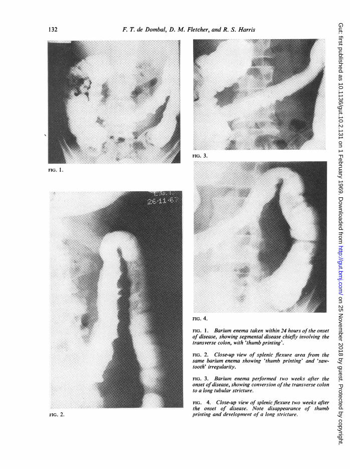
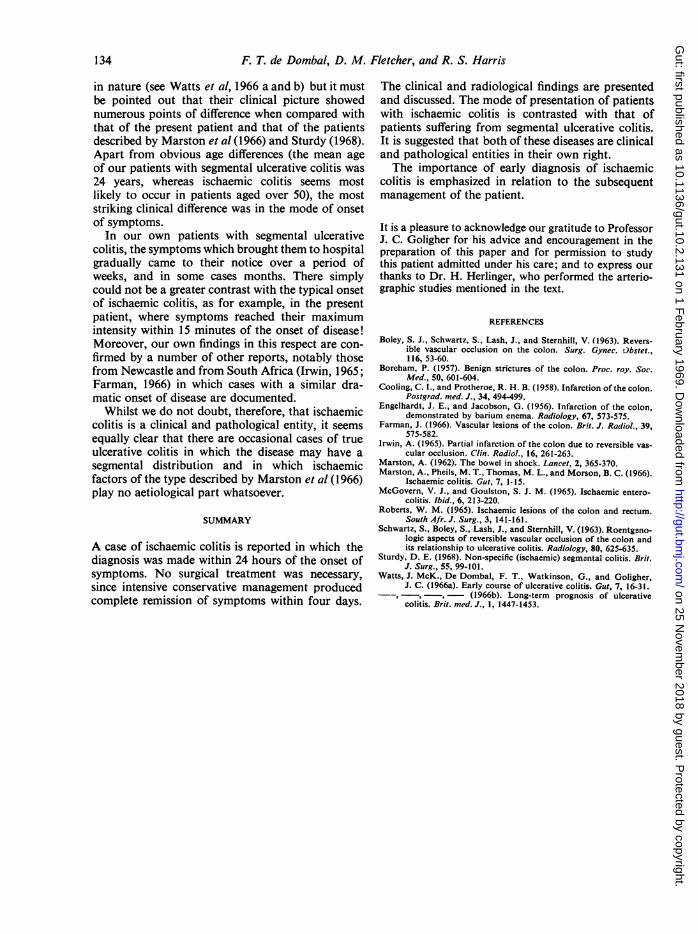
sweating but without any noticeable drop in blood pres-sure or other evidence of deterioration. Within 24 hoursof his admission, an urgent barium enema was performed(Figs. 1 and 2). This confirmed that the colon distal tothe splenic flexure was normal but showed changes highlysuggestive of ischaemic colitis in the transverse colon andat the region of the splenic flexure. Particularly wellshown on this investigation was the segmental nature ofthe lesions concerned, the 'thumb printing' and the 'sawtooth' mucosal irregularity described by Marston et al(1966).
In view of these findings an initial regime of con-servative management was instituted. The patient's oralintake was restricted to fluids, electrolytes were replacedintravenously, and large doses of penicillin were ad-ministered. On this regime the symptoms and physicalsigns rapidly settled, so that after four days the patientwas able to begin having a normal diet, the pyrexia andtachycardia had disappeared, and normal bowel actionsreturned.
Selective mesenteric arteriography carried out some10 days later failed to reveal any specific evidence oflocal arterial blockage. However, a further barium enemaexamination some 13 days after admission to hospitalrevealed a considerably changed state of affairs. The'thumb printing' and mucosal irregularity seen within48 hours of the onset of disease had rapidly resolved, tobe replaced by a tubular appearance of the bowel (Figs.3 and 4), again similar to that described by Marston andhis colleagues.The patient was allowed home after a total period in
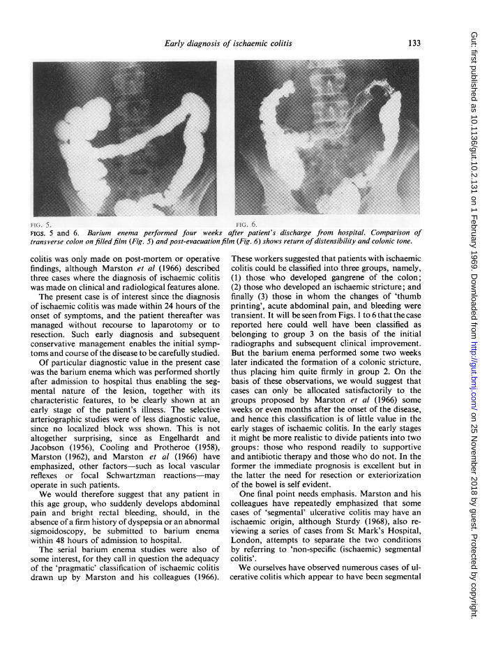
hospital of 15 days. At the time of discharge from hospitalthere were no symptoms or physical signs whatsoever, astate which has persisted to the present time. Repeatbarium enema studies performed some four weeks afterthe patient's discharge from hospital showed considerableimprovement, the most notable features being the returnof colonic distensibility and contractibility (Figs. 5 and 6).
DISCUSSION
The pathological features of ischaemic colitis, to-gether with the clinical and radiological findings inthe later stages of this disease, have been well docu-mented by Boreham (1957), Boley et al (1963),Schwartz et al (1963), McGovern and Goulston(1965), Roberts (1965), and Marston and his col-leagues (1966). However, in most of the casesdescribed by these authors the diagnosis of ischaemic
31
on 25 Novem
ber 2018 by guest. Protected by copyright.
http://gut.bmj.com
/G
ut: first published as 10.1136/gut.10.2.131 on 1 February 1969. D
ownloaded from
132 F. T. de Dombal, D. M. Fletcher, and R. S. Harris
.~~~~~~~~~~~~~~~~~~~~~~~~~M .... . ;..........
FIG. 1.
.... .......... ........ ...... ......FIG
t~~~~~~~~~~~FIG. 1. Barium enema taken within 24 hours of the onset.,.;,2e,,,,,,'S . _ ~~Of disease, showing segmental disease chiefly involving the
_ ~~~~transverse colon, with 'thumb printing'.
|i _ ~~~~~FIG.2. Close-up view of splenic flexure area from the-̂~~~~~~same barium enema showing 'thumb printing' and 'saw-
^ , _ ~~~~~~~tooth'irregularity.
[~~~~~~~FIG. 3. Barium enema performed two weeks after the[ _ ~~~~~~~~~onsetofdisease, showing conversion of the transverse colon
X- ^ _ ~~~~~~~~toa long tubular stricture.
.... FIG. 4. Close-up view of spenic.flexure two weeks afterthe onset of disease. Note disappearance of thumb
FIG. 2. printing and development of a long stricture.
on 25 Novem
ber 2018 by guest. Protected by copyright.
http://gut.bmj.com
/G
ut: first published as 10.1136/gut.10.2.131 on 1 February 1969. D
ownloaded from
Early diagnosis of ischaemic colitis
FIG. 5. FIG. 6.FIGS. 5 and 6. Barium enema performed four weeks after patient's discharge from hospital. Comparison oftransverse colon on filled film (Fig. 5) and post-evacuationfilm (Fig. 6) shows return of distensibility and colonic tone.
colitis was only made on post-mortem or operativefindings, although Marston et al (1966) describedthree cases where the diagnosis of ischaemic colitiswas made on clinical and radiological features alone.The present case is of interest since the diagnosis
of ischaemic colitis was made within 24 hours of theonset of symptoms, and the patient thereafter wasmanaged without recourse to laparotomy or toresection. Such early diagnosis and subsequentconservative management enables the initial symp-toms and course of the disease to be carefully studied.Of particular diagnostic value in the present case
was the barium enema which was performed shortlyafter admission to hospital thus enabling the seg-mental nature of the lesion, together with itscharacteristic features, to be clearly shown at anearly stage of the patient's illness. The selectivearteriographic studies were of less diagnostic value,since no localized block wss shown. This is notaltogether surprising, since as Engelhardt andJacobson (1956), Cooling and Protheroe (1958),Marston (1962), and Marston et al (1966) haveemphasized, other factors-such as local vascularreflexes or focal Schwartzman reactions-mayoperate in such patients.We would therefore suggest that any patient in
this age group, who suddenly develops abdominalpain and bright rectal bleeding, should, in theabsence of a firm history of dyspepsia or an abnormalsigmoidoscopy, be submitted to barium enemawithin 48 hours of admission to hospital.The serial barium enema studies were also of
some interest, for they call in question the adequacyof the 'pragmatic' classification of ischaemic colitisdrawn up by Marston and his colleagues (1966).
These workers suggested that patients with ischaemiccolitis could be classified into three groups, namely,(1) those who developed gangrene of the colon;(2) those who developed an ischaemic stricture; andfinally (3) those in whom the changes of 'thumbprinting', acute abdominal pain, and bleeding weretransient. It will be seen from Figs. 1 to 6 that the casereported here could well have been classified asbelonging to group 3 on the basis of the initialradiographs and subsequent clinical improvement.But the barium enema performed some two weekslater indicated the formation of a colonic stricture,thus placing him quite firmly in group 2. On thebasis of these observations, we would suggest thatcases can only be allocated satisfactorily to thegroups proposed by Marston et al (1966) someweeks or even months after the onset of the disease,and hence this classification is of little value in theearly stages of ischaemic colitis. In the early stagesit might be more realistic to divide patients into twogroups: those who respond readily to supportiveand antibiotic therapy and those who do not. In theformer the immediate prognosis is excellent but inthe latter the need for resection or exteriorizationof the bowel is self evident.One final point needs emphasis. Marston and his
colleagues have repeatedly emphasized that somecases of 'segmental' ulcerative colitis may have anischaemic origin, although Sturdy (1968), also re-viewing a series of cases from St Mark's Hospital,London, attempts to separate the two conditionsby referring to 'non-specific (ischaemic) segmentalcolitis'.We ourselves have observed numerous cases of ul-
cerative colitis which appear to have been segmental
133
on 25 Novem
ber 2018 by guest. Protected by copyright.
http://gut.bmj.com
/G
ut: first published as 10.1136/gut.10.2.131 on 1 February 1969. D
ownloaded from
134 F. T. de Dombal, D. M. Fletcher, and R. S. Harris
in nature (see Watts et al, 1966 a and b) but it mustbe pointed out that their clinical picture showednumerous points of difference when compared withthat of the present patient and that of the patientsdescribed by Marston et al (1966) and Sturdy (1968).Apart from obvious age differences (the mean ageof our patients with segmental ulcerative colitis was24 years, whereas ischaemic colitis seems mostlikely to occur in patients aged over 50), the moststriking clinical difference was in the mode of onsetof symptoms.
In our own patients with segmental ulcerativecolitis, the symptoms which brought them to hospitalgradually came to their notice over a period ofweeks, and in some cases months. There simplycould not be a greater contrast with the typical onsetof ischaemic colitis, as for example, in the presentpatient, where symptoms reached their maximumintensity within 15 minutes of the onset of disease!Moreover, our own findings in this respect are con-firmed by a number of other reports, notably thosefrom Newcastle and from South Africa (Irwin, 1965;Farman, 1966) in which cases with a similar dra-matic onset of disease are documented.
Whilst we do not doubt, therefore, that ischaemiccolitis is a clinical and pathological entity, it seemsequally clear that there are occasional cases of trueulcerative colitis in which the disease may have asegmental distribution and in which ischaemicfactors of the type described by Marston et al (1966)play no aetiological part whatsoever.
SUMMARY
A case of ischaemic colitis is reported in which thediagnosis was made within 24 hours of the onset ofsymptoms. No surgical treatment was necessary,since intensive conservative management producedcomplete remission of symptoms within four days.
The clinical and radiological findings are presentedand discussed. The mode of presentation of patientswith ischaemic colitis is contrasted with that ofpatients suffering from segmental ulcerative colitis.It is suggested that both of these diseases are clinicaland pathological entities in their own right.The importance of early diagnosis of ischaemic
colitis is emphasized in relation to the subsequentmanagement of the patient.
It is a pleasure to acknowledge our gratitude to ProfessorJ. C. Goligher for his advice and encouragement in thepreparation of this paper and for permission to studythis patient admitted under his care; and to express ourthanks to Dr. H. Herlinger, who performed the arterio-graphic studies mentioned in the text.
REFERENCES
Boley, S. J., Schwartz, S., Lash, J., and Sternhill, V. (1963). Revers-ible vascular occlusion on the colon. Surg. Gynec. Obstet.,116, 53-60.
Boreham, P. (1957). Benign strictures of the colon. Proc. roy. Soc.Med., 50, 601-604.
Cooling, C. L., and Protheroe, R. H. B. (1958). Infarction of the colon.Postgrad. med. J., 34, 494-499.
Engelhardt, J. E., and Jacobson, G. (1956). Infarction of the colon,demonstrated by barium enema. Radiology, 67, 573-575.
Farman, J. (1966). Vascular lesions of the colon. Brit. J. Radiol., 39,575-582.
Irwin, A. (1965). Partial infarction of the colon due to reversible vas-cular occlusion. Clin. Radio!., 16, 261-263.
Marston, A. (1962). The bowel in shock. Lancet, 2, 365-370.Marston, A., Pheils, M. T., Thomas, M. L., and Morson, B. C. (1966).
Ischaemic colitis. Gut, 7, 1-15.McGovern, V. J., and Goulston, S. J. M. (1965). Ischaemic entero-
colitis. Ibid., 6, 213-220.Roberts, W. M. (1965). Ischaemic lesions of the colon and rectum.
South Afr. J. Surg., 3, 141-161.Schwartz, S., Boley, S., Lash, J., and Sternhill, V. (1963). Roentgeno-
logic aspects of reversible vascular occlusion of the colon andits relationship to ulcerative colitis. Radiolcgy, 80, 625-635.
Sturdy, D. E. (1968). Non-specific (ischaemic) segmental colitis. Brit.J. Surg., 55, 99-101.
Watts, J. McK., De Dombal, F. T., Watkinson, G., and Goligher,J. C. (1966a). Early course of ulcerative colitis. Gut, 7, 16-31.-,__-,- (1966b). Long-term prognosis of ulcerativecolitis. Brit. med. J., 1, 1447-1453.
on 25 Novem
ber 2018 by guest. Protected by copyright.
http://gut.bmj.com
/G
ut: first published as 10.1136/gut.10.2.131 on 1 February 1969. D
ownloaded from