Early alterations of B cells in patients with septic shock
10
RESEARCH Open Access Early alterations of B cells in patients with septic shock Jorge Monserrat 1† , Raul de Pablo 1,2† , David Diaz-Martín 1 , Manuel Rodríguez-Zapata 1,3 , Antonio de la Hera 1 , Alfredo Prieto 1 and Melchor Alvarez-Mon 1,4* Abstract Introduction: It has recently been proposed that B lymphocytes are involved in sepsis pathogenesis. The goal of this study is to investigate potential abnormalities in a subset distribution and activation of circulating B lymphocytes in patients with septic shock. Methods: This observational prospective study was conducted in a medical-surgical ICU. All patients with septic shock were eligible for inclusion. B-cell phenotypes (CD19+CD69+, CD19+CD23+, CD19+CD5+, CD19+CD80, CD19+CD86+, CD19+CD40 and CD19+CD95+) were assessed by quantitative flow cytometry upon admission to the ICU and 3, 7, 14 and 28 d later. Results: Fifty-two patients were included. Thirty-six healthy volunteers matched for age and sex were used as controls. The patients had lymphopenia that was maintained during 28 d of follow-up. In patients with septic shock who died, the percentage of CD19+CD23+ was lower during the 7 d of follow-up than it was in survival patients. Moreover, the percentage of CD80+ and CD95+ expression on B cells was higher in patients who died than in survivors. Receiver operating characteristic curve analysis showed that a CD19+CD23+ value of 64.6% at ICU admission enabled discrimination between survivors and nonsurvivors with a sensitivity of 90.9% and a specificity of 80.0% (P = 0.0001). Conclusions: Patients with septic shock who survive and those who don’t have different patterns of abnormalities in circulating B lymphocytes. At ICU admission, a low percentage of CD23+ and a high of CD80+ and CD95+ on B cells were associated with increased mortality of patients with septic shock. Moreover, a drop in circulating B cells persisted during 28 d of ICU follow-up. Keywords: Apoptosis, B cells, Flow cytometry, Lymphocyte, Lymphocyte activation, Phenotype, Sepsis, Septic shock Introduction Several mechanisms of the innate and adaptive immune responses are involved in the pathogenesis of sepsis [1,2]. The bacteria and/or bacterial components released during infection may interact with, and induce abnormal activation of, different cell types of the immune system. The involvement of monocytes and phagocytic cells in the induction of inflammatory derangement of sepsis has been clearly established [3]. Our group and other authors have described that T lymphocytes and natural killer cells show several abnormalities in patients with septic shock [4-6]. Blymphocytes are a heterogeneous cell population with different functional and phenotypical properties [7-9]. The majority of B cells are classified as conventional B2 cells, including follicular B cells, which are characterized by high CD23 and low CD21 expression, and marginal B cells that express high amounts of CD21 [10,11]. The minority B-1 B-lymphocyte population is classified into B-1a and B-1b subsets based on cell-surface CD5 expres- sion. B-1a cells have an exclusive fetal origin and are characterized by CD5 expression (CD5+) and CD23 -/low , produce natural antibodies, IL-10 and inhibition of T cells. B-1b cells can be of adult origin, do not express * Correspondence: [email protected] † Contributed equally 1 Laboratory of Immune System Diseases and Oncology, National Biotechnology Center (CNB-CSIC) Associated Unit, Department of Medicine, University of Alcalá, Madrid, Spain Full list of author information is available at the end of the article Monserrat et al. Critical Care 2013, 17:R105 http://ccforum.com/content/17/3/R105 © 2013 Monserrat et al.; licensee BioMed Central Ltd. This is an Open Access article distributed under the terms of the Creative Commons Attribution License (http://creativecommons.org/licenses/by/2.0), which permits unrestricted use, distribution, and reproduction in any medium, provided the original work is properly cited.
Transcript of Early alterations of B cells in patients with septic shock

RESEARCH Open Access
Early alterations of B cells in patients withseptic shockJorge Monserrat1†, Raul de Pablo1,2†, David Diaz-Martín1, Manuel Rodríguez-Zapata1,3, Antonio de la Hera1,Alfredo Prieto1 and Melchor Alvarez-Mon1,4*
Abstract
Introduction: It has recently been proposed that B lymphocytes are involved in sepsis pathogenesis. The goal ofthis study is to investigate potential abnormalities in a subset distribution and activation of circulating Blymphocytes in patients with septic shock.
Methods: This observational prospective study was conducted in a medical-surgical ICU. All patients with septicshock were eligible for inclusion. B-cell phenotypes (CD19+CD69+, CD19+CD23+, CD19+CD5+, CD19+CD80,CD19+CD86+, CD19+CD40 and CD19+CD95+) were assessed by quantitative flow cytometry upon admission tothe ICU and 3, 7, 14 and 28 d later.
Results: Fifty-two patients were included. Thirty-six healthy volunteers matched for age and sex were used ascontrols. The patients had lymphopenia that was maintained during 28 d of follow-up. In patients with septicshock who died, the percentage of CD19+CD23+ was lower during the 7 d of follow-up than it was in survivalpatients. Moreover, the percentage of CD80+ and CD95+ expression on B cells was higher in patients who diedthan in survivors. Receiver operating characteristic curve analysis showed that a CD19+CD23+ value of 64.6% atICU admission enabled discrimination between survivors and nonsurvivors with a sensitivity of 90.9% and aspecificity of 80.0% (P = 0.0001).
Conclusions: Patients with septic shock who survive and those who don’t have different patterns of abnormalitiesin circulating B lymphocytes. At ICU admission, a low percentage of CD23+ and a high of CD80+ and CD95+ onB cells were associated with increased mortality of patients with septic shock. Moreover, a drop in circulatingB cells persisted during 28 d of ICU follow-up.
Keywords: Apoptosis, B cells, Flow cytometry, Lymphocyte, Lymphocyte activation, Phenotype, Sepsis, Septic shock
IntroductionSeveral mechanisms of the innate and adaptive immuneresponses are involved in the pathogenesis of sepsis[1,2]. The bacteria and/or bacterial components releasedduring infection may interact with, and induce abnormalactivation of, different cell types of the immune system.The involvement of monocytes and phagocytic cells inthe induction of inflammatory derangement of sepsishas been clearly established [3]. Our group and otherauthors have described that T lymphocytes and natural
killer cells show several abnormalities in patients withseptic shock [4-6].Blymphocytes are a heterogeneous cell population with
different functional and phenotypical properties [7-9].The majority of B cells are classified as conventional B2cells, including follicular B cells, which are characterizedby high CD23 and low CD21 expression, and marginal Bcells that express high amounts of CD21 [10,11]. Theminority B-1 B-lymphocyte population is classified intoB-1a and B-1b subsets based on cell-surface CD5 expres-sion. B-1a cells have an exclusive fetal origin and arecharacterized by CD5 expression (CD5+) and CD23-/low,produce natural antibodies, IL-10 and inhibition of Tcells. B-1b cells can be of adult origin, do not express
* Correspondence: [email protected]† Contributed equally1Laboratory of Immune System Diseases and Oncology, NationalBiotechnology Center (CNB-CSIC) Associated Unit, Department of Medicine,University of Alcalá, Madrid, SpainFull list of author information is available at the end of the article
Monserrat et al. Critical Care 2013, 17:R105http://ccforum.com/content/17/3/R105
© 2013 Monserrat et al.; licensee BioMed Central Ltd. This is an Open Access article distributed under the terms of the CreativeCommons Attribution License (http://creativecommons.org/licenses/by/2.0), which permits unrestricted use, distribution, andreproduction in any medium, provided the original work is properly cited.

CD5 (and CD23-/low) and respond to particulate antigensand polysaccharide [12].B cells play a pivotal role in both adaptive and innate
immune response [13]. During the immune responseagainst infectious agents, B lymphocytes play a relevantrole by different mechanisms, including the productionof antibodies and the presentation of microorganismantigens to T lymphocytes [14]. Furthermore, the inter-action of several bacterial products with B cells maycause their activation and cytokine secretory function[15-17].The role of B lymphocytes in the pathogenesis of sepsis
has not been established. It has been reported thatpatients who have recovered from an episode of invasivepneumococcal disease show defective B-cell activation[18]. It has been demonstrated that innate response acti-vator B (IRA-B) cells play a critical role in the response tosepsis [19,20]. It has been proposed that B cells mightcontribute to the immunosuppressive shift observed dur-ing sepsis [21].In this study, we investigated the presence of abnormal-
ities in the B-cell compartments of patients with septicshock and analyzed its relevance to the evolution of sepsisand the prognosis of these patients. In this study, we inves-tigated the number and distribution of B cells, as well astheir expression of activation/redistribution (CD69, CD23and CD5), costimulation (CD80, CD86 and CD40) andprogrammed cell death regulation (CD95) antigens in 52patients with septic shock at admission to the ICU at ourinstitution and at days 3, 7, 14 and 28 of follow-up. Sex-and age-matched healthy donors were studied in parallelas experimental normal controls.
Materials and methodsPatient eligibilityThe study was performed at the Principe de AsturiasUniversity Hospital over a period of 36 months. Thestudy was conducted according to the guidelines of the1975 Declaration of Helsinki. Approval was obtainedfrom the Hospital Universitario Príncipe de AstúriasInstitutional Ethics Committee. Written informed con-sent was obtained from each participant included in thestudy or from his or her relatives. The individualsenrolled were patients diagnosed with septic shock whohad clinical evidence of infection, defined as the presenceof a known source of infection, and who had been startedon parenteral antimicrobial therapy. Septic shock wasdefined as sepsis-induced hypotension despite adequatefluid resuscitation along with the presence of perfusionabnormalities that could include, but were not limited to,lactic acidosis, oliguria or an acute alteration in mentalstatus. Patients receiving inotropic or vasopressor agentsfor arterial hypotension were considered to be in septicshock [22]. All the patients received conventional
intensive care, and included patients were treated by phy-sicians who were not involved in this study. Patients trea-ted with hydrocortisone for refractory hypotension werewithdrawn from the study. No patient was treated withactivated protein C.The exclusion criteria were (1) subjects with immuno-
deficiency or who were being treated with any form ofimmunomodulation therapy, including low-dose corti-costeroids for septic shock; (2) autoimmune or hyper-sensitivity diseases; (3) disseminated malignancy; and (4)participation in another research study.Thirty-six age- and sex-matched healthy blood donors
were studied in parallel with the patients. They were stu-died to control for the adequacy of the cytometric techni-ques and cellular culture procedures, as well as forcharacterization of the normal range of the B-lymphocytecompartment parameters analyzed.
Blood samplesUpon patients’ admission to the ICU and after theirinformed consent had been obtained, blood samples werecollected into sterilized, silicone-coated glass tubes.Blood samples were also obtained from each includedpatient at days 3, 7, 14 and 28 of follow-up. In patientswho did not survive, the samples included in the analysiswere those obtained prior to the fatal outcome. Bloodsamples were prepared within 1 h of sample collectionfor flow cytometry.
Cell separationPeripheral blood mononuclear cells (PBMCs) werepurified from blood by Ficoll-Hypaque (LymphoprepAxis-Shield; PoC AS, Oslo, Norway) density gradientcentrifugation [23]. Cells were resuspended (106 cells/ml)in RPMI 1640 medium (BioWhittaker, Basel, Switzerland)supplemented with 10% heat-inactivated fetal bovineserum (Gibco/Invitrogen, Carlsbad, CA, USA), 25 mM 2-[4-(2-hydroxyethyl)piperazin-1-yl]ethanesulfonic acid(HEPES) and 1% penicillin-streptomycin (both fromBioWhittaker).
Flow cytometry analysisB cells were phenotypically analyzed in PBMCs by four-color flow cytometry in a FACSCalibur flow cytometerusing CellQuest 3.3 software (BD Biosciences, San Jose,CA, USA). PBMCs were incubated with combinations offluorescein isothiocyanate (FITC)-, phycoerythrin (PE)-and phycoerythrin-cyanine 5 (TC, Tricolor)-labeled mono-clonal antibodies (mAb). The mAb were CD3-PerCP,CD3-FITC, CD56-PE, CD95-FITC, CD69-FITC, CD80-PE, CD23-PE (BD Biosciences), CD5-FITC, CD19-TC(Caltag Laboratories, San Francisco, CA, USA) and CD86-FITC, CD40-PE (AbD Serotec, Kidlington, UK). For anadequate experimental staining control, the appropriate
Monserrat et al. Critical Care 2013, 17:R105http://ccforum.com/content/17/3/R105
Page 2 of 10

irrelevant anti-mouse isotype controls IgG1-FITC, PE, TCor IgG2a FITC, PE and TC (Caltag Laboratories) wereused.
Assessment of absolute number of lymphocytesThe absolute numbers of B-lymphocyte subsets were cal-culated according to standard flow cytometry criteria forlymphocyte subset identification. First, we calculated thepercentage of cells expressing CD19 in the total lympho-cytes gate defined by forward and side scatter in PBMCs.The absolute number of circulatory B lymphocytes wascalculated by determining the percentage of CD19+ cellsin peripheral blood lymphocytes multiplied by the totalnumber of lymphocytes per microliter measured using aCoulter LH instrument (Beckman Coulter, Fullerton,CA, USA). Next, we obtained the absolute number ofCD19+ (CD23+, CD69+, CD5+, CD80+, CD86+, CD40+or CD95+) by multiplying the total number of B lympho-cytes previously calculated by the percentage of positivecells for each one of these antigens in CD19+ B cells. Allabsolute numbers are expressed as cells per milliliter.
Statistical analysesAll statistical tests were performed using SPSS for Win-dows version 15.0 software (SPSS, Chicago, IL, USA). Dataare expressed as means ± SEM. Because most variablesdid not always fulfill the normality hypothesis, differencesbetween groups were analyzed using the Mann-WhitneyU-test for nonparametric data and analysis of variance fol-lowed by a Wilcoxon signed-rank test were used forwithin-group analyses. The reliability of the use of differ-ent phenotype markers concentrations or of main clinicalvariables to predict death due to septic shock was calcu-lated by plotting receiver operating characteristic (ROC)curves. The level of significance was set at P < 0.05.
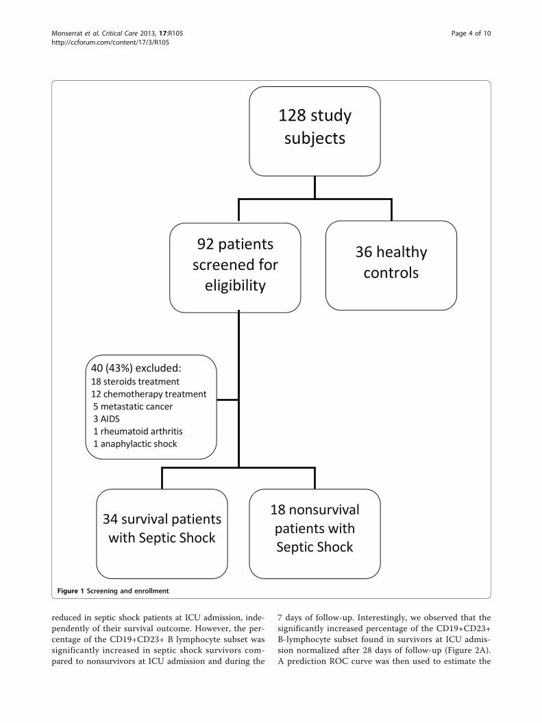
ResultsCharacteristics of patients with septic shockDuring the 36-month study period, a total of 92 patientswith septic shock treated at our institution were identi-fied (Figure 1). All patients were treated with vasopres-sors. Patients treated with hydrocortisone for refractoryhypotension were withdrawn from the study. Forty ofthese patients were excluded for the following reasons:3 patients had AIDS, 18 patients were on glucocorticoidtherapy, 12 patients were on chemotherapy, 5 patientshad metastatic cancer, 1 patient had rheumatoid arthritisand 1 patient was excluded due to anaphylactic shock.The mean age of the healthy controls was 62.0 ± 3.4 yr vs61.2 ± 3.2 yr in patients with septic shock (P > 0.05), andthe gender distribution was similar: 24 males (66.6%) and13 females in the healthy control group vs 36 males(69.2%) and 16 females in the group of patients with sep-tic shock (P > 0.05).
Table 1 provides demographic data for the 52 patientsultimately included in the study. Twenty-two patients(42.3%) had intraabdominal infections, 16 (30.7%) hadpneumonia, 4 (7.4%) had bacteremia of unknown origin,4 (7.4%) had pyelonephritis, 3 (5.7%) had soft-tissue andskin infections, 2 (3.8%) had surgical site infections and 1(1.9%) had mediastinitis. The diagnosis of infection wasmade on clinical grounds in 24 patients (46.1%), and posi-tive cultures were obtained from normally sterile sites in 28patients (53.9%). Gram-positive cocci were isolated in 11(21.1%) of these patients, Gram-negative organisms wereisolated in 10 patients (19.2%) and polymicrobial flora wereisolated in the remaining 7 patients (13.4%). The percentageof the 52 septic shock patients who had positive blood cul-ture was 30.2%. No patient had fungal sepsis or septicshock due to viruses. Bivariate analysis failed to demon-strate a correlation between the sources of infection, thepathogens isolated and mortality. There were no differencesin the lymphocyte profiles of patients who had Gram-posi-tive bacterial sepsis versus Gram-negative bacterial sepsis.
Severe lymphopenia, including B-lymphocyte population,in septic shockUpon ICU admission, total blood lymphocyte cell countwas significantly diminished in patients with septic shockcompared with healthy controls (1,144 ± 67 cells/μl vs2,095 ± 67 cells/μl, respectively; P < 0.05). This lympho-penia was maintained independently of survival outcomeduring 28 d of follow-up in patients with septic shock.The absolute number of CD19+ B cells was also signifi-
cantly lower in patients with septic shock than in normaldonors upon ICU admission (208 ± 45 cells/μl vs 238 ±13 cells/μl, respectively; P < 0.05). The numbers of thesecirculating lymphocyte populations remained signifi-cantly decreased during the 28 days of follow-up (183 ±45 cells/μl at day 3 and 116 ± 38 cells/μl at day 7, 175 ±54 cells/μl at day 14 and 120 ± 32 cells/μl at day 28; P <0.05). At the times analyzed (at ICU admission and atdays 3 and 7 of follow-up), because no additional patientdied between days 7 and 28, there were no differencesbetween survivors and nonsurvivors regarding the num-bers of the circulating CD19+ B lymphocytes.
Redistribution of B-lymphocyte subsets in septic shockpatientsNext, we investigated the subset distribution and activa-tion of circulating CD19+ B lymphocytes by means of ana-lysis of their expression of CD23+, CD69+ and CD5+antigens in patients with septic shock and in healthy con-trols. CD23 is expressed mainly by activated regulatory Bcells. CD69 is also expressed by early activated B cells.CD5+CD19+ represents the B1a subset of B cells.We observed that the circulating numbers of the
CD19+CD23+ B-lymphocyte subsets were significantly
Monserrat et al. Critical Care 2013, 17:R105http://ccforum.com/content/17/3/R105
Page 3 of 10
reduced in septic shock patients at ICU admission, inde-pendently of their survival outcome. However, the per-centage of the CD19+CD23+ B lymphocyte subset wassignificantly increased in septic shock survivors com-pared to nonsurvivors at ICU admission and during the
7 days of follow-up. Interestingly, we observed that thesignificantly increased percentage of the CD19+CD23+B-lymphocyte subset found in survivors at ICU admis-sion normalized after 28 days of follow-up (Figure 2A).A prediction ROC curve was then used to estimate the
Figure 1 Screening and enrollment.
Monserrat et al. Critical Care 2013, 17:R105http://ccforum.com/content/17/3/R105
Page 4 of 10

value of the percentage of CD19+CD23+ B cells to pre-dict death in patients with septic shock at admission. Asshown in Figure 3, for the percentage of CD19+CD23+B cells, a cut-off value of 64% showed sensitivity of90.9% (95% confidence interval (CI) = 75.6% to 98.0%)and specificity of 80.0% (95% CI = 61.4% to 92.0%) forpredicting the risk of death, with a positive predictivevalue of 89.4% and a negative predictive value of 82.7%.The area under the ROC curve was 0.818 ± 0.055 (95%CI = 0.701 to 0.904; P = 0.0001). Next, we selectedAPACHE II score for the comparative analysis of theoutcome because it was the best prognostic clinicalscore in our series. When we analyzed the ROC curveof the Acute Physiology and Chronic Health EvaluationII (APACHE II) score for predicting death in patientswith septic shock at admission, we found the area underthe ROC curve to be 0.721 ± 0.83 (95% CI = 0.559 to
Table 1 Demographic data of the study patients withseptic shock.
Parameters Survivors Nonsurvivors P
Number of patients (%) 34 (65.4%) 18 (34.6%)
Mean age (yr) 61.0 ± 3.2 64.5 ± 4.1 0.57
Men/women (n) 21/13 15/3 0.11
Medical/surgical patients 13/21 11/7 0.12
Mean APACHE II score 21.6 ± 1.3 29.2 ± 1.5 0.001
Mean MODS score 6.6 ± 0.5 9.5 ± 0.9 0.014
Mean Δ-MODS 2.2 ± 0.3 3.5 ± 0.7 0.169
Mean SOFA score 7.9 ± 0.6 10.6 ± 0.8 0.021
Mean Δ-SOFA 1.7 ± 0.4 2.9 ± 0.9 0.34
Categorical variables are expressed as number of patients, and continuousvariables are expressed as means ± SEM. APACHE II, Acute Physiology andChronic Health Evaluation II [39]; SOFA, Sepsis-Related Organ FailureAssessment [40]; MODS, Multiple Organ Dysfunction Score [41]; Δ-MODS is thedifference between the maximum and initial MODS scores [42], and Δ-SOFA isthe difference between the maximum and initial SOFA scores [43].
TIME (days)0 3 7 14 28
CD
19+C
D23
+ (%
of a
ll C
D19
+)
0
20
40
60
80
100
**
††
A*
**
†
TIME (days)0 3 7 14 28
CD
19+C
D69
+ (%
of a
ll C
D19
+)
0
5
10
15
20
*
**
B
***
*
*
TIME (days)0 3 7 14 28
CD
19+C
D5+
(% o
f all
CD
19+)
0
20
40
60
80
C
TIME (days)0 3 7 14 28
CD
19+C
D95
+ (%
of a
ll C
D19
+)
0
20
40
60
80
**
†
D
TIME (days)0 3 7 14 28
CD
19+C
D80
+ (%
of a
ll C
D19
+)
0
10
20
30
40
50
60
*†
E
Figure 2 Time course of the percentages of CD19+ lymphocytes that express CD23+ (A), CD69+ (B), CD5+ (C), CD95+ (D) and CD80+(E) antigens in patients with septic shock during their stay in the ICU. Data presented are for survivors (black triangles) and nonsurvivors(white triangles). The dotted line represents the mean value recorded in the healthy controls (n = 36). At ICU admission, study patients were 34survivors and 18 nonsurvivors; at day 3, 34 survivors and 13 nonsurvivors; at day 7, 34 survivors and 11 nonsurvivors; at day 14 and at day 28, 34survival patients were studied. All values are expressed as means ± SEM. *P < 0.05 for survivors or nonsurvivors vs healthy controls; †P < 0.05 forsurvivors vs nonsurvivors.
Monserrat et al. Critical Care 2013, 17:R105http://ccforum.com/content/17/3/R105
Page 5 of 10

0.883) and a cut-off value of 23 showed sensitivity of76.9% and specificity of 60.7% (P = 0.024).In contrast to the observed diminution of the absolute
number of total circulating CD19+ B lymphocytes andCD19+CD23+ B cells, we found that the CD19+CD69+B-cell subset count was normal in both survival andnonsurvival septic shock patients at ICU admission.Furthermore, the percentage of CD19+CD69+ PBMCswas elevated in both groups of septic shock patients at
ICU admission and during the 28 days of follow-up(Figure 2B).The number of circulating CD5+CD19+ B lymphocytes
was significantly reduced in patients with septic shock (atICU admission, 46 ± 11 cells/μl; at day 3, 46 ± 21 cells/μl;at day 7, 31 ± 14 cells/μl; at day 14, 50 ± 10 cells/μl; andat day 28, 41 ± 5 cells/μl in septic shock patients and63 ± 5 cells/μl in healthy controls). There were no differ-ences in the percentage of circulating CD5+CD19+ cells
A B C
CD19+CD23+
0 20 40 60 80 100
100
80
60
40
20
0
100-Specificity
Sensitivity
CD19+CD80+
0 20 40 60 80 100
100
80
60
40
20
0
100-Specificity
Sensitivity
CD19+CD95+
0 20 40 60 80 100
100
80
60
40
20
0
100-Specificity
Sensitivity
Figure 3 Receiver operating characteristic (ROC) curve analysis of percentages of CD19+CD23+, CD19+CD80+ and CD19+CD95+ forpredicting the risk of death at ICU admission. For CD19+CD23+, a cut-off value of 64% showed sensitivity of 90.9% (95% confidence interval(CI) = 75.6% to 98.0%) and specificity of 80.0% (95% CI = 61.4% to 92.0%) for predicting the risk of death, with a positive predictive value of89.4%, a negative predictive value of 82.7% and area under the ROC curve 0.818 ± 0.055 (95% CI = 0.701 to 0.904; P = 0.0001). For CD19+CD80+and CD19+CD95+, respectively, cut-off values of 20% and 17% showed sensitivity of 81.8% (95% CI = 64.5% to 92.8%) and 81.9% (95% CI =64.2% to 93.0%) and specificity of 70% (95% CI = 50.6% to 85.2%) and 75% (95% CI = 53.3% to 90.2%) for predicting the risk of death on day 28,with the area under the ROC curve 0.751 ± 0.061 (95% CI = 0.630 to 0.854) and 0.795 ± 0.058 (95% CI = 0.668 to 0.891) (P = 0.0001 for both).
Figure 4 Two representative CD19+CD23+ flow cytometry samples at basal time.
Monserrat et al. Critical Care 2013, 17:R105http://ccforum.com/content/17/3/R105
Page 6 of 10

between both groups of septic patients and healthydonors at baseline and during the follow-up (Figure 2C).We also investigated the expression of apoptotic sus-
ceptibility CD95 antigen on the surface of B lympho-cytes from patients and healthy donors. The absolutenumber of circulating CD19+CD95+ B cells found inseptic shock patients at ICU admission was similar tothat of healthy donors (Table 2). However, the percen-tage of CD19+CD95+ B cells was significantly higher innonsurvivors than in survivors at ICU admission andnormalized after 7 days of follow-up (Figure 2D).We also studied the expression of the CD80, CD86 and
CD40 on B lymphocytes from patients and healthy donors.CD80, CD86 and CD40 are membrane molecules that playa critical role in the stimulation of T lymphocytes by Blymphocytes acting as antigen-presenting cells. As shownin Table 2, the absolute number of circulating CD19+ Bcells expressing CD80+ or CD86+ was normal in septicshock patients at ICU admission. When we analyzed thebehavior of survivors and nonsurvivors, we found that thepercentage of CD19+CD80+ B cells was significantlyincreased in nonsurvival septic patients with respect tosurvivors at ICU admission and healthy controls (P <0.05). A normalization of this percentage was observedafter 3 d of follow-up (Figure 2E).A significant reduction of circulating CD19+CD40+ B
cells was found in septic shock patients at ICU admissioncompared to healthy donors (Table 2). There were nosignificant differences between the percentages of CD19+CD40+ B cells found in survivors and nonsurvivors.There were no significant differences in the number ofcirculating CD19+ B lymphocytes and in its cell subset
distribution during the 28 d of the study (Figure 2 anddata not shown).
DiscussionIn this paper, we report that septic shock patients have asevere retraction of circulating B lymphocytes. This lym-phopenia affects the B-cell subsets heterogeneously, withmarked reduction of CD19+CD23+ and CD19+CD5+ Bcells but normal numbers of CD19+CD69+ B cells.Furthermore, a different distribution of B cells subsets isfound in survivor and nonsurvivor septic shock patients.The percentage of CD19+CD23+ B lymphocytes appearsto be a biomarker for the prognosis of outcome of septicshock patients. These data support a role for the B-cellcompartment in septic shock patients and are in agree-ment with those published in previous studies [24-26].It is established that septic shock is associated with a
severe exhaustion and depletion of T lymphocytes [27].Our data support similar behavior in the B-lymphocytecompartment. We have found that the reduction of circu-lating B cells affects the different B-cell subsets heteroge-neously in septic shock patients and that those differentpatterns of involvement are observed in survivor and non-survivors. CD23 is a low-affinity receptor for IgE locatedat the surface of B cells [28]. CD23 is involved in differentregulatory functions, such as enhancing antigen presenta-tion, improving cell differentiation and growth and regu-lating IgE synthesis [28]. Some authors have reported thatCD23 is expressed on activated B cells, whereas othershave suggested that peripheral blood CD23 B cells resem-ble classic memory cells [28]. Our data presented hereinshow that circulating CD19+CD23+ B lymphocytes are
Table 2 Peripheral blood cell count of B-lymphocyte subsets and their percentage in the compartment of circulatingB lymphocytes in patients with septic shock and in healthy controls at ICU admissiona
Lymphocyte blood cell count (cells/μl) Subset Healthy controls Septic shock patients
CD19+ 238.5 ± 13.2 208.1 ± 45.4*
CD19+CD23+ 148.3 ± 11.2 102.5 ± 17.8*
CD19+CD69+ 5.4 ± 1.0 7.1 ± 2.5
CD19+CD5+ 63.5 ± 5.2 44.5 ± 7.8*
CD19+CD80+ 73.6 ± 6.5 60.5 ± 15.8
CD19+CD86+ 35.9 ± 3.4 47.6 ± 10.2
CD19+CD40+ 223.5 ± 15.5 130.7 ± 21.6*
CD19+CD95+ 37.8 ± 2.7 30.3 ± 4.7
Percentage of CD19+ (%) Subset Healthy controls Septic shock patients
CD19+CD23+ 57.4 ± 1.7 62.7 ± 4.6
CD19+CD69+ 2.6 ± 0.5 9.0 ± 2.1*
CD19+CD5+ 25.3 ± 1.5 24.6 ± 3.1
CD19+CD80+ 29.6 ± 1.7 32.4 ± 4.4
CD19+CD86+ 16.1 ± 1.2 36.7 ± 7.2*
CD19+CD40+ 94.6 ± 1.2 95.7 ± 1.5
CD19+CD95+ 16.1 ± 1.1 25.7 ± 4.7*aData are expressed as mean ± SE mean. *P < 0.05 between patients with septic shock and healthy controls.
Monserrat et al. Critical Care 2013, 17:R105http://ccforum.com/content/17/3/R105
Page 7 of 10
clearly decreased in septic shock patients because theirpercentage in the whole circulating B cell compartment isdifferent in survivors and nonsurvivors. We have foundthat higher percentages of circulating CD19+CD23+ areassociated with better clinical outcomes of patients withseptic shock. Interestingly, the number of CD19+CD69+early activated B cells in septic shock patients is similar tothat found in healthy donors and is not related to the clin-ical prognosis of the patient.Taking the behavior of both CD19+CD23+ and CD19+
CD69+ B lymphocytes together, it can be speculated thatthe depletion of B cells in septic shock patients is an eventthat preferentially occurs after the initiation of theirin vivo activation. The intensity of this event might corre-late with the observed reduction of activated B cells and isassociated with the clinical outcome of the patient. More-over, we have compared this finding with the APACHE IIscores, because APACHE II score was the best prognosticclinical variable that we analyzed. When we analyzed theROC curve of APACHE II score for predicting death inpatients with septic shock at admission, we found that B-lymphocyte data are significantly more accurate than theAPACHE II score for predicting death.In addition to the observed changes in the distribution
of the cell subsets of the circulating B-lymphocyte com-partment, we investigated the expression of the function-ally critical antigens CD80 and CD86 on these B cells. Inmurine studies of sepsis, an important role for CD80 andCD86 antigens in the response to sepsis has been estab-lished [29,30]. Our data presented herein show a higherpercentage of CD86 expression in circulating CD19+ Bcells in patients with septic shock than in healthy controls.Furthermore, at ICU admission, nonsurvivors showedmore elevated percentages of CD19+CD80+ B cells thanfound in survivors. These findings in B lymphocytes ofpatients with septic shock are consistent with theincreased expression of CD86 and decreased expression ofCD80 in dendritic cells found in human sepsis [31].Apoptosis is the process of programmed cell death that
occurs to limit damage of surrounding tissue. It is criticalfor the survival of many cells, including lymphocytes.Deregulated apoptotic immune cell death has been pro-posed to play a major role in immune dysfunction andmortality during sepsis [32,33]. Immunohistological stu-dies of different tissues have demonstrated increasedapoptosis of cells of the innate and adaptive immune sys-tem in sepsis [25,34,35]. In this work, we show that thetotal number of circulating B lymphocytes was low inpatients with septic shock at ICU admission and duringthe following 28 days. We also observe that circulating Bcells of septic shock patients showed increased expressionof CD95 antigen. Similar findings have been described incirculating T cells [36]. In support of the concept thatlymphocyte apoptosis is detrimental to host survival, a
number of studies have shown an inverse correlationbetween lymphocyte count and survival [34,36]. In agree-ment with this, we found a higher percentage of CD95expression on B cells from nonsurvivors than from survi-vor patients or healthy controls. This finding suggests thatthe increased expression of CD95 on B cells from septicshock patients might be involved in the mechanism of theobserved reduction of circulating B cells in these patients.Several limitations of our study should be noted. The per-
ipheral blood may not represent the situation in all lym-phoid compartments of the body, and these findings dosuggest that not all cell populations may play equal roles indriving inflammatory and anti-inflammatory responses inseptic shock [21]. All blood samples were collected uponICU admission within 12 hours after inclusion criteria weremet, but the length of time that the patients were in shockin the emergency room before admission or in the operat-ing room is a source of inaccuracy. Recently, early altera-tions of the innate and adaptive immune status accordingto the type of underlying infection in sepsis have beenreported [37]. In addition, other factors, such as host geneticpolymorphisms or the characteristics of the pathogen, mayalso have introduced variability. The average of age of ourpopulation was 61 years. It is known that the overall num-ber of B cells seems to moderately decline with age, but B-lymphocyte subset studies are frequently controversial [38].This is the reason why our control group was age-matched.CD23 is the low-affinity immunoglobulin E (IgE) receptorand binds both IgE and CD21 and, through these interac-tions, regulates the synthesis of IgE, the antibody isotypethat mediates the allergic response. The expression of CD23on B cells is higher in persons with asthma or atopy and inpatients with disorders characterized by chronic inflamma-tion [28]. We excluded patients who were undergoing glu-cocorticoid therapy or were in anaphylactic shock, butpatients with a personal history of chronic asthma withouthypersensitivity disease were included.
ConclusionsSeptic shock is associated with a severe abnormality of cir-culating B lymphocytes. At ICU admission, the expressionof CD23+, CD95+ or CD80+ on B cells was significantlyassociated with increased 28-day mortality. These resultshighlight the potential importance of B lymphocytes inseptic shock.
Key messages• Sepsis-induced immune dysfunction may contribute tomortality to a great degree. It is increasingly clear thatB-cell function beyond the production of immunoglobu-lins. However, little is known about how B cells affectinnate immunity during bacterial sepsis.• We found that patients with septic shock had B-cell
lymphopenia that was maintained during 28 days of
Monserrat et al. Critical Care 2013, 17:R105http://ccforum.com/content/17/3/R105
Page 8 of 10

follow-up. The increased expression of CD95 on B cellssuggests that apoptosis susceptibility is involved in thereduction of circulating B cells in these patients.• B-cell lymphopenia affects the B-cell subsets hetero-
geneously, with marked reduction of CD19+CD23+ Bcells (activated regulatory B cells) and CD19+CD5+ Bcells (natural responder B-1a cells), but with normalnumbers of CD19+CD69+ early activated B cells.• At ICU admission, a higher percentage (64%) of CD19
+CD23+, a marker of activation and regulation, appears tobe a reliable biomarker of good outcome for patients withseptic shock, whereas the percentages of CD80+ (a T-cellcostimulation marker) and CD95+ (a marker of apoptosissusceptibility) on B cells were significantly lower in survi-vors than in nonsurvivors.
AbbreviationsAIDS: acquired immunodeficiency syndrome; APACHE II: Acute Physiologyand Chronic Health Evaluation II; CD: cluster of differentiation; CI: confidenceinterval; FITC: fluorescein isothiocyanate; ICU: intensive care unit; IL-10:interleukin 10; IRA-B cells: innate response activator B cells; mAb: monoclonalantibodies; PBMC: peripheral blood mononuclear cell; PE: phycoerythrin;ROC: receiver-operating characteristic; SEM: standard error of the mean; TC:phycoerythrin-cyanine 5 tricolor.
Competing interestsThe authors have no direct or otherwise commercial association that mightlead to a conflict of interest.
Authors’ contributionsRP, JM, AP and MAM made substantial contributions to the conception anddesign of the study and the analysis and interpretation of data, and theywere involved in drafting and revising the manuscript. RP, JM and DDMmade substantial contributions to the acquisition, analysis and interpretationof data. MRZ and AH were involved in analysis and interpretation of dataand in drafting and revising the manuscript critically for importantintellectual content. All authors read and approved the final manuscript.
AcknowledgementsThe authors thank the valuable help of the nursing and medical staff of ourICU, the Department of Medicine and the University of Alcalá. This work waspartially funded by grants from Fondo de Investigación de la SeguridadSocial, Ministerio de Economia y Competitividad (MEC) (Spain), Consejeria deEducación, Comunidad de Madrid, MITIC-CM (S-2010/BMD-2502) andInstituto de Salud Carlos III, MEC (PI051871, CIBERehd).
Authors’ details1Laboratory of Immune System Diseases and Oncology, NationalBiotechnology Center (CNB-CSIC) Associated Unit, Department of Medicine,University of Alcalá, Madrid, Spain. 2Intensive Care Unit, Hospital UniversitarioPríncipe de Asturias, Alcalá de Henares, Madrid, Spain. 3Internal MedicineService, Hospital Universitario de Guadalajara, Guadalajara, Spain. 4ImmuneSystem Diseases and Oncology Service, Hospital Universitario Príncipe deAsturias, Alcalá de Henares, Madrid, Spain.
Received: 11 December 2012 Revised: 19 May 2013Accepted: 30 May 2013 Published: 30 May 2013
References1. Hotchkiss RS, Karl IE: The pathophysiology and treatment of sepsis. N Engl
J Med 2003, 348:138-150.2. Russell JA: Management of sepsis. N Engl J Med 2006, 355:1699-1713.3. Döcke WD, Randow F, Syrbe U, Krausch D, Asadullah K, Reinke P, Volk HD,
Kox W: Monocyte deactivation in septic patients: restoration by IFN-γtreatment. Nat Med 1997, 3:678-681.
4. de Pablo R, Monserrat J, Torrijos C, Martín M, Prieto A, Alvarez-Mon M: Thepredictive role of early activation of natural killer cells in septic shock.Crit Care 2012, 16:413.
5. Kasten KR, Tschöp J, Adediran SG, Hildeman DA, Caldwell CC: T cells arepotent early mediators of the host response to sepsis. Shock 2010,34:327-336.
6. Monserrat J, de Pablo R, Reyes E, Díaz D, Barcenilla H, Zapata MR, De laHera A, Prieto A, Alvarez-Mon M: Clinical relevance of the severeabnormalities of the T cell compartment in septic shock patients. CritCare 2009, 13:R26.
7. Frasca D, Blomberg BB: Aging affects human B cell responses. J ClinImmunol 2011, 31:430-435.
8. Hardy RR, Hayakawa K: B cell development pathways. Annu Rev Immunol2001, 19:595-621.
9. Sanz I, Wei C, Lee FE, Anolik J: Phenotypic and functional heterogeneityof human memory B cells. Semin Immunol 2008, 20:67-82.
10. Weller S, Braun MC, Tan BK, Rosenwald A, Cordier C, Conley ME, Plebani A,Kumararatne DS, Bonnet D, Tournilhac O, Tchernia G, Steiniger B,Staudt LM, Casanova JL, Reynaud CA, Weill JC: Human blood IgM“memory” B cells are circulating splenic marginal zone B cells harboringa prediversified immunoglobulin repertoire. Blood 2004, 104:3647-3654.
11. Zouali M, Richard Y: Marginal zone B-cells, a gatekeeper of innateimmunity. Front Immunol 2011, 2:63.
12. Haas KM, Poe JC, Steeber DA, Tedder TF: B-1a and B-1b cells exhibit distinctdevelopmental requirements and have unique functional roles in innateand adaptive immunity to S. pneumoniae. Immunity 2005, 23:7-18.
13. Kelly-Scumpia KM, Scumpia PO, Weinstein JS, Delano MJ, Cuenca AG,Nacionales DC, Wynn JL, Lee PY, Kumagai Y, Efron PA, Akira S, Wasserfall C,Atkinson MA, Moldawer LL: B cells enhance early innate immuneresponses during bacterial sepsis. J Exp Med 2011, 208:1673-1682.
14. Vaughan AT, Roghanian A, Cragg MS: B cells: masters of theimmunoverse. Int J Biochem Cell Biol 2011, 43:280-285.
15. Booth J, Wilson H, Jimbo S, Mutwiri G: Modulation of B cell responses byToll-like receptors. Cell Tissue Res 2011, 343:131-140.
16. Mauri C, Bosma A: Immune regulatory function of B cells. Annu RevImmunol 2012, 30:221-241.
17. Rawlings DJ, Schwartz MA, Jackson SW, Meyer-Bahlburg A: Integration of Bcell responses through Toll-like receptors and antigen receptors. Nat RevImmunol 2012, 12:282-294.
18. Darton TC, Wing JB, Lees A, Heath AW, Read RC: Adult survivors ofinvasive pneumococcal disease exhibit defective B cell function. ClinInfect Dis 2011, 52:1133-1136.
19. Rauch PJ, Chudnovskiy A, Robbins CS, Weber GF, Etzrodt M, Hilgendorf I,Tiglao E, Figueiredo JL, Iwamoto Y, Theurl I, Gorbatov R, Waring MT,Chicoine AT, Mouded M, Pittet MJ, Nahrendorf M, Weissleder R, Swirski FK:Innate response activator B cells protect against microbial sepsis. Science2012, 335:597-601.
20. Robbins CS, Swirski FK: Newly discovered innate response activator Bcells: crucial responders against microbial sepsis. Expert Rev Clin Immunol2012, 8:405-407.
21. Shubin NJ, Monaghan SF, Ayala A: Anti-inflammatory mechanisms ofsepsis. Contrib Microbiol 2011, 17:108-124.
22. Bone RC, Balk RA, Cerra FB, Dellinger RP, Fein AM, Knaus WA, Schein RM,Sibbald WJ: Definitions for sepsis and organ failure and guidelines forthe use of innovative therapies in sepsis. The ACCP/SCCM ConsensusConference Committee. American College of Chest Physicians/Society ofCritical Care Medicine. Chest 1992, 101:1644-1655.
23. Böyum AJ: Isolation of mononuclear cells and granulocytes from humanblood. Scand J Clin Lab Invest 1968, 21:77-89.
24. Venet F, Davin F, Guignant C, Larue A, Cazalis MA, Darbon R, Allombert C,Mougin B, Malcus C, Poitevin-Later F, Lepape A, Monneret G: Earlyassessment of leukocyte alterations at diagnosis of septic shock. Shock2010, 34:358-363.
25. Hotchkiss RS, Tinsley KW, Swanson PE, Schmieg RE Jr, Hui JJ, Chang KC,Osborne DF, Freeman BD, Cobb JP, Buchman TG, Karl IE: Sepsis-inducedapoptosis causes progressive profound depletion of B and CD4+ Tlymphocytes in humans. J Immunol 2001, 166:6952-6963.
26. Jiménez-Ibáñez EO, Castillejos-López M, Hernández A, Gorocica P, Alvarado-Vásquez N: High mortality associated with hyperglycemia, neutrophilia,and lymphopenia in critically ill patients. Tohoku J Exp Med 2012,226:213-220.
Monserrat et al. Critical Care 2013, 17:R105http://ccforum.com/content/17/3/R105
Page 9 of 10

27. Boomer JS, Shuherk-Shaffer J, Hotchkiss RS, Green JM: A prospectiveanalysis of lymphocyte phenotype and function over the course ofacute sepsis. Crit Care 2012, 16:R112.
28. Rosenwasser LJ, Meng J: Anti-CD23. Clin Rev Allergy Immunol 2005,29:61-72.
29. Nolan A, Kobayashi H, Naveed B, Kelly A, Hoshino Y, Hoshino S, Karulf MR,Rom WN, Weiden MD, Gold JA: Differential role for CD80 and CD86 inthe regulation of the innate immune response in murine polymicrobialsepsis. PLoS One 2009, 4:e6600.
30. Nolan A, Weiden M, Kelly A, Hoshino Y, Hoshino S, Mehta N, Gold JA: CD40and CD80/86 act synergistically to regulate inflammation and mortalityin polymicrobial sepsis. Am J Respir Crit Care Med 2008, 177:301-308.
31. Flohé SB, Agrawal H, Schmitz D, Gertz M, Flohé S, Schade FU: Dendriticcells during polymicrobial sepsis rapidly mature but fail to initiate aprotective Th1-type immune response. J Leukoc Biol 2006, 79:473-481.
32. van der Poll T, van Zoelen MA, Wiersinga WJ: Regulation of pro- and anti-inflammatory host responses. Contrib Microbiol 2011, 17:125-136.
33. Hotchkiss RS, Nicholson DW: Apoptosis and caspases regulate death andinflammation in sepsis. Nat Rev Immunol 2006, 6:813-822.
34. Hotchkiss RS, Swanson PE, Freeman BD, Tinsley KW, Cobb JP,Matuschak GM, Buchman TG, Karl IE: Apoptotic cell death in patients withsepsis, shock, and multiple organ dysfunction. Crit Care Med 1999,27:1230-1251.
35. Hotchkiss RS, Tinsley KW, Swanson PE, Grayson MH, Osborne DF,Wagner TH, Cobb JP, Coopersmith C, Karl IE: Depletion of dendritic cells,but not macrophages, in patients with sepsis. J Immunol 2002,168:2493-2500.
36. Roth G, Moser B, Krenn C, Brunner M, Haisjackl M, Almer G, Gerlitz S,Wolner E, Boltz-Nitulescu G, Ankersmit HJ: Susceptibility to programmedcell death in T-lymphocytes from septic patients: a mechanism forlymphopenia and Th2 predominance. Biochem Biophys Res Commun 2003,308:840-846.
37. Gogos C, Kotsaki A, Pelekanou A, Giannikopoulos G, Vaki I, Maravitsa P,Adamis S, Alexiou Z, Andrianopoulos G, Antonopoulou A, Athanassia S,Baziaka F, Charalambous A, Christodoulou S, Dimopoulou I, Floros I,Giannitsioti E, Gkanas P, Ioakeimidou A, Kanellakopoulou K, Karabela N,Karagianni V, Katsarolis I, Kontopithari G, Kopterides P, Koutelidakis I,Koutoukas P, Kranidioti H, Lignos M, Louis K, et al: Early alterations of theinnate and adaptive immune statuses in sepsis according to the type ofunderlying infection. Crit Care 2010, 14:R96.
38. Ongradi J, Kovesdi V: Numerical alterations of ageing B lymphocytesubsets. Acta Physiol Hung 2011, 98:99-104.
39. Knaus WA, Draper EA, Wagner DP, Zimmerman JE: APACHE II: a severity ofdisease classification system. Crit Care Med 1985, 13:818-829.
40. Vincent JL, de Mendonça A, Cantraine F, Moreno R, Takala J, Suter PM,Sprung CL, Colardyn F, Blecher S, Working Group on “Sepsis-RelatedProblems” of the European Society of Intensive Care Medicine: Use of theSOFA score to assess the incidence of organ dysfunction/failure inintensive care units: results of a multicenter, prospective study. Crit CareMed 1998, 26:1793-1800.
41. Marshall JC, Cook DJ, Christou NV, Bernard GR, Sprung CL, Sibbald WJ:Multiple organ dysfunction score: a reliable descriptor of a complexclinical outcome. Crit Care Med 1995, 23:1638-1652.
42. Jacobs S, Zuleika M, Mphansa T: The Multiple Organ Dysfunction Score asa descriptor of patient outcome in septic shock compared with twoother scoring systems. Crit Care Med 1999, 27:741-744.
43. Moreno R, Vincent JL, Matos R, de Mendonça A, Cantraine F, Thijs L,Takala J, Sprung C, Antonelli M, Bruining H, Willatts S, Working Group onSepsis-Related Problems of the ESICM: The use of maximum SOFA scoreto quantify organ dysfunction/failure in intensive care: results of aprospective, multicentre study. Intensive Care Med 1999, 25:686-696.
doi:10.1186/cc12750Cite this article as: Monserrat et al.: Early alterations of B cells inpatients with septic shock. Critical Care 2013 17:R105.
Submit your next manuscript to BioMed Centraland take full advantage of:
• Convenient online submission
• Thorough peer review
• No space constraints or color figure charges
• Immediate publication on acceptance
• Inclusion in PubMed, CAS, Scopus and Google Scholar
• Research which is freely available for redistribution
Submit your manuscript at www.biomedcentral.com/submit
Monserrat et al. Critical Care 2013, 17:R105http://ccforum.com/content/17/3/R105
Page 10 of 10