
E-Medicine - Herpes Simplex Encephalitis
24
Herpes Simplex Encephalitis Author: Wayne E Anderson, DO; Chief Editor: Karen L Roos, MD Background Despite advances in antiviral therapy over the past 2 decades, herpes simplex encephalitis (HSE) remains a serious illness with significant risks of morbidity and death. [1, 2, 3] Herpes simplex encephalitis occurs as 2 distinct entities: In children older than 3 months and in adults, HSE is usually localized to the temporal and frontal lobes and is caused by herpes simplex virus type 1 (HSV-1). In neonates, however, brain involvement is generalized, and the usual cause is herpes simplex virus type 2 (HSV-2), which is acquired at the time of delivery. Except where otherwise specified, this article describes HSE as it occurs in older children and adults (as opposed to neonatal HSE). HSE must be distinguished from herpes simplex meningitis, which is more commonly caused by HSV-2 than by HSV- 1 and which often occurs in association with a concurrent herpetic genital infection. Like other forms of viral meningitis , herpes simplex meningitis usually has a benign course and is not discussed in this article. Patients with HSV may require long-term antiviral treatment if they have recurrent lesions or if other organ systems are involved (as in herpes simplex keratitis). HSV remains dormant in the nervous system; rarely, it presents as encephalitis, possibly by direct transmission through peripheral nerves to the central nervous system (CNS). This encephalitis is a neurologic emergency and the most important neurologic sequela of HSV.
-
Upload
andrian-sitompul -
Category
Documents
-
view
135 -
download
1
Transcript of E-Medicine - Herpes Simplex Encephalitis
Herpes Simplex Encephalitis
Author: Wayne E Anderson, DO; Chief Editor: Karen L Roos, MD
Background
Despite advances in antiviral therapy over the past 2 decades, herpes simplex
encephalitis (HSE) remains a serious illness with significant risks of morbidity and
death.[1, 2, 3]
Herpes simplex encephalitis occurs as 2 distinct entities:
In children older than 3 months and in adults, HSE is usually localized to the
temporal and frontal lobes and is caused by herpes simplex virus type 1
(HSV-1).
In neonates, however, brain involvement is generalized, and the usual cause is
herpes simplex virus type 2 (HSV-2), which is acquired at the time of
delivery.
Except where otherwise specified, this article describes HSE as it occurs in older
children and adults (as opposed to neonatal HSE). HSE must be distinguished from
herpes simplex meningitis, which is more commonly caused by HSV-2 than by HSV-
1 and which often occurs in association with a concurrent herpetic genital infection.
Like other forms of viral meningitis, herpes simplex meningitis usually has a benign
course and is not discussed in this article.
Patients with HSV may require long-term antiviral treatment if they have recurrent
lesions or if other organ systems are involved (as in herpes simplex keratitis). HSV
remains dormant in the nervous system; rarely, it presents as encephalitis, possibly by
direct transmission through peripheral nerves to the central nervous system (CNS).
This encephalitis is a neurologic emergency and the most important neurologic
sequela of HSV.
Pathophysiology
The pathogenesis of HSE in humans is poorly understood. Neurons are quickly
overwhelmed by a lytic and hemorrhagic process distributed in an asymmetric
fashion throughout the medial temporal and inferior frontal lobes. Wasay et al
reported temporal lobe involvement in 60% of patients.[4]
Fifty-five percent of
patients demonstrated temporal and extratemporal pathology, and 15% of patients
demonstrated extratemporal pathology exclusively. Involvement of the basal ganglia,
cerebellum, and brainstem is uncommon.
The exact mechanism of cellular damage is unclear, but it may involve both direct
virus-mediated and indirect immune-mediated processes. The ability of HSV-1 to
induce apoptosis (programmed cell death, or “cellular suicide”) in neuronal cells, a
property not shared by HSV-2, might explain why the former causes virtually all
cases of herpes simplex encephalitis in immunocompetent older children and adults.[5,
6]
A vivid description of the temporal course of tissue destruction is given in an
immunohistologic autopsy study of patients succumbing to HSE over periods of days
to weeks in the era prior to acyclovir: The impression is of a rapidly spreading wave
of viral infection within limbic structures, probably starting on one side of the brain
and spreading within it and to the other side, lasting about 3 weeks and resulting in
severe necrosis and inflammation in infected parts of the brain.[7]
Brain infection is thought to occur by means of direct neuronal transmission of the
virus from a peripheral site to the brain via the trigeminal or olfactory nerve. Factors
that precipitate HSE are unknown. The prevalence of HSE is not increased in
immunocompromised hosts, but the presentation may be subacute or atypical in these
patients. HSV-2 may cause HSE in patients with HIV-AIDS.[8, 9, 10]
HSE represents a primary HSV infection in about one third of cases; the remaining
cases occur in patients with serologic evidence of preexisting HSV infection and are
due to reactivation of a latent peripheral infection in the olfactory bulb or trigeminal
ganglion or to reactivation of a latent infection in the brain itself. A substantial

number of neurologically asymptomatic individuals may have latent HSV in the
brain. In a postmortem study, HSV was present in the brains of 35% of patients with
no evidence of neurologic disease at the time of death.[11]
Neonatal HSE may occur as an isolated CNS infection or as part of disseminated
multiorgan disease.
Etiology
As noted (see Pathophysiology), HSE is caused by HSV, an enveloped, double-
stranded DNA virus. HSV-1 and HSV-2 are both members of the larger human
herpesvirus (HHV) family, which also includes varicella-zoster virus (VZV, or HHV-
3) and cytomegalovirus (CMV, or HHV-5). HSV-1, or HHV-1, is the more common
cause of adult encephalitis; it is responsible for virtually all cases in persons older
than 3 months. HSV-2, or HHV-2, is responsible for a small number of cases,
particularly in immunocompromised hosts.
HSV-1 causes oral lesions (so-called fever blisters); these are common and may
respond to antiviral medications, though they spontaneously remit in most cases.
HSV-2 causes genital lesions. It was previously thought to appear within 1-2 weeks
of primary infection, then to recur with lessening severity. That lesions may appear
clinically at any interval after primary infection is now known. HSV-2 may be treated
with antiviral medications.
In adults, the host immune response, combined with viral factors, determines
invasiveness and virulence. Mitchell et al showed that the invasiveness of HSV-1
glycoprotein variants is controlled by the host response.[12]
Geiger et al used
interferon-gamma–knockout mice to show how interferon-gamma protected against
HSV-1–mediated neuronal death.[13]
These data suggested that the presentation and
severity of encephalitis vary.
Evidence from a European study suggested that socioeconomic status and geography
might affect levels of virus seropositivity. However, clinical correlation is difficult,
because HSE can occur at any time, regardless of the patient’s socioeconomic status,
age, race, or sex.

In children, encephalitis often results from primary infection with HSV.
Approximately 80% of children with HSE do not have a history of labial herpes.
Cathomas et al report a case of HSE as a complication of chemotherapy for breast
cancer.[14]
Neonatal herpes simplex encephalitis
The predominant pathogen is HSV-2 (75% of cases), which is usually acquired by
maternal shedding (frequently asymptomatic) during delivery. A preexisting but
recurrent maternal genital herpes infection results in 8% risk of symptomatic
infection, usually transmitted at the second stage of labor via direct contact. Should
the mother acquire genital herpes during pregnancy, the risk increases to 40%.
The absence of a maternal history of prior genital herpes does not exclude risk; in
80% of cases of neonatal HSE, no maternal history of prior HSV infection is present.
Prolonged rupture of the membranes (>6 h) and intrauterine monitoring (eg,
attachment of scalp electrodes) are risk factors.
Epidemiology
In the United States, HSE is the most common nonepidemic encephalitis and the most
common cause of sporadic lethal encephalitis. Incidence is 2 cases per million
population per year. HSE may occur year-round. HSV-1 is ubiquitous, and HSV-2 is
also common. International incidence is similar to that in the United States.
Age-, sex-, and race-related demographics
HSE has a bimodal distribution by age, with the first peak occurring in those younger
than 20 years and a second occurring in those older than 50 years. HSE in younger
patients usually represents primary infection, whereas HSE in older persons typically
reflects reactivation of latent infection. One third of HSE cases occur in children.
Herpes affects both sexes equally, though genital herpes may be more apparent in the
male because of anatomy. No racial predilection exists.
In about 10% of cases, HSV (often type 1) is acquired post partum by contact with an
individual who is shedding HSV from a fever blister, finger infection, or other
cutaneous lesion.[15, 16]
Presentation - History
Herpes simplex encephalitis (HSE) is an acute or subacute illness that causes both
general and focal signs of cerebral dysfunction. It is sporadic and occurs without a
seasonal pattern. Although the presence of fever, headache, behavioral changes,
confusion, focal neurologic findings, and abnormal cerebrospinal fluid (CSF)
findings are suggestive of HSE, no pathognomonic clinical findings reliably
distinguish HSE from other neurologic disorders with similar presentations (see
Workup).[23]
Patients may have a prodrome of malaise, fever, headache, and nausea, followed by
acute or subacute onset of an encephalopathy whose symptoms include lethargy,
confusion, and delirium. The following are typically the most common symptoms of
HSE[24]
:
Fever (90%)
Headache (81%)
Psychiatric symptoms (71%)
Seizures (67%)
Vomiting (46%)
Focal weakness (33%)
Memory loss (24%)
Signs and symptoms of neonatal HSE develop about 6-12 days after delivery, at
which time lethargy, poor feeding, irritability, tremors, or seizures may be noted.
Those with disseminated disease also have abnormal liver function test results and
thrombocytopenia. In contrast to older patients, neonates often have herpetic skin
lesions.
The initial presentation may be mild or atypical in immunocompromised patients (eg,
those with HIV infection or those receiving steroid therapy).
Physical Examination
The most frequent findings on physical examination are fever and mental status
abnormalities. Meningeal signs may be present, but meningismus is uncommon.
Typical findings on presentation include the following[24]
:
Alteration of consciousness (97%)
Fever (92%)
Dysphasia (76%)
Ataxia (40%)
Seizures (38%) - Focal (28%); generalized (10%)
Hemiparesis (38%)
Cranial nerve defects (32%)
Visual field loss (14%)
Papilledema (14%)
A causal or temporal relationship between peripheral lesions (eg, herpes labialis) and
HSE does not exist. In addition, many febrile diseases may precipitate herpes labialis.
Therefore, the presence or absence of such lesions neither confirms nor excludes the
diagnosis.
Unusual presentations occur. Both herpes simplex virus type 1 (HSV-1) and herpes
simplex virus type 2 (HSV-2) may produce a more subacute encephalitis, apparent
psychiatric syndromes, and benign recurrent meningitis. Less commonly, HSV-1 may
produce a brain stem encephalitis, and HSV-2 may produce a myelitis.
Ku et al discussed the unique presentation of HSE in a bilingual patient, who
developed global aphasia for 1 language (his most recently learned language) but
retained most of his birth language ability.[25]
McGrath et al reported on 4 patients with confirmed HSE, each with an anterior
opercular syndrome, and observed that the syndrome (ie, paralysis of the masticatory,
facial, pharyngeal, and lingual muscles) occurred as the primary manifestation of
HSE in 2 patients and as part of the encephalitis picture in the other 2 patients.[26]
The
authors suggested that unique presentations (eg, anterior opercular syndrome), should
alert the clinician to the possibility of HSE.
Mondal et al reported basal ganglia involvement in a child with HSE, demonstrating
extrapyramidal symptoms.[27]
Li and Sax reported HSE-associated cerebral
hemorrhage in an HIV-positive person.[28]
Approach Considerations
A high index of suspicion is required to make the diagnosis of herpes simplex
encephalitis (HSE), and expeditious evaluation is indicated after the diagnosis is
considered. In the absence of any other identifiable cause, consider HSE in any
febrile patient with encephalopathy and CSF pleocytosis. Start empiric acyclovir
therapy promptly in patients with suspected HSE pending confirmation of the
diagnosis because acyclovir, the drug of choice, is relatively nontoxic and because the
prognosis for untreated HSE is poor.
Failure to consider the possibility of HSE can result in delayed diagnosis and
treatment, with subsequent increased risks of mortality and morbidity. A single-center
study from a high-volume academic emergency department (ED) reported that only
29% of patients with a presentation suggestive of viral encephalitis (fever,
neuropsychiatric abnormalities, cerebrospinal fluid [CSF] pleocytosis, and a negative
CSF Gram stain) received acyclovir in the ED.[37]
Magnetic Resonance Imaging
Magnetic resonance imaging (MRI) of the brain is the preferred imaging study.
Proton-density and T2 images may be more helpful than T1 images. MRI can
noninvasively establish many of the potential alternative diagnoses of HSE.
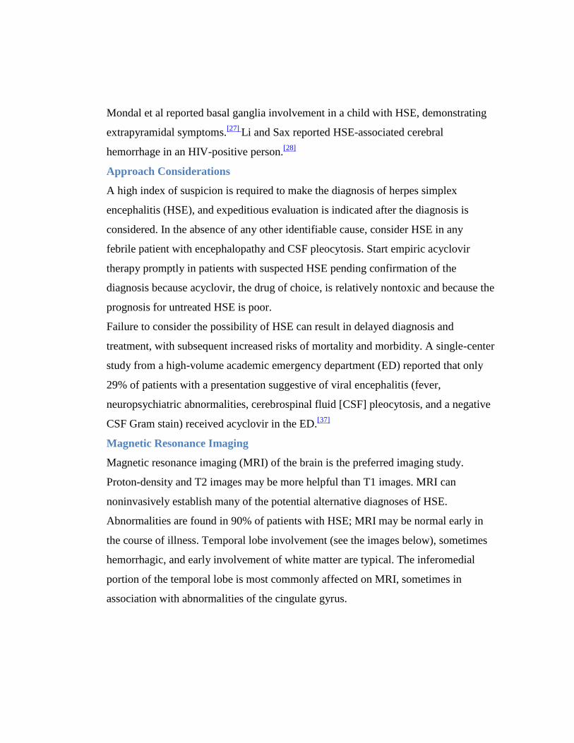
Abnormalities are found in 90% of patients with HSE; MRI may be normal early in
the course of illness. Temporal lobe involvement (see the images below), sometimes
hemorrhagic, and early involvement of white matter are typical. The inferomedial
portion of the temporal lobe is most commonly affected on MRI, sometimes in
association with abnormalities of the cingulate gyrus.
Axial proton density-weighted image in 62-year-old woman with confusion and
herpes encephalitis shows T2 hyperintensity involving right temporal lobe.
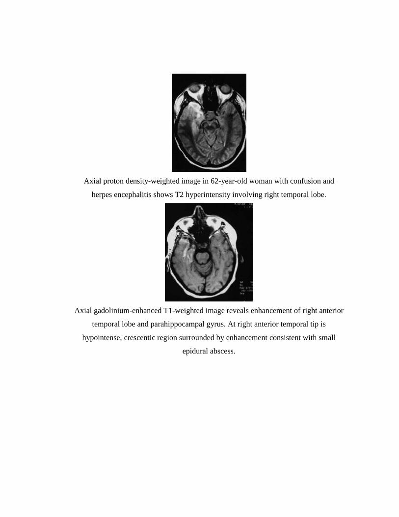
Axial gadolinium-enhanced T1-weighted image reveals enhancement of right anterior
temporal lobe and parahippocampal gyrus. At right anterior temporal tip is
hypointense, crescentic region surrounded by enhancement consistent with small
epidural abscess.
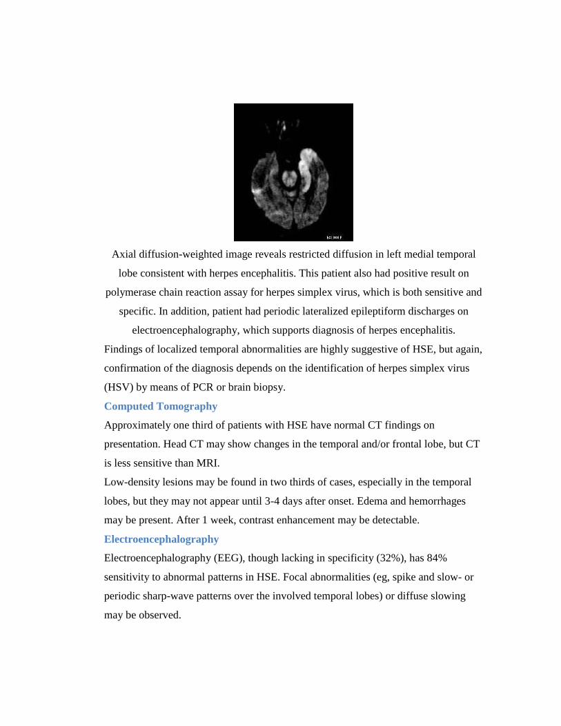
Axial diffusion-weighted image reveals restricted diffusion in left medial temporal
lobe consistent with herpes encephalitis. This patient also had positive result on
polymerase chain reaction assay for herpes simplex virus, which is both sensitive and
specific. In addition, patient had periodic lateralized epileptiform discharges on
electroencephalography, which supports diagnosis of herpes encephalitis.
Findings of localized temporal abnormalities are highly suggestive of HSE, but again,
confirmation of the diagnosis depends on the identification of herpes simplex virus
(HSV) by means of PCR or brain biopsy.
Computed Tomography
Approximately one third of patients with HSE have normal CT findings on
presentation. Head CT may show changes in the temporal and/or frontal lobe, but CT
is less sensitive than MRI.
Low-density lesions may be found in two thirds of cases, especially in the temporal
lobes, but they may not appear until 3-4 days after onset. Edema and hemorrhages
may be present. After 1 week, contrast enhancement may be detectable.
Electroencephalography
Electroencephalography (EEG), though lacking in specificity (32%), has 84%
sensitivity to abnormal patterns in HSE. Focal abnormalities (eg, spike and slow- or
periodic sharp-wave patterns over the involved temporal lobes) or diffuse slowing
may be observed.
Periodic complexes and periodic lateralizing epileptiform discharges (PLEDs), in the
proper clinical context, are strongly suggestive of HSE. However, Beneto et al
reported 9 patients with confirmed HSE who had no PLED activity or had other EEG
patterns.[29]
Analysis of Cerebrospinal Fluid
Once a space-occupying lesion has been excluded by imaging, lumbar puncture
always should be performed in suspected HSE. In general, CSF yield is proportional
to the volume analyzed; an adequate volume of CSF should be obtained (>10 mL).
Acutely, a typical “viral profile” is identified. Red blood cells (RBCs) and
xanthochromia may be seen. Patients typically have mononuclear pleocytosis of 10-
500 white blood cells (WBCs)/µL (average, 100 WBCs/µL). As a result of the
hemorrhagic nature of the underlying pathologic process, the RBC count may be
elevated (10-500/µL). Protein levels are elevated to the range of 60-700 mg/dL
(average, 100 mg/dL). Glucose values may be normal or mildly decreased (30-40
mg/dL).
In about 5-10% of patients, especially children, initial CSF results may be normal.[30]
However, on serial examinations, the cell counts and protein values increase.
Viral cultures of CSF are rarely positive and should not be relied on to confirm the
diagnosis. However, HSV can be cultured from the CSF in about one third of affected
neonates.
Polymerase chain reaction
CSF should be sent for HSV-1 and HSV-2 polymerase chain reaction (PCR) study.
PCR analysis of CSF for the detection of HSV DNA has virtually replaced brain
biopsy as the criterion standard for diagnosis.[31, 32]
Schloss and colleagues report that
whereas quantitative PCR is more rational than a nested PCR, the former has little
prognostic use.[33]
PCR is highly sensitive (94-98%) and specific (98-100%). Results become positive
within 24 hours of the onset of symptoms and remain positive for at least 5-7 days
after the start of antiviral therapy.

Clinical severity and outcome appear to correlate with viral load as assessed by
quantitative PCR techniques,[34]
but not all investigators have confirmed this
correlation.[35]
False-negative findings may occur early in the course of the disease when viral DNA
levels are low (within 72 hours of the onset of symptoms) or when blood is present in
the CSF, because hemoglobin may interfere with PCR.[36]
Pretest probability should be considered in interpretation of results. A negative result
obtained less than 72 hours after the onset of symptoms in a patient with a high
pretest probability (on the basis of fever, focal neurologic abnormalities, or CSF
pleocytosis) should be repeated.
False-positive test results are rare and usually reflect accidental contamination of the
specimen in the laboratory.
Brain Biopsy
Brain biopsy was once considered the only definitive means of diagnosing HSE. The
results of brain biopsy can also establish alternative diagnoses, both treatable (eg,
brain tumor) and nontreatable (eg, non-HSV viral encephalitis). Currently, with the
advent of PCR technology, the role of brain biopsy is diminishing. Studies have
demonstrated that PCR testing of CSF is as accurate as brain biopsy in confirming the
diagnosis of HSE.
When the diagnosis of HSE cannot be established by other means (eg, when lumbar
puncture is precluded or nondiagnostic), brain biopsy can yield a definitive diagnosis
and may be considered. However, with the availability of nontoxic and effective
antiviral medications, brain biopsy is rarely used today. The procedure carries a
complication rate of about 3%.
Orbitofrontal or limbic encephalitis may be seen. One hallmark of the condition is
significant hemorrhage in these locations. On pathology specimens, Cowdry A
inclusions are seen.
Serologic analysis
Serologic evaluation of blood or CSF may be useful for retrospective diagnosis, but it
has no role in the acute diagnosis and treatment of patients.
Strategies based on increases in antibody levels and on the ratio of antibody levels in
serum and CSF have not proven to be clinically useful.
Tzanck preparations
HSV can sometimes be confirmed by Tzanck preparations taken from vesicular
lesions in neonates with herpes simplex encephalitis.
Quantification of intrathecal antibodies
Intrathecal antibodies can be quantified, thus giving evidence for a central nervous
system (CNS) antibody response.
Approach Considerations
A high index of suspicion is required to make the diagnosis of herpes simplex
encephalitis (HSE), and expeditious evaluation is indicated after the diagnosis is
considered. In the absence of any other identifiable cause, consider HSE in any
febrile patient with encephalopathy and CSF pleocytosis. Start empiric acyclovir
therapy promptly in patients with suspected HSE pending confirmation of the
diagnosis because acyclovir, the drug of choice, is relatively nontoxic and because the
prognosis for untreated HSE is poor.
Failure to consider the possibility of HSE can result in delayed diagnosis and
treatment, with subsequent increased risks of mortality and morbidity. A single-center
study from a high-volume academic emergency department (ED) reported that only
29% of patients with a presentation suggestive of viral encephalitis (fever,
neuropsychiatric abnormalities, cerebrospinal fluid [CSF] pleocytosis, and a negative
CSF Gram stain) received acyclovir in the ED.[37]
Initial Management
Prehospital care consists of supportive management of the patient’s airway,
breathing, and circulation (ABCs). General nutritional and fluid support is important.
Universal precautions are appropriate. Monitor for increased intracranial pressure
(ICP) and seizures.
Intensive care unit (ICU) care may be required, especially if seizure activity or
increased ICP is present. Depending on the availability of local expertise (eg,
infectious disease, neurology, neurosurgery specialists), transfer to a tertiary care
facility may be appropriate. Hospitalization is not routine for uncomplicated herpes
simplex virus type 1 (HSV-1) or herpes simplex virus type 2 (HSV-2) infection.
Management of increased intracranial pressure
Treatment of brain edema ranges from simple measures (eg, elevating head of bed,
gentle diuresis with medication such as furosemide) to more complex measures (eg,
mannitol and steroids, intubation with hyperventilation).
Management of seizures
Behavioral manifestations of HSE may resemble seizures, which are also common.
Should seizure activity become apparent or should electroencephalography (EEG)
show evidence of nonconvulsive seizures, begin anticonvulsant therapy.
Benzodiazepines may be useful for aborting status epilepticus but, because of their
short duration, are ineffective at preventing further seizures. A longer-acting agent is
preferable.
Antiviral Therapy
Pharmacotherapy for HSE is available in the form of acyclovir. Patient outcome is
improved after treatment with this agent. Acyclovir is the treatment of choice for
HSE.[1, 3, 38]
When the diagnosis of HSE is suspected or has been established,
acyclovir (typically 30 mg/kg/d intravenously [IV] in adults) should be initiated
immediately.
Through a series of in vivo reactions catalyzed by viral and host cellular enzymes,
acyclovir is converted to acyclovir triphosphate, a potent inhibitor of HSV DNA
polymerase, without which viral replication cannot occur. Human cells are not
affected.
Acyclovir has relatively few serious adverse effects. Because of its high pH, IV
acyclovir may cause phlebitis and local inflammation if extravasation occurs.
Gastrointestinal (GI) disturbances, headache, and rash are among the more frequent
adverse reactions.
The drug is excreted by the kidney, and the dose should be reduced in patients with
renal dysfunction. Crystal-induced nephropathy may occur if the maximum solubility
of free drug is exceeded. Risk factors for this are IV administration, rapid infusion,
dehydration, concurrent use of nephrotoxic drugs, underlying renal disease, and high
doses. The risk of renal toxicity is reduced by adequately hydrating the patient (eg, 1
mL/d of fluid for each 1 mg/d of acyclovir).
Acyclovir is considered appropriate for serious infections during pregnancy. The
manufacturer cautions that it should be used in pregnancy only when the potential
benefits outweigh the potential risks. However, a prospective registry of acyclovir use
in pregnancy between 1984 and 1999, including 756 first-trimester exposures,
demonstrated a 3.2% rate of birth defects, similar to that expected in the general
population.[39]
In immunocompetent patients, viral resistance to acyclovir has been clinically
insignificant, with a reported prevalence of less than 1%.[40]
However, in
immunocompromised patients, this figure rises to 6%. Degree of immunosuppression
and duration of exposure to acyclovir appear to be the most important risk factors for
the development of resistant strains.
Since most relapses occur within 3 months of completing an initial course of IV
acyclovir, a prolonged course of an oral antiviral agent (eg, valacyclovir) has been
suggested after initial treatment. An ongoing clinical trial is currently evaluating a 90-
day course of valacyclovir versus placebo after treatment with acyclovir in patients
with HSE.[41]
A 2009 Cochrane database review of data from 17 trials that compared interventions
used for the prevention and treatment of HSV in patients being treated for cancer
concluded that acyclovir is effective in preventing and treating HSV infections.
Valacyclovir was not found to be more effective than acyclovir, nor did a higher dose
of valacyclovir make a difference. Some evidence indicated that placebo, as a
prophylaxis, is more effective than prostaglandin E, but the risk of bias was unclear in
all trials.[42]
If long-term suppressive therapy is needed, acyclovir or famciclovir can be used
orally.
Neonatal herpes simplex encephalitis
Acyclovir in doses of 20 mg/kg IV every 8 hours (60 mg/kg/d) is currently
recommended for neonatal HSE. This dosage is higher than that used in older
children and adults (30 mg/kg/d), but, in neonates, it has been shown to improve
mortality and morbidity when compared with the lower dosage. Because the higher
dosage is associated with neutropenia, the white blood cell (WBC) count should be
monitored closely.
Steroid Therapy
The role of steroids in the treatment of HSE remains uncertain. To the extent that
cellular damage in HSE is the result of immune-mediated inflammatory processes
triggered by the viral infection, the anti-inflammatory effects of steroids may be
beneficial. However, there is also concern that steroids might suppress immune
responses of the host that are necessary to limit viral replication.
Animal studies have demonstrated a beneficial effect of steroids on outcome, without
evidence of increased viral replication or dissemination.[43, 44]
Steroids have been used
to reduce cerebral edema in patients with severe HSE.
One nonrandomized, retrospective human study compared the outcomes of patients
with HSE who received steroids in addition to acyclovir with the outcomes of those
who received acyclovir alone.[45]
The steroid group had improved outcomes at 3
months. Although these results suggest a possible role for steroids in HSE, definitive
recommendations must await the results of larger prospective studies.
The German trial of Acyclovir and Corticosteroids in Herpes-simplex-virus-
Encephalitis (GACHE), a multicenter, randomized, placebo-controlled trial, is
currently enrolling patients with HSE in a study designed to assess the outcomes of
treatment with acyclovir against the outcomes of treatment with acyclovir plus
dexamethasone.[46]
Prevention
No measures are known to be effective for preventing HSE in adults and older
children. Person-to-person transmission does not occur. Prophylactic treatment of
close contacts and special isolation precautions are unnecessary.
Preventive measures for neonatal HSE include cesarean delivery in women with
active herpetic genital infections at the time of delivery and protection of neonates
from persons with active herpetic infections. Some authorities recommend a course of
suppressive acyclovir therapy near the time of delivery in mothers with a history of
genital herpes.
Consultations and Additional Care
HSE is a neurologic emergency. Consultation with a neurologist is required.
Neurosurgical consultation is helpful only if a brain biopsy is being considered. An
infectious disease consultation may be appropriate.
An evaluation for rehabilitation is often appropriate to deal with the long-term
neurologic sequelae of HSE. Depending on the nature and degree of any neurologic
deficits present, rehabilitation services may be required.
Medication Summary
The goals of therapy are to reduce morbidity, to shorten the clinical course of the
disease, to prevent complications, and to prevent recurrences. Pharmacotherapy for
herpes simplex encephalitis (HSE) is available in the form of acyclovir. Patient
outcome is improved when this agent is used for treatment.
Class Summary
The goals of using antivirals are to shorten the clinical course, prevent complications,
prevent development of latency and subsequent recurrences, decrease transmission,
and eliminate established latency.
Antivirals
Acyclovir (Zovirax)
Acyclovir is the drug of choice for HSE. It has demonstrated inhibitory activity
against both herpes simplex virus type 1 (HSV-1) and herpes simplex virus type 2
(HSV-2) and is taken up selectively by infected cells. Mortality from HSE before use
of acyclovir was 60-70%; since acyclovir, it is approximately 30%.
Famciclovir (Famvir)
After ingestion, drug is rapidly biotransformed into the active compound penciclovir
and phosphorylated by viral thymidine kinase. By competition with deoxyguanosine
triphosphate, penciclovir triphosphate inhibits viral polymerase, subsequently
inhibiting viral DNA synthesis/replication. Adjust the dose in patients with renal
insufficiency or hepatic disease.
Anticonvulsants
Class Summary
Anticonvulsants are used to terminate clinical and electrical seizure activity as rapidly
as possible and to prevent seizure recurrence.
Carbamazepine (Tegretol)
Carbamazepine is effective in treatment of complex partial seizures; it appears to act
by reducing polysynaptic responses and blocking posttetanic potentiation.
Phenytoin (Dilantin, Phenytek)
Phenytoin is a hydantoin. Its primary site of action appears to be the motor cortex,
where it may inhibit spread of seizure activity; it may reduce maximal activity of the
brain stem centers responsible for the tonic phase of grand mal seizures.
The dose should be individualized; if daily dosage cannot be divided equally, larger
dose should be given before bedtime. A phosphorylated formulation, fosphenytoin, is
available for parenteral use.
Diuretics
Class Summary
These agents are used for the management of increased intracranial pressure in
complications resulting from herpes simplex encephalitis.
Furosemide (Lasix)
Furosemide is a loop diuretic that increases the excretion of water by interfering with
the chloride-binding co-transport system, which, in turn, inhibits sodium and chloride
reabsorption in the ascending loop of Henle and distal renal tubule. It increases renal
blood flow without increasing the filtration rate. The onset of action generally is
within 1 hour. It increases potassium, sodium, calcium, and magnesium excretion.
Furosemide is used in the acute setting for reduction of increased ICP. The proposed
mechanisms in lowering ICP include following: (1) suppression of cerebral sodium
uptake, (2) carbonic anhydrase inhibition resulting in decreased CSF production, and
(3) inhibition of cellular membrane cation-chloride pump, thereby affecting the
transport of water into astroglial cells.
The dose must be individualized to the patient. Depending on the response,
administer at increments of 20-40 mg, no sooner than 6-8 hours after the previous
dose, until desired diuresis occurs. When treating infants, titrate with 1-mg/kg/dose
increments until a satisfactory effect is achieved.
Mannitol (Osmitrol)
Mannitol reduces cerebral edema with the help of osmotic forces, and it decreases
blood viscosity, resulting in reflex vasoconstriction and lowering of ICP.
Prognosis
Untreated HSE is progressive and often fatal in 7-14 days. A landmark study by
Whitley et al in 1977 revealed a 70% mortality in untreated patients and severe
neurologic deficits in most of the survivors.[17]
Mortality in patients treated with acyclovir was 19% in the trials that established its
superiority to vidarabine. Subsequent trials reported lower mortalities (6-11%),
perhaps because they included patients who were diagnosed by polymerase chain
reaction (PCR) rather than brain biopsy and who thus may have been identified
earlier with milder disease.[1, 3]
The mortality of neonatal HSE is substantial, even with treatment; 6% in patients
with isolated HSE and 31% in those with disseminated infection.
Sequelae among survivors are significant and depend on the patient’s age and
neurologic status at the time of diagnosis. Patients who are comatose at diagnosis
have a poor prognosis regardless of their age. In noncomatose patients, the prognosis
is age related, with better outcomes occurring in patients younger than 30 years.
Significant morbidity exists among those treated. Neurologic outcomes in survivors
treated with acyclovir are as follows:
No deficits or mild deficits - 38%
Moderate deficits - 9%
Severe deficits - 53%
Anterograde memory often is impaired even with successful treatment of HSE.
Retrograde memory, executive function, and language ability also may be impaired.
A study by Utley et al showed that patients who had a shorter delay (< 5 d) between
presentation and treatment had better cognitive outcomes.[18]
Elbers and colleagues followed properly treated children for 12 years after the HSE.
They found seizures in 44% of the children and developmental delay in 25% of the
children. They concluded that HSE continues to be associated with poor long-term
neurologic outcomes despite appropriate therapy.[19]
Shelley and colleagues reported a case of intracerebral hematoma occurring in a
patient successfully treated with a full course of acyclovir after apparent eradication
of the virus. The hematoma occurred in the region of the encephalitis.[20]
Marschitz and colleagues reported a case of chorea after HSE.[21]
Relapses after HSE have been reported to occur in 5-26% of patients, with most
relapses occurring within the first 3 months after completion of treatment. Relapses
are more frequent in children than adults. It is unclear whether such relapses represent
recurrence of viral infection or an immune-mediated inflammatory process. Some of
the relapses reported in earlier studies may have been due to inadequate duration of
treatment rather than true recurrences of HSE.
A long-term follow-up study of patients with HSE suggested that the pathogenic
mechanisms present during relapses differ from those present during the initial
infection.[22]
Serial measurements of inflammatory markers as well as HSV viral load
in the CSF of relapsing patients demonstrated increased inflammatory markers
without detectable HSV during relapses. These findings suggest that immune-
mediated events, rather than direct viral-mediated neuronal toxicity, may predominate
in relapses.
Patient Education
The belief that HSV-2 lesions appear initially 2 wk after primary infection can lead to
false accusations of infidelity. The physician should emphasize that the initial
outbreak of lesions may occur at any time after infection, possibly even years later.
Education may help reduce the spread of HSV-2.
For patient education resources, see the Teeth and Mouth Center and the Brain and
Nervous System Center, as well as Oral Herpes, Cold Sores, and Encephalitis.
REFERENCE
1. Whitley RJ. Herpes simplex encephalitis: adolescents and adults. Antiviral
Res. Sep 2006;71(2-3):141-8. [Medline].
2. Whitley RJ, Kimberlin DW. Herpes simplex encephalitis: children and
adolescents. Semin Pediatr Infect Dis. Jan 2005;16(1):17-23. [Medline].
3. Tyler KL. Herpes simplex virus infections of the central nervous system:
encephalitis and meningitis, including Mollaret's. Herpes. Jun 2004;11 Suppl
2:57A-64A. [Medline].
4. Wasay M, Mekan SF, Khelaeni B, Saeed Z, Hassan A, Cheema Z, et al. Extra
temporal involvement in herpes simplex encephalitis. Eur J Neurol. Jun
2005;12(6):475-9. [Medline].
5. Aurelian L. HSV-induced apoptosis in herpes encephalitis. Curr Top
Microbiol Immunol. 2005;289:79-111. [Medline].
6. DeBiasi RL, Kleinschmidt-DeMasters BK, Richardson-Burns S, Tyler KL.
Central nervous system apoptosis in human herpes simplex virus and
cytomegalovirus encephalitis. J Infect Dis. Dec 1 2002;186(11):1547-57.
[Medline].
7. Esiri MM. Herpes simplex encephalitis. An immunohistological study of the
distribution of viral antigen within the brain. J Neurol Sci. May
1982;54(2):209-26. [Medline].
8. Cinque P, Vago L, Marenzi R, Giudici B, Weber T, Corradini R, et al. Herpes
simplex virus infections of the central nervous system in human
immunodeficiency virus-infected patients: clinical management by
polymerase chain reaction assay of cerebrospinal fluid. Clin Infect Dis. Aug
1998;27(2):303-9. [Medline].
9. Fodor PA, Levin MJ, Weinberg A, Sandberg E, Sylman J, Tyler KL. Atypical
herpes simplex virus encephalitis diagnosed by PCR amplification of viral
DNA from CSF. Neurology. Aug 1998;51(2):554-9. [Medline].
10. Osih RB, Brazie M, Kanno M. Multifocal herpes simplex virus type 2
encephalitis in a patient with AIDS. AIDS Read. Feb 2007;17(2):67-70.
[Medline].
11. Baringer JR, Pisani P. Herpes simplex virus genomes in human nervous
system tissue analyzed by polymerase chain reaction. Ann Neurol. Dec
1994;36(6):823-9. [Medline].
12. Mitchell BM, Stevens JG. Neuroinvasive properties of herpes simplex virus
type 1 glycoprotein variants are controlled by the immune response. J
Immunol. Jan 1 1996;156(1):246-55. [Medline].
13. Geiger KD, Nash TC, Sawyer S, Krahl T, Patstone G, Reed JC, et al.
Interferon-gamma protects against herpes simplex virus type 1-mediated
neuronal death. Virology. Nov 24 1997;238(2):189-97. [Medline].
14. Cathomas R, Pelosi E, Smart J, Murray N, Simmonds P. Herpes simplex
encephalitis as a complication of adjuvant chemotherapy treatment for breast
cancer. Clin Oncol (R Coll Radiol). Jun 2005;17(4):292-3. [Medline].
15. Kohl S. Herpes Simplex Virus. In: Behrman RE, Kliegman RM, Jenson HB.
Behrman: Nelson Textbook of Pediatrics. 17th
ed. Philadelphia: Saunders;
2004.
16. Kimberlin D. Herpes simplex virus, meningitis and encephalitis in neonates.
Herpes. Jun 2004;11 Suppl 2:65A-76A. [Medline].
17. Whitley RJ, Soong SJ, Dolin R, Galasso GJ, Ch'ien LT, Alford CA. Adenine
arabinoside therapy of biopsy-proved herpes simplex encephalitis. National
Institute of Allergy and Infectious Diseases collaborative antiviral study. N
Engl J Med. Aug 11 1977;297(6):289-94. [Medline].
18. Utley TF, Ogden JA, Gibb A, McGrath N, Anderson NE. The long-term
neuropsychological outcome of herpes simplex encephalitis in a series of
unselected survivors. Neuropsychiatry Neuropsychol Behav Neurol. Jul
1997;10(3):180-9. [Medline].
19. Elbers JM, Bitnun A, Richardson SE, Ford-Jones EL, Tellier R, Wald RM, et
al. A 12-year prospective study of childhood herpes simplex encephalitis: is
there a broader spectrum of disease?. Pediatrics. Feb 2007;119(2):e399-407.
[Medline].
20. Shelley BP, Raniga SB, Al-Khabouri J. An unusual late complication of
intracerebral haematoma in herpes encephalitis after successful acyclovir
treatment. J Neurol Sci. Jan 31 2007;252(2):177-80. [Medline].
21. Marschitz I, Rödl S, Gruber-Sedlmayr U, Church A, Giovannoni G, Zobel G,
et al. Severe chorea with positive anti-basal ganglia antibodies after
herpesencephalitis. J Neurol Neurosurg Psychiatry. Jan 2007;78(1):105-7.
[Medline]. [Full Text].
22. Sköldenberg B, Aurelius E, Hjalmarsson A, Sabri F, Forsgren M, Andersson
B, et al. Incidence and pathogenesis of clinical relapse after herpes simplex
encephalitis in adults. J Neurol. Feb 2006;253(2):163-70. [Medline].
23. Whitley RJ, Cobbs CG, Alford CA Jr, Soong SJ, Hirsch MS, Connor JD, et al.
Diseases that mimic herpes simplex encephalitis. Diagnosis, presentation, and
outcome. NIAD Collaborative Antiviral Study Group. JAMA. Jul 14
1989;262(2):234-9. [Medline].
24. Whitley RJ, Soong SJ, Linneman C Jr, Liu C, Pazin G, Alford CA. Herpes
simplex encephalitis. Clinical Assessment. JAMA. Jan 15 1982;247(3):317-
20. [Medline].
25. Ku A, Lachmann EA, Nagler W. Selective language aphasia from herpes
simplex encephalitis. Pediatr Neurol. Sep 1996;15(2):169-71. [Medline].
26. McGrath NM, Anderson NE, Hope JK, Croxson MC, Powell KF. Anterior
opercular syndrome, caused by herpes simplex encephalitis. Neurology. Aug
1997;49(2):494-7. [Medline].
27. Mondal G, Kumar R, Ghosh JK, Basu K, Chatterjee S. Basal ganglia
involvement in a child with herpes simplex encephalitis. Indian J Pediatr. Jul
2009;76(7):749-50. [Medline].
28. Li JZ, Sax PE. HSV-1 encephalitis complicated by cerebral hemorrhage in an
HIV-positive person. AIDS Read. Apr 2009;19(4):153-5. [Medline].
29. Benetó A, Gómez E, Rubio P, Sobrino R, Esparza A, Gil M, et al. [Periodical
EEG pattern modifications in herpetic encephalitis treated with acyclovir].
Rev Neurol. Jul 1996;24(131):829-32. [Medline].
30. Mook-Kanamori B, van de Beek D, Wijdicks EF. Herpes simplex encephalitis
with normal initial cerebrospinal fluid examination. J Am Geriatr Soc. Aug
2009;57(8):1514-5. [Medline].
31. Lakeman FD, Whitley RJ. Diagnosis of herpes simplex encephalitis:
application of polymerase chain reaction to cerebrospinal fluid from brain-
biopsied patients and correlation with disease. National Institute of Allergy
and Infectious Diseases Collaborative Antiviral Study Group. J Infect Dis.
Apr 1995;171(4):857-63. [Medline].
32. Cinque P, Cleator GM, Weber T, Monteyne P, Sindic CJ, van Loon AM. The
role of laboratory investigation in the diagnosis and management of patients
with suspected herpes simplex encephalitis: a consensus report. The EU
Concerted Action on Virus Meningitis and Encephalitis. J Neurol Neurosurg
Psychiatry. Oct 1996;61(4):339-45. [Medline]. [Full Text].
33. Schloss L, Falk KI, Skoog E, Brytting M, Linde A, Aurelius E. Monitoring of
herpes simplex virus DNA types 1 and 2 viral load in cerebrospinal fluid by
real-time PCR in patients with herpes simplex encephalitis. J Med Virol. Aug
2009;81(8):1432-7. [Medline].
34. Domingues RB, Lakeman FD, Mayo MS, Whitley RJ. Application of
competitive PCR to cerebrospinal fluid samples from patients with herpes
simplex encephalitis. J Clin Microbiol. Aug 1998;36(8):2229-34. [Medline].
[Full Text].
35. Wildemann B, Ehrhart K, Storch-Hagenlocher B, Meyding-Lamadé U,
Steinvorth S, Hacke W, et al. Quantitation of herpes simplex virus type 1
DNA in cells of cerebrospinal fluid of patients with herpes simplex virus
encephalitis. Neurology. May 1997;48(5):1341-6. [Medline].
36. Weil AA, Glaser CA, Amad Z, Forghani B. Patients with suspected herpes
simplex encephalitis: rethinking an initial negative polymerase chain reaction
result. Clin Infect Dis. Apr 15 2002;34(8):1154-7. [Medline].
37. Benson PC, Swadron SP. Empiric acyclovir is infrequently initiated in the
emergency department to patients ultimately diagnosed with encephalitis. Ann
Emerg Med. Jan 2006;47(1):100-5. [Medline].
38. Rathmann K, Scott SA. Acyclovir. In: Drug Evaluation Monographs.
Micromedex:2005.
39. Stone KM, Reiff-Eldridge R, White AD, Cordero JF, Brown Z, Alexander
ER, et al. Pregnancy outcomes following systemic prenatal acyclovir
exposure: Conclusions from the international acyclovir pregnancy registry,
1984-1999. Birth Defects Res A Clin Mol Teratol. Apr 2004;70(4):201-7.
[Medline].
40. James SH, Kimberlin DW, Whitley RJ. Antiviral therapy for herpesvirus
central nervous system infections: neonatal herpes simplex virus infection,
herpes simplex encephalitis, and congenital cytomegalovirus infection.
Antiviral Res. Sep 2009;83(3):207-13. [Medline]. [Full Text].
41. National Institute of Allergy and Infectious Diseases. A Phase III Double-
Blind, Placebo-Controlled Trial of Long Term Therapy of Herpes Simplex
Encephalitis (HSE): An Evaluation of Valacyclovir (CASG-204).
ClinicalTrials.gov. Available at
http://clinicaltrials.gov/ct/show/NCT00031486?order=1. Accessed August 8,
2007.
42. Glenny AM, Fernandez Mauleffinch LM, Pavitt S, Walsh T. Interventions for
the prevention and treatment of herpes simplex virus in patients being treated
for cancer. Cochrane Database Syst Rev. Jan 21 2009;CD006706. [Medline].
43. Sergerie Y, Boivin G, Gosselin D, Rivest S. Delayed but not early
glucocorticoid treatment protects the host during experimental herpes simplex
virus encephalitis in mice. J Infect Dis. Mar 15 2007;195(6):817-25.
[Medline].
44. Thompson KA, Blessing WW, Wesselingh SL. Herpes simplex replication
and dissemination is not increased by corticosteroid treatment in a rat model
of focal Herpes encephalitis. J Neurovirol. Feb 2000;6(1):25-32. [Medline].
45. Kamei S, Sekizawa T, Shiota H, Mizutani T, Itoyama Y, Takasu T, et al.
Evaluation of combination therapy using aciclovir and corticosteroid in adult
patients with herpes simplex virus encephalitis. J Neurol Neurosurg
Psychiatry. Nov 2005;76(11):1544-9. [Medline]. [Full Text].
46. Martinez-Torres F, Menon S, Pritsch M, Victor N, Jenetzky E, Jensen K, et al.
Protocol for German trial of Acyclovir and corticosteroids in Herpes-simplex-
virus-encephalitis (GACHE): a multicenter, multinational, randomized,
double-blind, placebo-controlled German, Austrian and Dutch trial
[ISRCTN45122933]. BMC Neurol. Oct 29 2008;8:40. [Medline]. [Full Text].


![Immunology of Herpes Simplex Virus Infection: …...[CANCER RESEARCH 36, 836-844, February 1976] Immunology of Herpes Simplex Virus Infection: Relevance to Herpes Simplex Virus Vaccines](https://static.fdocuments.us/doc/165x107/5e3c207dedbcb80872726a41/immunology-of-herpes-simplex-virus-infection-cancer-research-36-836-844.jpg)


![Clinical characteristics and long-term prognosis of ... · other possible causes, such as viral encephalitis and herpes simplex encephalitis. Relapsing anti-NMDAR encephalitis [5]](https://static.fdocuments.us/doc/165x107/5f5cb978af3eab35a02f3630/clinical-characteristics-and-long-term-prognosis-of-other-possible-causes-such.jpg)


