Dysphagia - University of Texas Medical · PDF fileEvaluation of Dysphagia ......
46
Dysphagia Shashidhar Reddy, MD Faculty Advisor: Matthew W. Ryan, MD The University of Texas Medical Branch Department of Otolaryngology Grand Rounds Presentation November 21, 2001
Transcript of Dysphagia - University of Texas Medical · PDF fileEvaluation of Dysphagia ......

Dysphagia
Shashidhar Reddy, MD
Faculty Advisor: Matthew W. Ryan, MD
The University of Texas Medical Branch
Department of Otolaryngology
Grand Rounds Presentation
November 21, 2001

Physiology of Swallowing
• The act of swallowing involves three phases: Oral,
Pharyngeal, and Esophageal.
• Swallowing takes about 8-10 seconds
• Before swallowing begins, Oral Preparation of the
bolus must occur.

Physiology of Swallowing:
Oral Phase Pharyngeal Phase

Physiology of Swallowing Pharyngeal and Esophageal Phase:

Evaluation of Dysphagia
• History
• Review of Systems
• Physical Exam
• Imaging Studies

History
• Duration dietary changes, weight loss
• Odynophagia
• Solids or Liquids
• Level of sensation of dysphagia
• Past surgery to head and neck, trauma, ingestion of caustic substances
• Associated symptoms such as with GERD, voice changes, nasal leakage, otalgia
Review of Systems:
• Ask about common systemic processes
associated with dysphagia:
– Tobacco/Alcohol
– Medications – antihistamines, anticholinergics,
antidepressants, antihypertensives
– Osteoarthritis
– Systemic neuromuscular disorders
– Auto-Immune disorders
– Psychiatric state
Physical Exam:
• General: body habitus, mental status,
drooling, wheezing, dyspnea, voice quality
• Cranial nerves
• Inspection of the tongue and palate for
strength/symmetry
• Laryngeal Examination: pooled secretions,
vocal fold movement, interaretynoid area
Imaging Studies
• Should be chosen to suit the patient’s
symptoms and to confirm a finding.

Plain Film
• Uses:
– Suspected infectious cause of dysphagia with
gross displacement of structures.
Advantages Disadvantages
cheap Radiation
Fast Poor anatomic detail
No assessment of swallow

Plain Film
(Epiglottitis)

Barium Esophagram
• Uses: structural disorders, e.g. dysphagia
for solid foods. Can use air contrast.
Advantages Disadvantages
Good anatomic detail Radiation
Logistics in bedridden pts.
Cannot detect dynamic
disorders.

Air Contrast Barium Esophagram
Normal Fungal Plaques

Manometry
• Uses: disorders in which intraluminal
pressures must be measured (achalasia,
esophageal spasm, etc.)
Advantages Disadvantages
It is the only test of
pressure wave physiology
Cannot diagnose visible
lesions
Unpleasant for patient
Techincally demanding

Manometry

Bolus Scintigraphy
• Uses: follow improvement in a patient with
history of aspiration, patient with achalasia.
Advantages: Disadvantages:
Less radiation No anatomic details
Quantitative count of
particles
Single bolus, not
different consist. used

Bolus Scintigraphy

Ultrasound
• Uses: Portable tool for dynamic studies,
especially in children
Advantages: Disadvantages:
No radiation Not widely available
Portable Poor anatomic detail
Normal Food can be used

Ultrasound

Modified Barium Swallow
• Uses – excellent to evaluate dynamic (e.g.
neuromuscular, aspiration) swallow
disorders.
Advantages Disadvantages
Gives good anatomic detail Radiation
Evaluates all phases of
swallowing
Does not directly test
sensitivity
Logistics

Modified Barium Swallow
Neurogenic Dysphagia
Normal Barium Swallow http://www.hopkinsmedicine.org/gastroenterology_hepatology/
http://www.hopkinsmedicine.org/gastroenterology_hepatology/

Fiberoptic Endoscopic
Evaluation of Swallowing
• Uses – as a mobile tool that can be used in
training patients via biofeedback
Advantages Disadvantages
Portable Blind spot
Allows assessment of sensation Cannot evaluate
cricopharyngeus directly
Cheap Cannot eval. esophagus
Can be used for pt teaching
No radiation

Fiberoptic Endoscopic
Evaluation of Swallowing

Disorders that Cause Dysphagia
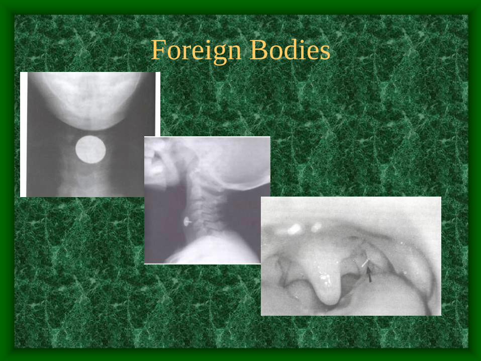
Foreign Bodies

Tracheostomy

Cricopharyngeal Achalasia

Cricopharyngeal Achalasia
Cricopharyngeal Myotomy:

Zenker’s Diverticulum

Zenker’s Diverticulum

Cervical Spine Disease

Esophageal Webs and Rings

Strictures / Caustic Ingestion

Achalasia

Diffuse Esophageal Spasm

Gastroesophageal Reflux Disease
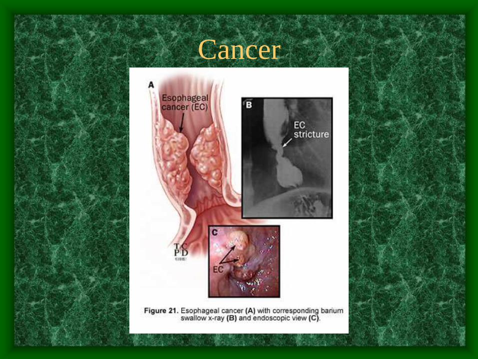
Cancer

Systemic Disorders that Cause
Dysphagia
• Stroke – present in up to 47%
• Amyotrophic Lateral Sclerosis
• Parkinson’s Disease
• Multiple Sclerosis
• Muscular Dystrophy
• Myasthenia Gravis
Autoimmune Disorders
• Systemic Sclerosis
• Systemic Lupus Erythematosis
• Dermatomyosits
• Mixed Connective Tissue Disease
• Mucosal Pemphigoid, Epidermolysis Bulosa
• Sjogren’s Syndrome (xerostomia)
• Rheumatoid Arthritis (cricoarytenoid joint
fixation)

Aging
• Dysphagia is present in 2% > 65
• Poor dentition
• Loss of tongue connective tissue
• Increased pharyngeal transit time

Dysphagia in Children
• Nasal obstruction
• Oral lesions – clefts, ranulas, mucoceles
• Laryngomalacia, laryngeal clefts, TE fistula
• Vascular rings, Foregut malformations
• Tumors – hemangiomas, lymphangiomas,
papillomas, leiomyomas, neurofibromas
Globus Hystericus
• Imagined dysphagia
• Somatization

Case Review
• 50 year old man presents with 6 month
history of progressive dysphagia.

Case Report • His dysphagia is worse for solid foods.
• Additionally he notes that he hears gurgling noises when he swallows, and occasionally chokes on his food.
• When he chokes, he often ends up “vomitting” his food back up.
• He has lost about 6 lbs over the past 6 months.
• He drinks socially but gave up tobacco x10yrs

Case Report
• Physical exam reveals a thin white
gentleman in no apparent distress.
• Neck exam reveals nothing unusual.
• Indirect Laryngoscopy is difficult because
of frothy secretions in his hypopharynx and
piriform sinus.

Case Report
Case Report Barium Esophagram
http://www.hopkinsmedicine.org/gastroenterology_hepatology/