Dynamics of Changes in Isometric Strength and Muscle...
9
Research Article Dynamics of Changes in Isometric Strength and Muscle Imbalance in the Treatment of Women with Low back Pain Jacek Wilczy ´ nski and Alicja Kasprzak Laboratory of Posturology, Collegium Medicum, Jan Kochanowski University, Kielce, Poland Correspondence should be addressed to Jacek Wilczy´ nski; [email protected] Received 24 September 2019; Revised 6 December 2019; Accepted 27 December 2019; Published 11 January 2020 Guest Editor: Mario Bernardo-Filho Copyright © 2020 Jacek Wilczy´ nski and Alicja Kasprzak. is is an open access article distributed under the Creative Commons Attribution License, which permits unrestricted use, distribution, and reproduction in any medium, provided the original work is properly cited. e aim of the study was to evaluate the dynamics of isometric changes in strength and muscular lumbar-pelvic imbalances in the treatment of women with low back pain. Forty-one women, nineteen in the study group (A) and twenty-two in the control group (B), participated in the study. Magnetic resonance imaging (MRI) was performed to assess the degree of degenerative changes in the lumbar spine. e diagnosis of isometric muscle strength and their imbalances was performed with the Tergumed 700 device. After six weeks of therapy in the study group (A), there was a significant improvement in the strength of all the examined muscle groups. However, in the control group (B), significant improvement occurred only in the strength of the lumbar flexor muscles and the flexor muscles on the left side. Furthermore, there was a significant intensification of the imbalance of left flexor muscle strength compared to right flexor strength in group B. Significant differences in favour of the study group (A) concerned the strength of the rotator muscles to the left, the strength of the extensor muscles of the lumbar spine, the strength of the flexors of the lumbar spine to the right, and the balance of the strength of the lumbar spine flexors to the left compared to the strength of the flexor muscles to the right. erapy with the Tergumed 700 system leads to an increase in the muscle strength of the lumbar and pelvic complex, compensating for its imbalance, bringing beneficial effects in the treatment of low back pain. 1. Introduction Low back pain is one of the most commonly diagnosed diseases of the osteoarticular system [1]. It is also the most frequently reported ailment [2] and the second main cause of sickness absence [3]. It is the most common cause of the inability to perform work [4] and is one of the main causes of physical disability of people below the age of 45 [5]. Pain syndromes can be divided into specific and nonspecific [6, 7]. Myofascial overloads, ligament injuries, and psy- chogenic factors are considered to be the causes of those nonspecific [8, 9]. Specific pains are most often caused by a herniated nucleus, spondylolisthesis, spinal canal stenosis, degenerative changes of the interappendix joints, vertebral fractures, spinal tumours, or inflammatory diseases [10, 11]. Pain sensations may be dull and diffuse but may also be shooting, stabbing, causing a burning or stinging sensation [12, 13]. In the population, nonspecific back pain is the most common, that is, the basis for which the specific pathology that causes the pain cannot be found [14, 15]. In acute pain, appearing for the first time in life, only 2% of patients can determine its cause [16, 17]. e incidence of this disease entity causes a significant burden on the state budget. e largest direct costs are generated by diagnostics, treatment, and rehabilitation, while indirect costs are disability pen- sions, benefits, and sick leaves at work [18, 19]. ere are many suggestions in the available literature for conservative treatment of low back pain [20, 21]. Properly targeted kinesiotherapy plays a dominant role [22, 23]. In recent years, various devices have been developed to create optimal conditions for conducting isolated exercises re- garding the lumbar region of the spine [24]. Tergumed is one such system used in the diagnosis and therapy of low back pain. Research conducted using this system, however, gen- erally took the short duration of therapy into account, which Hindawi BioMed Research International Volume 2020, Article ID 6139535, 8 pages https://doi.org/10.1155/2020/6139535
Transcript of Dynamics of Changes in Isometric Strength and Muscle...

Research ArticleDynamics of Changes in Isometric Strength and MuscleImbalance in the Treatment of Women with Low back Pain
Jacek Wilczynski and Alicja Kasprzak
Laboratory of Posturology, Collegium Medicum, Jan Kochanowski University, Kielce, Poland
Correspondence should be addressed to Jacek Wilczynski; [email protected]
Received 24 September 2019; Revised 6 December 2019; Accepted 27 December 2019; Published 11 January 2020
Guest Editor: Mario Bernardo-Filho
Copyright © 2020 Jacek Wilczynski and Alicja Kasprzak. 3is is an open access article distributed under the Creative CommonsAttribution License, which permits unrestricted use, distribution, and reproduction in anymedium, provided the original work isproperly cited.
3e aim of the study was to evaluate the dynamics of isometric changes in strength and muscular lumbar-pelvic imbalances in thetreatment of women with low back pain. Forty-one women, nineteen in the study group (A) and twenty-two in the control group(B), participated in the study. Magnetic resonance imaging (MRI) was performed to assess the degree of degenerative changes inthe lumbar spine. 3e diagnosis of isometric muscle strength and their imbalances was performed with the Tergumed 700 device.After six weeks of therapy in the study group (A), there was a significant improvement in the strength of all the examined musclegroups. However, in the control group (B), significant improvement occurred only in the strength of the lumbar flexor musclesand the flexor muscles on the left side. Furthermore, there was a significant intensification of the imbalance of left flexor musclestrength compared to right flexor strength in group B. Significant differences in favour of the study group (A) concerned thestrength of the rotator muscles to the left, the strength of the extensor muscles of the lumbar spine, the strength of the flexors of thelumbar spine to the right, and the balance of the strength of the lumbar spine flexors to the left compared to the strength of theflexor muscles to the right. 3erapy with the Tergumed 700 system leads to an increase in the muscle strength of the lumbar andpelvic complex, compensating for its imbalance, bringing beneficial effects in the treatment of low back pain.
1. Introduction
Low back pain is one of the most commonly diagnoseddiseases of the osteoarticular system [1]. It is also the mostfrequently reported ailment [2] and the secondmain cause ofsickness absence [3]. It is the most common cause of theinability to performwork [4] and is one of the main causes ofphysical disability of people below the age of 45 [5]. Painsyndromes can be divided into specific and nonspecific[6, 7]. Myofascial overloads, ligament injuries, and psy-chogenic factors are considered to be the causes of thosenonspecific [8, 9]. Specific pains are most often caused by aherniated nucleus, spondylolisthesis, spinal canal stenosis,degenerative changes of the interappendix joints, vertebralfractures, spinal tumours, or inflammatory diseases [10, 11].Pain sensations may be dull and diffuse but may also beshooting, stabbing, causing a burning or stinging sensation[12, 13]. In the population, nonspecific back pain is the most
common, that is, the basis for which the specific pathologythat causes the pain cannot be found [14, 15]. In acute pain,appearing for the first time in life, only 2% of patients candetermine its cause [16, 17]. 3e incidence of this diseaseentity causes a significant burden on the state budget. 3elargest direct costs are generated by diagnostics, treatment,and rehabilitation, while indirect costs are disability pen-sions, benefits, and sick leaves at work [18, 19].
3ere are many suggestions in the available literature forconservative treatment of low back pain [20, 21]. Properlytargeted kinesiotherapy plays a dominant role [22, 23]. Inrecent years, various devices have been developed to createoptimal conditions for conducting isolated exercises re-garding the lumbar region of the spine [24]. Tergumed is onesuch system used in the diagnosis and therapy of low backpain.
Research conducted using this system, however, gen-erally took the short duration of therapy into account, which
HindawiBioMed Research InternationalVolume 2020, Article ID 6139535, 8 pageshttps://doi.org/10.1155/2020/6139535

hindered its objective assessment. 3ere is also a lack ofresearch on the dynamics of changes in strength and muscleimbalance at individual periods of therapy, or comparison ofresults with the control group.3erefore, there was a need toconduct a comprehensive, objective, and controlled clinicalstudy as well as a thorough analysis of the dynamics ofchanges in muscle activity and their imbalances [25].
For stabilization, the spine needs both muscle strengthand stiffness, to which the muscles contribute. Due to therecognition of the close relationship between musclefunction and low back pain, a new paradigm has been de-veloped regarding the function and dysfunction of the deepmuscle system. Moreover, the characteristics of exercisesnecessary for the rehabilitation of patients with low backpain have also been determined. 3is model has contributedto the modification of programmes for the rehabilitation ofpatients with this type of ailment by introducing rotationand extension exercises [26–28]. Basically, there are twomain modes of action aimed at improving the protectivefunction of muscles in relation to the spine joints.3e first ofthem utilises the principle of minimising the forces affectingthe lumbar spine during basic motor activity, and the secondthe optimal control of the lumbar-pelvic complex. Patientssuffering from low back pain may demonstrate a lack ofmuscle tone normalization even after the pain subsides[29, 30]. 3erefore, in this study, the authors focused on theassessment of isometric muscle strength and muscle balance[31].
3e aim of the study was to assess the dynamics ofchanges in isometric strength and muscular imbalances ofthe lumbar and pelvic complex in the treatment of womenwith low back pain. 3e authors assumed that this therapy,by developing muscle strength and improving balance,improves the stabilization of the lumbar-pelvic complex anddemonstrates beneficial effects in the treatment of peoplewith low back pain.
2. Materials and Methods
Forty-one women aged 60–75 took part in the study(X� 65.3; SD� 6.5). 3ey were patients of the RehabilitationClinic who were diagnosed with low back pain. 3e in-clusion criteria were low back pain, degenerative changes ofthe lumbar spine visible in magnetic resonance imaging, age60–75 years, patient’s consent to participate in the study, andnot undergoing lumbar spine rehabilitation with a differentkinesiotherapy method than the one applied in the study atthe time of research.
3e exclusion criteria were less than 3 months from theonset of the acute discopathy phase, fresh fractures, shortremission intervals in the course of rheumatic diseases,inflammatory diseases at the stage of exacerbation, for ex-ample, ankylosing spondylitis, hernia (abdominal, inguinal),osteoporosis with mineral density of bones up to 80% of theaverage for a given age, cancer, and spinal deformities.
Magnetic resonance imaging (MRI) was performed toassess the degree of degenerative changes in the lumbarspine. Randomly tested using a computer number generator,the subjects were assigned to two groups. 3e study group
(A) comprised nineteen participants, while the controlgroup (B) totalled twenty-two subjects.
In the study group (A), central stabilization exercises andtherapy using the Tergumed 700 system were performed. Inthe control group (B), only central stabilization exerciseswere performed. 3e therapy lasted 6 weeks. All researchprocedures were carried out in accordance with the 1964Declaration of Helsinki and with the consent of the BioethicsCommittee at the Regional Medical Chamber in Krakow(Poland) No. 73/KBL/OIL/2016 from May 4, 2016.
2.1. Assessment of the Dynamics of Isometric Changes inMuscular Strength and Muscular Imbalances of the Lumbar-PelvicHipComplex. 3emuscle strength test of the lumbar-pelvic hip complex was performed using the Tergumed 700system.3is system was TUV Sud 0123 certified and met therequirements of Directive 93/42 EEC [32]. 3e test wasperformed in flexion, extension, lateral flexion (left/right),and rotation (left/right). 3e test took place in a seatedposition. Each device was adapted to the patient.3e authorsmade sure that the axis of motion was correct and that thesubject was well stabilized. Isometric strength of the muscleswas tested.3emeasurement was carried out using a built-indynamometer (Nm). On each of the four Tergumed devices,a strength and muscle imbalance test was performed. Onetest repetition and two research repetitions were carried out,from which the average value was calculated. 3e exami-nation was performed four times: before and after two, four,and six weeks of therapy.
2.1.1. Central Stabilization Training. Central stabilizationtraining was conducted on the basis of a scheme developedby Richardson et al. [33]. It consisted of three stages: trainingof local segmental control, of segmental control in a closedchain, and of segmental control in an open chain. A warm-up using cycloergometers and deep muscle activation in lowpositions was also performed.3e ability to properly activatemuscles was palpated, enabling the transition duringtraining to exercises in higher positions. Coordination andbalance exercises were carried out with the use of Swiss balls,aerodynamic discs, sphere segments, and elastic bands. Inaddition, exercises for stretching and relaxing contractedmuscles were used. Central stabilization training in bothgroups (A and B) was conducted equally by the samephysiotherapist and was applied for 30 minutes, 5 days aweek, and for 6 weeks from May 2016 to March 2017.
2.1.2. -erapy Using the Tergumed 700 System. Tergumed700 is a line of 4 devices for diagnosis and therapy of thelumbar spine. Each device was responsible for 4 main di-rections of spinal movement (extension, flexion, lateralbend, and rotation). Before therapy, a maximum musclestrength test was performed on each device. Based on thetest, an individual therapy plan using feedback was gener-ated. 3e therapy was aimed at improving strength andcompensating for muscular imbalances. 3e test alsoallowed for painless treatment. 3anks to programming the
2 BioMed Research International
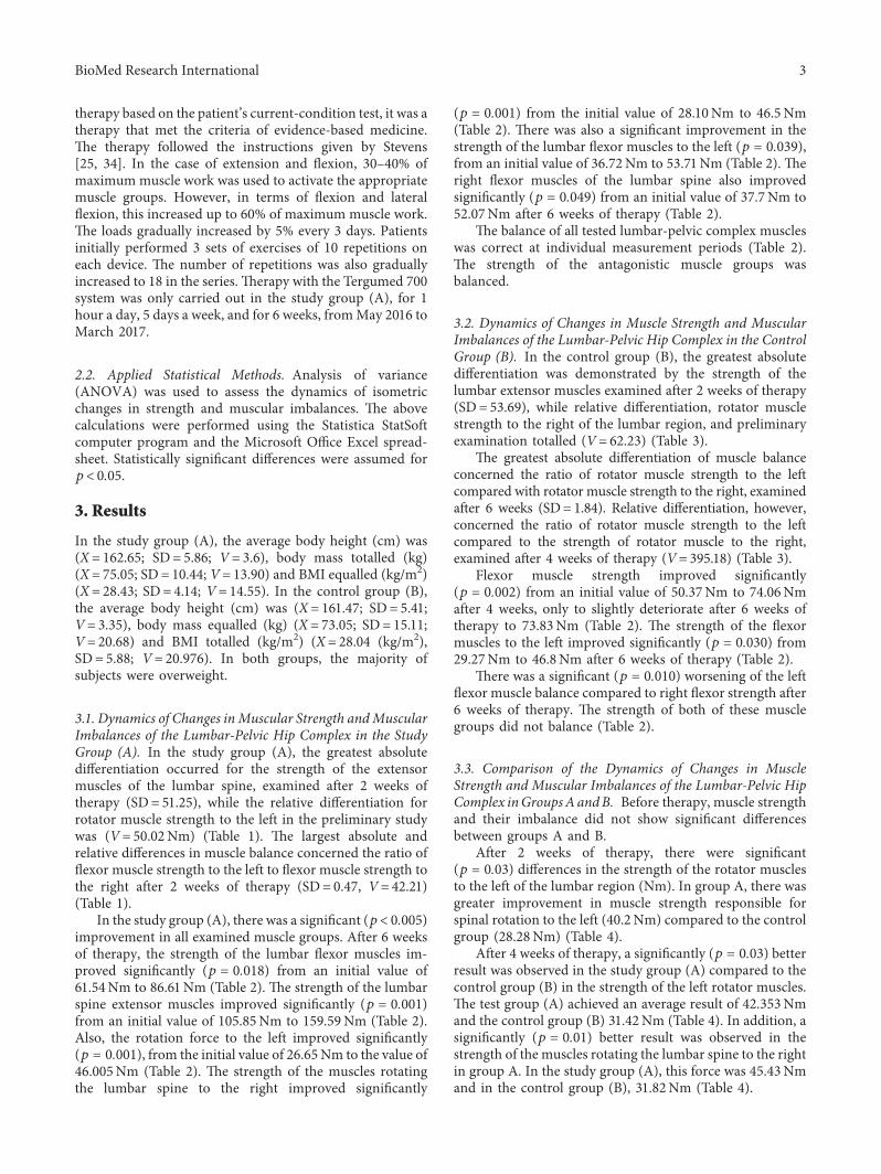
therapy based on the patient’s current-condition test, it was atherapy that met the criteria of evidence-based medicine.3e therapy followed the instructions given by Stevens[25, 34]. In the case of extension and flexion, 30–40% ofmaximum muscle work was used to activate the appropriatemuscle groups. However, in terms of flexion and lateralflexion, this increased up to 60% of maximum muscle work.3e loads gradually increased by 5% every 3 days. Patientsinitially performed 3 sets of exercises of 10 repetitions oneach device. 3e number of repetitions was also graduallyincreased to 18 in the series. 3erapy with the Tergumed 700system was only carried out in the study group (A), for 1hour a day, 5 days a week, and for 6 weeks, fromMay 2016 toMarch 2017.
2.2. Applied Statistical Methods. Analysis of variance(ANOVA) was used to assess the dynamics of isometricchanges in strength and muscular imbalances. 3e abovecalculations were performed using the Statistica StatSoftcomputer program and the Microsoft Office Excel spread-sheet. Statistically significant differences were assumed forp< 0.05.
3. Results
In the study group (A), the average body height (cm) was(X� 162.65; SD� 5.86; V� 3.6), body mass totalled (kg)(X� 75.05; SD� 10.44; V� 13.90) and BMI equalled (kg/m2)(X� 28.43; SD� 4.14; V� 14.55). In the control group (B),the average body height (cm) was (X� 161.47; SD� 5.41;V� 3.35), body mass equalled (kg) (X� 73.05; SD� 15.11;V� 20.68) and BMI totalled (kg/m2) (X� 28.04 (kg/m2),SD� 5.88; V� 20.976). In both groups, the majority ofsubjects were overweight.
3.1. Dynamics of Changes in Muscular Strength andMuscularImbalances of the Lumbar-Pelvic Hip Complex in the StudyGroup (A). In the study group (A), the greatest absolutedifferentiation occurred for the strength of the extensormuscles of the lumbar spine, examined after 2 weeks oftherapy (SD� 51.25), while the relative differentiation forrotator muscle strength to the left in the preliminary studywas (V� 50.02Nm) (Table 1). 3e largest absolute andrelative differences in muscle balance concerned the ratio offlexor muscle strength to the left to flexor muscle strength tothe right after 2 weeks of therapy (SD� 0.47, V� 42.21)(Table 1).
In the study group (A), there was a significant (p< 0.005)improvement in all examined muscle groups. After 6 weeksof therapy, the strength of the lumbar flexor muscles im-proved significantly (p � 0.018) from an initial value of61.54Nm to 86.61Nm (Table 2). 3e strength of the lumbarspine extensor muscles improved significantly (p � 0.001)from an initial value of 105.85Nm to 159.59Nm (Table 2).Also, the rotation force to the left improved significantly(p � 0.001), from the initial value of 26.65Nm to the value of46.005Nm (Table 2). 3e strength of the muscles rotatingthe lumbar spine to the right improved significantly
(p � 0.001) from the initial value of 28.10Nm to 46.5Nm(Table 2). 3ere was also a significant improvement in thestrength of the lumbar flexor muscles to the left (p � 0.039),from an initial value of 36.72Nm to 53.71Nm (Table 2). 3eright flexor muscles of the lumbar spine also improvedsignificantly (p � 0.049) from an initial value of 37.7Nm to52.07Nm after 6 weeks of therapy (Table 2).
3e balance of all tested lumbar-pelvic complex muscleswas correct at individual measurement periods (Table 2).3e strength of the antagonistic muscle groups wasbalanced.
3.2. Dynamics of Changes in Muscle Strength and MuscularImbalances of the Lumbar-Pelvic Hip Complex in the ControlGroup (B). In the control group (B), the greatest absolutedifferentiation was demonstrated by the strength of thelumbar extensor muscles examined after 2 weeks of therapy(SD� 53.69), while relative differentiation, rotator musclestrength to the right of the lumbar region, and preliminaryexamination totalled (V� 62.23) (Table 3).
3e greatest absolute differentiation of muscle balanceconcerned the ratio of rotator muscle strength to the leftcompared with rotator muscle strength to the right, examinedafter 6 weeks (SD� 1.84). Relative differentiation, however,concerned the ratio of rotator muscle strength to the leftcompared to the strength of rotator muscle to the right,examined after 4 weeks of therapy (V� 395.18) (Table 3).
Flexor muscle strength improved significantly(p � 0.002) from an initial value of 50.37Nm to 74.06Nmafter 4 weeks, only to slightly deteriorate after 6 weeks oftherapy to 73.83Nm (Table 2). 3e strength of the flexormuscles to the left improved significantly (p � 0.030) from29.27Nm to 46.8Nm after 6 weeks of therapy (Table 2).
3ere was a significant (p � 0.010) worsening of the leftflexor muscle balance compared to right flexor strength after6 weeks of therapy. 3e strength of both of these musclegroups did not balance (Table 2).
3.3. Comparison of the Dynamics of Changes in MuscleStrength and Muscular Imbalances of the Lumbar-Pelvic HipComplex inGroupsAandB. Before therapy, muscle strengthand their imbalance did not show significant differencesbetween groups A and B.
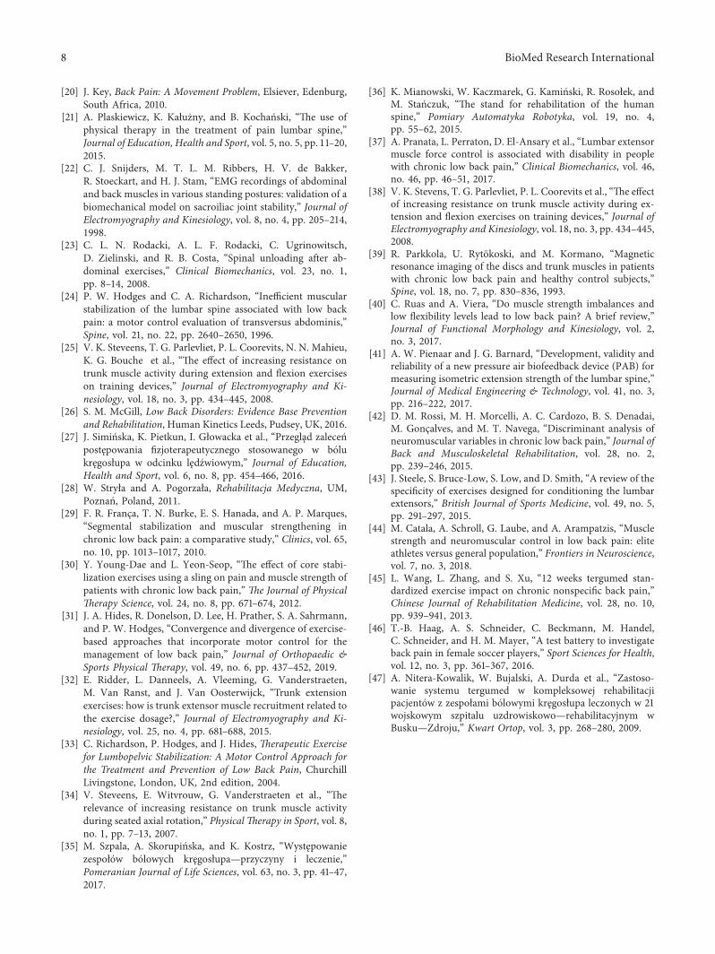
After 2 weeks of therapy, there were significant(p � 0.03) differences in the strength of the rotator musclesto the left of the lumbar region (Nm). In group A, there wasgreater improvement in muscle strength responsible forspinal rotation to the left (40.2Nm) compared to the controlgroup (28.28Nm) (Table 4).
After 4 weeks of therapy, a significantly (p � 0.03) betterresult was observed in the study group (A) compared to thecontrol group (B) in the strength of the left rotator muscles.3e test group (A) achieved an average result of 42.353Nmand the control group (B) 31.42Nm (Table 4). In addition, asignificantly (p � 0.01) better result was observed in thestrength of the muscles rotating the lumbar spine to the rightin group A. In the study group (A), this force was 45.43Nmand in the control group (B), 31.82Nm (Table 4).
BioMed Research International 3
After 6 weeks of therapy, the difference between groupsin left rotator muscle strength remained (p � 0.02) in favourof the study group (A). On average, in the study group (A),the result was 46.01Nm and in the control group (B),33.1Nm (Table 4). 3is also applies to extensor musclestrength (p � 0.02), which in the study group (A) increasedto 159.6Nm compared to the result in the control group (B)of 121.68Nm (Table 4). 3ere was also a significant
(p � 0.02) difference in lumbar spine flexor muscle strengthto the right in favour of the study group. In the study group(A), the average value of this force was 52.07Nm and in thecontrol group (B), 38.92Nm (Table 4).
A significant (p � 0.03) difference was also observed inthe balance of lumbar flexor muscle strength to the leftcompared with the right flexor muscle strength. Imbalancein the study group (A) remained within normal limits (1.05
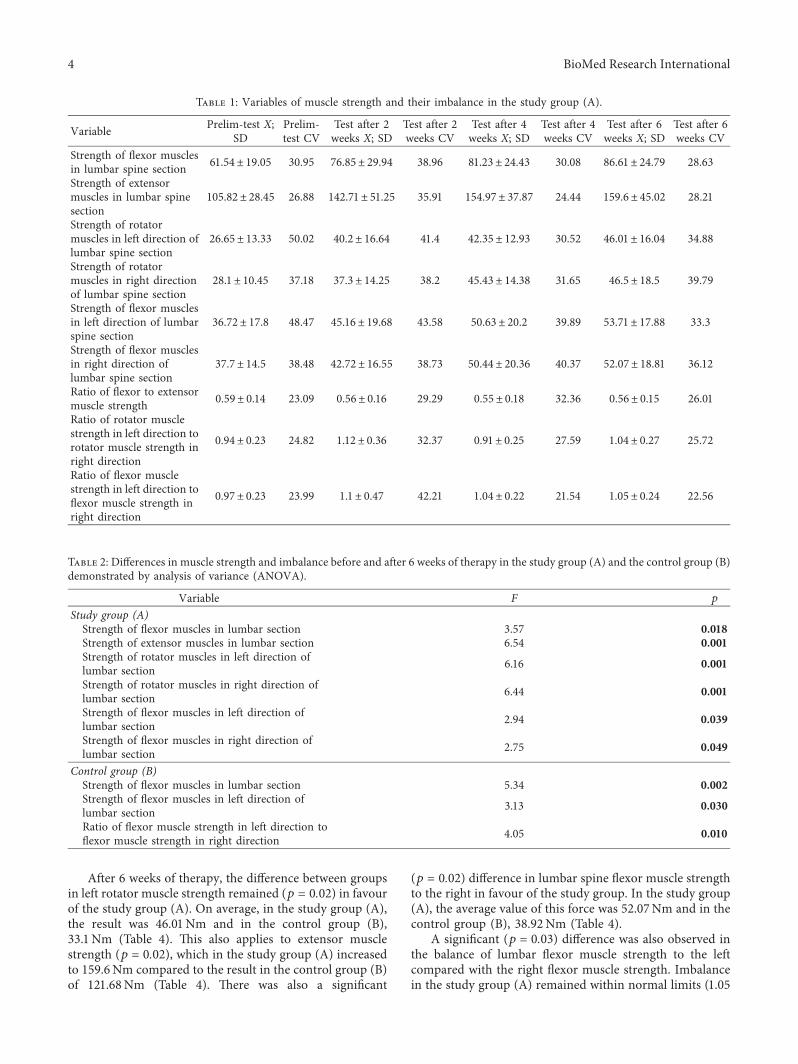
Table 2: Differences in muscle strength and imbalance before and after 6 weeks of therapy in the study group (A) and the control group (B)demonstrated by analysis of variance (ANOVA).
Variable F p
Study group (A)Strength of flexor muscles in lumbar section 3.57 0.018Strength of extensor muscles in lumbar section 6.54 0.001Strength of rotator muscles in left direction oflumbar section 6.16 0.001
Strength of rotator muscles in right direction oflumbar section 6.44 0.001
Strength of flexor muscles in left direction oflumbar section 2.94 0.039
Strength of flexor muscles in right direction oflumbar section 2.75 0.049
Control group (B)Strength of flexor muscles in lumbar section 5.34 0.002Strength of flexor muscles in left direction oflumbar section 3.13 0.030
Ratio of flexor muscle strength in left direction toflexor muscle strength in right direction 4.05 0.010
Table 1: Variables of muscle strength and their imbalance in the study group (A).
Variable Prelim-test X;SD
Prelim-test CV
Test after 2weeks X; SD
Test after 2weeks CV
Test after 4weeks X; SD
Test after 4weeks CV
Test after 6weeks X; SD
Test after 6weeks CV
Strength of flexor musclesin lumbar spine section 61.54± 19.05 30.95 76.85± 29.94 38.96 81.23± 24.43 30.08 86.61± 24.79 28.63
Strength of extensormuscles in lumbar spinesection
105.82± 28.45 26.88 142.71± 51.25 35.91 154.97± 37.87 24.44 159.6± 45.02 28.21
Strength of rotatormuscles in left direction oflumbar spine section
26.65± 13.33 50.02 40.2± 16.64 41.4 42.35± 12.93 30.52 46.01± 16.04 34.88
Strength of rotatormuscles in right directionof lumbar spine section
28.1± 10.45 37.18 37.3± 14.25 38.2 45.43± 14.38 31.65 46.5± 18.5 39.79
Strength of flexor musclesin left direction of lumbarspine section
36.72± 17.8 48.47 45.16± 19.68 43.58 50.63± 20.2 39.89 53.71± 17.88 33.3
Strength of flexor musclesin right direction oflumbar spine section
37.7± 14.5 38.48 42.72± 16.55 38.73 50.44± 20.36 40.37 52.07± 18.81 36.12
Ratio of flexor to extensormuscle strength 0.59± 0.14 23.09 0.56± 0.16 29.29 0.55± 0.18 32.36 0.56± 0.15 26.01
Ratio of rotator musclestrength in left direction torotator muscle strength inright direction
0.94± 0.23 24.82 1.12± 0.36 32.37 0.91± 0.25 27.59 1.04± 0.27 25.72
Ratio of flexor musclestrength in left direction toflexor muscle strength inright direction
0.97± 0.23 23.99 1.1± 0.47 42.21 1.04± 0.22 21.54 1.05± 0.24 22.56
4 BioMed Research International
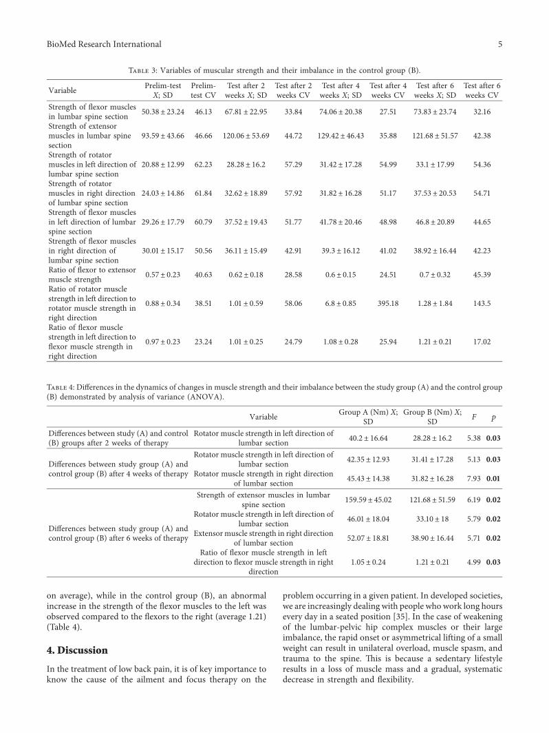
on average), while in the control group (B), an abnormalincrease in the strength of the flexor muscles to the left wasobserved compared to the flexors to the right (average 1.21)(Table 4).
4. Discussion
In the treatment of low back pain, it is of key importance toknow the cause of the ailment and focus therapy on the
problem occurring in a given patient. In developed societies,we are increasingly dealing with people who work long hoursevery day in a seated position [35]. In the case of weakeningof the lumbar-pelvic hip complex muscles or their largeimbalance, the rapid onset or asymmetrical lifting of a smallweight can result in unilateral overload, muscle spasm, andtrauma to the spine. 3is is because a sedentary lifestyleresults in a loss of muscle mass and a gradual, systematicdecrease in strength and flexibility.
Table 3: Variables of muscular strength and their imbalance in the control group (B).
Variable Prelim-testX; SD
Prelim-test CV
Test after 2weeks X; SD
Test after 2weeks CV
Test after 4weeks X; SD
Test after 4weeks CV
Test after 6weeks X; SD
Test after 6weeks CV
Strength of flexor musclesin lumbar spine section 50.38± 23.24 46.13 67.81± 22.95 33.84 74.06± 20.38 27.51 73.83± 23.74 32.16
Strength of extensormuscles in lumbar spinesection
93.59± 43.66 46.66 120.06± 53.69 44.72 129.42± 46.43 35.88 121.68± 51.57 42.38
Strength of rotatormuscles in left direction oflumbar spine section
20.88± 12.99 62.23 28.28± 16.2 57.29 31.42± 17.28 54.99 33.1± 17.99 54.36
Strength of rotatormuscles in right directionof lumbar spine section
24.03± 14.86 61.84 32.62± 18.89 57.92 31.82± 16.28 51.17 37.53± 20.53 54.71
Strength of flexor musclesin left direction of lumbarspine section
29.26± 17.79 60.79 37.52± 19.43 51.77 41.78± 20.46 48.98 46.8± 20.89 44.65
Strength of flexor musclesin right direction oflumbar spine section
30.01± 15.17 50.56 36.11± 15.49 42.91 39.3± 16.12 41.02 38.92± 16.44 42.23
Ratio of flexor to extensormuscle strength 0.57± 0.23 40.63 0.62± 0.18 28.58 0.6± 0.15 24.51 0.7± 0.32 45.39
Ratio of rotator musclestrength in left direction torotator muscle strength inright direction
0.88± 0.34 38.51 1.01± 0.59 58.06 6.8± 0.85 395.18 1.28± 1.84 143.5
Ratio of flexor musclestrength in left direction toflexor muscle strength inright direction
0.97± 0.23 23.24 1.01± 0.25 24.79 1.08± 0.28 25.94 1.21± 0.21 17.02
Table 4: Differences in the dynamics of changes in muscle strength and their imbalance between the study group (A) and the control group(B) demonstrated by analysis of variance (ANOVA).
Variable Group A (Nm) X;SD
Group B (Nm) X;SD F p
Differences between study (A) and control(B) groups after 2 weeks of therapy
Rotator muscle strength in left direction oflumbar section 40.2± 16.64 28.28± 16.2 5.38 0.03
Differences between study group (A) andcontrol group (B) after 4 weeks of therapy
Rotator muscle strength in left direction oflumbar section 42.35± 12.93 31.41± 17.28 5.13 0.03
Rotator muscle strength in right directionof lumbar section 45.43± 14.38 31.82± 16.28 7.93 0.01
Differences between study group (A) andcontrol group (B) after 6 weeks of therapy
Strength of extensor muscles in lumbarspine section 159.59± 45.02 121.68± 51.59 6.19 0.02
Rotator muscle strength in left direction oflumbar section 46.01± 18.04 33.10± 18 5.79 0.02
Extensormuscle strength in right directionof lumbar section 52.07± 18.81 38.90± 16.44 5.71 0.02
Ratio of flexor muscle strength in leftdirection to flexor muscle strength in right
direction1.05± 0.24 1.21± 0.21 4.99 0.03
BioMed Research International 5

Most studies available in the literature regarding phys-iotherapy in low back pain assessing individual methods ortherapies are based on the evaluation of the training pro-gramme. Patients are examined before and after therapy, andin some cases, additionally in the middle of the therapy [36].In this study, the authors evaluated the dynamics of changesin the strength of the lumbar-pelvic hip complex musclesand the equalization of their imbalances 4 times: beforetherapy, and after 2, 4, and 6 weeks of its duration.
Many authors have evaluated the effects of treating lowback pain similarly as in the authors’ research. Pranata et al.[37] investigated the coordination of lumbar extensormuscle work in patients with low back pain. Biofeedback wasused in the form of a sinusoid, on which the indicator of theisometric force with which the patient exercised moved.Maximum values during the exercise oscillated between 20%and 50% of the patient’s maximum isometric strength. Amuch smaller degree of sinusoid mapping (both voltageincrease and relaxation) was observed in comparison to thecontrol group without low back pain. 3ere were alsocorrelations between the increase in sinusoidal mappingduring the return to the starting position and the increase indisability measured by the Oswestry questionnaire. In thesestudies, it has been shown that the control of extensormuscles in the lumbar region is impaired among patientswith low back pain. Training using the Tergumed 700 systemapplied by the authors of this study was also based onbiofeedback in the form of a sinusoid, which, as it has beenshown in studies, has a positive effect on impaired musclecoordination.
França et al. [29] compared central stabilization trainingwith strength training in fighting back pain, reducing dis-ability and activating the transverse abdominal muscle. Bothtypes of training gave satisfactory results, but central sta-bilization training proved to be more effective, mainly in thearea of transverse abdominal muscle activation. However, asstated by Stevens et al. [38], strength-coordination trainingwith the use of the Tergumed 700 system also activates themultifunctional muscle, especially when loaded with 30% ofthe maximum extension force. In these studies, it has beenshown that strength training also affects deep muscle ac-tivity. In the research conducted by Parkkola et al. [39], itwas also indicated that patients with low back pain hadweakened muscles compared to the control group, asdemonstrated by the isometric force test. Ruas and Viera[40] conducted a study on muscle strength and imbalance inlow back pain, the results of which demonstrated that im-balance, mainly of flexor muscle strength relative to extensormuscle strength, may be associated with chronic lumbarspine pain.
Although pain syndromes are a complex and multi-factorial problem, many authors associate them with muscleweakness [41]. As reported by Rossi et al. [42], therapy ofback pain syndromes should include training of the effi-ciency and strength of muscles, mainly of the extensors. Asreported by Steele et al. [43], such training should beconducted with the pelvis stabilized so as to exclude theinvolvement of other muscles, for example, the hip exten-sors. From the research by Catala et al. [44], it may be
assumed that dorsal muscle training is beneficial in reducinglumbar pain among patients with low back pain. Patientswho experience lumbar pain due to lumbar pain syndromehave reduced strength in their trunk muscles, mainly theextensors [44]. 3e legitimacy of the authors’ research is alsoconfirmed by other authors. Wang et al. [45] conducted astudy regarding the impact of a 12-week standardizedtraining programme on patients with low back pain. 3eresults of the study showed significant improvement inmuscular strength as well as compensation of flexion andextensor muscles of the lumbar region. In this study, thepositive effect of training using the Tergumed system onmuscle strength has been exhibited. However, a disadvan-tage of this study was the lack of precise specification of thegroup of subjects. Haag et al. [46] used the Tergumed systemin their research to assess the strength of the dorsal musclesin athletes complaining of and not reporting pain in thelumbar spine. In the second group without pain, signifi-cantly higher isometric strength of the trunk muscles wasobserved. Nitera-Kowalik et al. [47] conducted studies onthe impact of comprehensive therapy using the Tergumedsystem on improving coordination, compensating formuscular imbalances, the degree of disability caused by lowback pain, and reducing pain sensations in patients treated atsanatoriums. Improvement in muscle imbalances was ob-served here for all of the examined muscle groups. However,this study did not include a control group.
In this study, in group A, in which the Tergumed 700system was additionally used, there was a significant im-provement in the strength of all the examinedmuscle groupsafter 6 weeks of therapy. Increased muscle strength re-sponsible for extension of the lumbar spine and rotation tothe left occurred after 2 weeks. 3is maintained after 4 and 6weeks of therapy. 3e strength of the muscles rotatingclockwise improved after 4 weeks and wasmaintained after 6weeks of therapy. On the other hand, the strength of thetrunk flexor muscles and those responsible for lateral flexionimproved in the final measurement period. 3is suggeststhat the 6-week training programme is optimal for achievingstrength improvement in all of the examined muscle groups.
3e definitely worse results obtained in muscle strengthin the control group (B) suggest that traditional centralstabilization training has less of an effect on muscle strength.3e differences between groups A and B became apparentafter 2 weeks of therapy in terms of the force of rotation tothe left. 3en, after 4 and 6 weeks of therapy, the differenceconcerned the strength of both-sided rotation, extension,and flexion to the right. Muscle imbalance of the lumbar-pelvis-hip complex in the study group (A) remained withinnormal limits (1.05 on average), while in the control group(B), an abnormal increase in the strength of the flexormuscles to the left compared with the flexors to the right(average 1.21) was observed.
3erapy using the Tergumed system, through its pro-gramming based on objective patient examination, is ef-fective in treating low back pain. Its use is also supported byeconomic considerations because one therapist can simul-taneously rehabilitate 4 patients according to individualprogrammes. It is also important that objective examination
6 BioMed Research International

before and after therapy allows verification of the appliedtherapeutic programme.
5. Conclusions
After 6 weeks of therapy in the study group (A), there was asignificant improvement in the strength of all examinedmuscle groups. However, in the control group (B), signifi-cant improvement only occurred in the strength of thelumbar flexor and flexor muscles on the left side. In addition,in group B, there was significant deterioration of imbalanceregarding the left flexor muscle strength compared to theright flexor strength. Significant differences in favour of thestudy group (A) concerned the strength of the rotatormuscles to the left, the strength of the extensor muscles ofthe lumbar spine, the strength of the flexors of the lumbarspine to the right, and the balance of strength of the flexors ofthe lumbar spine to the left compared with the strength ofthe flexor muscles to the right. 3erapy with the Tergumed700 system leads to an increase in the muscle strength of thelumbar-pelvic complex and compensation for its imbalance,which provides beneficial effects in the treatment of low backpain.
Data Availability
3e data and materials supporting the conclusions of thisarticle are included within the article.
Conflicts of Interest
3e authors declare no conflicts of interest regarding thepublication of this paper.
Authors’ Contributions
J. W. was responsible for conceptualisation, data collection,analysis, formal analysis, methodology, and writing theoriginal draft; A. K. was responsible for data collection,formal analysis, writing, reviewing, and editing.
Acknowledgments
3is project was supported within the framework of theprogramme funded by the Minister of Science and HigherEducation under the name “Regional Initiative of Excel-lence” in 2019–2022 (project number: 024/RID/2018/19).
References
[1] T. Vos, A. D. Flaxman, M. Naghavi et al., “Years lived withdisability (YLDs) for 1160 sequelae of 289 diseases and in-juries 1990–2010: a systematic analysis for the global burdenof disease study 2010,” -e Lancet, vol. 380, no. 9859,pp. 2163–2196, 2012.
[2] N. J. Manek and A. J. MacGregor, “Epidemiology of backdisorders: prevalence, risk factors, and prognosis,” CurrentOpinion in Rheumatology, vol. 17, no. 17, pp. 134–140, 2005.
[3] L. Lidgren, “3e bone and joint decade 2000–2010,” BullWorld Health Organ, vol. 81, no. 9, p. 629, 2003.
[4] D. I. Rubin, “Epidemiology and risk factors for spine pain,”Neurologic Clinics, vol. 25, no. 2, pp. 353–371, 2007.
[5] P. M. Kent and J. L. Keating, “3e epidemiology of low backpain in primary care,” Chiropractic & Osteopathy, vol. 13,no. 1, pp. 13–20, 2005.
[6] M. H. Halliday, A. N. Garcia, A. B. Amorim et al., “Treatmenteffect sizes of mechanical diagnosis and therapy for pain anddisability in patients with low back pain: a systematic review,”Journal of Orthopaedic & Sports Physical -erapy, vol. 49,no. 4, pp. 219–229, 2019.
[7] T. Kienbacher, E. Fehrmann, R. Habenicht et al., “Diagnosticvalue of trunk flexion-extension testing in old chronic lowback pain patients,” European Spine Journal, vol. 26, no. 2,pp. 510–517, 2017.
[8] P. Watson, C. Booker, C. Main, and A. Chen, “Surfaceelectromyography in the identification of chronic low backpain patients: the development of the flexion relaxation ratio,”Clinical Biomechanics, vol. 12, no. 3, pp. 165–171, 1997.
[9] A. Bergmark, “Stability of lumbar spine: a study in mechanicalengineering,” Acta Orthopaedica Scandinavica, vol. 60,no. 230, pp. 1–54, 1989.
[10] K. Rose-Dulcina, S. Genevay, D. E. Dominguez, S. Armand,and N. Vuillerme, “O 086–relation between the flexion-re-laxation phenomenon and back extensor endurance in non-specific chronic low back pain patients,” Gait & Posture,vol. 65, no. 1, pp. 176-177, 2018.
[11] Y. Javadian, M. Akbari, G. Talebi et al., “Influence of corestability exercise on lumbar vertebral instability in patientspresented with chronic low back pain: a randomized clinicaltrial,” Caspian Journal of Internal Medicine, vol. 6, no. 2,pp. 98–102, 2015.
[12] P. Li, Y. Nie, J. Chen, and N. Ning, “Application progress ofsurface electromyography and surface electromyographicbiofeedback in low back pain,” Chinese Journal of Reparativeand Reconstructive Surgery, vol. 31, no. 4, pp. 504–507, 2017.
[13] H. S. Desai and R. S. Bisen, “Lumbar flexion relaxationphenomenon in the patients with acute and subacute me-chanical low back pain and normal subjects,” InternationalJournal of Research in Medical Sciences, vol. 5, no. 3,pp. 1011–1014, 2017.
[14] C. Amroz, A. Scott, A. Ambroz, and E. O. Talbott, “Chroniclow back pain assessment using surface electromyography,”Journal of Occupational and Environmental Medicine, vol. 42,no. 6, pp. 660–669, 2000.
[15] F. Zhou, H. Li, G. Song et al., “Analysis of surface electro-myography of patients with low back pain based on differentmovement patterns,” in Proceedings of the 2016 IEEE Inter-national Conference on Information and Automation,pp. 1154–1158, Ningbo, China, 2016.
[16] J. Abboud, F. Nougarou, I. Page, V. Cantin, D.Massicotte, andM. Descarreaux, “Trunk motor variability in patients withnon-specific chronic low back pain,” European Journal ofApplied Physiology, vol. 114, no. 12, pp. 2645–2654, 2014.
[17] J. P. Correia, R. Oliveira, J. R. Vaz, L. Silva, and P. Pezarat-Correia, “Trunk muscle activation, fatigue and low back painin tennis players,” Journal of Science and Medicine in Sport,vol. 19, no. 4, pp. 311–316, 2016.
[18] A. Heydari, A. V. F. Nargol, A. P. C. Jones, A. R. Humphrey,and C. G. Greenough, “EMG analysis of lumbar paraspinalmuscles as a predictor of the risk of low-back pain,” EuropeanSpine Journal, vol. 19, no. 7, pp. 1145–1152, 2010.
[19] M. M. Panjabi, “3e stabilizing system of the spine: partI—function, dysfunction, adaptation, and enhancement,”Journal of Spinal Disorders, vol. 5, no. 4, pp. 383–389, 1992.
BioMed Research International 7
[20] J. Key, Back Pain: A Movement Problem, Elsiever, Edenburg,South Africa, 2010.
[21] A. Plaskiewicz, K. Kałuzny, and B. Kochanski, “3e use ofphysical therapy in the treatment of pain lumbar spine,”Journal of Education, Health and Sport, vol. 5, no. 5, pp. 11–20,2015.
[22] C. J. Snijders, M. T. L. M. Ribbers, H. V. de Bakker,R. Stoeckart, and H. J. Stam, “EMG recordings of abdominaland back muscles in various standing postures: validation of abiomechanical model on sacroiliac joint stability,” Journal ofElectromyography and Kinesiology, vol. 8, no. 4, pp. 205–214,1998.
[23] C. L. N. Rodacki, A. L. F. Rodacki, C. Ugrinowitsch,D. Zielinski, and R. B. Costa, “Spinal unloading after ab-dominal exercises,” Clinical Biomechanics, vol. 23, no. 1,pp. 8–14, 2008.
[24] P. W. Hodges and C. A. Richardson, “Inefficient muscularstabilization of the lumbar spine associated with low backpain: a motor control evaluation of transversus abdominis,”Spine, vol. 21, no. 22, pp. 2640–2650, 1996.
[25] V. K. Steveens, T. G. Parlevliet, P. L. Coorevits, N. N. Mahieu,K. G. Bouche et al., “3e effect of increasing resistance ontrunk muscle activity during extension and flexion exerciseson training devices,” Journal of Electromyography and Ki-nesiology, vol. 18, no. 3, pp. 434–445, 2008.
[26] S. M. McGill, Low Back Disorders: Evidence Base Preventionand Rehabilitation, Human Kinetics Leeds, Pudsey, UK, 2016.
[27] J. Siminska, K. Pietkun, I. Głowacka et al., “Przeglad zalecenpostepowania fizjoterapeutycznego stosowanego w bolukregosłupa w odcinku ledzwiowym,” Journal of Education,Health and Sport, vol. 6, no. 8, pp. 454–466, 2016.
[28] W. Stryła and A. Pogorzała, Rehabilitacja Medyczna, UM,Poznan, Poland, 2011.
[29] F. R. França, T. N. Burke, E. S. Hanada, and A. P. Marques,“Segmental stabilization and muscular strengthening inchronic low back pain: a comparative study,” Clinics, vol. 65,no. 10, pp. 1013–1017, 2010.
[30] Y. Young-Dae and L. Yeon-Seop, “3e effect of core stabi-lization exercises using a sling on pain and muscle strength ofpatients with chronic low back pain,” -e Journal of Physical-erapy Science, vol. 24, no. 8, pp. 671–674, 2012.
[31] J. A. Hides, R. Donelson, D. Lee, H. Prather, S. A. Sahrmann,and P. W. Hodges, “Convergence and divergence of exercise-based approaches that incorporate motor control for themanagement of low back pain,” Journal of Orthopaedic &Sports Physical -erapy, vol. 49, no. 6, pp. 437–452, 2019.
[32] E. Ridder, L. Danneels, A. Vleeming, G. Vanderstraeten,M. Van Ranst, and J. Van Oosterwijck, “Trunk extensionexercises: how is trunk extensor muscle recruitment related tothe exercise dosage?,” Journal of Electromyography and Ki-nesiology, vol. 25, no. 4, pp. 681–688, 2015.
[33] C. Richardson, P. Hodges, and J. Hides, -erapeutic Exercisefor Lumbopelvic Stabilization: A Motor Control Approach forthe Treatment and Prevention of Low Back Pain, ChurchillLivingstone, London, UK, 2nd edition, 2004.
[34] V. Steveens, E. Witvrouw, G. Vanderstraeten et al., “3erelevance of increasing resistance on trunk muscle activityduring seated axial rotation,” Physical -erapy in Sport, vol. 8,no. 1, pp. 7–13, 2007.
[35] M. Szpala, A. Skorupinska, and K. Kostrz, “Wystepowaniezespołow bolowych kregosłupa—przyczyny i leczenie,”Pomeranian Journal of Life Sciences, vol. 63, no. 3, pp. 41–47,2017.
[36] K. Mianowski, W. Kaczmarek, G. Kaminski, R. Rosołek, andM. Stanczuk, “3e stand for rehabilitation of the humanspine,” Pomiary Automatyka Robotyka, vol. 19, no. 4,pp. 55–62, 2015.
[37] A. Pranata, L. Perraton, D. El-Ansary et al., “Lumbar extensormuscle force control is associated with disability in peoplewith chronic low back pain,” Clinical Biomechanics, vol. 46,no. 46, pp. 46–51, 2017.
[38] V. K. Stevens, T. G. Parlevliet, P. L. Coorevits et al., “3e effectof increasing resistance on trunk muscle activity during ex-tension and flexion exercises on training devices,” Journal ofElectromyography and Kinesiology, vol. 18, no. 3, pp. 434–445,2008.
[39] R. Parkkola, U. Rytokoski, and M. Kormano, “Magneticresonance imaging of the discs and trunk muscles in patientswith chronic low back pain and healthy control subjects,”Spine, vol. 18, no. 7, pp. 830–836, 1993.
[40] C. Ruas and A. Viera, “Do muscle strength imbalances andlow flexibility levels lead to low back pain? A brief review,”Journal of Functional Morphology and Kinesiology, vol. 2,no. 3, 2017.
[41] A. W. Pienaar and J. G. Barnard, “Development, validity andreliability of a new pressure air biofeedback device (PAB) formeasuring isometric extension strength of the lumbar spine,”Journal of Medical Engineering & Technology, vol. 41, no. 3,pp. 216–222, 2017.
[42] D. M. Rossi, M. H. Morcelli, A. C. Cardozo, B. S. Denadai,M. Gonçalves, and M. T. Navega, “Discriminant analysis ofneuromuscular variables in chronic low back pain,” Journal ofBack and Musculoskeletal Rehabilitation, vol. 28, no. 2,pp. 239–246, 2015.
[43] J. Steele, S. Bruce-Low, S. Low, and D. Smith, “A review of thespecificity of exercises designed for conditioning the lumbarextensors,” British Journal of Sports Medicine, vol. 49, no. 5,pp. 291–297, 2015.
[44] M. Catala, A. Schroll, G. Laube, and A. Arampatzis, “Musclestrength and neuromuscular control in low back pain: eliteathletes versus general population,” Frontiers in Neuroscience,vol. 7, no. 3, 2018.
[45] L. Wang, L. Zhang, and S. Xu, “12 weeks tergumed stan-dardized exercise impact on chronic nonspecific back pain,”Chinese Journal of Rehabilitation Medicine, vol. 28, no. 10,pp. 939–941, 2013.
[46] T.-B. Haag, A. S. Schneider, C. Beckmann, M. Handel,C. Schneider, and H. M. Mayer, “A test battery to investigateback pain in female soccer players,” Sport Sciences for Health,vol. 12, no. 3, pp. 361–367, 2016.
[47] A. Nitera-Kowalik, W. Bujalski, A. Durda et al., “Zastoso-wanie systemu tergumed w kompleksowej rehabilitacjipacjentow z zespołami bolowymi kregosłupa leczonych w 21wojskowym szpitalu uzdrowiskowo—rehabilitacyjnym wBusku—Zdroju,” Kwart Ortop, vol. 3, pp. 268–280, 2009.
8 BioMed Research International

Stem Cells International
Hindawiwww.hindawi.com Volume 2018
Hindawiwww.hindawi.com Volume 2018
MEDIATORSINFLAMMATION
of
EndocrinologyInternational Journal of
Hindawiwww.hindawi.com Volume 2018
Hindawiwww.hindawi.com Volume 2018
Disease Markers
Hindawiwww.hindawi.com Volume 2018
BioMed Research International
OncologyJournal of
Hindawiwww.hindawi.com Volume 2013
Hindawiwww.hindawi.com Volume 2018
Oxidative Medicine and Cellular Longevity
Hindawiwww.hindawi.com Volume 2018
PPAR Research
Hindawi Publishing Corporation http://www.hindawi.com Volume 2013Hindawiwww.hindawi.com
The Scientific World Journal
Volume 2018
Immunology ResearchHindawiwww.hindawi.com Volume 2018
Journal of
ObesityJournal of
Hindawiwww.hindawi.com Volume 2018
Hindawiwww.hindawi.com Volume 2018
Computational and Mathematical Methods in Medicine
Hindawiwww.hindawi.com Volume 2018
Behavioural Neurology
OphthalmologyJournal of
Hindawiwww.hindawi.com Volume 2018
Diabetes ResearchJournal of
Hindawiwww.hindawi.com Volume 2018
Hindawiwww.hindawi.com Volume 2018
Research and TreatmentAIDS
Hindawiwww.hindawi.com Volume 2018
Gastroenterology Research and Practice
Hindawiwww.hindawi.com Volume 2018
Parkinson’s Disease
Evidence-Based Complementary andAlternative Medicine
Volume 2018Hindawiwww.hindawi.com
Submit your manuscripts atwww.hindawi.com