Dynamic Brain Changes in Autism: Review of Telencephalic...
22
Dynamic Brain Changes in Autism: Review of Telencephalic Structures Efrain C. Azmitia and Allyson Impallomeni Introduction This chapter will highlight postmortem studies that utilized immunocytochemical techniques to identify the morphological, cellular, and molecular differences that exist in individuals with autism compared to typically developing individuals. Two main themes emerge from our analysis of postmortem studies. (1) Many cellular changes of autism begin neonatally and persist throughout the adolescent years. (2) The changes globally affect cortical and subcortical centers, pathways, and multiple cell types located throughout the telencephalon. The pervasive overgrowth of the brain in children and adolescences was first recognized as a contributing factor to the pathology of autism nearly 15 years ago. Postmortem brains from 12 autistic donors, age 5–13, weighed significantly more by approximately 100–200 g (Kemper and Bauman 1998). However within the same study, they examined eight autistic adults, ages 18–54, and found no significant weight difference. More recent studies suggest that the fastest growth initiates during the first year of life and continues until toddler age, proceeded by a slowing or degeneration process beginning in the adolescent period (Courchesne et al. 2003, 2011a). Therefore, the active developmental abnormalities extend from birth to adolescence and involve accelerated growth and decline. Telencephalic regions associated with affective interpretation, social perspective, and communication have all been linked to the pathology of autism. This includes the frontal (including Broca’s language area, the motor speech center), temporal (including the auditory primary cortex), and parietal (including Wernicke’s language area) lobes as well as the subcortical areas of the amygdala, caudate, and basal ganglion. Major fiber pathways impli- cated in autism include the medial forebrain pathway, ansa lenticularis, internal capsule, and corpus callosum. There are reports on glutamate pyramidal neurons, E.C. Azmitia (*) • A. Impallomeni Department of Biology, New York University, New York, NY, USA e-mail: [email protected]; [email protected]; [email protected] V.B. Patel et al. (eds.), Comprehensive Guide to Autism, DOI 10.1007/978-1-4614-4788-7_33, # Springer Science+Business Media New York 2014 695
Transcript of Dynamic Brain Changes in Autism: Review of Telencephalic...

Dynamic Brain Changes in Autism: Reviewof Telencephalic Structures
Efrain C. Azmitia and Allyson Impallomeni
Introduction
This chapter will highlight postmortem studies that utilized immunocytochemical
techniques to identify the morphological, cellular, and molecular differences that
exist in individuals with autism compared to typically developing individuals. Two
main themes emerge from our analysis of postmortem studies. (1) Many cellular
changes of autism begin neonatally and persist throughout the adolescent years.
(2) The changes globally affect cortical and subcortical centers, pathways, and
multiple cell types located throughout the telencephalon. The pervasive overgrowth
of the brain in children and adolescences was first recognized as a contributing
factor to the pathology of autism nearly 15 years ago. Postmortem brains from 12
autistic donors, age 5–13, weighed significantly more by approximately 100–200 g
(Kemper and Bauman 1998). However within the same study, they examined eight
autistic adults, ages 18–54, and found no significant weight difference. More recent
studies suggest that the fastest growth initiates during the first year of life and
continues until toddler age, proceeded by a slowing or degeneration process
beginning in the adolescent period (Courchesne et al. 2003, 2011a). Therefore,
the active developmental abnormalities extend from birth to adolescence and
involve accelerated growth and decline. Telencephalic regions associated with
affective interpretation, social perspective, and communication have all been linked
to the pathology of autism. This includes the frontal (including Broca’s language
area, the motor speech center), temporal (including the auditory primary cortex),
and parietal (including Wernicke’s language area) lobes as well as the subcortical
areas of the amygdala, caudate, and basal ganglion. Major fiber pathways impli-
cated in autism include the medial forebrain pathway, ansa lenticularis, internal
capsule, and corpus callosum. There are reports on glutamate pyramidal neurons,
E.C. Azmitia (*) • A. Impallomeni
Department of Biology, New York University, New York, NY, USA
e-mail: [email protected]; [email protected]; [email protected]
V.B. Patel et al. (eds.), Comprehensive Guide to Autism,DOI 10.1007/978-1-4614-4788-7_33,# Springer Science+Business Media New York 2014
695

GABA interneurons, serotonin axons, and glial cell involvement (astrocytes, oli-
godendroglial, and microglial cells) in these postmortem studies.
The global cellular disturbances and its unusual developmental progression
require a fresh interpretation of this disorder based on neuroplasticity as well as
neurodevelopment principles. The heterogeneous nature of autism corresponds
with a wide variation in brain morphology, so focusing on a particular region can
only convey a small part of the disorder in its entirety (Amaral et al. 2008).
Unfortunately, neuroanatomical studies tend to be restrictive by their very nature.
One of the difficulties in writing this chapter is that the postmortem studies all
provide glimpses of the total pictures. We hope to promote the idea that the global
neuronal systems require attention in order to potentially provide the necessary
cohesion. Global neurons such as serotonin, dopamine, norepinephrine, and ace-
tylcholine have axonal projections throughout the telencephalon, which are
established early in development. These neurotransmitter neurons continue to
mature throughout the adolescent period as evidenced by increases in axonal pro-
jections and changes in receptor expression. These chemical neurotransmitters
regulate target cell proliferation in the subventricular zone and maturation of the
neurons in the cortical columns either directly or by modifying trophic compounds
such as BDNF and S100B (Azmitia 1999). In the autism brain, the serotonin
neurons have an acceleration of fiber growth in childhood (Azmitia et al. 2011a)
and have dystrophic fibers in adolescence (2011b). The dystrophic fibers appear
similar to those described in neurodegenerative diseases (Azmitia and Nixon 2008).
In this chapter we will attempt to provide a cohesive view of the autism changes
examined in the postmortem brain and identify both the advantages and disadvan-
tages of using this material. There is no substitute for careful microscopic exami-
nation of brain cells, and the studies by Bailey et al. (1998) and Wegiel et al. (2010)
are good examples of this type of descriptive analysis. However the use of post-
mortem tissue should be considered only as a necessary first step that requires
validation in imaging studies and in live patients, and can serve as a model for
animal research. However, identification of the cellular targets is first done with the
microscope and only later can therapeutic strategies be developed for use in
patients. To attempt pharmacological interventions without evidence of a cellular
or molecular target is not only random but can be counterproductive to the pro-
gression of the disease.
Cortical Minicolumnar Organization
Cortical minicolumns are the main functional organization pattern in cortex and
clearly reflect developmental conditions of brain growth. Neuronal organization and
specialization into minicolumns are abnormal in the autistic cortex (Casanova et al.
2006a). In a small sample of six patients with autism and six controls, ages 4–25,
significant alterations in minicolumns were detected. Brains were stained with
gallocyanin to identify the frontopolar cortex, orbitofrontal cortex, dorsolateral
prefrontal cortex (PFC), primary motor cortex, primary sensory cortex, fronto-insular
696 E.C. Azmitia and A. Impallomeni

cortex, ventrolateral cortex (part of Broca’s speech area), ACC, and primary visual
cortex. Neuropil space was significantly greater in both the frontopolar area and
anterior cingulate area. The two associated regions are considered regions of the
prefrontal lobe and typically linked through numerous connections. Neuropil space
was not significantly reduced in the dorsolateral PFC or the primary visual cortex.
In another study, minicolumnar abnormalities were found in cortical areas M1,
V1, frontal association cortex, and S1 in six autism patients with age-matched
controls (ages 4–25). Overall, the minicolumnar width was 27.2 um in controls and
25.7 in autism patients, a 5.54 % reduction (Casanova et al. 2006b). Mean neuron
cross section was 30.5 um2 smaller in the autistic cases; however, neuron density
was 23 % greater in autism as well. In the autism brains, the minicolumns appear to
contain more but smaller neurons that are more tightly packed. In general, these
studies support the idea that cell proliferation is increased in the cortex from autism
patients as old as 25 years of age.
Frontal Cortex
The cortical regions can be roughly divided into white (mainly axons and oligoden-
droglial cells) and gray (neuronal and glial cell bodies) matter. White matter in the
frontal lobe is typically enlarged in children with autism, whereas adults with autism
do not exhibit this increase. The changes in white matter of autism patients suggest
“decreased functional connectivity among brain areas, desynchronization of cortical
activity, and changes in the fractional anisotropy of the white matter” (Zikopoulos and
Barbas 2010, 14595). This postmortem observation is consistent with earlier
quantitative data gathered with the use of MRI scanning (Carper et al. 2002).
The data shows that the greatest amount of hyperplasia occurs in the white matter
of the frontal, temporal, and parietal cortices, as well as the gray matter of the frontal
and temporal cortices. White matter volume was significantly greater in autistic
children compared to normally developing controls at the age of two. These network
abnormalities including neuronal connectivity and excitability tend to be notably
detected within the frontal cortex. The disruption noted in the white matter may
reflect weak or disorganized long-range cortico-cortical pathways that connect the
frontal areas with other cortices in the brain. In order to test this idea, the inner
diameter of axons was measured using EM analysis of postmortem tissue. Five autism
brains, ages 30–34, and 4 age-matched typically developing controls were compared.
Based on the analysis, significantly fewer extra-large axons were observed in
area 32 of the ACC in the autistic samples (Zikopoulos and Barbas 2010).
The occurrence of brain overgrowth was quantified by counting neurons from
the PFC. The dorsolateral (DL) and mesial (M) subdivisions of the PFC were
observed from seven autistic and six control male children between the ages of
2 and 6 years. The mean brain weight of the autistic children (1,484 g) was
approximately 2.4 % greater than that of the control group (1,449 g) (Courchesne
et al. 2011b). Although this particular deviation was not significant, brain weight in
autistic samples did differ from the normative mean weight for their age group by
Dynamic Brain Changes in Autism: Review of Telencephalic Structures 697

17.6 %, while control brains only deviated by 0.2 %. There was a brain weight
deviation of 29.4 % beyond their age-based norms, in relation to the regression line
calculated according to controls. This emphasizes the brain growth spurt in the first
years of life.
The brain sections from these children were examined with stereological
methods. An optical fractionator method calculated the total number and size of
neurons within the PFC. A change in neuron number was found to be significant
(Fig. 1). In the DL-PFC, there were 79 % more neurons in the autistic population
and 29 %more neurons in the M-PFC. In total, there were 67 %more neurons in the
Total combined prefrontal neuron count
2.50
2.00
1.50
1.00
0.50Autism
Cadaveric Donors
Tota
l Pre
fron
tal N
euro
n C
ount
(in
Bill
ions
)
Control
2.50
2.00
1.50
1.00
0.50
0.00Autism
Cadaveric Donors
Dorsolateral prefrontal cortex neuron counta
c
b Mesial prefrontal cortex neuron countTo
tal D
L-P
FC
Neu
ron
Cou
nt (
in B
illio
ns)
Control
0.50
0.40
0.30
0.20
0.10
0.00Autism
Cadaveric Donors
Tota
l M-P
FC
Neu
ron
Cou
nt (
in B
illio
ns)
Control
Autistic casesWith intellectual disabilityWithout intellectual disabilityControl casesGroup mean
Fig. 1 *Dorsolateral (DL-PFC) and Mesial PFC (M-PFC) Neuron Counts in Autism vs Control
Group Cases: Error bars indicate 95 % CIs. For between-group comparisons, statistical tests were
as follows: p <.003 for panel A, p <.009 for panel B, and p <.002 for panel C. Autistic case withlowest neuron count value in panels A and C had a seizure disorder, adverse perinatal medical
conditions, and intellectual disability (Reprinted from Courchesne et al. 2011b, with permission)
698 E.C. Azmitia and A. Impallomeni

PFC of autistic children (1.94 billion in comparison to 1.16 billion in the controls).
Interestingly, six out of the seven autistic children had a neuron count that exceeded
the expected amount of neurons for their brain weight. A problem with this study is
that no account of cortical layer was noted, so the granular neurons in layer II and
IV were grouped with the larger pyramidal neurons in layers II and V–VI. The
decrease in cell size and the increase in cell density may reflect a greater proportion
of granule versus pyramidal neurons in the two populations.
Temporal Lobe
The temporal lobe is a key target for studies of autism pathology because this region
contains many key language centers including the primary auditory sensory region
in the superior temporal cortex and sensory speech region in Wernicke’s area.
Despite the importance of the superior temporal cortex to language expression,
there are no group postmortem studies on the cell bodies in this area. In a study of
one 24-year-old subject, numerous neurofibrillary tangles were found at autopsy in
the perirhinal and entorhinal cortices, where they were frequently grouped in nests
or clusters (Hof et al. 1991). A few neurofibrillary tangles were also observed in the
amygdala as well as the prepiriform and orbitofrontal cortex. In the cortex, tangles
were located in both layers II and III. This case report provides some preliminary
suggestion of neuropathology in the autism cortex.
The piriform cortex, a face recognition region, is known to be involved in autism
dysfunction. The size and number of neurons in this brain region were examined in
layers II–VI in the fusiform cortex of autistic donors (aged 4–23 years) and ten
control donors (4–65 years) (van Kooten et al. 2008). The main findings of the
present study include a significant reduction in the mean neuron density in layer III
(–13.1 %), a reduced mean total neuron number in layers III, V, and VI (�23.7 %,
�14.3 %, and �10.6 %, respectively), and a decreased mean perikaryal volume of
neurons in layers V and VI in the FG (�21.1 % and �13.4 %, respectively) in the
brains of patients with autism compared to the controls. No changes in neuron
number or size were found in the visual cortex when all layers were combined.
Another study by this group focused on pyramidal neurons in Brodmann areas
44 and 45 in the inferior frontal cortex (Jacot-Descombes et al. 2012). This study
used eight postmortem brains obtained from patients with autism and seven from
age-matched controls (age range 4–66 years). In this region, significantly smaller
pyramidal neurons were seen in patients with autism in comparison to controls,
although there was no difference in pyramidal neuron numbers or layer volumes.
The results showed significantly smaller pyramidal neuron volume in layer III
(�18 %), in layer V (�18.5 %), and in layer VI (�22 %) when comparing autism to
control donors. Although control neurons appeared to decrease in size over age, the
autism neuronal size was relatively constant from 4 to 60 years.While this type of layer
and cell type analysis is important, the lack of dynamics of the findings is puzzling.
There is one study on the hippocampus, which was performed 17 years ago.
Neurons within the cornu ammonis (CA) layers of the hippocampus were found to
Dynamic Brain Changes in Autism: Review of Telencephalic Structures 699

be smaller in autism brain samples (Raymond et al. 1996). Sections of the hippo-
campus were stained using the rapid Golgi method. Two autism cases were
examined, ages 9 and 7, with appropriate age-matched controls, specifically ages
8 and 13. Large pyramidal neurons were studied in hippocampal CA1 and CA4
regions. The investigators measured the perikaryon (somal) area of each nerve cell
as well as dendritic arborization. Unfortunately, only case one stained well for CA1
neurons. However, the cell bodies of CA1 neurons of the autism patient (551.0 +/�41.1 mm2) were significantly smaller at p < 0.01 in area in comparison to the age-
matched controls (757.1 +/� 113.9 mm2). The perikaryon area, which was
observed in the CA1 neurons of both autistic children, was not significantly
different than those of the controls. The CA4 and CA1 pyramidal neurons had
significantly less branching in the autism samples in comparison to the controls. No
other postmortem data could be found on hippocampal changes. Thus the pyramidal
neurons of the hippocampus appear less mature in size and branching in brains from
autistic donors.
Cingulate Cortex
Layer 5 of the ACC and fronto-insular cortex are regions where large bipolar or
spindle-shaped multipolar neurons termed Von Economo neurons (VENs) are
found. These neurons may be involved in social cognition and awareness. The
VENs were compared in nine male patients with autism and four male controls
between the ages of 19 and 55 (Simms et al. 2009). Stereological techniques were
used to measure size and packing density of the neurons in three cytoarchitectonic
subdomains of the ACC, including Brodmann’s area 24. Autism brains had
a decrease in cellular packing density within layers V and VI of area 24c as well
as a decrease in cellular area and volume in layers I–III and layers V–VI of area
24b. Three of the nine patients with autism had irregular lamination, and in two of
the autism brains there was an increase in density of neurons in subcortical white
matter. Thus the autism patients fell within two categories in which the VENs were
abnormally distributed or quantified as having a distinctive density. In the autism
brains, VENs were dispersed among layers V and VI but rarely in the subcortical
white matter. Antithetically, control subjects did show VEN in subcortical white
matter. Of these large VEN in older patients with autism, there was a decrease in
cell size and a decrease in neuron density.
Von Economo neurons were also studied in the fronto-insular cortex of young
autism patients (Santos et al. 2011). They found neuronal overgrowth as well as an
increase in VENs to pyramidal neuron ratio (Fig. 2). VENs were observed in four
autism brains between the ages of 4 and 14, along with three age-matched controls.
A gallocyanin stain was used to distinguish between pyramidal neurons and VENs,
which typically appear lighter than the pyramidal neurons in comparison. After
medsagittal separation, three right hemispheres and four left hemispheres
were analyzed using stereology. The VENs found in the three youngest autism
patients were coiled with undulating dendrites, swollen soma, and clusters of
700 E.C. Azmitia and A. Impallomeni

oligodendroglia situated in close proximity (Santos et al. 2011). These VENs cell
bodies were also fairly contiguous to one another. Patients with autism also had
a significantly greater ratio of VENs to pyramidal neurons than the controls. The
significance of these observations remains to be firmly established.
Ventricular Zones
Evidence of a brain size increase in young autism patients followed by
a contingent decrease should be evident in areas of the brain associated with
cell proliferation. This region was examined across a very large time (4–60 years)
in brains from 13 autism patients and 14 age-matched controls (Wegiel et al.
2010). Brain sections from these brains were stained with cresyl violet and
examined for defects in cell proliferation, neuronal migration, and dysplastic
alterations. Two autistic subjects, the most described being a 7-year-old male,
expressed a sevenfold increase in the thickness of the subependymal cell layer,
Fig. 2 Photomicrographs
showing the typical
morphology of pyramidal
cells (a) and VENs (b) incontrol subjects, and atypical
morphologies of VENs in
patients with autism (c–f).Scale bar ¼ 30 mm (Reprinted
from Santos et al. 2011, with
permission)
Dynamic Brain Changes in Autism: Review of Telencephalic Structures 701

which was characterized by plentiful nodules, depicting a subependymal nodular
dysplasia. Larger nodules could be observed within the white matter and sub-
merged into the lumen of the ventricles. Thus, the ventricles appeared narrower.
The ventricular wall also contained tubers of dysplastic neurons with modified
morphology of pyramidal, multipolar, or bipolar large neurons. Those neurons
located in the small nodules were small and more homogenized. These brains with
subependymal nodular dysplasia showed a tuberous expansion of the caudate
nucleus, separated by the ependyma.
Heterotopias were observed in four young autism brain subjects. This included
subcortical heterotopia in the white matter of the anterior cingulate gyrus (5 years
old) and the inferior frontal gyrus (11 years old), periventricular heterotopias in the
wall of the lateral ventricle (7 years old), and a single heterotopia in the stratum
oriens of the hippocampus. These heterotopias were comprised of poorly differen-
tiated oval and multipolar neurons that lacked spatial orientation and had an
abnormal laminar organization. Multifocal neocortical dysplasia detected within
the four autism brain samples was accompanied by a local loss of vertical and
horizontal organization of the neocortex, abnormal layer formation, deficient ori-
entation of neurons, and a thickening of the associated cortical ribbon. In the
dentate gyrus of young autism patients, granule neurons appeared to have abnor-
mally migrated into the molecular layer forming an additional granule cell layer or
granule cells forming irregular circles and loops. Within the CA1 sector of the
hippocampus of a 13-year-old male donor, dysplastic changes resulted in abnormal
shape, differentiation, and size of pyramidal neurons.
Examples of multifocal disorganization were observed in both gray and white
matter; these alterations manifested in subependymal nodular dysplasia,
heterotopia, and dysplastic changes in the neo- and archicortex, as well as devel-
opmental abnormalities within the hippocampus (Wegiel et al. 2010). Within the
entorhinal cortex, focal dysplasia was detected in the 23- and 60-year-old autistic
subjects. A lack of giant multinuclear neurons and large, ballooned glial cells
typical of focal cortical dysplasia indicated that the observed developmental
changes in neocortex and archicortex reflect a more subtle cortical malformation,
classified usually as focal cortical microdysgenesis, localized regions of apparent
development problem. This is a clear example of neuropathology in adult autism
patients.
Neuropathological differences throughout the cerebral cortex were observed in
six postmortem brains (ages 4, 20, 23, 24, 24, and 27) along with age-matched
controls (Bailey et al. 1998). Immunocytochemistry for GFAP as well as phosphor-
ylated neurofilaments was conducted. Neuronal counts were then taken from the
medial region of the superior frontal gyrus and temporal gyrus. This chapter pro-
vides a case by case analysis of the six brains. Of note was the larger raphe nucleus
site of serotonergic neurons in the youngest sample. Within the cerebral cortex,
there were observations of irregular laminar organization within the superior
temporal gyrus and superior frontal gyrus of autistic samples. Although most of
the brains were macrocephalic when compared to the control donors, no change in
neuronal density and neuron frequency were detected.
702 E.C. Azmitia and A. Impallomeni

Subcortical Telencephalic Regions
Amygdala
The amygdala is a large group of nuclei that serve important functions in
fear-associated memories. To examine the cell density of neurons in the amygdala
of autistic and control postmortem brain samples, neurons were counted and
measured within the amygdala of nine autistic male brains, ages 10 – 44, and ten
aged-matched male controls. To receive an accurate account of pathological changes
in neuronal density, measurements were taken of neuron number, regional volume,
and mean neuronal cross-sectional area. Nissl staining was applied to the tissue
samples and measured with stereological techniques (Schumann et al. 2006). The
amygdala was partitioned into five sections: lateral nucleus, basal nucleus, accessory
basal nucleus, central nucleus, and remaining nuclei. No statistically significant
difference was found in regard to total volume of the amygdala or its subdivisions.
However, there were significantly fewer neurons in the amygdala from autistic donors
in comparison to controls (Fig. 3). This decrease in neuron number was most
pronounced in autism patients over the age of 20. This finding should be compared
with reports that found a greater number of neurons in young autism patients. This
stark contrast provides further support for the notion of dramatic plastic changes
occurring in many regions of the brain over a wide age range in autism.
Subcortical Pathways
Serotonin
The serotonin system has been implicated in autism for nearly 35 years. Although
there is little postmortem work on serotonin, Bailey and coworkers in 1998 noted
that in a 4-year-old autistic boy, the midbrain was unusually small; however, the
controlautism
88
9
10
11
12
13
14
12 16 20 24 28Age (years)
Tota
l Am
ygda
loid
Neu
ron
# (x
106 )
32 36 40 44
Fig. 3 Bivariate scattergram
of the number of neurons in
the total amygdala of autism
(dark) and control (light)brains by age (Reprinted from
Schumann et al. 2006, with
permission)
Dynamic Brain Changes in Autism: Review of Telencephalic Structures 703

periaqueductal gray matter and raphe nuclei, the region in which serotonergic
neurons are located, were disproportionately large. The serotonin neurons are the
first brainstem system to reach the telencephalon; serotonin fibers are especially
dense in the supraependymal layer (Brusco et al. 1998). In human brains, the
serotonin fibers travel within the ventricles and also use two ascending pathways
that originate in the midbrain raphe nuclei and extend into the forebrain cortical
structures. These are the medial forebrain bundle(MFB), which receives fibers from
the median raphe nucleus, and the lateral forebrain bundle (LFB) that encompasses
the ansa lenticularis, which receives fibers from the dorsal raphe nucleus
(Azmitia et al. 2011a). The MFB fibers project into the cortex through the septum
and stria terminalis, whereas the LFB fibers reach the cortex with connections to the
globus pallidus and amygdala (Azmitia et al. 2011a). Both these pathways were
enlarged in telencephalic sections from autism donors (Fig. 4). Ten autistic children
(ages 2.9–29) and nine control donors (ages 2.1–25.6) were selected for this study.
The serotonin axons in both pathways as well as their extensive terminal projections
were much denser with 5-HTT immunoreactive fibers in the autistic samples than
those of the controls (Fig. 4).
The serotonergic pathways in autistic donors contained thicker axons, whereas the
control samples typically have straight, fine, and heavily varicose fibers. In young
Fig. 4 The series of pictures are grouped by matching age of control:autism matches: 2.1 versus
2.8 years respectively. The number of 5-HTT immunoreactive fibers was greater in the brain from
an autism donor than in brains from the control NKD donor. There is no evidence of dystrophic
fibers (Reprinted from Azmitia et al. 2011a, with permission)
704 E.C. Azmitia and A. Impallomeni

samples, there is a great increase in the density of the fibers in the projection
pathways. This increase in fiber density is observed at every age group studied.
5-HTT-IR axons from layer I to layer VI of the cortex, taken from a 25-year-old
control donor, appeared denser in the upper layers, especially layer I (Azmitia et al.
2011a). In comparison, the cortical section from a 29-year-old autism donor
expressed denser 5-HTT-IR axons in every layer. These fibers appear to originate
from the ventral white matter. A large increase in immunoreactive axons was seen in
the MFB, ansa lenticularis, and stria terminalis, as well as the 5-HTT-IR axons in the
globus pallidus. In the upper layer (I and II) of the superior temporal cortex, which is
involved in the processing of language and social attentiveness, there was a consistent
increase in the number of axons per unit section as well as in the total area fraction.
Serotonin axons are fine, unmyelinated axons; they have been shown to be
sensitive to environmental toxins and to have a dystrophic appearance in the
postmortem cortex of neurodegenerative patients over the age of 60 (Azmitia and
Nixon 2008). Similar evidence of pathology and dystrophic fibers were observed in
layer III of the autism brain beginning in adolescence (Fig. 5) (Azmitia et al.
2011b). The cause of this decline is most probably related to the earlier accelerated
development (Azmitia 2001), which would be expected to result in a decline in
receptor number, a fall in trophic availability, and a subsequent loss of normal
morphology (Whitaker-Azmitia et al. 1997). More work on the anatomy and
receptors of this chemical system needs to be done.
GABA Neurotransmitter
GABA interneurons are an important component of cortical circuitry. These cells
have a very different developmental history from the neurons in the cortical
columns. Glutamic acid decarboxylase (GAD) 65 and 67 kDa proteins, which are
responsible for the conversion of glutamate into GABA, were studied in the parietal
tissue, Brodmann’s area 40, of postmortem autistic subjects (Fatemi et al. 2002).
Five autism brains and four controls were examined with a mean age that was
between 21.6 and 23.5. Tissue samples were treated with a GAD antibody and then
processed with SDS-PAGE and Western blotting. GAD65 levels were 61 % lower
in autistic brain samples than in controls, although this was not significant.
However, GAD67 values in the autistic samples were significantly reduced
by 61 %. GAD67 is crucial to the non-vesicular release of GABA and is
thus required for the synthesis of GABA for more general metabolic activity.
The non-vesicular function of this neurotransmitter may be more closely associated
with global network activation, such as serotonin.
Oblak et al. (2009) examined GABAA receptors, a binding site for the agonist
pharmaceutics benzodiazepines (BZD). The ACC was examined for distribution of
these receptors in seven autism and ten control brains, ages 19–22. The ligand (3H)
muscimol and (3H) flunitrazepam were used for the GABAA receptors and BZD
binding sites, respectively. The sections were also Nissl stained to expose the
cytoarchitecture of the ACC. Both GABAA receptors and BZD binding site densities
Dynamic Brain Changes in Autism: Review of Telencephalic Structures 705

Serotonin Axons
Typical Control Amygdala 2.1 yr. Typical Control Amygdala 25 yr.
Autism Amygdala 2.8 yrs. Autism Amygdala 8 yrs.
Autism STC 14 yrs. Autism STC 14 yrs.
Autism Cortex 17 yrs.
a e
f
g
h
b
c
d
Autism Cortex 29 yrs.
Fig. 5 (continued)
706 E.C. Azmitia and A. Impallomeni

were reduced in the supra- and infragranular layers of the autistic samples. In the
autistic sample, the supragranular layers of the ACC had a 46.8 % reduction in
GABAA receptors, and the infragranular layers had a 20.2 % reduction of GABAA
receptors (p¼ .04). This significant difference in receptor number was not associated
with binding affinity. All but one autistic case fell below the average of BZD binding
sites in regards to average total receptors (Fig. 6). Within the supragranular layers,
there was a 28.7 % (p < .003) reduction in concentration of receptors, and a 16.4 %
(p < .04) reduction of receptors was noted in the infragranular layers. These
decreases in receptor number for GABAA in older subjects may have important
functional consequences in inhibitory circuits in the brains of autistic adults.
Neurotrophic Factors
Neurotrophic factors are believed to be a critical factor in neuronal development
and survival. Increases in neurotrophic factor NT-3 were found in the cortical area
corresponding to Wernicke’s area and subcortical putamen from two young autistic
donors, one child age 9 and an adolescent age 15 (Sajdel-Sulkowska et al. 2011).
NT-3 levels were assayed with an enzyme-linked immunosorbent assay (ELISA)
kit. Pairs of subjects included a regressive autistic Caucasian male, age 14.1,
compared to a control Caucasian male at the age of 14.6, and a non-regressive
autistic African American male, age 8.8, compared to a control Caucasian male
donor, age 7.8. In the control brains, NT-3 levels ranged between 8.1 pg/g in the
putamen and 115.8 pg/g in the orbitofrontal cortex for the adolescent. In the ASD
samples, NT-3 levels ranged from 36.1 pg/g in the cingulate gyrus to 109.2 pg/g in
Wernicke’s area. In comparison to the controls, the adolescent ASD proband’s
NT-3 levels were elevated only in the dorsolateral PFC but lower in the
orbitofrontal cortex, Wernicke’s area, corpus callosum, hippocampus, and caudate
nucleus. However, in the child ASD case, levels of NT-3 were higher in Wernicke’s
area and cingulate gyrus. There are too few subjects to allow firm conclusions to be
made from this chapter. However, it does provide a glimpse into what might
become an important area for study. The detection of steroid and protein growth
factors in postmortem tissue is subject to many variables due to the solubility of
these factors. Therefore a reasonable strategy is to study the cells that produce these
trophic factors. Postmortem analysis is suitable to follow cellular changes with glial
cells, such as astrocytes and microglial cells.
�
Fig. 5 This figure shows 5-HTT immunoreactive axons in the variousterminal areas including the
amygdala, superior temporal cortex (STC), and in the fusiform cortex. (a) Typical control
amygdale 2.1 years; (b) autism amygdala 2.8 years; (c) autism STC 1 years; (d) autismcortex
17 years; (e) typical control amygdale 25 years; (f) autismamygdala 8 years; (g) autism STC
14 years; (h) autism cortex 29 years. Note the relative absence of dystrophic fibers in the amygdala
of control donors at both (a) and (e) and (b). Dystrophic profiles immunoreactive to 5-HTT
antibodies are seen in amygdala, STC, and fusiform cortex in autism donors 8–29 years of age
(Reprinted from Azmitia et al. 2011b with permission)
Dynamic Brain Changes in Autism: Review of Telencephalic Structures 707

Astrocytes
Important functions of astrocytes include the regulation of trophic factor S100B,
increases in extracellular K, uptake of excitatory amino acids, alterations in blood
vessel diameter, and regulation of trophic factors, including neurotrophin brain-
derived neurotrophic factor (BDNF) and nerve growth factor (NGF) (Kimelberg
2010; Donato et al. 2009). GFAP, a marker of astrocyte activation, is elevated in
patients with autism (Laurence and Fatemi 2005). This report studied six autistic
brains and ten controls, ages 19–30. Tissue samples were analyzed for GFAP with
a GFAP or beta-actin primary antibody. Proteins were separated through SDS-PAGE
and Western blotting techniques. Brodmann’s area 9 (dorsal lateral PFC) of the
superior frontal cortex and Brodmann’s area 40 (supramarginal gyrus) of the parietal
cortex were observed for this study. GFAP levels were increased by 45 % and 75 % in
both the frontal and parietal cortices respectively. Although there was no significant
difference in beta-actin values, the GFAP to beta-actin ratio was higher in this adult
autism population. This study is consistent with the idea of some type of
neurodegeneration in adult autism patients.
Specific Binding in the Infragranular Layers
Sp
ecif
ic B
ind
ing
(fm
ol/m
g p
rote
in)
Sp
ecif
ic B
ind
ing
(fm
ol/m
g p
rote
in)
[3H]Muscimol (M)
1.0x10⁻¹⁰ 1.0x10−09 1.0x10−08 1.0x10−07 1.0x10−06
Specific Binding in the Supragranular Layers
Autism
Control
AutismControl
0
200
400
600
800
1000
[3H]Muscimol (M)
a
b
1.0x10⁻¹⁰ 1.0x10−09 1.0x10−08 1.0x10−07 1.0x10−06
0
200
400
600
800
1000
Fig. 6 Examples of
individual [3H] muscimol
binding curves. Specific
binding of [3H]-muscimol to
the supragranular (a) andinfragranular (b) layers of theACC in seven autistic and
nine control subjects. Smooth
curves indicate fits to the
hyperbolic binding equation
(Reprinted from Oblak et al.
2009, p. 211, with
permission)
708 E.C. Azmitia and A. Impallomeni

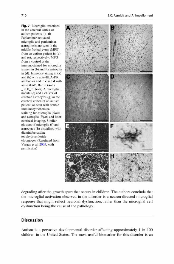
Microglial Cells
These cells of mesodermal origin have been implicated in neurodegenerative
diseases, although their function during development is unclear. These cells are
increased in all areas of the autism brain examined. Fixed brain tissues from the
middle frontal gyrus (MFG) and anterior cingulate gyrus (ACG) were selected from
brains obtained at autopsy of autistic (n ¼ 9; 5–44 years) and control (n ¼ 6)
patients (7–46 years). Immunocytochemical studies showed marked activation of
microglia and astroglia (Fig. 7). Cytokine profiling indicated that macrophage
chemoattractant protein (MCP)-1 and tumor growth factor-1, derived from neuro-
glia, were the most prevalent cytokines in brain tissues (Vargas et al. 2005). The
authors suggest that the increased neuroglial responses are most likely a part of
a neuroinflammatory reaction associated with the CNS innate immune system in
which microglial activation is the main cellular response to CNS dysfunction. The
microglial responses in autism resemble those seen in neurodegenerative disorders
and are similar to those seen in dementia associated with the human immunodefi-
ciency virus (HIV) infection. In chronic conditions, the microglial activation
facilitates the production of neurotoxic mediators.
Increased microglial cells were seen using stereological methods on the fronto-
insular and occipital cortices of postmortem brains stained with Iba1 as a marker
for microglial cells. Eleven autism subjects, aged 3–22 years, and 12 controls,
aged 2–23 years, were examined. An increased density of microglia was found in
both the fronto-insular and visual cortex in people with autism. The extensive
nature of microglial activation at both extremes of the cerebral cortex substanti-
ates the other postmortem studies that have demonstrated widespread alterations
in autism. However, there is one report that the neuronal changes seen in the
piriform cortex of postmortem tissue from autism brains are not seen in the visual
cortex (van Kooten et al. 2008).
There was one unique autism subject (12-year-old male UMB4305), which
showed microglial cells in the normal range. Although the ADI-R scores for this
case are in the autistic range, he was the only one among all subjects to be treated for
psychosis, including administration of the drugs quetiapine, olanzapine, and
risperdal. It is tempting to suggest that the treatment with these atypical serotonergic
active drugs may have alleviated the microglial activation (see Azmitia et al. 2011a).
The study of the microglial cells at different ages produced a very interesting
finding. The clustering of microglial cells around neurons was examined using 13
male postmortem brains from autistic subjects (aged 3–41 years) and nine controls
(aged 1–44 years) (Morgan et al. 2012). There is a close anatomical association
between microglial cells and neurons in both controls and autism children. This
special relationship implies a functional interaction. However, the degree of these
associations becomes much more frequent in the autism brains than in the control
donors during adolescence (Fig. 8). The authors of this chapter conclude that at
least some microglial activation in the dorsolateral prefrontal cortex in autism
is associated with a neuron-specific reaction. The neuron in the adolescent brain
is triggering the microglial cells to react. This suggests the neuronal organization is
Dynamic Brain Changes in Autism: Review of Telencephalic Structures 709
degrading after the growth spurt that occurs in children. The authors conclude that
the microglial activation observed in the disorder is a neuron-directed microglial
response that might reflect neuronal dysfunction, rather than the microglial cell
dysfunction being the cause of the pathology.
Discussion
Autism is a pervasive developmental disorder affecting approximately 1 in 100
children in the United States. The most useful biomarker for this disorder is an
Fig. 7 Neuroglial reactions
in the cerebral cortex of
autism patients. (a–d)Panlaminar activated
microglia and panlaminar
astrogliosis are seen in the
middle frontal gyrus (MFG)
from an autism patient in (a)and (c), respectively. MFG
from a control brain
immunostained for microglia
is seen in (b) and for astroglia
in (d). Immunostaining in (a)and (b) with anti–HLA-DR
antibodies and in c and d with
anti-GFAP. Bar in (a–d)_ 200_m. (e–h) A microglial
nodule (e) and a cluster of
reactive astrocytes (g) in the
cerebral cortex of an autism
patient, as seen with double
immunocytochemical
staining for microglia (dark)and astroglia (light) and laser
confocal imaging. Similar
clusters of microglia (f) andastrocytes (h) visualized with
diaminobenzidine
tetrahydrochloride
chromogen (Reprinted from
Vargas et al. 2005, with
permission)
710 E.C. Azmitia and A. Impallomeni

increase in plasma serotonin (Anderson 1990). Children diagnosed with autism
typically experience difficulty in learning, expressing language appropriately, and
empathizing with others. This disorder is characterized by stereotypy, a repetitive
or ritualistic movement, posture, or utterance, usually exhibited at an early age,
characterized by stimming, flapping, lack of appropriate gaze, sensory acuteness,
nervousness, and heightened sensitivity to neutral stimuli. This behavioral depic-
tion is consistent with an accelerated development of telencephalon described in
this chapter. In general, the primary centers of sensory processing are attenuated as
indicated by evidence of increase cell proliferation and fiber projections. Later in
the disorder, problems that are based on higher-order brain functions, such as those
associated with communication, appear. It can be rationalized that an increase in
early regional development can disrupt the assembly of a more complex function
requiring the synchrony of many brain regions.
Attention, social interactions, and emotions are associated with the frontal cortex.
The orbitofrontal cortex has been connected with decisionmaking within the realm of
moral consciousness, as well as the process of reinforced associations made during
learning, including those created during reward and punishment scenarios. Thus,
a disruption in neuronal connectivity within these areas due to a reduction in large
axonal frequency as well as excessive branching may help explain the deficits
involved in affective recognition in individuals with autism.
00 20 40 60
Distance from Seed Cell (µm)
CS
R-c
orre
cted
Clu
ster
ing
Rat
io Neuron-Neuron Clustering
Control
Autism
80 100 120
0.2
0.4
0.6
0.8
1
1.2
00 20 40 60
Distance from Seed Cell (µm)
CS
R-c
orre
cted
Clu
ster
ing
Rat
io Microglia-Neuron Clustering
Control
Autism
80 100 120
0.2
0.4
0.6
0.8
1.2
1
1.4 a
b
Fig. 8 (a) Microglia–neuron
interaction is significantly
increased in autism at 25 mm(p ¼.006), 75 mm (p ¼.005),
and 100 mm (p ¼.005). The
profile is marked by a spike in
spatial clustering in the
10–30 mm range that is more
pronounced in the autism
group. (b) Neuron–neuronspatial clustering is not
significantly different at any
distance interval, and lacks
prominent features across the
distance range in either
diagnostic group.
Abbreviations: CSR complete
spatial randomness
(Reprinted from Morgan et al.
2010, with permission)
Dynamic Brain Changes in Autism: Review of Telencephalic Structures 711

The lateral PFC is involved in executive functions, comprised of predicting out-
comes, resolving a conflict, processing affect, and determining right from wrong. The
inability to problem solve or respond to emotive stimuli is a compromising symptom
of autism that could be traced to this brain region. Since autism is characterized by
repetitive stereotype behaviors, the orbitofrontal cortex has been observed due to their
involvement with obsessive–compulsive actions. The ACC is associated with “condi-
tioned emotional learning, vocalizations associated with expressing internal states,
assessments of motivational content and assigning emotional valence to internal and
external stimuli, and maternal-infant interactions” (Devinsky 1995). Some of the
phenotypic stereotypes that characterize autism could correspond with this region.
Wernicke’s area is the region crucial to receptive language. Recent findings
suggest the existence of increased neurotrophic factors, enhancing neuronal growth,
within regions, including Wernicke’s area. The ACC, believed to be involved in
cognitive behaviors, theory of mind, and motor activity, is involved in a powerful
network within the limbic system as well as circuitry associated with joint attention
and social interaction. The development of language at appropriate stages is typically
delayed or nonexistent in children with autism. Such deficits could be linked to the
excessive growth observed within the temporal lobe during development.
Previous studies using single-photon emission computed tomography as well as
a 5HT2A receptor ligand, observed reduced 5HT2A receptor density in the anterior
and posterior cingulate gyrus, frontal lobe, superior temporal gyrus, and left parietal
lobes in autism brains (Simms et al. 2009). The VENs provide compelling evidence
of structural cytoarchitectural abnormalities in autism. VENs first appear in gesta-
tion around the 35th week and do not finish fully emerging throughout development
until the age of 4. This trajectory of growth is accelerated in children with autism,
specifically in the frontal cortex. Since these neurons are involved in socio-
emotional and other cognitive processes, they could be a candidate for the observed
behavioral malfunctioning in children with autism.
Serotonin, a brain neurotransmitter known to be increased in the plasma of many
autism patients, functions as a trophic factor (Whitaker-Azmitia 2005). In animal
studies, serotonin results in increased cell proliferation (Dizeyi et al. 2005) and
neuronal and astrocyte maturation (Azmitia 2001) and inhibits microglial activation
(Krabbe et al. 2012). Selective serotonin reuptake inhibitors increase cell prolifer-
ation and neural progenitor cells (NPCs) in the subgranular zone (SGZ) of the
dentate gyrus in humans and mice (DG; Boldrini et al. 2012, 2009). Two recent
findings in autistic children and adolescents are relevant and possibly related:
(1) neuron number, head circumference, and brain size are increased (Courchesne,
2012) and (2) serotonin axons in the temporal cortex are increased (Azmitia et al.
2011). The following two findings lead us to formulate an integrative and highly
innovative hypothesis that the macrocephaly in autistic children is produced by
serotonin-mediated augmentation of cell proliferation of neuroprecursor cells.
Early inhibition of the 5-HT1A receptors may correct this abnormality.
S100B is a calcium-binding protein secreted by astrocytes onto the cytoskeleton
of neurons and glial cells. “The extracellular effect of S100B. . .depends on its
concentration, since it is neurotrophic at pico and nanomolar levels and apoptotic at
712 E.C. Azmitia and A. Impallomeni

micromolar levels” (Azmitia 2001). The research pertaining to brain weight in
autistic children describes an abnormal growth trajectory in which there is an
abnormal increase in size within the first 2 years and then a steady decrease until
the years of puberty. It appears that the brain grows in about 3 years the amount it is
suppose to grow over the course of 16. If S100B is associated with this growth, it
would make sense that an abnormally high level would be a possible cause of the
eventual decline in brain weight, since it is apoptotic after a certain concentration.
As observed in the temporal lobe of autistic adults, neurotrophic factors decline and
neuron frequency is reduced.
Other approaches to the study of autism are available. Patient study is limited by
the constraints that no agreed upon biomarkers exist. The most reliable marker
available is the increase in serotonin levels in the plasma that is observed in about
29–100 % of patients with autism. Medical ethics for the treating and examining of
the autism patients should serve as a barrier to invasive procedures and use of
experimental or off-label drugs. Animal research has great promise if a biological
system can be identified and isolated. Although there has been great hope in
selecting a single gene or system for study, in general these approaches have met
with many problems, not the least of which is translating the predicted findings to
the human population. Trophic factors and the serotonin system are two avenues
where both patient studies and animal research can focus. And most importantly,
autism needs to be considered as a developmental disorder with neuroplastic
changes occurring early and throughout the progression of the disease. It is
a dynamic condition in which the age of the patient is crucial for understanding
the state of the disorder as well as how the patient should be treated.
Key Terms
Dystrophy. Injured axons having an abnormal appearance. Evidence of neuropa-
thology and predictive of neurodegenerative changes that may lead to cell death.
Cortical minicolumns. The vertical assembly of the layers of neurons including
pyramidal (III, V, and VI) and granule neurons (II and IV). These are believed to
be the main functional organization of the cellular elements of the cortex. Does
not include the inhibitory GABA interneurons or monoamine innervations.
Heterotopia. A clump of gray matter that is located in the wrong part of the brain.
The cells in heterotopia have a normal appearance except for their position.
Telencephalon. The most rostral end of the neural tube. Gives rise to all of cortex
and the main subcortical centers of basal ganglion (caudate, putamen, and globus
pallidus), amygdala, and septum.
Glial cells. Consist of the bipolar glia, astrocytes, microglial cells, and oligoden-
droglial cells. Do not have action potential and are involved in many precursor,
supportive, and phagocytic processes to neighboring neurons.
Pyramidal neurons. Among the largest neurons of the brain having a pyramidal
shape and receiving information from the smaller granule neurons. Involved in
directing electrical activity outside individual cortical columns.
Dynamic Brain Changes in Autism: Review of Telencephalic Structures 713

Granule neurons. Smaller receptive neurons that are usually tightly clustered.
Project to the dendrites of pyramidal neurons.
Von Economo neurons. Located in layer 5 of the ACC and fronto-insular cortex.
The neurons are very large, bipolar, or spindle-shaped multipolar neurons.
Key Facts
• Autism brains are macroencephalic in children and toddlers.
• Pyramidal neurons are smaller in older autism brains.
• Increased number of neurons in children and toddlers.
• Serotonin fibers are increased in children and adults.
• Serotonin fibers are dystrophic in adolescence.
• Ependymal zone shows marked neuropathology.
• Microglial cells and astrocytes are increased throughout all cortical regions in
adolescence and adult.
• Neuron and microglial clustering is increased in adolescence.
• GABAA receptors are decreased in parietal cortex and anterior cingulate cortex
in adult.
• BDNF levels are reduced in adult Wernicke’s area.
Summary Points
• The size of the brains of children with autism are larger in children and
adolescents and then declines to control levels. The most rapid period
of growth is the first year of life and the growth appears to stop at the start of
adolescence.
• The increase in cell proliferation is seen in both cortical and subcortical regions.
The cells come from the ependymal layer and account for the changes seen in
cortical column.
• Astrocytes and microglial cells are trophic sources in early life and become
detrimental when activated in adolescence and adults.
• Serotonin fibers entering the telencephalon are increased at the earliest age
studied. Serotonin functions as a trophic factor and projects to supraependymal
and all cortical and subcortical structures.
• The microglial cells are increased in all brain regions studied. The clustering of
microglial cells around neurons peaks at early adolescents.
• GABAA and 5-HT2 receptors are downregulated and BDNF levels are reduced
in adult autism.
• There is evidence of neuropathology in adolescent and adult brains including
smaller and fewer pyramidal and subcortical neurons, dystrophic serotonin
fibers, and glial activation.
714 E.C. Azmitia and A. Impallomeni

References
Amaral DG, Schumann CM, Nordahl CW. Neuroanatomy of autism. Trends Neurosci.
2008;31:137–45.
Anderson GM, Horne WC, Chatterjee D, Cohen DJ. The hyperserotonemia of autism. Ann N Y
Acad Sci. 1990;600:331–42.
Azmitia EC. Serotonin neurons, neuroplasticity, and homeostasis of neural tissue. Neuropsycho-
pharmacology. 1999;21(2 Suppl):33S–45.
Azmitia EC. Modern views on an ancient chemical: serotonin effects on cell proliferation,
maturation, and apoptosis. Brain Res Bull. 2001;56:413–24.
Azmitia EC, Nixon R. Dystrophic serotonergic axons in neurodegenerative diseases. Brain
Resour. 2008;1217:185–94.
Azmitia EC, Singh JS, Whitaker-Azmitia PM. Increased serotonin axons (immunoreactive to
5-HT transporter) in postmortem brains from young autism donors. Neuropharmacology.
2011a;60:1347–54.
Azmitia EC, Singh JS, Hou XP, Wegiel J. Dystrophic serotonin axons in postmortem brains from
young autism patients. Anat Records. 2011b;294:1653–62.
Bailey A, Luthert P, Dean A, Harding B, Janota I, Montgomery M, Rutter M,
Lantos P. A clinicopathological study of autism. Brain. 1998;121:889–905.
Boldrini M, Underwood MD, Hen R, Rosoklija GB, Dwork AJ, John Mann J,. . . Arango V.
Antidepressants increase neural progenitor cells in the human hippocampus.
Neurpsychopharm. 2009;34:2376–89
Boldrini M, Hen R, Arango V. Hippocampal angiogenesis and progenitor cell proliferation are
increased with antidepressant use in major depression. Biol Psychiatry. 2012.
Brusco A, Lopez-Costa JJ, Tagliaferro P, Pecci SJ. Serotonergic ependymal fibres in rat and
monkey: light and electron microscopic immunocytochemical study. Biocell. 1998;22:115–22.
Casanova MF, van Kooten IAJ, Switala AE, van Engeland H, Heinsen H, Steinbusch HWM,
Schmitz C. Abnormalities of cortical minicolumnar organization in the prefrontal lobes of
autism patients. Clin Neurosci Res. 2006a;6(3–4):127–33.
Casanova MF, van Kooten IAJ, Switala AE, van Engeland H, Heinsen H, Steinbusch HWM,
Hof PR, Trippe J, Stone J, Schmitz C. Minicolumnar abnormalities in autism. Acta
Neruopathol. 2006b;112:287–303.
Courchesne E, Carper R, Akshoomoff N. Evidence of brain overgrowth in the first year of life in
autism. J Am Med Assoc. 2003;290:337–44.
Courchesne E, Campbell K, Solso S. Brain growth across the life span in autism: age-specific
changes in anatomical pathology. Brain Res. 2011a;1380:138–45.
Courchesne E, Mouton PR, Calhoun ME, Semendeferi K, Ahrens-Barbeau C, Hallet MJ,
Barnes CC, Pierce K. Neuron number and size in PFC of children with autism. J Am Med
Assoc. 2011b;306:2001–10.
Devinsky O, Morrell MJ, Vogt BA. Contributions of ACC to behaviour. Brain.
1995;118(1):279–306.
Dizeyi N, Bjartell A, Hedlund P, Tasken KA, Gadaleanu V, Abrahamsson PA. Expression of
serotonin receptors 2B and 4 in human prostate cancer tissue and effects of their antagonists on
prostate cancer cell lines. Eur Urol. 2005;47(6):895–900.
Donato R, Sorci G, Riuzzi F, Arcuri C, Bianchi R, Brozzi F, Tubaro C, Giambanco I. S100B’s double
life: intracellular regulator and extracellular signal. Biochim Biophys Acta. 2009;1793:1008–22.
Fatemi SH, Halt AR, Stary JM, Kanodia R, Schulz SC, Realmuto GR. Glutamic acid decarbox-
ylase 65 and 67 kDa proteins are reduced in autistic parietal and cerebellar cortices. Biol
Psychiatry. 2002;52(8):805–10.
Hof PR, Knabe R, Bovier P, Bouras C. Neuropathological observations in a case of autism
presenting with self-injury behavior. Acta Neuropathol. 1991;82:321–6.
Dynamic Brain Changes in Autism: Review of Telencephalic Structures 715

Jacot-Descombes S, Uppal N, Wicinski B, Santos M, Schmeidler J, Giannakopoulos P,
Heinsein H, Schmitz C, Hof PR. Decreased pyramidal neuron size in brodmann areas
44 and 45 in patients with autism. Acta Neuropathol. 2012;124:67–79.
Kemper TL, Bauman M. Neuropathology of infantile autism. J Neuropath Exp Neur. 1998;
57(7):645–52.
Kimelberg HK. Functions of mature mammalian astrocytes: a current view. Neuroscientist.
2010;16:79–106.
Krabbe G, Matyash V, Pannasch U, Mamer L, Boddeke HW, Kettenmann H. Activation of
serotonin receptors promotes microglial injury-induced motility but attenuates phagocytic
activity. Brain Behav Immun. 2012;26:419–28.
Laurence JA, Fatemi SH. Glial fibrillary acidic protein is elevated in superior frontal, parietal and
cerebellar cortices of autistic subjects. Cerebellum. 2005;4(3):206–10.
Morgan JT, Chana G, Pardo CA, Achim C, Semendeferi K, Buckwalter J, Courchesne E,
Everall IP. Microglial activation and increased microglial density observed in the dorsolateral
prefrontal cortex in autism. Biol Psychiatry. 2010;68(4):368–76.
Morgan JT, Chana G, Abramson I, Semendeferi K, Courchesne E, Everall IP. Abnormal
microglial-neuronal spatial organization in the dorsolateral prefrontal cortex in autism. Brain
Res. 2012;1456:72–81.
Oblak A, Gibbs TT, Blatt GJ. Decreased GABAA receptors and benzodiazepine binding sites in
the anterior cingulate cortex in autism. Autism Res. 2009;2(4):205–19.
Raymond GV, Bauman ML, Kemper TL. Hippocampus in autism: a Golgi analysis. Acta
Neuropathol. 1996;91(1):117–9.
Sajdel-Sulkowska EM, Xu M, McGinnis W, Koibuchi N. Brain region-specific changes in oxidative
stress and neurotrophin levels in autism spectrum disorders (ASD). Cerebellum. 2011;10(1):43–8.
Santos M, Uppal N, Butti C, Wicinski B, Schmeidler J, Giannakopoulos P, Heinsen H, Schmitz C,
Hof PR. Von Economo neurons in autism: a stereologic study of the frontoinsular cortex in
children. Brain Res. 2011;1380:206–17.
Schumann CM, Amaral DG. Stereological analysis of amygdala neuron number in autism.
J Neurosci. 2006;26(29):7674–9.
SimmsML, Kemper TL, Timbie CM, Bauman ML, Blatt GJ. The ACC in autism: heterogeneity of
qualitative and quantitative cytoarchitectonic features suggests possible subgroups. Acta
Neuropathol. 2009;118(5):673–84.
van Kooten IA, Palmen SJ, von Cappeln P, Steinbusch HW, Korr H, Heinsen H, Hof PR, van
Engeland H, Schmitz C. Neurons in the fusiform gyrus are fewer and smaller in autism. Brain.
2008;131:987–99.
Vargas DL, Nascimbene C, Krishnan C, Zimmerman AW, Pardo CA. Neuroglial activation and
neuroinflammation in the brain of patients with autism. Ann Neurol. 2005;57(1):67–81.
Wegiel J, Kuchna I, Nowicki K, Imaki H, Wegiel J, Marchi E, Ma SY, Chauhan A, Chauhan V,
Bobrowicz TW, de Leon M, Louis LA, Cohen IL, London E, Brown WT, Wisniewski T. The
neuropathology of autism: defects of neurogenesis and neuronal migration, and dysplastic
changes. Acta Neuropathol. 2010;119(6):755–70.
Whitaker-Azmitia PM. Behavioral and cellular consequences of increasing serotonergic activity
during brain development: a role in autism? Int J Dev Neurosci. 2005;23(1):75–83.
Whitaker-Azmitia PM, Wingate M, Borella A, Gerlai R, Roder J, Azmitia EC. Transgenic mice
overexpressing the neurotrophic factor S-100 beta show neuronalcytoskeletal and behavioral
signs of altered aging processes: implications forAlzheimer’s disease and Down’s syndrome.
Brain Res. 1997;776:51–60.
Zikopoulos B, Barbas H. Changes in prefrontal axons may disrupt the network in autism.
J Neurosci. 2010;30(44):14595–609.
716 E.C. Azmitia and A. Impallomeni