Poster Culture of the microalgae Dunaliella salina in rejected brine water

Plant Physiol. (1 995) 107: 885-893
Chloroplast Response in Dunaliella salina to I r rad iance St ress'
Effect on Thylakoid Membrane Protein Assembly and Function
Michael R. Webb and Anastasios Melis*
Department of Plant Biology, University of California, Berkeley, CA 94720-31 02
The chloroplast response in the green alga Dunaliella salina to irradiance stress was investigated. Cells were grown under low light (11) at 100 pmol photons m-'s-' or high light (HL) at 2000 pmol photons m-* s-' incident intensity. 11-grown cells had a low chlo- rophyll (Chl) a/b ratio, an abundance of light-harvesting complex II proteins (LHC-II), and a large Chl antenna size. HL-grown cells had a higher Chl a/b ratio, relatively fewer LHC-II, and a small Chl antenna size. The more abundant higher molecular m a s subunits of the LHC-II (approximately 31 kD) were selectively depleted from the thylakoid membrane of HL-grown cells. Light-shift experiments defined the kinetics of change in the subunit composition of the LHC-II and suggested distinct mechanisms in the acclimation of thylakoids to HL or LL conditions. The results showed that irradi- ance exerts a differential regulation on the expression of various Lhcb genes. The specific polyclonal antibodies used in this work, raised against the purified LHC-II, cross-reacted with a polypeptide of approximately 20 kD in HL-grown samples. In this work we examined the dynamics of induction of this nove1 protein and discuss its function in terms of a chloroplast response to the level of irradiance.
LHC-I1 contains a minimum of six polypeptide subunits with molecular masses in the 25- to 31-kD region. These proteins are encoded by a nuclear family of genes now named Lhcb (formerly cab) (Jansson et al., 1992). The genes of this family show a high degree of homology to each other and to a lesser degree to the Lkca genes, which code for the subunit proteins of the LHC-I. Both Lkca and Lhcb genes are members of a larger superfamily of genes, which includes the genes coding for the so-called ELIPs (Grimm et al., 1989), the Cbr protein (Lers et al., 19911, and the recently identified PsbS gene (Kim et al., 1992; Wedel et al., 1992). AI1 members of this superfamily appear to be nu- clear-encoded genes. The mature products of these genes a11 contain highly conserved transmembrane helices and display varying degrees of immunological cross-reactivity with one another. Their amino acid sequences diverge sharply, however, in their amino-terminal regions. There also appears to be considerable difference in the Chl-
This work was supported by grant DCB-9003928 from the
* Corresponding author; e-mail melis8nature.berkeley.edu; fax National Science Foundation.
1-51 O - 642- 4995. 885
carotene-xanthophyll-binding properties of the various apoproteins.
The level of irradiance during plant growth modulates the size and composition of the light-harvesting antenna of the photosystems (Anderson, 1986; Melis, 1991). In general, LL intensity promotes a larger Chl antenna size for both PSI and PSII (larger photosynthetic unit size). HL condi- tions elicit a smaller Chl antenna size. This adjustment in the Chl antenna size of the photosystems comes about because of changes in the size of the auxiliary LHC-I1 and LHC-I (Leong and Anderson, 1984; Larsson et al., 1987; Sukenik et al., 1988; Smith et al., 1990). Mechanistically, these changes are implemented through the association of variable amounts of Lhcb proteins with the light-harvest- ing antenna of PSII (Larsson et al., 1987; Morrissey et al., 1989; Mawson et al., 1994). The response appears to be highly conserved in a11 photosynthetic organisms exam- ined. However, the amplitude of the response differs sig- nificantly among different species. Green algae display a significantly greater amplitude of this response than higher plants, making them a better model organism in such stud- ies. Recent work by LaRoche et al. (1991) suggested that changes in the abundance of LHC-I1 are preceded by changes in the abundance of the mRNA coding for these proteins. The regulation of these phenomena at the molec- ular and membrane levels is currently unknown.
When the green alga Dunaliella salina is grown at a high irradiance (2000 pmol photons m-' s-'), a number of changes occur in the structural and functional organization of its thylakoid membranes, including a decrease in Chl/ cell, an increase in the Chl a/Chl b ratio, and a decrease in the LHC-I1 antenna size (Smith et al., 1990). It is also known that variations in the level of irradiance during cell growth bring about changes in the expression of ELIP genes in pea (Adamska et al., 1993) and of Cbr genes in Dunaliella bardawil (Levy et al., 1992). The relationship between light- harvesting antenna size in chloroplasts and the regulated
Abbreviations: Cbr, carotene biosynthesis related; ELIP, early light-inducible proteins; HL, high light; LHC, light-harvesting complex; LHC-I, Chl a-b light-harvesting complex of photosystem I; LHC-11, Chl a-b light-harvesting complex of photosystem 11; LL, low light; SLL and SHL, slopes of the logarithmically linear growth phase of LL and HL cells, respectively.
Dow
nloaded from https://academ
ic.oup.com/plphys/article/107/3/885/6069087 by guest on 22 N
ovember 2021
886 Webb and Melis Plant Physiol. Vol. 107, 1995
expression of ELIP and/or Cbr genes has not been addressed.
In this report, we examine the dynamics of LHC-I1 as- sembly as a function of irradiance. We used light-shift experiments in which D. salina cultures were switched from LL to HL conditions and vice versa. This approach helps us to identify the LHC-I1 subunits that change the most in response to irradiance. Furthermore, we describe the presence and dynamics of an approximately 20-kD nove1 protein found in association with the chloroplast in the green alga D. salina. This protein is immunochemically recognized by polyclonal antibodies specific for the LHC-I1 and appears to be induced under conditions of irradiance stress.
MATERIALS AND METHODS
Cell Crowth Conditions
Dunaliella salina cultures were grown in an artificial hy- persaline medium similar to that used by Pick et al. (1986) containing 1.5 M NaC1,5 mM MgSO,, 0.3 mM CaCl,, 0.1 mM KHJ’O,, 20 mM EDTA, 2 mM FeCl,, 5 mM NH,C1, and 40 mM Tris-HC1, pH 7.5, and supplemented with a mixture of micronutrients. Carbon was supplied as NaHCO, in the growth medium at an initial concentration of 25 mM. Cul- tures were grown in flat bottles (optical pathlength = 3 cm) at 30°C under illumination at 100 pmol photons m-’ s-* (LL) or at 2000 pmol photons m-’ s-* (HL). Care was exercised, by means of shaking and by the use of reflectors, to ensure as uniform illumination to the culture as possible. Cells were grown to the late ln phase (ln = -2.0 for LL-grown cultures, ln A,,, = -3.3 for HL-grown cultures) and treatments (transfer to different irradiance conditions and/or addition of antibiotics) were made. In some exper- iments, aliquots of cells were harvested at different stages of growth, e.g. from the early logarithmic phase to the late stationary phase of culture development.
of the cell suspension (1n A,,,) was measured at room temperature using the technique of Shibata (1958). An Aminco (Urbana, IL) DW 2a spectrophotometer was used with a 3-nm half-band width and opal glass cuvettes of 1 cm optical pathlength placed directly in front of the photomultiplier tube to minimize light-scattering effects.
The
Thylakoid Membrane lsolation
Cells were harvested by centrifugation at 3000g for 3 min at 4°C. Pellets were resuspended in 0.5 mL of fresh growth medium and stored frozen at -20°C until a11 samples were ready for processing. Samples were thawed on ice and diluted with sonication buffer containing 50 mM Tris-HC1, 0.1% sodium ascorbate, 1 mM aminocaproic acid, and 1 mM aminobenzamidine, pH 8.5. Cells were disrupted by soni- cation for 30 s in a Branson (Danbury, CT) Sonifier (Cell Disruptor 200) operated in the pulsed mode with a 50% duty cycle and an output power setting of 5. Unbroken cells and other large cell fragments were removed by cen- trifugation at 3000g for 3 min at 4°C. The supernatant was then centrifuged at 46,OOOg for 30 min at 4°C. The thylakoid membrane pellet was resuspended in 200 mM Tris, 20%
glycerol, pH 6.6, and Chl concentrations were cletermined in 80% acetone according to the method of Arnon (1949).
Membranes were solubilized in buffer contairing 75 mM Tris-HC1, 3.5% SDS, 10% glycerol, and 4 M urea, pH 6.8, and incubated at room temperature for 15 mim This ap- proach (denaturation of thylakoid membrane proteins at room temperature by SDS-urea) was chosen tci avoid ag- gregation of the hydrophobic light-harvestinl; polypep- tides. In our experience, heat denaturation resulted in the formation of high mo1 wt aggregates that appeared either as a smear in the stacking gel and upper portion of the running gel or as a high mo1 wt band of protein immobi- lized at the stacking/running gel interface. Prior to elec- trophoresis, samples were centrifuged in a microcentrifuge for 5 min to remove unsolubilized material. tl-Mercapto- ethanol was added to samples to give a final concentration of 10% and samples were diluted accordingly to yield equal Chl concentrations. Samples were stored on ice until used or otherwise stored at -80°C.
Thylakoid Membrane Protein Analysis
Thylakoid membrane proteins were resolved by SDS- PAGE using the discontinuous buffer system of Laemmli (1970) with 15% acrylamide and 0.41 % bis-acrylamide, with or without 4 M urea. The stacking gel con tained 4.5% acrylamide (and 1 M urea, as appropriate). The gel lanes were loaded with 2 to 4 nmol Chl (a plus b) fo1, SDS-PAGE and immunoblot analysis unless otherwise indicated. Elec- trophoresis on 0.15- X 14- X 16-cm slab gels was performed at 2°C at a constant current of 8 mA for 18 E.. Gels were stained with 0.1% Coomassie brilliant blue R for protein visualization.
lmmunochemical Analysis
Identification of LHC (Lkcb gene) polypept des was ac- complished with immunoblot analysis using s pecific poly- clonal antibodies raised in this laboratory in raobits against proteins of LHC-11. Antigens from spinach wl2re obtained from resolved membranes of the grana partition regions (the so-called BBY particles) (Berthold et al., 1981) follow- ing a modification of the method of Burke (1978). The isolated LHC-I1 was precipitated upon incubation with MgC1, and washed twice (Burke, 1978). Its purity was tested by SDS-PAGE analysis. The antigen, cclntaining the isolated LHC-I1 in its native form, was injected subcutane- ously in rabbits. This approach prevented antjgen contam- ination by other thylakoid membrane proteins and resulted in the formation of monospecific polyclonal antibodies (Melis, 1992).
Electrophoretic transfer of the SDS-PAGE resolved D. salina thylakoid membrane polypeptides to nitrocellulose, and the subsequent incubations with the antibodies and with alkaline phosphate-conjugated antibodies were per- formed as described previously (Smith et al., 1990). Cross- reaction was quantitated by scanning the nitrocellulose membranes with an LKB-Pharmacia XL laser densitometer.
Dow
nloaded from https://academ
ic.oup.com/plphys/article/107/3/885/6069087 by guest on 22 N
ovember 2021
Chloroplast Response to Irradiance Stress 887
RESULTS
Cell Growth and Chl Content under LL and HL Conditions
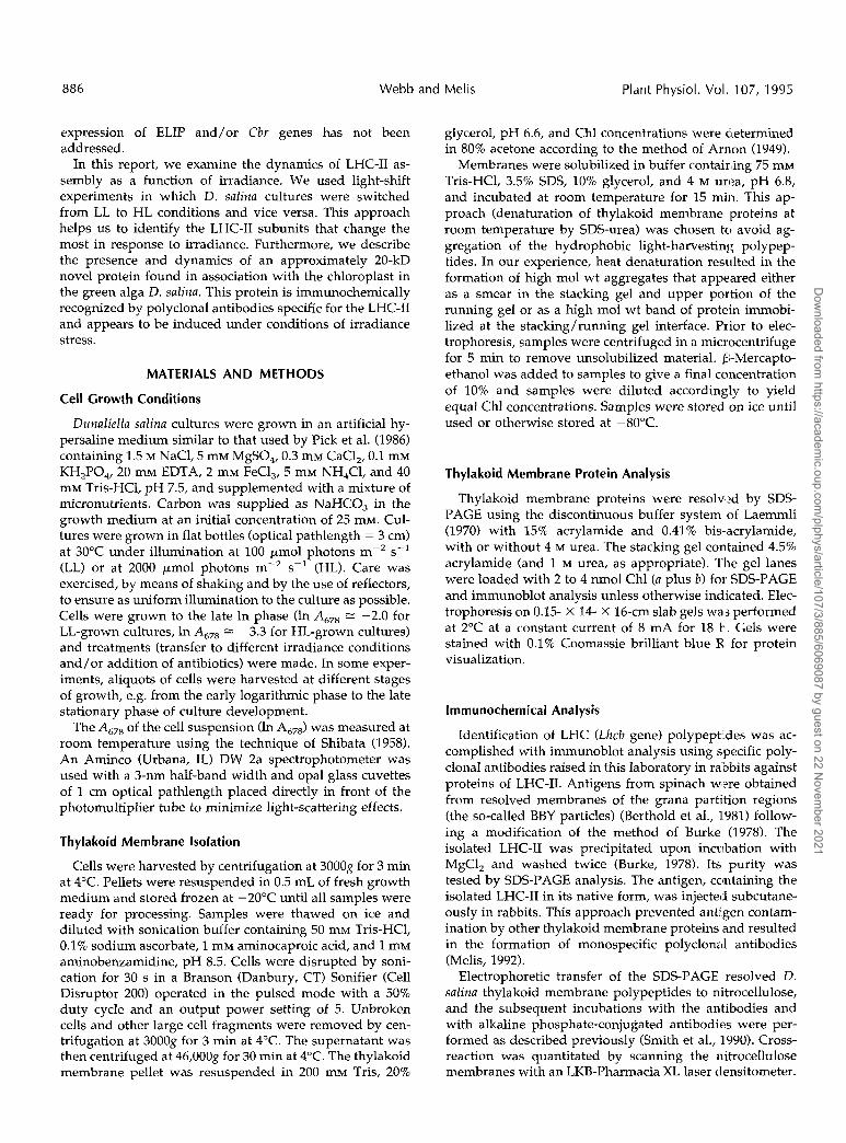
D. salina was grown under uniform illumination either at100 /^mol photons m~2 s"1 (LL conditions) or at 2000 /xmolphotons m~2 s"1 (HL conditions). Cell growth was moni-tored by measuring In A67S. We determined that the Chl/cell ratio remained fairly constant under continuous LL orHL growth conditions (Naus and Melis, 1991). Therefore,the In Ah76, when plotted as a function of time after cultureinoculation (Fig. 1), provides a measure of Chl accumula-tion and of cell growth under LL and HL conditions. Figure1 shows a logarithmically linear phase of growth and agradually slower phase, followed by the stationary phase.
Rates of growth in the LL and HL culture were estimatedfrom the growth curves, defined by the slope at the loga-rithmically linear phase. Our analyses indicated an SLL =1.3 ± 0.2 d"1 and an SHL = 0.9 ± 0.2 d"1, suggesting aslightly faster cell growth under LL than HL conditions. Itis known that D. salina, grown under optimal light inten-sities, is capable of higher rates of growth at the logarith-mic-linear phase, approaching slopes of about 5 d"1 at anincident intensity of about 500 /imol photons m~2 s"1
(Smith et al., 1990). Thus, under our LL conditions, an SLL= 1.3 d^1 suggests growth limited by the intensity ofillumination. Under our HL conditions, an SHL = 0.9 d~ T
indicates irradiance stress and a chronic photoinhibitioncondition (Kim et al., 1993) that limit photosynthesis andcell growth.
LL and HL cultures reached approximately the same celldensity (approximately 2 X 10⁶ cells mL"1) in the station-ary phase (Kim et al., 1993). However, they differed signif-icantly in the Chl fl/Chl b ratio (approximately 4:1 for theLL cells and approximately 17:1 for the HL cells). They alsodiffered in the Chl/cell ratio, a fact that is illustrated inFigure 1 by the significantly lower In /1678 values in the HLculture relative to the LL culture at all comparable stages ofgrowth. In the stationary phase, the LL-grown culture con-tained about 4 x 10"13 mol of Chl/cell, whereas the HL-grown cells reached approximately 0.8 X 10"13 mol of
60 90Time, h
120 150
Figure 1. Growth curve of D. salina under LL and HL conditions. TheI" Ab?8 is plotted as a function of time during cell growth. LL cells (O)were grown under 100 ^mol photons rrT2 s"'. HL cells (•) weregrown under 2000 jiimol photons irT2 s~'.
LHC II
LL HL LL HL
— 66
-45
t~31
-21
— 14kD
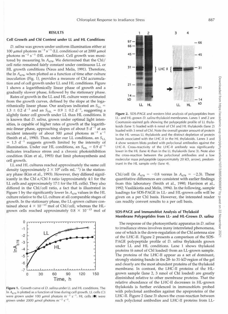
Figure 2. SDS-PACE and western blot analysis of polypeptides fromLL- and HL-grown D. salina thylakoid membranes. Lanes 1 and 2 areCoomassie-stained gels showing the polypeptide profile of LL thyla-koids (lane 1) loaded with 6 nmol of Chl and HL thylakoids (lane 2)loaded with 3 nmol of Chl. Note the overall greater amount of proteinin the HL versus LL thylakoids and the distinct depletion of proteinbands associated with the LHC-II in the HL thylakoids. Lanes 3 and4 show western blots probed with polyclonal antibodies against theLHC-II. Cross-reactivity of the LHC-II antibody was significantlylower in the HL (lane 4) than in the LL thylakoids (lane 3). Note alsothe cross-reaction between the polyclonal antibodies and a lowmolecular mass polypeptide (approximately 20 kD, arrow), predom-inant in the HL sample only (lane 4).
Chl/cell (In A67S = -0.8 versus In A67S = -2.3). Thesequantitative differences are consistent with earlier findingsfrom this laboratory (Smith et al., 1990; Harrison et al.,1992; Vasilikiotis and Melis, 1994). In the following, sampleloadings for SDS-PAGE in LL- and HL-grown cells will begiven on a per Chl basis. However, the interested readercan readily convert results to a per cell basis.
SOS-PAGE and Immunoblot Analysis of ThylakoidMembrane Polypeptides from LL- and HL-Grown D. salina
The response of the photosynthetic apparatus in D. salinato irradiance stress involves many interrelated phenomena,one of which is the down-regulation of the Chl antenna sizeof the LHC-II. Figure 2 presents a comparison of the SDS-PAGE polypeptide profile of D. salina thylakoids grownunder LL and HL conditions. Lane 1 shows thylakoidproteins (6 nmol of Chl loaded) from an LL-grown sample.The proteins of the LHC-II appear as a set of dominant,strongly staining bands in the 28- to 31-kD region of the geland clearly are the most abundant proteins of the thylakoidmembrane. In contrast, the LHC-II proteins of the HL-grown sample (lane 2, 3 nmol of Chl loaded) are greatlydiminished relative to other membrane proteins. That therelative abundance of the LHC-II decreases in HL-grownthylakoids is further evidenced in immunoblots probedwith polyclonal antibodies against the apoproteins of theLHC-II. Figure 2 (lane 3) shows the cross-reaction betweensuch polyclonal antibodies and LHC-II proteins from LL-
Dow
nloaded from https://academ
ic.oup.com/plphys/article/107/3/885/6069087 by guest on 22 N
ovember 2021

888 Webb and Melis Plant Physiol. Vol. 107, 1995
grown D. salina (equivalent of 6 nmol of Chl). Figure 2 (lane4) shows the cross-reaction between the polyclonal anti-bodies and proteins from HL-grown D. salina (equivalentof 3 nmol of Chl loaded). It is evident that considerablyfewer LHC-II apoproteins are associated with the thylakoidmembrane of HL-grown D. salina. This observation is con-sistent with the results of other investigators (Sukenik etal., 1988; Smith et al., 1990) and suggests that one adaptiveresponse of green algae to high irradiance stress is thedown-sizing of the Chl a-b LHC-II antenna by an as-yetunknown mechanism.
The western blot analysis (Fig. 2, lanes 3 and 4) revealedthat the more abundant higher molecular mass proteins ofthe LHC-II (approximately 31 kD) were selectively de-pleted from the thylakoid membrane under HL growthconditions. Conversely, an LHC-II protein at approxi-mately 28 kD was preferentially retained in the thylakoidmembrane of HL-grown D. salina. These results suggestthat the level of irradiance during cell growth exerts adifferential regulation on the expression of Lhcb genesand/or on the assembly of the LHC-II polypeptides in theChl a-b light-harvesting antenna. A more detailed analysison this differential regulation of LHC-II apoproteins byirradiance is presented below.
Of interest in the results of Figure 2 (lane 4, HL sample)is the cross-reaction of the LHC-II polyclonal antibodieswith an approximately 20-kD polypeptide (marked by ar-row). Such a cross-reaction is much less pronounced, orabsent, with proteins from the LL sample (lane 3). Thisnovel protein (approximately 20-kD protein) was consis-tently present in the samples isolated from the HL-growncells. Occasionally, a second cross-reaction was detectedwith a band in the approximately 18-kD region (not shownhere).
To explore the kinetics of the differential expression ofLhcb genes and of the appearance of the approximately20-kD protein, we conducted light-shift experiments inwhich LL-grown algae were switched to HL conditions andvice versa.
IE 24 36 48 h
20 40 60 80Time, h
100 120
Figure 3. Effect of irradiance change from LL to HL on Chl accumu-lation and growth of D. salina. Cultures were grown under LL con-ditions at 100 /xmol photons nrT2 s~' and either maintained at thisirradiance (O) or switched to HL conditions (2000 /xmol photons m~2
s"1) at 40 h (•). Cell growth was measured as the In Ah7B.
LHC II
97 —66 — '
45 —
31 —
21 —
14 —kD
Figure 4. SDS-PAGE profile of solubilized membrane proteins fromthylakoids of D. salina transferred from LL to HL growth conditions asshown in Figure 3. Cells were harvested at the time of transition (0 h)and at subsequent time intervals as indicated. Thylakoid membranesfrom these cells were isolated, solubilized, and loaded at 3.8 nmol ofChl per lane.
Effect of Irradiance Change (LL-»HL shift) on D. salinaThylakoid Membrane Protein Profile
When LL-grown cultures, still in the linear logarithmicphase of growth, are switched to HL conditions they grad-ually lose their deep green color and acquire the greenish-yellow appearance of HL cells. Figure 3 shows the growthcurve of an LL culture of D. salina (open circles) and that ofa replicate LL culture that was switched to HL conditionsin the mid-logarithmic phase of growth (solid circles, 40 h,In A678 = -2.6). It can be seen that the LL control culturecontinues to accumulate Chl and reaches the stationaryphase approximately 100 h later at In A678 = -0.9. TheLL-»HL culture shows a lag of about 20 h in Chl accumu-lation after the irradiance change. Subsequently, Chl accu-mulation resumes at a slower rate than that of the LLcontrol. The LL—»HL culture reaches the stationary phaseat a lower density In A67S = -2.0, which is typical for HL-grown cells (Fig. 1). We have determined that during thistransition period, cell division continues, albeit at a slowerrate. The results show that the rate of Chl biosynthesis islowered promptly upon transfer to HL conditions. More-over, these results underline changes in the amount andorganization of the LHC-II in chloroplasts, occurring uponthe LL—>HL transition (see below).
Thylakoid membrane proteins from cells that had beentransferred from LL to HL conditions were examined bySDS-PAGE. In this experiment, an LL culture of D. salinawas transferred to HL conditions, cells were harvestedafter different periods of exposure to HL, thylakoids wereisolated and solubilized, and proteins were resolved basedon equal Chl loading (3.8 nmol per lane) as describedpreviously. In Figure 4, a Coomassie-stained gel from suchan experiment shows that the amount of total protein per
Dow
nloaded from https://academ
ic.oup.com/plphys/article/107/3/885/6069087 by guest on 22 N
ovember 2021

Chloroplast Response to Irradiance Stress 889
lane increases but the relative amount of LHC-II proteindecreases with time of exposure to HL, a consequence ofthe decrease in the Chl/protein ratio occurring as the Chlantenna of PSII is down-sized under the HL conditions.
Figure 5 shows a western blot analysis of such samplesusing polyclonal antibodies to the LHC-II. It can be seenthat gradual changes occur in the relative subunit compo-sition of the LHC-II as a function of time in HL. Highermolecular mass subunits of the LHC-II (the approximately31-kD region) specifically decrease in abundance relative tothe lower molecular mass subunit at approximately 28 kD.This probably reflects a shift in the relative subunit com-position of the LHC-II, occurring concomitantly with theadjustment in the LHC-II antenna size.
Important in the results of Figure 5 is the appearance ofa protein band in the approximately 20-kD region thatcross-reacts with the anti-LHC-II antibodies. This protein(Fig. 5, arrow) accumulates steadily as a function of time inHL from nondetectable levels in LL samples (0 h) to levelsafter 48 h of HL exposure that are sufficient to give across-reaction with the LHC-II antibody comparable inintensity with that of the LHC-II subunits. It appears thatthe transition to HL causes either the de novo biosynthesisof an approximately 20-kD protein or its generation fromexisting proteins in the thylakoid membrane, or both.
Figure 6 is the result of densitometric scans of westernblots similar to those shown in Figure 5. The results showa 2-fold increase in the relative amount of the smallerLHC-II subunits (Fig. 6, 28 kD) and a corresponding de-cline in the amount of the larger subunits. As a result, therelative amount of the total LHC-II in the thylakoid mem-brane remained fairly constant during the 48-h period in
45 —
31 ~~ HHHMB-lLHC"1 'i , HI! ' . I '
21 —
14 —kD
Figure 5. Western blot analysis of thylakoid membrane proteins fromLL-grown D. salina cells after transition to HL. Cells were grownunder LL conditions and then transferred to HL conditions as shownin Figure 3. Cells were harvested at zero time after the light shift andat subsequent time intervals as indicated. Thylakoid membraneswere isolated and analyzed by SDS-PAGE as described in Figure 4.Polyclonal antibodies against the LHC-II were used to obtain theimmunoblot shown.
10 20 30 40Time in HL, h
Figure 6. Quantitation of cross-reactions following a transition of D.salina cells from an LL to an HL environment. The relative amount ofthe total LHC-II (O), the lower (28 kD) molecular mass subunits (D),and the approximately 20-kD proteins (0) were measured from thedensitometric analysis of western blots similar to that shown in Fig-ure 5.
HL (Fig. 6, LHC-II). Note also the kinetics of the approxi-mately 20-kD protein increase as a function of culture timeinHL.
Effect of Irradiance Change (HL-*LL shift) on D. salinaThylakoid Membrane Protein Profile
In the converse experiment, in which an HL-grown cul-ture was transferred to LL conditions, the greenish-yellowtypical of the HL cultures changed to deep green withinhours of exposure to LL conditions. Figure 7 shows thegrowth curve of an HL-grown culture (open circles) and anHL culture transferred to LL conditions at 40 h and an InA678 = -3.5. Here, the HL culture reached maximal densityat In A67g = —1.9, whereas the HL—»LL culture reached amaximum In A678 value of —1.0. We observed that, upontransition to LL conditions, enhanced accumulation of Chlin the culture is prompt and approaches a rate comparableto that of the rate encountered in LL-grown cultures.
Thylakoid membrane proteins from cells that had beentransferred from HL to LL conditions were examined bySDS-PAGE. Cells were harvested after different periods ofexposure to LL, membranes were isolated and solubilized,and proteins were resolved based on equal Chl loading (4nmol per lane). Figure 8 shows a Coomassie-stained gelfrom such an experiment in which it can be seen that theamount of total protein per lane decreases with increasingtime of exposure to LL, a consequence of the increase in theChl/protein ratio expected as the Chl antenna size in-creases under the LL conditions. Moreover, there is a grad-ual increase in the amount of the LHC-II present, as well aschanges in the relative subunit composition of the LHC-IIas a function of time in LL.
Figure 9 shows a western blot analysis of such samplesusing polyclonal antibodies raised against the LHC-II. Itcan be seen that during a 48-h period in LL there is agradual change in the amount and subunit composition ofthe LHC-II. At 0 h, the LHC-II of the HL-grown cells
Dow
nloaded from https://academ
ic.oup.com/plphys/article/107/3/885/6069087 by guest on 22 N
ovember 2021

890 Webb and Melis Plant Physiol. Vol. 107, 1995
40 60 80Time, h
100 120
Figure 7. Effect of irradiance change from HL to LL on the growth ofD. salina. Cultures were grown under HL conditions (2000 jiimolphotons rrT2 s"1) and either maintained at this irradiance (O) orswitched to LL conditions (100 jxmol photons m~Cell growth was measured as the In A678.
appears to contain only two subunits migrating at approx-imately 28 and 28.5 kD. After 2 h in LL, a higher molecularmass subunit of approximately 31 kD is clearly present.This is followed after 6 h by the appearance of minoramounts of a lower molecular mass subunit at approxi-mately 27 kD. With increasing time in LL the relativeintensity of the LHC-II subunits is seen to increase. The rateof increase, however, is faster for the approximately 31-kDand slower for the 28- and 28.5-kD subunits.
Figure 9 also shows that, in a complementary fashion tothe LL-^HL experiment, the amount of the approximately20-kD protein decreases gradually over time in LL. Thesample from the HL-grown culture (0 h lane) containssignificant levels of the approximately 20-kD protein, giv-ing a cross-reaction with the polyclonal LHC-II antibodies
0 2 6 12 24 48 h
97 —66 —
45 —
31 — LHCII
kD
Figure 8. SDS-PAGE profile of solubilized membrane proteins fromthylakoids of D. salina transferred from HL to LL as shown in Figure6. Cells were harvested at zero time after the light shift (0 h) and atsubsequent time intervals as indicated. Thylakoid membranes fromthese cells were isolated, solubilized, and loaded at 4 nmol of Chlper lane.
0 2 6 12 24 46 h
97 —66 —f*-»*» ««••«•« M>'f •«•.»•»»•• i •
45 —
21 —
14 —kD
Figure 9. Western blot analysis of thylakoid membrane proteins fromHL-grown D. salina cells after transition to LL. Cells were grownunder HL conditions and then transferred to LL conditions as shownin Figure 7. Following the light shift, cells were harvested at the timeintervals indicated and thylakoid membranes were isolated and an-alyzed by SDS-PAGE as described in Figure 8. Polyclonal antibodiesto the LHC-II were used to obtain the immunoblot shown. Thecross-reacting proteins visible in the 27- to 31-kD region are those ofthe LHC-II. The approximately 20-kD protein appears as a cross-reacting band (arrow) and clearly decreases in abundance withincreasing time of exposure to LL.
that is considerably more intense than that of the LHC-IIitself. During time of exposure to LL, however, this rela-tionship gradually becomes reversed as the relative level ofthe approximately 20-kD protein decreases significantlyand the level of LHC-II increases.
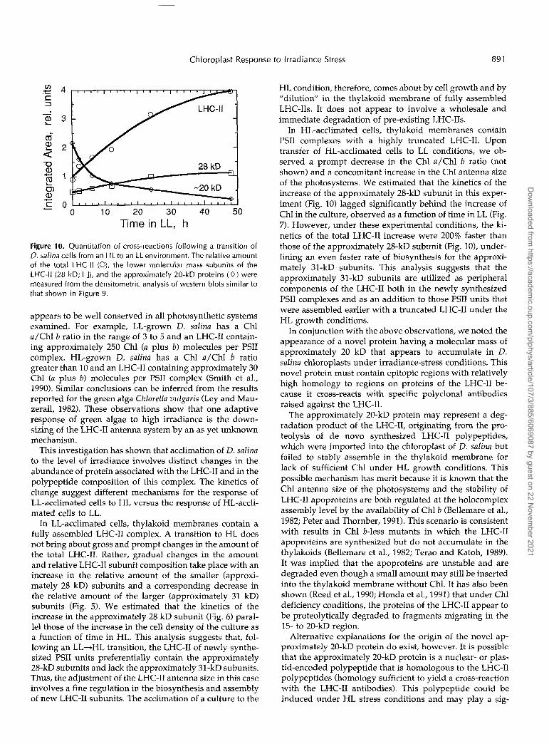
Figure 10 presents the result of densitometric scans ofwestern blots similar to those shown in Figure 9. Theresults show a 2-fold increase in the relative amount of thesmaller LHC-II subunits during the 48-h period in HL (Fig.10, 28 kD). However, there is 4-fold increase in the amountof the total LHC-II in the thylakoid membrane (Fig. 10,LHC-II), reflecting a significantly greater increase in theamount of the higher molecular mass proteins of theLHC-II (approximately 31 kD). Note also the kinetics of thedecrease in the approximately 20-kD protein as a functionof cell time in LL.
DISCUSSION
Both Chl a and Chl b are used by higher plants and greenalgae as the main light-harvesting pigments; however,these pigments are not homogeneously distributed amongthe various Chl-binding proteins. Since the majority of Chlb is associated with the LHC-II, the Chl a /Chl b ratioprovides information concerning the size of the LHC-II thatstably assembles relative to the Chl a-binding proteins ofthe PSII core. Evidence in the literature (Anderson, 1986;Melis, 1991) suggests that variations in the Chl a/Chl bratio occur naturally in higher plants and green algae inresponse to long-term changes in irradiance. This response
Dow
nloaded from https://academ
ic.oup.com/plphys/article/107/3/885/6069087 by guest on 22 N
ovember 2021
Chloroplast Response to lrradiance Stress 89 1
l i m e in LL, h
Figure 10. Quantitation of cross-reactions following a transition of O. salina cells from an HL to an LL environment. The relative amount of the total LHC-II (O), the lower molecular mass subunits of the LHC-II (28 kD; O), and the approximately 20-kD proteins ( O ) were measured from the densitometric analysis of western blots similar to that shown in Figure 9.
appears to be well conserved in a11 photosynthetic systems examined. For example, LL-grown D. salina has a Chl a/Chl b ratio in the range of 3 to 5 and an LHC-I1 contain- ing approximately 250 Chl (a plus b) molecules per PSII complex. HL-grown D. salina has a Chl a/Chl b ratio greater than 10 and an LHC-I1 containing approximately 30 Chl (a plus b) molecules per PSII complex (Smith et al., 1990). Similar conclusions can be inferred from the results reported for the green alga Chlorella vulgaris (Ley and Mau- zerall, 1982). These observations show that one adaptive response of green algae to high irradiance is the down- sizing of the LHC-I1 antenna system by an as yet unknown mechanism.
This investigation has shown that acclimation of D. salina to the level of irradiance involves distinct changes in the abundance of protein associated with the LHC-I1 and in the polypeptide composition of this complex. The kinetics of change suggest different mechanisms for the response of LL-acclimated cells to HL versus the response of HL-accli- mated cells to LL.
In LL-acclimated cells, thylakoid membranes contain a fully assembled LHC-I1 complex. A transition to HL does not bring about gross and prompt changes in the amount of the total LHC-11. Rather, gradual changes in the amount and relative LHC-I1 subunit composition take place with an increase in the relative amount of the smaller (approxi- mately 28 kD) subunits and a corresponding decrease in the relative amount of the larger (approximately 31 kD) subunits (Fig. 5). We estimated that the kinetics of the increase in the approximately 28 kD subunit (Fig. 6) paral- lel those of the increase in the cell density of the culture as a function of time in HL. This analysis suggests that, fol- lowing an LL-+HL transition, the LHC-I1 of newly synthe- sized PSII units preferentially contain the approximately 28-kD subunits and lack the approximately 31-kD subunits. Thus, the adjustment of the LHC-I1 antenna size in this case involves a fine regulation in the biosynthesis and assembly of new LHC-I1 subunits. The acclimation of a culture to the
HL condition, therefore, comes about by cell growth and by "dilution" in the thylakoid membrane of fully assembled LHC-11s. It does not appear to involve a wholesale and immediate degradation of pre-existing LHC-11s.
In HL-acclimated cells, thylakoid membranes contain PSII complexes with a highly truncated LHC-11. Upon transfer of HL-acclimated cells to LL conditions, we ob- served a prompt decrease in the Chl a/Chl b ratio (not shown) and a concomitant increase in the Chl antenna size of the photosystems. We estimated that the kinetics of the increase of the approximately 28-kD subunit in this exper- iment (Fig. 10) lagged significantly behind the increase of Chl in the culture, observed as a function of time in LL (Fig. 7). However, under these experimental conditions, the ki- netics of the total LHC-I1 increase were 200% faster than those of the approximately 28-kD subunit (Fig. 101, under- lining an even faster rate of biosynthesis for the approxi- mately 31-kD. subunits. This analysis suggests that the approximately 31-kD subunits are utilized as peripheral components of the LHC-I1 both in the newly synthesized PSll complexes and as an addition to those PSII units that were assembled earlier with a truncated LHC-I1 under the HL growth conditions.
In conjunction with the above observations, we noted the appearance of a novel protein having a molecular mass of approximately 20 kD that appears to accumulate in D. salina chloroplasts under irradiance-stress conditions. This novel protein must contain epitopic regions with relatively high homology to regions on proteins of the LHC-I1 be- cause it cross-reacts with specific polyclonal antibodies raised against the LHC-11.
The approximately 20-kD protein may represent a deg- radation product of the LHC-11, originating from the pro- teolysis of de novo synthesized LHC-I1 polypeptides, which were imported into the chloroplast of D. salina but failed to stably assemble in the thylakoid membrane for lack of sufficient Chl under HL growth conditions. This possible mechanism has merit because it is known that the Chl antenna size of the photosystems and the stability of LHC-I1 apoproteins are both regulated at the holocomplex assembly level by the availability of Chl b (Bellemare et al., 1982; Peter and Thornber, 1991). This scenario is consistent with results in Chl b-less mutants in which the LHC-I1 apoproteins are synthesized but do not accumulate in the thylakoids (Bellemare et al., 1982; Terao and Katoh, 1989). It was implied that the apoproteins are unstable and are degraded even though a small amount may still be inserted into the thylakoid membrane without Chl. It has also been shown (Reed et al., 1990; Honda et al., 1991) that under Chl deficiency conditions, the proteins of the LHC-I1 appear to be proteolytically degraded to fragments migrating in the 15- to 20-kD region.
Alternative explanations for the origin of the novel ap- proximately 20-kD protein do exist, however. It is possible that the approximately 20-kD protein is a nuclear- or plas- tid-encoded polypeptide that is homologous to the LHC-I1 polypeptides (homology sufficient to yield a cross-reaction with the LHC-I1 antibodies). This polypeptide could be induced under HL stress conditions and may play a sig-
Dow
nloaded from https://academ
ic.oup.com/plphys/article/107/3/885/6069087 by guest on 22 N
ovember 2021
892 Webb and Melis Plant Physiol. Vol. 107, 1995
nificant functional role in the stressed chloroplast. Both ELIP-type proteins and the Cbr gene product exhibit a high degree of homology to the proteins of the LHC and appear to play unique roles in the chloroplast defense against light-induced stress (Green and Pichersky, 1994).
ELIPs are nuclear-encoded membrane proteins that oc- cur transiently in the early stages of the light-induced development of chloroplasts from etioplasts (Grimm et al., 1989) and in mature plants in response to light stress (Adamska et al., 1993). Once integrated into the thylakoid membrane, ELIPs appear to associate with PSII, although they have not been shown to bind Chl. It has been pro- posed that, in the early stages of photosystem assembly (etioplast greening), they act as temporary substitutes for the LHC proteins, which do not accumulate to significant levels until after the appearance of the ELIPs (Adamska and Kloppstech, 1991). The appearance of stable, mem- brane-associated ELIPs in mature plants under light-stress conditions or in the presence of carotene synthesis inhibi- tors suggests that they may play a role in photoprotection of PSII (Adamska et al., 1993).
An interesting, and possibly related, response of the green alga D. bardawil to HL stress is the accumulation of high levels of p-carotene, presumably as a protective mea- sure against photooxidative damage. The nuclear gene des- ignated Cbr was isolated from an HL-induced cDNA ex- pression library from D. bardawil (Levy et al., 1992). The fact that Cbr transcription and p-carotene biosynthesis show coincidental modes of induction and that certain nucleotide sequences upstream of Cbr match consensus sequences of the sterol-response elements coding for me- valonic acid biosynthesis suggested that Cbr is co-regulated with genes crucial for p-carotene biosynthesis. The pre- dicted amino acid sequence of Cbr shows a high degree of homology to the ELIP proteins and, to a lesser degree, to the proteins of the LHC. Analysis by nondenaturing PAGE indicated that Cbr proteins are integrated into the thyla- koid membrane and exist in association with the LHC-11. It seems likely, based on these facts, that Cbr proteins are also involved in photoprotection. More recently, evidence was presented that shows that the Cbr protein specifically binds zeaxanthin, further supporting a possible anti-photooxida- tive role for this protein (Levy et al., 1993). Interestingly, antibodies raised against the predicted Cbr gene product have been shown to cross-react strongly with a thylakoid membrane protein from the chloroplasts of nutrient- stressed D. salina, even though the latter does not accumu- late p-carotene to significant levels under such stress.
The gene product of the nuclear gene PsbS, which has been reported in higher plants and cyanobacteria, is known to be a 22-kD protein and a member of the LHC superfam- ily (Green and Pichersky, 1994). Unlike the other members of this family of proteins, the PsbS protein contains an extra transmembrane helix in addition to the normal three but still includes large regions of highly conserved amino acid sequences. It too appears to be associated closely with the PSII and binds both Chl a and Chl b (Funk et al., 1994), but it has not been shown to be involved in a stress response.
Other 20- to 22-kD chloroplast-localized proteins are in- duced under stress (particularly heat stress) conditions. The so-called low-mol-wt heat-shock proteins a -e members of another large superfamily of proteins shown to occur in higher plants in response to heat-stress conditions. A chlo- roplastic heat-shock protein of approximately i 1-kD (Vier- ling, 1991) has been well characterized in severa1 plant species and, although this group of proteins appears to bear no structural similarity to the LHC family of proteins, their induction and localization in the chloroplast under conditions similar to those described here bears not- ing. A further detailed characterization of the nove1 ap- proximately 20-kD protein is currently underway in our laboratory.
Received October 11, 1994; accepted November 29, 1994. Copyright Clearance Center: 0032-0889/95/107/08e5/09.
LITERATURE ClTED
Adamska I, Kloppstech K (1991) Evidence for an association of the early light-inducible protein (ELIP) of pea with pliotosystem 11. Plant Mo1 Biol 16: 209-223
Adamska I, Kloppstech K, Ohad I(1993) The early light inducible protein in pea is stable during light stress but is degraded during recovery at low light intensity. J Biol Chem 268:
Anderson JM (1986) Photoregulation of the compos :tion, function and structure of thylakoid membranes. Annu Rev Plant Physiol
Arnon DI (1949) Copper enzymes in isolated chloroplasts. Poly- phenoloxidase in Betn vulgnris. Plant Physiol 24: 1-15
Bellemare GS, Bartlet SG, Chua NH (1982) Biosynthesis of chlo- rophyll a/b-binding polypeptides in wild-type and chlorina f2 mutant of barley. J Biol Chem 257: 7762-7767
Berthold DA, Babcock GT, Yocum CF (1981) A highly resolved oxygen-evolving photosystem-I1 preparation froni spinach thy- lakoid membranes. FEBS Lett 1 3 4 231-234
Burke JJ, Ditto CL, Arntzen CJ (1978) 1nvolvemer.t of the light- harvesting complex in cation regulation of exc .tation energy distribution in chloroplasts. Arch Biochem Biophy s 187 252-263
Funk C, Schroder WP, Green BR, Renger G, Andersson B (1994) The intrinsic 22 kDa protein is a chlorophyll-binding subunit of photosystem-11. FEBS Lett 342 261-266
Green BR, Pichersky E (1994) Hypothesis for thi? evolution of three-helix Chl n/b and Chl n/c light-harvesting antenna proteins from two-helix and four-helix ancestors. Photosynth Res 39:
Grimm B, Kruse E, Kloppstech K (1989) Transieiitly expressed early light-inducible thylakoid proteins share transmembrane domains with light-harvesting chlorophyll binsiing proteins. Plant Mo1 Biol 1 3 583-593
Harrison MA, Melis A, Allen JF (1992) Restoratior of irradiance- stressed Dunaliella salinn (green alga) to physio'.ogical growth conditions: changes in antenna size and compos ition of photo- system-11. Biochim Biophys Acta 1100: 83-91
Honda T, Ito H, Tanaka Y, Tsuji H (1991) Proteolyk digestion of apo-proteins of light-harvesting chlorophyll a/lr-protein com- plexes in barley leaves In JH Argyroudi-Akoyunoglou, ed, Reg- ulation of Chloroplast Biogenesis. Plenum Press, New York, pp
Jansson S, Pichersky E, Bassi R, Green BR, Ikeuclii M, Melis A, Simpson DJ, Spangfort M, Staehelin LA, Thornber JP (1992) A nomenclature for the genes encoding the chlorophyll a/b-bind- ing proteins of higher plants. Plant Mo1 Biol Rep 10: 242-253
Kim JH, Nemson JA, Melis A (1993) Photosystem I1 reaction center damage and repair in Dunaliella salina (green alga). Plant Physiol 103: 181-189
5438-5444
37: 93-136
149-162
337-341
Dow
nloaded from https://academ
ic.oup.com/plphys/article/107/3/885/6069087 by guest on 22 N
ovember 2021

Chloroplast Response to lrradiance Stress 893
Kim S , Sandusky P, Bowlby NR, Aebersold R, Green BR, Vla- hakis S, Yocum CF, Pichersky E (1992) Characterization of a spinach PsbS cDNA encoding the 22 kD protein of photosystem- 11. FEBS Lett 314: 67-71
Laemmli U (1970) Cleavage of structural proteins during the as- sembly of the head of bacteriophage T4. Nature 227: 680485
LaRoche J, Mortain-Bertrand A, Falkowski PG (1991) Light in- tensity induced changes in cab mRNA and light-harvesting com- plex I1 apoprotein levels in the unicellular Chlorophyte Du- naliella tertiolecta. Plant Physiol 97: 147-153
Larsson UK, Anderson JM, Andersson B (1987) Variations in the relative content of the peripheral and inner light-harvesting chlorophyll a/b-protein complex (LHC-11) subpopulations dur- ing thylakoid light adaptation and development. Biochim Bio- phys Acta 894: 69-75
Leong TA, Anderson JM (1984) Adaptation of the thylakoid mem- branes of pea chloroplasts to light intensities. I. Study on the distribution of chlorophyll-protein complexes. Photosynth Res
Lers A, Levy H, Zamir A (1991) Co-regulation of a gene homolo- gous to early light-induced genes in higher plants and p-caro- tene biosynthesis in the alga Dunaliella baudawil. J Biol Chem 266
Levy H, Gokhman I, Zamir A (1992) Regulation and light-harvest- ing complex I1 association of a Dunaliella protein homologous to early light-induced proteins in higher plants. J Biol Chem 267:
Levy H, Tal T, Shaish A, Zamir A (1993) Cbr, an alga1 homolog of plant early light-induced proteins, is a putative zeaxanthin bind- ing protein. J Biol Chem 268 20892-20896
Ley AC, Mauzerall DC (1982) Absolute absorption cross-section for photosystem-I1 and the minimum quantum requirement for photosynthesis in Chlorella vulgaris. Biochim Biophys Acta 680:
Mawson BT, Morrissey PJ, Gomez A, Melis A (1994) Thylakoid membrane development and differentiation: assembly of the chlorophyll a-b light-harvesting complex and evidence for the origin of M,=19,17.5 and 13.4 kD proteins. Plant Cell Physiol35
Melis A (1991) Dynamics of photosynthetic membrane composi-
Melis A (1992) Modification of chloroplast development by irra-
5 105-115
13698-13705
18831-1 8836
95-106
341 -35 1
tion and function. Biochim Biophys Acta 1058: 87-106
diance. In JH Argyroudi-Akoyunoglou, ed, Regulation of Chlo- roplast Biogenesis. Plenum Press, New York, pp 491-498
Morrissey PJ, Glick RE, Melis A (1989) Supramolecular assembly and function of subunits associated with the chlorophyll-a-b light harvesting complex I1 (LHC-11) in soybean chloroplasts. Plant Cell Physiol 30: 335-344
Naus J, Melis A (1991) Changes of photosystem stoichiometry during cell growth in Dunaliella salina cultures. Plant Cell Physiol 3 2 1-7
Peter GF, Thornber JP (1991) Biochemical composition and orga- nization of higher plant photosystem-I1 light-harvesting pig- ment-proteins. J Biol Chem 266 16745-16754
Pick U, Karni L, Avron M (1986) Determination of ion content and ion fluxes in the halotolerant alga Dunaliella salina. Plant Physiol 81: 92-96
Reed JE, Cline K, Stephens LC, Bacut KO, Viitanen PV (1990) Early events in the import/assembly pathway of an integral thylakoid protein. Eur J Biochem 194: 33-42
Shibata K (1958) Spectrophotometry of biological materials. J Bio- chem 45: 559-604
Smith BM, Morrissey PF, Guenther TE, Nemson JA, Harrison MA, Allen JF, Melis A (1990) Response of the photosynthetic apparatus in Dunaliella salina (green algae) to irradiance stress. Plant Physiol93: 1433-1440
Sukenik A, Bennett J, Falkowski P (1988) Changes in the abun- dance of individual apo-proteins of light-harvesting chlorophyll a/b-protein complexes of photosystem I and I1 with growth irradiance in the marine chlorophyte Dunaliella teutiolecta. Bio- chim Biophys Acta 932: 206-215
Terao T, Katoh S (1989) Synthesis and breakdown of the apo- proteins of light-harvesting chlorophyll a/b-proteins in chloro- phyll b-deficient mutants of rice. Plant Cell Physiol 30: 571-580
Vasilikiotis C, Melis A (1994) Photosystem-I1 reaction center damage and repair cycle-chloroplast acclimation strategy to irradiance stress. Proc Natl Acad Sci USA 91: 7222-7226
Vierling E (1991) The role of heat-shock proteins in plants. Annu Rev Plant Physiol Plant Mo1 Biol 4 2 579-620
Wedel N, Klein R, Ljungberg U, Andersson B, Hermann RG (1992) The single-copy gene PsbS codes for a phylogenetically intriguing 22 kD polypeptide of photosystem-11. FEBS Lett 314: 61-66
Dow
nloaded from https://academ
ic.oup.com/plphys/article/107/3/885/6069087 by guest on 22 N
ovember 2021