DSSP and STRIDE Topic 9 Chapter 19, Du and Bourne “Structural Bioinformatics”
37
DSSP and STRIDE Topic 9 r 19, Du and Bourne “Structural Bioinformatics”
-
Upload
rodrigo-alles -
Category
Documents
-
view
218 -
download
2
Transcript of DSSP and STRIDE Topic 9 Chapter 19, Du and Bourne “Structural Bioinformatics”

DSSP and STRIDE
Topic 9Chapter 19, Du and Bourne “Structural Bioinformatics”

Why Secondary Structure Assignments?
A key step in protein classification
--class, fold…..
It has functional implications
Useful in protein structure comparison and protein structure prediction
-- Some protein structure alignment programs use SSE (secondary structure element)
-- In protein threading, secondary structures are used to define “cores”, more later…
It is an intuitive means of visualizing
and understanding protein structures
CAF Andersen, and B Rost (2002) “Secondary structure assignment”
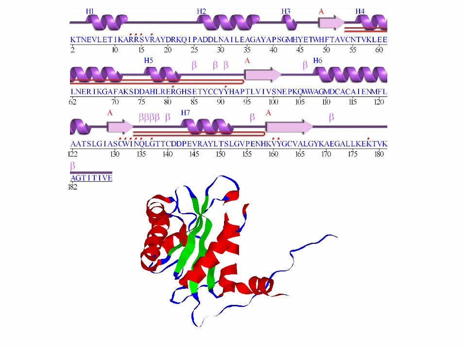
lysozyme

How do we extract 2o structure info?

Secondary Structure Annotations from PDB File
Types of helices and sheets:Helix: Right-handed alpha (default) 1 Right-handed omega 2 Right-handed pi 3 Right-handed gamma 4 Right-handed 310 5
………………
Sheets: Sense of strand with respect to previous strand in the sheet. first strand 0 Parallel 1anti-parallel -1
They are assigned by crystallographers, but how? Will come back to this later……

Secondary Structure Assignments
What are the two main structural properties when we talk about secondary structures?
1. Hydrogen bond patterns
“Knowledge-based protein secondary structure assignment”. Frishman D, Argos P. (1995). Proteins 23(4):566-79
2. Backbone geometry (main-chain dihedral
angles)

Hydrogen Bond Identification
1. A simple way: angle-distance hydrogen bond assignment:
A hydrogen bond is assigned when: 1. q > 120O
AND 2. rHO < 2.5 Å
Baker, E. N. & Hubbard, R. E. (1984).

A better H-bond potential
Dahiyat BI, Gordon DB, and Mayo SL (1997). Automated design of the surface positions of protein helices. Protein Science. 6:1333-1337.
Repulsive Attractive

But when used in practice, it isn’t without problems
Allosteric response is both conserved and variable across three CheY orthologs.Mottonen JM, Jacobs DJ, Livesay DR (2010). Biophysical Journal, 99:2245-2254.

Kabsch W, Sander C (1983). Dictionary of protein secondary structure: pattern recognition of hydrogen-bonded and geometrical features. Biopolymers. 1983 Dec;22(12):2577-637
2. Hydrogen bond identification is based on Coulomb energy
DSSP: Definition (Dictionary) of Secondary Structure of Proteins
)1111
(21NCHOHCNO rrrr
qfqE
Where: f = 332 kcal/mol q1= 0.42, q2=0.2
A hydrogen bond is identified if this energy E is less than -0.5 kcal/mol
Hydrogen Bond Identification-DSSP
**Hydrogen issue

Kabsch W, Sander C (1983). Dictionary of protein secondary structure: pattern recognition of hydrogen-bonded and geometrical features. Biopolymers. 1983 Dec;22(12):2577-637
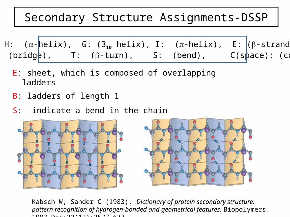
Secondary Structure Assignments-DSSP
H: two consecutive amino acids have i and i+4 hydrogen bonds, and ends likewise with two consecutive i-4 and i hydrogen bonds.
** Similarly for G and I assignments. ** The helix definition does not assign the edge residue having the initial and
final hydrogen bonds in the helix.
T: single helix hydrogen bonds.
H: (-helix), G: (310 helix), I: (-helix), E: (-strand), B: (bridge), T: (-turn), S: (bend), C(space): (coil)

Kabsch W, Sander C (1983). Dictionary of protein secondary structure: pattern recognition of hydrogen-bonded and geometrical features. Biopolymers. 1983 Dec;22(12):2577-637
Secondary Structure Assignments-DSSP
H
G
I

Kabsch W, Sander C (1983). Dictionary of protein secondary structure: pattern recognition of hydrogen-bonded and geometrical features. Biopolymers. 1983 Dec;22(12):2577-637
Secondary Structure Assignments-DSSP
T

Kabsch W, Sander C (1983). Dictionary of protein secondary structure: pattern recognition of hydrogen-bonded and geometrical features. Biopolymers. 1983 Dec;22(12):2577-637
Secondary Structure Assignments-DSSP
Beta Structure Definitions:• Kabsch and Sander define all beta structure in terms of `bridges' which
are either parallel or antiparallel. • Where two or more bridges of the same type are consecutive, the
structure is termed a ladder. • Finally, overlapping ladders are amalgamated into sheets. Additional
complications arise because ladders may have discontinuities in them, and ladders may consists of just a single bridge.
• These aspects of protein structure make the coding of beta-structure less straightforward than for helix.
Kabsch W, Sander C (1983). Dictionary of protein secondary structure: pattern recognition of hydrogen-bonded and geometrical features. Biopolymers. 1983 Dec;22(12):2577-637
Secondary Structure Assignments-DSSP
E: sheet, which is composed of overlapping ladders
B: ladders of length 1
S: indicate a bend in the chain
H: (-helix), G: (310 helix), I: (-helix), E: (-strand), B: (bridge), T: (-turn), S: (bend), C(space): (coil)

Kabsch W, Sander C (1983). Dictionary of protein secondary structure: pattern recognition of hydrogen-bonded and geometrical features. Biopolymers. 1983 Dec;22(12):2577-637
Secondary Structure Assignments-DSSP

DSSP Sample Output

DSSP Sample Output
Note: lower case for SS-bridge CYS

DSSP Sample Output
N-H-->O etc. hydrogen bonds;
e.g. -3,-1.4 means: if this residue is residue i then N-H of i is h-bonded to C=O of i-3 with an electrostatic H-bond energy of -1.4 kcal/mol.
There are two columns for each type of H-bond, to allow for bifurcated H-bonds.
Bifurcated HBs

DSSP Sample Output
**TCO: cosine of angle between C=O of residue I and C=O of residue I-1. α-helices: near +1; β-sheets: near -1. (Not used for structure definition)**KAPPA: virtual bond angle (bend angle) defined by the three Cα atoms of residues I- 2, I, I+2. Used to define bend (structure code 'S'). **ALPHA: virtual torsion angle (dihedral angle) defined by the four Cα atoms of residues I-1, I, I+1, I+2. Used to define chirality (structure code '+' or '-'). **PHI PSI**X, Y, Z coordinates of C

Kabsch W, Sander C (1983). Dictionary of protein secondary structure: pattern recognition of hydrogen-bonded and geometrical features. Biopolymers. 1983 Dec;22(12):2577-637
Kappa and Alpha-DSSP
k a

1ATP
α
310
β
Turn
SS Assignment of PDB Files using DSSP

An aside: ACC from DSSP
ACC = water exposed surface (Å2). But what is the problem with doing this???

Relative solvent accessibility (Range = 0-1)
G G X G GX = Lys
Reference =

STRIDE: secondary STRuctural IDEntification
STRIDE uses two criteria:
1. Hydrogen Bond Energy 2. dihedral angle probabilities
Knowledge-based protein secondary structure assignment. Frishman D, Argos P. (1995). Proteins 23(4):566-79

Hydrogen Bond Identification-STRIDE
distance dependent term
two angular dependent terms
Knowledge-based protein secondary structure assignment. Frishman D, Argos P. (1995). Proteins 23(4):566-79
Empirical hydrogen bond calculation:
E p = cos2 ()
E t =
[0.9 + 0.1 sin(2ti )] cos(to )
K1 [K2 - cos2 (ti ) ] cos(to)
0
0 < ti 9090 < ti 110
110 ti
r is N-O distance
68 r
D
r
CEr
o22
o61 110cos,110cos/9.0 where KK

Torsion angles propensities for alpha-helix and beta-sheet
Dihedral Angle Probabilities-STRIDE
Knowledge-based protein secondary structure assignment. Frishman D, Argos P. (1995). Proteins 23(4):566-79

Secondary Structure Assignments-STRIDE
Recognition of -helices: similar to DSSP--have two consecutive hydrogen bonds between k and k+4
For edge residues:
Recognition of -sheets: similar to DSSP--have two consecutive hydrogen bonds
Five parameters
Four parameters
Knowledge-based protein secondary structure assignment. Frishman D, Argos P. (1995). Proteins 23(4):566-79
Optimized based on a dataset with author’s assignments
1
421
4, )2
1( TPP
WWE kkkkhb
352 and TPTP kk
elAntiparallelAntiparallhb
elAntiparallelAntiparallhb
TCONFWWE
TCONFWWE
)1(
)1(
212
211
ParallelPparallelhb
ParallelParallelhb
TCONFWWE
TCONFWWE
)1(
)1(
212
211
OR 2
)( 21
IntIntInt P
PPCONF

STRIDE Sample Output

STRIDE Sample Output

STRIDE Sample Output

http://webclu.bio.wzw.tum.de/cgi-bin/stride/stridecgi.py

DSSP vs STRIDE
Knowledge-based protein secondary structure assignment. Frishman D, Argos P. (1995). Proteins 23(4):566-79
+
STRIDE better, 58%
DSSP better, 31%
Same Assignment, 11%
226 chains based on authors’ three state Assignments, helix, extended, coil
** <14% difference for individual proteins

DSSP vs STRIDE
Although DSSP is the older method and continues to be the most commonly used, the original STRIDE definition reported it to give a more satisfactory structural assignment in at least 70% of cases. In particular, STRIDE was observed to correct for the propensity of DSSP to assign shorter secondary structures than would be assigned by an expert crystallographer, usually due to the minor local variations in structure that are most common near the termini of secondary structure elements. Knowledge-based protein secondary structure assignment. Frishman D, Argos P. (1995). Proteins 23(4):566-579.
Using a sliding-window method to smooth variations in assignment of single terminal residues, current implementations of STRIDE and DSSP are reported to agree in up to 95.4% of cases.Protein secondary structure assignment revisited: a detailed analysis of different assignment methods. Martin J, et al (2005). BMC Structural Biology 5:17.
Both STRIDE and DSSP, among other common secondary structure assignment methods, are believed to under predict pi helices.Occurrence, conformational features and amino acid propensities for the pi-helix. Fodje MN, Al-Karadaghi S (2002). Protein Engineering 15(5):353-358.

Comparison of Methods for Secondary Structure Assignment
Fourrier et al. BMC Bioinformatics 2004 5:58
Other Programs:
DEFINE – uses a distance criteria between C atoms which varies slightly for each secondary structure type; allows modifications for curvature
DSSP is widely used and a generally accepted method

Secondary Structure Annotations from PDB File
Types of helices and sheets:Helix: Right-handed alpha (default) 1 Right-handed omega 2 Right-handed pi 3 Right-handed gamma 4 Right-handed 310 5
………………
Sheets: Sense of strand with respect to previous strand in the sheet. first strand 0 Parallel 1anti-parallel -1
Crystallographers’ assignments-- angle-distance simple hydrogen bonding pattern-- more complex distance and geometric-- hydrogen pattern + mainchain dihedral angles-- mainchain dihedral angles only-- DSSP algorithm -- a combination of several methods-- visual inspection


![STRIDE-based Threat Modeling for Cyber-Physical Systems · STRIDE-based threat modeling can be performed in two possible ways [22]: (i) STRIDE-per-element and (ii) STRIDE-per-interaction.](https://static.fdocuments.us/doc/165x107/5ec0069865be937c564c10b3/stride-based-threat-modeling-for-cyber-physical-systems-stride-based-threat-modeling.jpg)