Dr./ Wafaa Abd El-ghany Assistant Professor of poultry...
73
Colibacillosis Dr./ Wafaa Abd El-ghany Assistant Professor of poultry dis., Fac. Vet. Med., Cairo Univ.
Transcript of Dr./ Wafaa Abd El-ghany Assistant Professor of poultry...

Colibacillosis
Dr./ Wafaa Abd El-ghany
Assistant Professor of poultry dis.,
Fac. Vet. Med., Cairo Univ.

DefinitionInfectious disease of birds in which E.coli is
either the primary cause of a group of
affections or secondary cause (combined
with other complicating factors as bacteria,
viruses, management, etc….) so,
colibacillosis is a part of the disease
complex rather than a disease itself.
(E.coli is either the primary or secondary
pathogen).

Economic importance
1. High mortality rate (95%) especially in
baby chicks.
2. Poor feed conversion rate.
3. Growth retardation (loss of weight).
4. Down grade of the carcasses.
5. Low fertility and hatchability.
6. Zoonotic importance (infantile diarrhea).

Causative agent• E. coli species of genus Escherichia, Family
Enterobacteriacea are Gram negative bacilli,
non spore forming, aerobic, motile by
peritrichous flagellae.
(few member are non motile) and lactose
fomenter.
• E. coli grows well on ordinary media at 18-44 C
,the colonies on agar after 24 hours at 37 C are
convex, smooth, colorless colonies, while on
MacConky agar, the colonies are bright pink


Antigenic structure• E.coli has unique antigenic structure, It has 7
antigenic structure. Only 4 structures are dominant and have important significance:
• Somatic (O) antigen: polysacchride phospholipid complex with protein resistant to boiling and heat stable (165 serotype).
• Capsular or enveloped ( K ) antigen: I is a polymeric acid contain reducing sugar and associated with the virulence of the cell surface. On the basis of heat stability it is subdivided into (L , A and B ) form. include (89 serotype). It interferes with O antigen and removed by heating for 1 hr.

Antigenic structure• Flagellar (H) antigen: it is a protein and
destroyed at 100C. It is not used as antigen identification of E. coli. It is not related to pathogenicity and absent in non-pathogenic serotypes. It is about 49 serotypes.
• Fimbrial or pilus (F) antigen: it is related to the virulence in mammals, but has no significance in poultry. It is protein in nature and heat labile. It is involved in the cell attachment either (mannose sensitive haemagglutinating) or (mannose resistant haemagglutinating). It is 17 serotypes.
•

• Presence of a lost of different E.coli serotypes creates many problems in the prevention and control of the disease:
1. Difficult to prepare a common antigen, so
A. It is difficult to detect the carriers.
B. Difficult to prepare international vaccine.
C. Difficult treatment due to great differences in their sensitivity to antibiotics (sensitivity test is the must).
Antigenic structure

Epizootiology• E.coli is widely spread in nature as it
present in dust, soil, litter, droppings,
water, feathers, etc…
• E.coli is normal inhabitant of the intestinal
tract in man and animals at a conc. 106
organism/gm of the intestinal contents.
• Recovered birds shed organism in feces
for long time.

• E.coli can live in the respiratory tract as a normal inhabitant organism.
• E.coli strains differs in their pathogenicity from pathogenic, opportunistic or non pathogenic.
• E.coli persist in the dry conditions for long time, wetness of the environment inactivate the bacteria.
• Pathogencity of the organism is affected by host susceptibility factors, nature and degree of stress factors.
Epizootiology

Mode of infection
• Ingestion of contaminated food and water
(oral) .
• Inhalation of dust in houses and hatchery
(Respiratory).
• Direct contact with infected or chronic
carrier birds.

Mode of transmission
I Vertical transmission
True (transoverian)False (egg shell contamination)

During laying from contaminated cloacae.
Contaminated incubators and hatcheries.
Contaminated hands of workers during eggs
collection.
• (the organism can enter egg through egg pores
by its motility or negative pressure).
Mode of transmission
egg shell contamination

II Horizontal (lateral transmission)
Contaminated food, water and air dust.
Mechanically by contaminated
equipments, workers, utensils, and feed
bags.
Free living birds, pets and rodents.
Contaminated environment around the
hatcheries or farms.
Mode of transmission

III Stress factors
Bad ventillation (Increase ammonia and
Co2 levels).
Chilling.
Vitamin A deficiency.
Parasitism.
Malnutrition.
Mode of transmission

1. Colisepticaemia
2. Air sac disease (CRD).
3. Panothalmitis.
4. Arthritis.
5. Salpingitis.
6. Omphalitis.
7. Coligranuloma.
8. Swollen head syndrome
9. Enteritis

Colisepticaemia
1. Respiratory-origin colisepticemia:
• This type occurred following damage of
respiratory system by virus, bacteria and /
or environmental factors to form air sac
disease , chronic respiratory disease (
CRD ) or Complicated chronic respiratory
disease (CCRD).

2. Enteric-origin colisepticemia:• It occurs following intestinal damage
especially at turkey hemorrhagic enteritis.
• Enteric signs (diarrhea) is commonly
observed.
Colisepticaemia

Colisepticaemia
3. Neonatal Colisepticemia:
• Occurred in chicks following navel
infection at first 1-2 days after hatching as
mortality can reach 10-20 % in 2-3 weeks.

4. Acute septicemia of layer
chickens and turkeys:• The infection occurred at the onset of
egg production and spread to older birds
with sudden death and mortality 5 -100
%.
Colisepticaemia

Colisepticaemia
1. Respiratory signs (sneezing, nasal discharge, sinusitis, cough, rals, etc…).
2. Enteric signs (diarrhea).
3. Omphalitis.
4. High mortality rate.
Signs

1. Serous to fibrinous pericarditis,
perihepatitis and airsacculitis.
2. Cattharal to serous sinusitis, laryngitis and
tracheitis and pneumonia.
3. Septicaemic picture.
4. Enteritis.
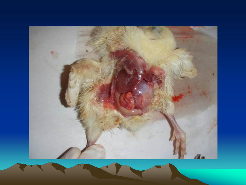
5. Un-absorbed yolk sac.
Colisepticaemia
Lesions

Severely congested liver
Foci of necrosis on liver

Sequel of
Colisepticeamia
Death
Recovery with localized lesions
Complete
Recovery

Air sac disease
Chronic respiratory disease
(CRD)
• Severe respiratory signs and high
mortalities are found in broiler chickens (3-
8) weeks old.
• Drop in feed consumption and in egg
production with low mortalities in adult
birds.

1. Serous to fibrinous (caseous) pericarditis,
perihepatitis and airsacculitis.
2. Cattharal to serous sinusitis, laryngitis
and tracheitis.
3. Lung congestion and pneumonia.
Air sac disease
Chronic respiratory disease
(CRD)


Pericarditis and perihepatitisPeritonitis

Fibrinous pericarditis

Pericarditis and perihepatitis

Airsacculitis


Pneumonia
Airsacculitis

Panophthalmitis• Localized severe inflammation of eye ball.
• Affected bird shows:
1. Conjunctivitis.
2. Keratitis.
3. Complete destruction of the retina.
4. Conjunctivitis.
5. Closed eyes and blindness.
6. Emaciation.

Arthritis• This infection follows colisepticaemia or as a
result of joint injuries.
• Affected birds show:
1. Locomotor disturbance and lamness.
2. Unable to sand.
3. Uni or bilateral swelling of the joints and they
become, hot and painful.
4. Death due to the bird is un-ables to reach the
food sites.


Salpingitis• Inflammation of the oviduct occurs as a result
from ascending infection from the cloacae or follow air sac infection.
• It occurs at the onset of egg production and inflamed with hormonal changes that occur at this period.
• Affected birds showed marked drop in egg production, decrease in fertility and hatchability.
• Sudden death due to abdominal distension and severe peritonitis

• The oviduct become inflamed, thick and
narrow.
• Presence of caseous material (core) that
occlude the oviduct.
• Rupture of the oviduct and presence of
yolk material in the abdominal cavity (yolk
peritonitis).
Salpingitis


Omphalitis• Inflammation of the navel may occure due
to contamination of the egg shell by the
droppings followed by entering of the
organism to the embryo causing high
embryonic mortalities during incubation.
• Hatched chicks may infected in the
hatchery causing high mortalities
especially at the first week of life.

• Affected chicks with omphalitis show:
1. General weakness and tendency to
huddle around the heat sources.
2. Opened, wet, un-healed and inflamed
navel.
3. Swollen, fluffy and flabbiness of the
abdominal muscles (mushy chick).
4. Diarrhea and death within 1 week.
Omphalitis

• Dead chicks showed lesions of:
1. Opened, wet, un-healed and inflamed navel.
2. Swollen, fluffy and flabbiness of the abdominal muscles (mushy chick).
3. Un absorbed yolk sac (thick wall and contained reddish caseous contents with offensive odour).
4. Ascitis.
Omphalitis

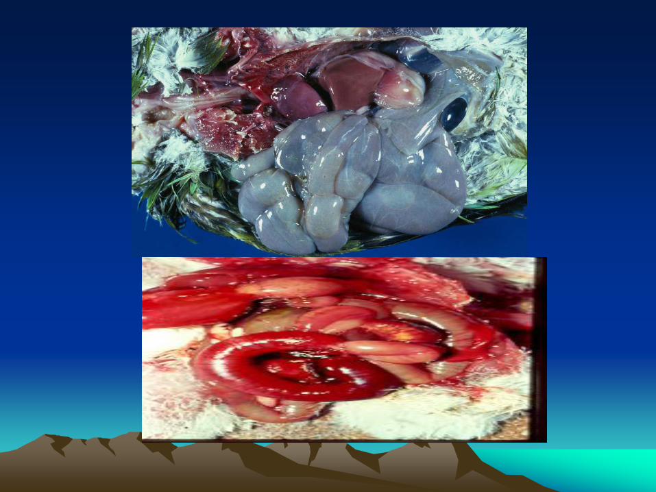
Coligranuloma
(Hjarr’s Disease)• Sporadic conditions occur in layers towards the
end of laying period.
• It caused by mucoid strain of E.coli following the
intestinal damage by ectoparasites.
• Clinical signs is vague, just emaciation and
unthriftness.
• There were granulomatous nodules (smooth and
grayish white) on the cacum, intestine, liver,
spleen and gizzard.


Swollen head syndrome
(SHS)• It is acute or sub-acute cellulitis involving
per-orbital and the subcutanous tissues of the head.
• The predisposing factors for this condition may be infection with pneumovirus, poor ventilation, high ammonia level or infection with IBV.
• Infection may occur through the Eustachian tube of the ear.


Enteritis
• It is uncommon form in poultry rather than
animals.
• It may be occur as a result from intestinal
damage by coccidiosis and intestinal
parasites.
• Affected birds show diarrhea and catarrhal
to haemorrhagic enteritis.

• Samples should be taken under complete aseptic conditions either from freshly dead or ailing birds.
• Samples could be taken from the gall bladder, liver and bone marrow, and also swabs from the yolk sac, umbilicus, heart blood, pericardial sac and intestine.
• Samples could be taken from any organ showing lesions (joints, eye, oviduct, etc….).
• Suspected or contaminated feed, water, hatchery and incubator or environmental samples could be taken.
Diagnosis

• Samples are streaked on MacConkey agar media and incubated at 37C for 24-48 hrs under aerobic or facultative anaerobic conditions. The colonies appears rose-pink in colour (lactose fermenting strains), smooth, glossy and translucent.
• Colonies on blood agar media are colourless (white), glistening, raised and sometimes surrounded with zone of haemolysis.
• Colonies on nutrient agar are circular (1-3mm) in diameter, smooth, low convex, colourless to gray and translucent.
Diagnosis

• Colonies on XLD (xylose-lysine deoxycholate) media are yellow.
• Colonies on CLED (cystine-lactose electrolyte deficient) media with bromothymol blue are smooth, circular and yellow (If lactose fermenting strains) while blue (If non-lactose fermenting strains).
• Colonies on EMB (Eosin-methylene-blue) media are green with black center.
• In broth, E. coli produces diffuse cloudiness and heavy sediment.
Diagnosis

• The growth of E. coli on DCA (Deoxycholate-citrate agar) media is inhibited by addition o0f citrate.
• Growth of E. coli is inhibited by sodium selenite, sodium tetrathionate and brilliant green which are selective for Salmonella and Shegilla isolation.
• Growth of E. coli is inhibited by 7% Nacl in salt media which used for isolation of Staphylococci.
Diagnosis

• Gram staining of E. coli colonies revealed Gram negative, medium size bacilli, non spore former, non acid fast, capsulated (on Congo red media) , and motile (most strains) with peritrichus fagellae.
• Most strains are fimbrate type 1 (either haemagglutinating mannose sensitive adhesive to the epithelium or eluting haemagglutinating mannose resistant adhesive to the epithelium.
• Some strains produce loose slime on sugar media.
Diagnosis

• E. coli is -, +, -, + (IMVC) as it used for differentiation between E. coli and other enterobacteriacae like Klebsiella and enterococci.
• It doesn’t liquefy gelatin, not produce H2S and negative urease.
• Positive oxidase, catalase, nitrate reduction lysine and ornithine decarboxylase.
• Most E. coli strains are lactose fermenter, also ferment glucose, galactose, mannitol and fructose with production of acids and gas. Fermentation of sucrose, salicin and dulcitol is variable.
Diagnosis

• Slide agglutination test:
Emulsify pure E. coli colonies with saline,
then put one drop of the suspension with
one drop of polyvalent antisera specific to E.
coli on the glass slide. Agglutination
(follicules) indicate positive reaction (detect
the antigen).
Using of monovalent antisera for detection
of serotype.
Diagnosis

• Serological examination using specific antisera is of value only in case of epidemiological examination due to the followings:
1. will not distinguish infecting organisms from other faecal contaminants.
2. There were many pathogenic E. coli strains don’t belong any serotype (Un agglutinable strains).
3. Antigenic complexity of E. coli (difficult to prepare antigen).
4. It is costly.
5. Delay the diagnosis (time consuming).
Diagnosis


• Pathogenicity test:
1. Inoculation of 4 weeks old chickens with the
suspected material induces death within 72 h.
2. Inoculation of yolk sac of day old chick with
stains causing embryonic mortalities induces
death within 72 h.
• Survived birds should be examined for specific
lesions.
• Isolation of the organism from the infected organs
Diagnosis

PreventionI. Good eggs sanitation:
1. The eggs used for hatching should be originated from colibacillosis free breeders.
2. Frequent collection and fumigation of the eggs on the farm prior to storage.
3. Stored under ideal conditions (cool and clean place) for short time sitting.
4. Only clean eggs used in hatching.

PreventionI. Good eggs sanitation:
5. Clean and disinfected utensils and egg containers.
6. Adequate number of clean nests should be provided.
7. Precaution of persons during egg collection
8. Dipping of the eggs in germicidal solution.

II. Hatchery sanitation:
1. Thorough cleaning and disinfection of the incubators and hatcheries after each cycle.
2. Fumigation of the incubators and eggs after sitting to minimize shell contamination.
3. Hatchery and its equipments should be monitored by periodic culture.
Prevention

III. Strict sanitation and sound management in the farm (biosecurity measures):
1. Thorough cleaning and disinfection.
2. Provide good environmental conditions (temp., RH, ventilation).
3. Avoid predisposing stress factors.
4. Eradicate pets, insects and rodents.
5. Avoid mechanical transmission by workers, persons, etc…
Prevention

6. Using gamma globulins (immunoglobulins)
Serum from animals or egg yolk ppt is used in
the drinking water or by injection.
7. Using of the probiotics, prepibiotics and synbiotics.
They are competitive exclusion products containing
beneficial safe bacteria or fungi, applied in the food or the
drinking water at the first days of life as they
compete the pathogenic organisms by blocking the
receptor sites or flourish normal flora which inhibit
the organism colonization.
Prevention

8. There is no commercial vaccine as it is
difficult to be prepared due to presence
of multiple serotypes. Local (autogenous)
vaccine could be prepared from the
predominant serotype in certain area to
give protection in this area only.
Prevention

Control1. Strict biosecurity measures.
2. Using of weak organic acids (lactic, acetic
and probionic) as they produce acidic pH
unsuitable for the growth of E.coli and
increase the intestinal flora that inhibit
E.coli colonization.
3. Treatment with antibiotics according to the
sensitivity test results (antibiogram) due to:

1. Presence of multiple serotypes of E.coli.
2. Presence of R-Factor (resistant factor) which
induces certain phenomenon characterizing
E.coli called transferable infectious drug
resistance problem.
• This problem caused by the miss or hazardous
use of antibiotics in the field which leads to
appearance of mono or multiple resistant strains
which transfer R-factor partially or completely to
other E.coli sensitive strain.
Control

• R- Factor can transmit from resistant E.coli generation to the sensitive one by
• Transformation: A small portion of genetic material (DNA) from dead bacterial cell enters another living one.
• Transduction: Transfer of pieces of genetic material of cell to other by phage in which it multiplied.
• Sexual recombination: Under certain condition sexual reproduction occurred by which genetic factor transferred from cell to other.
Control