Dr. Saidunnisa Professor of Biochemistry Carbohydrates-II.
39
Dr. Saidunnisa Dr. Saidunnisa Professor of Biochemistry Carbohydrates-II
-
Upload
sherilyn-boone -
Category
Documents
-
view
229 -
download
3
Transcript of Dr. Saidunnisa Professor of Biochemistry Carbohydrates-II.

Dr. Saidunnisa Dr. Saidunnisa
Professor of BiochemistryCarbohydrates-II
Learning ObjectivesLearning ObjectivesAt the end of the session student shall
be able to:Define PolysaccharidesList homopolysaccharides and explains
the biomedical importance of each.List biologically important sugar
derivatives with its clinical importance. Define and lists various
Glycosaminoglycans and explains the biomedical importance of each and correlate to lysosomal storage disorders.

PolysaccharidesPolysaccharides They contain more than ten
monosaccharide units. These are of two types;1. Homo polysaccharides.2. Hetero polysaccharides. ( GAGS)
Homo polysaccharides.Homo polysaccharides.
Made up of same single monosaccharide units.
Examples:1. Starch2. Cellulose3. Glycogen4. Inulin5. Dextrans6. Chitin
StarchStarch
Carbohydrate reserve of the plants.
Important dietary source of man.
High content is present in cereals, roots, tubers.
It is made up of multiple units of alpha-D Glucose.
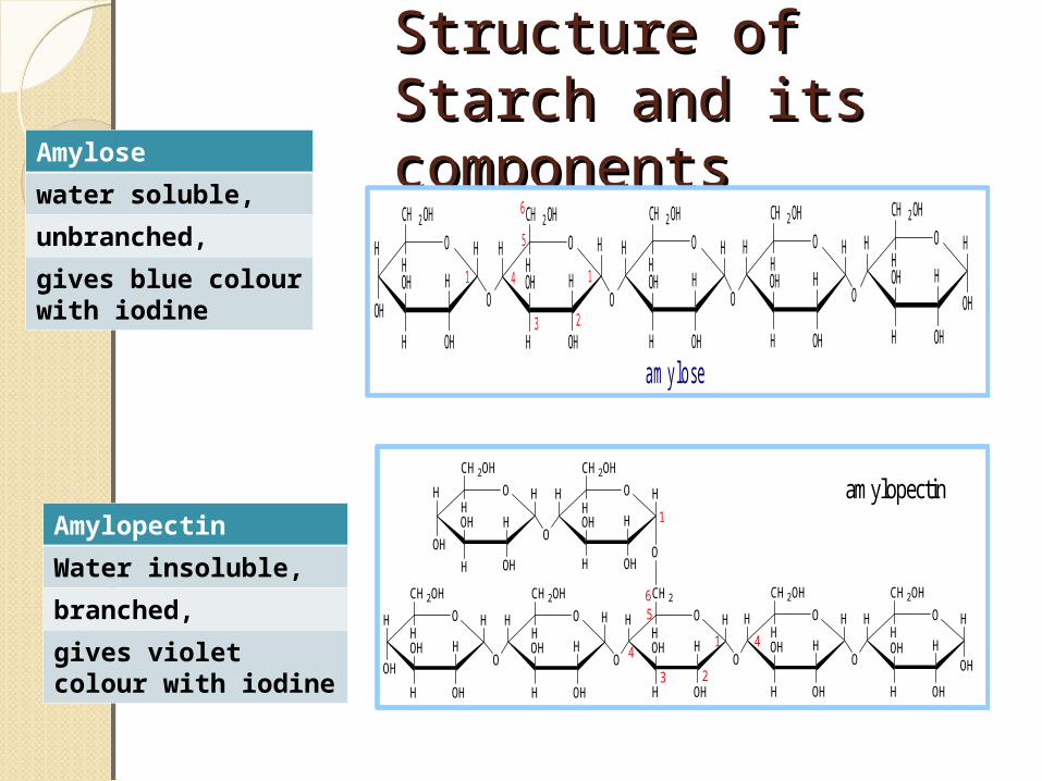
Structure of Starch Structure of Starch and its componentsand its components
H O
OH
H
OHH
OH
CH 2 OH
HO H
H
OHH
OH
CH 2 OH
H
O
HH H O
OH
OHH
OH
CH 2 OH
HH H O
H
OHH
OH
CH 2 OH
H
OH
HH O
OH
OHH
OH
CH 2 OH
H
O
H
1
6
5
4
3
1
2
a m y lo s e
H O
OH
H
OHH
OH
CH2OH
HO H
H
OHH
OH
CH2OH
H
O
HH H O
OH
OHH
OH
CH2
HH H O
H
OHH
OH
CH2OH
H
OH
HH O
OH
OHH
OH
CH2OH
H
O
H
O
1 4
6
H O
H
OHH
OH
CH2OH
HH H O
H
OHH
OH
CH2OH
HH
O1
OH
3
4
5
2
amylopectin
Amylose
water soluble,
unbranched,
gives blue colour with iodine
Amylopectin
Water insoluble,
branched,
gives violet colour with iodine
DegradationDegradation Starches are hydrolyzed by amylase to
liberate dextrin's.
Dextrin's are finally converted to Maltose and glucose
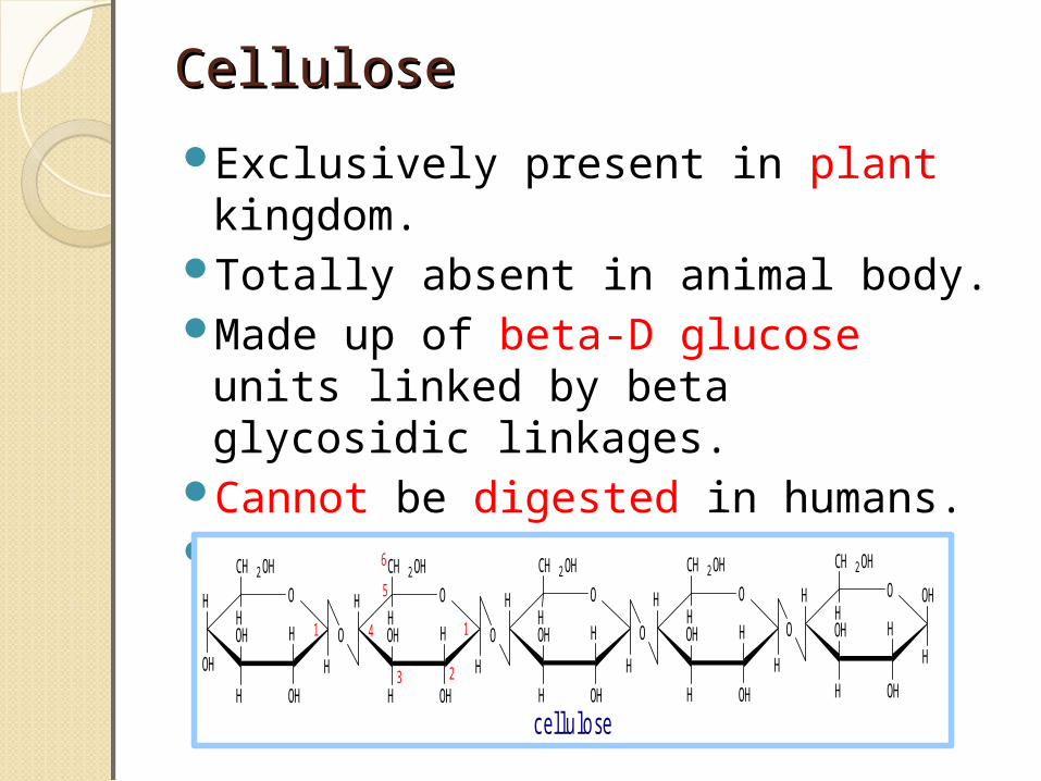
Cellulose Cellulose
Exclusively present in plant kingdom.
Totally absent in animal body.Made up of beta-D glucose units
linked by beta glycosidic linkages.
Cannot be digested in humans.Major constituent of dietary fiber.
c e l lu lo s e
H O
OH
H
OHH
OH
CH 2 OH
HO
H
OHH
OH
CH 2 OH
HO
H H O
O H
OHH
OH
CH 2 OH
HH O
H
OHH
OH
CH 2 OH
H
H
OHH O
O H
OHH
OH
CH 2 OH
HO
H H H H
1
6
5
4
3
1
2
Learning Check?Learning Check?Cellulose though not digested,
has great importance in human nutrition why?

1. Improves glucose tolerance2. Decresaes blood cholesterol 3. Relives constipation by
increasing the bulk of the feces.
GlycogenGlycogenAnimal reserve of the body.Present in high concentration in
liver, muscle and brain.Similar to Amylopectin, made up
of repeated units of alpha-D-Glucose linked by glycosidic linkages.
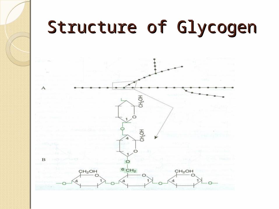
Structure of GlycogenStructure of Glycogen
InulinInulinPolymer of fructose.Present in garlic, onion, dahlia
bulbs etc.Low molecular weight easily
soluble in water.Not utilized by the body.Used for assessing kidney
function through GFR.
DextransDextransHigh molecular weight.Polymer of Alpha-D glucose
linked by glycosidic linkages.Used as an plasma expander.When given I.V in cases of blood
loss (Hemorrhage), it increase the blood volume.

Learning checkLearning checkDextrose, Dextrin, and Dextran
are same or different molecules?
ChitinChitinComposed of N-acetyl-D-
glucosamine.Found in exoskeleton of some
invertebrates.
HeteroHetero polysaccharides polysaccharides
Glycosaminoglycans (GAGs) important constituents of ECM (extra cellular matrix).
Extra cellular matrix Extra cellular matrix (ECM)(ECM)The ECM is often referred to as the
connective tissue made up of:Ground substancesCartilages, Tendons, ligaments, Vascular wall, Skin, LungCornea

FunctionsFunctions
Mechanical supportLubricationCushioning
Extracellular matrix (ECM)Extracellular matrix (ECM)The ECM is composed of 3 major
classes of biomolecules: 1. Structural proteins: collagen
and elastin.2. Specialized proteins: e.g.
fibrillin, fibronectin, and laminin.3. Proteoglycans: GAGS

Synthesis and Synthesis and Degradation Degradation These are synthesized in ER and
modifications like sulfation takes place in Golgi complex.
These are degraded by lysosomal enzymes.
Inability to degrade leads to a set of disease known as Lysosomal storage disease or mucopolysaccharidoses.
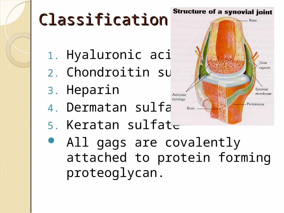
ClassificationClassification
1. Hyaluronic acid2. Chondroitin sulfate3. Heparin4. Dermatan sulfate.5. Keratan sulfate All gags are covalently attached
to protein forming proteoglycan.

Resilience of GAGS Resilience of GAGS
There are negatively charged COO- and SO4
– groups on the GAG which bind to positively charged hydrogen ions of water molecules thereby creating a hydrated gel.
Negative charge imparts high viscosity and low compressibility which makes them ideal for lubricating fluid in the joints.

Constituents of GAGS are Constituents of GAGS are repeating sugar derivatives repeating sugar derivatives
These units contain a
1. Amino sugar:
(N acetyl glucosamine / galactosamine)
&
2.Uronic acid:
(Glucuronic / iduronic acid).
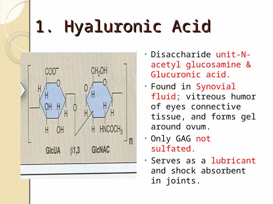
1. Hyaluronic Acid1. Hyaluronic Acid
• Disaccharide unit-N-acetyl glucosamine & Glucuronic acid.
• Found in Synovial fluid; vitreous humor of eyes connective tissue, and forms gel around ovum.
• Only GAG not sulfated.• Serves as a lubricant
and shock absorbent in joints.
Learning CheckLearning CheckWhat is Hyaluronidase? (hint:
fertilization)
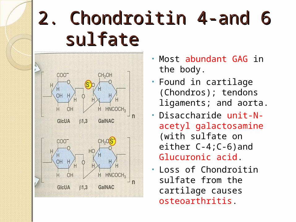
2. Chondroitin 4-and 6 2. Chondroitin 4-and 6 sulfatesulfate
• Most abundant GAG in the body.
• Found in cartilage (Chondros); tendons ligaments; and aorta.
• Disaccharide unit-N-acetyl galactosamine (with sulfate on either C-4;C-6)and Glucuronic acid.
• Loss of Chondroitin sulfate from the cartilage causes osteoarthritis.
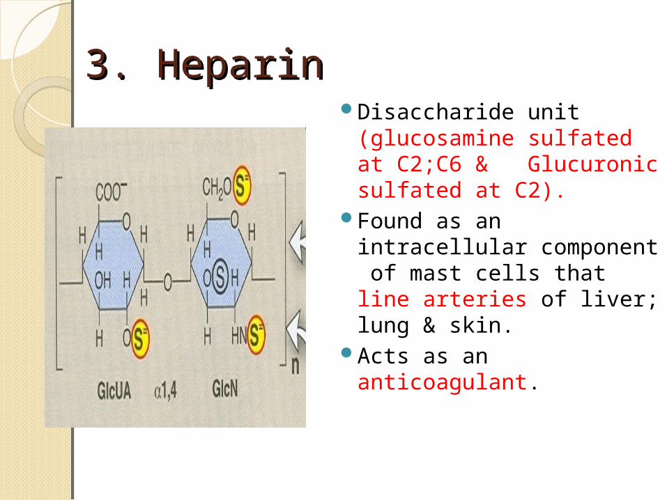
3. Heparin3. HeparinDisaccharide unit
(glucosamine sulfated at C2;C6 & Glucuronic sulfated at C2).
Found as an intracellular component of mast cells that line arteries of liver; lung & skin.
Acts as an anticoagulant.
CaseCaseA 76-year old woman who is bed
bound in a nursing home begins to develop swelling of her left leg. She is evaluated for deep vein thrombosis; she is immediately started on heparin to further prevent the clot.
What is Heparin and its components ?
What is its action?
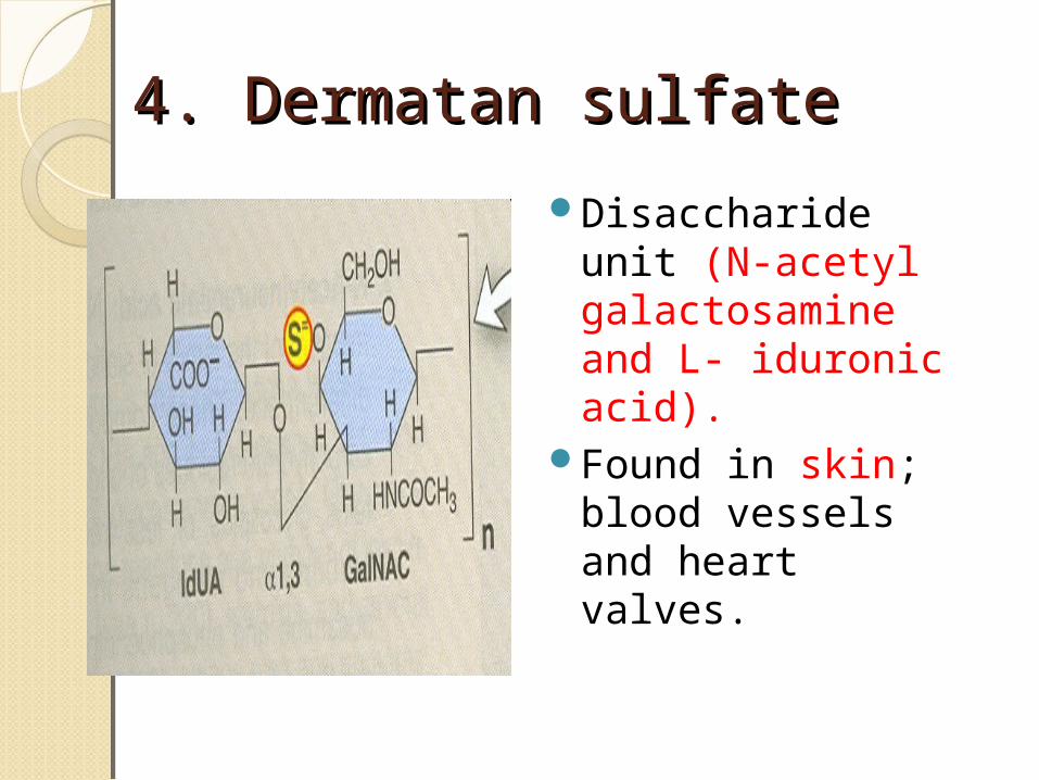
4. Dermatan sulfate4. Dermatan sulfate
Disaccharide unit (N-acetyl galactosamine and L- iduronic acid).
Found in skin; blood vessels and heart valves.
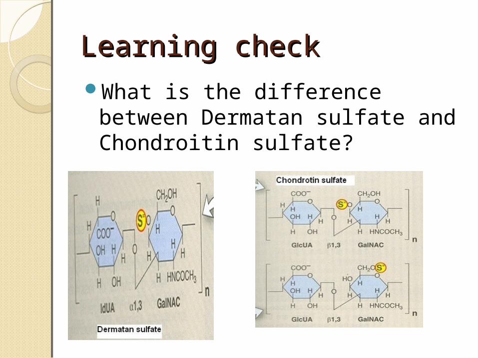
Learning checkLearning checkWhat is the difference between
Dermatan sulfate and Chondroitin sulfate?

5. Keratan sulfate5. Keratan sulfate
Disaccharide unit (N acetyl glucosamine& galactose; sulfate may be present on C6 of either sugar) does not contain uronic acid.
Found in cornea responsible for transparency of cornea.

Summary Summary GAG Repeating Disaccharide Tissue
distribution
Hyaluronic Acid
N-acetyl glucosamine (Not sulfated)
Glucuronic acid.
Synovial fluid
Chondroitin 4-and 6 sulfate
N-acetyl galactosamine(sulfated)
Glucuronic acid
Cartilages
Heparin glucosamine (sulfated )
Glucuronic sulfated
line arteries
Dermatan sulfate
N-acetyl galactosamine
L- iduronic acid
Skin
Keratan sulfate
N acetyl glucosamine& galactose; sulfate
does not contain uronic acid
cornea
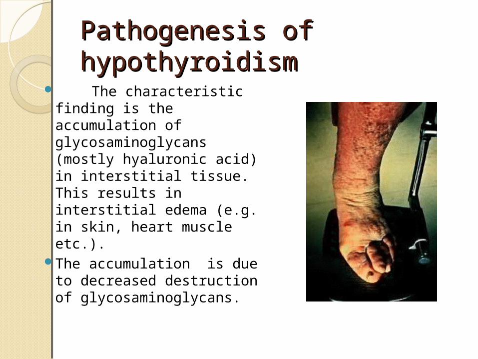
Pathogenesis of Pathogenesis of hypothyroidism hypothyroidism
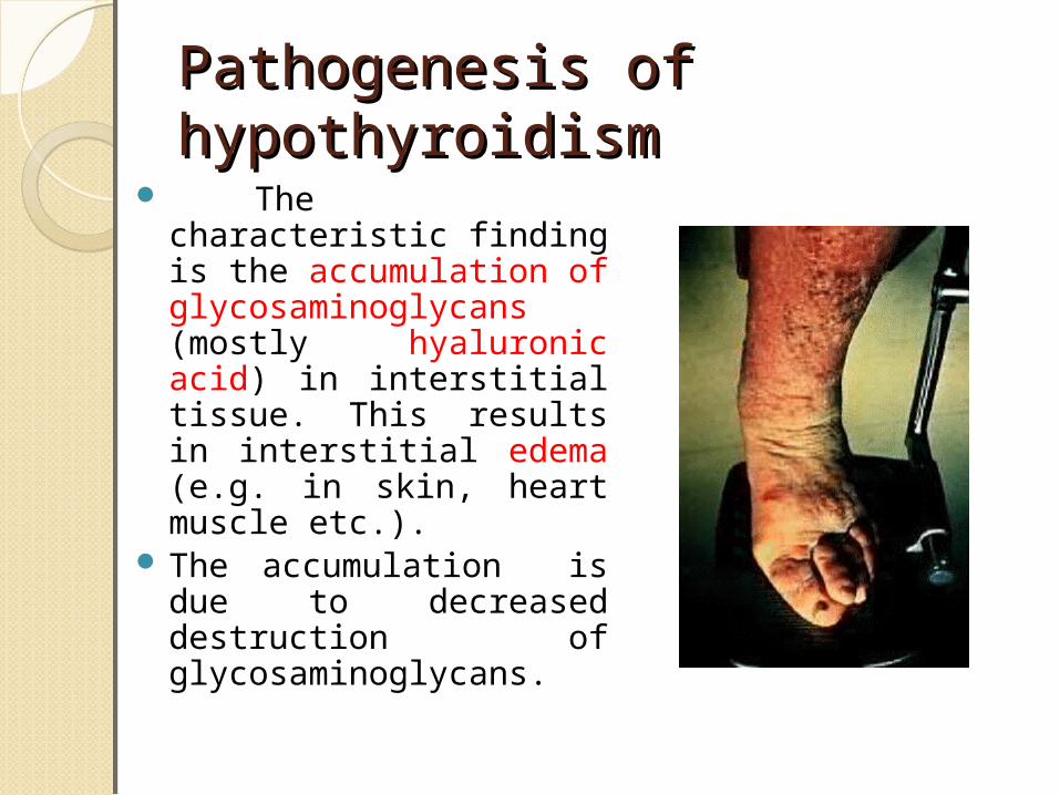
The characteristic finding is the accumulation of glycosaminoglycans (mostly hyaluronic acid) in interstitial tissue. This results in interstitial edema (e.g. in skin, heart muscle etc.).
The accumulation is due to decreased destruction of glycosaminoglycans.
Pathogenesis of Pathogenesis of hypothyroidism hypothyroidism
The characteristic finding is the accumulation of glycosaminoglycans (mostly hyaluronic acid) in interstitial tissue. This results in interstitial edema (e.g. in skin, heart muscle etc.).
The accumulation is due to decreased destruction of glycosaminoglycans.

Structure of ProteoglycansStructure of Proteoglycans
The Proteoglycans are essential parts of extracellular matrix,
The majority of GAGs in the body are linked to core proteins, forming Proteoglycans .
The linkage of GAGs to the protein core involves a specific trisaccharide composed of two Galactose residues and a xylose residue (GAG-GalGalXyl-O-CH2-protein). This is linked to the protein core through an O-glycosidic bond.
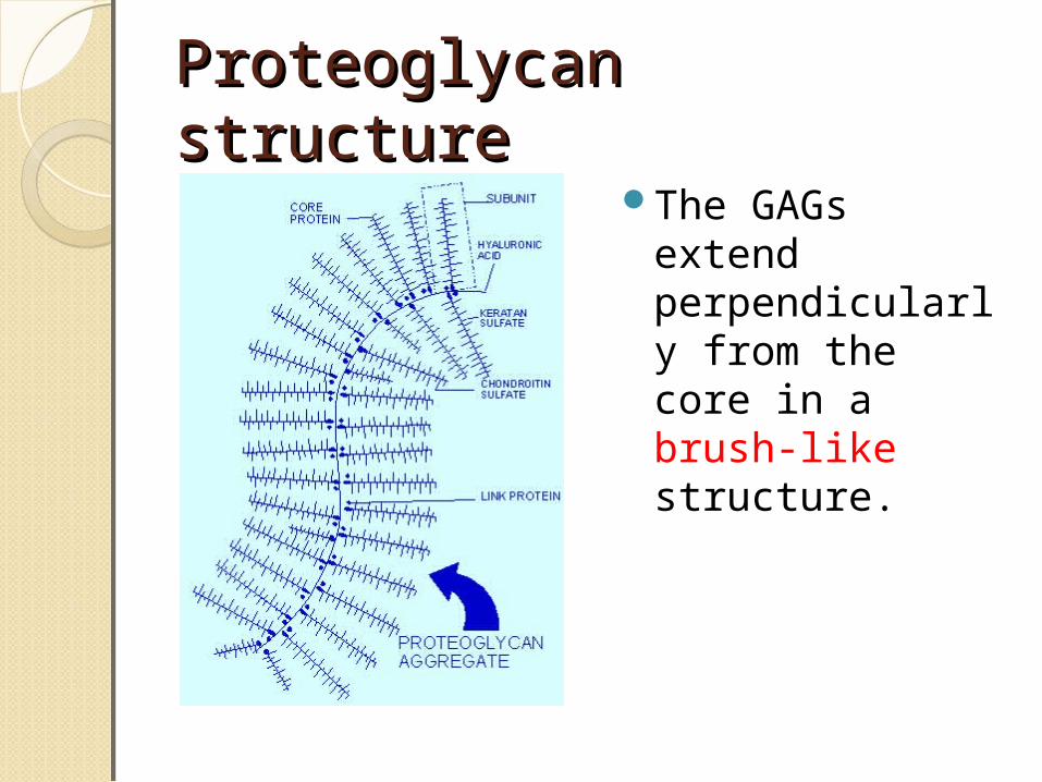
Proteoglycan structure Proteoglycan structure The GAGs extend
perpendicularly from the core in a brush-like structure.

Lysosomal storage disorders or MPS

DiagnosisDiagnosisWide spread
deposits in tissues of a particular GAGS.
Excessive excretion of GAG in Urine.
Detection of GAG in urine:
Cetyl trimethyl ammonium bromide test.