Dr Majid Al-Homiedan Head ER/Trauma Radiology division … · 2019-10-13 · I. Technique Learn the...
64
Dr Majid Al-Homiedan Head ER/Trauma Radiology division Cardiothoracic and abdominal subspecialty consultant
Transcript of Dr Majid Al-Homiedan Head ER/Trauma Radiology division … · 2019-10-13 · I. Technique Learn the...

Dr Majid Al-Homiedan
Head ER/Trauma Radiology division
Cardiothoracic and abdominal subspecialty consultant

I. Technique
Learn the difference between PA vs. AP CXR
Learn the utility of a lateral decubitus CXR
Understand the terms inspiration, penetration, and rotation as they apply to determining a technically adequate film
II. Anatomy
Learn the basic anatomy of the fissures of the lungs, heart borders, bronchi, and vasculature that can be seen on a chest x-ray
III. Interpretation
Develop a consistent and thorough technique for reading images
Learn how the silhouette sign can help localize pathology
IV. Pathology
Objectives

PositioningPA

Positioning lateral
Always read together

positioning

PositioningPA vs AP
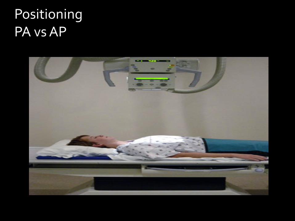
PositioningPA vs AP
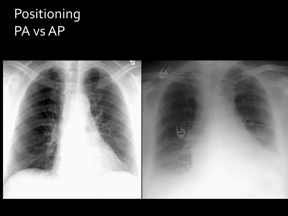
PositioningPA vs AP

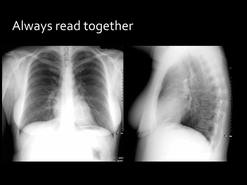
PositioningLateral decubitus

Technically adequateinspiration
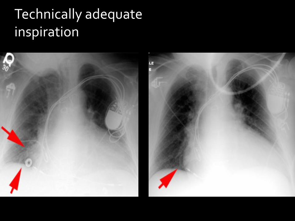
Technically adequateinspiration

Technically adequatePenetration

Technically adequatePenetration

Technically adequatePenetration

Technically adequateRotation

Technically adequateRotation

Technically adequateRotation

AnatomyLobes and Fissures

AnatomyLobes and Fissures

AnatomyLobes and Fissures

AnatomyLobes and Fissures
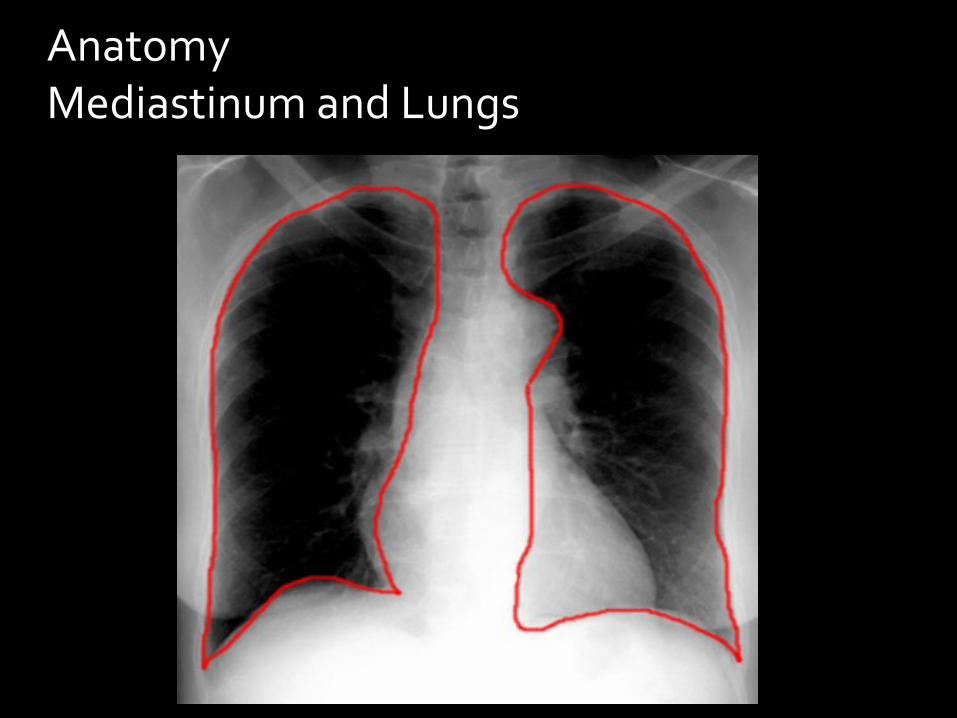
AnatomyMediastinum and Lungs

Anatomy

Anatomy Bronchi

Pulmonary vasculature
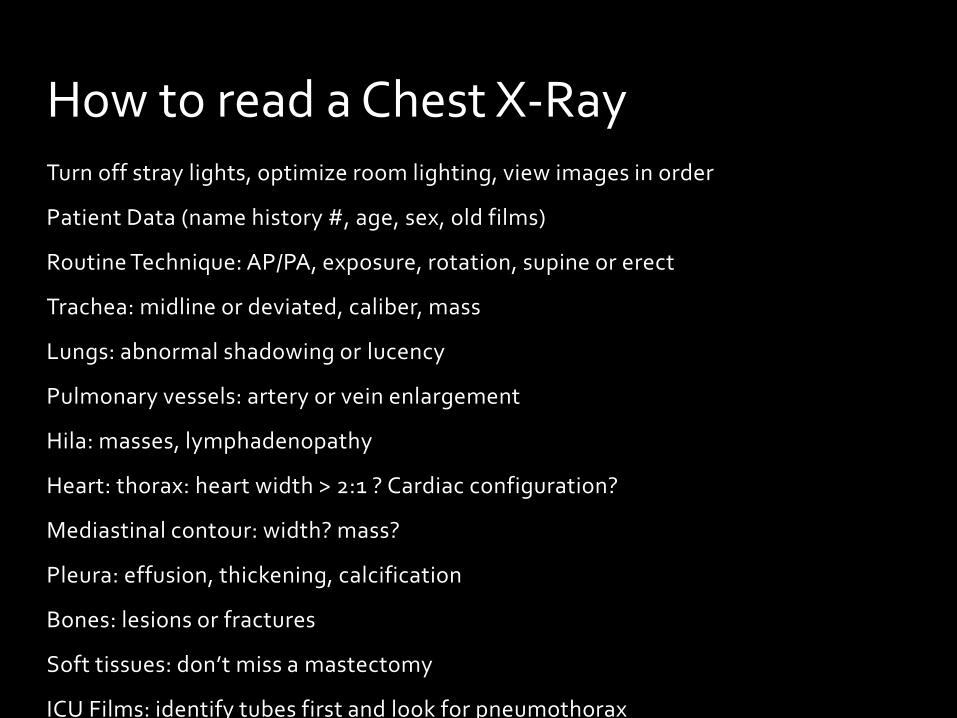
Turn off stray lights, optimize room lighting, view images in order
Patient Data (name history #, age, sex, old films)
Routine Technique: AP/PA, exposure, rotation, supine or erect
Trachea: midline or deviated, caliber, mass
Lungs: abnormal shadowing or lucency
Pulmonary vessels: artery or vein enlargement
Hila: masses, lymphadenopathy
Heart: thorax: heart width > 2:1 ? Cardiac configuration?
Mediastinal contour: width? mass?
Pleura: effusion, thickening, calcification
Bones: lesions or fractures
Soft tissues: don’t miss a mastectomy
ICU Films: identify tubes first and look for pneumothorax
How to read a Chest X-Ray

Your eye gaze should scan all portions of the film, follow lung/mediastinal interfaces and look again carefully in areas where you know that mistakes are easily made, such as over the spine on the lateral view and in the apex on the PA view.
Looking for abnormalities

Looking for abnormalities

Looking for abnormalities
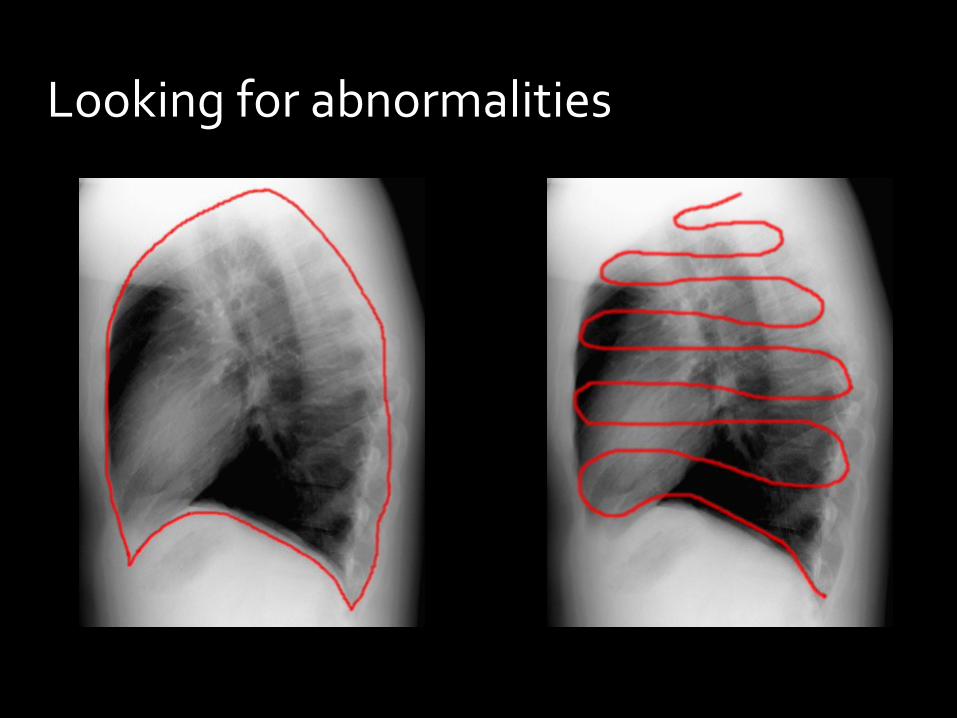
Looking for abnormalities

Silhouette sign

Air Bronchogram
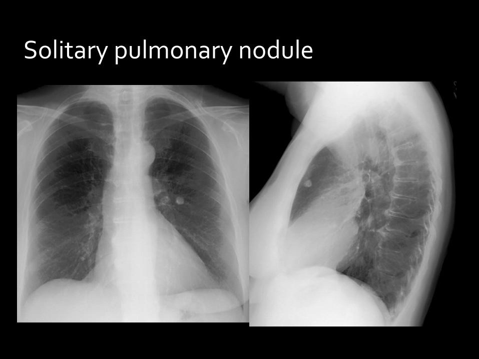
Solitary pulmonary nodule

Cont.

PathologyAtelectasis

PathologyAtelectasis

left lower lobe atelectasis followed by partial resolution
PathologyAtelectasis

Note the elevation of the left hemidiaphragm

PathologyAtelectasis

PathologyAtelectasis
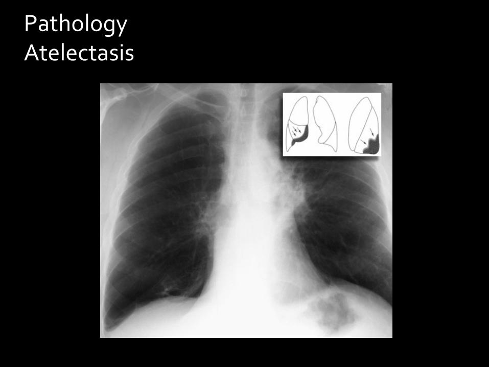
PathologyAtelectasis
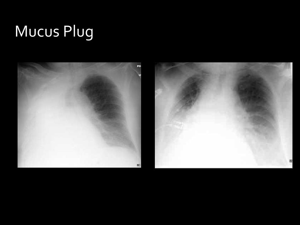
Mucus Plug

Pulmonary edema

Pulmonary edema

Kerley B lines

Pneumonia

Atelectasis Pneumonia
Volume Loss
Associated Ipsilateral Shift
Linear, Wedge-Shaped
Apex at Hilum
Normal or Increased Volume
No Shift, or if Present Then
Contralateral
Consolidation, Air Space Process
Not Centered at Hilum
Air bronchograms can occur in both.

Pulmonary hemorrhage

Pulmonary embolism
What is the most common chest X-ray finding in PE?

Pulmonary embolism

Pleural effusion

Pneumothorax

PTX deep sulcus sign
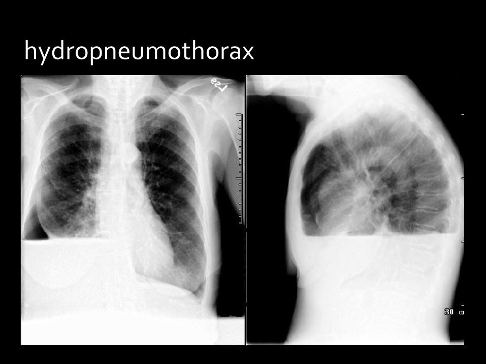
hydropneumothorax

Emphysema
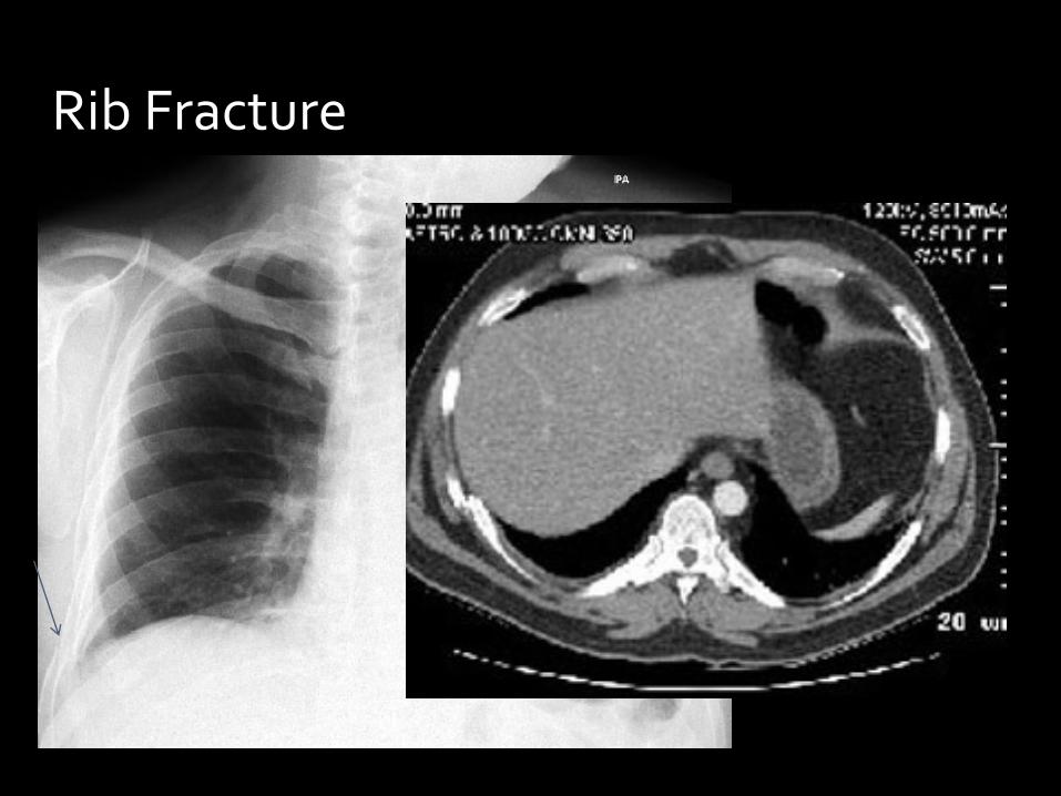
Rib Fracture

Flail Chest

Anterior Mediastinal Mass

Pneumomediastinum


Hiatal Hernia

THANK YOU…..