Dr. Fowler's 12 Lead EKG Interpretation Part 1
26
12 Lead EKG Interpretation E s s e n t i a l l y E s s e n t i a l l y s p e a k i n g . . . s p e a k i n g . . . Copyright 2002, Ray Fowler, M.D., FACEP Ray Fowler, M.D., FACEP Assistant Professor of Emergency Medicine The University of Texas Southwestern [email protected] Copyright 2002, Ray Fowler, M.D., FACEP Objectives of this program: 1. Become familiar with cardiac anat omy as it relates to the electrocardiogram 2. Develop an initial understanding of “Axis” 3. Understand how to interpret hypertrophy 4. Recognize Bundle Branch Blocks 5. Understand how to interpret EKG’s for signs of myocardial infarction 6. Understand the “grouped leads” concept Copyright 2002, Ray Fowler, M.D., FACEP
-
Upload
queenmom09 -
Category
Documents
-
view
222 -
download
0
Transcript of Dr. Fowler's 12 Lead EKG Interpretation Part 1

8/3/2019 Dr. Fowler's 12 Lead EKG Interpretation Part 1
http://slidepdf.com/reader/full/dr-fowlers-12-lead-ekg-interpretation-part-1 1/26
12 Lead
EKG Interpretation
E s s e n t i a l l y
E s s e n t i a l l y s p e a k i n g ...
s p e a k i n g ...
Copyright 2002, Ray Fowler, M.D., FACEP
Ray Fowler, M.D., FACEP
Assistant Professor of Emergency Medicine
The University of Texas Southwestern
Copyright 2002, Ray Fowler, M.D., FACEP
Objectives of this program:
1. Become familiar with cardiac anatomy
as it relates to the electrocardiogram
2. Develop an initial understanding of “Axis”
3. Understand how to interpret hypertrophy
4. Recognize Bundle Branch Blocks
5. Understand how to interpret EKG’s for
signs of myocardial infarction
6. Understand the “grouped leads” concept
Copyright 2002, Ray Fowler, M.D., FACEP
8/3/2019 Dr. Fowler's 12 Lead EKG Interpretation Part 1
http://slidepdf.com/reader/full/dr-fowlers-12-lead-ekg-interpretation-part-1 2/26
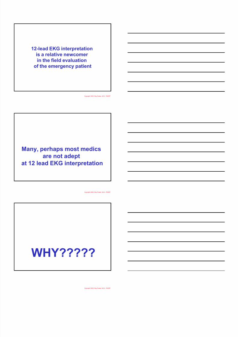
12-lead EKG interpretation
is a relative newcomer
in the field evaluationof the emergency patient
Copyright 2002, Ray Fowler, M.D., FACEP
Many, perhaps most medics
are not adept
at 12 lead EKG interpretation
Copyright 2002, Ray Fowler, M.D., FACEP
WHY?????
Copyright 2002, Ray Fowler, M.D., FACEP

8/3/2019 Dr. Fowler's 12 Lead EKG Interpretation Part 1
http://slidepdf.com/reader/full/dr-fowlers-12-lead-ekg-interpretation-part-1 3/26
Because many EKG courses
are too long,
too boring,
and teach absolutely unnecessaryand unrememberable stuff
to medics
who will never use
that information
Copyright 2002, Ray Fowler, M.D., FACEP
Fowler’s handy
and dandy program
will give you ALL
you need to know
about 12 lead EKG’s
in an absolutely rememberable format
(GOOD LUCK, right?!?!?)
Copyright 2002, Ray Fowler, M.D., FACEP
What am I NOT talking about?
Advanced rhythm assessment
Ventricular tachycardia assessment
Vtach vs. SVT assessment
Block
Copyright 2002, Ray Fowler, M.D., FACEP

8/3/2019 Dr. Fowler's 12 Lead EKG Interpretation Part 1
http://slidepdf.com/reader/full/dr-fowlers-12-lead-ekg-interpretation-part-1 4/26
Copyright 2002, Ray Fowler, M.D., FACEP
This EKG is the
REASON that
12 EKG Machines
are in the field
Copyright 2002, Ray Fowler, M.D., FACEP
Acute Anterior
Myocardial Infarction
Copyright 2002, Ray Fowler, M.D., FACEP

8/3/2019 Dr. Fowler's 12 Lead EKG Interpretation Part 1
http://slidepdf.com/reader/full/dr-fowlers-12-lead-ekg-interpretation-part-1 5/26
Rhythm strip interpretation
has been a standardsince almost the beginning of EMS
Copyright 2002, Ray Fowler, M.D., FACEP
Basic Rhythm
Strip Interpretation
•Rate
•Rhythm
•P Waves
•PR Interval•QRS Complex
•ST Segment
•T Wave
•U Wave
•Summary
Copyright 2002, Ray Fowler, M.D., FACEP
Rate
Rhythm
Axis
Hypertrophy
Infarction
Axis
Hypertrophy
Infarction
P
PR
QRS
ST
T
U
Assessment
P
PR
QRS
ST
T
U
Assessment
Copyright 2002, Ray Fowler, M.D., FACEP

8/3/2019 Dr. Fowler's 12 Lead EKG Interpretation Part 1
http://slidepdf.com/reader/full/dr-fowlers-12-lead-ekg-interpretation-part-1 6/26
Since serious rhythm
disturbances are
the most important issue
(like VF, VT, asystole),
then if you see a serious
rhythm disturbance
proceed with
rhythm strip interpretation
FIRST
Copyright 2002, Ray Fowler, M.D., FACEP
Fowler’s Prime Directive
of Cardiac Emergencies:
Some systole is better
than no systole at all!
Copyright 2002, Ray Fowler, M.D., FACEP
Pulseless Rhythms
Shock x 3, Intubate withCPR, Epi q 3, Shock,
Lidocaine then Who Knows
Copyright 2002, Ray Fowler, M.D., FACEP

8/3/2019 Dr. Fowler's 12 Lead EKG Interpretation Part 1
http://slidepdf.com/reader/full/dr-fowlers-12-lead-ekg-interpretation-part-1 7/26
Pulseless Rhythms
Shock x 3, Intubate with
CPR, Epi q 3, Shock,
Lidocaine then Who Knows
Shock x 3, Intubate withCPR, Epi q 3, Shock,
Lidocaine then Who Knows
Copyright 2002, Ray Fowler, M.D., FACEP
Pulseless Rhythms
Shock x 3, Intubate with
CPR, Epi q 3, Shock,
Lidocaine then Who Knows
Shock x 3, Intubate with
CPR, Epi q 3, Shock,
Lidocaine then Who Knows
Intubate, IV, Epi q 3,
Consider Atropine,Look for cause
Copyright 2002, Ray Fowler, M.D., FACEP
Second point:
Much of what we call
“12 lead interpretation”
is in fact actually
rhythm strip interpretation.
…such as, for example,
the evaluation of AV block,
which can usually be done
in one, or at most, two leads
Copyright 2002, Ray Fowler, M.D., FACEP

8/3/2019 Dr. Fowler's 12 Lead EKG Interpretation Part 1
http://slidepdf.com/reader/full/dr-fowlers-12-lead-ekg-interpretation-part-1 8/26
Third point:AXIS INTERPRETATION
IS BORING!!H e n c e , I w i l l m a k e i t V E R Y s h o r t !
Copyright 2002, Ray Fowler, M.D., FACEP
Copyright 2002, Ray Fowler, M.D., FACEP
Copyright 2002, Ray Fowler, M.D., FACEP

8/3/2019 Dr. Fowler's 12 Lead EKG Interpretation Part 1
http://slidepdf.com/reader/full/dr-fowlers-12-lead-ekg-interpretation-part-1 9/26
Copyright 2002, Ray Fowler, M.D., FACEP
Copyright 2002, Ray Fowler, M.D., FACEP
Copyright 2002, Ray Fowler, M.D., FACEP

8/3/2019 Dr. Fowler's 12 Lead EKG Interpretation Part 1
http://slidepdf.com/reader/full/dr-fowlers-12-lead-ekg-interpretation-part-1 10/26
Copyright 2002, Ray Fowler, M.D., FACEP
Copyright 2002, Ray Fowler, M.D., FACEP
Copyright 2002, Ray Fowler, M.D., FACEP

8/3/2019 Dr. Fowler's 12 Lead EKG Interpretation Part 1
http://slidepdf.com/reader/full/dr-fowlers-12-lead-ekg-interpretation-part-1 11/26
Copyright 2002, Ray Fowler, M.D., FACEP
Positive
Copyright 2002, Ray Fowler, M.D., FACEP
Positive
As the lead sees the impulse growing
(or “coming toward it”),
the machine records
an upward deflection
Copyright 2002, Ray Fowler, M.D., FACEP

8/3/2019 Dr. Fowler's 12 Lead EKG Interpretation Part 1
http://slidepdf.com/reader/full/dr-fowlers-12-lead-ekg-interpretation-part-1 12/26
Positive Positive
As the lead
sees the impulse growing
(or “coming toward it”),the machine records
an upward deflection
Copyright 2002, Ray Fowler, M.D., FACEP
Positive Positive
As the lead
sees the impulse growing
(or “coming toward it”),
the machine records
an upward deflection
Copyright 2002, Ray Fowler, M.D., FACEP
Positive
Copyright 2002, Ray Fowler, M.D., FACEP

8/3/2019 Dr. Fowler's 12 Lead EKG Interpretation Part 1
http://slidepdf.com/reader/full/dr-fowlers-12-lead-ekg-interpretation-part-1 13/26
Positive Positive
As the lead sees the
impulse coming then going
(or “going by the lead”),the machine records
an isoelectric deflection
Copyright 2002, Ray Fowler, M.D., FACEP
Positive Positive
As the lead sees the
impulse coming then going
(or “going by the lead”),
the machine records
an isoelectric deflection
Copyright 2002, Ray Fowler, M.D., FACEP
Copyright 2002, Ray Fowler, M.D., FACEP

8/3/2019 Dr. Fowler's 12 Lead EKG Interpretation Part 1
http://slidepdf.com/reader/full/dr-fowlers-12-lead-ekg-interpretation-part-1 14/26
Copyright 2002, Ray Fowler, M.D., FACEP
Copyright 2002, Ray Fowler, M.D., FACEP
Copyright 2002, Ray Fowler, M.D., FACEP

8/3/2019 Dr. Fowler's 12 Lead EKG Interpretation Part 1
http://slidepdf.com/reader/full/dr-fowlers-12-lead-ekg-interpretation-part-1 15/26
Copyright 2002, Ray Fowler, M.D., FACEP
Copyright 2002, Ray Fowler, M.D., FACEP
II
IIII IIIIII
++
++ ++
The EKG Basic Limb Leads
Copyright 2002, Ray Fowler, M.D., FACEP

8/3/2019 Dr. Fowler's 12 Lead EKG Interpretation Part 1
http://slidepdf.com/reader/full/dr-fowlers-12-lead-ekg-interpretation-part-1 16/26
Lead I is “horizontal”, and is arbitrarily
established at “0 Degrees”
Lead II is 60 degrees
down from Lead 1
and is arbitrarily
established at
“Positive 60 Degrees”
Lead III is
120 degrees
from Lead I, and
is arbitrarily
established at
“Positive
120 Degrees”
++
++++
Copyright 2002, Ray Fowler, M.D., FACEP
The Leads may be moved
to the center of the chest
II
IIII IIIIII
II
IIIIIIIIII
++
++++
++
++ ++
Copyright 2002, Ray Fowler, M.D., FACEP
II
IIIIIIIIII
++
++ ++
Axis is based on the
direction of the heart’s
depolarization
Copyright 2002, Ray Fowler, M.D., FACEP

8/3/2019 Dr. Fowler's 12 Lead EKG Interpretation Part 1
http://slidepdf.com/reader/full/dr-fowlers-12-lead-ekg-interpretation-part-1 17/26
II
IIIIIIIIII
++
++ ++
II
IIII
IIIIII
Copyright 2002, Ray Fowler, M.D., FACEP
II
IIIIIIIIII
++
++ ++
II
IIII
IIIIII
Copyright 2002, Ray Fowler, M.D., FACEP
II
IIII
IIIIII
++
++ ++
II
IIII
IIIIII
Copyright 2002, Ray Fowler, M.D., FACEP

8/3/2019 Dr. Fowler's 12 Lead EKG Interpretation Part 1
http://slidepdf.com/reader/full/dr-fowlers-12-lead-ekg-interpretation-part-1 18/26
Is this a Normal EKG???Is this a Normal EKG???
Copyright 2002, Ray Fowler, M.D., FACEP
Left Axis DeviationLeft Axis Deviation
Copyright 2002, Ray Fowler, M.D., FACEP
Ventricular Hypertrophy
Enlargement of
ventricles
Copyright 2002, Ray Fowler, M.D., FACEP

8/3/2019 Dr. Fowler's 12 Lead EKG Interpretation Part 1
http://slidepdf.com/reader/full/dr-fowlers-12-lead-ekg-interpretation-part-1 19/26
Left Ventricular
Hypertrophy
Copyright 2002, Ray Fowler, M.D., FACEP
Left Ventricular
Hypertrophy
Left Axis Deviation
Deep S wave in V1
Large R wave in V5
Copyright 2002, Ray Fowler, M.D., FACEP
Left Ventricular
Hypertrophy
Left Axis Deviation
Deep S wave in V1
Large R wave in V5
V1 plus V5 adds up
to more than 35 millimeters
Copyright 2002, Ray Fowler, M.D., FACEP

8/3/2019 Dr. Fowler's 12 Lead EKG Interpretation Part 1
http://slidepdf.com/reader/full/dr-fowlers-12-lead-ekg-interpretation-part-1 20/26
Left Ventricular
Hypertrophy
Left Axis DeviationDeep S wave in V1
Large R wave in V5
Left Axis DeviationDeep S wave in V1
Large R wave in V5
V1 plus V5 adds up
to more than 35 millimeters
V1 plus V5 adds up
to more than 35 millimeters
Copyright 2002, Ray Fowler, M.D., FACEP
Right Ventricular
Hypertrophy
Copyright 2002, Ray Fowler, M.D., FACEP
Right Ventricular
Hypertrophy
Look to the RIGHT side
of the heart to find it,
namely V1
Copyright 2002, Ray Fowler, M.D., FACEP

8/3/2019 Dr. Fowler's 12 Lead EKG Interpretation Part 1
http://slidepdf.com/reader/full/dr-fowlers-12-lead-ekg-interpretation-part-1 21/26
Right Ventricular
Hypertrophy
Look to the RIGHT side
of the heart to find it,
namely V1
Look to the RIGHT side
of the heart to find it,
namely V1
Copyright 2002, Ray Fowler, M.D., FACEP
Finding Ventricular
Hypertrophy
Copyright 2002, Ray Fowler, M.D., FACEP
Finding Ventricular
Hypertrophy
Always look at
Lead V1
Copyright 2002, Ray Fowler, M.D., FACEP

8/3/2019 Dr. Fowler's 12 Lead EKG Interpretation Part 1
http://slidepdf.com/reader/full/dr-fowlers-12-lead-ekg-interpretation-part-1 22/26
Finding Ventricular
Hypertrophy
Large R wave in V1 = RVH
Deep S wave in V1 = LVH
Copyright 2002, Ray Fowler, M.D., FACEP
Finding Ventricular
Hypertrophy
Large R wave in V1 = RVH
Deep S wave in V1 = LVH
Corollary: If the complex is
wider than 0.12 seconds,
this is probably a bundle branch blockand not ventricular hypertrophy
Copyright 2002, Ray Fowler, M.D., FACEP
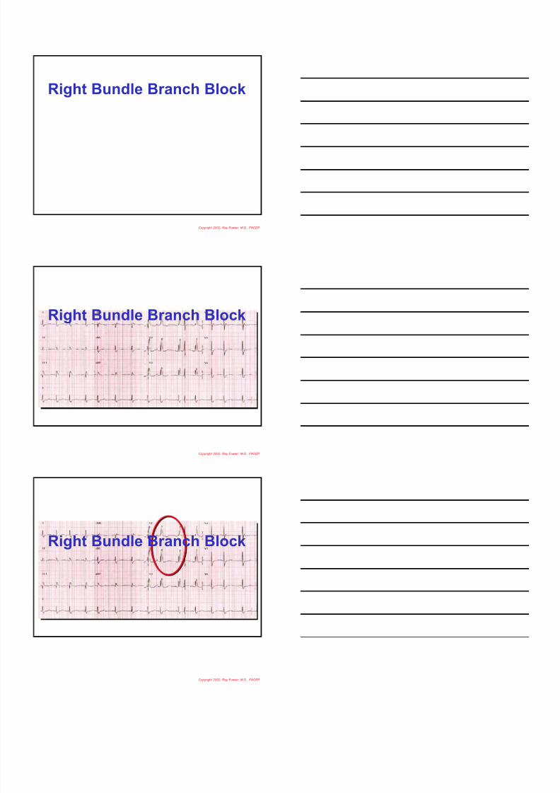
Bundle Branch Block
Positive Deflection
Rabbit Ears in V1
with wide complex
Right
Bundle
Branch
Block
Positive Deflection
in V6
with wide complex
Left
Bundle
Branch
Block
Copyright 2002, Ray Fowler, M.D., FACEP
8/3/2019 Dr. Fowler's 12 Lead EKG Interpretation Part 1
http://slidepdf.com/reader/full/dr-fowlers-12-lead-ekg-interpretation-part-1 23/26
Right Bundle Branch Block
Copyright 2002, Ray Fowler, M.D., FACEP
Right Bundle Branch Block
Copyright 2002, Ray Fowler, M.D., FACEP
Right Bundle Branch Block
Copyright 2002, Ray Fowler, M.D., FACEP

8/3/2019 Dr. Fowler's 12 Lead EKG Interpretation Part 1
http://slidepdf.com/reader/full/dr-fowlers-12-lead-ekg-interpretation-part-1 24/26
Left Bundle Branch Block
Copyright 2002, Ray Fowler, M.D., FACEP
Left Bundle Branch Block
Copyright 2002, Ray Fowler, M.D., FACEP
Left Bundle Branch Block
Copyright 2002, Ray Fowler, M.D., FACEP

8/3/2019 Dr. Fowler's 12 Lead EKG Interpretation Part 1
http://slidepdf.com/reader/full/dr-fowlers-12-lead-ekg-interpretation-part-1 25/26
Left Bundle Branch Block
Copyright 2002, Ray Fowler, M.D., FACEP
Left Bundle Branch Block
Copyright 2002, Ray Fowler, M.D., FACEP
Left Anterior Hemiblock
Copyright 2002, Ray Fowler, M.D., FACEP

8/3/2019 Dr. Fowler's 12 Lead EKG Interpretation Part 1
http://slidepdf.com/reader/full/dr-fowlers-12-lead-ekg-interpretation-part-1 26/26
This is the END of
Part 1
of 12 Lead EKG
Interpretation!
Please join us for part 2!
Copyright 2002, Ray Fowler, M.D., FACEP