Dr. Alex Ng
99
Dr. Alex Ng Consultant, PWH Clinical Associate Professor (Honorary), CUHK The Chinese University of Hong Kong Department of Imaging and Interventional Radiology Prince of Wales Hospital
Transcript of Dr. Alex Ng

Dr. Alex NgConsultant, PWH
Clinical Associate Professor (Honorary), CUHK
The Chinese University of Hong Kong
Department of Imaging and Interventional Radiology
Prince of Wales Hospital

Date: 7 April 2016, (Thursday)
Time: 1.00pm -2.00pm
Venue: Conference Room, 2/F, DIIR, Main Clinical Building, New Block, Prince of Wales Hospital, Shatin
Topic: Dual Energy CT - MSK Applications
Speaker: Professor Hugue Ouellette, University of British Columbia, Canada
,
Ms. Mandy Cheng: 26321189For Professor James GriffithDepartment of Imaging and Interventional RadiologyThe Chinese University of Hong Kong

Wrist coil

Where is pain located
For how long
Trauma– if so, what and when

Don’t mention any feature without grading it
Qualitative measure:
Minimal, mild, moderate, severe
Quantitative measure:
Small, medium, large (mm wide x mm deep x mm long)

TFCC/Ligaments:
TFCC injury
Intrinsic ligament injury (DISI VISI)
Other carpal ligament (ganglion cyst)
Muscle and Tendon:
Tendinosis (Dequervain’s disease)
Tenosynovitis
Tear
Bone: Scaphoid fracture
(SNAC),
Lunate (Kienbock)
Ulnocarpal impaction
Joint and cartilage RA/arthritis
Degeneration/Cartilage
Nerve and vessels: Carpal tunnel syndrome
Guyon’s tunnel syndrome


vRUL/dRUL attachment
Anatomy
radial attachment
Peripheral /proximal attachment:Foveal attachment (proximal lamina)Ulnar attachment (distal lamina)
Distal attachment: ulno-triquetral/lunate ligament
Meniscal homologue

meniscal homologue
Ulno-lunateulno-triquetralligaments
Articulardisc
Foveal attachmentUlnar attachment
Radial attachment
MRI Anatomy

TFCC Maintain the DRUJ
stability Prevent sublux when
wrist pronate and supinate
Torn DRUJ arthritis, UCJ arthritis
Symptoms: pain, click and Limited range of movement
Asymptomatic :degenerative tear
Schematic diagram: Diagnostic imaging
RC joint
Mid carpal joint
UC joint
DRUJ

TFCC tear
location
1A: Central/pararadial
1B: Peripheral: Ulnar/foveal
1C: Ulno-triquetral/UL tear
Volar or dorsal sides
1D: radial
Type 1: Traumatic

Pararadial tear: type 1A (partial thickness, undersurface)
Type 1 TFC tear
Pararadial tear: type 1AFull thickness tear, measuring xxmmxmmin size
Pararadial tear: type 1AContour irregularity suggestive of partial tear, no gap seen

Foveal and ulnar tear (type 1B), partial tear
Normal for comparison

Foveal and ulnar tear (type 1B)
Proximal lamina full thickness tearDistal lamina partial tear (type1B)

Non-union of ulnar styloid processThe ulnar attachment of TFCC shows partial tear (type 1B)
Type 1=Tips: TFCC intact, DRUJ stableType 2=Base of styloid: TFCC disrupted,
DRUJ unstable
Full thickness tear pararadial region (type 1a)

Distal attachment tear (type 1C)

Large radial avulsion (type 1D), retracted peripherally and the gap measures mmxmmxmmThe TFC is lax (loss of trampoline effect)

TFCC tear at the dorsal attachmentof the dRUL
TFCC partial tear at vRUL>dRUL attachment
Mild dorsal DRUJ subluxation

TFCC tear
location
Type 2: Degenerative
Central/pararadial
• More irregular versus sharp edge• TFCC thinning/degeneration• Ulnar positive


Type 2 TFC tear
Type 2C/D Type 2E Type 2C
40% perforation in 50+ with NO symptoms

Foveal and ulnar tear (type 1b)?fibrous tissue or partial tear
MR ARTHROGRAM Full thickness tear

• Injection at RCJ and DRUJ• Gold standard for communicating (full thickness) tear

Suspicious tear (Type 2C) Intact thin disc (Type 2A)
Distend the joint or Traction

Proximal /membranous
Volar dorsal
Copied from Diagnostic imaging
Anatomy

INTRINSIC LIGAMENT Maintain the stability of the proximal carpal row (SL
instability and LT instability) Torn Arthritis, pain, click and degeneration
Features of torn ligament Morphology distortion (thinning, irregularities)
Abnormal signal (difficult to detect)
Discontinuity of fibres
Complete absence of ligament
Secondary dissociation of SL interval (>3mm)
Ganglion cyst formation

MRI anatomy
Schematic diagram: Diagnostic imaging
membranousVolar Dorsal

sL T
MRI anatomy
L S
Schematic diagram: Diagnostic imaging
SL: dorsal side strongerLT: volar side stronger

Membranous portion: Central perforation (full thickness) of the SL ligament
Membranous portion: Flap tear from the scaphoid attachment (full thickness) of the SL ligament
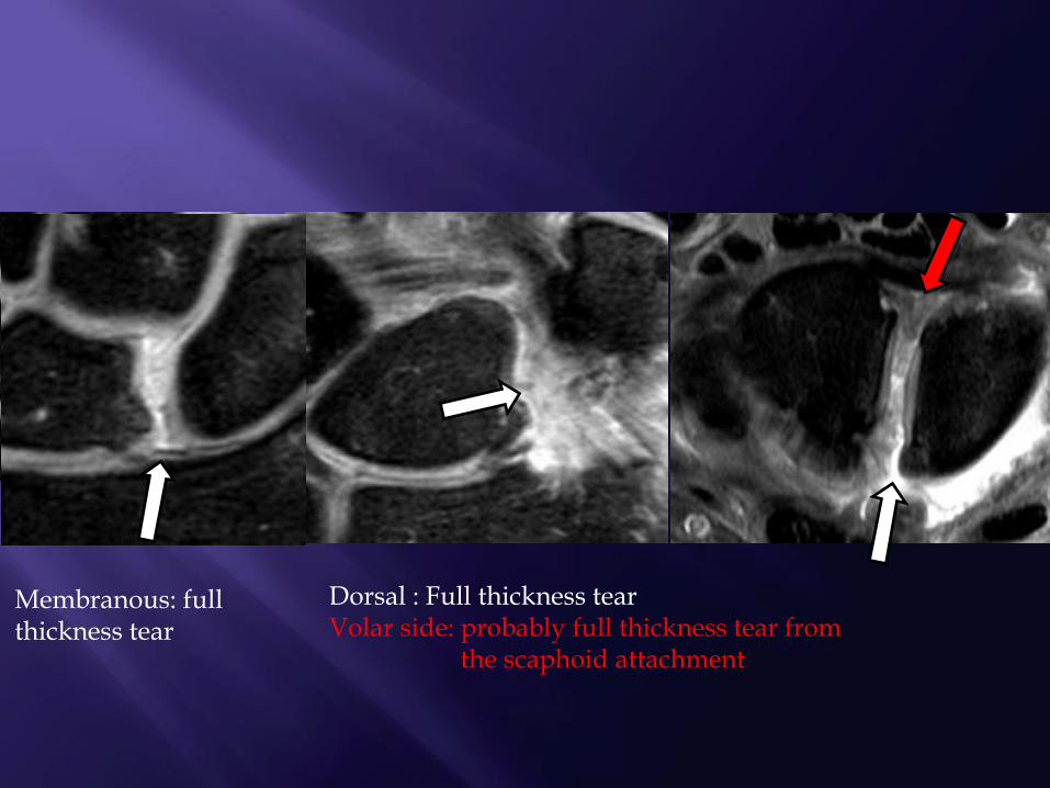
Dorsal : Full thickness tearVolar side: probably full thickness tear from
the scaphoid attachment
Membranous: full thickness tear

Dorsal component: partial thickness tearVolar component: Swollen and distorted
configuration: partial tear
Membranous : full thickness tear

Full thickness tear of the SL ligament in the dorsal and membranous portionsSecondary dissociation of the SL interval >3mm
DISI deformity ?

DISI= dorsal intercalated segmental instability
VISI= Volar
SL tear DISI
Scaphoid palmar flex and L-T dorsiflex
LT tear VISI
Scaphoid-lunate palmar flex and T dorsiflex

Scapholunate angle Capitolunate angle
Normal 30-60 0-30 (20)
DISI >60 (60-80 borderline) >30
VISI <30 >30
DISI = Dorsal intercalated segment instabilityVISI = Volar intercalated segment instability
**

Capitolunate angle
0-30 (20)
>30
DISI deformity
*

SL ligament tear
SLAC
SLAC1: radial styloid and distal scaphoid2: whole RSJ3: Proximal migration of scaphoid and scaphoid-capitate articulation4: radiolunate articulation
12
3
4
SLAC 4


LT tear
VISI

MR arthrogram: Most accurate test
Communicating tear and non-communicating tear
MRI Pitfall in SL/LT tear

Partial tear Full thickness tear/communicating tear
Post-Traction
MRI Pitfall in SL/LT tear

Intact Tear
Post-Traction

Anatomy RSC, RLT RSL (not a true ligament;
Ligament of Testut) Short radiolunate (not much
concern) UT, UL Arcuate/deltoid/V
ligament: Ulnar arm: THC (triquetral-
hamate-capitate) Radial arm: SC (scaphoid-
capitate) joining the proximal and
distal carpal rows
dRCL, DIC, dUTL
Theumann N et al, Radiology 2003

volar dorsalvolar
MRI Anatomy


Maintain the carpal stability (simplified version)
Radiocarpal: mainly RSC, RLT
Midcarpal : Tear/insufficiency of Arcuate ligament
SC ligament and THC ligament (radial arm and ulnar arm)
SL, LT instability: SL/LT ligaments
(DRUJ: TFCC)

Abnormalities GRADE BEST TO DESCRIBE
Acute sprain Grade 1 Periligamentous edema
Partial tear Grade 2 • Partial tears• Weakening with
thickening due to periligamentous and intraligamentousedema (sprain??)
Complete tear Grade 3 Complete disruptions
Traction related avulsivecystic change at site of osseous attachment
soft tissue ganglia from capsular injuries or ligament degeneration
Abnormalities related to carpal ligament injuries
Chhabra et al. 2012 Radiographics

RSC/RLT
Carpal ligament partial tear with ganglion cyst
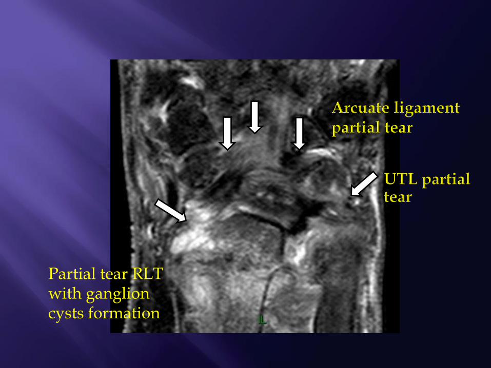
Partial tear RLT with ganglion cysts formation

dRCL and dICL RLT RLT dRCL and dICL

• Unilocular or multilocular• Inhomogeneity –mucin• Narrow stalk/pedicle– joint e.g. PTJ, STT, SL interval- prevent
recurrent cyst• Tendon sheath communication• Compression on neurovascular bundle
Ganglion cyst


Carpal ligament tear with ganglion cyst
DIC ligament RLT ligament

EPBAPL
ED/EI EPLEDM
ECU ECRL
ECRB
Schematic diagram: Diagnostic imaging
Anatomy
FCU FCR
FDS and FDP
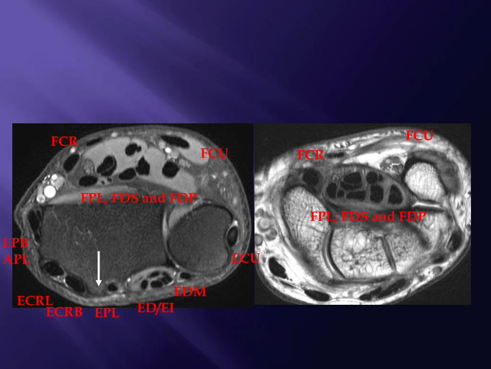
EPBAPL
ED/EIEPL
EDM
ECU
ECRLECRB
FCU FCR
FPL, FDS and FDP
FPL, FDS and FDP
FCUFCR

ECU moderate tendinosis with adjacent soft tissue inflammation
ECU mild tenosynovitisTendon signal within normal limit
Severe inflammation with enhancement
ECU Tendinosis/Tenosynovitis
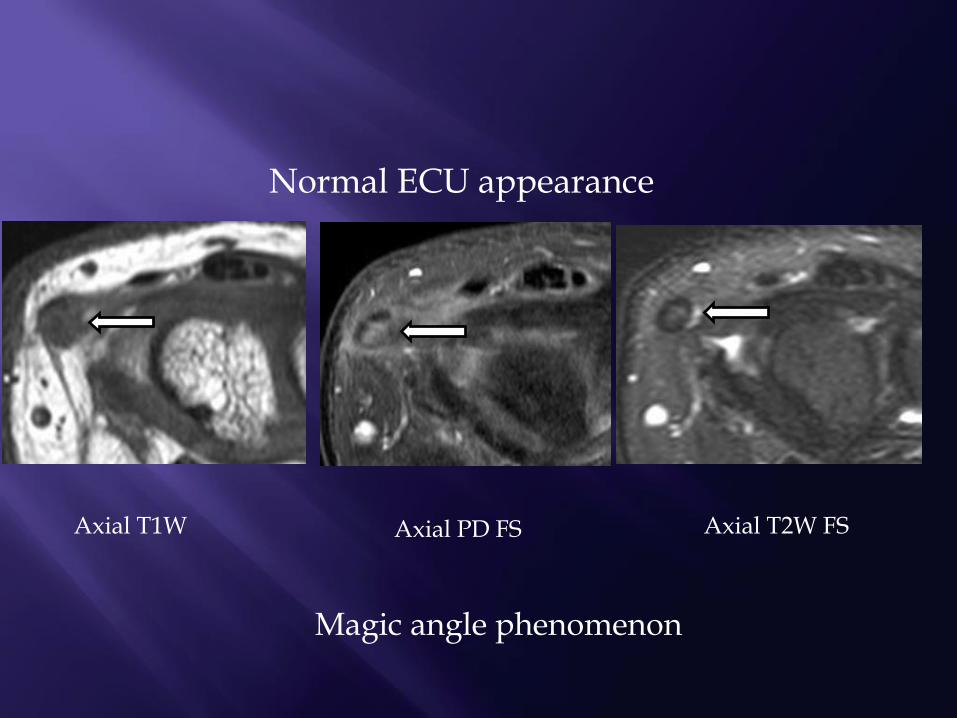
Normal ECU appearance
Axial PD FS
Magic angle phenomenon
Axial T2W FSAxial T1W

ECU severe tendinosis paratendinous soft tissue inflammation
TIP

Moderate tendinosis with longitudinal split tearDorsal subluxation of the DRUJ Subluxation of the ECU
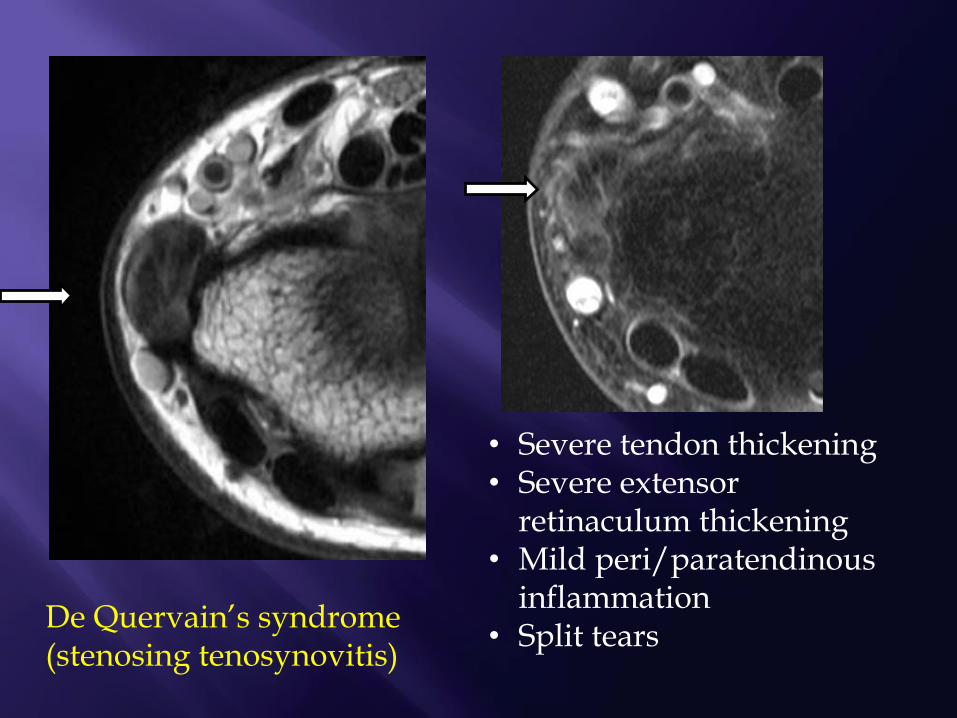
• Severe tendon thickening• Severe extensor
retinaculum thickening• Mild peri/paratendinous
inflammation• Split tears
De Quervain’s syndrome (stenosing tenosynovitis)
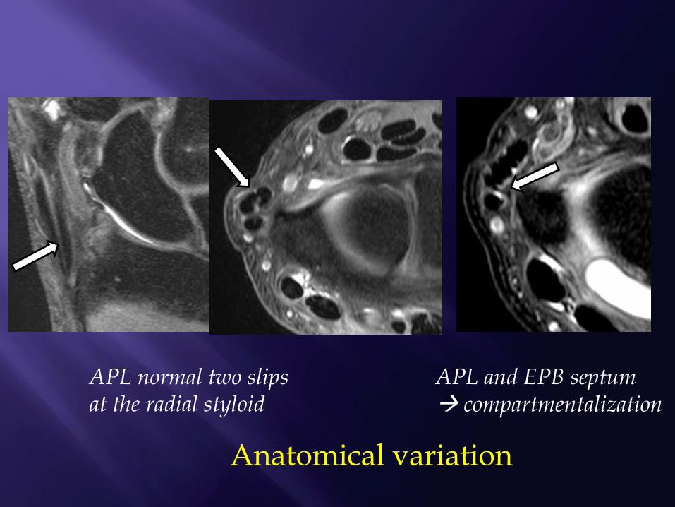
APL normal two slips at the radial styloid
APL and EPB septum compartmentalization
Anatomical variation

EPB partial tear

• Compartment 3 crossing the compartment 2 at the wrist level• Tendinosis- swelling and hyperintense T2W signal• Paratendinous soft tissue oedema• Tendon sheath effusion

Normal EPL appearance: magic angle phenomenon

Flexor compartment tendons
Schematic diagram: Diagnostic imaging
Radial and ulnar bursa anatomy
Radial bursaUlnar bursa
Communication of flexor tendon sheaths

Tenosynovitis- TB
Radial bursa Ulnar bursa

FCR: stenosing tenosynovitis Focal loculated fluid collection in the
tendon sheath
Due to chronic friction with the carpal bone
Other causes of tenosynovitis: Inflammatory joint disease
Metabolic deposits: gout, pseudogout

Anatomy and fracture classification
Schematic diagram: Diagnostic imaging
Scaphoid
Distal pole or 1/3=10%
Tuberosity: volar prominence
Proximal pole or 1/3=10%
Middle 1/3 (waist)=80%

Same day
1 month
6monthColleague suspicious of scaphoid bone pain after injury
Occult fracture (10-20%)

Associated injury

SNAC III
Scaphoid nonunion and SNAC
Scaphoid non-union (10%)
SNAC stages:1: OA between distal pole of scaphoid and radial styloid process2: scaphoid and capitate3: Lunate and capitate
SNAC

SNAC II/III
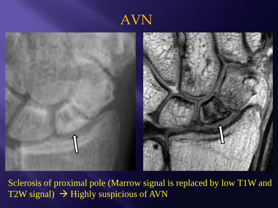
Sclerosis of proximal pole (Marrow signal is replaced by low T1W and
T2W signal) Highly suspicious of AVN
AVN

Proximal pole collapse, fragmentation and resorption
compatible with AVN

T1W:homogeneous low signal in the proximal and distal fragments without collapse.
T2W FS: homogeneous low signal in the proximal fragmentPoor vascularity
T1W FS+Gd: No contrast enhancement poor vascularity.
DCE :flat perfusion curve poor vascularity
Surgery confirmed

DCE showed poor vascularity
T1W: Preserved normal bone marrow fat signal good vascularity
T2W FS:Mild increased signal intensity adjacent to the fracture site
Post-Gd:No significant contrast enhancement near the fracture marginpoor vascularity
Surgery confirmed

T1W: homogeneous diffuse low signal
T2W FS: homogeneous isointense signal intensity Poorvascularity.
Post-Gd: Good enhancement good vascularity
DCE: steep enhancement slope with enhancement similar to the distal pole good vascularity
Surgery confirmed

T1W: homogeneous low signal in the proximal scaphoid fragment
T2W FS: homogeneous increased signal intensity at the with peri-fracture cyst formation Poor vascularity
Post-Gd: homogeneous enhancement good vascularity.
DCE : Flat slope fair vascularity
Surgery confirmed

Kienbock disease
• Ulna negative (78%)• Uniform hypointense T1W
signal• Oedema or sclerosis
• Cystic changes on T2W• Fracture line
• Fragmentation/Collapse of lunate
• Proximal migration of capitate + scaphoid hyperflexion (degeneration of mid carpal and RCJ)

Impingement of lateral ulna against TFCC and proximal carpal row
Features: Ulna positive variance
(rarely can be neutral or negative)
TFCC tear/degeneration
Eccentric bone oedema, subchondral cyst, sclerosis
Cartilage loss
LT tear
Ulnocarpal impaction

Hamatolunate impaction
Type 2 lunate: additional facet with hamate (55%)

• Chondral injury
• Oedema tip of hamate
(subchondral oedema)
• Sclerosis

Schematic diagram: Diagnostic imaging
Anatomy
RC joint• Scaphoid fossa• Lunate fossa
Mid carpal joint
UC joint
STT/triscaphe joint
CMC joint
DRUJ
PT joint

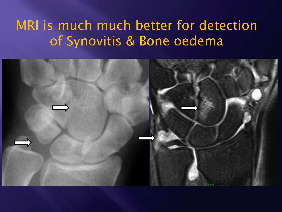
• Bone erosions• Bone oedema• Synovitis• Pannus: synovial
mass causing erosions
• Tenosynovitis• TFCC/ligament/te
ndon tear

Before Treatment After Treatment
• Pain decreased after medications• less oedema• less synovitis/joint effusion


Better depiction
Arthrogram or
traction


Triscaphe arthritis
• Scaphoid-trapezoid (most common)• Scaphoid-trapezium• Trapezium-Trapezoid

Schematic diagram: Diagnostic imaging
Carpal tunnel syndrome

MRI Anatomy
At tunnel inletProximal to tunnel
At tunnel outlet Distal to tunnel
HTRAP
SCAP
PISI

CTS: MRI No established role??CTS (controversial)
CSAp >15mm2
CSAnormal <12mm2
At tunnel inletProximal to tunnel
At tunnel outlet Distal to tunnel
BRi >4mm
FR >3??
BRo >3mm
CSAd >15mm2
CSAdnormal
<13mm2
Hyperintense
yes
Thickened tenosynovium
yes

Gouty tophus Neurogenic tumour
Ganglion cyst Lipoma
Persistent median artery

One year Three monthsPreop
More bowingLess oedema
Less bowingReformed TCL

CTS recurrence
Incomplete release
Perineural scarrings with tethering

Guyon’s tunnel syndrome
Guyon’s tunnel lipoma
Causes:• Carpal tunnel fracture• OA• Ganglion• Anomalous muscle• Ulnar artery aneurysm
Pisi & H
Palmar carpal ligament
Transverse carpal ligament

Guyon’s tunnel fibrosis
Answer the clinical questions
Good communication with clinicians
Familiar with the anatomy
Describe the abnormalities
Using a checklist
Correlate all the planes