DpaceMICCAIAbdominalWorkshop Final
8
Sliding Geometries in Deformable Image Registration Danielle F. Pace 1 , Marc Niethammer 2 , and Stephen R. Aylward 1 1 Kitware Inc., Clifton Park NY and Carrboro NC, USA danielle.pace@kitwa re.com, stephen.aylward@ki tware.com 2 The University of North Carolina at Chapel Hill, Chapel Hill NC, USA [email protected] Abstract. Regularization is used in deformable image registration to enco urage plausible displ acement field s, and signi fican tly impac ts the derived correspondences. Sliding motion, such as that between the lungs and chest wall and between the abdominal organs, complicates regis- tration because many regularizations are global smoothness constraints that produce errors at object boundaries. We present locally adaptive regularizations that handle sliding objects with locally planar and tubu- lar geometries. These regularizations allow discontinuities to develop in the displacement field at sliding interfaces and increase the independence with which regions surrounding distinct geometric structures can behave. Validation is performed by registering inhale and exhale abdominal com- puted tomography (CT) images and artificial images of a sliding tube. The sliding registration methods produce more realistic correspondences that may better reflect the underlying physical motion, while performing as well as the diffusive regularization with respect to image match. 1 In troduction Within many clinical workflows, the goal of deformable image registration is to (1) quantify treatment effectiveness by measuring change within longitudinal datasets or across subjects, (2) map surgical plans from preoperative images onto intraoperative images for guidance, or (3) align atlases with patient images to map auxilliary information from the atlas, such as expected functional site localizations, into the patient. To successfully accomplish thse tasks, it is critical to establish accurate correspondence between the images. However, establishing correspondence using a deformable image registration method that optimizes image match alone is ill-posed, i.e. there exist many displacement fields that produce the same tran sformed mov ing image [5]. To obtain sensible correspondences using deformable image registration, a regularization term is typically introduced to encourage a smooth displacement field. The registration result is therefore a compromise between image similarity and spatial regularity, and the regularization forms a strong prior on the final transformation. This is true for any non-corner or point structure due to spatial ambiguity (the so-called aperture effect). Registration results are dependent on
-
Upload
marcniethammer -
Category
Documents
-
view
218 -
download
0
Transcript of DpaceMICCAIAbdominalWorkshop Final

8/3/2019 DpaceMICCAIAbdominalWorkshop Final
http://slidepdf.com/reader/full/dpacemiccaiabdominalworkshop-final 1/8
Sliding Geometries in Deformable Image
Registration
Danielle F. Pace1, Marc Niethammer2, and Stephen R. Aylward1
1 Kitware Inc., Clifton Park NY and Carrboro NC, [email protected], [email protected]
2 The University of North Carolina at Chapel Hill, Chapel Hill NC, USA [email protected]
Abstract. Regularization is used in deformable image registration toencourage plausible displacement fields, and significantly impacts thederived correspondences. Sliding motion, such as that between the lungs
and chest wall and between the abdominal organs, complicates regis-tration because many regularizations are global smoothness constraintsthat produce errors at object boundaries. We present locally adaptiveregularizations that handle sliding objects with locally planar and tubu-lar geometries. These regularizations allow discontinuities to develop inthe displacement field at sliding interfaces and increase the independencewith which regions surrounding distinct geometric structures can behave.Validation is performed by registering inhale and exhale abdominal com-puted tomography (CT) images and artificial images of a sliding tube.The sliding registration methods produce more realistic correspondencesthat may better reflect the underlying physical motion, while performingas well as the diffusive regularization with respect to image match.
1 Introduction
Within many clinical workflows, the goal of deformable image registration isto (1) quantify treatment effectiveness by measuring change within longitudinaldatasets or across subjects, (2) map surgical plans from preoperative imagesonto intraoperative images for guidance, or (3) align atlases with patient imagesto map auxilliary information from the atlas, such as expected functional sitelocalizations, into the patient. To successfully accomplish thse tasks, it is criticalto establish accurate correspondence between the images. However, establishingcorrespondence using a deformable image registration method that optimizesimage match alone is ill-posed, i.e. there exist many displacement fields thatproduce the same transformed moving image [5].
To obtain sensible correspondences using deformable image registration, a
regularization term is typically introduced to encourage a smooth displacementfield. The registration result is therefore a compromise between image similarityand spatial regularity, and the regularization forms a strong prior on the finaltransformation. This is true for any non-corner or point structure due to spatialambiguity (the so-called aperture effect). Registration results are dependent on
8/3/2019 DpaceMICCAIAbdominalWorkshop Final
http://slidepdf.com/reader/full/dpacemiccaiabdominalworkshop-final 2/8
relatively sparse features and regularization drives the estimation of otherwiseunobservable deformations within homogeneous regions.
The focus of this paper is on accurately registering images that depict slidingmotion between multiple structures. This includes the sliding of the abdominalorgans and lungs due to respiration. Here, the assumption of a globally smoothdisplacement field is inappropriate, as globally smoothing regularizations, such asthe diffusive regularization, cannot recover the motion discontinuities that ariseat the interfaces between sliding structures. Locally adaptive regularizations [2,9] vary spatially and can more accurately capture complex deformations.
Several regularizations aiming to handle sliding motion have been presented,but have typically been applied to the alignment of lung images. These includeinvestigations of non-quadratic regularizers [4], a volume-preserving regulariza-tion that allows shear discontinuities at sliding interfaces [7], and regularizationsbased on anisotropic diffusion that use supplied organ segmentations [6, 8, 10].We take this last approach here. Yin et al. [10] tailor their regularization towards
the biomechanics of lung lobar fissures using spatially-varying isotropic smooth-ing. Schmidt et al. [8] implement sliding motion by smoothing the displacementcomponents tangential to the sliding interface separately for a sliding objectand within its background, using Neumann boundary conditions. We present aregularization that instead applies separate diffusion tensors to the normal andtangential displacements of the entire displacement field. This regularization na-tively handles multiple sliding organs and has been evaluated preliminarily usingsynthetic and phantom lung images [6].
The sliding regularizations based on anisotropic diffusion described above,including our own work [6], solely handle displacement field discontinuities atroughly planar interfaces between two sliding organs. By also adding notions of tubular and point-like structures, we present a regularization that handles slidingmotion of planes and tubes and that smooths displacement fields according to
local structure classifications. We refer to registration methods including thisregularization as “geometry conditional registration methods”.
We begin by outlining our sliding organ formulation, followed by a descrip-tion of its extension to a geometry conditional deformable registration methodthat also considers tubular and point-like structures. Validation is conductedby performing intra-subject registrations between inhale and exhale computedtomography (CT) images and between artificial images depicting a sliding tube.These assessments demonstrate the increased plausibility of the resulting dis-placement field, which encapsulates the correspondence necessary to use regis-tration in clinical tasks. Additionally, these results demonstrate the advantagesof considering sliding motion throughout the abdomen.
2 Methods2.1 Deformable non-parametric image registration
Given a target image T and a moving image M on the domain Ω, deformablenon-parametric image registration aims to find a displacement field u that maps
8/3/2019 DpaceMICCAIAbdominalWorkshop Final
http://slidepdf.com/reader/full/dpacemiccaiabdominalworkshop-final 3/8
the moving image onto the target such that T (x) ≈M (x−u(x)) [5]. This is oftenperformed by minimizing a cost function of the form C (u) = D[T,M ;u]+αS (u).D[T,M ;u] is an image match distance measure between T and M under thecurrent estimate of the displacement field u, and S (u) is the regularization thatpenalizes unrealistic displacement fields.
2.2 Principles of sliding motion
Writing a regularization for use in registering images depicting sliding organscan be guided by decomposing the displacement field u into normal (u⊥) andtangential (u) displacements with respect to the organ boundary along whichsliding is expected to occur. One can then consider the following principles [6,8] close to sliding organ boundaries:
1. Sliding motion: Sliding causes the tangential displacements to be discon-tinuous along the normal direction(s).
2. Intra-organ smoothness: The displacements must be smooth along thetangential direction(s) to encourage smooth movement within individualstructures.
3. Inter-organ coupling: Discontinuities in the normal displacements in thesurface normal direction(s) are penalized to ensure organs do not pull apart(a valid assumption for many medical images).
2.3 Sliding organ registration
For sliding organ registration, we encapsulate these rules in a regularizer basedon anisotropic diffusion [6]. Here, diffusion tensors specify the direction andstrength of the intra-organ smoothness (IOS ) and inter-organ coupling (IOC )
constraints:
S sliding(u) =1
2
l=x,y,z
x∈Ω
DIOS (x)ul(x) + DIOC (x)u⊥l (x)2 (1)
where is the gradient operator, ul(x) is the lth scalar component of the dis-placement field, and u⊥l (x) is the lth scalar component of the normal componentof the displacement field. If n(x) is the normal to the organ boundary derivedfrom a surface model of the organ and nl(x) is its lth scalar component, thenu⊥l (x) =
u(x)T n(x)
nl(x).
The diffusion tensorP (x) = n(x)n(x)T (2)
smooths in the normal direction alone, while the diffusion tensor I − P (x)
smooths in the tangential plane. We therefore define the intra-organ smooth-ness and inter-organ coupling diffusion tensors as:
DIOS (x) = I −w(x)P (x)
DIOC (x) = w(x)P (x)(3)
8/3/2019 DpaceMICCAIAbdominalWorkshop Final
http://slidepdf.com/reader/full/dpacemiccaiabdominalworkshop-final 4/8
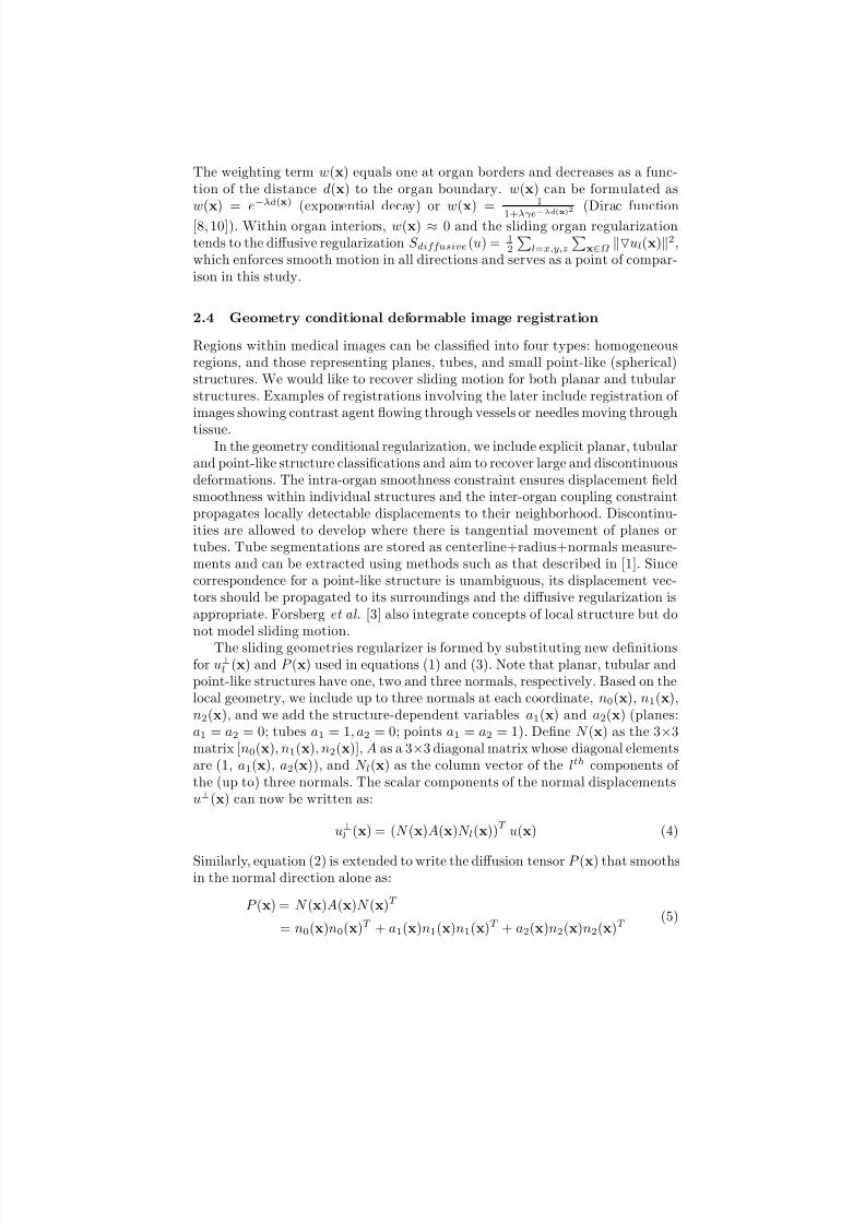
The weighting term w(x) equals one at organ borders and decreases as a func-tion of the distance d(x) to the organ boundary. w(x) can be formulated asw(x) = e−λd(x) (exponential decay) or w(x) = 1
1+λγe−λd
(x
)2 (Dirac function
[8, 10]). Within organ interiors, w(x) ≈ 0 and the sliding organ regularizationtends to the diffusive regularization S diffusive(u) = 1
2
l=x,y,z
x∈Ω ul(x)2,
which enforces smooth motion in all directions and serves as a point of compar-ison in this study.
2.4 Geometry conditional deformable image registration
Regions within medical images can be classified into four types: homogeneousregions, and those representing planes, tubes, and small point-like (spherical)structures. We would like to recover sliding motion for both planar and tubularstructures. Examples of registrations involving the later include registration of images showing contrast agent flowing through vessels or needles moving through
tissue.In the geometry conditional regularization, we include explicit planar, tubular
and point-like structure classifications and aim to recover large and discontinuousdeformations. The intra-organ smoothness constraint ensures displacement fieldsmoothness within individual structures and the inter-organ coupling constraintpropagates locally detectable displacements to their neighborhood. Discontinu-ities are allowed to develop where there is tangential movement of planes ortubes. Tube segmentations are stored as centerline+radius+normals measure-ments and can be extracted using methods such as that described in [1]. Sincecorrespondence for a point-like structure is unambiguous, its displacement vec-tors should be propagated to its surroundings and the diffusive regularization isappropriate. Forsberg et al. [3] also integrate concepts of local structure but donot model sliding motion.
The sliding geometries regularizer is formed by substituting new definitionsfor u⊥l (x) and P (x) used in equations (1) and (3). Note that planar, tubular andpoint-like structures have one, two and three normals, respectively. Based on thelocal geometry, we include up to three normals at each coordinate, n0(x), n1(x),n2(x), and we add the structure-dependent variables a1(x) and a2(x) (planes:a1 = a2 = 0; tubes a1 = 1, a2 = 0; points a1 = a2 = 1). Define N (x) as the 3×3matrix [n0(x), n1(x), n2(x)], A as a 3×3 diagonal matrix whose diagonal elementsare (1, a1(x), a2(x)), and N l(x) as the column vector of the lth components of the (up to) three normals. The scalar components of the normal displacementsu⊥(x) can now be written as:
u⊥l (x) = (N (x)A(x)N l(x))T u(x) (4)
Similarly, equation (2) is extended to write the diffusion tensorP (x) that smoothsin the normal direction alone as:
P (x) = N (x)A(x)N (x)T
= n0(x)n0(x)T + a1(x)n1(x)n1(x)T + a2(x)n2(x)n2(x)T (5)

8/3/2019 DpaceMICCAIAbdominalWorkshop Final
http://slidepdf.com/reader/full/dpacemiccaiabdominalworkshop-final 5/8
This formulation approximates the diffusive regularization at point-like struc-tures, where a1(x) = a2(x) = 1, and within organ interiors, where w(x) ap-proximates zero. Near planar objects, the regularization equals the sliding organregularization presented earlier.
2.5 Optimization
We use a gradient descent scheme to minimize the cost function C (u) using theregularization defined in equation (1). Without loss of generality, and since wefocus on monomodal registration of CT images, we use the sum of squared dif-ferences image similarity metric for D[T,M ;u]. On each registration iteration,the sliding geometries regularization induces an update to the displacement fieldequal to −uS (u)t at each coordinate x, where uS (u) is the gradient of equa-tion (1) with respect to a small perturbation and t is the time step. Droppingthe (x) notation,
uS (u) = −
l=x,y,z
div((I −wP )T ((I − wP )ul + wP u⊥l ))el
+ div(wP T ((I −wP )ul + wP u⊥l ))NAN l
(6)
where el is the lth canonical unit vector (ex. ex = [1, 0, 0]T ). At convergence, thisgradient must be balanced with the gradient from the image similarity measure.
3 Validation
3.1 Inhale/exhale abdominal CT registration
Respiration induces significant and complex deformations to the abdominal or-gans, including sliding motions between several semi-independent organs. Anarterial abdominal CT image acquired at inhale (target) was registered with ahepatic-venous abdominal CT image (moving) from the same patient acquiredat exhale (size 240×170 × 150; spatial resolution 1.5mm3). Segmentations of theliver, left and right kidneys, and bones were created semi-automatically in thetarget image and used to generate surface models defining organ boundaries andnormals. Following initial rigid alignment between the two images, the slidingorgan deformable registration algorithm described in Section 2.3 was applied ina multi-resolution framework using two resolution levels, the first downsamplingby a factor of two and the second operating at native resolution. The regis-trations took 1000 and 3000 iterations, respectively, with time step 0.046875s, empirically-determined parameters λ = 0.2 (for exponential decay) and α =
24/8, and were repeated using the diffusive regularization for comparison.Figure 1 shows the alignment between the target and deformed moving im-
ages following sliding organ registration. The alignment was validated quantita-tively by segmenting the liver, kidneys and bones in the moving image, warpingthe resulting label map by the registration displacement field, and calculating
8/3/2019 DpaceMICCAIAbdominalWorkshop Final
http://slidepdf.com/reader/full/dpacemiccaiabdominalworkshop-final 6/8
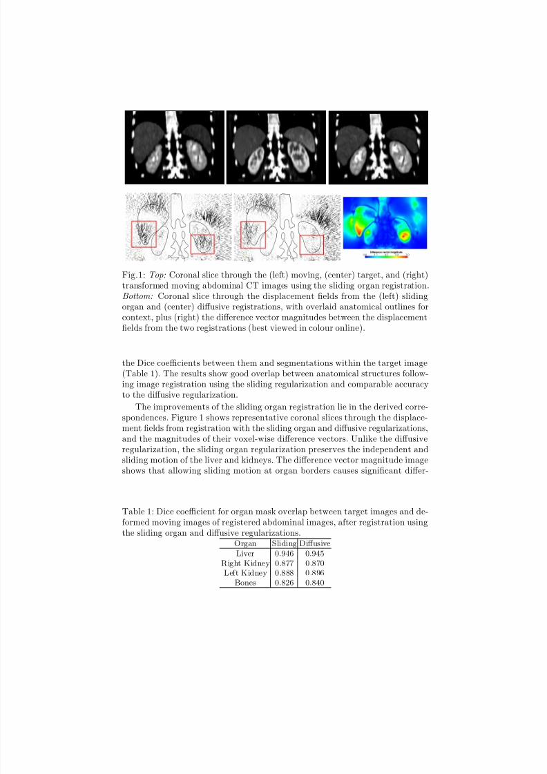
Fig.1: Top: Coronal slice through the (left) moving, (center) target, and (right)transformed moving abdominal CT images using the sliding organ registration.Bottom: Coronal slice through the displacement fields from the (left) slidingorgan and (center) diffusive registrations, with overlaid anatomical outlines forcontext, plus (right) the difference vector magnitudes between the displacementfields from the two registrations (best viewed in colour online).
the Dice coefficients between them and segmentations within the target image(Table 1). The results show good overlap between anatomical structures follow-ing image registration using the sliding regularization and comparable accuracyto the diffusive regularization.
The improvements of the sliding organ registration lie in the derived corre-spondences. Figure 1 shows representative coronal slices through the displace-ment fields from registration with the sliding organ and diffusive regularizations,and the magnitudes of their voxel-wise difference vectors. Unlike the diffusiveregularization, the sliding organ regularization preserves the independent andsliding motion of the liver and kidneys. The difference vector magnitude imageshows that allowing sliding motion at organ borders causes significant differ-
Table 1: Dice coefficient for organ mask overlap between target images and de-formed moving images of registered abdominal images, after registration usingthe sliding organ and diffusive regularizations.
Organ Sliding DiffusiveLiver 0.946 0.945
Right Kidney 0.877 0.870Left Kidney 0.888 0.896
Bones 0.826 0.840
8/3/2019 DpaceMICCAIAbdominalWorkshop Final
http://slidepdf.com/reader/full/dpacemiccaiabdominalworkshop-final 7/8
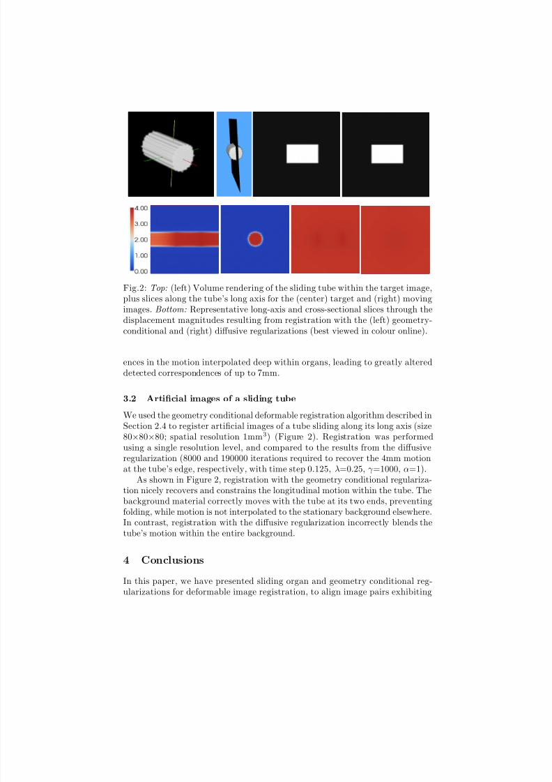
Fig.2: Top: (left) Volume rendering of the sliding tube within the target image,plus slices along the tube’s long axis for the (center) target and (right) movingimages. Bottom: Representative long-axis and cross-sectional slices through thedisplacement magnitudes resulting from registration with the (left) geometry-conditional and (right) diffusive regularizations (best viewed in colour online).
ences in the motion interpolated deep within organs, leading to greatly altereddetected correspondences of up to 7mm.
3.2 Artificial images of a sliding tube
We used the geometry conditional deformable registration algorithm described inSection 2.4 to register artificial images of a tube sliding along its long axis (size80×80×80; spatial resolution 1mm3) (Figure 2). Registration was performedusing a single resolution level, and compared to the results from the diffusiveregularization (8000 and 190000 iterations required to recover the 4mm motionat the tube’s edge, respectively, with time step 0.125, λ=0.25, γ =1000, α=1).
As shown in Figure 2, registration with the geometry conditional regulariza-tion nicely recovers and constrains the longitudinal motion within the tube. Thebackground material correctly moves with the tube at its two ends, preventingfolding, while motion is not interpolated to the stationary background elsewhere.In contrast, registration with the diffusive regularization incorrectly blends thetube’s motion within the entire background.
4 Conclusions
In this paper, we have presented sliding organ and geometry conditional reg-ularizations for deformable image registration, to align image pairs exhibiting
8/3/2019 DpaceMICCAIAbdominalWorkshop Final
http://slidepdf.com/reader/full/dpacemiccaiabdominalworkshop-final 8/8
large and complex deformations with a focus on handling sliding motion. Bytaking advantage of local structure information modeled as surfaces and tubularstructures, these registration techniques increase the plausibility of the result-ing displacement fields while maintaining registration accuracy as measured byimage match. In the domain of abdominal imaging, such improved correspon-dence detection has implications for more accurate image-guided interventions,longitudinal and intersubject analysis, and application of atlas information toindividuals. Future work includes additional quantitative validation of corre-spondence accuracy, application of the geometry conditional regularization toclinical images, and investigations into alternatives to requiring a detailed priorsegmentation.
The registration software is available at http://public.kitware.com/Wiki/TubeTK. This work was sponsored in part by: (1) NIH/NCI 1R01CA138419-01;(2) NIH/NIBIB 2U54EB005149-06; (3) NIH/NCI 1R41CA153488-01; (4) NSFEECS-0925875; (5) NIH/NIMH 1R01MH091645-01A1; (6) NIH/NIBIB
5P41EB002025-27; (7) NIH/NCI 1R41CA153488-01.
References
1. Aylward, S., Bullitt, E.: Initialization, noise, singularities, and scale in height ridgetraversal for tubular object centerline extraction. IEEE Transactions on MedicalImaging 21(2), 61–75 (2002)
2. Cahill, N., Noble, J., Hawkes, D.: A Demons algorithm for image registration withlocally adaptive regularization. In: G.Z. Yang et al. (ed.) MICCAI 2010, LNCS,vol. 5761, pp. 574–581. Springer-Verlag, Berlin Heidelberg (2009)
3. Forsberg, D., Andersson, M., Knutsson, H.: Adaptive anisotropic regularizationof deformation fields for non-rigid registration using the morphon framework. In:Proc. IEEE ICASSP. pp. 473–476 (2010)
4. Heinrich, M., Jenkinson, M., Brady, M., Schnabel, J.: Discontinuity preserving
regularization for variational optical-flow registration using the modified lp norm.In: B. van Ginneken et al. (ed.) Medical Image Analysis for the Clinic - A GrandChallenge, workshop MICCAI 2010, pp. 185–194 (2010)
5. Modersitzki, J.: Numerical methods for image registration. Oxford University Press(2004)
6. Pace, D., Enquobahrie, A., Yang, H., Aylward, S., Niethammer, M.: Deformableimage registration of sliding organs using anisotropic diffusive regularization. In:Proc. IEEE ISBI, pp. 407–413 (2011)
7. Ruan, D., Esedoglu, S., Fessler, J.: Discriminative sliding preserving regularizationin medical imaging registration. In: Proc. IEEE ISBI. pp. 430–433 (2009)
8. Schmidt-Richberg, A., Ehrhardt, J., Werner, R., Handels, H.: Slipping objects inimage registration: Improved motion field estimation with direction-dependent reg-ularization. In: G.Z. Yang et al. (ed.) MICCAI 2009, LNCS, vol. 5761, pp. 755–762.Springer-Verlag, Berlin Heidelberg (2009)
9. Stefanescu, R., Pennec, X., Ayache, N.: Grid powered nonlinear image registrationwith locally adaptive regularization. Medical Image Analysis 8, 325–342 (2004)10. Yin, A., Hoffman, E., Lin, C.: Lung lobar slippage assessed with the aid of image
registration. In: T. Jiang et al. (ed.) MICCAI 2010, LNCS, vol. 6362, pp. 578–585.Springer-Verlag, Berlin Heidelberg (2010)